– Smooth (airways of the lungs, blood vessels, the digestive, urinary, and reproductive tracts)• controlled unconsciously (involuntary)
• Prefixes– sarco- “flesh”
• sarcoplasm = cytoplasm of a muscle cell (fiber)– my- “muscle”
• myocyte = muscle fiber
Characteristics of Muscle Tissue
• Irritability– the ability to receive and respond to stimuli
• Conductivity– the ability to conduct an electrical impulse called an
action potential along the cell membrane• an action potential (AP) is caused by the diffusion
of ions (typically Na+ and K+) across the cell membrane through opened gated ion channels
• Contractility– the ability to shorten forcibly
• Extensibility– the ability to be stretched or extended
• Elasticity– the ability to recoil after being stretched
Microscopic Anatomy of a Skeletal Muscle Fiber
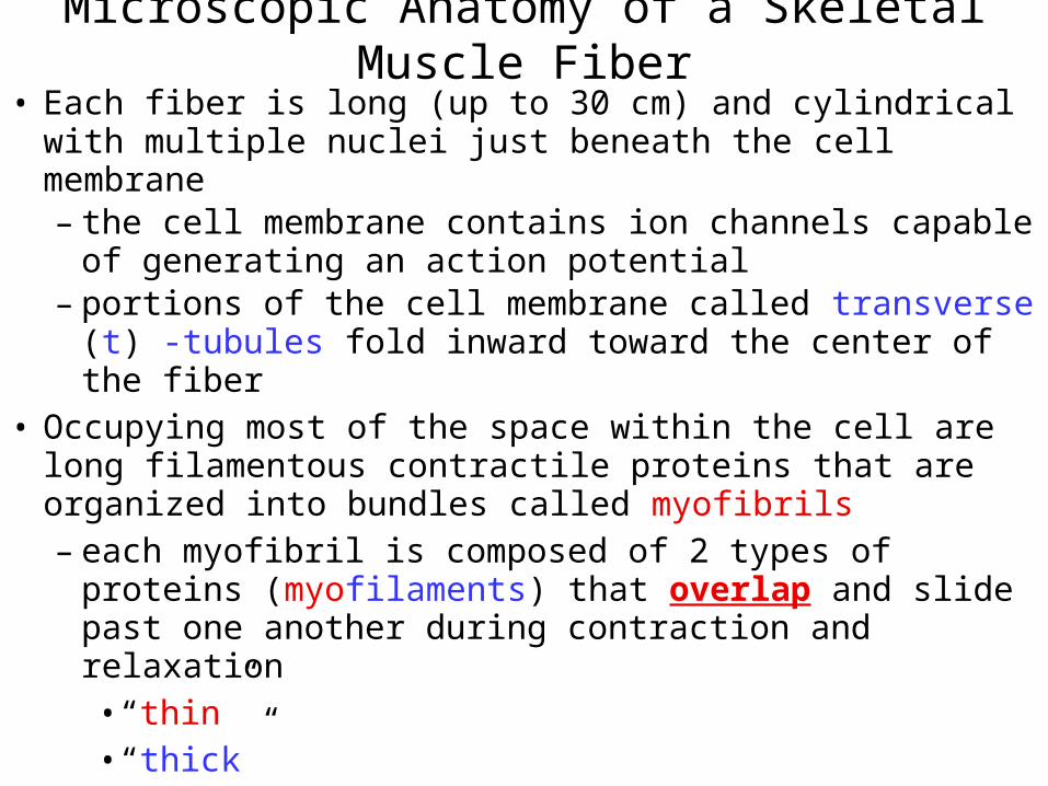
• Each fiber is long (up to 30 cm) and cylindrical with multiple nuclei just beneath the cell membrane – the cell membrane contains ion channels capable of
generating an action potential– portions of the cell membrane called transverse (t) -
tubules fold inward toward the center of the fiber• Occupying most of the space within the cell are long
filamentous contractile proteins that are organized into bundles called myofibrils– each myofibril is composed of 2 types of proteins
(myofilaments) that overlap and slide past one another during contraction and relaxation• “thin”• “thick”
Microscopic Anatomy of a Skeletal Muscle Fiber
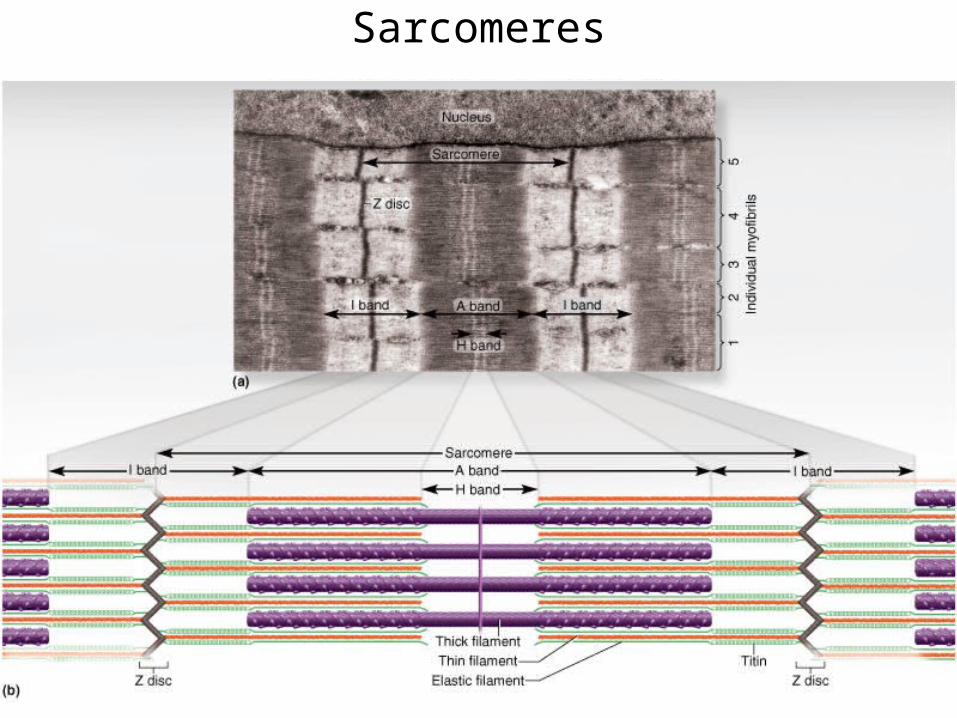
Striations of Skeletal Muscle• When viewed longitudinally, the overlapping
arrangement of myofilaments creates a repeating pattern of dark and light striations (stripes) called sarcomeres– the contractile unit of skeletal (and cardiac) muscle
Sarcomeres
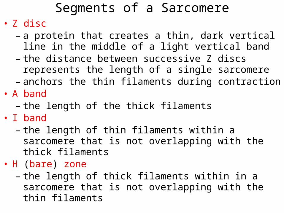
Segments of a Sarcomere• Z disc
– a protein that creates a thin, dark vertical line in the middle of a light vertical band
– the distance between successive Z discs represents the length of a single sarcomere
– anchors the thin filaments during contraction• A band
– the length of the thick filaments • I band
– the length of thin filaments within a sarcomere that is not overlapping with the thick filaments
• H (bare) zone– the length of thick filaments within in a sarcomere
that is not overlapping with the thin filaments
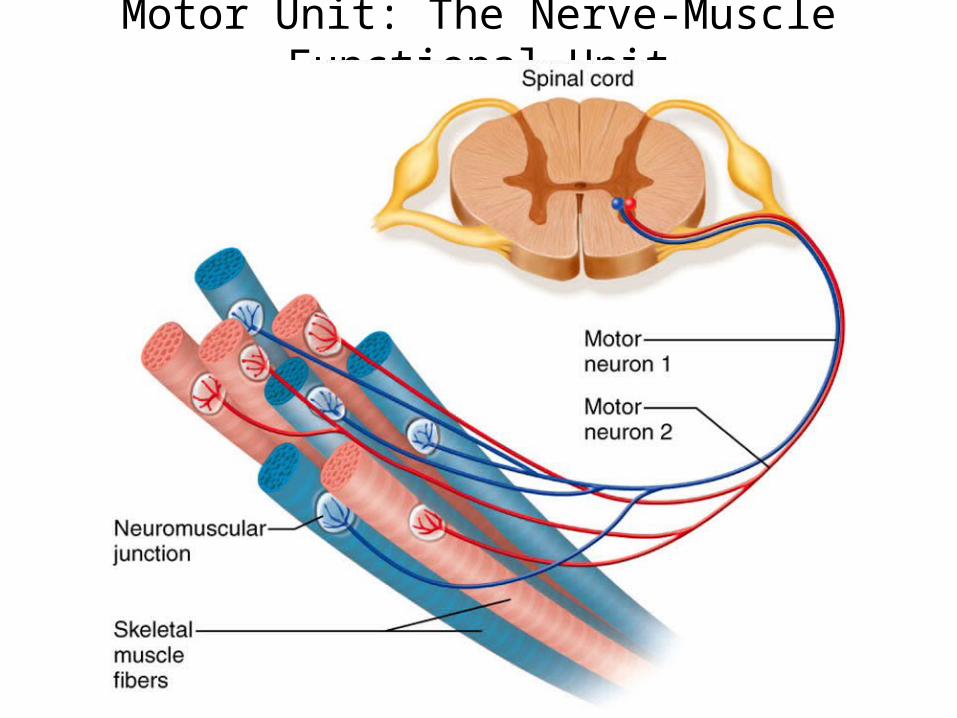
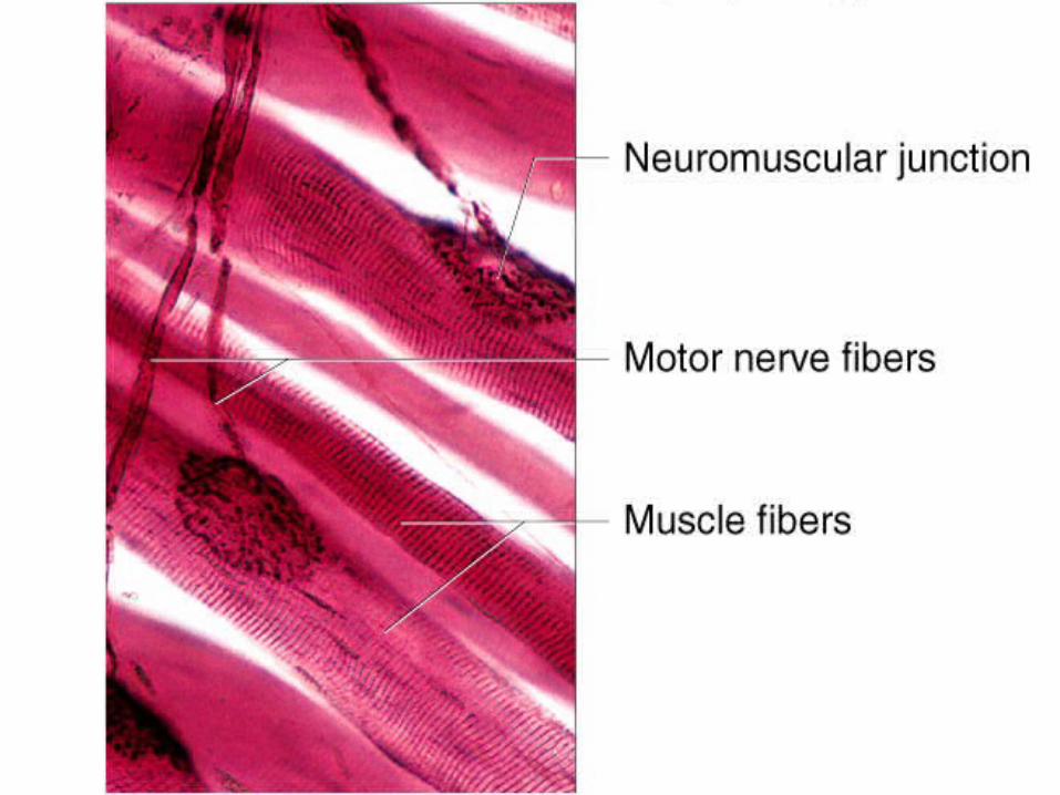
Motor Unit: The Nerve-Muscle Functional Unit
• In order to contract, a skeletal muscle must be stimulated by a motor neuron
• The location where the end of a motor neuron and a skeletal muscle fiber meet is called the neuromuscular junction (NMJ)
• A single motor neuron is capable of stimulating multiple skeletal muscle fibers to contract simultaneously– one neuron branches allowing it to stimulate
multiple muscle fibers simultaneously– the anatomical relationship between a single motor
neuron and all skeletal fibers that it controls is called a motor unit
Motor Unit: The Nerve-Muscle Functional Unit
Motor Unit: The Nerve-Muscle Functional Unit
• The number of muscle fibers per motor unit can range:– few (small motor unit)
• control fine, precise movements (fingers, eyes)– several hundred (large motor unit)
• control gross movements (arms, legs)• large weight-bearing muscles (back)
Muscle Twitch
• The contraction followed by the relaxation of a muscle fiber to a single, brief stimulus by a motor neuron is called a twitch
• There are three phases of a muscle twitch – Latent (lag) period
• time between the stimulation by a motor neuron and the beginning of contraction (few milliseconds)
– Contractile period• contractile proteins within the fiber hydrolyze ATP
causing the fiber to shorten resulting in an increase in tension (force)
– Relaxation period• fiber lengthens causing tension to decrease
Muscle Twitch
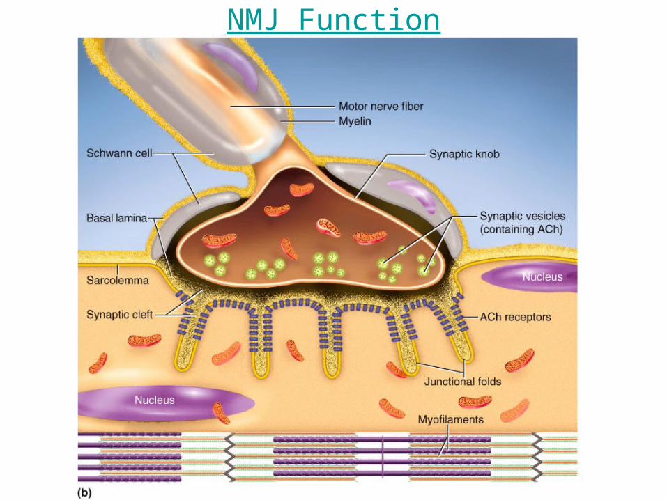
The Neuromuscular Junction• Between the motor neuron and the skeletal muscle
fiber is a small space called a symaptic cleft • A motor neuron stimulates the contraction of a skeletal
muscle fiber by exocytosing a chemical messenger called a neurotransmitter into the synaptic cleft
• The specific neurotransmitter released onto skeletal muscle fibers is called acetylcholine (ACh)
• Acetylcholine diffuses through the ECF within the cleft and binds to integral membrane proteins of the skeletal muscle fiber called ACh receptors
• The binding of ACh to ACh receptors creates an action potential in the cell membrane of the skeletal muscle fiber which will ultimately cause the cell to elicit a twitch
• Linking the action potential to the contraction of a muscle fiber is called excitation-contraction coupling
NMJ Function
Muscle Fiber Relaxation
• After ACh creates an action potential in the fiber it is rapidly hydrolyzed into acetate and choline by the enzyme Acetylcholine esterase located in the synaptic cleft of the NMJ– prevents prolonged stimulation (and contraction) of
a skeletal muscle fiber allowing it to relax
• Skeletal muscle fibers contain an elaborate, smooth sarcoplasmic (endoplasmic) reticulum (SR) which is the storage site of intracellular calcium (Ca+2)
• Action potentials travel along the sarcolemma into the t-tubules which open Ca2+ channels in the SR to open resulting in the diffusion of Ca2+ out of the SR into the sarcoplasm
Sliding Filament Model of Contraction
• An increase in the amount of Ca2+ in the sarcoplasm, allows the thick filaments to pull the thin filaments toward the center of the sarcomere causing the sarcomere to shorten
• As all of the sarcomeres in a muscle shortens, the entire muscle shortens
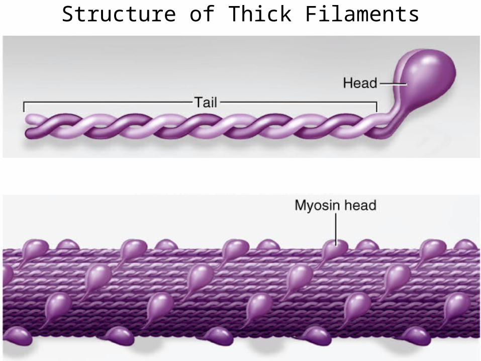
Structure of Thick Filaments
• Thick filaments are composed of many molecules of the protein myosin
• Each myosin protein has a rodlike tail and two heads– Myosin heads:
• hydrolyze a molecule of ATP–uses the chemical energy to contract
• attach to and pull on the protein actin of thin filaments causing the sarcomere to shorten
Structure of Thick Filaments
Structure of Thin Filaments
• Thin filaments are composed of 3 proteins– F (fibrous) Actin is a helical polymer of G
(globular) actin protein subunits• each subunit contains a binding site for the
protein myosin of the thick filaments– Tropomyosin blocks the interaction between actin
and myosin • prevents an unstimulated muscle from
contracting– Troponin C is attached to tropomyosin
• binds to Ca2+ in the sarcoplasm during contraction
Structure of Thin Filaments
Excitation-Contraction Coupling• Ca2+ in the sarcoplasm binds to troponin C
– changes the position of troponin C• moves tropomyosin away from the myosin
binding site on actin promoting the interaction between myosin and actin (CONTRACTION)
• Linking the action potential to the contraction of a muscle fiber is called excitation-contraction coupling
Molecular Events of Contraction• Myosin pulls on actin in a repetitive (cyclic) fashion
progressively moving the thin filaments toward the center of the sarcomere
• Each cycle consists of 4 steps1. Activation of the myosin head
• a molecule of ATP is hydrolyzed and the energy is used by the myosin head to change the shape of myosin into the high-energy state
3. Power stroke• myosin head pivots and pulls thin filament
4. Cross bridge detachment • the binding of a molecule of ATP to the myosin
head causes it to detach from actin
(Cross Bridge Cycling)
Muscle Fiber Relaxation• Within the membrane of the SR is a primary active
transporting pump called the Ca2+-ATPase • The Ca2+-ATPase constantly pumps Ca2+ out of the
sarcoplasm into the SR• During an action potential, Ca2+ diffuses into the
sarcoplasm faster than the Ca2+-ATPase can remove it. However, when the action potential is over the Ca2+-ATPase pumps the Ca2+ back into the SR ending contraction
Contraction of Skeletal Muscle
• The two types of muscle contractions are:– Isometric contraction = “same length”
• muscle contracts and produces tension, but does not shorten
• trying to lift a car– Isotonic contraction = “same tension”
• muscle contracts and produces tension • shortens as it contracts • lifting a pencil
– muscle contracts and produces tension, but the muscle but does not shorten or lengthen
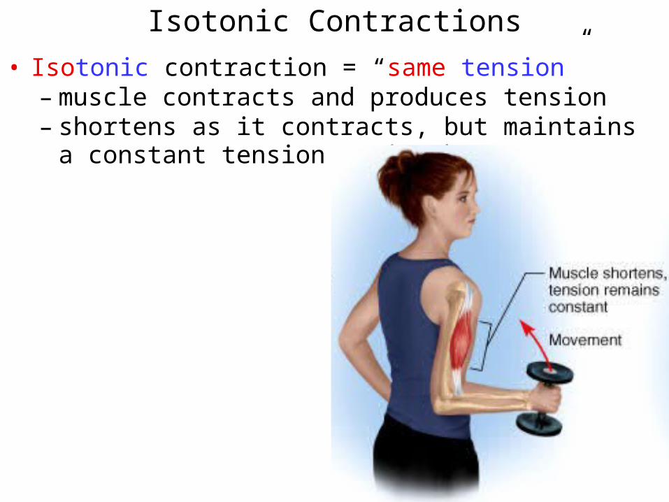
Isotonic Contractions
• Isotonic contraction = “same tension” – muscle contracts and produces tension – shortens as it contracts, but maintains a constant
tension as it shortens
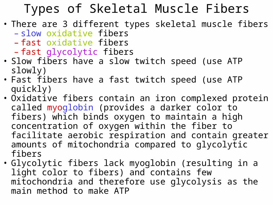
Types of Skeletal Muscle Fibers• There are 3 different types skeletal muscle fibers
– slow oxidative fibers– fast oxidative fibers– fast glycolytic fibers
• Slow fibers have a slow twitch speed (use ATP slowly)• Fast fibers have a fast twitch speed (use ATP quickly)• Oxidative fibers contain an iron complexed protein
called myoglobin (provides a darker color to fibers) which binds oxygen to maintain a high concentration of oxygen within the fiber to facilitate aerobic respiration and contain greater amounts of mitochondria compared to glycolytic fibers
• Glycolytic fibers lack myoglobin (resulting in a light color to fibers) and contains few mitochondria and therefore use glycolysis as the main method to make ATP
Characteristics of Skeletal Muscle Fiber Types• Slow oxidative fibers:
– muscle fibers used to maintain posture – high resistance to fatigue since they can make lots
of ATP and use it somewhat slowly• Fast oxidative fibers:
– muscle fibers used for non-exertive movement– moderate resistance to fatigue since they can make
lots of ATP but use it somewhat quickly• Fast glycolytic fibers:
– muscle fibers used for powerful movements– low resistance to fatigue since they make little ATP
and use it very quickly• Skeletal muscles of your body contain a combination
of all three fiber types, but their ratio determines the overall function of that muscle
• Weakening of contracting muscle caused by:– the rate of ATP hydrolysis exceeds the rate of
protein function– motor neurons run out of acetylcholine
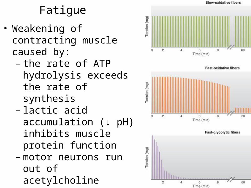
Fatigue
Fatigue
• Weakening of contracting muscle caused by:– the rate of ATP hydrolysis
exceeds the rate of synthesis
– lactic acid accumulation (↓ pH) inhibits muscle protein function
– motor neurons run out of acetylcholine
Variety of Muscle Responses
• Variations in the force of muscle contraction is required for proper control of skeletal movement– moving a pencil vs. a textbook with your hand uses
the same muscles, but requires a different amount of force
• Skeletal muscle contractions are varied by:– altering the number of muscle fibers that contract
• determined by the number of motor units that are actively stimulating muscle fibers
– altering the frequency of muscle stimulation • determined by how often the motor neuron
releases ACh onto the muscle fiber
Motor Unit Recruitment
• Slow oxidative fibers are first stimulated to contract– provide basal muscle
tension (tone)• If additional muscle
tension is required, fast oxidative fibers are stimulated to contract– recruitment
• Finally, the fast glycolytic fibers are stimulated to bring muscle tension to maximum
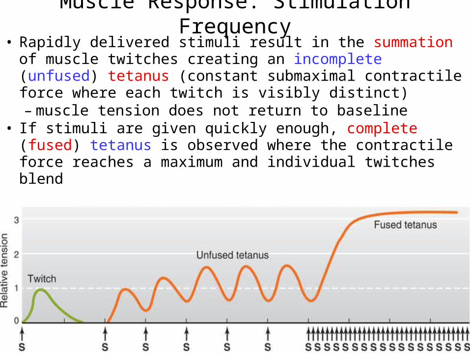
Muscle Response: Stimulation Frequency• Rapidly delivered stimuli result in the summation of
muscle twitches creating an incomplete (unfused) tetanus (constant submaximal contractile force where each twitch is visibly distinct)– muscle tension does not return to baseline
• If stimuli are given quickly enough, complete (fused) tetanus is observed where the contractile force reaches a maximum and individual twitches blend