50
Muscle Tissue / Muscles of the Body
| Date post: | 22-Dec-2015 |
| Category: |
Documents |
| Upload: | derek-carr |
| View: | 238 times |
| Download: | 2 times |
Muscle Tissue / Muscles of the Body
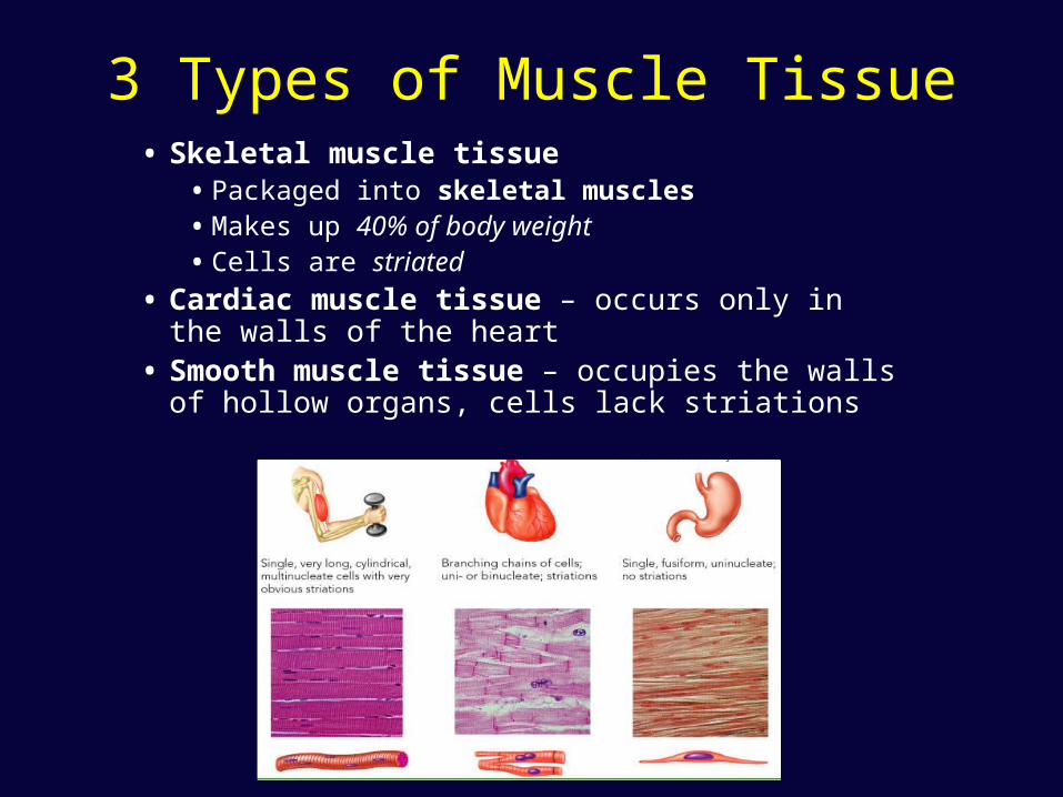
3 Types of Muscle Tissue• Skeletal muscle tissue
• Packaged into skeletal muscles• Makes up 40% of body weight• Cells are striated
• Cardiac muscle tissue – occurs only in the walls of the heart
• Smooth muscle tissue – occupies the walls of hollow organs, cells lack striations

Functions Of Muscle Tissue• Produce movement - movement of the body as a whole, propels
blood, food, wastes, and babies, eye movement
• Maintain posture and body position - muscles continuously contract to help you maintain your body posture
• Support soft tissues - soft tissues such as the organs in your abdominal and pelvic cavity are supported by skeletal muscle
• Control entrances and exits - openings to the urinary and digestive tracts have muscles that allow you to control swallowing, defecation, and urination
• Maintain body temperature - contractions produce heat through shivering
Functional Features of Muscles
• Contractility - long cells shorten and generate pulling force
• Excitability - electrical nerve impulse stimulates the muscle cell to contract
• Extensibility - can be stretched back to its original length by contraction of an opposing muscle
• Elasticity - can recoil after being stretched
Similarities of Muscle Tissue• Cells of muscles are known as fibers
• Plasma membrane is called a sarcolemma
• Cytoplasm is called sarcoplasm
• Muscle contraction• Depends on two types of myofilaments (contractile
proteins) called actin and myosin
• These two proteins generate contractile force
Skeletal Muscle
• Each muscle is an organ • Consists mostly of muscle tissue• Skeletal muscle also contains
• Connective tissue• Blood vessels• Nerves
• Each skeletal muscle supplied by branches of• One nerve• One artery• One or more veins
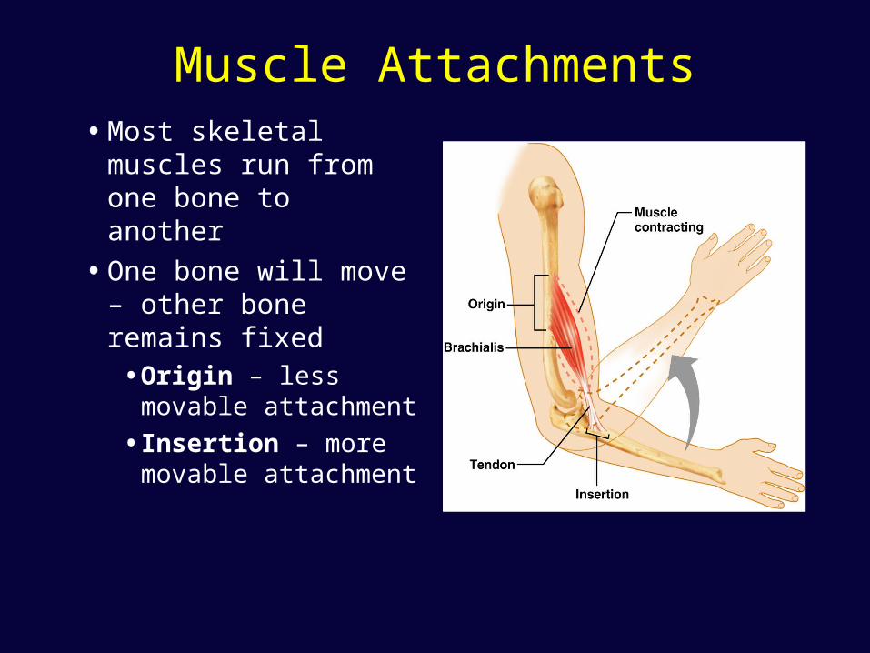
Muscle Attachments• Most skeletal
muscles run from one bone to another
• One bone will move – other bone remains fixed• Origin – less
movable attachment
• Insertion – more movable attachment
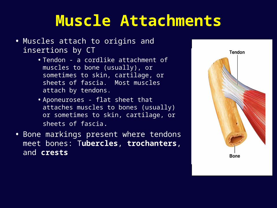
Muscle Attachments• Muscles attach to origins and insertions
by CT• Tendon - a cordlike attachment of
muscles to bone (usually), or sometimes to skin, cartilage, or sheets of fascia. Most muscles attach by tendons.
• Aponeuroses - flat sheet that attaches muscles to bones (usually) or sometimes
to skin, cartilage, or sheets of fascia.
• Bone markings present where tendons meet bones: Tubercles, trochanters, and crests

Connective Tissue And Fascicles
• Connective tissue sheaths bind a skeletal muscle and its fibers together
• Epimysium – dense irregular connective tissue surrounding entire muscle
• Perimysium – surrounds each fascicle (group of muscle fibers)
• Endomysium – a fine sheath of connective tissue wrapping each muscle cell
• Connective tissue sheaths are continuous with tendons

Histology of Skeletal Muscle Tissue• The skeletal muscle fiber
• Fibers are long and cylindrical• Are huge cells – diameter is 10–100µm
• Length – several centimeters to dozens of centimeters
• Cells are multinucleate
• Nuclei are peripherally located

Myofibrils and Sarcomeres• Striations result from internal structure of myofibrils• Myofibrils
• Long rods within cytoplasm • Make up 80% of the cytoplasm • Are a specialized contractile organelle found in muscle tissue • A long row of repeating segments called sarcomeres
(functional unit of Skeletal MT)
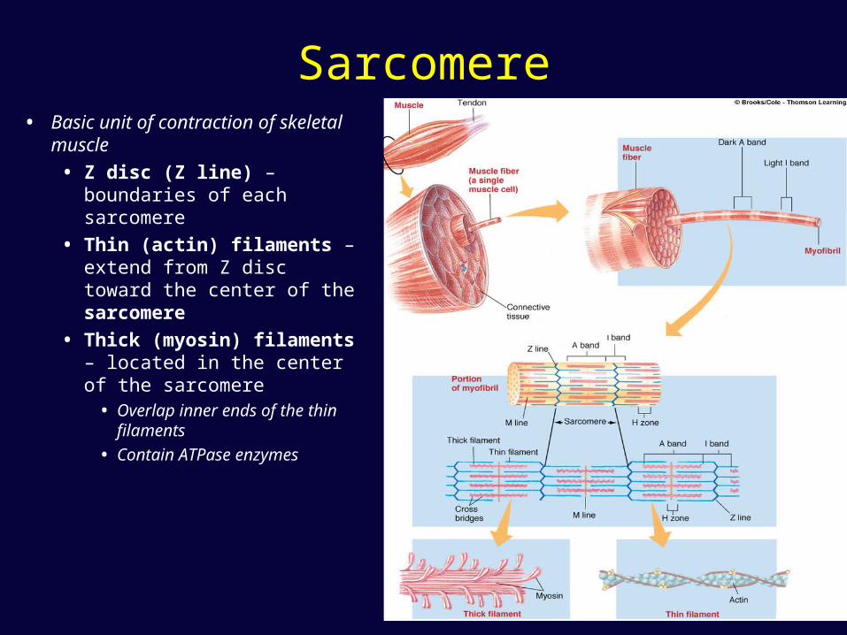
Sarcomere• Basic unit of contraction of
skeletal muscle
• Z disc (Z line) – boundaries of each sarcomere
• Thin (actin) filaments – extend from Z disc toward the center of the sarcomere
• Thick (myosin) filaments – located in the center of the sarcomere
• Overlap inner ends of the thin filaments
• Contain ATPase enzymes
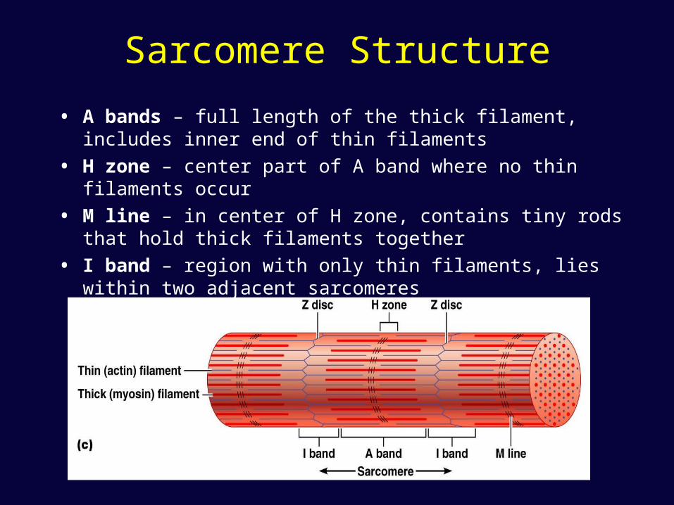
Sarcomere Structure
• A bands – full length of the thick filament, includes inner end of thin filaments
• H zone – center part of A band where no thin filaments occur
• M line – in center of H zone, contains tiny rods that hold thick filaments together
• I band – region with only thin filaments, lies within two adjacent sarcomeres
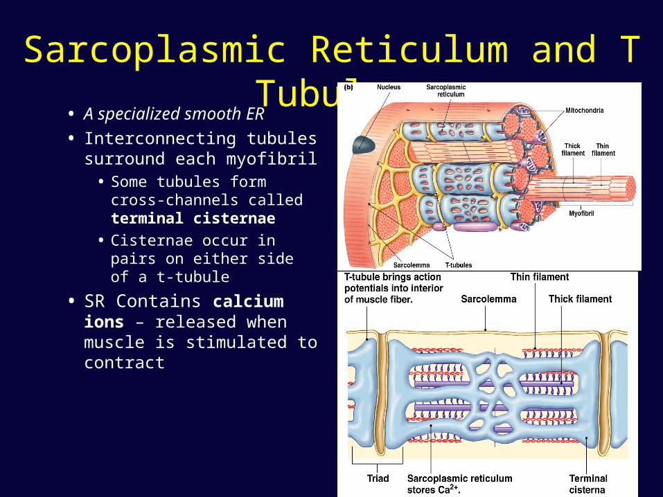
Sarcoplasmic Reticulum and T Tubules
• A specialized smooth ER
• Interconnecting tubules surround each myofibril
• Some tubules form cross-channels called terminal cisternae
• Cisternae occur in pairs on either side of a t-tubule
• SR Contains calcium ions – released when muscle is stimulated to contract
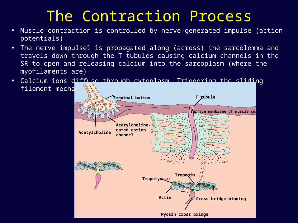
The Contraction Process• Muscle contraction is controlled by nerve-generated impulse (action potentials)
• The nerve impulsel is propagated along (across) the sarcolemma and travels down through the T tubules causing calcium channels in the SR to open and releasing calcium into the sarcoplasm (where the myofilaments are)
• Calcium ions diffuse through cytoplasm, Triggering the sliding filament mechanism
Terminal button
Acetylcholine-gated cationchannel
Acetylcholine
T tubule
Surface membrane of muscle cell
TropomyosinTroponin
Cross-bridge binding
Myosin cross bridge
Actin
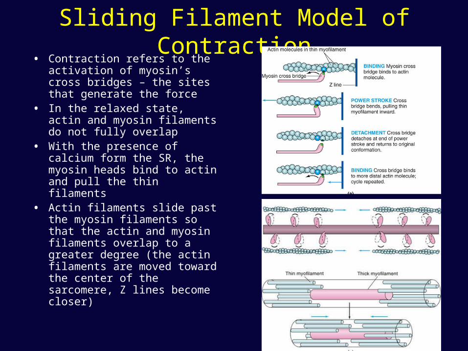
Sliding Filament Model of Contraction
• Contraction refers to the activation of myosin’s cross bridges – the sites that generate the force
• In the relaxed state, actin and myosin filaments do not fully overlap
• With the presence of calcium form the SR, the myosin heads bind to actin and pull the thin filaments
• Actin filaments slide past the myosin filaments so that the actin and myosin filaments overlap to a greater degree (the actin filaments are moved toward the center of the sarcomere, Z lines become closer)
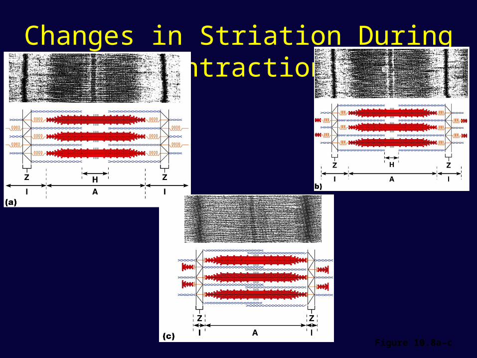
Changes in Striation During Contraction
Figure 10.8a–c
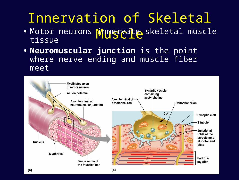
Innervation of Skeletal Muscle• Motor neurons innervate skeletal muscle
tissue• Neuromuscular junction is the point where
nerve ending and muscle fiber meet
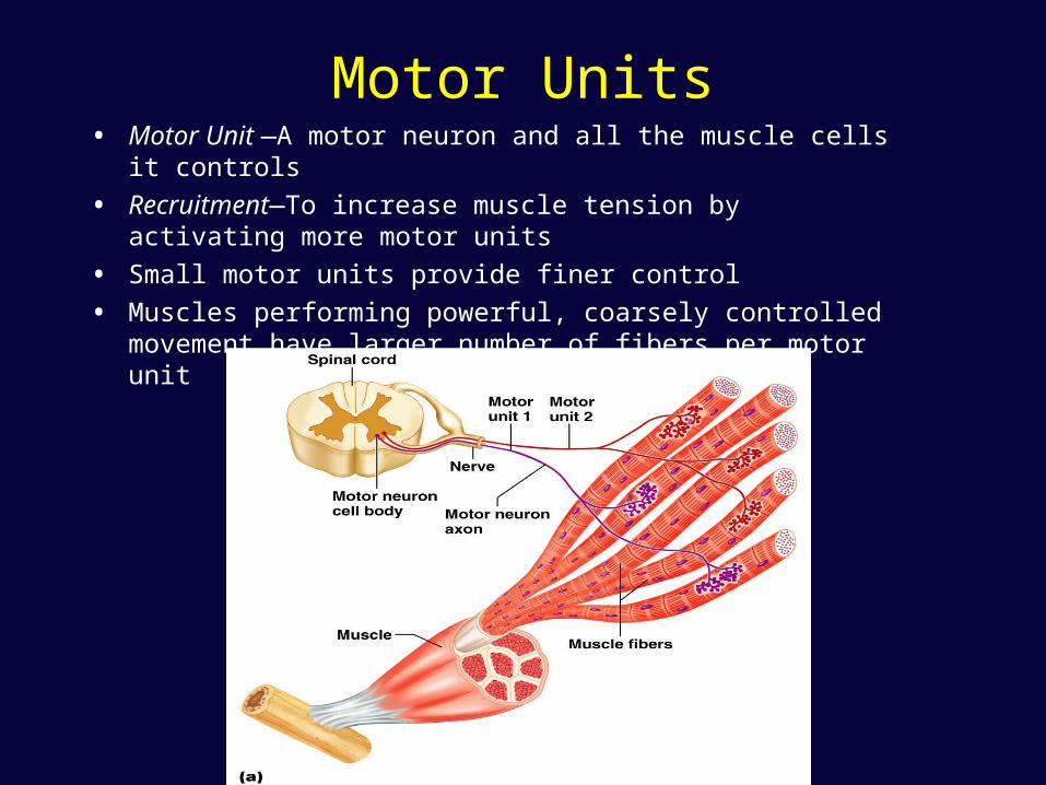
Motor Units• Motor Unit —A motor neuron and all the muscle cells it controls
• Recruitment—To increase muscle tension by activating more motor units
• Small motor units provide finer control
• Muscles performing powerful, coarsely controlled movement have larger number of fibers per motor unit
Types of Skeletal Muscle Fibers
• Skeletal muscle fibers are categorized according to• How they manufacture energy (ATP)• How quickly they contract
• Divided into 3 classes• Slow oxidative fibers (Type I)
• Red Slow twitch
• Fast glycolytic fibers (Type IIx)• White fast-twitch
• Fast oxidative fibers (Type IIa)• Intermediate fibers
Types of Skeletal Muscle Fibers
• Slow oxidative fibers (Type I)
• Red color due to abundant myoglobin
• Obtain energy from aerobic metabolic reactions
• Contain a large number of mitochondria
• Richly supplied with capillaries
• Contract slowly and resistant to fatigue
• Fibers are small in diameter
Types of Skeletal Muscle Fibers
• Fast glycolytic fibers (Type IIx)
• Contain little myoglobin and few mitochondria
• About twice the diameter of slow-oxidative fibers
• Contain more myofilaments and generate more power
• Depend on anaerobic pathways
• Contract rapidly and tire quickly
Types of Skeletal Muscle Fibers
• Fast oxidative fibers (Type IIa)
• Have an intermediate diameter
• Contract quickly like fast glycolytic fibers
• Are oxygen-dependent
• Have high myoglobin content and rich supply of capillaries
• Somewhat fatigue-resistant
• More powerful than slow oxidative fibers
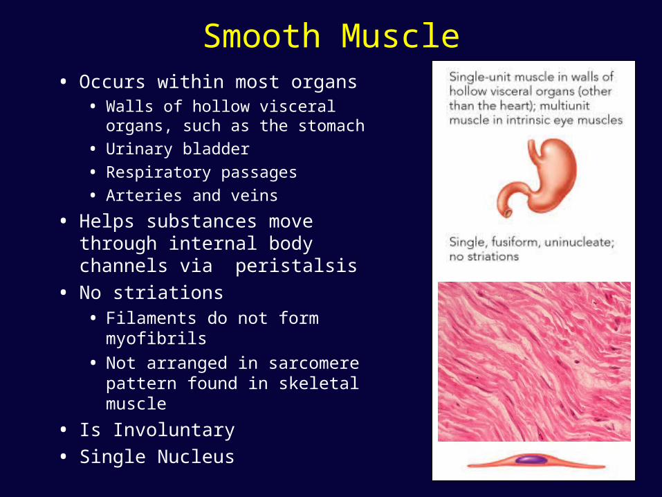
Smooth Muscle• Occurs within most organs
• Walls of hollow visceral organs, such as the stomach
• Urinary bladder
• Respiratory passages
• Arteries and veins
• Helps substances move through internal body channels via peristalsis
• No striations• Filaments do not form myofibrils
• Not arranged in sarcomere pattern found in skeletal muscle
• Is Involuntary
• Single Nucleus
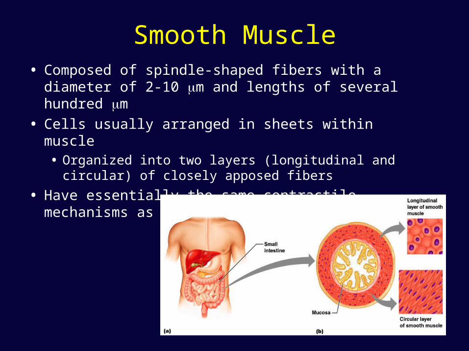
Smooth Muscle• Composed of spindle-shaped fibers with a diameter of
2-10 m and lengths of several hundred m
• Cells usually arranged in sheets within muscle• Organized into two layers (longitudinal and circular) of
closely apposed fibers
• Have essentially the same contractile mechanisms as skeletal muscle
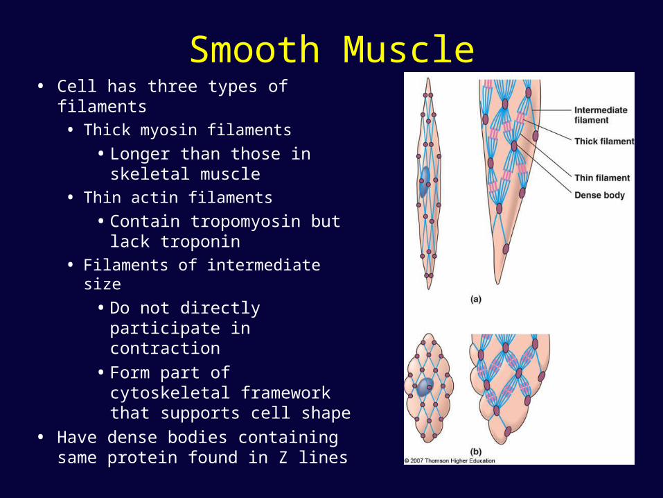
Smooth Muscle• Cell has three types of filaments
• Thick myosin filaments
• Longer than those in skeletal muscle
• Thin actin filaments
• Contain tropomyosin but lack troponin
• Filaments of intermediate size
• Do not directly participate in contraction
• Form part of cytoskeletal framework that supports cell shape
• Have dense bodies containing same protein found in Z lines
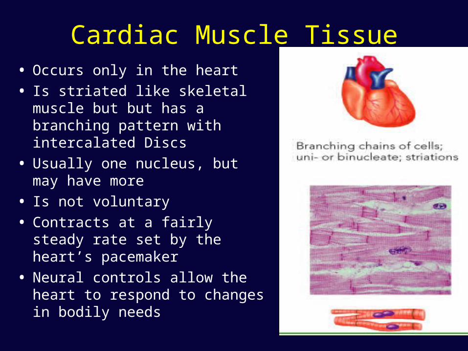
Cardiac Muscle Tissue• Occurs only in the heart
• Is striated like skeletal muscle but but has a branching pattern with intercalated Discs
• Usually one nucleus, but may have more
• Is not voluntary
• Contracts at a fairly steady rate set by the heart’s pacemaker
• Neural controls allow the heart to respond to changes in bodily needs
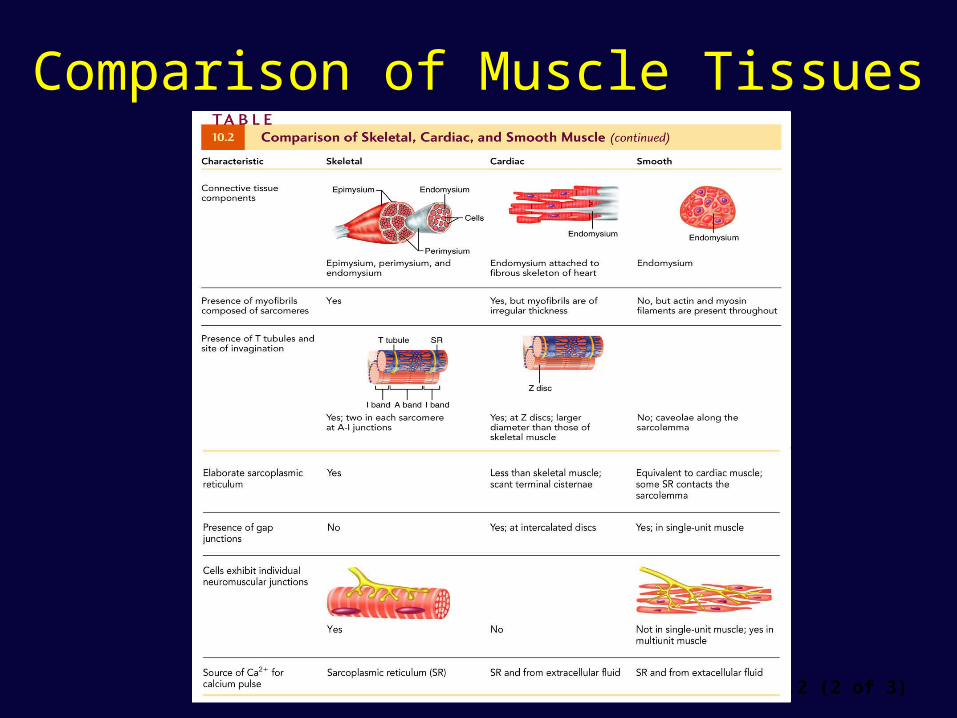
Comparison of Muscle Tissues
Table 10.2 (2 of 3)
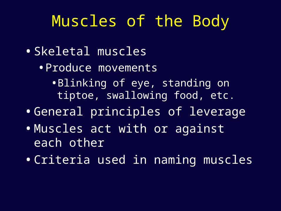
Muscles of the Body
• Skeletal muscles
• Produce movements
• Blinking of eye, standing on tiptoe, swallowing food, etc.
• General principles of leverage
• Muscles act with or against each other
• Criteria used in naming muscles
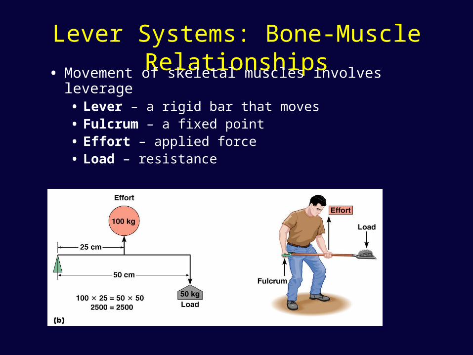
Lever Systems: Bone-Muscle Relationships
• Movement of skeletal muscles involves leverage• Lever – a rigid bar that moves • Fulcrum – a fixed point• Effort – applied force• Load – resistance
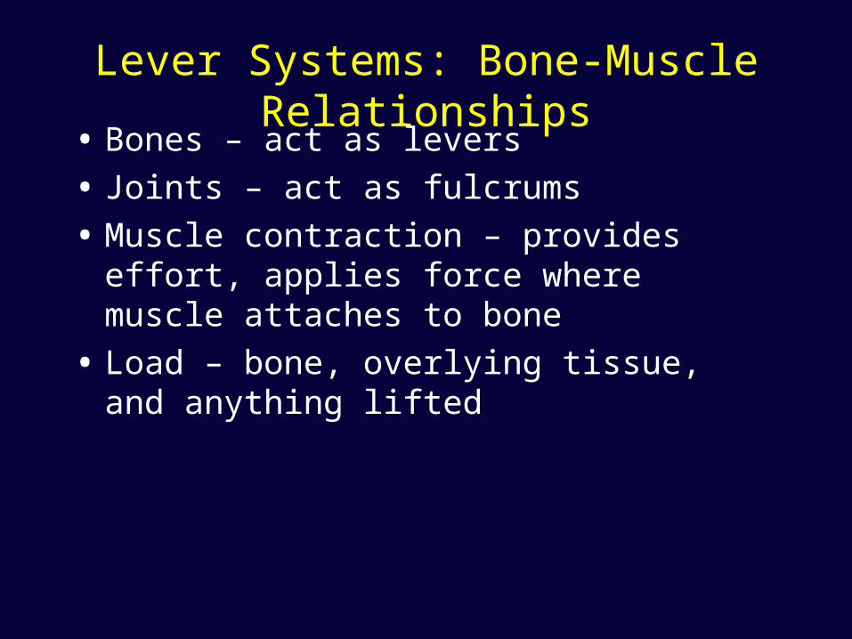
Lever Systems: Bone-Muscle Relationships
• Bones – act as levers
• Joints – act as fulcrums
• Muscle contraction – provides effort, applies force where muscle attaches to bone
• Load – bone, overlying tissue, and anything lifted
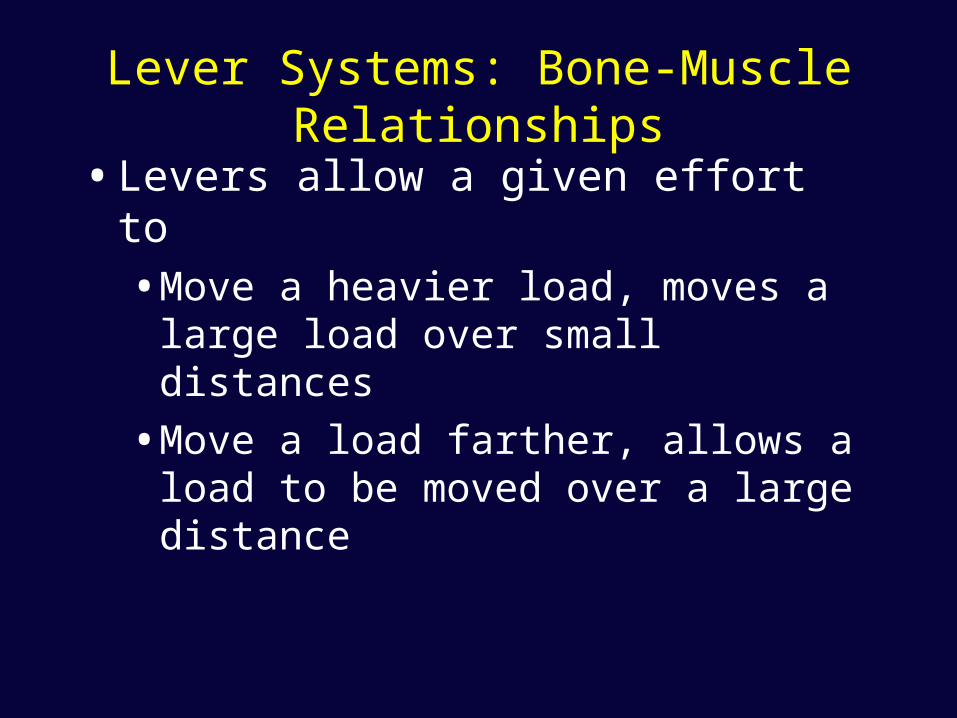
Lever Systems: Bone-Muscle Relationships
• Levers allow a given effort to
• Move a heavier load, moves a large load over small distances
• Move a load farther, allows a load to be moved over a large distance
Lever Systems: Bone-Muscle Relationships
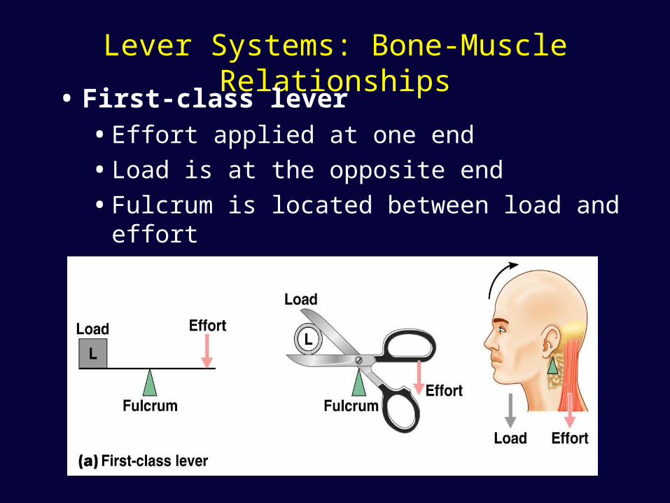
• First-class lever• Effort applied at one end
• Load is at the opposite end
• Fulcrum is located between load and effort
Lever Systems: Bone-Muscle Relationships
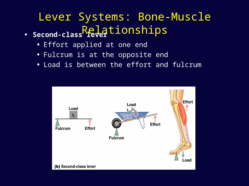
• Second-class lever
• Effort applied at one end
• Fulcrum is at the opposite end
• Load is between the effort and fulcrum
Lever Systems: Bone-Muscle Relationships
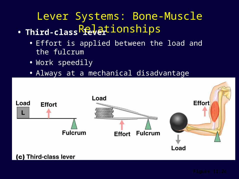
• Third-class lever• Effort is applied between the load and the fulcrum
• Work speedily
• Always at a mechanical disadvantage
Figure 11.2c
Lever Systems: Bone-Muscle Relationships
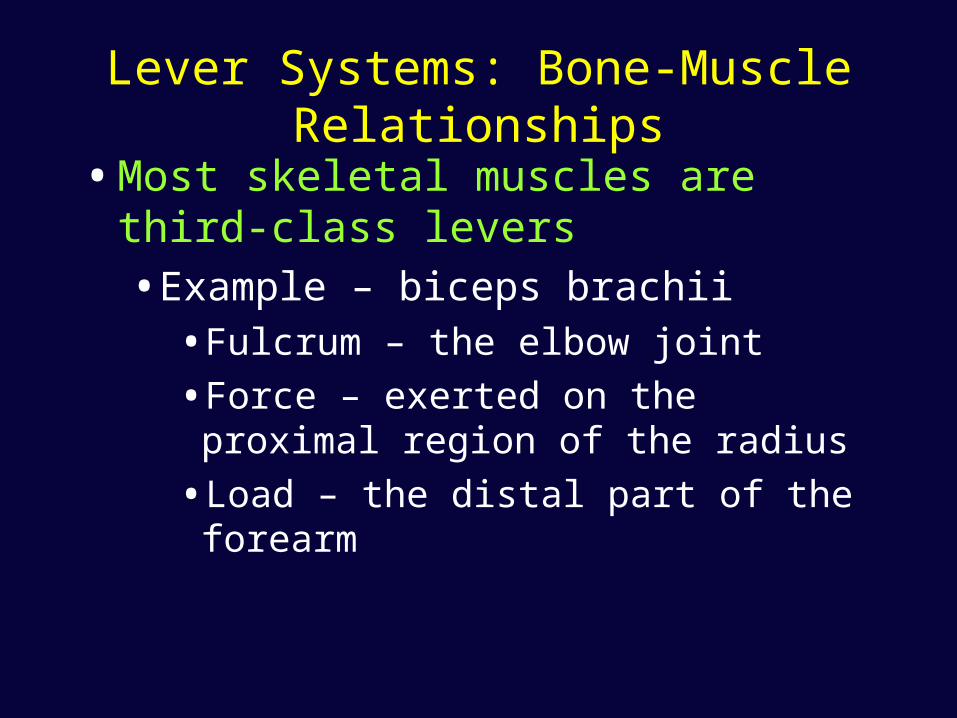
• Most skeletal muscles are third-class levers
• Example – biceps brachii
• Fulcrum – the elbow joint
• Force – exerted on the proximal region of the radius
• Load – the distal part of the forearm

Interactions of Skeletal Muscles in the Body
• A muscle cannot reverse the movement it produces
• Another muscle must undo the action
• Muscles with opposite actions lie on opposite sides of a joint
Muscles Classified into Several Functional Groups
• Prime mover (agonist)• Has major responsibility for a certain movement
• Antagonist• Opposes or reverses a movement
• Synergist – helps the prime mover • By adding extra force • By reducing undesirable movements • Fixator - a type of synergist that holds a bone firmly in
place
Arrangement of Fascicles in Muscles
• Skeletal muscles – consist of fascicles
• Fascicles – arranged in different patterns
• Fascicle arrangement – tells about action of a muscle
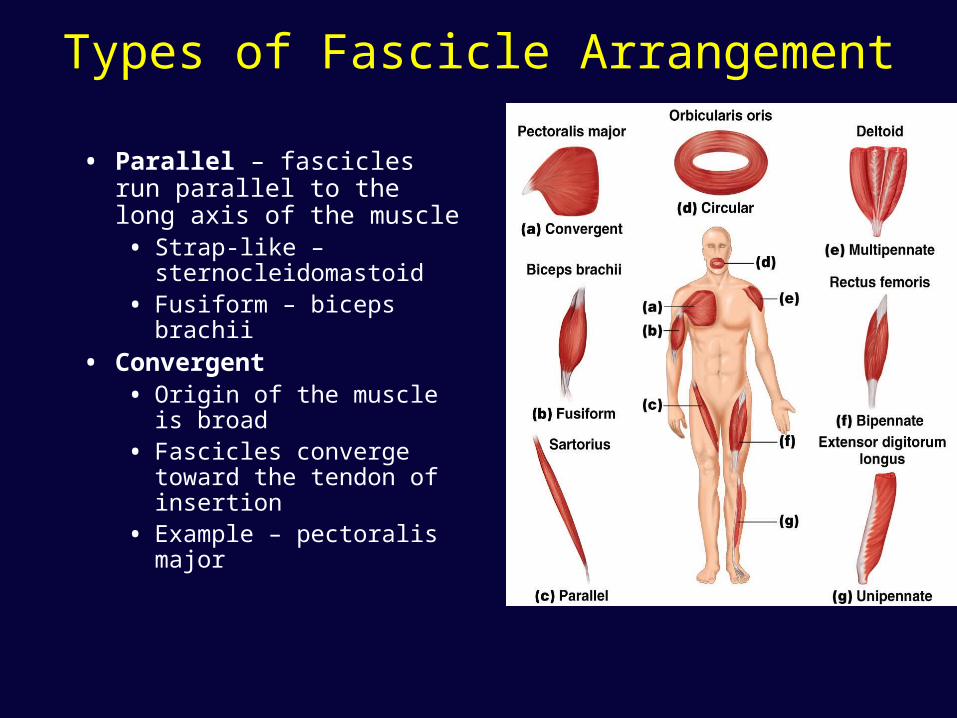
Types of Fascicle Arrangement
• Parallel – fascicles run parallel to the long axis of the muscle• Strap-like –
sternocleidomastoid• Fusiform – biceps brachii
• Convergent • Origin of the muscle is
broad• Fascicles converge toward
the tendon of insertion• Example – pectoralis
major
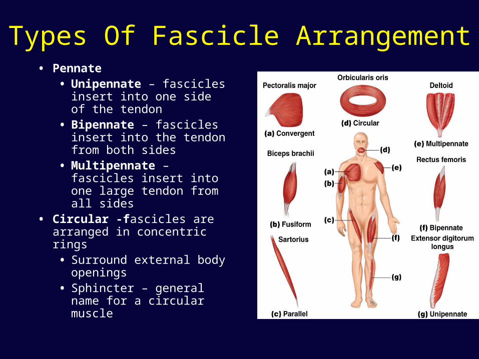
Types Of Fascicle Arrangement• Pennate
• Unipennate – fascicles insert into one side of the tendon
• Bipennate – fascicles insert into the tendon from both sides
• Multipennate – fascicles insert into one large tendon from all sides
• Circular -fascicles are arranged in concentric rings • Surround external body
openings• Sphincter – general
name for a circular muscle
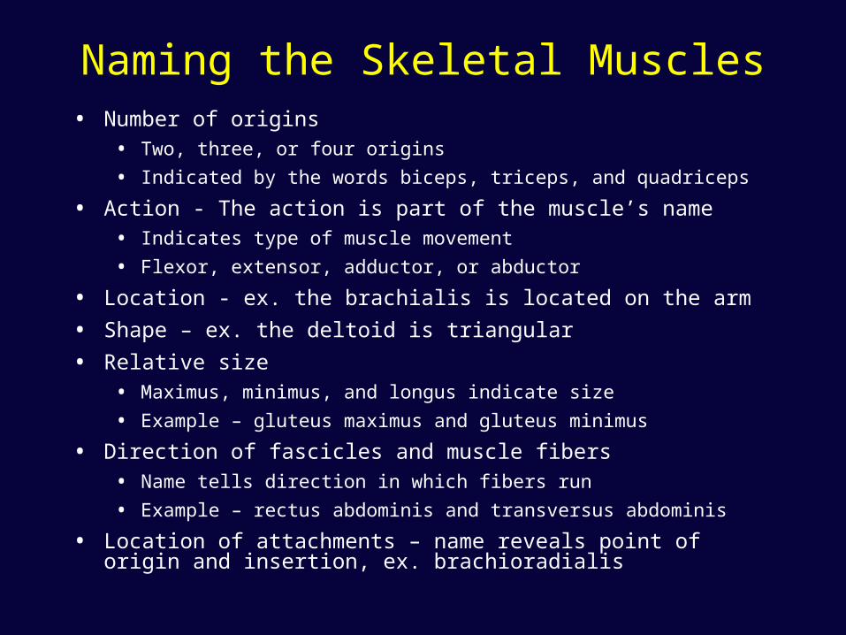
Naming the Skeletal Muscles• Number of origins
• Two, three, or four origins
• Indicated by the words biceps, triceps, and quadriceps
• Action - The action is part of the muscle’s name
• Indicates type of muscle movement
• Flexor, extensor, adductor, or abductor
• Location - ex. the brachialis is located on the arm
• Shape – ex. the deltoid is triangular
• Relative size
• Maximus, minimus, and longus indicate size
• Example – gluteus maximus and gluteus minimus
• Direction of fascicles and muscle fibers
• Name tells direction in which fibers run
• Example – rectus abdominis and transversus abdominis
• Location of attachments – name reveals point of origin and insertion, ex. brachioradialis
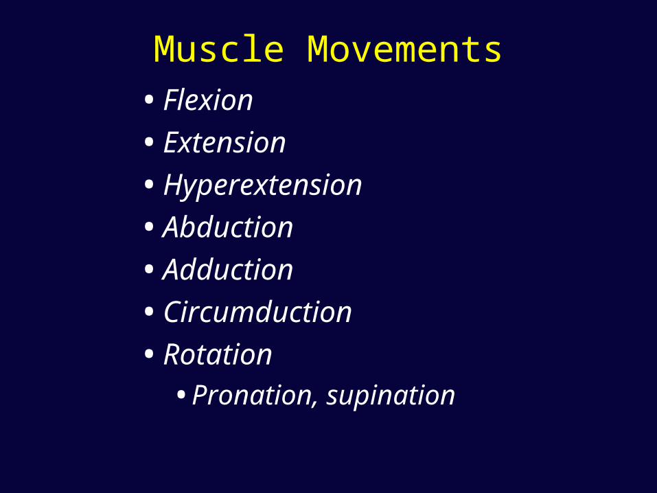
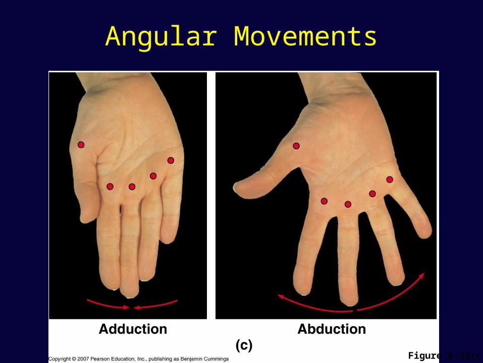
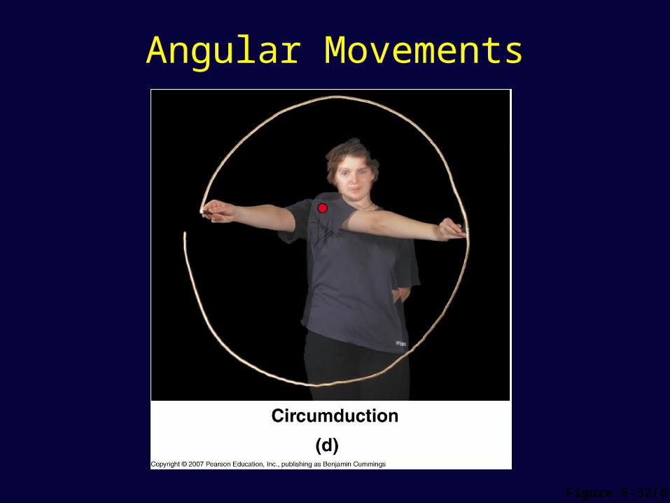
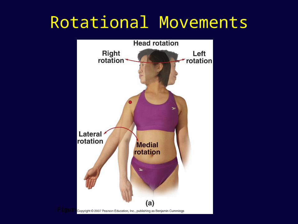
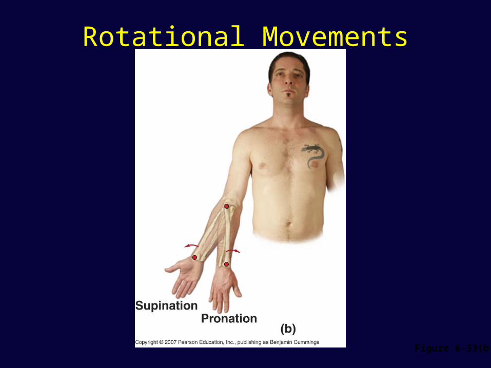
Muscle Movements• Flexion
• Extension
• Hyperextension
• Abduction
• Adduction
• Circumduction
• Rotation
• Pronation, supination
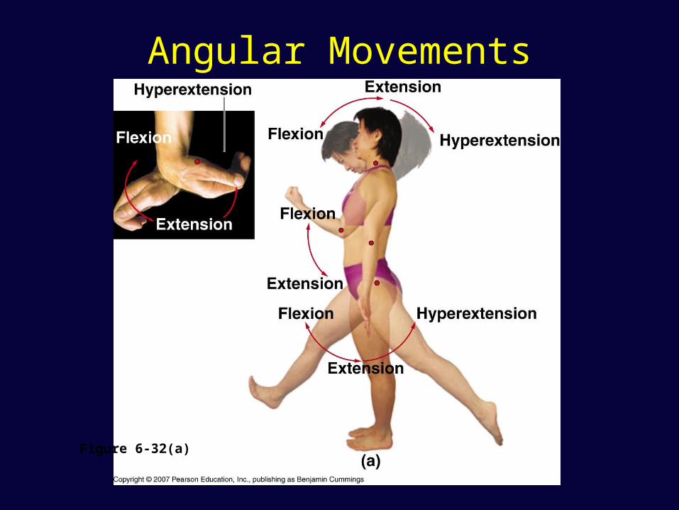
Angular Movements
Figure 6-32(a)
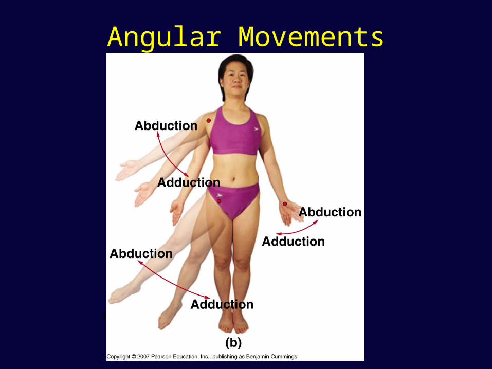
Angular Movements
Figure 6-32(b)
Angular Movements
Figure 6-32(c)
Angular Movements
Figure 6-32(d)
Rotational Movements
Figure 6-33(a)
Rotational Movements
Figure 6-33(b)
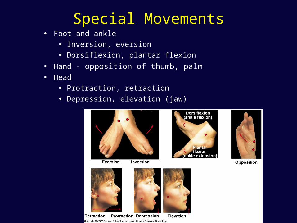
Special Movements• Foot and ankle
• Inversion, eversion
• Dorsiflexion, plantar flexion
• Hand - opposition of thumb, palm
• Head
• Protraction, retraction
• Depression, elevation (jaw)


