35
Muscles of the Forearm Dr. Sama ul Haque
| Date post: | 31-Dec-2015 |
| Category: |
Documents |
| Upload: | cadman-potter |
| View: | 38 times |
| Download: | 2 times |
Muscles of the Forearm
Dr. Sama ul Haque

Objectives
• Identify the muscles in the anterior and posterior compartments of the forearm in terms of their origin, insertion, nerve supply and actions.
• Know the neurovascular structures in the anterior and posterior compartments of the forearm.
• Discuss the functions of the muscles in the anterior and posterior compartment of the forearm.
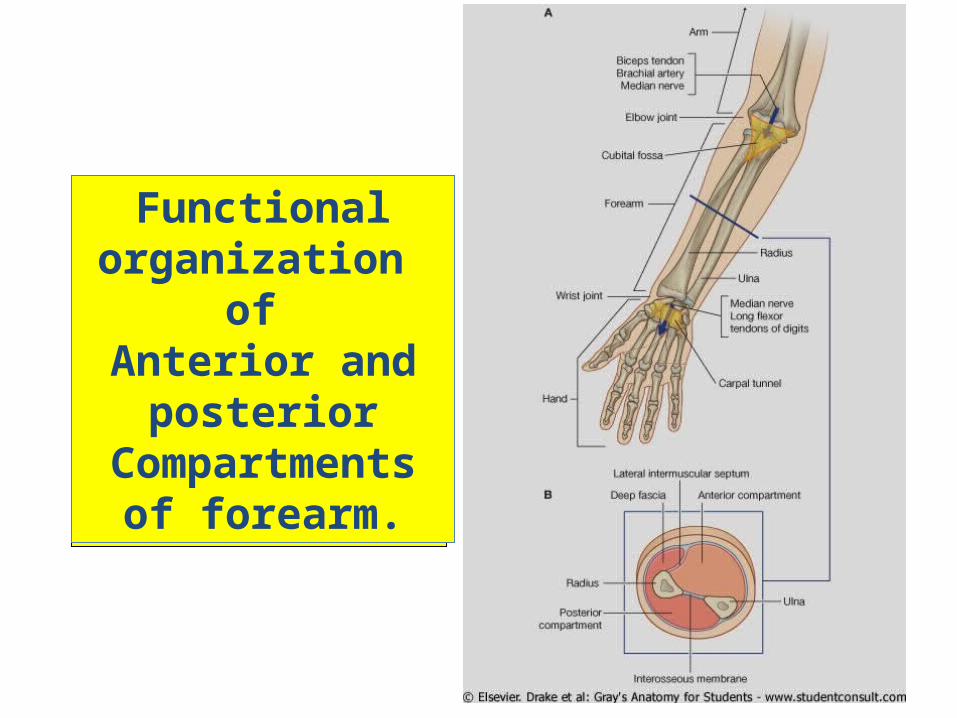
Functional organization
of Anterior and
posterior Compartments of
forearm.
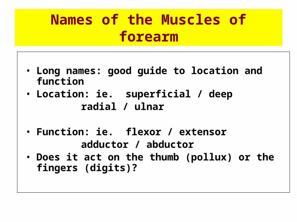
Names of the Muscles of forearm
• Long names: good guide to location and function• Location: ie. superficial / deep
radial / ulnar
• Function: ie. flexor / extensor adductor / abductor
• Does it act on the thumb (pollux) or the fingers (digits)?
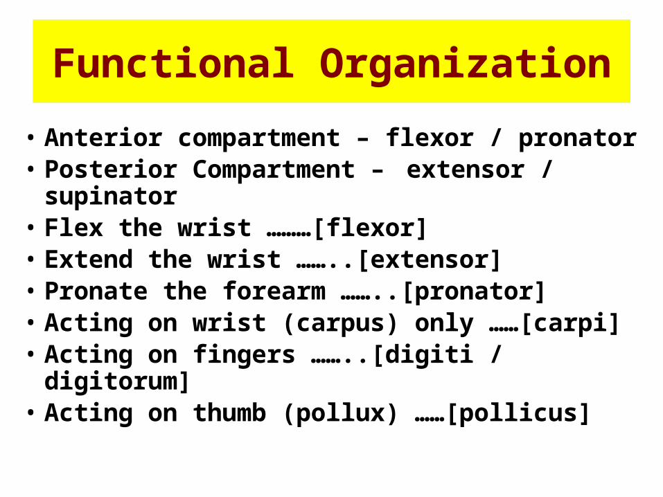
Functional Organization
• Anterior compartment – flexor / pronator• Posterior Compartment – extensor /
supinator• Flex the wrist ………[flexor]• Extend the wrist ……..[extensor]• Pronate the forearm ……..[pronator]• Acting on wrist (carpus) only ……[carpi]• Acting on fingers ……..[digiti / digitorum]• Acting on thumb (pollux) ……[pollicus]
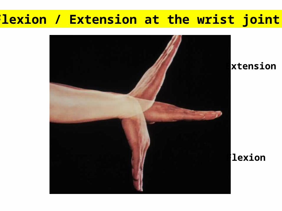
Flexion / Extension at the wrist joint.
Flexion
Extension
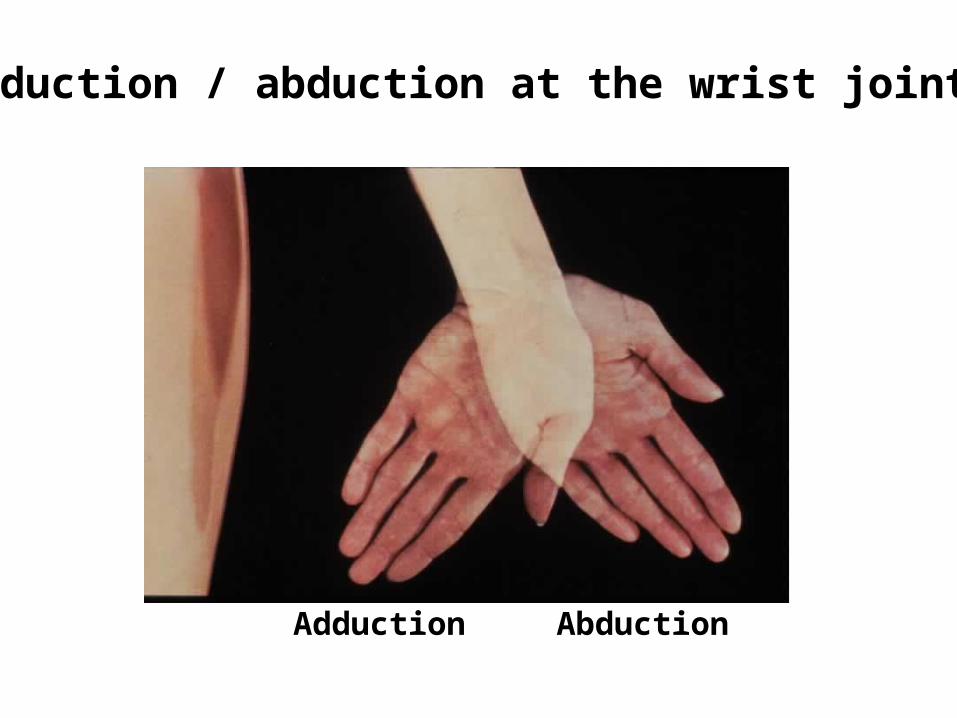
Adduction / abduction at the wrist joint.
Adduction Abduction
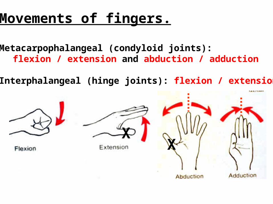
Movements of fingers.
Metacarpophalangeal (condyloid joints):flexion / extension and abduction / adduction
Interphalangeal (hinge joints): flexion / extension
XX
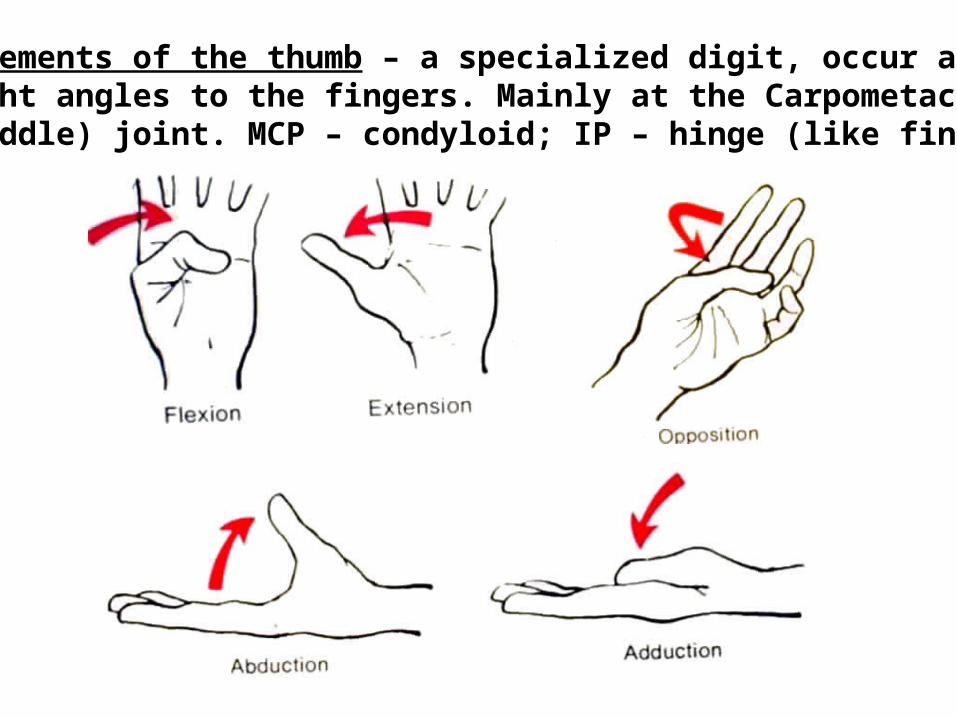
Movements of the thumb – a specialized digit, occur atright angles to the fingers. Mainly at the Carpometacarpal(saddle) joint. MCP – condyloid; IP – hinge (like fingers).
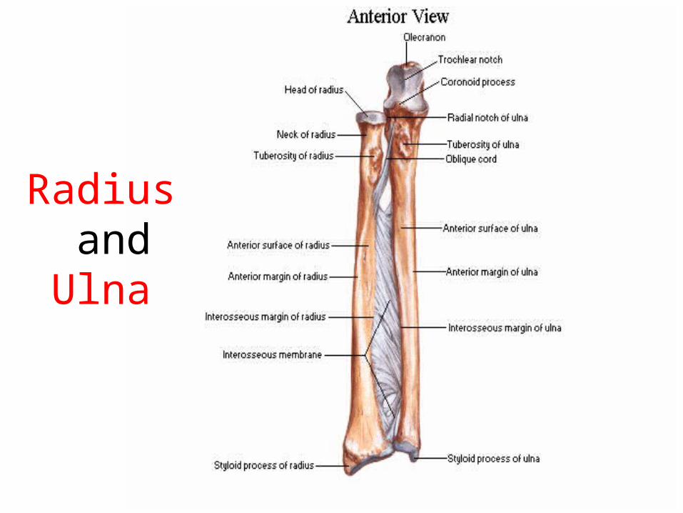
Radius and Ulna
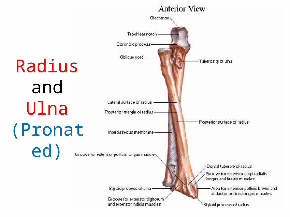
Radius and Ulna
(Pronated)
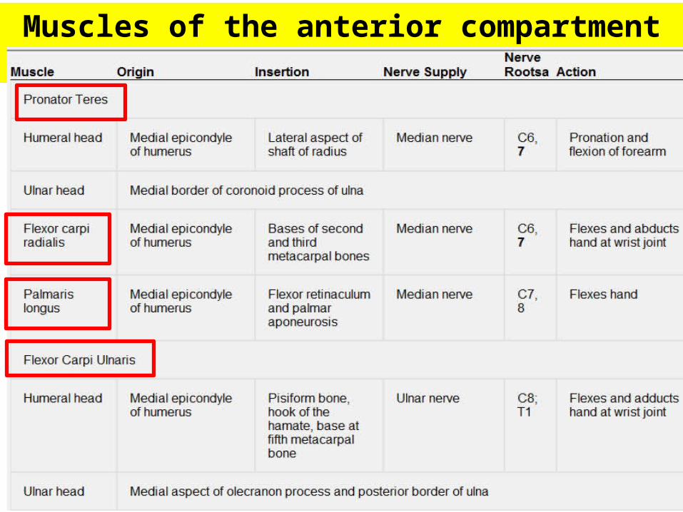
Muscles of the anterior compartment of forearm
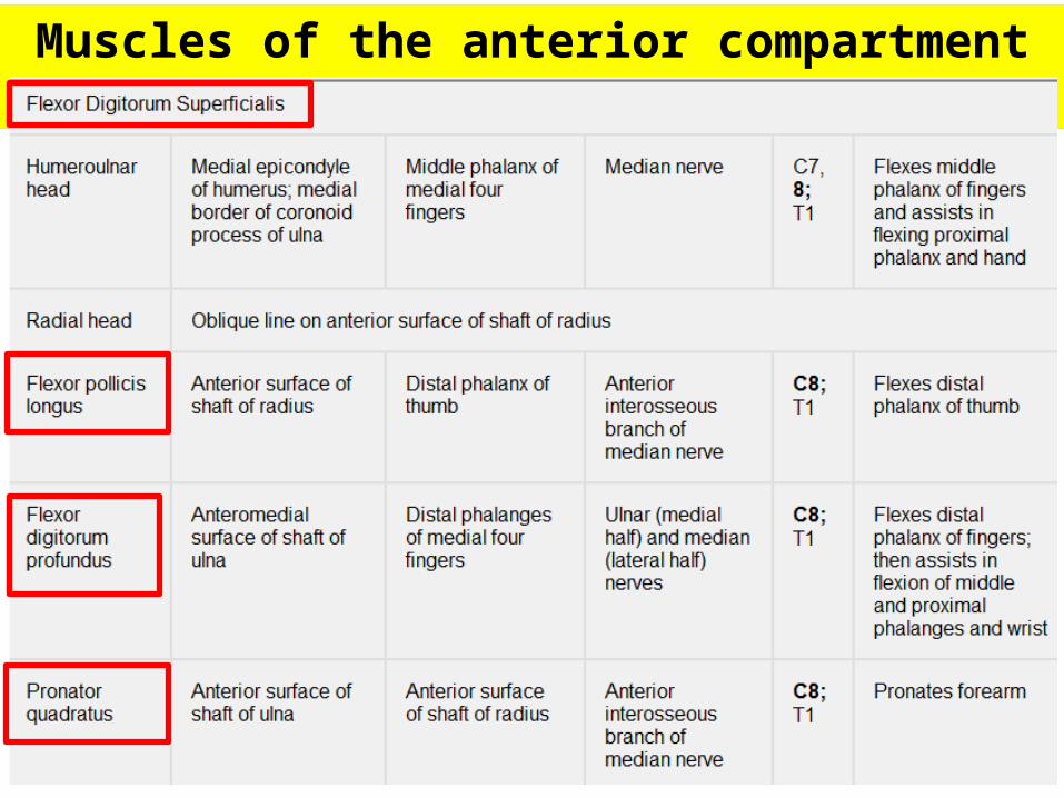
Muscles of the anterior compartment of forearm
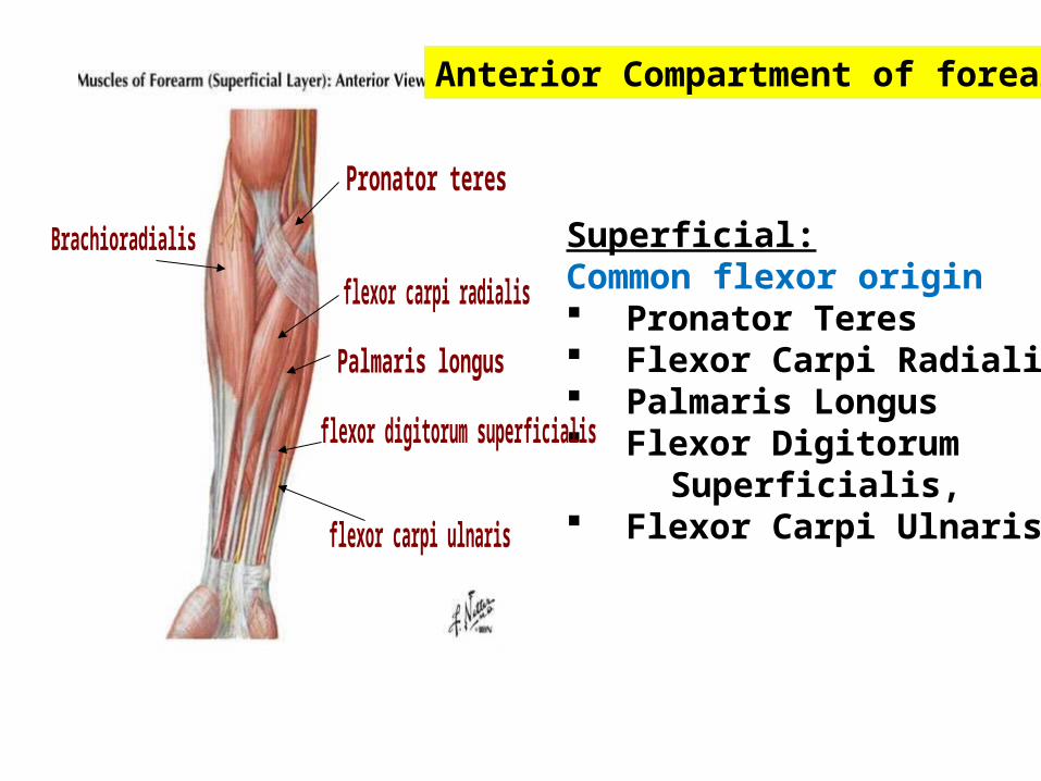
Anterior Compartment of forearm
Superficial:Common flexor origin Pronator Teres Flexor Carpi Radialis Palmaris Longus Flexor Digitorum Superficialis, Flexor Carpi Ulnaris

Functional organization:
Only flex at the wrist.
Flexor carpi radialis Palmaris longus Flexor carpi ulnaris
Anterior Compartment of forearm
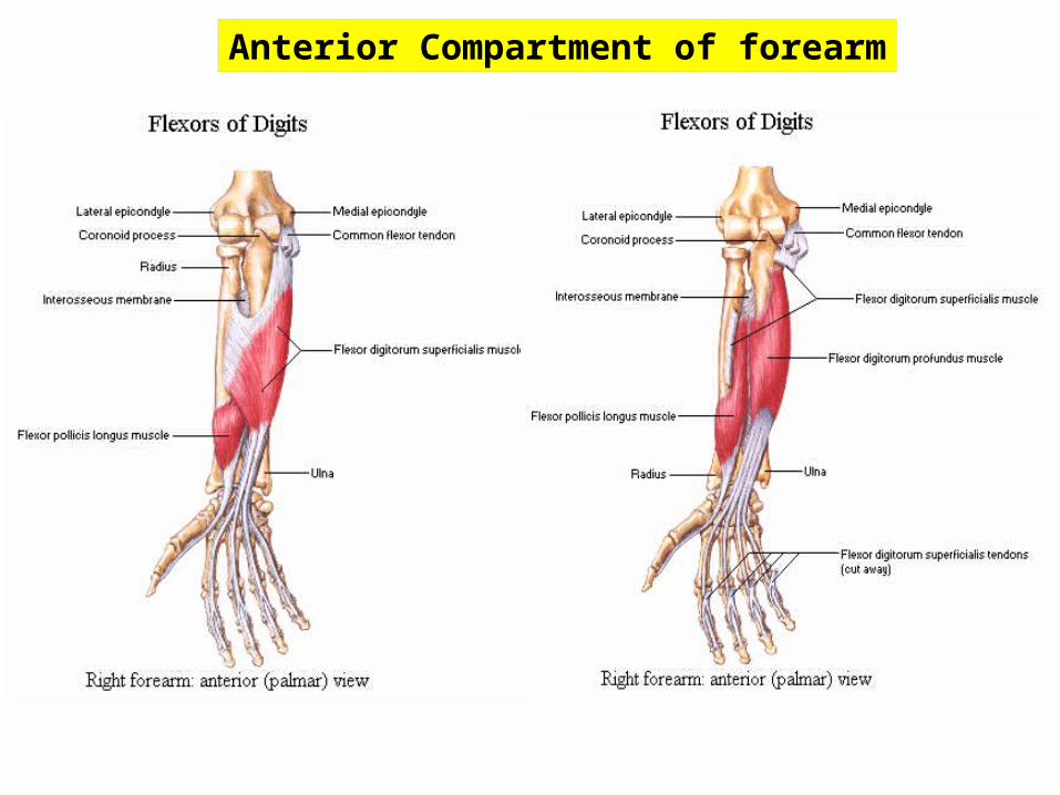
Anterior Compartment of forearm
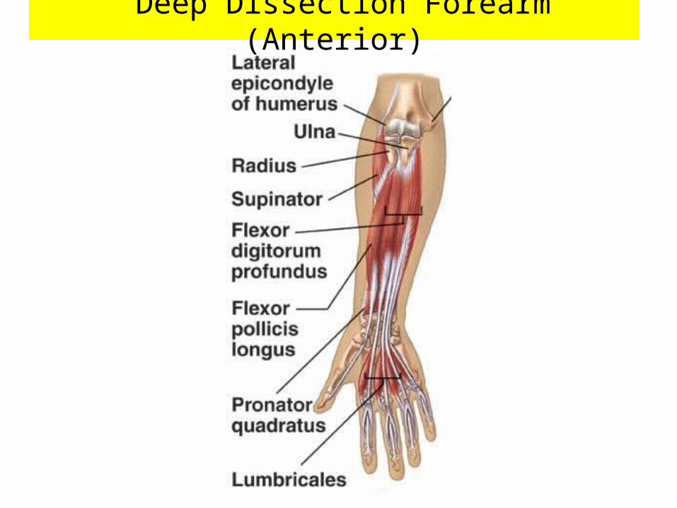
Deep Dissection Forearm (Anterior)
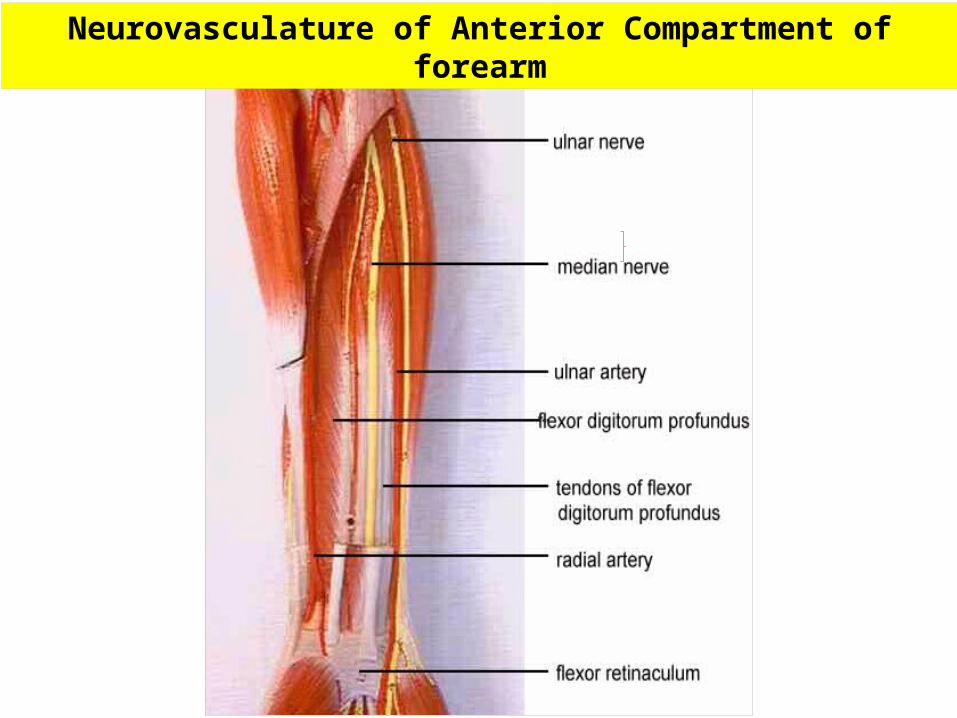
Neurovasculature of Anterior Compartment of forearm
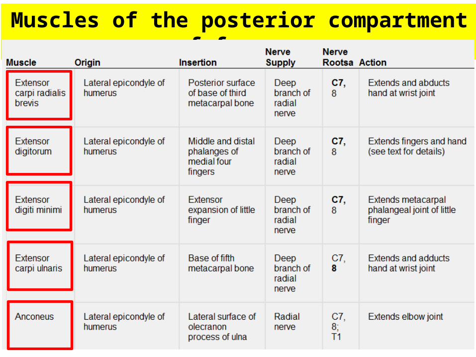
Muscles of the posterior compartment of forearm
Muscles of the posterior compartment of forearm
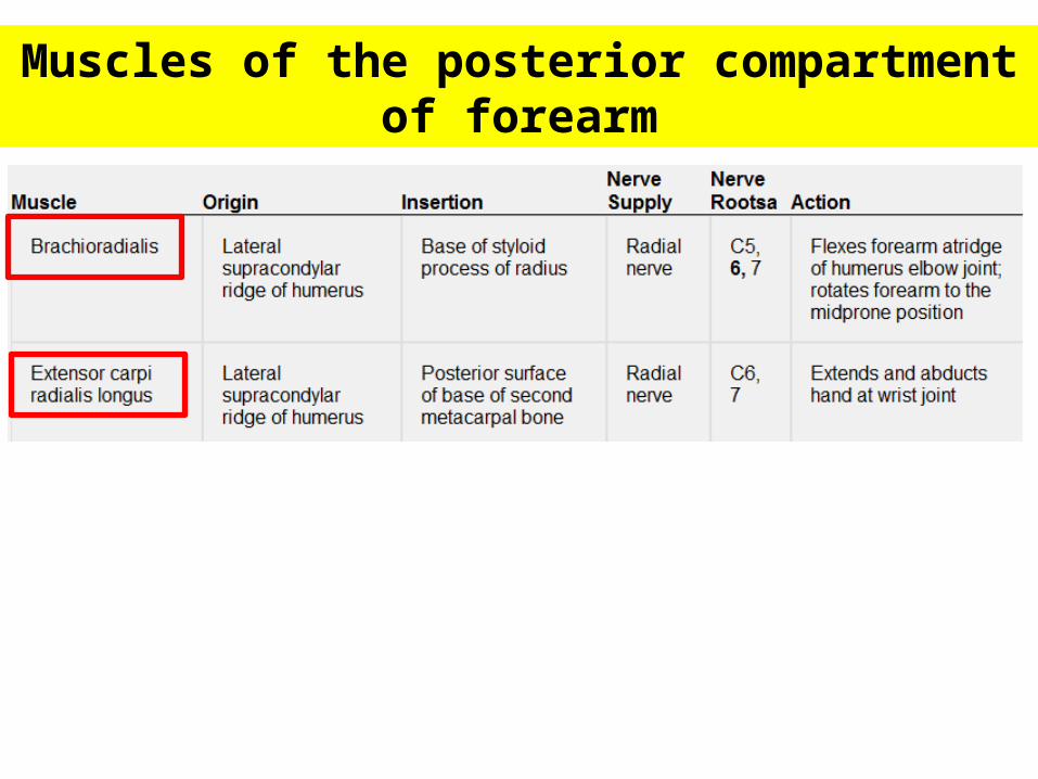
Muscles of the posterior compartment of forearm
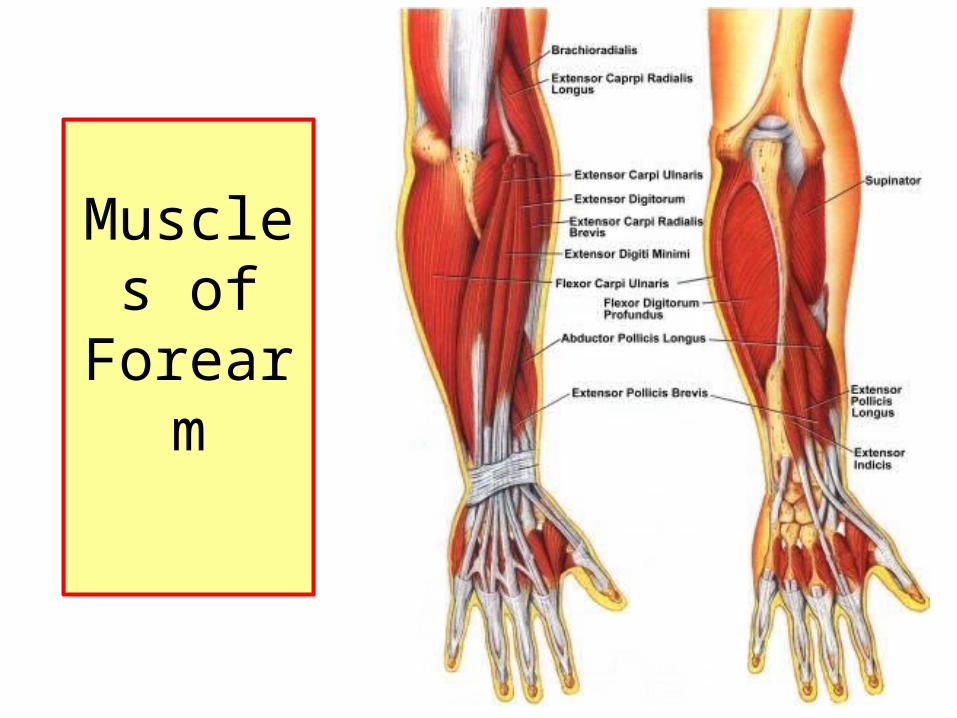
Muscles of
Forearm
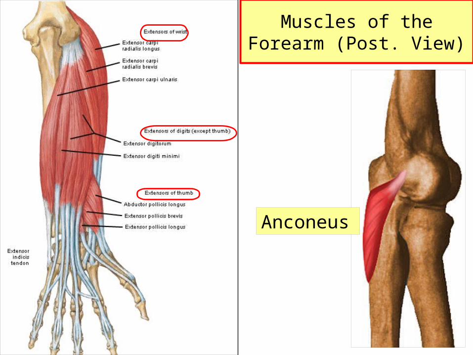
Anconeus
Muscles of the Forearm (Post. View)
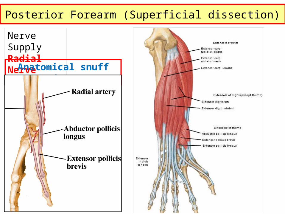
Posterior Forearm (Superficial dissection)Nerve SupplyRadial Nerve
Anatomical snuff box
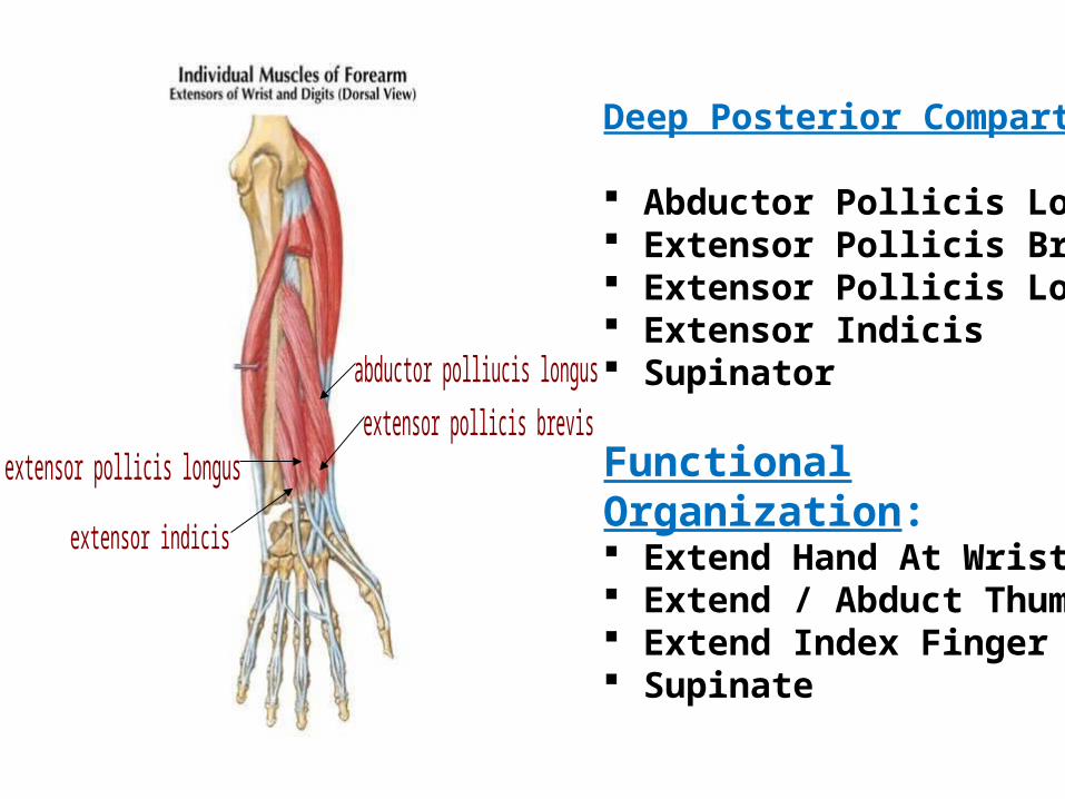
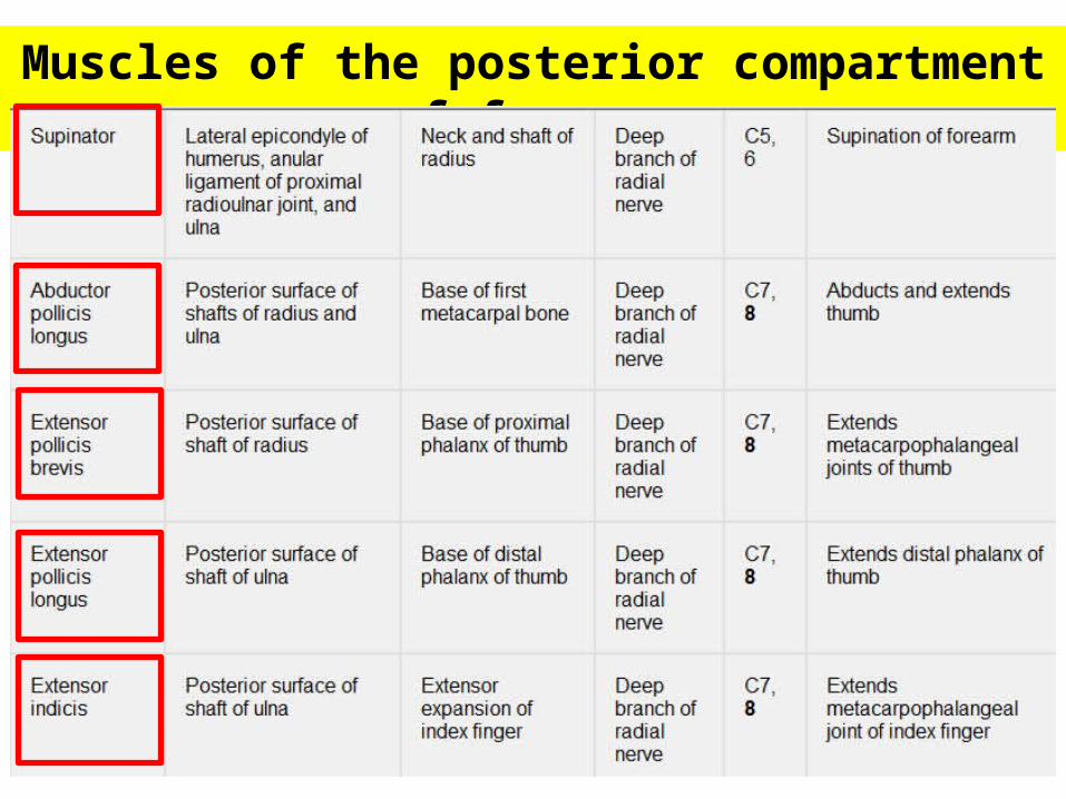
Deep Posterior Compartment
Abductor Pollicis Longus Extensor Pollicis Brevis Extensor Pollicis Longus Extensor Indicis Supinator
Functional Organization: Extend Hand At Wrist Extend / Abduct Thumb Extend Index Finger Supinate
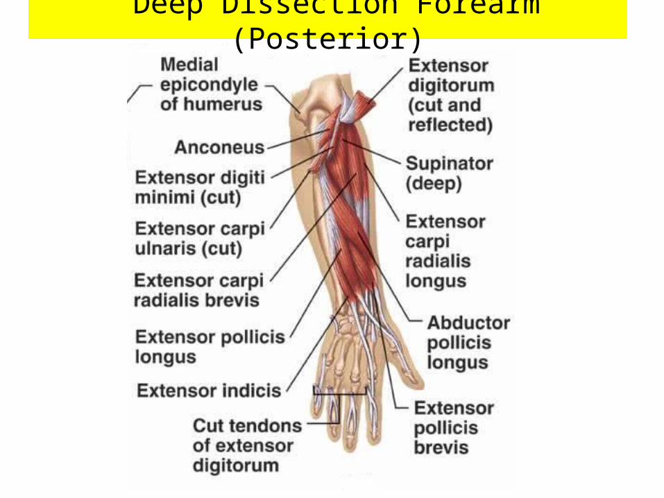
Deep Dissection Forearm (Posterior)
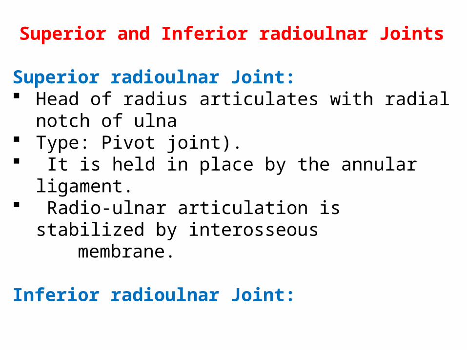
Superior and Inferior radioulnar Joints
Superior radioulnar Joint: Head of radius articulates with radial notch of ulna Type: Pivot joint). It is held in place by the annular ligament. Radio-ulnar articulation is stabilized by interosseous membrane.
Inferior radioulnar Joint:
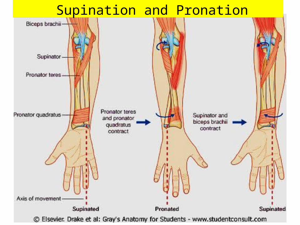
Supination and Pronation

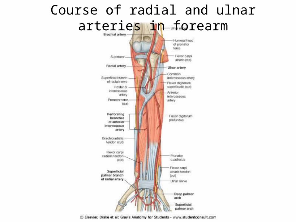
Course of radial and ulnar arteries in forearm
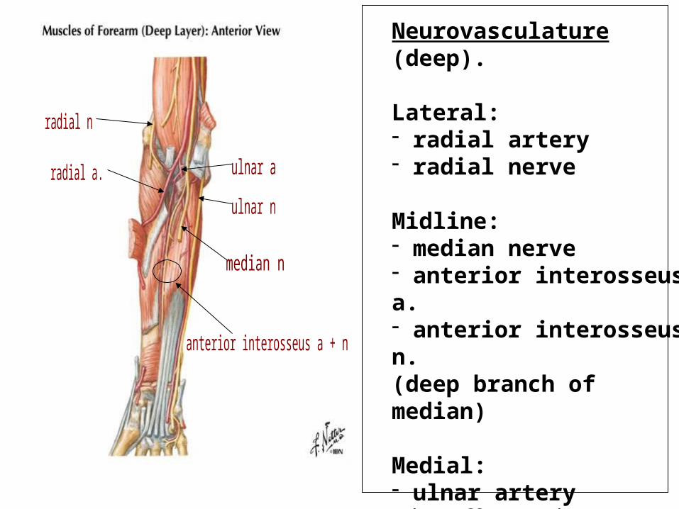
Neurovasculature (deep).
Lateral: - radial artery - radial nerve
Midline: - median nerve- anterior interosseus a. - anterior interosseus n.(deep branch of median)
Medial: - ulnar artery(gives off common interosseus artery divides into anterior and posterior branches)
- ulner nerve

Neurovasculature.
Radial nerve and its branches supplyall muscles in posterior compartment, including Brachioradialis (!).
- superficial radial nerve- deep radial nerve - posterior interosseous nerve.
Posterior interosseous artery runs between superficial and deep muscles

Radial & Ulnar Arteries
mediallateral
Ulnar artery
Common interosseousAnteriorPosterior
Dorsal and palmer carpal branches
Radial artery
superficial (deep)palmar arches
Deep (superficial) palmar arches
Dorsal and palmer carpal branches
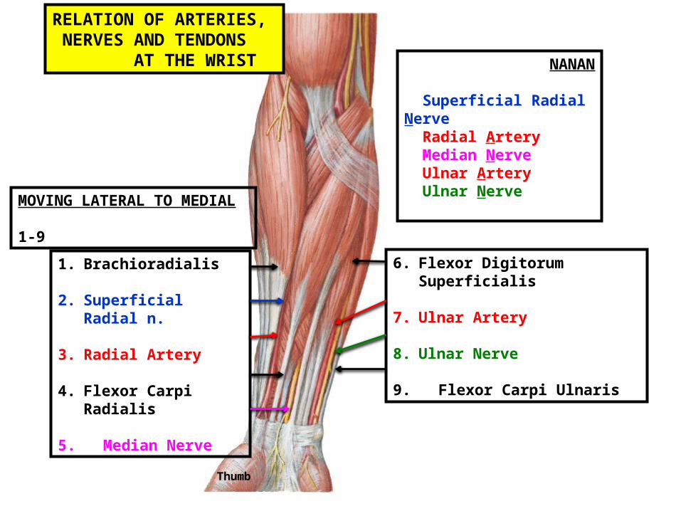
RELATION OF ARTERIES, NERVES AND TENDONS AT THE WRIST NANAN
Superficial Radial Nerve Radial Artery Median Nerve Ulnar Artery Ulnar Nerve
1. Brachioradialis
2. Superficial Radial n.
3. Radial Artery
4. Flexor Carpi Radialis
5. Median Nerve
6. Flexor Digitorum Superficialis
7. Ulnar Artery
8. Ulnar Nerve
9. Flexor Carpi Ulnaris
MOVING LATERAL TO MEDIAL 1-9
Thumb

Thank you


