135
Muscular System 2012-2013
| Date post: | 16-Dec-2015 |
| Category: |
Documents |
| Upload: | stella-whitehead |
| View: | 219 times |
| Download: | 2 times |

Muscular System
2012-2013

Vocab development
• Calat- something inserted
• Erg- work• Fasc- bundle • -gram- something written• Hyper- over, more• inter;- between • Iso-equal• Laten- hidden• Myo- muscle
• Reticul- a net• Sarco- flesh• Syn- together• Tetan- stiff• -tonic- stretched• -troph- well fed • Voluntar- of one’s free
will

Introduction
• Muscles are organs made of cells that use chemical energy stored in nutrients to exert a force on the structures they are attached to.
• Muscle actions provide:– Muscle tone– Propel body fluids and food– Generate the heartbeat– Distribute heat
Introduction
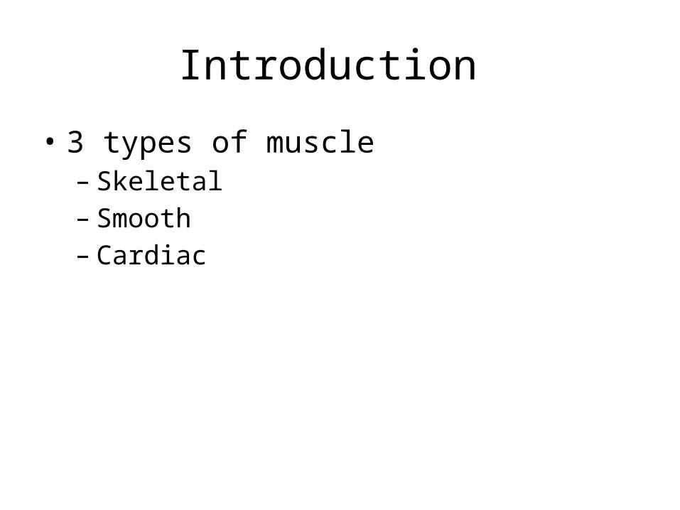
• 3 types of muscle– Skeletal– Smooth– Cardiac

Structure of Skeletal Muscle
• Composed mostly of skeletal muscle tissue, nervous tissue, blood, and other connective tissues
• Layers of connective tissue enclose and separate all parts of a skeletal muscle allowing the parts to move somewhat independently.

Skeletal Muscle: Connective Tissue Coverings
• Fascia– Separates a muscle from its adjacent muscles;
covers the whole muscle• Tendon– Connect a muscle to a bone
• Aponeuroses – connects muscle to bone and other muscles
Skeletal Muscle: Connective Tissue Coverings
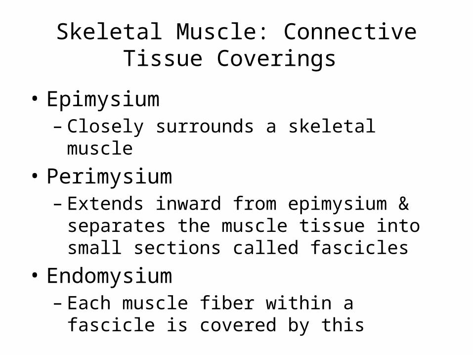
• Epimysium– Closely surrounds a skeletal muscle
• Perimysium– Extends inward from epimysium & separates the
muscle tissue into small sections called fascicles • Endomysium – Each muscle fiber within a fascicle is covered by
this
Skeletal Muscle Fibers
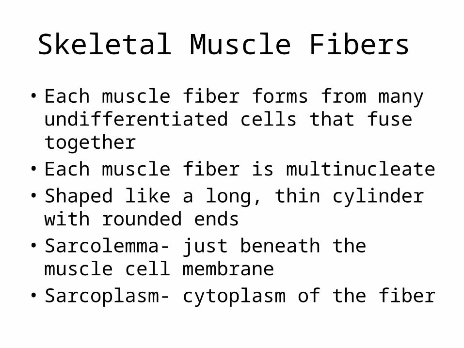
• Each muscle fiber forms from many undifferentiated cells that fuse together
• Each muscle fiber is multinucleate• Shaped like a long, thin cylinder with rounded
ends• Sarcolemma- just beneath the muscle cell
membrane• Sarcoplasm- cytoplasm of the fiber

Skeletal Muscle Fibers
• Myofibrils – Bundles of threadlike structures found within muscle
fibers– Fundamental in the muscle contraction mechanism – Consist of 2 types of proteins
• Myosin- thick filaments• Actin- thin filaments
– Alternating of the myosin & actin causes the striations found in skeletal muscle • Sarcomeres- repeating patterns of striations along each
muscle fiber

Skeletal Muscle Fibers

Skeletal Muscle Fibers

Skeletal Muscle Fibers
• Sarcoplasmic reticulum – Within the sarcoplasm of a muscle fiber – Network of channels that surrounds each
myofibril

Skeletal Muscle Contraction
• Complex interaction of cellular and chemical pieces
• The result is movement within the myofibrils where the filaments of actin and myosin slide past each other causing the sarcomere to shorten

Skeletal Muscle Contraction
• Energy Sources – ATP • Muscle fiber only has enough ATP to contract briefly so
it must be able to regenerate ATP
– Creatine Phosphate • Initial source of energy to regenerate ATP • Much more abundant in muscle fibers than ATP, but it
cannot supply energy directly to the cell
– Cellular Respiration
Skeletal Muscle Contraction
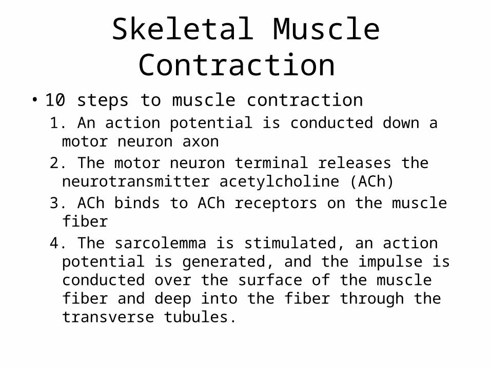
• 10 steps to muscle contraction1. An action potential is conducted down a motor
neuron axon 2. The motor neuron terminal releases the
neurotransmitter acetylcholine (ACh)3. ACh binds to ACh receptors on the muscle fiber4. The sarcolemma is stimulated, an action potential
is generated, and the impulse is conducted over the surface of the muscle fiber and deep into the fiber through the transverse tubules.

Skeletal Muscle Contraction
5. The impulse reaches the sarcoplasmic reticulum, and calcium channels open.
6. Calcium ions diffuse from the sarcoplasmic reticulum into the sarcoplasm and bind to tropin molecules.
7. Tropomyosin molecules move and expose specific sites on actin.
8. Actin and myosin link, forming cross-bridges.9. Thin (actin) filaments are pulled toward the center of the
sarcomere by myosin cross-bridges increasing the overlap of the thin and thick filaments.
10. The muscle fiber contracts.

Skeletal Muscle Relaxation
• 1. Acetylcholinesterase decomposes acetylcholine, and the muscle fiber membrane is no longer stimulated.
• 2. Calcium ions are actively transported into the sarcoplasmic reticulum.
• 3. ATP breaks linkages between actin and myosin filaments without breakdown of ATP itself
• 4. Breakdown of ATP “cocks” the myosin heads. • 5. Troponin and tropomyosin molecules inhibit the
interaction between myosin and actin filaments.• 6. Muscle fiber remains relaxed until it is stimulated again.

Muscle Fatigue
• Caused by– Decreased blood flow– Ion imbalances due to repeated stimulation– Psychological loss of desire to continue – Lactic acid accumulation – Oxygen debt
• Lactic acid accumulation – Accumulates in the muscles when ATP production
goes from aerobic to anaerobic

Muscular Responses
• Threshold Stimulus – A muscle fiber remains unresponsive until a
certain strength of stimulation is reached, once this is reached an action potential is generated and the process of muscle contraction begins

Muscular Responses
• Recording a Muscle Contraction

Muscular Responses
• Summation–

Muscular Responses
• Types of Contractions– Isotonic Contractions (equal force –change in
length)—allow you to move things • Concentric-muscle contracts with greater force than
resistance and shortens • Eccentric- muscle contracts with less force than
resistance and lengthens
– Isometric Contractions – (equal length- change in force) – allow you to sit and hold your posture

Muscular Responses
• Fast & Slow Twitch Muscle Fibers– 3 types
• Slow twitch fibers (red fibers) – Produce ATP from oxygen making them more resistant to fatigue – These fibers can contract for long periods of time without fatigue
• Fast twitch fibers (white fibers) – Produce ATP primarily through glycolysis – Can contract rapidly but also fatigue rapidly as lactic acid
accumulates in them
• Intermediate Fibers (white fibers) – Can contract rapidly and also have a larger respiratory capacity
so they don’t fatigue like fast-twitch fibers

Smooth Muscles
• Smooth muscles lack striations• Cells have only one nucleus• 2 major types of smooth muscles– Multiunit – Visceral

Smooth Muscles
• Multiunit Smooth Muscle – Muscle fibers function as separate units – Found in the irises of the eyes & walls of large
blood vessels – Contract after stimulation by neurons or certain
hormones
Smooth Muscles
• Visceral Smooth Muscle– Fibers respond as a single unit – Found in the walls of hollow organs (intestines, stomach,
bladder, uterus) – Two features- conduction of impulses and rythmicity produce
peristalsis• Peristalsis- wavelike motion of contraction
– Peristalsis is what help your body move food from organ in the digestive system to the next
– Vascular smooth muscle• Found in the walls of small blood vessels where it helps control
blood pressure and blood flow

Cardiac Muscle
• Found only in the heart • Composed of striated cells joined end to end• Opposite ends of cardiac cells are connected
by intercalated discs– Help join cells, transmit the force of contraction, &
diffuse ions from cell to cell

Skeletal Muscle Actions
• Skeletal action depends on – Type of joint it is associated with– The way the muscle is attached on either side of
the joint

Skeletal Muscle Actions
• Body Movement – When a body part moves bones and muscles
interact as a lever – 3 types of levers• 1st class- resistance-fulcrum, force (seesaw; when the
arm straightens at the elbow) • 2nd class- fulcrum- resistance- force (wheelbarrow;
when you chew something up) • 3rd class- resistance-force-fulcrum (tweezers- when the
arm bends at the elbow)

Skeletal Muscle Actions
• Origin and Insertion– Origin- less moveable end of the muscle– Insertion- more moveable end of the muscle – When a muscle contracts • Insertion is pulled toward its origin • Head of the muscle is the part closest to its origin

Skeletal Muscle Action

Skeletal Muscle Actions
• Interaction of Skeletal Muscles– Agonist- muscle that causes an action – Synergists- muscles that work together– Prime mover- muscle that does most of the work
during an action – Antagonists- muscle that opposes action

Major Skeletal Muscles
• Muscles of Facial Expression – Innervated by the facial nerve (CN VII) – Lack of symmetry in facial expression may indicate
nerve damage

Muscles of Facial Expression
• Orbicularis oculi– orbicular= circular– Oculi= eye – Origin: orbital rim,
frontal & maxillary bones– Insertion: lateral region
of eye, some encircle the eye
– Action: closing the eyelid– Expression: form’s crows
feet

Muscles of Facial Expression
• Corrugator– Origin: frontal bone– Insertion: eyebrow– Action: draws eyebrow
medially & inferiorly– Expression: frowning &
suffering

Muscles of Facial Expression
• Procerus– Origin: fascia covering
the lower nasal bone & upper lateral nasal cartilage
– Insertion: skin between and above the eyebrows
– Action: causes transverse wrinkles over the bridge of the nose
– Expression: squinting

Muscles of Facial Expression
• Nasalis – Circles the opening of
the nostrils– Has 2 parts:
• Dilator naris • Compressor naris
– Action: dilates & compresses nostrils • Wiggles your nostrils

Muscles of Facial Expression
• Epicranius– Origin: occipital bone – Insertion: skin around
the eye & orbicularis oculi
– Action: elevates eyebrows, moves scalp forward & backward
– Expression: surprise

Muscles of Facial Expression
• Orbicularis Oris– Oribicular= circle– oris = mouth – Origin: encircles mouth– Insertion: angle of mouth – Action: encloses &
protrudes up; helps keep food on occlusal surfaces during chewing
– Expression: closing or pursing lips

Muscles of Facial Expression
• Quadratus Labii Superioris – 4 muscles of the upper
lip • Levator labii superioris
alaeque nasi• Levator labii superioris• Zygomaticus minor• Zygomaticus major
– Allow you to frown and smile

Muscles of Facial Expression
• Quadratus Labii Superioris cont…– Levator labii superioris
alaeque nasi• Origin: maxilla• Insertion: nose• Action: dilates nostrils &
raises upper lip

Muscles of Facial Expression
• Quadratus Labii Superioris – Levator labii superioris
• Origin: maxilla• Insertion: upper lip• Action: raises upper lip • Expression: scorn

Muscles of Facial Expresssion
• Quadratus Labii Superioris Cont…– Zygomaticus minor
• Origin: zygomatic bone• Insertion: upper lip• Action: raises upper lip• Expression: scorn
– Zygomaticus major • Origin: zygomatic bone• Insertion: angle of mouth• Action: elevates the corner
of the mouth • Expression: smiling

Muscles of Facial Expression • Levator Anguli Oris– Origin: canine fossa (on
the maxilla)– Insertion: orbicularis oris– Action: elevates the
angle of the mouth – Expression: smiling
(laughing)

Muscles of Facial Expression
• Smiling– Produced by the
contraction of 2 facial muscles: • Zygomaticus major • Oribicularis oculi

Muscles of Facial Expression
• Risorius– Origin: fasica superficial
to masseter muscle– Insertion: angle of the
mouth– Action: pulls angle of the
mouth laterally– Expression: smiling
widely; grinning

Muscles of Facial Expression
• Depressor labii inferioris– Origin: mandible– Insertion: lower lip– Action: depresses the
angle of the mouth – Expression: sadness;
grief

Muscles of Facial Expression
• Depressor Anguli Oris – A.K.A triagularis – Origin: mandible– Insertion: angle of the
mouth– Action: depresses angle
of the mouth– Expression: frowning

Muscles of Facial Expression
• Mentalis – Origin: mandible near
the incisive fossa– Insertion: skin of the
chin– Action: pulls skin of chin
upward; protrudes lower lip; raise lower lip
– Expression: doubt; disdain
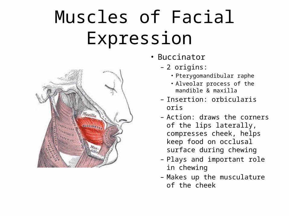
Muscles of Facial Expression • Buccinator
– 2 origins: • Pterygomandibular raphe • Alveolar process of the mandible
& maxilla
– Insertion: orbicularis oris– Action: draws the corners of
the lips laterally, compresses cheek, helps keep food on occlusal surface during chewing
– Plays and important role in chewing
– Makes up the musculature of the cheek

Muscles of Facial Expresion
• Laughter– Muscle that form the core
of the laughter of exhilartion: • Zygomatic major• Oribicularis oculi
– Muscles used to enhance laughter:• Levator labii superioris• Risorius • Mentalis• Depressor anguli oris• Orbicularis oris

Muscles of Facial Expression
• Auriculares– 3 small muscles around
the auricle of the ear– Not well developed in
man– Allow you to wiggle your
ears

Muscles of Facial Expression
• Platysma – Broad, thin, superficial
muscle – Origin: fascia below clavicle– Insertion: lower border of
mandible from canine to second molar
– Action: depresses angle of the mouth, wrinkles the skin of the neck & upper chest
– Expression: dejection, horror, grimacing
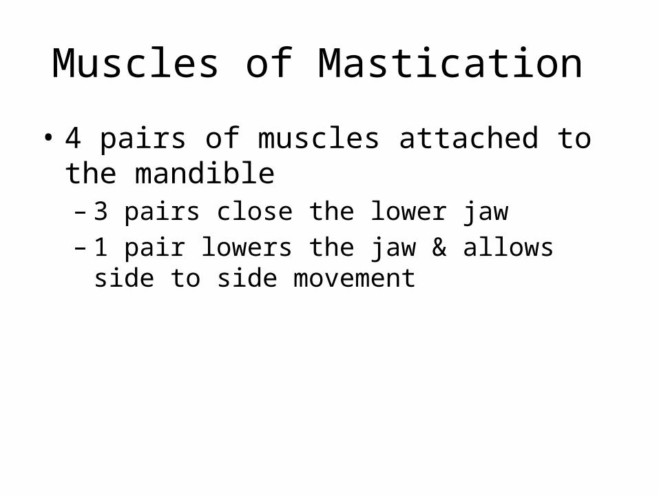
Muscles of Mastication
• 4 pairs of muscles attached to the mandible – 3 pairs close the lower jaw – 1 pair lowers the jaw & allows side to side
movement
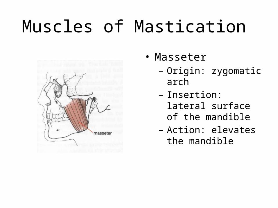
Muscles of Mastication
• Masseter – Origin: zygomatic arch – Insertion: lateral surface
of the mandible – Action: elevates the
mandible

Muscles of Mastication
• Temporalis – Origin: temporal fossa – Insertion: coronoid fossa
of the mandible – Action: elevates the
mandible; retraction
Muscles of Mastication
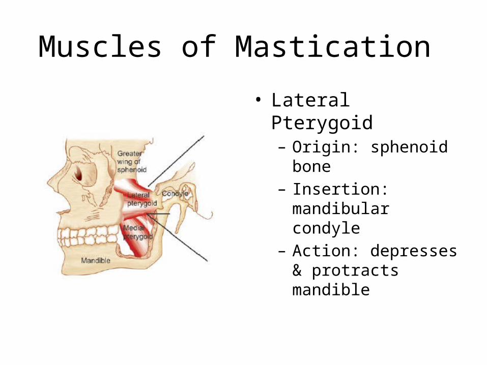
• Lateral Pterygoid– Origin: sphenoid bone– Insertion: mandibular
condyle– Action: depresses &
protracts mandible

Muscles of Mastication
• Medial Pterygoid– Origin: sphenoid,
palatine, & maxilla – Insertion: medial surface
of the mandible – Action: elevates
mandible; moves it from side to side

Muscles That Move the Head and Vertebral Column
• Sternocleidomastoid– Origin:sternum & collar
bone – Insertion: temporal bone– Action: pulls head to one
side, flexes neck or elevates the sternum

Muscles That Move the Head & Vertebral Column
• Splenis Capitis– Origin: spinous process
of lower cervical & upper thoracic vertebrae
– Insertion: occipital bone – Action: rotates head,
bends head to one side, or extends neck

Muscles That Move the Head & Vertebral Column
• Semispinalis capitis– Origin: processes of
lower cervical & upper thoracic vertebrae
– Insertion: occipital bone – Action: elevates head &
rotates the head

Muscles that Move the Head & Vertebral Column
• Quadratus lumborum – Origin: iliac crest– Insertion: upper lumbar
vertebrae & twelfth rib – Action: aids in breathing,
extends lumbar region of vertebral column

Muscles That Move the Head & Vertebral Column
• Erector Spinae – Origin & Insertion at
many locations on the axial skeleton
– Action: extend & rotate the head & maintain the erect position of the vertebral column

Muscles That Move the Pectoral Girdle
• Work closely with the muscles that move the arm
• Connect the scapula to near by bones & help move the scapula up, down, forward, & backward

Muscles That Move the Pectoral Girdle
• Trapezius – Origin: occipital bone &
spines of the cervical & thoracic vertebrae
– Insertion: clavicle, spine, & acromion process of scapula
– Action: rotates scapula; shrugs shoulders

Muscles That Move the Pectoral Girdle
• Rhomboid Major – Origin: spines of upper
thoracic vertebrae– Insertion: medial border
of the scapula – Action: retracts,
elevates, & rotates the scapula

Muscles That Move the Pectoral Girdle
• Rhomboid Minor – Origin: spines of the
lower cervical vertebrae– Insertion: medial border
of the scapula – Action: retracts &
elevates the scapula

Muscles That Move the Pectoral Girdle
• Levator Scapulae – Origin: transverse
process of the cervical vertebrae
– Insertion: medial margin of the scapula
– Action: elevates scapula

Muscles That Move the Pectoral Girdle
• Serratus Anterior – Origin: outer surfaces of
upper ribs– Insertion: ventral surface
of scapula – Action: pulls scapula
anteriorly & downward

Muscles That Move the Pectoral Girdle
• Pectoralis Minor – Origin: sternal ends of
upper ribs – Insertion: coracoid
process of scapula – Action: pulls scapula
forward and downward to raise ribs

Muscles That Move the Forearm
• Most forearm muscle movements are produced by muscles that connect the radius or ulna to the humerus or pectoral girdle.
• Muscles that move the forearm are grouped into three categories: – Flexors- – Extensors– Rotators

Muscles That Move the Forearm
• Flexor: – Biceps Brachii
• Origin: above the glenoid cavity of the scapula
• Insertion: radius• Action: flexes elbow &
rotates the hand laterally (turning a doorknob or screw driver)

Muscles That Move the Forearm
• Flexor– Brachialis
• Origin: anterior shaft of the humerus
• Insertion: coronoid process of ulna
• Action: Flexes elbow – Strongest flexor of the
elbow

Muscles That Move the Forearm
• Flexor:– Brachioradialis
• Origin: distal lateral end of humerus
• Insertion: lateral surface of the radius above the styloid process
• Action: flexes elbow

Muscles That Move the Forearm
• Extensor – Triceps Brachii
• Origin: below glenoid cavity & lateral & medial surfaces of the humerus
• Insertion: olecranon process of the ulna
• Action: extends elbow • This is the only muscle on
the back of the arm.

Muscles That Move the Forearm
• Rotators:– Supinator
• Origin: lateral epicondyle of humerus & ulna
• Insertion: lateral surface of radius
• Action: rotates forearm laterally and supinates the hand (palm facing upward)

Muscles That Move the Forearm
• Rotators:– Pronator teres
• Origin: medial epicondyle of humerus and the ulna
• Insertion: lateral surface of radius
• Action: rotates forearm medially and pronates the hand

Muscles That Move the Forearm
• Rotator:– Pronator Quadratus
• Origin: anterior distal end of ulna
• Insertion: anterior distal end of radius
• Action: rotates forearm medially and pronates hand
Muscles That Move the Hand
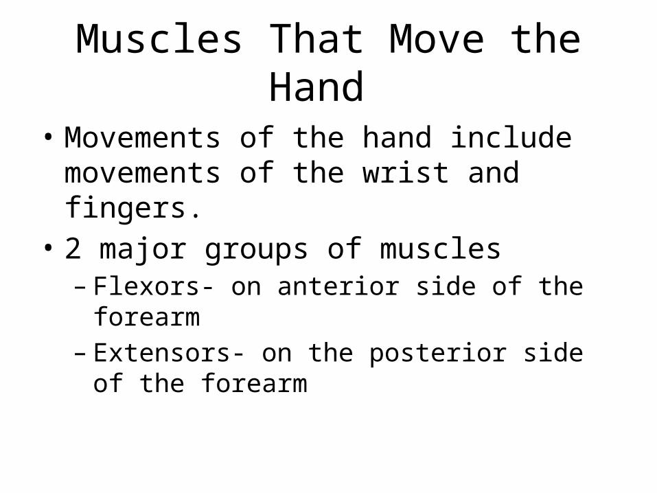
• Movements of the hand include movements of the wrist and fingers.
• 2 major groups of muscles– Flexors- on anterior side of the forearm – Extensors- on the posterior side of the forearm

Muscles That Move the Hand
• Flexors – Flexor carpi radialis
• Origin: medial epicondyle of the humerus
• Insertion: base of the 2nd & 3rd metacarpals
• Action: flexes wrist & abducts hand

Muscles That Move the Hand
• Flexor– Flexor carpi ulnaris
• Origin: medial epicondyle of the humerus
• Insertion: carpals & metacarpals
• Action: flexes the wrist & adducts the hand

Muscles that Move the Hand
• Flexors– Palmaris longus
• Origin: medial epicondyle of humerus
• Insertion: fascia of the palm
• Action: flexes wrist; like you are telling someone to come here

Muscles That Move the Hand
• Flexors– Flexor Digitorum
Profundus • Origin: anterior surface of
the ulna • Insertion: bases of distal
phalanges in fingers 2-5 • Action: flexes distal joints
of fingers

Muscles that Move the Hand
• Flexor– Flexor digitorum
superficialis • Origin: humerus • Insertion: tendons of
fingers • Action: flexes the fingers
and wrist

Muscles that Move the Hand
• Extensor – Extensor Carpi Radialis
Longus • Origin: distal end of the
humerus • Insertion: base of 2nd
metacarpal • Action: extends wrist and
abducts the hand

Muscles that Move the Hand
• Extensor– Extensor carpi radialis
brevis • Origin: lateral epicondyle
of the humerus • Insertion: base of 2nd & 3rd
metacarpals • Action: extends wrist &
abducts hand

Muscles that Move the Hand
• Extensors – Extensor carpi ulnaris
• Origin: lateral epicondyle of humerus
• Insertion: base of the 5th metacarpal
• Action: extends wrist & adducts hand

Muscles that Move the Hand
• Extensor – Extensor Digitorum
• Origin: lateral epicondyle of the humerus
• Insertion: posterior surface of phalanges in fingers 2-5
• Action: extends fingers

Muscles that Move the Arm
• Flexors – Coracobrachialis
• Origin: coracoid process of the scapula
• Insertion: shaft of the humerus
• Action: flexes & adducts the arm
Muscles that Move the Arm
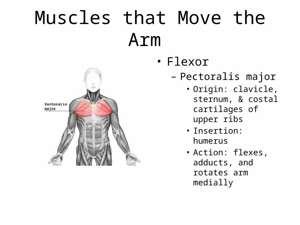
• Flexor– Pectoralis major
• Origin: clavicle, sternum, & costal cartilages of upper ribs
• Insertion: humerus • Action: flexes, adducts,
and rotates arm medially

Muscles that Move the Arm
• Extensor– Teres Major
• Origin: lateral border of scapula
• Insertion: humerus • Action: extends, adducts,
and rotates the arm medially

Muscles that Move the Arm
• Extensor– Latissimus Dorsi
• Origin: spines of scral, lumbar, & lower thoracic vertebrae, iliac crest, & lower ribs
• Insertion: humerus • Action: extends, adducts,
and rotates the arm medially, or pulls the should downward & back

Muscles that Move the Arm
• Abductors – Supraspinatus
• Origin: posterior surface of scapula above spine
• Insertion: humerus• Action: abducts the arm

Muscles that Move the Arm
• Abductors – Deltoid
• Origin: acromion process, spine of the scapula, & clavicle
• Insertion: humerus • Action: abducts, extends,
& flexes the arm

Muscles that Move the Arm
• Rotators – Subscapularis
• Origin: Anterior surface of scapula
• Insertion: humerus• Action: rotates arm
medially

Muscles that Move the Arm
• Rotators – Infraspinatus
• Origin: posterior surface of scapula below spine
• Insertion: humerus • Action: rotates arm
laterally

Muscles that Move the Arm
• Rotators – Teres Minor
• Origin: lateral border of scapula
• Insertion: humerus • Action: rotates arm
laterally

Muscles of the Abdominal Wall
• Muscles of the abdominal wall connect the rib cage & vertebral column to the pelvic girdle
• Linea alba- band of tough connective tissue that extends from the xiphoid process of the sternum to the pubic symphysis & provides attachment for some of the abdominal muscles
• Contraction of these muscles helps move air out of the lungs during forceful exhalation & other everyday functions of the body

Muscles of the Abdominal Wall
• External oblique – Origin- outer surfaces of
the lower ribs – Insertion- Outer lip of
iliac crest & linea alba – Action- Tenses
abdominal wall & compresses abdominal contents

Muscles of the Abdominal Wall
• Internal Oblique – Origin- crest of ilium &
inguinal ligament – Insertion- cartilages of
the lower ribs, linea alba, & crest of the pubis
– Action- Tenses abdominal wall & compresses abdominal contents

Muscles of the Abdominal Wall
• Transversus abdominis – Origin- costal cartilages of
the lower ribs, processes of the lumbar vertebrae, lip of iliac crest, & inguinal ligament
– Insertion- linea alba & crest of pubis
– Action- tenses abdominal wall & compresses abdominal contents

Muscles of the Abdominal Wall
• Rectus Abdominis – Origin- Crest of the pubis
& pubic symphysis – Insertion- xiphoid
process of sternum & costal cartilage
– Action- tenses the abdominal wall & compresses abdominal contents & also flexes the vertebral column

Muscles that Move the Thigh
• Muscles that move the thigh are attached to the femur & to part of the pelvic girdle – Important exceptions: sartorius & rectus femoris
• Muscles can be separated into 2 groups:– Anterior- primarily flexes the thigh; advance the
lower limb when walking – Posterior- primarily extends, abducts, or rotates
the thigh

Muscles that Move the Thigh: Anterior Group
• Psoas major– Origin: lumbar
intervertebral discs; bodies and transverse processes of lumbar vertebrae
– Insertion: lesser trochanter of the femur
– Action: flexes the thigh

Muscles that Move the Thigh: Anterior Group
• Iliacus – Origin: Illiac fossa of
ilium – Insertion: lesser
trochanter of the femur – Action: Flexes thigh

Muscles that Move the Thigh: Posterior Group
• Gluteus maximus – Origin: sacrum, coccyx, &
posterior surface of the ilium
– Insertion: posterior surface of the femur & fascia of the thigh
– Action: extends hip; helps straighten the lower limb at the hip when you walk, run, or climb

Muscles that Move the Thigh: Posterior Group
• Gluteus minimus – Origin: lateral surface of
the ilium– Insertion: greater
trochanter of the femur – Action: abducts &
rotates the thigh medially

Muscles that Move the Thigh: Posterior Group
• Gluteus medius – Origin: lateral surface of
the ilium– Insertion: greater
trochanter of the femur – Action: abducts &
rotates thigh medially

Muscles that Move the Thigh: Posterior Group
• Piriformis – Origin: anterior surface
of the sacrum – Insertion: greater
trochanter of the femur – Action: abducts &
rotates the thigh medially ; stabilizes the hip

Muscles that Move the Thigh: Posterior Group
• Tensor fasciae latae – Origin: anterior iliac
crest– Insertion: greater
trochanter of the femur – Action: abducts, flexes,
& rotates thigh medially

Muscles that Move the Thigh: Adductors
• Pectineus – Origin: spine of the pubis – Insertion: femur distal to
lesser trochanter– Action: Flexes & adducts
thigh

Muscles that Move the Thigh: Adductors
• Adductor brevis – Origin: pubic bone – Insertion: posterior
surface of femur – Action: adducts & flexes
thigh

Muscles that Move the Thigh: Adductors
• Adductor longus – Origin: pubic bone near
the pubic symphysis– Insertion: posterior
surface of the femur – Action: adducts & flexes
the thigh

Muscles that Move the Thigh: Adductors
• Adductor magnus – Origin: Ischial tuberosity – Insertion: posterior
surface of the femur– Action: adducts thigh,
posterior portion extends & anterior portion flexes thigh

Muscles that Move the Thigh: Adductors
• Gracilis – Origin: Lower edge of
pubic symphysis– Insertion: medial surface
of the tibia – Action: adducts thigh &
flexes knee

Muscles that Move the Leg
• Connect the tibia or fibula to the femur or pelvic girdle.
• Two major groups:– Flexors– Extensors

Muscles that Move the Leg
• Hamstring Group – Biceps femoris
• Origin: ischial tuberosity & linea aspera
• Insertion: head of fibula & lateral condyle of tibia
• Action: flexes knee, rotates leg laterally & extends thigh

Muscles that Move the Leg
• Hamstring Group – Semitendinosus
• Origin: ischial tuberosity • Insertion: medial surface
of the tibia • Action: flexes knee,
rotates leg medially & extends thigh

Muscles that Move the Leg
• Hamstring Group– Semimembranosus
• Origin: ischial tuberosity• Insertion: medial condyle
of tibia • Action: Flexes the knee,
rotates the leg medially & extends the thigh

Muscles that Move the Leg
• Sartorius – Origin: anterior superior
iliac spine – Insertion: medial surface
of tibia – Action: flexes knee &
hip, abducts & rotates thigh laterally

Muscles that Move the Leg
• Quadriceps Group– Rectus Femoris
• Origin: spine of the illium & margin of the acetabulum
• Insertion: patella by tendon, which continues as the patellar ligament to the tibia
• Action: extends knee, flexes thigh

Muscles that Move the Leg
• Quadriceps Group– Vastus Lateralis
• Origin: greater trochanter & posterior surface of the femur
• Insertion: patella by tendon, which continues as patellar ligament to the tibia
• Action: extends knee

Muscles that Move the Leg
• Quadriceps Group– Vastus medialis
• Origin: medial surface of the femur
• Insertion: patella by tendon, which continues as patellar ligament to the tibia
• Action: extends knee

Muscles that Move the Leg
• Quadriceps Group – Vastus intermedius
• Origin: anterior & lateral surfaces of femur
• Insertion: patella by tendon, which continues as patellar ligament to the tibia
• Action: extends knee

Muscles that Move the Foot
• Movements of the foot include movements of the ankle & toes
• Attach to the femur, tibia, & fibula to bones of the foot
• Move the foot upward (dorsiflexion) or downward (plantar flexion) and turn the foot so the plantar surface faces medially (inversion) or laterally (eversion)
• 4 types: dorsal flexors, plantar flexors, invertor, evertor

Muscles that Move the Foot
• Dorsal Flexor – Tibialis Anterior
• Origin: lateral condyle & lateral surface of the tibia
• Insertion: tarsal bone & first metatarsal
• Action: dorsiflexion & inversion of foot

Muscles that Move the Foot
• Dorsal Flexor – Fibularis Tertius
• Origin: anterior surface of the tibia
• Insertion: dorsal surface of the 5th metatarsal
• Action: dorsiflexion & eversion of the foot

Muscles that Move the Foot
• Dorsal Flexor – Extensor Digitorum
Longus • Origin: lateral condyle of
tibia & anterior surface of the fibula
• Insertion: dorsal surfaces of 2nd & 3rd phalanges of the 4 lateral toes
• Action: dorsiflexion & eversion of the foot, extends toes

Muscles that Move the Foot
• Dorsal Flexor – Extensor Hallucis Longus
• Origin: anterior surface of the fibula
• Insertion: distal phalanx of the big toe
• Action: extends big toe, dorsiflexion & inversion of foot

Muscle that Move the Foot
• Plantar Flexor– Gastrocnemius
• Origin: lateral & medial condyles of femur
• Insertion: posterior surface of calcaneus
• Action: plantar flexion of foot, flexes knee

Muscles that Move the Foot
• Plantar Flexor – Soleus
• Origin: head & shaft of fibula & posterior surface of the tibia
• Insertion: posterior surface of the calcaneus
• Action: plantar flexion of the foot

Muscles that Move the Foot
• Plantar Flexion– Plantaris
• Origin: femur• Insertion: calcaneus • Action: plantar flexion of
foot, flexes knee

Muscles that Move the Foot
• Plantar Flexor – Flexor Digitorum Longus
• Origin: posterior surface of the tibia
• Insertion: distal phalanges of four lateral toes
• Action: plantar flexion & inversion of foot, flexes four lateral toes

Muscles that Move the Foot
• Invertor – Tibialis Posterior
• Origin: lateral condyle & posterior surface of tibia & posterior surface of fibula
• Insertion: tarsal & metatarsal bones
• Action: plantar flexion & inversion of foot

Muscles that Move the Foot
• Evertor – Fibularis Longus
• Origin: lateral condyle of tibia & head & shaft of the fibula
• Insertion: Tarsal & metatarsal bones
• Action: plantar flexion & eversion of foot, supports arch


