42
Muscular System Muscle Tissue and Organization
| Date post: | 17-Dec-2015 |
| Category: |
Documents |
| Upload: | jeffrey-foster |
| View: | 227 times |
| Download: | 0 times |
Muscular System
Muscle Tissue and Organization
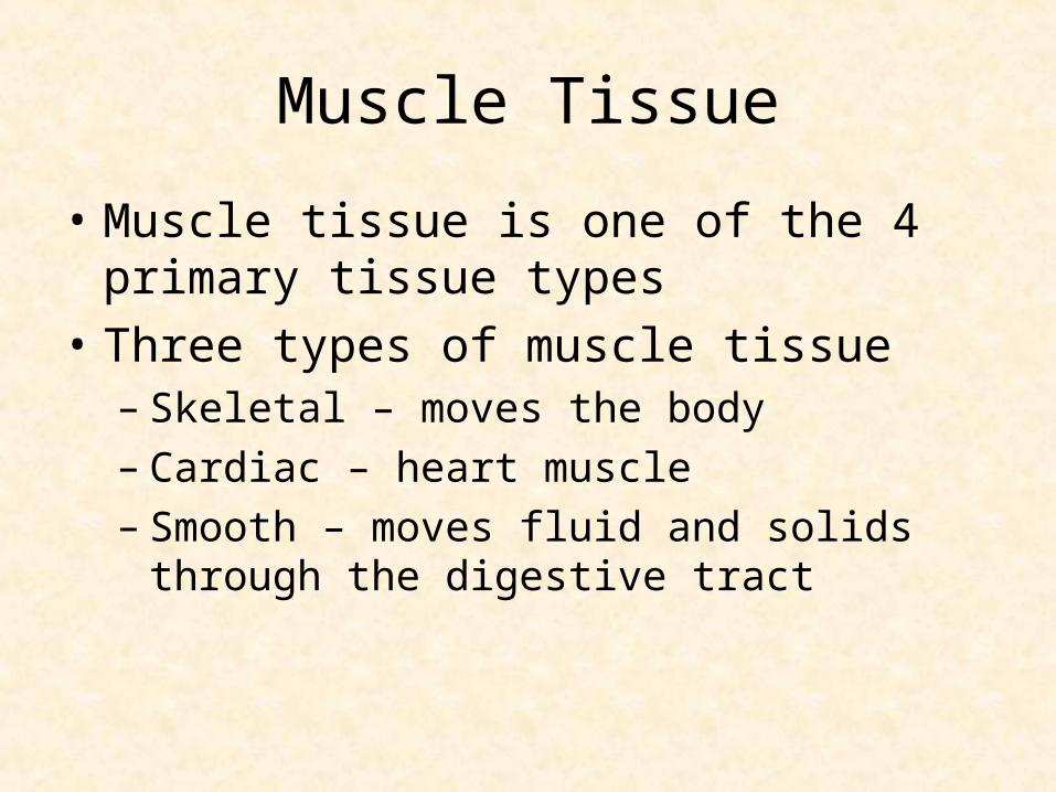
Muscle Tissue
• Muscle tissue is one of the 4 primary tissue types
• Three types of muscle tissue– Skeletal – moves the body– Cardiac – heart muscle– Smooth – moves fluid and solids through the
digestive tract
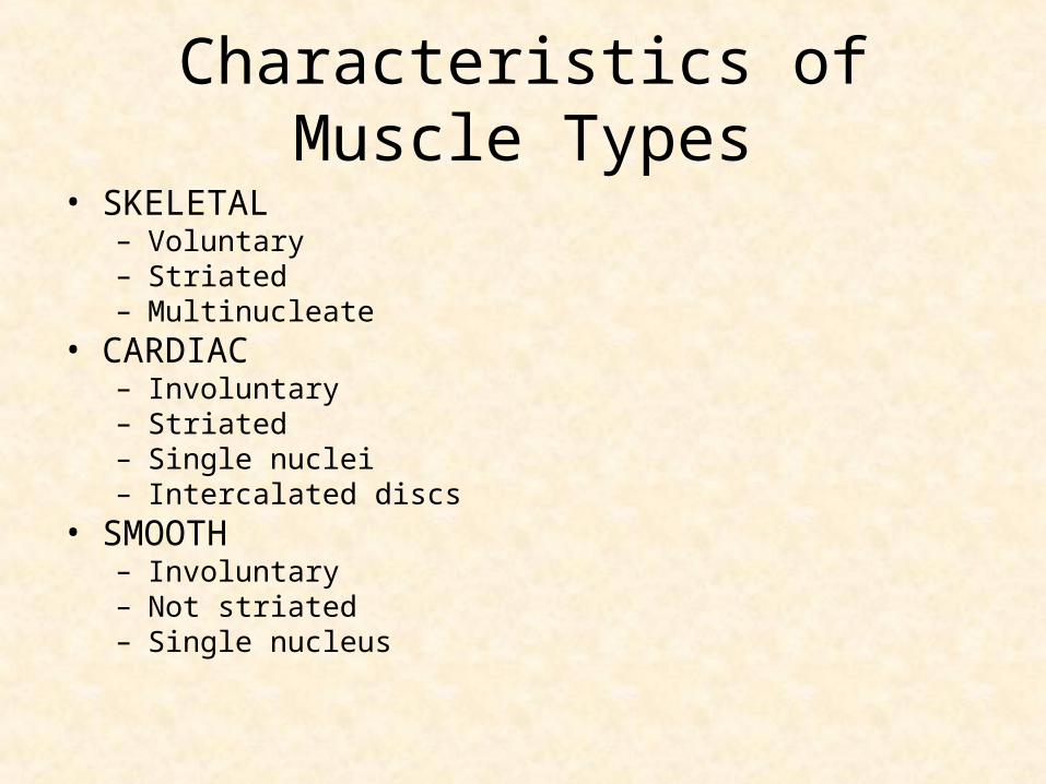
Characteristics of Muscle Types
• SKELETAL– Voluntary– Striated– Multinucleate
• CARDIAC– Involuntary– Striated– Single nuclei– Intercalated discs
• SMOOTH– Involuntary– Not striated– Single nucleus
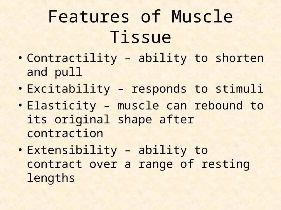
Features of Muscle Tissue
• Contractility – ability to shorten and pull
• Excitability – responds to stimuli
• Elasticity – muscle can rebound to its original shape after contraction
• Extensibility – ability to contract over a range of resting lengths
Functions of Skeletal Muscle
• Movement
• Posture
• Stabilize joints
• Support soft tissue
• Generation of heat
• Regulate entrances and exits (orifices)
Muscle Attachment
• TENDONS attach muscle to bone
• Dense regular CT
• Each muscle has an ORIGIN and INSERTION, and a specific ACTION
• The origin remains stationary while the insertion moves

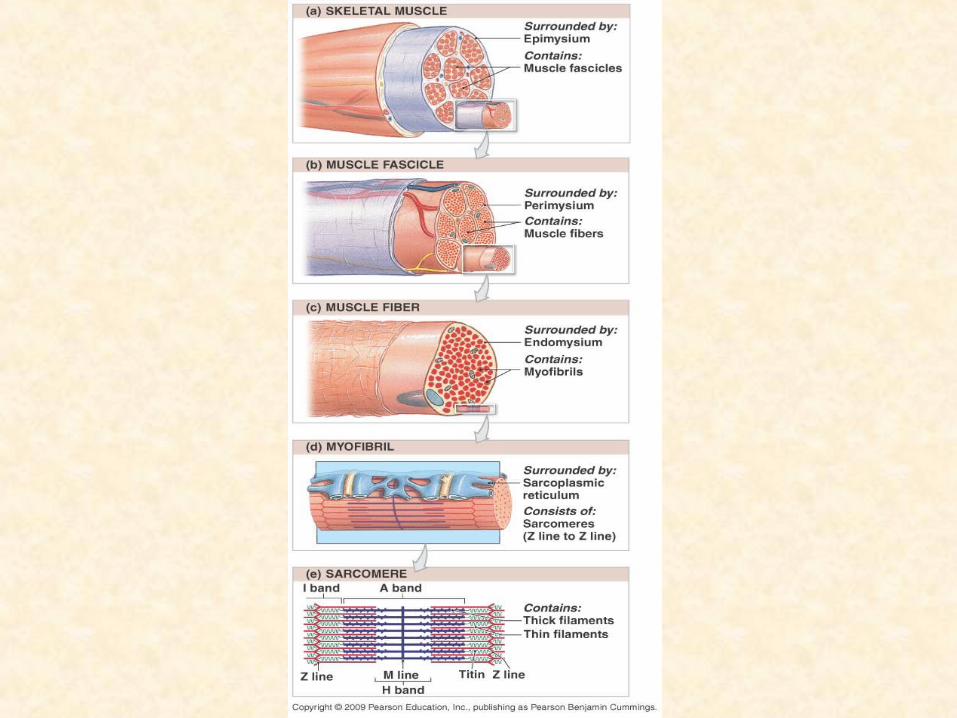
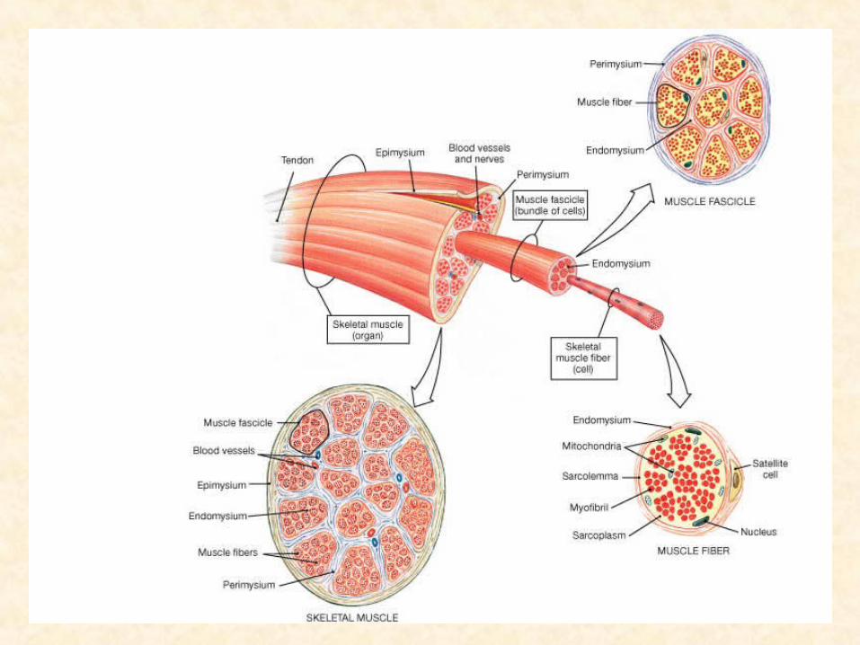
Connective Tissue of Muscle
• Skeletal muscle has three layers of connective tissue
• 1. EPIMYSIUM – dense irregular CT that surrounds the entire muscle
• 2. PERIMYSIUM – divides muscle into compartments or bundles of muscle fibers called FASCICLES
• 3. ENDOMYSIUM – surrounds each muscle fiber (muscle cell)
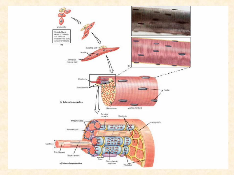
Muscle Cell Terminology
• Muscle cells are very long; muscle fibers• SARCOLEMMA – cell membrane• SARCOPLASM – cytoplasm• Sarcoplasm is filled with thousands of
MYOFIBRILS that are responsible for contraction
• Myofibrils are composed of MYOFILAMENTS• Myofilaments are composed of the proteins
ACTIN and MYOSIN
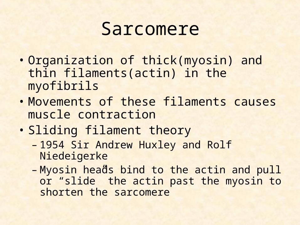
Sarcomere
• Organization of thick(myosin) and thin filaments(actin) in the myofibrils
• Movements of these filaments causes muscle contraction
• Sliding filament theory– 1954 Sir Andrew Huxley and Rolf Niedeigerke– Myosin heads bind to the actin and pull or
“slide” the actin past the myosin to shorten the sarcomere
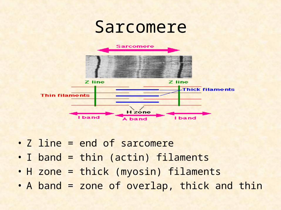
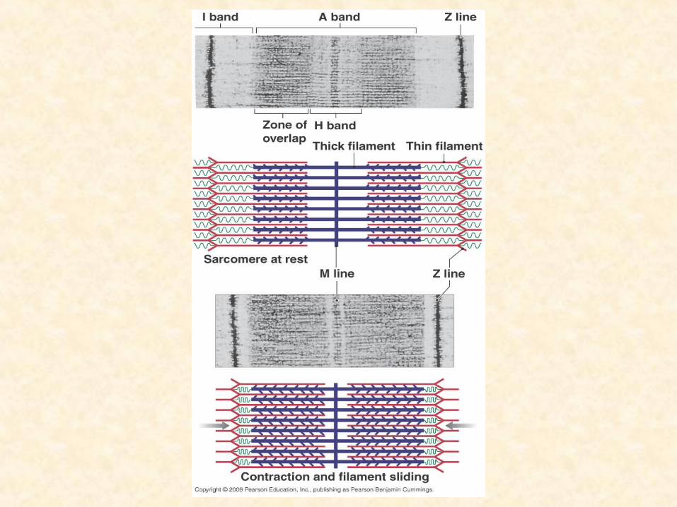
Sarcomere
• Z line = end of sarcomere• I band = thin (actin) filaments• H zone = thick (myosin) filaments• A band = zone of overlap, thick and thin
Thin Filaments
• Twisted strands of globular G actin molecules
• Each molecule of G actin has an active site that can bind to a myosin molecule
• Thin filaments also have two other proteins associated it– Tropomyosin – covers active sites on actin– Troponin - holds tropomyosin in place
Thick filaments
• Bundles of myosin molecules
• About 500 myosin molecules per bundle
• Myosin molecules have heads that can cross bridge to actin active sites
• The binding of myosin heads to actin result in muscle contraction
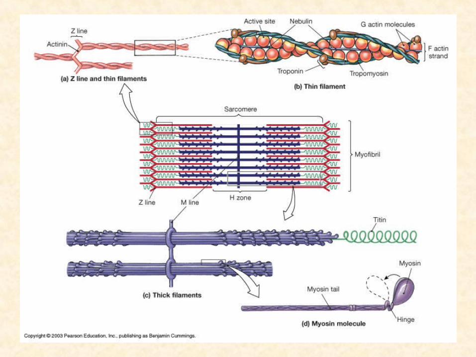
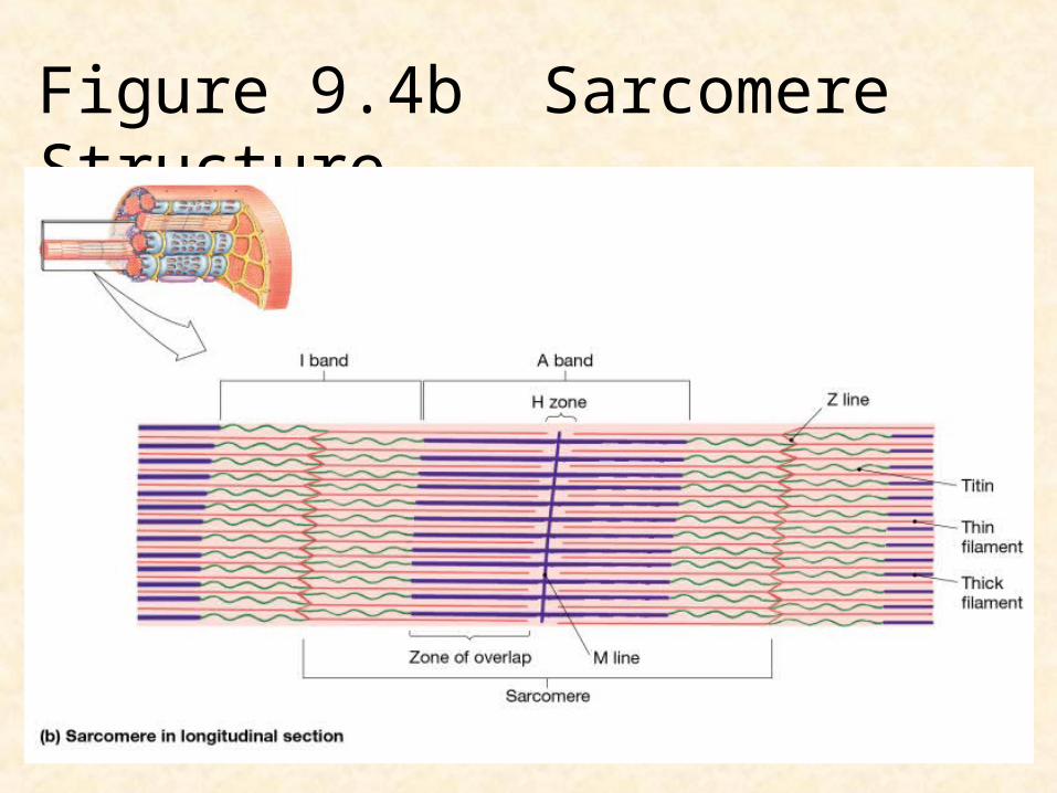
Figure 9.4b Sarcomere Structure
Muscle contraction-Sliding filament theory
• Contraction exerts a pull – tension• Interaction between actin and myosin
triggered by calcium ions and presence of ATP
• Sliding filament theory:– H band and I band get smaller– Zone of overlap gets larger– Z lines move closer together– Width of A band remains constant
Sliding Filament Theory
• Myosin heads cross bridge to the actin active sites
• Myosin attachment “pulls” the actin toward the center of the sarcomere
• Contraction begins with release of Ca2+
from the terminal cisternae of the sarcoplasmic reticulum
• The release of ions is the result of electrical stimulation of the muscle fiber
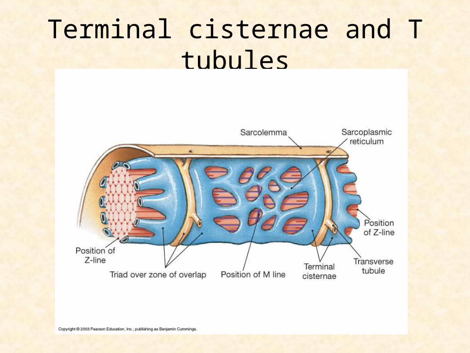
Terminal cisternae and T tubules
T-tubules
• The t-tubules distribute the electrical signal for contraction deep into the muscle fiber
• As the signal travels the terminal cisternae release calcium ions
• Release of calcium cause the troponin molecule to change shape
• Change in troponin causes a change in the position of tropomyosin, myosin can bind to action and contraction occurs!
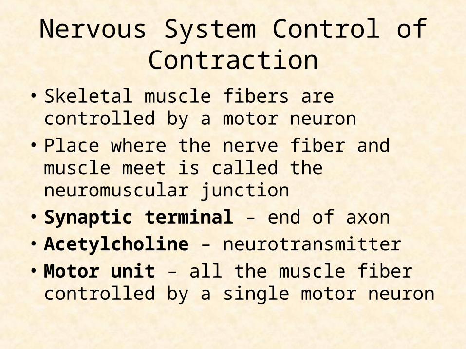
Nervous System Control of Contraction
• Skeletal muscle fibers are controlled by a motor neuron
• Place where the nerve fiber and muscle meet is called the neuromuscular junction
• Synaptic terminal – end of axon
• Acetylcholine – neurotransmitter
• Motor unit – all the muscle fiber controlled by a single motor neuron
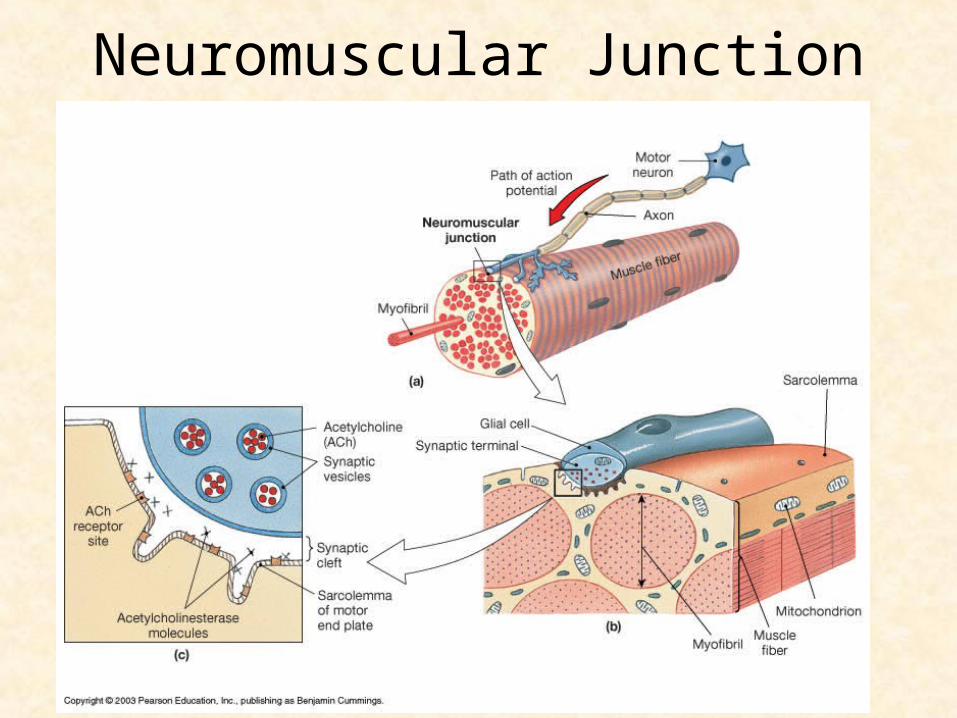
Neuromuscular Junction

Neuromuscular junction
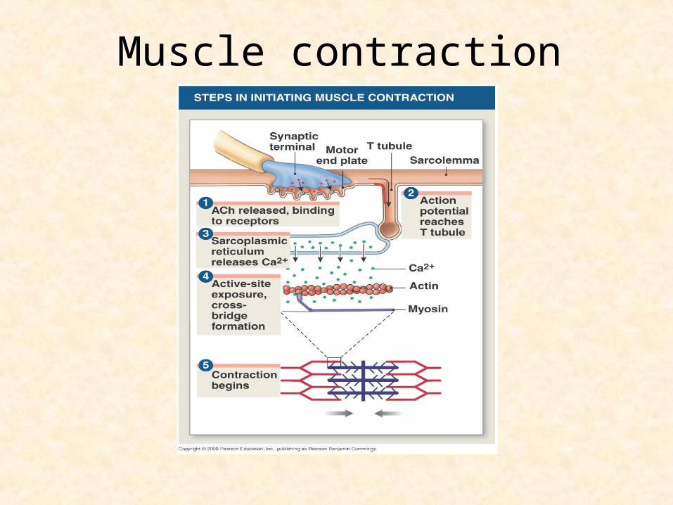
Muscle contraction
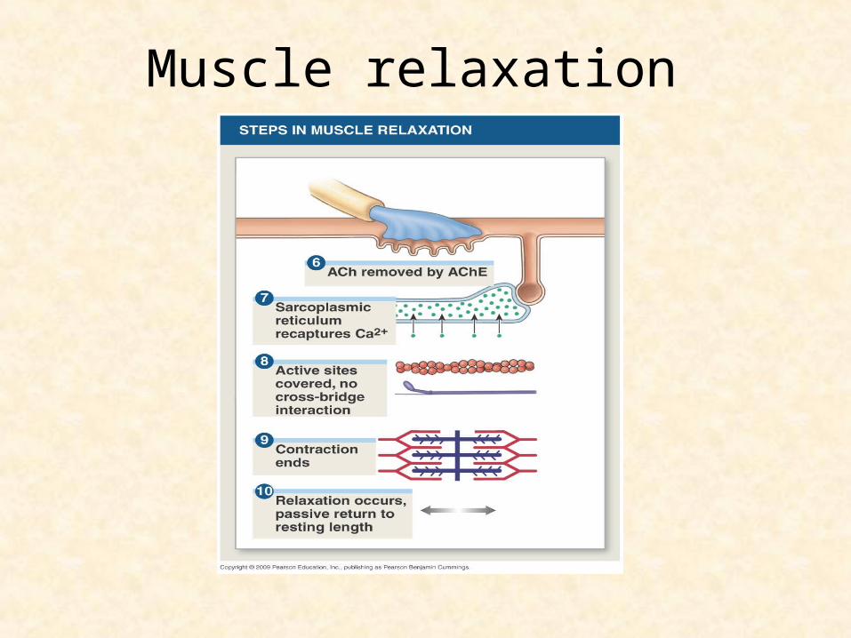
Muscle relaxation

Sarcomere contraction
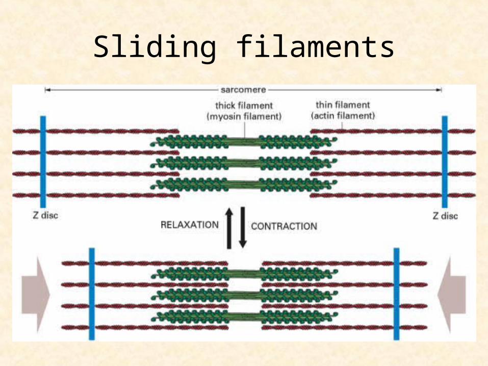
Sliding filaments
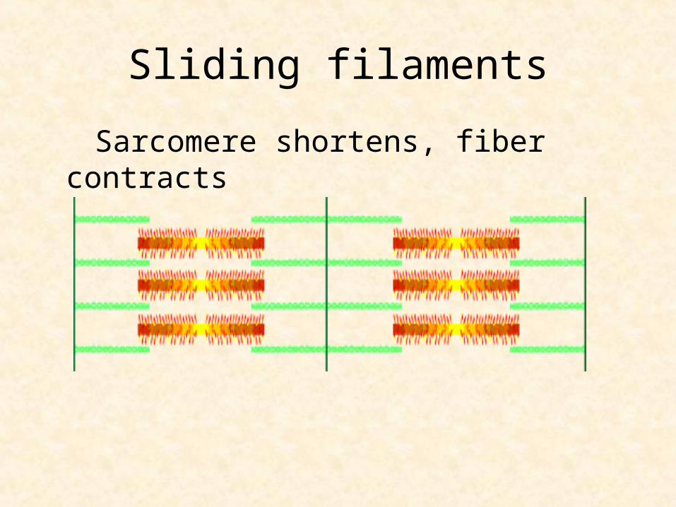
Sliding filaments
Sarcomere shortens, fiber contracts
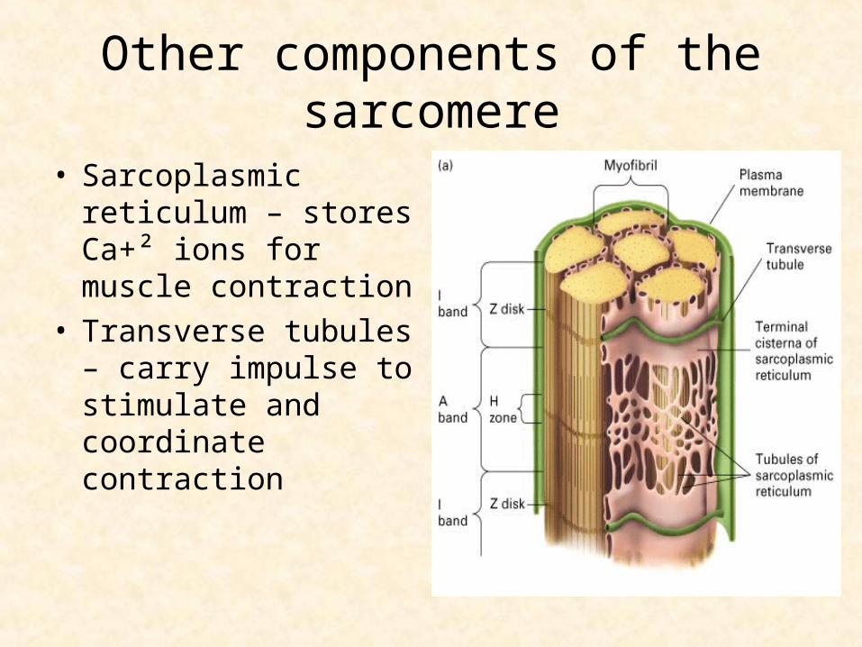
Other components of the sarcomere
• Sarcoplasmic reticulum – stores Ca+² ions for muscle contraction
• Transverse tubules – carry impulse to stimulate and coordinate contraction
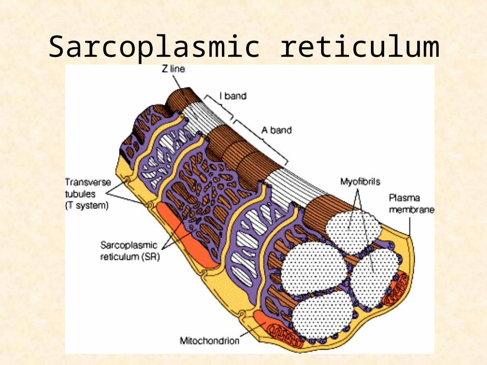
Sarcoplasmic reticulum
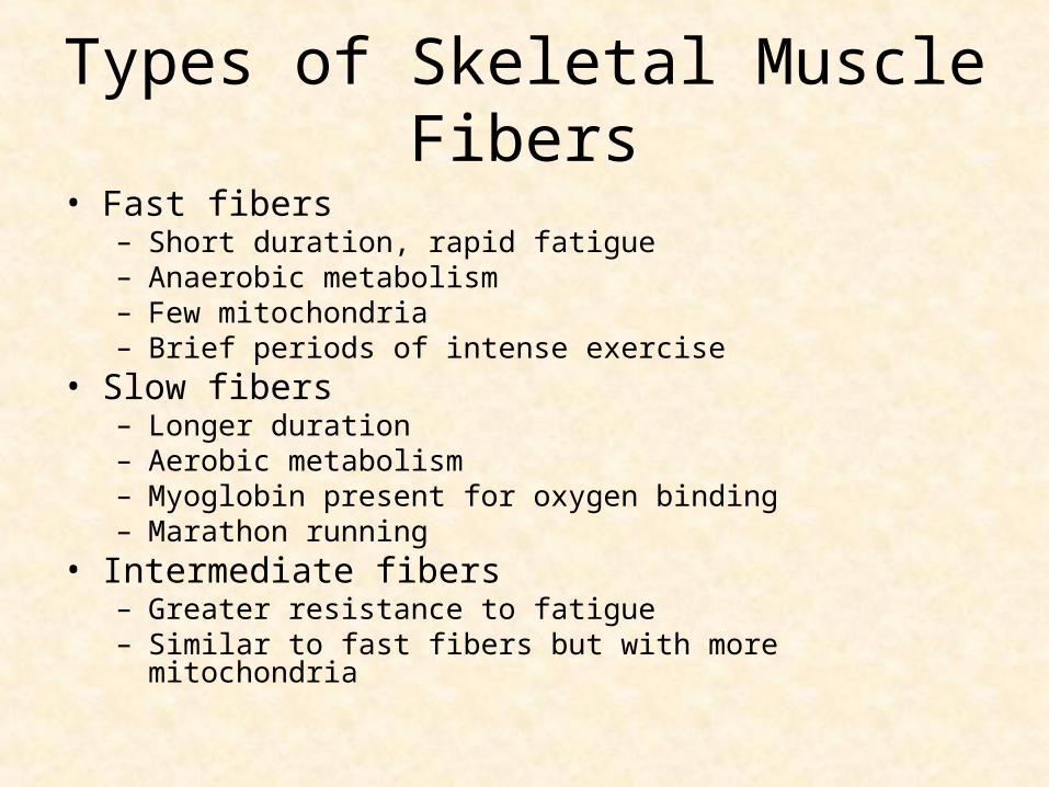
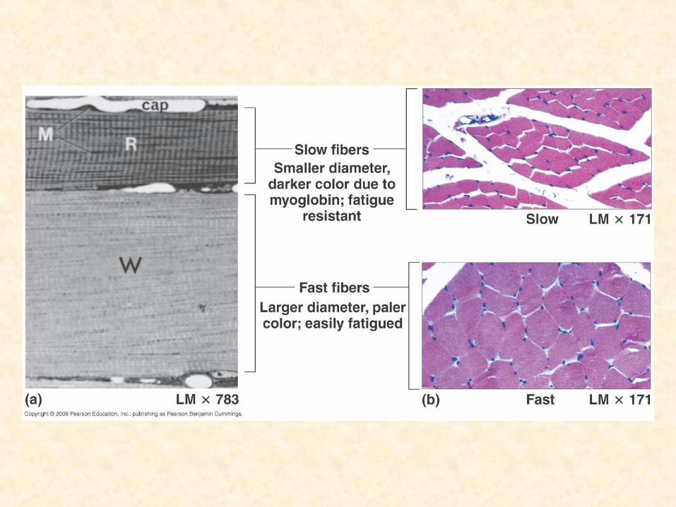
Types of Skeletal Muscle Fibers
• Fast fibers– Short duration, rapid fatigue– Anaerobic metabolism– Few mitochondria– Brief periods of intense exercise
• Slow fibers– Longer duration– Aerobic metabolism – Myoglobin present for oxygen binding– Marathon running
• Intermediate fibers– Greater resistance to fatigue– Similar to fast fibers but with more mitochondria
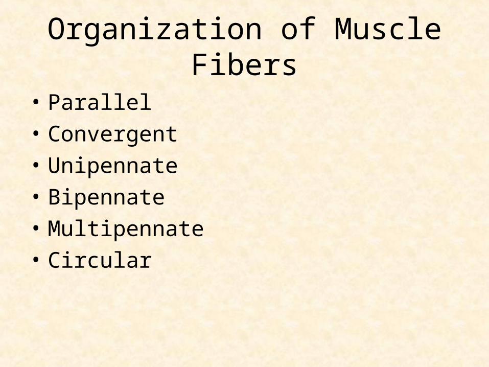
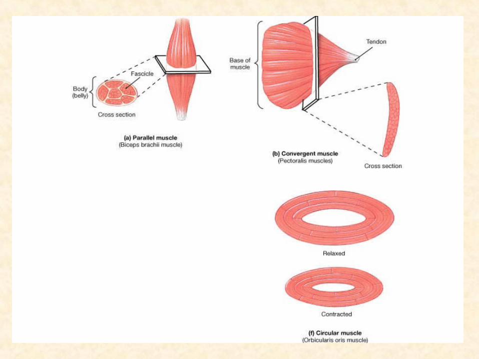
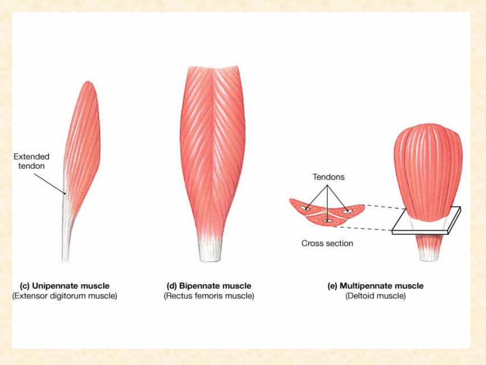
Organization of Muscle Fibers
• Parallel
• Convergent
• Unipennate
• Bipennate
• Multipennate
• Circular
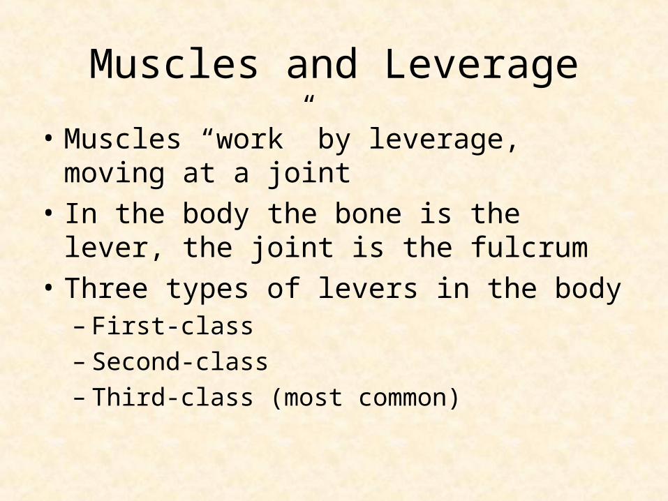
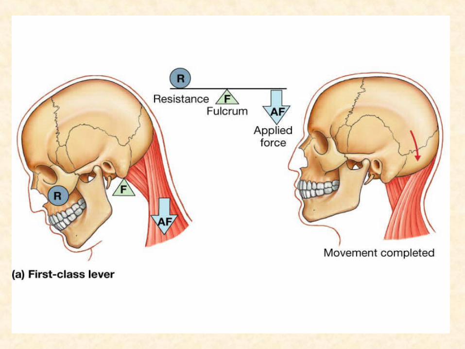
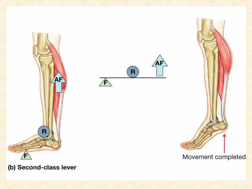
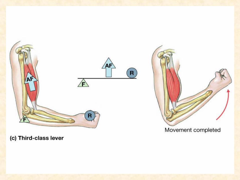
Muscles and Leverage
• Muscles “work” by leverage, moving at a joint
• In the body the bone is the lever, the joint is the fulcrum
• Three types of levers in the body– First-class– Second-class– Third-class (most common)
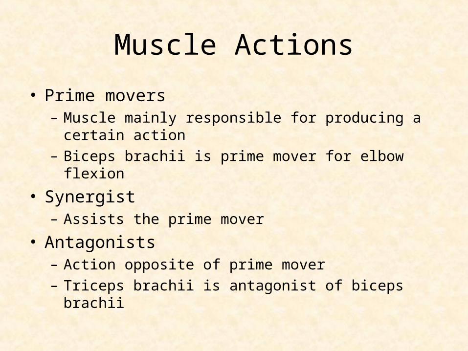
Muscle Actions
• Prime movers– Muscle mainly responsible for producing a certain
action– Biceps brachii is prime mover for elbow flexion
• Synergist– Assists the prime mover
• Antagonists– Action opposite of prime mover– Triceps brachii is antagonist of biceps brachii


