Mutant SOD1 in neuronal mitochondria causes toxicity and mitochondrial dynamics abnormalities Jordi Magrane ´ § , Isabel Hervias §,{ , Matthew S. Henning, Maria Damiano { , Hibiki Kawamata and Giovanni Manfredi Department of Neurology and Neuroscience, Weill Medical College of Cornell University, New York, NY 10065, USA Received May 29, 2009; Revised and Accepted August 28, 2009 Amyotrophic lateral sclerosis (ALS) is a fatal neurological disorder characterized by motor neuron degener- ation. Mutations in Cu,Zn-superoxide dismutase (SOD1) are responsible for 20% of familial ALS cases via a toxic gain of function. In mutant SOD1 transgenic mice, mitochondria of spinal motor neurons develop abnor- mal morphology, bioenergetic defects and degeneration, which are presumably implicated in disease patho- genesis. SOD1 is mostly a cytosolic protein, but a substantial portion is associated with organelles, including mitochondria, where it localizes predominantly in the intermembrane space (IMS). However, whether mito- chondrial mutant SOD1 contributes to disease pathogenesis remains to be elucidated. We have generated NSC34 motor neuronal cell lines expressing wild-type or mutant SOD1 containing a cleavable IMS targeting signal to directly investigate the pathogenic role of mutant SOD1 in mitochondria. We show that mitochond- rially-targeted SOD1 localizes to the IMS, where it is enzymatically active. We prove that mutant IMS-targeted SOD1 causes neuronal toxicity under metabolic and oxidative stress conditions. Furthermore, we demon- strate for the first time neurite mitochondrial fragmentation and impaired mitochondrial dynamics in motor neurons expressing IMS mutant SOD1. These defects are associated with impaired maintenance of neuritic processes. Our findings demonstrate that mutant SOD1 localized in the IMS is sufficient to determine mito- chondrial abnormalities and neuronal toxicity, and contributes to ALS pathogenesis. INTRODUCTION Amyotrophic lateral sclerosis (ALS) is a devastating neurode- generative disease resulting in a rapidly progressive paralysis due to degeneration of motor neurons. Sporadic ALS rep- resents 90% of the cases, whereas familial ALS accounts for the remaining 10%. Among the familial forms, 20% are caused by mutations in the gene encoding Cu,Zn-superoxide dismutase (SOD1). The mechanism underlying the selective degeneration and death of motor neurons in SOD1 familial ALS are still largely unknown, but it is clear that mutant SOD1 exerts a toxic gain of function. There are several hypotheses for mutant SOD1 toxicity, which are non-mutually exclusive (1), including the contribution of mitochondrial dysfunction (2) and axonal transport abnormalities (3). Mice expressing G93A mutant human SOD1 (hSOD1) develop mitochondrial bioenergetic impairment in the spinal cord (4–6). In brain and spinal cord of mutant SOD1 transgenic mice, there is decreased mitochondrial Ca 2þ capacity early on in the course of the disease (7). Mitochondrial dysfunction has also been observed in cultured cells expressing mutant SOD1 (8). Furthermore, mitochondrial morphological abnormalities are early signs of mutant SOD1 toxicity, and appear both in the cell bodies (9) and in the terminal axons of motor neurons (10). Accumulation of abnormal mitochondria may be caused by a block of axonal transport into proximal neur- ites (11) or impairment of mitochondria recycling and dynamics (12). A substantial amount of SOD1 is found in mitochondria (5,13 – 21), predominantly in the spinal cord (22). Accumu- lation of mutant SOD1 is associated with mitochondrial § The authors wish it to be known that, in their opinion, the first two authors should be regarded as joint First Authors. † Present address: Department of Pharmacology, University of Navarra, Pamplona, Spain. ‡ Present address: URA CEA-CNRS 2210, Service MIRCen, Institut d’Imagerie Biome ´dicale, CEA, Fontenay aux Roses, France. To whom correspondence should be addressed at: Department of Neurology and Neuroscience, Weill Medical College of Cornell University, 525 E. 68th Street, A-505, New York, NY 10065, USA. Tel: þ1 2127464605; Fax: þ1 2127468276; Email: [email protected]# The Author 2009. Published by Oxford University Press. All rights reserved. For Permissions, please email: [email protected]Human Molecular Genetics, 2009, Vol. 18, No. 23 4552–4564 doi:10.1093/hmg/ddp421 Advance Access published on September 24, 2009 Downloaded from https://academic.oup.com/hmg/article-abstract/18/23/4552/665440 by guest on 24 November 2018
Transcript
Mutant SOD1 in neuronal mitochondria causestoxicity and mitochondrial dynamics abnormalities
Jordi Magrane§, Isabel Hervias§,{, Matthew S. Henning, Maria Damiano{, Hibiki Kawamata
and Giovanni Manfredi�
Department of Neurology and Neuroscience, Weill Medical College of Cornell University, New York, NY 10065, USA
Received May 29, 2009; Revised and Accepted August 28, 2009
Amyotrophic lateral sclerosis (ALS) is a fatal neurological disorder characterized by motor neuron degener-ation. Mutations in Cu,Zn-superoxide dismutase (SOD1) are responsible for 20% of familial ALS cases via atoxic gain of function. In mutant SOD1 transgenic mice, mitochondria of spinal motor neurons develop abnor-mal morphology, bioenergetic defects and degeneration, which are presumably implicated in disease patho-genesis. SOD1 is mostly a cytosolic protein, but a substantial portion is associated with organelles, includingmitochondria, where it localizes predominantly in the intermembrane space (IMS). However, whether mito-chondrial mutant SOD1 contributes to disease pathogenesis remains to be elucidated. We have generatedNSC34 motor neuronal cell lines expressing wild-type or mutant SOD1 containing a cleavable IMS targetingsignal to directly investigate the pathogenic role of mutant SOD1 in mitochondria. We show that mitochond-rially-targeted SOD1 localizes to the IMS, where it is enzymatically active. We prove that mutant IMS-targetedSOD1 causes neuronal toxicity under metabolic and oxidative stress conditions. Furthermore, we demon-strate for the first time neurite mitochondrial fragmentation and impaired mitochondrial dynamics in motorneurons expressing IMS mutant SOD1. These defects are associated with impaired maintenance of neuriticprocesses. Our findings demonstrate that mutant SOD1 localized in the IMS is sufficient to determine mito-chondrial abnormalities and neuronal toxicity, and contributes to ALS pathogenesis.
INTRODUCTION
Amyotrophic lateral sclerosis (ALS) is a devastating neurode-generative disease resulting in a rapidly progressive paralysisdue to degeneration of motor neurons. Sporadic ALS rep-resents 90% of the cases, whereas familial ALS accounts forthe remaining 10%. Among the familial forms, 20% arecaused by mutations in the gene encoding Cu,Zn-superoxidedismutase (SOD1).
The mechanism underlying the selective degeneration anddeath of motor neurons in SOD1 familial ALS are stilllargely unknown, but it is clear that mutant SOD1 exerts atoxic gain of function. There are several hypotheses formutant SOD1 toxicity, which are non-mutually exclusive(1), including the contribution of mitochondrial dysfunction(2) and axonal transport abnormalities (3). Mice expressing
G93A mutant human SOD1 (hSOD1) develop mitochondrialbioenergetic impairment in the spinal cord (4–6). In brainand spinal cord of mutant SOD1 transgenic mice, there isdecreased mitochondrial Ca2þ capacity early on in thecourse of the disease (7). Mitochondrial dysfunction has alsobeen observed in cultured cells expressing mutant SOD1 (8).Furthermore, mitochondrial morphological abnormalities areearly signs of mutant SOD1 toxicity, and appear both in thecell bodies (9) and in the terminal axons of motor neurons(10). Accumulation of abnormal mitochondria may becaused by a block of axonal transport into proximal neur-ites (11) or impairment of mitochondria recycling anddynamics (12).
A substantial amount of SOD1 is found in mitochondria(5,13–21), predominantly in the spinal cord (22). Accumu-lation of mutant SOD1 is associated with mitochondrial
§The authors wish it to be known that, in their opinion, the first two authors should be regarded as joint First Authors.†Present address: Department of Pharmacology, University of Navarra, Pamplona, Spain.‡Present address: URA CEA-CNRS 2210, Service MIRCen, Institut d’Imagerie Biomedicale, CEA, Fontenay aux Roses, France.
�To whom correspondence should be addressed at: Department of Neurology and Neuroscience, Weill Medical College of Cornell University, 525E. 68th Street, A-505, New York, NY 10065, USA. Tel: þ1 2127464605; Fax: þ1 2127468276; Email: [email protected]
# The Author 2009. Published by Oxford University Press. All rights reserved.For Permissions, please email: [email protected]
Human Molecular Genetics, 2009, Vol. 18, No. 23 4552–4564doi:10.1093/hmg/ddp421Advance Access published on September 24, 2009
Dow
nloaded from https://academ
ic.oup.com/hm
g/article-abstract/18/23/4552/665440 by guest on 24 Novem
ber 2018
swelling and degeneration in neurons of transgenic mice(18,19). The mechanisms that regulate SOD1 mitochondrialimport are complex and involve the redox state of the cell,the intracellular distribution of the copper chaperone forSOD1 (CCS), and the folding of SOD1 (20). Indeed, increasedlocalization of mutant SOD1 in mitochondria induced by CCSoverexpression in mice causes early loss of mitochondrialfunction and accelerates the disease course (23).
Despite the evidence that a portion of mutant SOD1 loca-lizes to mitochondria, it still remains to be proven that itcauses mitochondrial dysfunction directly. A large amountof mitochondrial SOD1 is concentrated in the intermembranespace (IMS) (5,14,15,19–21). Therefore, in this study we haveevaluated the impact of wild-type (WT) or two mutant SOD1(G93A and G85R) targeted to the IMS on neuronal survival,mitochondrial bioenergetics, mitochondrial dynamics, andneuritic outgrowth of motor neuronal cells, and compared itwith the effects of the corresponding untargeted (canonical)forms of SOD1. We demonstrate that mutant SOD1 localizedin the IMS causes neuronal toxicity and abnormalities in mito-chondrial morphology and dynamics, indicating that mutantSOD1 accumulated in mitochondria contributes to diseasepathogenesis.
RESULTS
Targeting of SOD1 to the mitochondrial IMS in motorneuronal NSC34 cells
To direct the import of hSOD1 to the mitochondrial IMS weconstructed fusion proteins consisting of WT or mutant(G93A or G85R) hSOD1 appended in-frame at their N-terminus to the mitochondrial import signal of Saccharomycescerevisiae cytochrome b2 (CytB2). The CytB2 targetingpeptide is comprised of 80 amino acids: the first 31 directthe import into the mitochondrial matrix, where they arecleaved by matrix metalloproteases (MMP), whereas theremaining 49 amino acids direct the export of the resultingpeptide to the IMS, where they are cleaved by the intermem-brane space proteases (IMP) (24). In our final construct, weadded four residues corresponding to the mature CytB2 tothe N-terminus of SOD1 to ensure proper recognition byIMP (Fig. 1A).
We first characterized the subcellular localization of differ-ent WT and two mutant IMS hSOD1 in transiently transfectedCOS cells by confocal microscopy, and demonstrated target-ing to the mitochondria, as indicated by colocalization withthe mitochondrial dye Mitotracker Red (Fig. 1B). In contrast,untargeted WT or mutant hSOD1 appeared diffusely distribu-ted in the cytosol, with limited colocalization with mitochon-dria (not shown).
In order to study the effects of mitochondrial SOD1 in adisease-relevant cell line, we generated stably transfectedNSC34 cells expressing IMS-targeted or untargeted SOD1.NSC34 is a hybrid motor neuron-like cell line produced byfusion of N2A neuroblastoma cells and embryonic mousespinal cord neurons, which displays several typical propertiesof motor neurons (25,26). In cell homogenates from NSC34cells expressing WT or G93A IMS-targeted hSOD1, westernblot using a polyclonal anti-SOD1 antibody (Fig. 2A), that
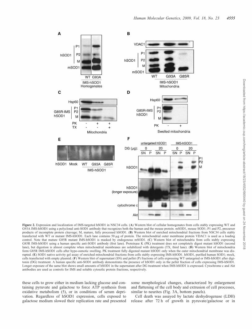
recognized both human and mouse SOD1 (mSOD1), indicatedthat, in addition to mature hSOD1 (M), there was a proportionof IMS-hSOD1 that was either uncleaved (i.e. full-length, P1)or only cleaved at the first site (i.e. 231 amino acids, P2).Enriched mitochondrial fractions showed the coexistence ofall three products, P1, P2 and mature protein (Fig. 2B). Thepresence of IMS-targeted G85R hSOD1 in mitochondria wasdemonstrated using human-specific monoclonal antibodies(Fig. 2C, first lane), because this mutant co-migrates withmouse endogenous SOD1.
We took advantage of the known sensitivity of G85R SOD1to proteinase K digestion (19) to investigate the intramitochon-drial localization of IMS-hSOD1. To test for mitochondrialintegrity, we used Hsp60, a matrix protein. Hsp60 was par-tially digested after proteinase K treatment (Fig. 2C, secondlane), suggesting that mitochondrial membranes were partiallypermeant to proteinase K. Nevertheless, a portion of both P1precursor and mature G85R peptides were protected fromdigestion, indicating that they were enclosed within intactmitochondrial membranes (Fig. 2C, second lane). Upon deter-gent solubilization, all of Hsp60 and most of the IMS-hSOD1were digested (Fig. 2C, third lane). Upon hypo-osmotic swel-ling to rupture the outer membrane and allow the access ofproteinase K to the IMS, all of IMS-targeted G85R hSOD1was digested, whereas the matrix protein Hsp60 was partiallypreserved (Fig. 2D). Since virtually no hSOD1 was resistant toproteinase K digestion, we concluded that no hSOD1 accumu-lated into the mitochondrial matrix (Fig. 2D).
Native gel SOD1 activity assays in isolated mitochondriashowed that both IMS-targeted WT and G93A hSOD1 dis-played increased enzymatic activity as compared with mock-transfected cells (Fig. 2E), whereas mitochondria expressingIMS-G85R hSOD1 did not show increased activity, becausethis mutant is inactive (27).
Because we wanted to compare the effects of IMS-targetedwith those of untargeted mutant SOD1, NSC34 cells werealso stably transfected with canonical WT or mutanthSOD1. By cell fractionation and western blot analyses,hSOD1 was found in both cytosolic and mitochondrial frac-tions of cells transfected with WT, G93A and G85R (datanot shown).
We also wanted to ensure that recombinant hSOD1 wasmostly localized in mitochondria and not in the cytosol inIMS-hSOD1 expressing cells. Typical mitochondria isolationmethods are not completely appropriate for this purpose,because some mitochondria are prone to break and releaseIMS-hSOD1 to the cytosol (data not shown). Therefore, wetreated hSOD1 expressing cells with digitonin under con-ditions that permeabilize the plasma membrane and not theouter mitochondrial membrane. As shown in Figure 2F, Aktshifted from the pellet fraction to the supernatant fractionupon digitonin treatment and centrifugation; although cyto-chrome c remained in the pellet fraction. Untargeted hSOD1was largely released to the supernatant after digitonin treat-ment (Fig. 2F), which indicated that it was mainly localizedin the cytosol. Instead, WT IMS-hSOD1 was retained in thepellet, demonstrating that it was mainly localized in the mito-chondria. Longer exposure of the same blots revealed that avery small amount of IMS-hSOD1 (estimated to be ,1%)was in the cytosol (Fig. 2F). The mitochondrial localization
Human Molecular Genetics, 2009, Vol. 18, No. 23 4553
Dow
nloaded from https://academ
ic.oup.com/hm
g/article-abstract/18/23/4552/665440 by guest on 24 Novem
ber 2018
of G93A and G85R mutant IMS-targeted hSOD1 was similarto WT (data not shown).
We compared the effects of constitutively expressingIMS-targeted or untargeted hSOD1 in NSC34 cells by asses-sing cell toxicity. Under normal growth conditions, therewas no difference in viability and cell morphology between
cells expressing hSOD1 and mock-transfected cells. Untreatedcells expressing either WT or mutant IMS-hSOD1 had roundmorphology and similar diameters, as shown in the represen-tative micrographs of NSC34 cells, in Figure 3A (toppanels). Moreover, mitochondrial ATP synthesis, cytochromec oxidase, and citrate synthase activities were unchanged incells expressing either IMS-targeted or canonical mutant andWT hSOD1, as compared with mock-transfected cells (datanot shown). We then subjected cells to modified conditionsthat induce metabolic stress. First, we tested the ability of
Figure 1. Targeting of SOD1 to mitochondria in cultured cells. (A) Schematic representation of the fusion protein containing human SOD1 (hSOD1) appendedin-frame at its N-terminus to the mitochondrial import signal of cytochrome b2 (CytB2). MMP, matrix metalloproteases; IMP, intermembrane space proteases.(B) Transient transfection of WT or mutant (G93A and G85R) IMS-hSOD1 in COS cells. Recombinant SOD1 is immuno-labeled in green and mitochondriastained with the fluorescent dye Mitotracker red. The overlay of the two images indicates that IMS-h SOD1 localizes within mitochondria. Colocalization ofthe two fluorochromes is further confirmed by z-section reconstruction (insets).
4554 Human Molecular Genetics, 2009, Vol. 18, No. 23
Dow
nloaded from https://academ
ic.oup.com/hm
g/article-abstract/18/23/4552/665440 by guest on 24 Novem
ber 2018
these cells to grow either in medium lacking glucose and con-taining pyruvate and galactose to force ATP synthesis fromoxidative metabolism (5), or in conditions of serum depri-vation. Regardless of hSOD1 expression, cells exposed togalactose medium slowed their replication rate and presented
some morphological changes, characterized by enlargementand flattening of the cell body and extrusion of cell processes,similar to neurites (Fig. 3A, bottom panels).
Cell death was assayed by lactate deshydrogenase (LDH)release after 72 h of growth in pyruvate/galactose or in
Figure 2. Expression and localization of IMS-targeted hSOD1 in NSC34 cells. (A) Western blot of cellular homogenates from cells stably expressing WT andG93A IMS-hSOD1 using a polyclonal anti-SOD1 antibody that recognizes both the human and the mouse protein. mSOD1, mouse SOD1; P1 and P2, precursorproducts of incomplete protein cleavage; M, mature, fully processed hSOD1. (B) Western blot of enriched mitochondrial fractions from NSC34 cells stablytransfected with WT or mutant IMS-hSOD1. Each lane contains 50 mg of protein. The mitochondrial outer membrane protein VDAC1 is used as a loadingcontrol. Note that mature G85R mutant IMS-hSOD1 is masked by endogenous mSOD1. (C) Western blot of mitochondria from cells stably expressingG85R IMS-hSOD1 using a human specific anti-SOD1 antibody (first lane). Proteinase K (PK) treatment does not completely digest mutant hSOD1 (secondlane), but digestion is almost complete when mitochondrial membranes are solubilized with detergents (TX, third lane). (D) Western blot of mitochondriaform G85R IMS-hSOD1 cells after hypo-osmotic swelling. PK treatment fully digested mutant hSOD1 only when the outer mitochondrial membrane was dis-rupted. (E) SOD1 native activity gel assay of enriched mitochondrial fractions from cells stably expressing IMS-hSOD1. hSOD1, purified human SOD1; mock,cells transfected with empty plasmid. (F) Western blot of supernatant (SN) and pellet (P) fractions of cells expressing WT untargeted or IMS-hSOD1 after digi-tonin (DG) treatment. A human specific anti-SOD1 antibody demonstrates the presence of hSOD1 only in the pellet fraction of cells expressing IMS-hSOD1.Longer exposure of the same blot shows small amounts of hSOD1 in the supernatant after DG treatment when IMS-hSOD1 is expressed. Cytochrome c and Aktantibodies are used as controls for IMS and soluble cytosolic protein fractions, respectively.
Human Molecular Genetics, 2009, Vol. 18, No. 23 4555
Dow
nloaded from https://academ
ic.oup.com/hm
g/article-abstract/18/23/4552/665440 by guest on 24 Novem
ber 2018
serum-deprived medium. Under these metabolic stressors,LDH release was highest in cells expressing either untargetedor IMS-targeted mutant hSOD1, whereas WT hSOD1expression did not significantly affect cell viability, as com-pared with control mock-transfected cells (Fig. 3B). Loss ofviability in cells expressing mutant hSOD1 exposed to eithergalactose or lack of serum was confirmed by MTT assay(data not shown). These results suggest that IMS-targetedmutant hSOD1 is capable of sensitizing neuronal cells to meta-bolic stress.
We also tested the sensitivity of mutant hSOD1 cells to oxi-dative stress. We first induced ROS production by adding acombination of xantine and xantine-oxidase (X/XO) in thegrowth medium (28). Exposure to X/XO for 6 h significantlyincreased toxicity in cells expressing either IMS-targeted oruntargeted mutant hSOD1, as measured by LDH (Fig. 3D)and by MTT assays (data not shown). We then treated cellsfor 72 h with the complex I inhibitor rotenone with theaddition of the gluthatione depleting agent ethacrynic acid(EA) during the last 16 h of rotenone treatment. We determined
Figure 3. Metabolic stress in NSC34 cells expressing IMS-targeted hSOD1. (A) Representative phase contrast micrographs of NSC34 cells either mock trans-fected or stably expressing IMS-targeted hSOD1. Untreated, normal growth conditions; galactose, galactose medium induces morphologic changes in all cells,including extrusion of cell processes, similar to neurites (arrows). (B, C, D) Quantification of cell death by LDH release in cells grown in galactose medium (n ¼7), serum-deprived medium (n ¼ 8) and xantine oxidase (X/XO) medium (n ¼ 8). (E) Cell viability by WST-1 assay (n ¼ 8) in cells treated with the combinationof rotenone (50 nM) for 72 h, plus ethacrynic acid (100 mM). �P , 0.05 compared with mock-transfected controls. Error bars represent the SEM.
4556 Human Molecular Genetics, 2009, Vol. 18, No. 23
Dow
nloaded from https://academ
ic.oup.com/hm
g/article-abstract/18/23/4552/665440 by guest on 24 Novem
ber 2018
that 100 mM EA for 16 h does not result in cell death inuntransfected NSC-34 cells, but longer treatments cause celldeath regardless of SOD1 expression (data not shown).Because in cultured cells rotenone at high doses may exerttoxic effects independent of mitochondrial dysfunction (29),we used a low concentration (50 nM) that is not lethal tocells, when administered in the absence EA, but causes anincrease in ROS production from complex I of the respiratorychain (29). The combination of rotenone and EA resulted inloss of cell viability, as determined by WST-1 assay, in cellsexpressing IMS-targeted or untargeted mutant hSOD1(Fig. 3E). These results suggest that mitochondrial accumu-lation of mutant SOD1 is responsible for the increased sensi-tivity of neuronal cells to oxidative stress.
IMS-targeted mutant hSOD1 causes mitochondrialmorphological changes in differentiated NSC34 cells
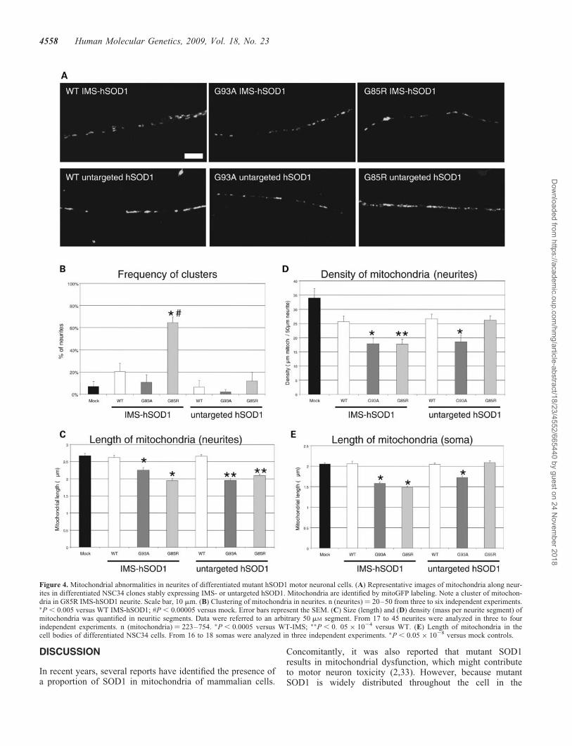
Mutant SOD1 causes mitochondrial abnormalities in ALSpatients (11) and transgenic mouse models (9,10,18,30).Furthermore, reduction of average mitochondrial length(i.e. mitochondrial fragmentation) has been reported inmotor neuron axons of transgenic SOD1 mutant mice (31).Mitochondrial fragmentation was also found in undifferen-tiated NSC34 cells expressing mutant SOD1 (8,32).However, undifferentiated NSC34 lack the principal structuralfeatures of motor neurons, because they are round and devoidof neurites. Therefore, we induced differentiation in NSC34cells for 6 days, in differentiation medium, containing 1:1DMEM/Ham’s F12 supplemented with 1% fetal bovineserum (FBS), 1% P/S and 1% modified Eagle’s medium non-essential amino acids, followed by transfection with mitoGFP(i.e. GFP targeted to the mitochondrial matrix) and imaging36 h post-transfection. Individual mitochondria could beeasily identified in neurites (Fig. 4A). Mitochondria werehomogeneously distributed along neurites with occasionalclustering, which in the case of IMS-targeted G85R hSOD1expressing cells was markedly increased as compared withmock-transfected cells, affecting �60% of neurites(Fig. 4B). The average mitochondrial length in neurites wassignificantly reduced in cells expressing IMS-targeted oruntargeted mutant SOD1, as compared with controls(Fig. 4C), indicating mitochondrial fragmentation. Thedensity of mitochondria in neurites was calculated as theaverage length of all the mitochondria contained within a50 mm neuritic segment. Mitochondrial density was decreasedin cells expressing both IMS-targeted mutant hSOD1 forms,whereas only G93A mutant untargeted hSOD1 showeddecreased density, as compared with controls (Fig. 4D). Thedecrease in density reflects the smaller size of mitochondria,which is not balanced by an increase in the number of mito-chondria. This abnormality is specific to differentiated cells,because undifferentiated mutant hSOD1 cells did not have areduction in mitochondrial mass, as evidenced by normalcontent of mitochondrial enzymes (data not shown). Averagemitochondrial length was also significantly reduced in thesoma of differentiated cells expressing both IMS-targetedmutant hSOD1 forms, whereas only G93A mutant untargetedhSOD1 showed decreased mitochondrial length (Fig. 4E).
Mitochondrial dynamics in neurites are abnormalin differentiated IMS-targeted mutant SOD1expressing NSC34 cells
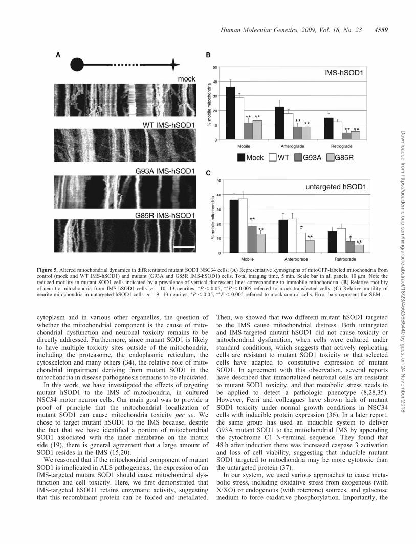
Mutant SOD1 causes an impairment of mitochondrial axonaltransport in primary cultured neurons (31). To assesswhether IMS-targeted mutant SOD1 is capable of disruptingmitochondrial neurite transport, NSC34 cells were inducedto differentiate and transfected with mitoGFP as describedabove. Mitochondrial transport along neuritic processes wasmeasured by time-lapse in vivo microscopy. Kymographsobtained from the recorded movies (Fig. 5A) were used forquantification of the proportion of moving mitochondria ineach direction (anterograde and retrograde). Approximately37% of all mitochondria were mobile during the course ofthe recording (5 min). The proportion of mobile mitochondriawas unchanged in NSC34 cells expressing untargeted orIMS-targeted WT hSOD1, whereas both untargeted andIMS-targeted G93A and G85R mutant hSOD1 caused a sig-nificant reduction of mobile mitochondria (Fig. 5B and C).Mitochondrial anterograde and retrograde transport wereequally affected in untargeted or IMS-targeted hSOD1mutant cells. Note that in Figure 5C and D the sum of antero-grade plus retrograde mitochondrial movements is slightlyhigher than the total, because mitochondria that movedbi-directionally in the course of the recording were countedin both groups.
These results indicate that the presence of mutant hSOD1 inthe IMS is sufficient to cause mitochondrial transport defects,which may contribute to the functional and morphologicalmitochondrial abnormalities observed in differentiatedmutant SOD1 NSC34 cells.
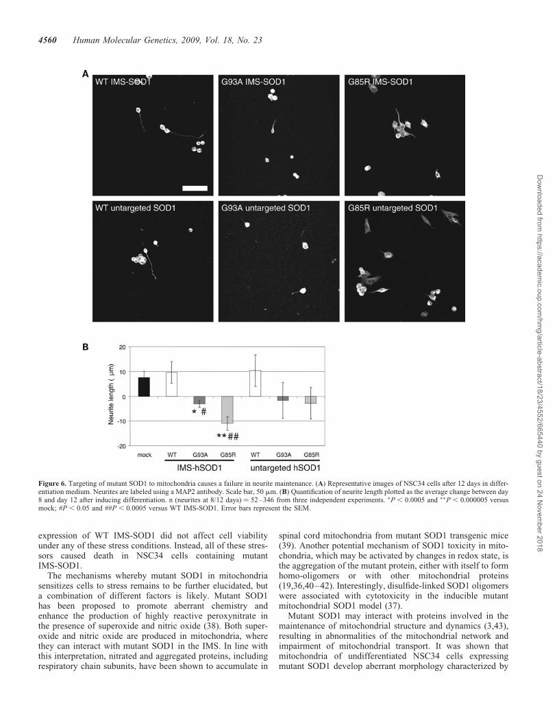
To assess the consequences of impaired mitochondrial mor-phology and dynamics on the fate of the neurite, NSC34cells were induced to differentiate for 8 or 12 days prior tomorphometric analyses of neurite length. The panels inFigure 6A are representative of the various NSC34 linesafter 12 days of differentiation. Approximately 30% of thecells projected neurites of various lengths and there was nodifference in the number of differentiated cells or in theaverage neurite length (40.1 mm+ 1.6) after 8 days amongthe lines (data not shown). The increase in average neuritelength between days 8 and 12 was used as a measure ofneurite viability. The average neurite length increased by�10 mm in mock-transfected and WT hSOD1-expressingcells. However, in IMS-targeted mutant hSOD1-expressingcells the average neurite length decreased by day 12 andwas significantly shorter than in mock-transfected andIMS-targeted WT hSOD1 cells (Fig. 6B). Neurites of cellsexpressing untargeted mutant hSOD1 were not significantlyshorter than controls.
These data suggest that IMS-targeted mutant SOD1impairs the maintenance of viable neuronal processes bydisrupting the dynamics of the axonal mitochondrialnetwork.
Human Molecular Genetics, 2009, Vol. 18, No. 23 4557
Dow
nloaded from https://academ
ic.oup.com/hm
g/article-abstract/18/23/4552/665440 by guest on 24 Novem
ber 2018
DISCUSSION
In recent years, several reports have identified the presence ofa proportion of SOD1 in mitochondria of mammalian cells.
Concomitantly, it was also reported that mutant SOD1results in mitochondrial dysfunction, which might contributeto motor neuron toxicity (2,33). However, because mutantSOD1 is widely distributed throughout the cell in the
Figure 4. Mitochondrial abnormalities in neurites of differentiated mutant hSOD1 motor neuronal cells. (A) Representative images of mitochondria along neur-ites in differentiated NSC34 clones stably expressing IMS- or untargeted hSOD1. Mitochondria are identified by mitoGFP labeling. Note a cluster of mitochon-dria in G85R IMS-hSOD1 neurite. Scale bar, 10 mm. (B) Clustering of mitochondria in neurites. n (neurites) ¼ 20–50 from three to six independent experiments.�P , 0.005 versus WT IMS-hSOD1; #P , 0.00005 versus mock. Error bars represent the SEM. (C) Size (length) and (D) density (mass per neurite segment) ofmitochondria was quantified in neuritic segments. Data were referred to an arbitrary 50 mM segment. From 17 to 45 neurites were analyzed in three to fourindependent experiments. n (mitochondria) ¼ 223–754. �P , 0.0005 versus WT-IMS; ��P , 0. 05 � 1024 versus WT. (E) Length of mitochondria in thecell bodies of differentiated NSC34 cells. From 16 to 18 somas were analyzed in three independent experiments. �P , 0.05 � 1028 versus mock controls.
4558 Human Molecular Genetics, 2009, Vol. 18, No. 23
Dow
nloaded from https://academ
ic.oup.com/hm
g/article-abstract/18/23/4552/665440 by guest on 24 Novem
ber 2018
cytoplasm and in various other organelles, the question ofwhether the mitochondrial component is the cause of mito-chondrial dysfunction and neuronal toxicity remains to bedirectly addressed. Furthermore, since mutant SOD1 is likelyto have multiple toxicity sites outside of the mitochondria,including the proteasome, the endoplasmic reticulum, thecytoskeleton and many others (34), the relative role of mito-chondrial impairment deriving from mutant SOD1 in themitochondria in disease pathogenesis remains to be elucidated.
In this work, we have investigated the effects of targetingmutant hSOD1 to the IMS of mitochondria, in culturedNSC34 motor neuron cells. Our main goal was to provide aproof of principle that the mitochondrial localization ofmutant SOD1 can cause mitochondria toxicity per se. Wechose to target mutant hSOD1 to the IMS because, despitethe fact that we have identified a portion of mitochondrialSOD1 associated with the inner membrane on the matrixside (19), there is general agreement that a large amount ofSOD1 resides in the IMS (15,20).
We reasoned that if the mitochondrial component of mutantSOD1 is implicated in ALS pathogenesis, the expression of anIMS-targeted mutant SOD1 should cause mitochondrial dys-function and cell toxicity. Here, we first demonstrated thatIMS-targeted hSOD1 retains enzymatic activity, suggestingthat this recombinant protein can be folded and metallated.
Then, we showed that two different mutant hSOD1 targetedto the IMS cause mitochondrial distress. Both untargetedand IMS-targeted mutant hSOD1 did not cause toxicity ormitochondrial dysfunction, when cells were cultured understandard conditions, which suggests that actively replicatingcells are resistant to mutant SOD1 toxicity or that selectedcells have adapted to constitutive expression of mutantSOD1. In agreement with this observation, several reportshave described that immortalized neuronal cells are resistantto mutant SOD1 toxicity, and that metabolic stress needs tobe applied to detect a pathologic phenotype (8,28,35).However, Ferri and colleagues have shown lack of mutantSOD1 toxicity under normal growth conditions in NSC34cells with inducible protein expression (36). In a later report,the same group has used an inducible system to deliverG93A mutant SOD1 to the mitochondrial IMS by appendingthe cytochrome C1 N-terminal sequence. They found that48 h after induction there was increased caspase 3 activationand loss of cell viability, suggesting that inducible mutantSOD1 targeted to mitochondria may be more cytotoxic thanthe untargeted protein (37).
In our system, we used various approaches to cause meta-bolic stress, including oxidative stress from exogenous (withX/XO) or endogenous (with rotenone) sources, and galactosemedium to force oxidative phosphorylation. Importantly, the
Figure 5. Altered mitochondrial dynamics in differentiated mutant SOD1 NSC34 cells. (A) Representative kymographs of mitoGFP-labeled mitochondria fromcontrol (mock and WT IMS-hSOD1) and mutant (G93A and G85R IMS-hSOD1) cells. Total imaging time, 5 min. Scale bar in all panels, 10 mm. Note thereduced motility in mutant SOD1 cells indicated by a prevalence of vertical fluorescent lines corresponding to immobile mitochondria. (B) Relative motilityof neuritic mitochondria from IMS-hSOD1 cells. n ¼ 10–13 neurites, �P , 0.05, ��P , 0.005 referred to mock-transfected cells. (C) Relative motility ofneurite mitochondria in untargeted hSOD1 cells. n ¼ 9–13 neurites, �P , 0.05, ��P , 0.005 referred to mock control cells. Error bars represent the SEM.
Human Molecular Genetics, 2009, Vol. 18, No. 23 4559
Dow
nloaded from https://academ
ic.oup.com/hm
g/article-abstract/18/23/4552/665440 by guest on 24 Novem
ber 2018
expression of WT IMS-SOD1 did not affect cell viabilityunder any of these stress conditions. Instead, all of these stres-sors caused death in NSC34 cells containing mutantIMS-SOD1.
The mechanisms whereby mutant SOD1 in mitochondriasensitizes cells to stress remains to be further elucidated, buta combination of different factors is likely. Mutant SOD1has been proposed to promote aberrant chemistry andenhance the production of highly reactive peroxynitrate inthe presence of superoxide and nitric oxide (38). Both super-oxide and nitric oxide are produced in mitochondria, wherethey can interact with mutant SOD1 in the IMS. In line withthis interpretation, nitrated and aggregated proteins, includingrespiratory chain subunits, have been shown to accumulate in
spinal cord mitochondria from mutant SOD1 transgenic mice(39). Another potential mechanism of SOD1 toxicity in mito-chondria, which may be activated by changes in redox state, isthe aggregation of the mutant protein, either with itself to formhomo-oligomers or with other mitochondrial proteins(19,36,40–42). Interestingly, disulfide-linked SOD1 oligomerswere associated with cytotoxicity in the inducible mutantmitochondrial SOD1 model (37).
Mutant SOD1 may interact with proteins involved in themaintenance of mitochondrial structure and dynamics (3,43),resulting in abnormalities of the mitochondrial network andimpairment of mitochondrial transport. It was shown thatmitochondria of undifferentiated NSC34 cells expressingmutant SOD1 develop aberrant morphology characterized by
Figure 6. Targeting of mutant SOD1 to mitochondria causes a failure in neurite maintenance. (A) Representative images of NSC34 cells after 12 days in differ-entiation medium. Neurites are labeled using a MAP2 antibody. Scale bar, 50 mm. (B) Quantification of neurite length plotted as the average change between day8 and day 12 after inducing differentiation. n (neurites at 8/12 days) ¼ 52–346 from three independent experiments. �P , 0.0005 and ��P , 0.000005 versusmock; #P , 0.05 and ##P , 0.0005 versus WT IMS-SOD1. Error bars represent the SEM.
4560 Human Molecular Genetics, 2009, Vol. 18, No. 23
Dow
nloaded from https://academ
ic.oup.com/hm
g/article-abstract/18/23/4552/665440 by guest on 24 Novem
ber 2018
abnormal cristae and fragmented network (32). Furthermore,impaired mitochondrial axonal transport was demonstratedin cultured motor neurons from SOD1 mutant rodents (31).
Taking advantage of the potential of NSC34 cells to differen-tiate, we investigated for the first time the effects ofIMS-targeted mutant SOD1 on mitochondrial transport andmorphology in neuritic processes. Mutant IMS-SOD1 impairsmitochondrial motility both in anterograde and retrograde direc-tions, resulting in organelle fragmentation and decreaseddensity of mitochondria along neurites. Impaired mitochondrialtransport was associated with inability to maintain viable neur-ites, as demonstrated by neuritic shortening over time. Theseobservations are consistent with the notion that SOD1 familialALS develops as a dying-back neuropathy that initiates and pro-gresses from the distal to the proximal portions of the motorneuron (44), and causes paralysis even in the absence of cellbody degeneration in the spinal cord (10).
Mitochondrial transport abnormalities can result in failureof mitochondria originated in the cell body to reach the synap-tic terminals and altered retrograde transport of mitochondriafrom the periphery to the cell body for degradation. Mitochon-drial transport abnormalities may be critical in motor neurons,where cellular components have to move long distancesthroughout axons, and may contribute to explain the selectivedegeneration of MN in ALS (3). The mechanisms wherebymutant IMS-SOD1 impairs mitochondrial axonal dynamicsremain to be elucidated, but they are likely to involve defec-tive mitochondrial bioenergetics and aberrant interactionswith the machinery that regulates mitochondrial dynamics (3).
Further in vivo studies in transgenic mouse models expres-sing IMS-targeted mutant SOD1 are warranted for the future.Nevertheless, our observations in motor neuronal cellsstrongly support the role of mutant SOD1 localized in theIMS in causing mitochondrial dysfunction and neuronaldegeneration in familial ALS.
MATERIALS AND METHODS
Plasmids
WT and G93A or G85R mutant forms of hSOD1 were cloned atthe BamHI site of the mammalian expression vector pIRESNeo3(Clontech, Mountain View, CA, USA). To target SOD1 to themitochondrial IMS, we used a cDNA encoding the first84 amino acid of the S. cerevisiae CytB2, which contains themitochondrial targeting sequence, plus four amino acids of themature protein (provided by Dr Carla Koheler, University ofCalifornia, Los Angeles, CA, USA). CytB2 was cloned intopIRESNeo3 at the BamHI site, and then the cDNA encodingfor either WT or mutant SOD1 was cloned in-frame with the C-terminus of the CytB2 pre-sequence using NotI and BstXI sitesgenerated by PCR. The final construct was confirmed by DNAsequencing. A mitoGFP construct with targeted expression ofGFP to the mitochondrial matrix (45) was used for labeling ofmitochondria in morphology and transport studies.
Cell culture and transfection
COS cells (from ATCC, American Type Culture Collection,Manassas, VA, USA) were maintained in Dulbecco’s modified
Eagle’s medium (DMEM, Invitrogen, Carlsbad, CA, USA)supplemented with 5% v/v FBS and 1% antibiotic anti-mycoticsolution (Invitrogen). The motor neuron-like cell line NSC34(provided by Dr Neil Cashman, University of Toronto,Toronto, ON, Canada) were maintained in DMEM sup-plemented with 10% v/v FBS and 1% antibiotic anti-mycoticsolution. When differentiation was required, cells were platedonto poly-D-lysine-coated plates and grown in differentiationmedium, which contains 1:1 DMEM/Ham’s F12 (Invitrogen)supplemented with 1% FBS, 1% P/S and 1% modified Eagle’smedium non-essential amino acids (Invitrogen).
Transient transfections of COS cells were performed with3 ml of the FuGENE 6 transfection reagent (Roche, Indianapo-lis, IN, USA) mixed with 1 mg of plasmid DNA, as rec-ommended by the manufacturer. For the generation of stablecell lines expressing SOD1 constructs, NSC34 cells weretransfected as above, and after 24 h selection was startedwith 500 mg/ml of the neomycin analog G418 (Invitrogen).G418-selected cells were constantly maintained in the pres-ence of the selective drug. For live-cell imaging studies on dif-ferentiated cells, NSC34 motor neurons were subjected todifferentiation medium for 6 days, and then transfected with0.75 mg mitoGFP and 1 ml of Lipofectamine 2000 (Invitrogen)and analyzed 36 h later.
ATP synthesis
Mitochondrial ATP synthesis was measured in digitonin per-meabilized NSC34 cells, using a luciferase/luciferin basedkinetic assay, as previously described, with malate and pyru-vate as respiratory chain substrates (46).
Mitochondrial isolation from cultured cells
NSC34 cells stably expressing hSOD1 constructs were washedwith PBS, trypsinized, and resuspended in 10 ml of cold NKMbuffer (130 mM NaCl, 5 mM KCl, 7.5 mM MgCl2) containing1 mM DTT and a cocktail of protease inhibitors (Sigma,Saint Louis, MO, USA). Cells were then centrifuged at3000g for 3 min at 48C and the resulting cell pellet was resus-pended in 3 ml of RSB buffer (10 mM Tris–HCl pH 7.6,10 mM KCl, 0.15 mM MgCl2, 1 mM DTT and PI), incubated4 min on ice, and homogenized in glass–glass Potter–Elveh-jem style homogenizer (pestle B) on ice. Then, 0.5 ml of 2 M
sucrose was added and the homogenates were centrifuged at600g for 10 min at 48C. The resulting supernatants were cen-trifuged at 15 000g for 15 min at 48C. The crude mitochon-drial pellet was incubated with 150 mM KCl for 5 min on iceand centrifuged at 15 000g for 15 min at 48C. The washedmitochondrial pellet was resuspended in 125 ml of chilledbuffer containing 250 mM sucrose, 10 mM Tris–HCl, 10 mM
EDTA 1 mM DTT and PI. Sample aliquots were snap-frozenin liquid nitrogen and stored at 2808C. Protein concentrationin the samples were determined by a colorimetric assay(BioRad DC, Hercules, CA, USA).
Immunofluorescence and mitochondrial labeling
For immunocytochemistry, COS cells were transiently trans-fected on glass slides with untargeted or IMS-targeted WT,
Human Molecular Genetics, 2009, Vol. 18, No. 23 4561
Dow
nloaded from https://academ
ic.oup.com/hm
g/article-abstract/18/23/4552/665440 by guest on 24 Novem
ber 2018
G93A and G85R SOD1 constructs. After 48 h, cells wereincubated for 30 min with 250 nM of the mitochondrial-specific fluorescent dye MitoTracker Red (Invitrogen). Cellswere then fixed in a 4% paraformaldehyde solution, permeabi-lized with 0.1% Triton-X100, and immunostained with mousemonoclonal anti-SOD1 antibodies (Santa Cruz Biotechnology,Santa Cruz, CA, USA). Secondary Cy2-conjugated anti-mouseIgG (Jackson Immunochemicals, West Grove, PA, USA) wereused for fluorescence visualization.
Differentiated NSC34 cells stably expressing SOD1 pro-teins were transfected with mitoGFP to label mitochondria,and fixed and immunostained with antibodies against andMAP2 (Chemicon, Temecula, CA, USA) to reveal the entireneurite length. Images were collected using a Leica SP5spectral confocal microscope (Leica Microsystems Inc,Bannockburn, IL, USA) equipped with a HCX PL APO CS63�/NA1.4 oil objective, and optical sections were acquiredevery 0.5 mm and standard pinhole of 1 AU to achieve highresolution of mitochondria.
SOD1 immunodetection
For western blots, cytosolic and mitochondrial fractions fromNSC34 cells were separated by 12% SDS-PAGE and immuno-blotted onto PVDF membranes (BioRad). Immunoreactivebands were detected with polyclonal antibodies that recognizeboth human and mSOD1 (Calbiochem, San Diego, CA, USA)or monoclonal antibodies (Santa Cruz Biotechnology) againsthSOD1.
For assays of protease protection of mitochondrial proteins,isolated mitochondria from NSC34 cells expressingIMS-targeted G85R hSOD1 were treated with 20 mg/ml pro-teinase K (Roche Applied Sciences, Indianapolis, IN, USA)on ice for 20 min either in the presence or the absence of0.5% Triton-X100, as described (19). After inactivation ofproteinase K with PMSF, the residual content of mitochondrialproteins was analyzed by western blot using specific anti-bodies. Alternatively, mitochondria were treated with hypo-tonic buffer (1 mM Tris–HCl) to swell and rupture the outermembrane and allow proteinase K to access the IMS.
To confirm the enrichment of SOD1 in mitochondria,1 million cells were treated with or without 20 mg digitonin(Sigma) for 1 min at room temperature. Supernatant (contain-ing soluble proteins) and pellet (containing membrane-boundorganelles, including mitochondria) fractions were thenobtained by centrifugation at 22 000g for 15 min at 48C.
Other antibodies were used against VDAC1 (Invitrogen),Hsp60 (Assay Designs, Ann Arbor, MI, USA), Akt (SantaCruz Biotechnology) and cytochrome c (BD Biosciences,San Jose, CA, USA).
Stress treatments and cell viability assays
Stable NSC34 cell lines expressing untargeted or IMS-targetedhSOD1 were seeded onto 48-well plates at 12 500 cells/well.The next day, cells were subjected to the following stress con-ditions: culture medium deprived of FBS; culture mediumwith 5 mM galactose instead of glucose; and treatment with50 nM rotenone (a respiratory chain complex I inhibitor) for72 h plus depletion of glutathione stores with EA (100 mM),
during the last 16 h of treatment. In these stress paradigms,cell viability was assessed after 72 h. In another set of exper-iments, 10 000 cells were seeded onto 12-well plates, and200 mM xanthine plus 10 mU/ml xanthine oxidase (Sigma)were added the next day to produce exogenous superoxide;cell viability was assessed after 6 h.
To determine cell survival after exposure to oxidativeand metabolic stressors, several assays were used. 3-(4,5-Dimethylthiazol-2-yl)-2,5-diphenyltetrazolium bromide(MTT, Sigma) assay was used following manufacture’sinstructions. Absorbance was measured in duplicates in aplate reader (Packard Instrument Company, Downers Grove,IL, USA) at 570 nm. Alternatively, a similar Cell ProliferationReagent WST-1 assay (Roche) was used to measure cell via-bility, according to the manufacturer’s protocol. LDHrelease into the culture media was measured with a Cytotox-icity Detection kit (Roche) according to the manufacturer’sprotocol. The value of released LDH is expressed as the pro-portion of total cellular LDH content measured after lysing thecells with 2% Triton X-100.
Analysis of mitochondrial morphology, densityand clustering
Metamorph software (Universal Imaging) was used for quan-titative analysis of mitochondria measurements. For morpho-logical quantification in neurites, z-sections were merged(using maximal projection) and mitoGFP-labeled mitochon-dria marked by longitudinal regions covering their entirelength (from tip to tip). In cell bodies, mitochondria lengthwas measured in each z-section covering the entire soma.The total number of mitochondria analyzed ranged from 223to 754 for each group. For density quantification, the lengthof the entire neurite immunostained with anti-MAP2 anti-bodies was measured, and the sum of all mitochondriallengths was divided by neurite length and referred to an arbi-trary 50 mm segment. Only neurites longer than 40 mm wereanalyzed. Clustering of mitochondria was defined as anaccumulation of several mitochondria that cannot be measuredindividually, having the cluster an irregular shape (nottubular), and more than 2 mm thick and more than 5 mmlong. The number of neurites analyzed ranged from 14 to 50in each category. Three to six independent experiments wereperformed.
Live-cell imaging and analysis mitochondria dynamics
For time-lapse microscopy, images were collected using aZeiss LSM510 Laser Scanning Confocal Microscope (ZeissMicroimaging, Thornwood, NY, USA) with a 63x lens.A fully equipped live imaging station (CTI-Controller 3700and incubator S, Leica Microsystems) allowed for thecontrol of CO2 content, humidity and temperature of boththe stage (through a heated stage; Heating Insert P, PeCon)and the air during the curse of the experiment. Preliminaryobservation of the cells with either epifluorescence or488-nm laser was performed at low light/laser intensities toprevent photo-toxicity. We selected neurites from 4 to 5 differ-entiated mitoGFP-positive NSC34 cells for each experiment(3–4 independent experiments), and mitochondrial transport
4562 Human Molecular Genetics, 2009, Vol. 18, No. 23
Dow
nloaded from https://academ
ic.oup.com/hm
g/article-abstract/18/23/4552/665440 by guest on 24 Novem
ber 2018
was followed for 5 min with frames taken every 5 s. Pinholeopening was set at 2 AU.
We counted the number of mitochondria that presented ret-rograde or anterograde movements or remained stationary(293 for mock, 424 for WT, 222 for G93A, 402 for G85R;183 for WT IMS, 130 for G93A IMS and 353 for G85RIMS). Movement was defined as changes in mitochondrionposition in at least four consecutive time frames. A movingmitochondrion during the course of an experiment wasscored as one movement event if no change in directionoccurred; otherwise it was scored as x þ 1 movements(where x is the number of changes in direction). Number ofmobile mitochondria was referred to the total number of mito-chondria present during the course of the experiment. Differ-ences were measured by analysis of variance to comparemutant hSOD1 cells with WT hSOD1 and mock-transfectedcontrols.
Analysis neurite length
To evaluate neurite maintenance over time in culture, 15 000cells were plated on coated glass coverslips, and differen-tiation medium was added after 24–48 h. Cells were fixed 8and 12 days later and stained with antibodies against MAP2to label neurites. Only differentiated cells with one clearlydefined neurite (no thread-like neurites, no multiple neuritespresent) were quantified. Images were taken from randomfields using a HCX PL APO CS 40x/NA1.25 oil objective,and pinhole was opened maximally to allow the entire thick-ness of the neurite to be imaged. The total number of neuritesanalyzed ranged from 53 to 346 in each category. Neuritemaintenance was represented as the average change inneurite length between day 8 and day 12 of differentiation.At least three independent experiments were performed.
Data analysis
Data from cell viability assays were analyzed by two-wayANOVA. In this case, WT or mutant hSOD1 was used asthe between-subjects factor. Comparisons between groups(WT or mutant hSOD1) were made using one-way ANOVAand Tukey’s test. All quantified results were expressed asthe mean+SE. Statistical significance for mitochondria mor-phology and transport, and neurite length was determined bythe Student’s two-tailed, unpaired t-test, and a P-value,0.05 was considered indicative of a significant difference.All statistical P-values in this study were determined usingANOVA or t-test from experiments repeated a minimum ofthree times, unless stated otherwise.
ACKNOWLEDGEMENTS
We thank Dr. Carla Koehler for providing the yeast cyto-chrome b2 construct used in this study, and Dr. NeilR. Cashman for providing the NSC34 cells. We also thankDr. Roy Smith and Dr. Cristofol Vives-Bauza for criticallyreading the manuscript.
Conflict of Interest statement. None declared.
FUNDING
This work was supported by National Institute of Health(grants P01-NS011766, NS051419, to G.M.); the MuscularDystrophy Association (to J.M.); the Robert Packard ALSResearch Center ‘The New York Community Trust’ (toG.M.); the Hiller Foundation (to G.M.); and a Ministerio deEducacion y Ciencia postdoctoral fellowship from theSpanish Government (to I.H.).
REFERENCES
1. Bruijn, L.I., Miller, T.M. and Cleveland, D.W. (2004) Unraveling themechanisms involved in motor neuron degeneration in Als. Annu. Rev.Neurosci., 27, 723–749.
2. Hervias, I., Beal, M.F. and Manfredi, G. (2006) Mitochondrialdysfunction and amyotrophic lateral sclerosis. Muscle Nerve, 33,598–608.
3. Magrane, J. and Manfredi, G. (2009) Mitochondrial function,morphology, and axonal transport in amyotrophic lateral sclerosis.Antioxid. Redox Signal., 11, 1615–1626.
4. Jung, C., Higgins, C.M. and Xu, Z. (2002) A quantitative histochemicalassay for activities of mitochondrial electron transport chain complexes inmouse spinal cord sections. J. Neurosci. Methods, 114, 165–172.
5. Mattiazzi, M., D’Aurelio, M., Gajewski, C.D., Martushova, K., Kiaei, M.,Beal, M.F. and Manfredi, G. (2002) Mutated human SOD1 causesdysfunction of oxidative phosphorylation in mitochondria of transgenicmice. J. Biol. Chem., 277, 29626–29633.
6. Kirkinezos, I.G., Bacman, S.R., Hernandez, D., Oca-Cossio, J., Arias, L.J.,Perez-Pinzon, M.A., Bradley, W.G. and Moraes, C.T. (2005) Cytochromec association with the inner mitochondrial membrane is impaired in theCNS of G93A-SOD1 mice. J. Neurosci., 25, 164–172.
7. Damiano, M., Starkov, A.A., Petri, S., Kipiani, K., Kiaei, M., Mattiazzi,M., Flint Beal, M. and Manfredi, G. (2006) Neural mitochondrial Ca2þ
capacity impairment precedes the onset of motor symptoms in G93A Cu/Zn-superoxide dismutase mutant mice. J. Neurochem., 96, 1349–1361.
8. Menzies, F.M., Cookson, M.R., Taylor, R.W., Turnbull, D.M.,Chrzanowska-Lightowlers, Z.M., Dong, L., Figlewicz, D.A. and Shaw,P.J. (2002) Mitochondrial dysfunction in a cell culture model of familialamyotrophic lateral sclerosis. Brain, 125, 1522–1533.
9. Kong, J. and Xu, Z. (1998) Massive mitochondrial degeneration in motorneurons triggers the onset of amyotrophic lateral sclerosis in miceexpressing a mutant SOD1. J. Neurosci., 18, 3241–3250.
10. Gould, T.W., Buss, R.R., Vinsant, S., Prevette, D., Sun, W., Knudson,C.M., Milligan, C.E. and Oppenheim, R.W. (2006) Complete dissociationof motor neuron death from motor dysfunction by Bax deletion in a mousemodel of ALS. J. Neurosci., 26, 8774–8786.
11. Sasaki, S. and Iwata, M. (1996) Impairment of fast axonal transport in theproximal axons of anterior horn neurons in amyotrophic lateral sclerosis.Neurology, 47, 535–540.
13. Weisiger, R.A. and Fridovich, I. (1973) Superoxide dismutase. Organellespecificity. J. Biol. Chem., 248, 3582–3592.
14. Sturtz, L.A., Diekert, K., Jensen, L.T., Lill, R. and Culotta, V.C. (2001)A fraction of yeast cu,zn-superoxide dismutase and its metallochaperone,ccs, localize to the intermembrane space of mitochondria. a physiologicalrole for sod1 in guarding against mitochondrial oxidative damage. J. Biol.Chem., 276, 38084–38089.
15. Okado-Matsumoto, A. and Fridovich, I. (2001) Subcellular distribution ofsuperoxide dismutases (SOD) in rat liver: Cu,Zn-SOD in mitochondria.J. Biol. Chem., 276, 38388–38393.
16. Jaarsma, D., Rognoni, F., van Duijn, W., Verspaget, H.W., Haasdijk, E.D.and Holstege, J.C. (2001) CuZn superoxide dismutase (SOD1)accumulates in vacuolated mitochondria in transgenic mice expressingamyotrophic lateral sclerosis-linked SOD1 mutations. Acta Neuropathol.(Berl), 102, 293–305.
17. Higgins, C.M., Jung, C., Ding, H. and Xu, Z. (2002) Mutant Cu, Znsuperoxide dismutase that causes motoneuron degeneration is present inmitochondria in the CNS. J. Neurosci., 22, RC215.
Human Molecular Genetics, 2009, Vol. 18, No. 23 4563
Dow
nloaded from https://academ
ic.oup.com/hm
g/article-abstract/18/23/4552/665440 by guest on 24 Novem
ber 2018
18. Higgins, C.M., Jung, C. and Xu, Z. (2003) ALS-associated mutantSOD1G93A causes mitochondrial vacuolation by expansion of theintermembrane space and by involvement of SOD1 aggregation andperoxisomes. BMC Neurosci., 4, 16.
19. Vijayvergiya, C., Beal, M.F., Buck, J. and Manfredi, G. (2005) Mutantsuperoxide dismutase 1 forms aggregates in the brain mitochondrialmatrix of amyotrophic lateral sclerosis mice. J. Neurosci., 25, 2463–2470.
20. Kawamata, H. and Manfredi, G. (2008) Different regulation of wild-typeand mutant Cu,Zn superoxide dismutase localization in mammalianmitochondria. Hum. Mol. Genet., 17, 3303–3317.
21. Vande Velde, C., Miller, T.M., Cashman, N.R. and Cleveland, D.W.(2008) Selective association of misfolded ALS-linked mutant SOD1 withthe cytoplasmic face of mitochondria. Proc. Natl. Acad. Sci. USA, 105,4022–4027.
22. Liu, J., Lillo, C., Jonsson, P.A., Velde, C.V., Ward, C.M., Miller, T.M.,Subramaniam, J.R., Rothstein, J.D., Marklund, S., Andersen, P.M. et al.(2004) Toxicity of familial ALS-linked SOD1 mutants from selectiverecruitment to spinal mitochondria. Neuron, 43, 5–17.
23. Son, M., Puttaparthi, K., Kawamata, H., Rajendran, B., Boyer, P.J.,Manfredi, G. and Elliott, J.L. (2007) Overexpression of CCS inG93A-SOD1 mice leads to accelerated neurological deficits with severemitochondrial pathology. Proc. Natl. Acad. Sci. USA, 104, 6072–6077.
24. Ono, H., Gruhler, A., Stuart, R.A., Guiard, B., Schwarz, E. and Neupert,W. (1995) Sorting of cytochrome b2 to the intermembrane space ofmitochondria. Kinetic analysis of intermediates demonstrates passagethrough the matrix. J. Biol. Chem., 270, 16932–16938.
25. Cashman, N.R., Durham, H.D., Blusztajn, J.K., Oda, K., Tabira, T., Shaw,I.T., Dahrouge, S. and Antel, J.P. (1992) Neuroblastoma x spinal cord(NSC) hybrid cell lines resemble developing motor neurons. Dev. Dyn.,194, 209–221.
26. Eggett, C.J., Crosier, S., Manning, P., Cookson, M.R., Menzies, F.M.,McNeil, C.J. and Shaw, P.J. (2000) Development and characterisation of aglutamate-sensitive motor neurone cell line. J. Neurochem., 74, 1895–1902.
28. Pasinelli, P., Borchelt, D.R., Houseweart, M.K., Cleveland, D.W. andBrown, R.H. Jr (1998) Caspase-1 is activated in neural cells and tissuewith amyotrophic lateral sclerosis-associated mutations in copper-zincsuperoxide dismutase [published erratum appears in Proc Natl Acad SciUSA 1999 Mar 16;96(6):3330]. Proc. Natl. Acad. Sci. USA, 95, 15763–15768.
29. Barrientos, A. and Moraes, C.T. (1999) Titrating the effects ofmitochondrial complex I impairment in the cell physiology. J. Biol.Chem., 274, 16188–16197.
30. Wong, P.C., Pardo, C.A., Borchelt, D.R., Lee, M.K., Copeland, N.G.,Jenkins, N.A., Sisodia, S.S., Cleveland, D.W. and Price, D.L. (1995) Anadverse property of a familial ALS-linked SOD1 mutation causes motorneuron disease characterized by vacuolar degeneration of mitochondria.Neuron, 14, 1105–1116.
32. Raimondi, A., Mangolini, A., Rizzardini, M., Tartari, S., Massari, S.,Bendotti, C., Francolini, M., Borgese, N., Cantoni, L. and Pietrini, G.(2006) Cell culture models to investigate the selective vulnerability ofmotoneuronal mitochondria to familial ALS-linked G93ASOD1.Eur. J. Neurosci., 24, 387–399.
33. Xu, Z., Jung, C., Higgins, C., Levine, J. and Kong, J. (2004)Mitochondrial degeneration in amyotrophic lateral sclerosis. J. Bioenerg.
Biomembr., 36, 395–399.
34. Pasinelli, P. and Brown, R.H. (2006) Molecular biology of amyotrophiclateral sclerosis: insights from genetics. Nat. Rev. Neurosci., 7, 710–723.
35. Rizzardini, M., Mangolini, A., Lupi, M., Ubezio, P., Bendotti, C. andCantoni, L. (2005) Low levels of ALS-linked Cu/Zn superoxide dismutaseincrease the production of reactive oxygen species and causemitochondrial damage and death in motor neuron-like cells. J. Neurol.
Sci., 232, 95–103.36. Ferri, A., Cozzolino, M., Crosio, C., Nencini, M., Casciati, A., Gralla,
E.B., Rotilio, G., Valentine, J.S. and Carri, M.T. (2006) FamilialALS-superoxide dismutases associate with mitochondria and shift theirredox potentials. Proc. Natl. Acad. Sci. USA, 103, 13860–13865.
37. Cozzolino, M., Pesaresi, M.G., Amori, I., Crosio, C., Ferri, A., Nencini,M. and Carri, M.T. (2009) Oligomerization of mutant SOD1 inmitochondria of motoneuronal cells drives mitochondrial damage and celltoxicity. Antioxid. Redox Signal., 11, 1547–1558.
38. Estevez, A.G., Crow, J.P., Sampson, J.B., Reiter, C., Zhuang, Y.,Richardson, G.J., Tarpey, M.M., Barbeito, L. and Beckman, J.S. (1999)Induction of nitric oxide-dependent apoptosis in motor neurons byzinc-deficient superoxide dismutase. Science, 286, 2498–2500.
39. Martin, L.J., Liu, Z., Chen, K., Price, A.C., Pan, Y., Swaby, J.A. andGolden, W.C. (2007) Motor neuron degeneration in amyotrophic lateralsclerosis mutant superoxide dismutase-1 transgenic mice: mechanisms ofmitochondriopathy and cell death. J. Comp. Neurol., 500, 20–46.
40. Deng, H.X., Shi, Y., Furukawa, Y., Zhai, H., Fu, R., Liu, E., Gorrie, G.H.,Khan, M.S., Hung, W.Y., Bigio, E.H. et al. (2006) Conversion to theamyotrophic lateral sclerosis phenotype is associated with intermolecularlinked insoluble aggregates of SOD1 in mitochondria. Proc. Natl. Acad.
Sci. USA, 103, 7142–7147.41. Kawamata, H., Magrane, J., Kunst, C., King, M.P. and Manfredi, G.
(2008) Lysyl-tRNA synthetase is a target for mutant SOD1 toxicity inmitochondria. J. Biol. Chem., 283, 28321–28328.
42. Pasinelli, P., Belford, M.E., Lennon, N., Bacskai, B.J., Hyman, B.T.,Trotti, D. and Brown, R.H. Jr (2004) Amyotrophic lateralsclerosis-associated SOD1 mutant proteins bind and aggregate with Bcl-2in spinal cord mitochondria. Neuron, 43, 19–30.
43. Zhang, F., Strom, A.L., Fukada, K., Lee, S., Hayward, L.J. and Zhu, H.(2007) Interaction between familial ALS-linked SOD1 mutants and thedynein complex: Implications of retrograde axonal transport in ALS.J. Biol. Chem., 282, 16691–16699.
44. Fischer, L.R., Culver, D.G., Tennant, P., Davis, A.A., Wang, M.,Castellano-Sanchez, A., Khan, J., Polak, M.A. and Glass, J.D. (2004)Amyotrophic lateral sclerosis is a distal axonopathy: evidence in mice andman. Exp. Neurol., 185, 232–240.
45. Chiesa, A., Rapizzi, E., Tosello, V., Pinton, P., de Virgilio, M., Fogarty,K.E. and Rizzuto, R. (2001) Recombinant aequorin and green fluorescentprotein as valuable tools in the study of cell signalling. Biochem. J., 355,1–12.
46. Manfredi, G., Yang, L., Gajewski, C.D. and Mattiazzi, M. (2002)Measurements of ATP in mammalian cells. Methods, 26, 317–326.
4564 Human Molecular Genetics, 2009, Vol. 18, No. 23
Dow
nloaded from https://academ
ic.oup.com/hm
g/article-abstract/18/23/4552/665440 by guest on 24 Novem


![Mutant SOD1 Transgenic Rodents as Models of FALS NDI G93A [b].pdfVariables in the Clinical Phenotype of the Drexel University School of Medicine G93A SOD1 Transgenic Mouse Colony1](https://static.documents.pub/doc/80x56/5f59b9b96e1b173fdc4ad324/mutant-sod1-transgenic-rodents-as-models-of-ndi-g93a-bpdf-variables-in-the-clinical.jpg)
