Direct Whole-Genome Sequencing of Sputum AccuratelyIdentifies Drug-Resistant Mycobacterium tuberculosis Fasterthan MGIT Culture Sequencing
Ronan M. Doyle,a,b Carrie Burgess,a Rachel Williams,a Rebecca Gorton,c Helen Booth,d,e James Brown,f,l
Josephine M. Bryant,g Jackie Chan,h Dean Creer,f Jolyon Holdstock,h Heinke Kunst,i Stefan Lozewicz,j Gareth Platt,c
Erika Yara Romero,a Graham Speight,h Simon Tiberi,i Ibrahim Abubakar,k Marc Lipman,f,l Timothy D. McHugh,c Judith Breuera
aDivision of Infection and Immunity, University College London, London, United KingdombMicrobiology, Virology and Infection Control, Great Ormond Street Hospital NHS Foundation Trust, London,United Kingdom
cCentre for Clinical Microbiology, Division of Infection and Immunity, Royal Free Campus, UCL, London, UnitedKingdom
dUniversity College London Hospitals NHS Foundation Trust, London, United KingdomeNorth Central London TB Service-South Hub, Whittington Hospital NHS Trust, London, United KingdomfRoyal Free London NHS Foundation Trust, London, United KingdomgMolecular Immunity Unit, MRC Laboratory of Molecular Biology, Department of Medicine, University ofCambridge, Cambridge, United Kingdom
hOxford Gene Technology, Oxford Begbroke Science Park, Begbroke, Oxfordshire, United KingdomiBlizard Institute, Barts and The London School of Medicine and Dentistry, Queen Mary University of London,London, United KingdomjNorth Middlesex University Hospital NHS Trust, London, United KingdomkUCL Institute for Global Health, University College London, London, United KingdomlUCL Respiratory, Division of Medicine, University College London, London, United Kingdom
ABSTRACT The current methods available to diagnose antimicrobial-resistant Myco-bacterium tuberculosis infections require a positive culture or only test a limitednumber of resistance-associated mutations. A rapid accurate identification of antimi-crobial resistance enables the prompt initiation of effective treatment. Here, we de-termine the utility of whole-genome sequencing (WGS) of M. tuberculosis directlyfrom routinely obtained diagnostic sputum samples to provide a comprehensive re-sistance profile compared to that from mycobacterial growth indicator tube (MGIT)WGS. We sequenced M. tuberculosis from 43 sputum samples by targeted DNA en-richment using the Agilent SureSelectXT kit, and 43 MGIT positive samples fromeach participant. Thirty two (74%) sputum samples and 43 (100%) MGIT samplesgenerated whole genomes. The times to antimicrobial resistance profiles and con-cordance were compared with Xpert MTB/RIF and phenotypic resistance testingfrom cultures of the same samples. Antibiotic susceptibility could be predicted fromWGS of sputum within 5 days of sample receipt and up to 24 days earlier than WGSfrom MGIT culture and up to 31 days earlier than phenotypic testing. Direct sputumresults could be reduced to 3 days with faster hybridization and if only regions en-coding drug resistance are sequenced. We show that direct sputum sequencing hasthe potential to provide comprehensive resistance detection significantly faster thanMGIT whole-genome sequencing or phenotypic testing of resistance from cultures ina clinical setting. This improved turnaround time enables prompt appropriate treat-ment with associated patient and health service benefits. Improvements in samplepreparation are necessary to ensure comparable sensitivities and complete resis-tance profile predictions in all cases.
Received 23 April 2018 Returned formodification 15 May 2018 Accepted 25 May2018
Accepted manuscript posted online 30 May2018
Citation Doyle RM, Burgess C, Williams R,Gorton R, Booth H, Brown J, Bryant JM, Chan J,Creer D, Holdstock J, Kunst H, Lozewicz S, PlattG, Romero EY, Speight G, Tiberi S, Abubakar I,Lipman M, McHugh TD, Breuer J. 2018. Directwhole-genome sequencing of sputumaccurately identifies drug-resistantMycobacterium tuberculosis faster than MGITculture sequencing. J Clin Microbiol56:e00666-18. https://doi.org/10.1128/JCM.00666-18.
Editor Alexander Mellmann, UniversityHospital Münster
Tuberculosis (TB) infection is a global emergency associated with an increasingburden of drug-resistant Mycobacterium tuberculosis complex infections (1). Phe-
notypic testing for antimicrobial resistance detection is slow, with results typically amonth to 6 weeks after initial culture confirmation, leading to the potential forprolonged suboptimal antibiotic treatment. Molecular assays such as the Xpert MTB/RIF(Cepheid), MTBDRplus, and MTBDRsl (Hain Lifescience) can rapidly detect a limitednumber of first- and second-line drug resistance mutations (2). However, none arecurrently able to identify the full range of antibiotic resistance mutations needed forappropriately targeted therapy in people with multidrug-resistant (MDR) TB. Further,these assays recognize only a fixed number of target mutations, missing less commonresistance mutations (3), while Xpert MTB/RIF can only detect DNA mutations andcannot predict amino acid changes, resulting in potential false positives (4).
Whole-genome sequencing (WGS) of M. tuberculosis enables a comprehensiveidentification of all known drug-resistant mutations for all classes of TB drugs and alsocan provide valuable contact tracing information (5). Recently, the sequencing oforganisms cultured in mycobacterial growth indicator tubes (MGITs) has been shown tobe both an accurate method for detecting first- and second-line resistance mutationsacross the genome and less expensive than present routine diagnostic workflows (6).Although it is being rolled out in England (7), it relies on bacterial culture, which candelay the time to result by several weeks.
We previously described a successful method for capturing M. tuberculosis DNAdirectly from sputum samples using biotinylated RNA baits (8). This protocol providesa possible faster alternative to sequencing M. tuberculosis whole genomes and couldtherefore offer a quicker diagnosis of antibiotic resistance, leading to tailored treatmentregimens with less use of antimicrobials and associated toxicity, fewer days in thehospital, reduced cost, and improved outcomes.
Mixed-strain infections of M. tuberculosis are well documented (9) and may lead topoor treatment outcomes and the possible emergence of minority drug-resistantstrains (10–12). Culture of M. tuberculosis is known to impact negatively the detectionof mixtures and minority variant mutations (13), with short-term MGIT culture beingparticularly poor at identifying mixed infections (14).
The aims of this study were (i) to compare the utility of performing WGS directlyfrom routinely obtained diagnostic sputum with that from MGIT samples taken fromthe same participant (time to diagnosis plus their ability to predict antimicrobialresistance [AMR]) and (ii) to identify mixed infections and minority populations withinsamples.
MATERIALS AND METHODSStudy recruitment. Individuals aged 16 years or older attending a TB service with suspected
pulmonary TB at seven clinics in London, UK, were invited to take part in this study.DNA extraction. DNA was extracted from 1-ml clinical samples and MGIT cultures using mechanical
ribolysis and an automated DNA extraction workflow. Samples were centrifuged for 30 min at 16,200 �g, and the supernatant was discarded. For MGIT cultures only, a saline prewash method was utilized toreduce the human nucleic acid component of the sample (15). One milliliter of sterile saline was addedto the pellet (0.9% [wt/vol]), and the pellet was resuspended and centrifuged for 15 min at maximalspeed (16,200 � g). The supernatant was discarded and the process was repeated. For sputum and MGITcultures, approximately 50 �l of glass beads (425 to 600 �m) was added to each sample pellet, andribolysis was performed on a FastPrep24 platform for 45 s at 6.4 m/s. Two hundred forty microliters ofextraction buffer 2 and 10 �l of proteinase K were added to each of the samples, which were vortexedand then incubated at 56°C for 10 min. DNA was extracted from samples lysates on the Diasorin IXT(Arrow) automated platform using DNA extraction cartridges eluting into 100 �l.
Quantification of extracted Mycobacterium tuberculosis DNA. The Xpert MTB/RIF (Cepheid) assaywas performed on sputum samples as per the manufacturer’s instructions; the M. tuberculosis quantitywas reported as either very low, low, medium, or high alongside threshold cycle (CT) values. The XpertMTB/RIF assay also reported rifampin resistance as “detected” or “not detected.” Drug susceptibilitytesting was based on phenotypic cultures for first-line drugs on solid media using the resistance ratio
Doyle et al. Journal of Clinical Microbiology
August 2018 Volume 56 Issue 8 e00666-18 jcm.asm.org 2
method and was carried out by the National Mycobacterium Reference Service using their standardprotocols. A second M. tuberculosis-specific quantitative PCR (qPCR) targeting the 16S rRNA gene (rrs) wasutilized to quantify the M. tuberculosis DNA extracted from sputum samples and MGIT cultures. For MGITculture extracts, a 1/1,000 dilution was prepared prior to qPCR analysis. qPCR was performed usingforward primer 5=-GTGATCTGCCCTGCACCTC-3= and reverse primer 5=-ATCCCACACCGCTAAAGCG-3= witha TaqMan probe ROX-AGGACCACGGGATGCATGTCTTGT-BHQ2 (16). The M. tuberculosis-specific qPCRmixtures consisted of 12.5 �l of Quantitect Multiplex NoROX mix (Qiagen), 0.2 �M primers and probes,and 5 �l template per reaction mixture in a total volume of 25 �l. Reactions were performed in duplicateson a Rotor-gene 8000 platform. The PCR cycling conditions were as follows: 50°C for 30 min, 95°C for 15min, and 40 cycles of 94°C for 45 s and 60°C for 45 s. Standards were prepared from commercially sourcedM. tuberculosis genomic DNA (Vircell), reconstituted as directed by the manufacturer.
Sequencing library preparation and whole-genome sequencing. Total DNA was quantified insputum and MGIT extracts using the Qubit high sensitivity DNA assay (Life Technologies). Carrier humangenomic DNA (Promega) was added where needed to obtain a total of 200 ng of DNA input for librarypreparation. All DNA samples were sheared using a Covaris S2 ultrasonicator for 150 s (peak incidentpower [PIP], 175; duty factor, 5; 200 cycles per burst using frequency sweeping). Sputum samples wereprepared using the SureSelectXT target enrichment system for the Illumina paired-end sequencinglibrary protocol (Agilent Technologies). End repair, the 3= addition of adenosine, and the ligation ofadapters were all carried out according to Agilent’s protocol. Prior to hybridization, 12 cycles ofprecapture PCR were performed using primers provided in the SureSelectXT kit. Hybridization of M.tuberculosis DNA to the streptavidin-coated beads was carried out using am M. tuberculosis-specific baitset described previously (8). Briefly, 120-mer RNA baits were designed to provide nonredundantcoverage of the entire length of the positive strand of the H37Rv reference genome; they weresynthesized by Agilent Biotechnologies. The baits can be purchased from Agilent and the bait sequencesare available upon request from the authors. Eighteen cycles of postcapture PCR were performed withindexing primers provided in the SureSelectXT kit. All Agilent-recommended quality control steps werecarried out. To compare the effect of target enrichment on MGIT sequencing, the first 14 MGIT samplesunderwent library preparation using SureSelectXT, and all subsequent MGIT samples had DNA librariesprepared using the NEBNext Ultra II DNA Library Prep kit (NEB) as per the manufacturer’s protocol. Theresulting DNA libraries were run on either a MiSeq or NextSeq sequencer (Illumina) using either a V2500-cycle or 500/550 Mid output 300-cycle kit, respectively.
Optimized sequencing method. Three samples were prepared using a SureSelectXT fast targetenrichment system (Agilent Technologies) as per the manufacturer’s protocol. A reduced bait set wasdesigned to capture only genes associated with drug resistance, and information for spoligotyping wasused in the hybridization step. The reduced set of 120-mer RNA baits was synthesized by AgilentTechnologies in the same way as the full set, except that it only included baits that were complementaryto the genes and regions in Table 1 of the H37Rv reference genome.
Bioinformatics analysis. Sequencing reads weretrimmed for adapter content and quality using TrimGalore, keeping reads longer than 100 bp. Trimmed reads were deduplicated and mapped to the H37Rv(accession number NC_000962) reference genome using BBmap, allowing only successfully mappedpaired reads at the 99% equivalent minimum identity across the entire read and a maximum insert sizeof 500 bp. Duplicate mapped reads were removed using Picard tools, and variants against the referencegenome were called with freebayes, keeping only variants with a minimum of 10 supporting reads,greater than 2% frequency, mapping quality greater than 20, and a base quality score greater than 30,with reads present on both the forward and reverse strands and on both the 5= and 3= ends of reads.Variants found in and within 100 bp of Pro-Glu (PE) and Pro-Pro-Glu (PPE) genes, mobile elements, andrepeat regions were discarded. For resistance calling, single nucleotide variants (SNVs) were annotated
TABLE 1 Target genes/regions and reason for inclusion in the reduced M. tuberculosis baitset
Gene target and/or region Region property
gyrB/gyrA Fluoroquinolone resistancerpoB/rpoC Rifampin resistancerpsL Streptomycin resistancerrs Streptomycin, amikacin, and kanamycin resistancegidB Streptomycin resistancemabA-fabG1, inhA, and promoter Isoniazid and ethionamide resistancekatG Isoniazid resistancekasA Isoniazid resistanceaphC-oxyR Isoniazid resistancetlyA Capreomycin resistancepncA and promoter Pyrazinamide resistanceeis and promoter Kanamycin resistancethyA PAS resistanceembC Ethambutol resistanceembB Ethambutol resistanceethA Ethionamide resistanceDirect repeat locus Spoligotyping
WGS of Drug-Resistant M. tuberculosis from Sputum Journal of Clinical Microbiology
August 2018 Volume 56 Issue 8 e00666-18 jcm.asm.org 3
using ANNOVAR (17). A maximum likelihood phylogeny was also inferred from 1,113 core genome SNVspresent in 64 samples representing 32 participants using RAxML (v. 8.2.1) (18) with 99 bootstrapreplicates. The SNV distance between pairs was calculated using R package seqinr. The same filteringconditions were also applied to the variants for the minor variant analysis. The number of reads acrosseach variant position was normalized between pairs of samples from the same patient to adjust for theeffect of read depth on variant frequency. Minor variants were filtered from the data set if found on readswith greater sequence identity to a different M. tuberculosis complex species. To further control possiblecontamination of paired samples with low frequency variants, MGIT and sputum samples from each pairwere extracted on separate days, prepared in separate sequencing libraries, and sequenced on differentruns.
Ethics approval and consent to participate. Samples were collected with informed consent frompatients attending a TB clinic setting at the participating hospitals. Approval for the study was grantedby the NRES Committee East Midlands–Nottingham 1 (REC reference 15/EM/0091). All samples werepseudoanonymized and allocated unique identification numbers.
Data availability. All sequence data associated with this study have been deposited in the EuropeanNucleotide Archive under study accession number PRJEB21685.
RESULTSGenomic coverage. Sixty-three participants were prospectively enrolled. Paired
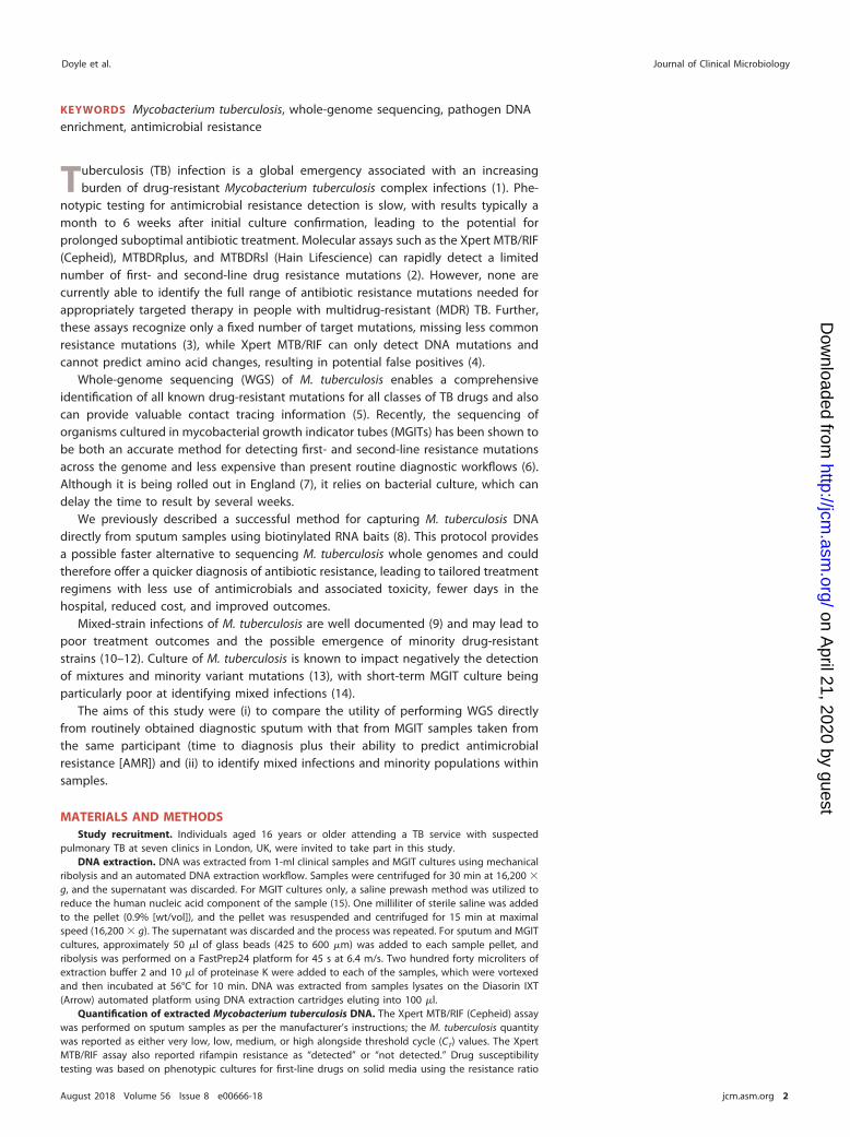
sputum and MGIT samples were sequenced from 43 patients. This is due to 10participants not having an MGIT sample collected for sequencing, another eightsamples being smear and Xpert MTB/RIF negative, and two where the volume ofsputum was insufficient for DNA extraction. Samples sequenced from MGIT cultureshad a higher reference genome coverage than those obtained directly from sputum(Fig. 1), and this was correlated to the increased M. tuberculosis DNA available from theformer (see Fig. S1 in the supplemental material). The enrichment of MGIT culturesamples using the M. tuberculosis probes also enhanced the quality and depth of thesequences (Fig. S1).
We next evaluated whether bacterial load, as measured by smear and Xpert MTB/RIF, could be used to predict the success of whole-genome sequencing. From 43patients, 32 sputum samples (74.4%) and 43 MGIT samples (100%) generated wholegenomes (�85% coverage against reference genome) (Fig. 2A). Sputum sequencingsuccess was linked to estimated input pathogen copy number. We stratified partici-pants into 16 with high (3� smear result, Xpert MTB/RIF high), 18 with medium (2�
smear result, Xpert MTB/RIF medium), and 9 with low (scanty or 1� smear result, XpertMTB/RIF low) bacterial loads and found that 87.5% of sputum samples with highbacterial load samples generated complete genomes compared to 72.2% with mediumand 55.5% with low bacterial loads (Fig. 2B and C). We were also able to recover partial
FIG 1 Comparison of the percentages of reference genomes with at least one sequence read covering a position on the y axis by the median depth of coverageon the x axis for each individual sample. This is stratified by whether original sample material was a sputum or MGIT culture.
Doyle et al. Journal of Clinical Microbiology
August 2018 Volume 56 Issue 8 e00666-18 jcm.asm.org 4
genomes for two sputum samples that were reported as negative by smear microscopybut Xpert MTB/RIF positive.
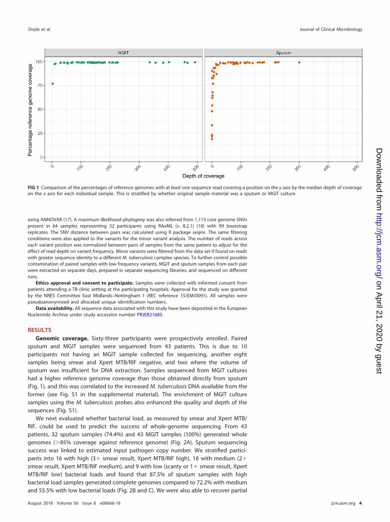
MGIT and sputum sequence variation. A comparison of the 32 patients with bothcomplete sputum and MGIT genomes available showed no unique consensus sequencevariation between the pairs. The identity between sputum and MGIT consensus se-quences is shown in a heat map (Fig. 3) and phylogenetic tree (see Fig. S2). Twenty-three MGIT and sputum pairs showed no SNV between them at the consensus level,while nine patients’ sample pairs differed by one or two single nucleotides. In all ninepatients, the consensus polymorphism was present as a minority variant in the matchedsputum or MGIT sample (see Table S1).
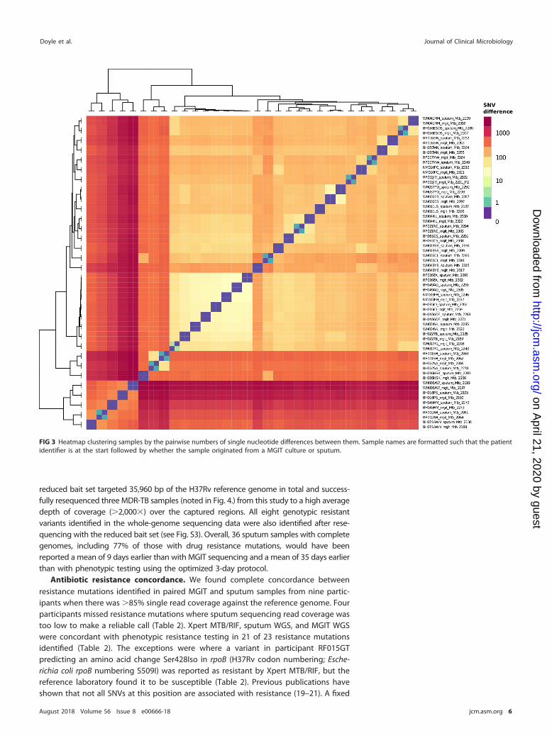
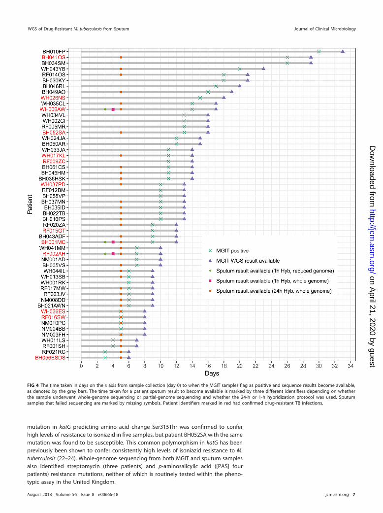
Time to antibiotic resistance prediction. Allowing for sample batching, which wasonly carried out for study purposes, the direct sequencing of sputum using targetedenrichment reduced the time to antibiotic susceptibility prediction initially to 5 days,compared with a mean of 11 (standard deviation [SD], 6) days for MGIT sequencing (Fig. 4).This was reduced further by protocol optimization to 3 days when a reduced bait set wasused that captures only the regions with putative resistance mutations (Table 1 and Fig. 4).Hybridization optimization also reduced the whole-genome protocol to 4 days (Fig. 4). The
FIG 2 (A) Bar plot showing the percentages of reference genome coverage (a single read covering each genome position) for patients with both sputum andMGIT samples sequenced. The plot is annotated with both Xpert MTB/RIF (GX) and smear microscopy (Smear) results. For GX: ****, high; ***, medium; **, low;*, very low. For smear: ***, 3�; **, 2�; *, 1�; S, scanty; �, negative. Where a result is missing, the test was not carried out. Boxplots showing median depthsof coverage for sputum samples stratified by both the quantitative Xpert MTB/RIF measures (B) and semiquantitative smear results (C).
WGS of Drug-Resistant M. tuberculosis from Sputum Journal of Clinical Microbiology
August 2018 Volume 56 Issue 8 e00666-18 jcm.asm.org 5
reduced bait set targeted 35,960 bp of the H37Rv reference genome in total and success-fully resequenced three MDR-TB samples (noted in Fig. 4.) from this study to a high averagedepth of coverage (�2,000�) over the captured regions. All eight genotypic resistantvariants identified in the whole-genome sequencing data were also identified after rese-quencing with the reduced bait set (see Fig. S3). Overall, 36 sputum samples with completegenomes, including 77% of those with drug resistance mutations, would have beenreported a mean of 9 days earlier than with MGIT sequencing and a mean of 35 days earlierthan with phenotypic testing using the optimized 3-day protocol.
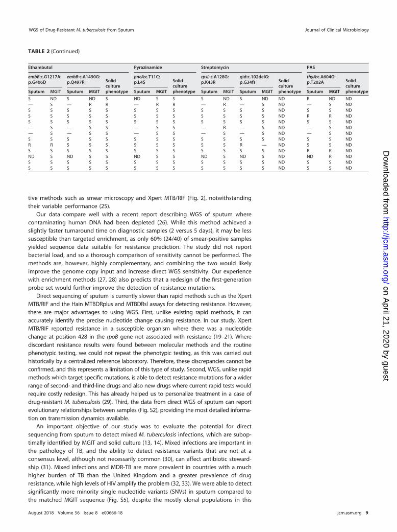
Antibiotic resistance concordance. We found complete concordance betweenresistance mutations identified in paired MGIT and sputum samples from nine partic-ipants when there was �85% single read coverage against the reference genome. Fourparticipants missed resistance mutations where sputum sequencing read coverage wastoo low to make a reliable call (Table 2). Xpert MTB/RIF, sputum WGS, and MGIT WGSwere concordant with phenotypic resistance testing in 21 of 23 resistance mutationsidentified (Table 2). The exceptions were where a variant in participant RF015GTpredicting an amino acid change Ser428Iso in rpoB (H37Rv codon numbering; Esche-richia coli rpoB numbering S509I) was reported as resistant by Xpert MTB/RIF, but thereference laboratory found it to be susceptible (Table 2). Previous publications haveshown that not all SNVs at this position are associated with resistance (19–21). A fixed
FIG 3 Heatmap clustering samples by the pairwise numbers of single nucleotide differences between them. Sample names are formatted such that the patientidentifier is at the start followed by whether the sample originated from a MGIT culture or sputum.
Doyle et al. Journal of Clinical Microbiology
August 2018 Volume 56 Issue 8 e00666-18 jcm.asm.org 6
mutation in katG predicting amino acid change Ser315Thr was confirmed to conferhigh levels of resistance to isoniazid in five samples, but patient BH052SA with the samemutation was found to be susceptible. This common polymorphism in katG has beenpreviously been shown to confer consistently high levels of isoniazid resistance to M.tuberculosis (22–24). Whole-genome sequencing from both MGIT and sputum samplesalso identified streptomycin (three patients) and p-aminosalicylic acid ([PAS] fourpatients) resistance mutations, neither of which is routinely tested within the pheno-typic assay in the United Kingdom.
FIG 4 The time taken in days on the x axis from sample collection (day 0) to when the MGIT samples flag as positive and sequence results become available,as denoted by the gray bars. The time taken for a patient sputum result to become available is marked by three different identifiers depending on whetherthe sample underwent whole-genome sequencing or partial-genome sequencing and whether the 24-h or 1-h hybridization protocol was used. Sputumsamples that failed sequencing are marked by missing symbols. Patient identifiers marked in red had confirmed drug-resistant TB infections.
WGS of Drug-Resistant M. tuberculosis from Sputum Journal of Clinical Microbiology
August 2018 Volume 56 Issue 8 e00666-18 jcm.asm.org 7
Mixed infections and minority variants. No mixed infections were detected. Usingdata normalized for read depth, from 32 matched sputa-MGIT samples, minor fre-quency variation was low, with only 88 minority biallelic sites meeting the qualitycriteria identified in all samples, representing 0.002% unique variable positions acrossthe genomes. We undertook stringent procedures to exclude sequencing error, thepresence of closely related M. tuberculosis complex species in sputum, contamination inand between sequencing runs, and lab contamination after sample collection aspotential causes for the findings. None of the variant alleles were at positions known tobe associated with antimicrobial resistance. In 41% (13) of cases, directly sequencedsputa had higher numbers of novel minority variants identified than the matched MGITsamples, compared to 19% (6) of MGIT samples with more minority variants in thesputum samples (see Fig. S4).
To control for the potential influence of the SureSelectXT step, we analyzed theproportions of minority variants shared between sputum and eight SureSelectXTenriched MGIT samples (40%), comparing between sputum and 16 nonenriched sam-ples (38%), and found no statistically significant difference (P � 0.854). Overall, 37.2%of minority variants were concordant between sputum and MGIT samples, and the readfrequencies with which they occurred were weakly correlated (see Fig. S5). Thiscorrelation was skewed by one patient (WH044IL), in whom both MGIT and sputumsamples were more variable than other samples (Fig. S4) and in whom seven variantswere at much higher frequencies in the MGIT than in sputum (at 30% to 45% versus�5%, respectively) (Fig. S5 and S6). However, there was no evidence of mixed geno-types. The five synonymous minor variants and two nonsynonymous variants occurringin two genes of unknown function (Rv3529c and Rv3888c) were distributed across thegenome and were not shared across any other pairs of samples.
DISCUSSION
We have shown that using target enrichment WGS methodology directly fromdiagnostic sputum samples generates resistance data, at most, up to 24 days earlierthan MGIT culture WGS and up to 31 days faster than phenotypic testing of Mycobac-terium tuberculosis. Sputum sequencing only achieved whole-genome sequences suit-able for predicting resistance mutations in 32/43 (74%) samples, though this includeda smear-negative sputum sample. Our demonstration that the quality of sequence datais strongly correlated with the input level of TB DNA (see Fig. S1 in the supplementalmaterial) means that the success of sequencing can be predicted using semiquantita-
TABLE 2 Antimicrobial resistance profiles from 13 patients with evidence of resistance from direct sputum sequencing using whole-genome bait set
BH001MC � Sa NDb S ND S ND ND S S ND S ND S ND SBH041OS � —c S — S — Rd R R — R — S — S RBH052SA ��� S S S S S S S S R R S S S S SBH056ESDS � S S S S S S S S S S S S S S SRF002AH � S S S S S S ND S R R S S S S RRF009ZC Neg — S — S — S S S — R — S — S RRF015GT � — S — S — S R S — S — S — S SRF016SW �� S S S S S S S S R R S S S S RWH006AW �� R R S S S S R R S S R R R R RWH017KL � S S S S S S S S S S S S S S RWH026NS Neg ND S ND S ND S ND S ND S ND S ND S SWH036ES Neg S S S S S S S S S S R R S S RWH037PD �� — S — R — S R R R R S S S S RaS, susceptible.bND, sample or result not available.c—, low sequence read coverage at position.dR, resistant.
Doyle et al. Journal of Clinical Microbiology
August 2018 Volume 56 Issue 8 e00666-18 jcm.asm.org 8
tive methods such as smear microscopy and Xpert MTB/RIF (Fig. 2), notwithstandingtheir variable performance (25).
Our data compare well with a recent report describing WGS of sputum wherecontaminating human DNA had been depleted (26). While this method achieved aslightly faster turnaround time on diagnostic samples (2 versus 5 days), it may be lesssusceptible than targeted enrichment, as only 60% (24/40) of smear-positive samplesyielded sequence data suitable for resistance prediction. The study did not reportbacterial load, and so a thorough comparison of sensitivity cannot be performed. Themethods are, however, highly complementary, and combining the two would likelyimprove the genome copy input and increase direct WGS sensitivity. Our experiencewith enrichment methods (27, 28) also predicts that a redesign of the first-generationprobe set would further improve the detection of resistance mutations.
Direct sequencing of sputum is currently slower than rapid methods such as the XpertMTB/RIF and the Hain MTBDRplus and MTBDRsl assays for detecting resistance. However,there are major advantages to using WGS. First, unlike existing rapid methods, it canaccurately identify the precise nucleotide change causing resistance. In our study, XpertMTB/RIF reported resistance in a susceptible organism where there was a nucleotidechange at position 428 in the rpoB gene not associated with resistance (19–21). Wherediscordant resistance results were found between molecular methods and the routinephenotypic testing, we could not repeat the phenotypic testing, as this was carried outhistorically by a centralized reference laboratory. Therefore, these discrepancies cannot beconfirmed, and this represents a limitation of this type of study. Second, WGS, unlike rapidmethods which target specific mutations, is able to detect resistance mutations for a widerrange of second- and third-line drugs and also new drugs where current rapid tests wouldrequire costly redesign. This has already helped us to personalize treatment in a case ofdrug-resistant M. tuberculosis (29). Third, the data from direct WGS of sputum can reportevolutionary relationships between samples (Fig. S2), providing the most detailed informa-tion on transmission dynamics available.
An important objective of our study was to evaluate the potential for directsequencing from sputum to detect mixed M. tuberculosis infections, which are subop-timally identified by MGIT and solid culture (13, 14). Mixed infections are important inthe pathology of TB, and the ability to detect resistance variants that are not at aconsensus level, although not necessarily common (30), can affect antibiotic steward-ship (31). Mixed infections and MDR-TB are more prevalent in countries with a muchhigher burden of TB than the United Kingdom and a greater prevalence of drugresistance, while high levels of HIV amplify the problem (32, 33). We were able to detectsignificantly more minority single nucleotide variants (SNVs) in sputum compared tothe matched MGIT sequence (Fig. S5), despite the mostly clonal populations in this
S ND S ND S ND S S S ND S ND ND R ND ND— S — R R — R R — R — S ND — S NDS S S S S S S S S S S S ND S S NDS S S S S S S S S S S S ND R R NDS S S S S S S S S S S S ND S S ND— S — S S — S S — R — S ND — S ND— S — S S — S S — S — S ND — S NDS S S S S S S S S S S S ND S S NDR R S S S S S S S S R — ND S S NDS S S S S S S S S S S S ND R R NDND S ND S S ND S S ND S ND S ND ND R NDS S S S S S S S S S S S ND S S NDS S S S S S S S S S S S ND S S ND
WGS of Drug-Resistant M. tuberculosis from Sputum Journal of Clinical Microbiology
August 2018 Volume 56 Issue 8 e00666-18 jcm.asm.org 9
study and the greater read depths achieved from MGIT sequencing. The origin of thisheterogeneity remains unconfirmed, although we rigorously excluded contaminationand methodological error. SNVs could be due to the presence in sputum of nontuber-culous mycobacteria and other species which are known to have sequence homologywith M. tuberculosis and may theoretically be detected by targeted enrichment. How-ever, our use of highly stringent sequence mapping and the fact that the SNVs weredetected across the genome and not concentrated in regions generally associated withcross hybridization suggest that they are real. In case WH044IL, seven SNVs present insputum increased in frequency in the MGIT culture, possibly reflecting a selectivegrowth advantage for this haplotype, particularly as one nonsynonymous SNV occurredin the Rv3888c gene, which has been shown to be essential for mycobacterial in vitrogrowth (34). This result confirms suggestions that diversity is lost and that culture-related selection of some variants can occur even during limited MGIT culture. Thus,MGIT culture may not be representative of the original sample and could potentiallyreduce the likelihood of identifying low-level resistance mutations and mixed infectionsthat may act as a reservoir for resistance development.
The standard diagnostic workflows for M. tuberculosis are costly and time consum-ing. Ground breaking work within Public Health England has demonstrated thatsequencing M. tuberculosis whole genomes from positive MGIT cultures is faster andless expensive (6), and where the sputum pathogen DNA concentration is low, earlyMGIT sequencing could still be the best possible workaround (15). Direct sequencing ofM. tuberculosis from sputum has the potential to reduce the time to antimicrobialresistance detection within a clinically relevant time frame (26). We show here that itssuccess is critically dependent on the input genome copies of pathogen DNA. Whileenrichment increases the cost of pathogen sequencing, this could be offset, as dem-onstrated in our study, by only enriching areas of interest on the genome. It isimportant to note that the infrastructure and expertise for rapid, high-throughput,targeted enrichment sequencing directly from clinical material of M. tuberculosis andother pathogens already exist in genomic centers where cancer and genetic diseasesequencing uses this methodology. We believe, therefore, that effective scale-up andimplementation of this rapid and accurate technology is relatively easy, once thetechnique has been optimized.
SUPPLEMENTAL MATERIAL
Supplemental material for this article may be found at https://doi.org/10.1128/JCM.00666-18.
SUPPLEMENTAL FILE 1, PDF file, 0.9 MB.
ACKNOWLEDGMENTSWe thank all participants who agreed to take part in this study. We also thank Lusha
Kellgren, Maria McEwan, Narinder Boparai, Jacqui White, Neil Jones, Stephen Morris-Jones, Surjo De, and Trupti Patel for collecting samples at The Whittington Health NHSTrust, University College London Hospitals, and the TB Service North Central London,Nirmala Ghimire and Devan Vaghela for collecting samples at Bart’s Health NHS Trust,and Josephine Silles, Pragna Patel, Marinos Christofi, Thomas Rendal, Yemi Martins, andEsmeralda Sicat for collecting samples at North Middlesex University Hospital.
This work was funded by the UCLH/UCL NIHR Biomedical Resource Centre (grantnumber BRC/176/III/JB/101350) and the PATHSEEK European Union’s Seventh Programmefor research, technological development and demonstration (grant number 304875).
We declare no competing interests.
REFERENCES1. WHO. 2017. Global tuberculosis report 2017. WHO, Geneva, Switzerland.
2. Scott LE, McCarthy K, Gous N, Nduna M, Van Rie A, Sanne I, Venter WF,
Duse A, Stevens W. 2011. Comparison of Xpert MTB/RIF with othernucleic acid technologies for diagnosing pulmonary tuberculosis in ahigh HIV Prevalence setting: a prospective study. PLoS Med 8:e1001061.https://doi.org/10.1371/journal.pmed.1001061.
Doyle et al. Journal of Clinical Microbiology
August 2018 Volume 56 Issue 8 e00666-18 jcm.asm.org 10
4. Ocheretina O, Byrt E, Mabou M-M, Royal-Mardi G, Merveille Y-M, RouzierV, Fitzgerald DW, Pape JW. 2016. False-positive rifampin resistant resultswith Xpert MTB/RIF version 4 assay in clinical samples with a lowbacterial load. Diagn Microbiol Infect Dis 85:53–55. https://doi.org/10.1016/j.diagmicrobio.2016.01.009.
5. Witney AA, Gould KA, Arnold A, Coleman D, Delgado R, Dhillon J, PondMJ, Pope CF, Planche TD, Stoker NG, Cosgrove CA, Butcher PD, HarrisonTS, Hinds J. 2015. Clinical application of whole-genome sequencing toinform treatment for multidrug-resistant tuberculosis cases. J Clin Mi-crobiol 53:1473–1483. https://doi.org/10.1128/JCM.02993-14.
6. Pankhurst LJ, del Ojo Elias C, Votintseva AA, Walker TM, Cole K, Davies J,Fermont JM, Gascoyne-Binzi DM, Kohl TA, Kong C, Lemaitre N, NiemannS, Paul J, Rogers TR, Roycroft E, Smith EG, Supply P, Tang P, Wilcox MH,Wordsworth S, Wyllie D, Xu L, Crook DW, COMPASS-TB Study Group.2016. Rapid, comprehensive, and affordable mycobacterial diagnosiswith whole-genome sequencing: a prospective study. Lancet Respir Med4:49 –58. https://doi.org/10.1016/S2213-2600(15)00466-X.
7. Walker TM, Cruz ALG, Peto TE, Smith EG, Esmail H, Crook DW. 2017.Tuberculosis is changing. Lancet Infect Dis 17:359 –361. https://doi.org/10.1016/S1473-3099(17)30123-8.
8. Brown AC, Bryant JM, Einer-Jensen K, Holdstock J, Houniet DT, Chan JZM,Depledge DP, Nikolayevskyy V, Broda A, Stone MJ, Christiansen MT,Williams R, McAndrew MB, Tutill H, Brown J, Melzer M, Rosmarin C,McHugh TD, Shorten RJ, Drobniewski F, Speight G, Breuer J. 2015. Rapidwhole-genome sequencing of Mycobacterium tuberculosis isolates di-rectly from clinical samples. J Clin Microbiol 53:2230 –2237. https://doi.org/10.1128/JCM.00486-15.
9. Cohen T, van Helden PD, Wilson D, Colijn C, McLaughlin MM, AbubakarI, Warren RM. 2012. Mixed-strain Mycobacterium tuberculosis infectionsand the implications for tuberculosis treatment and control. Clin Micro-biol Rev 25:708 –719. https://doi.org/10.1128/CMR.00021-12.
10. van Rie A, Victor TC, Richardson M, Johnson R, van der Spuy GD, MurrayEJ, Beyers N, Gey van Pittius NC, van Helden PD, Warren RM. 2005.Reinfection and mixed infection cause changing Mycobacterium tuber-culosis drug-resistance patterns. Am J Respir Crit Care Med 172:636 – 642.https://doi.org/10.1164/rccm.200503-449OC.
11. Baldeviano-Vidalón GC, Quispe-Torres N, Bonilla-Asalde C, Gastiaburú-Rodriguez D, Pro-Cuba JE, Llanos-Zavalaga F. 2005. Multiple infectionwith resistant and sensitive M. tuberculosis strains during treatment ofpulmonary tuberculosis patients. Int J Tuberc Lung Dis 9:1155–1160.
12. Zetola NM, Shin SS, Tumedi KA, Moeti K, Ncube R, Nicol M, Collman RG,Klausner JD, Modongo C. 2014. Mixed Mycobacterium tuberculosis com-plex infections and false-negative results for rifampin resistance byGeneXpert MTB/RIF are associated with poor clinical outcomes. J ClinMicrobiol 52:2422–2429. https://doi.org/10.1128/JCM.02489-13.
13. Martín A, Herranz M, Ruiz Serrano MJ, Bouza E, García de Viedma D.2010. The clonal composition of Mycobacterium tuberculosis in clinicalspecimens could be modified by culture. Tuberculosis (Edinb) 90:201–207. https://doi.org/10.1016/j.tube.2010.03.012.
14. Hanekom M, Streicher EM, Van de Berg D, Cox H, McDermid C, BosmanM, Gey van Pittius NC, Victor TC, Kidd M, van Soolingen D, van HeldenPD, Warren RM. 2013. Population structure of mixed Mycobacteriumtuberculosis infection is strain genotype and culture medium dependent.PLoS One 8:e70178. https://doi.org/10.1371/journal.pone.0070178.
15. Votintseva AA, Pankhurst LJ, Anson LW, Morgan MR, Gascoyne-Binzi D,Walker TM, Quan TP, Wyllie DH, Del Ojo Elias C, Wilcox M, Walker AS,Peto TEA, Crook DW. 2015. Mycobacterial DNA extraction for whole-genome sequencing from early positive liquid (MGIT) cultures. J ClinMicrobiol 53:1137–1143. https://doi.org/10.1128/JCM.03073-14.
16. Honeyborne I, McHugh TD, Phillips PPJ, Bannoo S, Bateson A, Carroll N,Perrin FM, Ronacher K, Wright L, van Helden PD, Walzl G, Gillespie SH.2011. Molecular bacterial load assay, a culture-free biomarker for rapidand accurate quantification of sputum Mycobacterium tuberculosis bac-illary load during treatment. J Clin Microbiol 49:3905–3911. https://doi.org/10.1128/JCM.00547-11.
17. Yang H, Wang K. 2015. Genomic variant annotation and prioritizationwith ANNOVAR and wANNOVAR. Nat Protoc 10:1556 –1566. https://doi.org/10.1038/nprot.2015.105.
18. Stamatakis A. 2014. RAxML version 8: a tool for phylogenetic analysisand post-analysis of large phylogenies. Bioinformatics 30:1312–1313.https://doi.org/10.1093/bioinformatics/btu033.
20. Sekiguchi J, Miyoshi-Akiyama T, Augustynowicz-Kopec E, Zwolska Z,Kirikae F, Toyota E, Kobayashi I, Morita K, Kudo K, Kato S, Kuratsuji T, MoriT, Kirikae T. 2007. Detection of multidrug resistance in Mycobacteriumtuberculosis. J Clin Microbiol 45:179 –192. https://doi.org/10.1128/JCM.00750-06.
21. Zenteno-Cuevas R, Zenteno JC, Cuellar A, Cuevas B, Sampieri CL, RivieraJE, Parissi A. 2009. Mutations in rpoB and katG genes in Mycobacteriumisolates from the Southeast of Mexico. Mem Inst Oswaldo Cruz 104:468 – 472. https://doi.org/10.1590/S0074-02762009000300012.
22. Wengenack NL, Uhl JR, St Amand AL, Tomlinson AJ, Benson LM, NaylorS, Kline BC, Cockerill FR, Rusnak F. 1997. Recombinant Mycobacteriumtuberculosis KatG(S315T) is a competent catalase-peroxidase with re-duced activity toward isoniazid. J Infect Dis 176:722–727. https://doi.org/10.1086/514096.
23. Saint-Joanis B, Souchon H, Wilming M, Johnsson K, Alzari PM, Cole ST.1999. Use of site-directed mutagenesis to probe the structure, functionand isoniazid activation of the catalase/peroxidase, KatG, from Mycobac-terium tuberculosis. Biochem J 338(Pt 3):753–760.
24. Pym AS, Saint-Joanis B, Cole ST. 2002. Effect of katG mutations on thevirulence of Mycobacterium tuberculosis and the implication for trans-mission in humans. Infect Immun 70:4955– 4960. https://doi.org/10.1128/IAI.70.9.4955-4960.2002.
25. Devonshire AS, O’Sullivan DM, Honeyborne I, Jones G, Karczmarczyk M,Pavšic J, Gutteridge A, Milavec M, Mendoza P, Schimmel H, Van Heuver-swyn F, Gorton R, Cirillo DM, Borroni E, Harris K, Barnard M, HeydenrychA, Ndusilo N, Wallis CL, Pillay K, Barry T, Reddington K, Richter E,Mozioglu E, Akyürek S, Yalçınkaya B, Akgoz M, Žel J, Foy CA, McHugh TD,Huggett JF. 2016. The use of digital PCR to improve the application ofquantitative molecular diagnostic methods for tuberculosis. BMC InfectDis 16:366. https://doi.org/10.1186/s12879-016-1696-7.
26. Votintseva AA, Bradley P, Pankhurst L, Del Ojo Elias C, Loose M, Nilgiri-wala K, Chatterjee A, Smith EG, Sanderson N, Walker TM, Morgan MR,Wyllie DH, Walker AS, Peto TEA, Crook DW, Iqbal Z. 2017. Same-daydiagnostic and surveillance data for tuberculosis via whole genomesequencing of direct respiratory samples. J Clin Microbiol 55:1285–1298.https://doi.org/10.1128/JCM.02483-16.
27. Depledge DP, Palser AL, Watson SJ, Lai IY-C, Gray ER, Grant P, Kanda RK,Leproust E, Kellam P, Breuer J. 2011. Specific capture and whole-genomesequencing of viruses from clinical samples. PLoS One 6:e27805. https://doi.org/10.1371/journal.pone.0027805.
28. Christiansen MT, Brown AC, Kundu S, Tutill HJ, Williams R, Brown JR,Holdstock J, Holland MJ, Stevenson S, Dave J, Tong CYW, Einer-Jensen K,Depledge DP, Breuer J. 2014. Whole-genome enrichment and sequenc-ing of Chlamydia trachomatis directly from clinical samples. BMC InfectDis 14:591. https://doi.org/10.1186/s12879-014-0591-3.
29. Nimmo C, Doyle R, Burgess C, Williams R, Gorton R, McHugh TD, BrownM, Morris-Jones S, Booth H, Breuer J. 2017. Rapid identification of aMycobacterium tuberculosis full genetic drug resistance profile throughwhole genome sequencing directly from sputum. Int J Infect Dis 62:44 – 46. https://doi.org/10.1016/j.ijid.2017.07.007.
30. Witney AA, Bateson ALE, Jindani A, Phillips PPJ, Coleman D, Stoker NG,Butcher PD, McHugh TD, RIFAQUIN Study Team. 2017. Use of whole-genome sequencing to distinguish relapse from reinfection in a com-pleted tuberculosis clinical trial. BMC Med 15:71. https://doi.org/10.1186/s12916-017-0834-4.
31. Cohen T, Chindelevitch L, Misra R, Kempner ME, Galea J, Moodley P, WilsonD. 2016. Within-host heterogeneity of Mycobacterium tuberculosis infectionis associated with poor early treatment response: a prospective cohortstudy. J Infect Dis 213:1796–1799. https://doi.org/10.1093/infdis/jiw014.
32. Bifani PJ, Mathema B, Kurepina NE, Kreiswirth BN. 2002. Global dissemina-tion of the Mycobacterium tuberculosis W-Beijing family strains. TrendsMicrobiol 10:45–52. https://doi.org/10.1016/S0966-842X(01)02277-6.
33. Eldholm V, Rieux A, Monteserin J, Lopez JM, Palmero D, Lopez B, RitaccoV, Didelot X, Balloux F. 2016. Impact of HIV co-infection on the evolutionand transmission of multidrug-resistant tuberculosis. Elife 5:e16644.https://doi.org/10.7554/eLife.16644.