This article was downloaded by: [University of Nebraska, Lincoln] On: 07 September 2013, At: 15:21 Publisher: Taylor & Francis Informa Ltd Registered in England and Wales Registered Number: 1072954 Registered office: Mortimer House, 37-41 Mortimer Street, London W1T 3JH, UK Avian Pathology Publication details, including instructions for authors and subscription information: http://www.tandfonline.com/loi/cavp20 Mycoplasma infection of Geese I. Incidence of Mycoplasmas and Acholeplasmas in Geese L. Stipkovits a , A.A. El‐Ebeedy a , J. Kisary a & Lea Varga a a Veterinary Medical Research Institute of the Hungarian Academy of Sciences, Budapest, Hungary Published online: 17 Sep 2008. To cite this article: L. Stipkovits , A.A. El‐Ebeedy , J. Kisary & Lea Varga (1975) Mycoplasma infection of Geese I. Incidence of Mycoplasmas and Acholeplasmas in Geese, Avian Pathology, 4:1, 35-43 To link to this article: http://dx.doi.org/10.1080/03079457509353848 PLEASE SCROLL DOWN FOR ARTICLE Taylor & Francis makes every effort to ensure the accuracy of all the information (the “Content”) contained in the publications on our platform. However, Taylor & Francis, our agents, and our licensors make no representations or warranties whatsoever as to the accuracy, completeness, or suitability for any purpose of the Content. Any opinions and views expressed in this publication are the opinions and views of the authors, and are not the views of or endorsed by Taylor & Francis. The accuracy of the Content should not be relied upon and should be independently verified with primary sources of information. Taylor and Francis shall not be liable for any losses, actions, claims, proceedings, demands, costs, expenses, damages, and other liabilities whatsoever or howsoever caused arising directly or indirectly in connection with, in relation to or arising out of the use of the Content.
Transcript
This article was downloaded by: [University of Nebraska, Lincoln]On: 07 September 2013, At: 15:21Publisher: Taylor & FrancisInforma Ltd Registered in England and Wales Registered Number: 1072954Registered office: Mortimer House, 37-41 Mortimer Street, London W1T3JH, UK
Avian PathologyPublication details, including instructions forauthors and subscription information:http://www.tandfonline.com/loi/cavp20
Mycoplasma infection of GeeseI. Incidence of Mycoplasmasand Acholeplasmas in GeeseL. Stipkovits a , A.A. El‐Ebeedy a , J. Kisary a & Lea
Varga aa Veterinary Medical Research Institute of theHungarian Academy of Sciences, Budapest,HungaryPublished online: 17 Sep 2008.
To cite this article: L. Stipkovits , A.A. El‐Ebeedy , J. Kisary & Lea Varga (1975)Mycoplasma infection of Geese I. Incidence of Mycoplasmas and Acholeplasmas inGeese, Avian Pathology, 4:1, 35-43
To link to this article: http://dx.doi.org/10.1080/03079457509353848
PLEASE SCROLL DOWN FOR ARTICLE
Taylor & Francis makes every effort to ensure the accuracy of allthe information (the “Content”) contained in the publications on ourplatform. However, Taylor & Francis, our agents, and our licensorsmake no representations or warranties whatsoever as to the accuracy,completeness, or suitability for any purpose of the Content. Any opinionsand views expressed in this publication are the opinions and views ofthe authors, and are not the views of or endorsed by Taylor & Francis.The accuracy of the Content should not be relied upon and should beindependently verified with primary sources of information. Taylor andFrancis shall not be liable for any losses, actions, claims, proceedings,demands, costs, expenses, damages, and other liabilities whatsoeveror howsoever caused arising directly or indirectly in connection with, inrelation to or arising out of the use of the Content.
This article may be used for research, teaching, and private studypurposes. Any substantial or systematic reproduction, redistribution,reselling, loan, sub-licensing, systematic supply, or distribution in any formto anyone is expressly forbidden. Terms & Conditions of access and use canbe found at http://www.tandfonline.com/page/terms-and-conditions
I. INCIDENCE OF MYCOPLASMAS AND ACHOLEPLASMAS IN GEESE
L. STIPKOVITS, A.A. EL-EBEEDY, J. KISARY andLea VARGA
Veterinary Medical Research Institute of the HungarianAcademy of Sciences, Budapest, Hungary
SUMMARY
This paper presents data about the isolation of members of the orderMycoplasmatales from material of goose origin.
Acholeplasma laidlawii strains were isolated from 2 to 8 day old goslingswith heavy fibrinous airsacculitis, peritonitis and perihepatitis. Lossesreached 30% of the flock by the end of the 8th week of age. Achole-plasma axanthum strains were detected in goose-embryos that died onthe 13th day of incubation. A significant loss (up to 60%) of embryoswas observed in the flock and some layers died showing fibrinous peri-tonitis, salpingitis and abdominal airsacculitis. Mycoplasma gallinarumalso was isolated from goose-embryo fibroblast tissue cultures.
All strains except A. laidlawii caused cytoplasmic vacuolization andintracytoplasmic inclusion bodies in goose-embryo fibroblast tissuecultures. The alteration observed in chicken-embryo fibroblast cellcultures were similar; in addition, the A. laidlawii caused a markedpycnosis of the cells.
INTRODUCTION
Since mycoplasma were first isolated from poultry (Nelson, 1936) several Myco-plasma species including M. gallinarum (Edward and Freundt, 1956), M. gallisepti-cum (Edward and Kanarek, 1960),Af. iners (Edward and Kanarek, 1960),Af.synoviae (Olson et al, 1964), Ai. meleagridis (Yamamoto et al, 1965), A. laidlawiivar. inocuum (Adler et al, 1961),M anatis (Roberts, 1964) and avian serogroups(C, D, F, I, J, K, L, M, N, O, P, Q and R) (Dierks et al, 1967) have been described.Some of them have a definite etiological role in chronic respiratory diseases, arthri-tis and other diseases.
However, we could find no data in the literature on mycoplasma infection in geese.The development of intensive breeding and keeping of geese was accompanied by newdisorders in which, on the basis of clinical symptoms and pathological lesions, apathological role of mycoplasmas and acholeplasmas seemed possible. Therefore we
Received 6 May 1974
Dow
nloa
ded
by [
Uni
vers
ity o
f N
ebra
ska,
Lin
coln
] at
15:
21 0
7 Se
ptem
ber
2013
36 L. Stipkovits, A.A. El-Ebeedy, J. Kisary and Lea Varga
attempted to isolate organisms of the order of Mycoplasmatales from geese, goose-embryos and goose-embryo fibroblast tissue cultures.
MATERIALS AND METHODS
Specimen collectionSamples for culture were obtained from 3 sources: 1. From samples of fibrinousthoracic and abdominal airsacs of 2 weeks old goslings with heavy clinical symptomsof respiratory disease which were removed, minced and tenfold dilutions prepared inmycoplasma media. (Flock No.l). 2. From goose-embryos which died on the 13th dayof incubation and from fresh goose eggs and oviducts of geese (Flock No.2). Suspen-sions prepared from these samples were inoculated into mycoplasma media. 3. Fromgoose-embryo fibroblast tissue cultures after 2—5 days of cultivation. Tissue cultureswere frozen and thawed 3 times before inoculation of mycoplasma media.
MediaFor primary isolation and cultivation Medium B (Ern^and Stipkovits, 1973), VF-medium and Medium B enriched with 10% fresh pig serum and 0.01% reduced DPNwere used. Biochemical examinations of the strains were performed in various modi-fications of Medium B (Ern¿> and Stipkovits, 1973).
Cultural methodsInoculated liquid media were incubated at 37°C for at least 14 days. After inoculationthe media were plated onto solid agar every second day. A part of it was incubatedaerobically, the other part in the presence of 5% CO2 and 95% N2 . The growth ofcolonies was checked regularly. Agar blocks containing colonies were transferred intoliquid medium, and after 5 days of incubation the liquid cultures were filtered througha Millipore filter (Millipore HAWP 01300,100 ea. HA 0.45 /urn). Filtrates were streak-ed onto solid media, and if the cultivation was successful, a single colony was pickedfor further subculture. Each strain was cloned three times.
Biochemical and serological characterizationAll strains were examined for criteria of the order of Mycoplasmatales (Subcommitteeon the Taxonomy of Mycoplasmatales, 1972) (Penicillin resistance, filtration, morpho-logy, absence of bacterial reversion), for classification into family (cholesterol require-ment, sensitivity to sodium polyanethol sulphonate and digitonin), and determinationof species (Ern¿ and Stipkovits, 1973). Growth inhibition tests were performed usingthe following antisera: M hominis(?G2l),M. oralel. (CH19219),M. oralell.(CH20247),M salivarum (PG20),M fermentons (PG18),M pneumoniae (MAC),M. primatum (Navel),M arthritidis (PG6),M. neurolyticum (Sabin A),Af. pulmonis(Ash),M canis (PG14),M felis (CO),M. histotropicum (ATCC 23115),M edwardii(PG24),M.feliminitum(BEN),M. spumans (PG13),M. leonis.M. lipophylum(MABY),Ai. maculosum (PG15),M hyorhinis(PG29),M. hyosynoviae(AMRC/104),A. granularum (Friend),,4. laidlawiiA. (PG8),Af. agalactiae subsp. bovis (Donetta),M. bovirhinis (PG43),M. arginini (G230),M. sp. PG50 (Leach's 7 serogroup),M. my-coides subsp. mycoides (PG l),M. mycoides subsp. capri(?G3),M. bovigenitalium(PG11),M. sp. B 144P(Al-Aubaidi's L-serogroup),/l. modicum (Squire), A axanthum(ATCC 25176), M. conjunctivae (MHRC581),Af. caviae.M. agalactiae subsp. agalac-tiae (PG2), M. gallinarum (PG 16), M. gateae (CS), M. gallisepticum (X95), M. melea-gridia (17529),M. anatis (1340),M iners (PG30). The mycoplasma reference strainswere obtained from the FAO/WHO Reference Centre for Animal Mycoplasmas (Medi-cal Microbiology, University of Aarhus, Aarhus, Denmark, Prof. A.E. Freundt). All
Dow
nloa
ded
by [
Uni
vers
ity o
f N
ebra
ska,
Lin
coln
] at
15:
21 0
7 Se
ptem
ber
2013
Mycoplasma infection of geese 3 7
antiserum used were prepared in our laboratory.
Examination of strains in tissue cultureStrains. The following strains of goose origin were used: No.598 (M. gallinarum),No.606 (A. laidlawii), Nos.609 and 611 (A. axanthum). For comparison strain No.613(M. galliseptiaim) isolated from the sinus fluid of a 5 months old turkey sufferingfrom heavy sinusitis, airsacculitis and peritonitis, as well as strain No.618 (M. melea-gridis) from the airsac of a day-old turkey poult with airsacculitis, pericarditis andperitonitis were included in the experiments. For tissue culture inoculation the strainswere propagated in medium B without thallium acetate and the horse serum was re-placed by PPLO-Serum Fraction (DIFCO).
Tissue cultures. 14 days old goose and 11 days old chicken embryos were used tomake fibroblast cell cultures by standard methods. The growth medium was TCM-199(DIFCO) supplemented with 0.5% lactalbumine hydrolysate and 5% inactivated foetalcalf serum. pH was adjusted to 7.2 by 1 g/1. NaHCO3. Earle's TC solution (DIFCO)served as maintenance medium with 2% foetal calf serum and 2.2 g/1. NaHCO3.100units of penicillin/ml, were added to both media. Four tubes per strains were infected.
All media and ingredients were checked for mycoplasma contamination before use inexperiments. Tissue cultures infected by mycoplasmas and acholeplasmas, as well asthe controls, were monitored daily. Cultures showing any alterations were immediate-ly stained by haematoxylin and eosin (H & E), and those without cytopathic effect(c.p.e.) were similarly treated on the 7th day after infection.
RESULTS
Field observations1. Flock No. 1. consisted of 8025 goslings. Goslings were treated at 3 days of age with2.0 ml hyperimmune serum produced with strain B of gosling virus (so-called "gos-ling influenza", "gosling hepatitis", etc.). During the first 2 weeks no significantlosses (2%) were observed. At the end of the third week about 100 goslings died daily.The losses increased, reaching 30% of the total flock by the end of the 8th week. Thediseased goslings showed clinical symptoms of respiratory disease. At postmortemexamination fibrinous airsacculitis, peritonitis, perihepatitis and enlarged spleen wereobserved almost in all dead goslings.
2. FlockNo.2. consisted of 2000 laying geese. Significant embryonic mortality wasobserved in the flock. Gradual decrease of hatchability was recorded from Februaryreaching the highest loss (60%) in March and early April. No significant losses (2%)were observed in adult birds. No special clinical symptoms were found in the layinggeese. The postmortem examination of some dead birds revealed mostly fibrinousperitonitis, salpingitis and abdominal airsacculitis.
Isolation and characterization of strainsA total of 23 specimens was examined. Four of 10 samples from airsacs of goslings(Flock No.l) (Strain Nos.603, 604, 605 and 606), 4 dead embryos (Strain Nos.609,610, 611 and 612) (Flock No.2) and 1 of 3 samples of goose-embryo fibroblast tissueculture (Strain No.598) contained microorganisms belonging to the order Mycoplas-matales. Four samples of fresh goose eggs and 2 samples from the oviducts of layersproved to be negative.
All isolated strains were filterable through Millipore filter (HAWP 01300 100 ea. HA0.45 ism) without a significant decrease of CFU. None of them produced bacterial
Dow
nloa
ded
by [
Uni
vers
ity o
f N
ebra
ska,
Lin
coln
] at
15:
21 0
7 Se
ptem
ber
2013
38 L. Stipkovits, A.A. El-Ebeedy, J. Kisary and Lea Varga
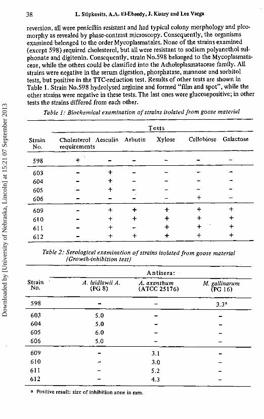
reversion, all were penicillin resistant and had typical colony morphology and pleo-morphy as revealed by phase-contrast microscopy. Consequently, the organismsexamined belonged to the order Mycoplasmatales. None of the strains examined(except 598) required cholesterol, but all were resistant to sodium polyanethol sul-phonate and digitonin. Consequently, strain No.598 belonged to the Mycoplasmata-ceae, while the others could be classified into the Acholeplasmataceae family. Allstrains were negative in the serum digestion, phosphatase, mannose and sorbitoltests, but positive in the TTC-reduction test. Results of other tests are shown inTable 1. Strain No.598 hydrolysed arginine and formed "film and spot", while theother strains were negative in these tests. The last ones were glucosepositive; in othertests the strains differed from each other.
Table 1: Biochemical examination of strains isolated from goose material
Table 2: Serological examination of strains isolated from goose material(Growth-inhibition test)
Antisera:
Strain A. laidlawii A. A. axanthum M. gallinarumNo. (PG8) (ATCC 25176) (PG 16)
598 - - 3.3a
603604605606
609610611612
5.05.06.05.0
---_
----
3.13.05.24.3
a Positive result: size of inhibition zone in mm.
Dow
nloa
ded
by [
Uni
vers
ity o
f N
ebra
ska,
Lin
coln
] at
15:
21 0
7 Se
ptem
ber
2013
Mycoplasma infection of geese 39
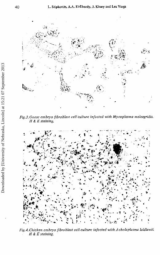
Goose embryo fibroblast cell culture infected with Mycoplasma gallinarum.H & E staining.
Goose embryo fibroblast cell culture infected with Acholeplasma axanthum.H & E staining.
Dow
nloa
ded
by [
Uni
vers
ity o
f N
ebra
ska,
Lin
coln
] at
15:
21 0
7 Se
ptem
ber
2013
40 L. Stipkovits, A.A. El-Ebeedy, J. Kisary and Lea Varga
\> »«y
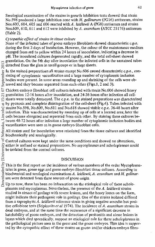
.3. Goose embryo fibroblast cell culture infected with Mycoplasma meleagridis.H & E staining.
Fig.4.Chicken embryo fibroblast cell culture infected with Acholeplasma laidlawii.H & E staining.
Dow
nloa
ded
by [
Uni
vers
ity o
f N
ebra
ska,
Lin
coln
] at
15:
21 0
7 Se
ptem
ber
2013
Mycoplasma infection of geese 41
Serological examination of the strains in growth inhibition tests showed that strainNo.598 produced a large inhibition zone withM. gallinarwn (PG16) antiserum, strainsNos.603, 604, 605 and 606 reacted with/I. laidlawii A (PG8) antiserum and strainsNos.609, 610, 611 and 612 were inhibited by A. axanthum (ATCC 25176) antiserum(Table 2).
Cytopathic effect of strains in tissue cultureNone of the infected tubes of goose embryo fibroblasts showed characteristic c.p.e.during the first 3 days of incubation. However, the colour of the maintenance mediumchanged from red to yellow within 24 hours of inoculation, indicating a decrease inpH. The inoculated cultures degenerated rapidly, and the total cell-sheet showedgranulation. On the 5th day after inoculation the infected cells in the unstained tubesdetached from the glass in small groups or in large sheets.
In the stained preparations all strains except No.606 caused characteristic c.p.e., con-sisting of cytoplasmic vacuolization and a large number of cytoplasmic inclusionbodies were present. In some areas rounding up and shrinking of the cells were ob-served and they became separated from each other (Figs.l, 2 and 3).
Chicken embryo fibroblast cell cultures infected with strain No.606 showed heavygranulation 12-16 hours after inoculation, and 24-36 hours after infection all cell-sheets were totally destroyed. The c.p.e. in the stained preparations was characterizedby pycnosis and complete disintegration of the cell-sheet (Fig.4). Tubes infected withstrains No.598, No.609, No.611 and No.618 showed visible c.p.e. 36-48 hours afterinfection. This was characterized by rounding up of cells at a located area; later thecells became elongated and separated from each other. By staining these cultures be-tween 48-72 hours after infection a large number of cytoplasmic inclusion bodies andvacuolization were seen as in goose embryo fibroblast cells.
All strains used for inoculation were reisolated from the tissue cultures and identifiedbiochemically and serologically.
Control cultures were kept under the same conditions and showed no alterations,either in unfixed or stained preparations. No mycoplasmas and acholeplasmas couldbe isolated from the control cultures.
DISCUSSION
This is the first report on the incidence of various members of the order Mycoplasma-tales in geese, goose eggs and goose-embryo fibroblast tissue cultures. According tobiochemical and serological examinations,4. laidlawii, A. axanthum andM. gallinar-wn were detected from these sources of goose origin.
Up to now, there has been no information on the etiological role of these achole-plasmas and mycoplasmas. Nevertheless, the presence of the A. laidlawii strainsstudied in airsacs of goslings with severe lesions, and the heavy losses on the farm,might indicate their pathogenic role in goslings. One of the strains isolated differedfrom a saprophytic A laidlawii reference strain in giving negative aesculin but posi-tive cellobiose tests (Stipkovits et al 1974). The incidence oí A. axanthum strains indead embryos, and at the same time the occurrence of a significant decrease inhatchability of goose embryos, and the detection of peritonitis and airsac lesions inlayers which died sporadically, support an etiological role for these acholeplasmas inthe pathological picture seen in the geese and the goose embryos. This idea is suppor-ted by the cytopathic effect of these strains on goose- and/or chicken-embryo fibro-
Dow
nloa
ded
by [
Uni
vers
ity o
f N
ebra
ska,
Lin
coln
] at
15:
21 0
7 Se
ptem
ber
2013
42 L. Stipkovits, A.A. El-Ebeedy, J. Kisary and Lea Varga
blast tissue cultures, which was characterized by rapid degeneration of the cell sheet,pycnosis and production of inclusion bodies in the cytoplasm of cells, even thoughthe strains did not split arginine.
M. gallinarum has not been isolated from geese or goose eggs, but on the basis of itspresence in goose-embryo fibroblast tissue cultures we might presume its occurrencein geese also. This mycoplasma can produce cytopathic effects in goose and chicken-embryo fibroblast tissue cultures also, characterized by elongation and separation ofthe cells and cytoplasmic vacuolization due to its arginine requirement (Kisary andStipkovits, 1974).
Because goose-embryos used for preparing cell cultures, and goose material used forvirological examination, could be infected with various species of mycoplasmas andacholeplasmas, mycoplasma should be excluded as a cause of cytoplasmic alterationsbefore the latter are attributed to a virus infection.
REFERENCES
Adler, H.E., Shifrine, M. and Ortmayer, H. (1961). Mycoplasma inocuum sp.n.a saprophyte fromchickens. Journal of Bacteriology, 82: 239-240.
Barber, T.L. and Fabricant, J. (1962). Primary isolation of mycoplasma organisms (PPLO) frommammalian sources. Journal of Bacteriology, 83: 1269-1273.
Dierks, R.E., Newman, J.A. and Pomeroy, B.S. (1967). Characterization of avian Mycoplasma.Annals New York Academy of Sciences, 143: 170-189.
Edward, D.G.ff. and Kanarek, A.D. (1956). The classification and nomenclature of organisms ofthe pleuropneumonia group. Journal of General Microbiology, 14: 197-204.
Edward, D.G.ff. and Kanarek, A.D. (1960). Organisms of the pleuropneumonia group of avianorigin. Their classification into species. Annals New York Academy of Sciences, 79:696-702.
Ernφ, H. and Stipkovits, L. (1973). Bovine Mycoplasmas: Cultural and biochemical studies 1-2.Acta Veterinaria Scandinavica, 14: 436-449. 450-463.
Kisary, J. and Stipkovits, L. The effect of Mycoplasma gallinarum on the in vitro replication ofgoose parvo-virus strain "B". Acta Microbiologica Academiae Scientiarum Hungaricae (inpress).
Nelson, J.B. (1936). Studies on an uncomplicated coryza of the domestic fowl. 7. Cultivation ofthe coccobacilli-form bodies in fertile eggs and in tissue cultures. Journal of ExperimentalMedicine, 64: 749-758.
Olson, N.O., Kerr, K.M. and Campbell, A. (1964). Control of infectious synovitis. 13. Antigenstudy of three strains. Avian Diseases, 8: 209-214.
Roberts, D.H. (1964). Serotypes of avian mycoplasma. Journal of Comparative Pathology andTherapeutics, 74: 447-456.
Stipkovits, L., Schimmel, D. and Varga Lea (1974). Study of Acholeplasma strains isolatedfrom swine. Acta Veterinaria A cademiae Scientiarum Hungaricae, 23: 30 7-313.
Subcommittee on the Taxonomy of Mycoplasmatales (1972). Proposal for minimal standardsfor descriptions of new species of the order Mycoplasmatales. International Journal ofSystematic Bacteriology, 22: 184-188.
Yamamoto, R., Bigland, C.H. and Ortmayers, H.B. (1965). Characteristics of Mycoplasmameleagridis sp. n. isolated from turkeys. Journal of Bacteriology, 90: 47-49.
Dow
nloa
ded
by [
Uni
vers
ity o
f N
ebra
ska,
Lin
coln
] at
15:
21 0
7 Se
ptem
ber
2013
Mycoplasma infection of geese 43
RESUME
L'infection mycoplasmaire de l'oie. 1. Incidence demycoplasmes et d'acholeplasmes chez l'oie.
Les auteurs présentent leurs observations sur l'isolation d'organismes de l'ordre Myco-plasmatales dans les tissus de l'oie.
Des souches d'Acholeplasma laidlawii ont été isolées dans des oisons de 2 â 8 jours,atteints d'airsacculite, de péritonite et d'hépatite. La perte d'oisons, sur 8 semaines,s'élevait â 30%. Des souches d'Acholeplasma axanthum ont été isolées dans des em-bryons décédés au treizième jour d'incubation. Une perte significative, s'élevantjusqu'à 60% a été observée, mais seules quelques pondeuses sont mortes de périton-ite fibrineuse et de sacculite abdominale. Le Mycoplasma gallinarum a également étécultivé sur des cellules d'embryon d'oie. Toutes les souches, sauf A. laidlawii, ontcausé des vacuoles cytoplasmiques et des corps d'inclusion dans les cellules d'embryon.Les lésions observées étaient similaires. En outre, A. laidlawii a provoqué une pycnosemarquée des cellules.
ZUSAMMENFASSUNG
Mykoplasma Infektion der Gänse. 1. Vorkommen von Mykoplasma undAcholeplasmastammen bei Gänsen.
Untersuchungen über die Isolation von Organismen, die der Ordnung der Mycoplas-matales angehören und in Gänsen gefunden wurden.
Von Tieren, die 2-8 Tage alt waren und an schwerer Luftsackentzündung, Peritonitisund Perihepatitis litten, wurden Acholeplasma laidlawii Stämme isoliert. Nach 8Wochen waren 30% des gesamten Tierbestandes eingegangen. In den 13 Tage langbebrüteten Gänseembryonen Konnten Acholeplasma axanthum Stämme nachge-wiesen werden. Ein erheblicher Verlust (60%) von Embryonen wurde vermerkt, je-doch wiesen nur wenige Legegänse die Symptome der fibrinösen, Peritonitis,Salpingitis und abdominalen Luftsackentzündung auf. Zusätzlich isolierte manMycoplasma gallinarum aus Zellkulturen, die von Gänseembryonen stammten. In denvon den Gänseembryonen abstammenden Fibroblastkulturen verursachten alleStamme, außer A. laidlawii, die Bildung von zytoplasmatischen Einschluβkörpern.
Ähnliche Veränderungen wurden auch in Kulturen von Hühnerembryonen festge-stellt. A. laidlawii verursacht hier zusätzlich eine ausgeprägte Zellkernpyknose.