Case ReportMyelodysplastic Syndrome Clinically Presenting with the(Classic TTP Pentad)
Santiago Fabián Moscoso Martínez,1 Evelyn Carolina Polanco Jácome,2
Elizabeth Guevara,1 and Vijay Mattoo1
1Department of Hematology and Oncology, The Brooklyn Hospital Center, 121 Dekalb Ave, New York, NY 11201, USA2Department of Pathology, Hofstra Northwell Health School of Medicine, 6 Ohio Drive, New Hyde Park, NY 11042, USA
Correspondence should be addressed to Santiago Fabian Moscoso Martınez; [email protected]
Received 26 August 2016; Revised 6 January 2017; Accepted 16 January 2017; Published 1 February 2017
The clinical presentation of myelodysplastic syndrome (MDS) is not specific. Many patients can be asymptomatic and can bedetected only due to an abnormal complete blood cell count (CBC) on routine exam or for other reasons while others can besymptomatic as a consequence of underlying cytopenias. Thrombotic thrombocytopenic purpura (TTP) usually is suspectedunder the evidence of microangiopathic hemolytic anemia (MAHA) and thrombocytopenia and because it is a life-threateningcondition (medical emergency) immediate initiation of plasmapheresis could be life-saving. The following case illustrates anunusual presentation of MDS in a patient who came in to the emergency room with the classic TTP “pentad” of fever, renalinvolvement, MAHA, mental status changes, and thrombocytopenia. We will focus our discussion in the clinical presentation ofthis case.
1. Introduction
MDS is a clonal stem-cell disorder characterized by dyshe-matopoiesis and dysplasia of single or multiple blood celllines causing progressive cytopenias. As a result, MDS, ifsymptomatic, is manifested with signs and symptoms relatedto the severity of the underlying cytopenias (e.g., fever, infec-tion, pallor, fatigue, petechiae, ecchymosis, bleeding, andhematomas). However, MDS in an elderly patient presentingas full-blown TTP is unusual. We present a case of a sixty-six-year-old woman coming to the emergency room with theclassic TTP “pentad” and ended up with a diagnosis of MDS.
2. Case Report
A 66-year-old African American female without signifi-cant personal or family medical history and who has notbeen followed by any physician for years came in to theemergency department due to acute onset altered mentalstatus changes. As per patient’s daughter, she was in goodhealth until the night prior to admission after a two-milewalk. In the morning she was noticed to be drowsy and
confused “trying to drink her hair as if it were a glass ofwater”. She could not identify her daughter and she wascomplaining of generalized weakness as well as right groinpain. As the morning went by she was progressively get-ting worse becoming somnolent and stopped responding toverbal commands. She was brought to the emergency roomwhere she was found to be febrile (as high as 106 degreesFahrenheit), tachycardic (145 beats per minute), and tachyp-neic (25 breaths per minute) with a normal blood pressure.She was only responsive to painful stimuli with nomeningealsigns. No palpable organomegaly or cervical, supraclavicular,axillary, or inguinal lymphadenopathy was found.
Complete blood cell count showed normocytic anemia(Hb 6.6 g/dL; MCV 91), thrombocytopenia (platelet count77,000), and slight leukocytosis with neutrophilia (WBC13,400. ANC 11,800). Further testing revealed an elevatedLDH1248U/L (125–220U/L), reticulocyte count 3.5% (1-2%),mild indirect hyperbilirubinemia (total bilirubin 1.4mg/dLand direct bilirubin 0.3mg/dL), and mildly elevated creati-nine (1.2mg/dL). Vitamin B12 level, folate level, prothrombintime, partial thromboplastin time, and INR were all normalas well as fibrinogen level (402mg/dL). Serum iron was 6
HindawiCase Reports in HematologyVolume 2017, Article ID 4619406, 6 pageshttps://doi.org/10.1155/2017/4619406
Figure 1: Peripheral blood smear shows fragmented helmet shaped rbcs (red arrows) (2-3 per high power field).
(a) (b)
Figure 2: Peripheral blood smear (Wright Giemsa stain at 100x magnification) shows dysplastic neutrophils characterized by nuclearhypolobation (Pseudo-Pelger Huet) and decreased granules (a). Neutrophil with a nonlobated nucleus (b).
mcg/dL (50–170mcg/dL), total iron binding capacity was257mcg/dL (179–378mcg/dL), and ferritin was 378 ng/mL(10–204 ng/mL). Urinalysis showed microscopic hematuriaand proteinuria (30mg/dL). Antibodies to human immun-odeficiency virus were negative. Peripheral smear showedmarked schistocytosis (Figure 1) and dysplastic neutrophils(Figures 2(a) and 2(b)).
Chest X-ray and computed tomography (CT) of thehead were unremarkable. Because of the compelling evidenceof microangiopathic hemolytic anemia (MAHA) associatedwith thrombocytopenia, alter mental status, fever, and renalinvolvement thrombotic thrombocytopenic purpura (TTP)was strongly considered. ADAMTS-13 was sent and shewas started on daily plasmapheresis and steroids. Plateletcount dropped even further in the next couple of days(nadir 9,000) and there was no improvement in the MAHA(including the persistent marked schistocytosis) Table 1. ESRwas 5 (normal range: 0–20 MM/HR). Three days after thebeginning of plasmapheresis the test results for haptoglobinas well as for ADAMTS-13 activity came back and they were120 (14–250mg/dL) and 52% (normal: 68–163%), respectively.Plasmapheresis was stopped. Urine culture and blood cul-tures were negative for infection. CT chest, abdomen, andpelvis with contrast as well as CT angiogram of the lowerextremities were unremarkable. Flow cytometry showedno evidence of B-cell lymphoma, T-cell lymphoma, acuteleukemia, or increase in blasts.
She underwent to bone marrow aspiration and biopsywhich showed markedly hypercellular marrow, hyperplasticand dysplastic megakaryocytic, and dyspoietic erythroidelements with occasional binucleate forms and irregularcontour, and myeloid elements showed slight left shift.There was mild increase in reticulin fiber content making amyelophthisic process as the underlying cause of schistocy-tosis unlikely (Figures 3–7).There was no evidence of plasmacell disorder. Iron stain on biopsy showed mild increase instainable iron (3+) and no ring sideroblasts. There was noincrease in blasts.
Karyotype analysis in twenty metaphases was performedand revealed a complex karyotype in eight metaphases(Figures 8 and 9). Twelve metaphases were chromosomallynormal. Due to all these findings the final diagnosis wasMDSwith multilineage dysplasia [1] and she was referred for aclinical trial.
3. Discussion
This is a case of an elderly patient with MDS clinicallypresenting with the “classic TTP pentad.” To the best of theauthors’ knowledge this is the first case reported of MDSpresenting as full-blown TTP in an elderly person. There arethree cases in the twenties reporting the association betweenTTP and MDS [2–4]. Our case illustrates that MDS can
Case Reports in Hematology 3
Table 1: Schematic representation of hemoglobin, platelets, schistocytes, and plasmapheresis.
Day 1 Day 2 Day 3 Day 4Hemoglobin (g/dL) 6.6 6.8 7.7 (transfused one unit) 7.0Platelets (K/cmm) 77 50 12 9Schistocytes Marked Marked Marked MarkedPlasmapheresis Started Continued Continued Stopped
Figure 3: Bone Marrow Core Biopsy: low power magnification, highly cellular bone marrow.
(a) (b)
Figure 4: BoneMarrow Core Biopsy: picture (intermediate magnification, H&E) shows a hypercellular bonemarrow with increased numberof dysplastic megakaryocytes. Dysplastic features include nuclear hypolobation (a) and micromegakaryocytes (b).
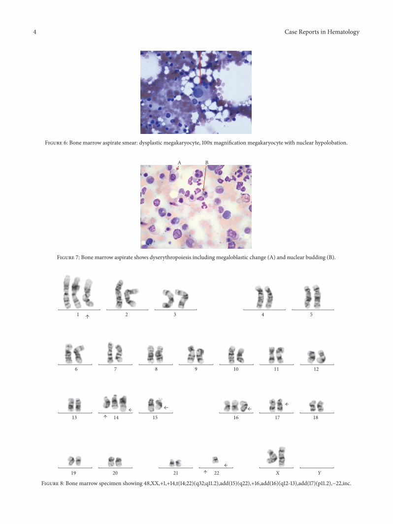
Figure 6: Bone marrow aspirate smear: dysplastic megakaryocyte, 100x magnification megakaryocyte with nuclear hypolobation.
A B
Figure 7: Bone marrow aspirate shows dyserythropoiesis including megaloblastic change (A) and nuclear budding (B).
1 2 3 4 5
1211109876
13 14 15 16 17 18
YX22212019
Figure 8: Bone marrow specimen showing 48,XX,+1,+14,t(14;22)(q32;q11.2),add(15)(q22),+16,add(16)(q12-13),add(17)(p11.2),−22,inc.
Case Reports in Hematology 5
1 2 3 4 5
1211109876
13 14 15 16 17 18
YX22212019
Figure 9: Bone marrow specimen showing 47,XX,+1,add(3)(q21),der(6)t(6;10)(q25;q11.2),+8,t(8;22)(q24.1;q11.2),−10,add(15)(q22),+16,add(16)(q12-13),add(17)(p11.2),add(19)(q13.1),−22.
clinically present as TTP and life-saving treatment such thatplasmapheresis in this acute clinical setting should not bedelayed until further testing to clarify the diagnosis is inprocess.
MDS generally affects older adults (median age 76 years)[5]. Clinically MDS can be incidentally found in a work-updone for other reasons (CBC showing anemia, leukopenia, orthrombocytopenia) and being asymptomatic usually in halfof the cases [6] or it can be symptomatic with clinical featuresrelated to the severity of the underlying cytopenias which inturn are due to ineffective hematopoiesis.
At least 80–90%will have anemia at the time of diagnosis;it is usually normocytic or macrocytic, and half of them willhave an Hb less than 10 g/dL [7]. Anemia can be manifestedas fatigue, generalized weakness, exercise intolerance, angina,dizziness, or alter mental status [8–11]. In the present caseher Hb on admission was 6.6 g/dL explaining her fatigue,generalized weakness, and at least in part her alter mentalstatus.
Thrombocytopenia is present in 30–45% of cases andapproximately 40% are neutropenic at the time of diag-nosis [7]. As a result of them and less often patients canhave petechial rash, easy bruising, inappropriate bleed-ing, hematomas, or infection. Our patient did not haveclear evidence of infection by physical exam, cultures,and imaging studies. After approximately a week she wasfound to have a right thigh abscess that was drained. The
cause of fever appears to be due to underlying infection.Weight loss is rare and represents a late manifestation ofMDS.
On physical exam there are not specific findings related toMDS. Pallor is seen in 60% of the cases and rash and purpurain 26%. Lymphadenopathy, splenomegaly, and hepatomegalyare uncommon [9, 12].
MDS can be suspected when there is unexplainedmonocytosis, cytopenias, macrocytosis (even with normalhemoglobin), or cellular atypia and dysplasia. In our case thepresence of cytopenias and cellular dysplasia were suggestiveof MDS.
Peripheral blood smear findings described in the eryth-roid line in MDS include spherocytes, macroovalocytes,acanthocytes, elliptocytes, stomatocytes, teardrop cells,nucleated erythrocytes, basophilic stippling, and howell-jollybodies. However, schistocytes have been rarely described andto the best of our knowledge, only 4 cases have been report-ed in the English language literature [13–15]. In the presentcase, schistocytosis could be due to an underlying dissemi-nated intravascular coagulation (DIC) perhaps due to anunderlying infection (thigh abscess).
The initial presentation of acute onset and rapidly pro-gressive worsening alter mental status changes, fever, anemiawith marked schistocytosis (MAHA), thrombocytopenia,and renal involvement made TTP occupy the top in the listof the differential diagnosis in this unusual case.
6 Case Reports in Hematology
Diagnosis ofMDS relies on themorphological assessmentof bone marrow aspiration and biopsy as well as on the cyto-genetic analysis. In some occasions bone marrow aspirationand biopsy need to be repeated more than once since MDScan affect the bone marrow unevenly and it can cause falsenegatives.
4. Conclusion
In an elderly patient who presents with anemia and markedschistocytosis (microangiopathic hemolytic anemia) associ-ated with thrombocytopenia MDS should be considered inthe differential diagnosis. As illustrated in the present case,MDS can even present with the classic TTP “pentad” of fever,renal involvement, anemia (MAHA), mental status changes,and lowplatelets (thrombocytopenia). In this clinical context,MDS becomes a diagnosis of exclusion and life-threateningconditions such that TTP needs to be addressed first withappropriate evaluation and initiation of pertinent treatmentuntil this medical emergency is being ruled out.
Competing Interests
The authors declare that there is no conflict of interestsregarding the publication of this paper.
References
[1] D. A. Arber, A. Orazi, R. Hasserjian et al., “The 2016 revisionto the World Health Organization classification of myeloidneoplasms and acute leukemia,” Blood, vol. 127, no. 20, pp. 2391–2405, 2016.
[2] N. Sasaki, J. Kuroda, E. Kawata et al., “Thrombotic thrombo-cytopenic purpura associated with myelodysplastic syndrome,”International Journal of Hematology, vol. 88, no. 4, pp. 457–459,2008.
[3] I. Doudenko-Pirozzolo and R. Booth, “Pathologic quiz case: a20-year-old man with a history of hemoptysis and purpura,”Archives of Pathology and Laboratory Medicine, vol. 125, no. 6,pp. 835–837, 2001.
[4] G. Leone, S. Sica, V. De Stefano et al., “Acute onset of juve-nile myelodysplastic syndrome mimicking thrombotic throm-bocytopenic purpura and rapidly evolving in overt myeloidleukemia,” American Journal of Hematology, vol. 41, no. 1, pp.64–65, 1992.
[5] X. Ma, M. Does, A. Raza, and S. T. Mayne, “Myelodysplasticsyndromes,” Cancer, vol. 109, no. 8, pp. 1536–1542, 2007.
[6] V. Santini, “Anemia as the main manifestation of myelodysplas-tic syndromes,” Seminars in Hematology, vol. 52, no. 4, pp. 348–356, 2015.
[7] D. P. Steensma and J. M. Bennett, “The myelodysplastic syn-dromes: diagnosis and treatment,”Mayo Clinic Proceedings, vol.81, no. 1, pp. 104–130, 2006.
[8] S. L. Goldberg, E. Chen,M. Corral et al., “Incidence and clinicalcomplications of myelodysplastic syndromes among UnitedStates Medicare beneficiaries,” Journal of Clinical Oncology, vol.28, no. 17, pp. 2847–2852, 2010.
[9] K. Foucar, R. M. Langdon II, J. O. Armitage, D. B. Olson, andT. J. Carroll Jr., “Myelodysplastic syndromes. A clinical and
pathologic analysis of 109 cases,” Cancer, vol. 56, no. 3, pp. 553–561, 1985.
[10] A. J. G. Jansen, M.-L. Essink-Bot, E. A. M. Beckers, W. C.J. Hop, M. R. Schipperus, and D. J. Van Rhenen, “Qualityof life measurement in patients with transfusion-dependentmyelodysplastic syndromes,” British Journal of Haematology,vol. 121, no. 2, pp. 270–274, 2003.
[11] C. A. Meyers, M. Albitar, and E. Estey, “Cognitive impairment,fatigue, and cytokine levels in patients with acute myelogenousleukemia or myelodysplastic syndrome,” Cancer, vol. 104, no. 4,pp. 788–793, 2005.
[12] H. P. Koeffler and D. W. Golde, “Human preleukemia,” Annalsof Internal Medicine, vol. 93, no. 2, pp. 347–353, 1980.
[13] J. L. Rummens, C. Verfaillie, A. Criel et al., “Elliptocytosisand schistocytosis in myelodysplasia: report of two cases,” ActaHaematologica, vol. 75, no. 3, pp. 174–177, 1986.
[14] M. F. Zahid, N. Khan, J. Pei, J. R. Testa, and E. Dulaimi,“Genomic imbalances in peripheral blood confirm the diagno-sis of myelodysplastic syndrome in a patient presenting withnon-immune hemolytic anemia,” Leukemia Research Reports,vol. 5, pp. 23–26, 2016.
[15] J.W.Hartz,D.H. Buss,D. R.White,M.G. Bond, andM. Scharyj,“Marked elliptocytosis and schistocytosis in hematopoieticdysplasia,” American Journal of Clinical Pathology, vol. 82, no.3, pp. 354–359, 1984.


![PlasmablasticLymphomainanImmunocompetentPatientwith MDS ...downloads.hindawi.com/journals/crihem/2018/2525070.pdf · increasedapoptosisofthenormalhematopoieticprecursors [15],butnodirectassociationbetweenMDSandimmu-nosuppressionhasbeenreported.However,acausalre-](https://static.documents.pub/doc/80x56/5f0631327e708231d416c1cb/plasmablasticlymphomainanimmunocompetentpatientwith-mds-increasedapoptosisofthenormalhematopoieticprecursors.jpg)