Page 1
8/12/2019 Myocardial Perfusion Imaging (MPI),
http://slidepdf.com/reader/full/myocardial-perfusion-imaging-mpi 1/11
Introduction:
Over the past three decades myocardial perfusion
single photon emission computed tomography
(SPECT) has emerged as a robust tool for the
noninvasive assessment of both myocardial
perfusion and function. Since its inception in the
1970s, many advances have been made that have
enhanced the diagnostic and prognostic strength
of this modality, including develop-ment of new,
technetium 99m (99 mTc)-based Isotopes,
imple-mentation of SPECT, multidetector
cameras, computerized quantification,
attenuation correction, and electrocardiographic
(ECG) gating for the assessment of left
ventricular (LV) function. These advances allow
for very high diagnostic sensitivity and specificity.
In addition, there is a wealth of data supporting
the strength of this technique as a prognostic
tool, not only in the general population, but also
in many very important patient subgroups, such
as women, patients with diabetes mellitus, in
post revascularization patients, and as a
preoperative assessment prior to noncardiac
surgery.
Review Articles
Myocardial Perfusion Imaging (MPI),
An Overview
A Azam1
, WA Jahan2
, M Rahman2
, MA Kauser 3
, Noor F4
1 Department of Nuclear Cardiology, NICVD, 2 Department of Radiology, NICVD, 3 NCCRF&HD,4 Department of Pharmacology, Dhaka Dental College
Abstract:
The clinical utility of radiotracer study of heart in nuclear cardiology must always be considered in
the of context of other cardiac diagnostic procedures, including ECG, Echo, ETT,CAG and cardiac
enzymes or serum cardiac protein. So one should not be a hop cardiologist.
Two modalities in Nuclear Cardiology:
1).Radionuclide Angiography (RNA) or Radionuclide Ventriculography.
2).Myocardial Perfusion Imaging (MPI).
Radionuclide Angiography (RNA) or Radionuclide Ventriculography: This procedure is
designed to provide measurement of LVEF in patient of coronary artery disease, valvular heart
disease or cardiomyopathy. In our setting we determine EF from MPI, so Radionuclide Angiography
routinely not do in many centre of world including NICVD, Dhaka.
Myocardial Perfusion Imaging (MPI): MPI, more specifically myocardial perfusion single photon
emission computed tomography (SPECT) is a nice tool for the noninvasive assessment of myocardial
perfusion, ejection fraction, wall motion, and wall thickness.
Why not perform cardiac catheterization and coronary angiography in all patients suspected of
having coronary artery diseases?
The contrast coronary angiogram displays the anatomic extent of epicardial coronary artery disease,
the severity of luminal narrowing, and the number of diseased vessels. Stress radionuclide myocardial
perfusion imaging, on the other hand, displays the downstream functional consequences of epicardial
coronary artery disease in the myocardium. It also may visualize the regional effects of micro
vascular endothelial dysfunction and impairment of regional coronary flow reserve.
Application of MPI: The diagnosis of coronary artery disease remain common application of MPI
,but it is increasingly being used for the diagnosis of acute MI, risk stratification after infarction,
and assessment of viable myocardium versus scar in patients in chronic coronary artery disease.
(Cardiovasc. j. 2010; 3(1) : 55-65)
Keywords:
Myocardial
Perfusion
Imaging (MPI).
Myocardial
viability,
Radiophar-
maceutical
Address of Correspondence : Dr Abu Azam, Department of Nuclear Cardiology, NICVD, Dhaka, Bangladesh.
Page 2
8/12/2019 Myocardial Perfusion Imaging (MPI),
http://slidepdf.com/reader/full/myocardial-perfusion-imaging-mpi 2/11
Gating of MPI:
(i) Gating is a very simple way of making the MPI
data synchronized with cardiac cycle, so that we
get a MPI record in systole and diastole in discipline
manner for easy interpretation.
(ii) Gating can be achieved by attaching gatingmonitor with patient through three limb leads.
Virtually it is similar to synchronization during
DC shock.
Methods:
Single photon emission computed tomography has
emerged as the standard clinical method of
performing nuclear cardiology studies. Compared
with its predecessor, planar imaging, it improves
image contrast and resolution. SPECT imaging is
based on the theory that a three-dimensional image
of the heart can be created from two-dimensionalplanar images acquired at 30 to 64 projections
around the patient’s chest over a 180° arc.
Following data acquisition, the images are filtered
and reconstructed. Finally the trans-axial images
are reoriented into short axis images and the
images are then displayed.
Initially, myocardial perfusion studies were
performed using planar imaging. However, planar
imaging is hampered by the overlap of normal and
abnormal myocardium as well as adjacent soft
tissue, interfering with accurate localization and
assessment of defect size and severity. Unlike
planar imaging, SPECT is technically demanding
and requires strict attention to quality control.
Gated SPECT myocardial perfusion imaging (MPI)
was introduced in the early 1990s and has become
a standard part of myocardial perfusion SPECT
imaging. Around the same time came the
development of multidetector gamma cameras.
With the increasing use of dual-detector cameras
(with heads mounted at 90°) cardiac studies could
be acquired in half of the time required using a
single-detector system, reducing patient discomfort
and motion, as well as increasing laboratory
throughout.1
Radiopharmaceuticals
Thallium 201 (201Tl) was the first clinically useful
radio-isotope to assess regional myocardial blood
flow. Its initial distribution is proportional to
myocardial blood flow, thus imaging early (within
the first hour) after its injection shows deficits in
regions where blood flow is relatively reduced.
Over time, 201T1 undergoes redistribution as it
attains con-centration equilibrium between the
extracellular and intra-cellular compartments. The
redistribution of 201T1 results in the “wash in” or
normalization of its concentration in the initially
underperfused regions. Generally, this can be
demonstrated 3 to 4 hours after the injection of 201T1; how-ever, in severely hypoperfused
segments this may require imaging 24 to 48 hours
later.
Currently there are two 99mTc-based
radiopharmaceuticals approved for myocardial
perfusion SPECT: 99mTc sestamibi and 99mTc
tetrofosmin. The shorter half-life of 99mTc (6
hours) compared with 201TI (73 hours) allows the
administration of a larger dose of the technetium
agents: eight to 10 times that of 201Tl. 99mTc alsohas the advantage of having a higher photopeak
energy, resulting in improved counting statistics
and less soft tissue attenua-tion, therefore offering
enhanced image quality. There is good correlation
of 201T1 and 99mTc-based radiopharmaceuticals
with myocardial blood flow over its normal
phys-iologic range. However, there is an
underestimation of flow at higher flow rates for
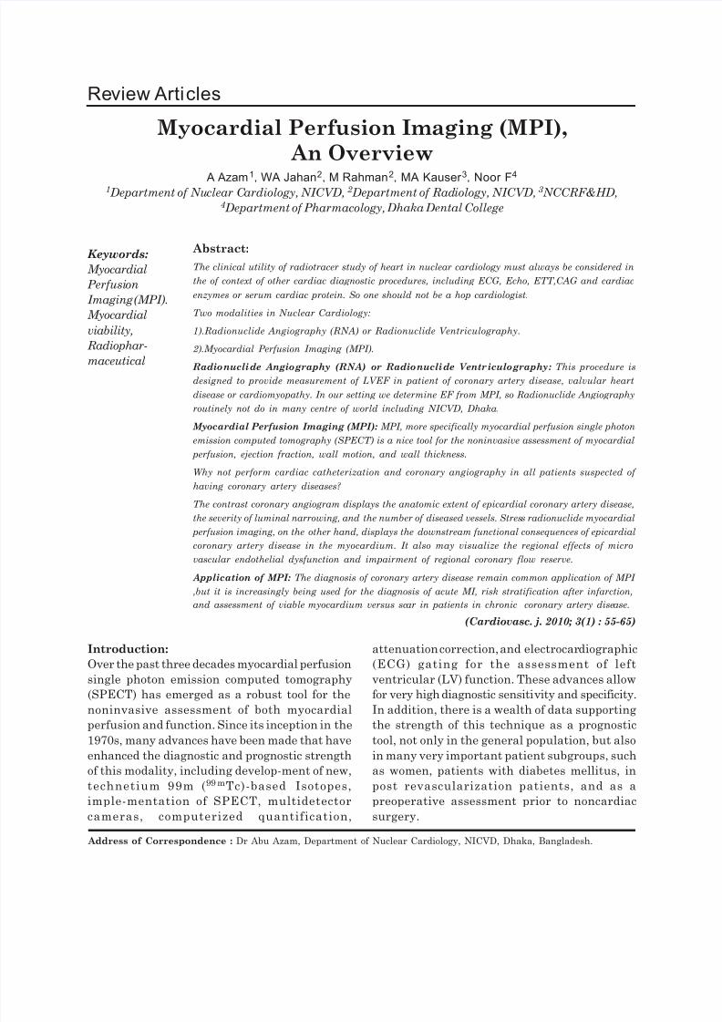
Fig.-1: Pathophysiology underlying radionuclide
myocardial perfusion imaging. Coronary blood flow
in each of the coronary artery branches at rest and
during stress is shown in graph.(From: Atlas of
Nuclear Cardiology.)
Cardiovascular Journal Volume 3, No. 1, 2010
56
Page 3
8/12/2019 Myocardial Perfusion Imaging (MPI),
http://slidepdf.com/reader/full/myocardial-perfusion-imaging-mpi 3/11
both of the 99mTc tracers. Further-more, 99mTc-
based agents have clinically insignificant washout,
thus requiring separate injections at rest and stress
to assess blood flow under these two different
physiologic states.
Positron imaging tracers currently used include
oxygen 15 (t1/2, 2 min), nitrogen 13 (t1
/2, 10 min),
carbon 11 (t1/2 , 20 min), and fluorine 18
fluorodeoxyglucose (18F FDG) ( t1/2, 110 min).
These tracers require a cyclotron for production.
Rubidium 82 (t1/2 , 5 sec) does not require a
cyclotron and can be delivered directly by an on-
she generator. Tracers used to assess myocardial
perfusion are rubidium 82, nitrogen 13 ammonia,
and oxygen 15 water. Metabolic tracers include18F FDG, carbon 11-labeled fatty acids, and carbon
11 acetate.
Acquisition Protocols
Several different protocols have been described
using 201TI (especially for myocardial viability
assessment), 99mTc-labeled agents, and both 201Tl
and 99mTc-labeled agents. At the present time,
approximately 20% of myocardial perfusion SPECT
studies are performed with 201Tl alone, 40% with99tnTc-sestamibi alone, 20% with rest 201Tl/stress99mTc sestamibi, and 20% using 99mTc tetrofosmin
either alone or in combination with 201T1.2
When 201Tl is used alone, the most common
acquisi-tion protocol uses some combination of stress with redis-tribution and/or reinjection
imaging. The latter involves obtaining an additional
image in patients with nonreversible (fixed)
perfusion defects following injection of 50% of the
dose used at stress, with imaging performed 15 to
30 minutes thereafter.3. This protocol has been
shown to improve the ability to detect viable
myocardium over standard stress/4-hour
redistribution imaging.4 An alternate protocol that
is gaining popularity is to give sublingual
nitroglycerin prior to reinjection with 201Tl.5 From
a practical standpoint, 201Tl offers the advantage
of use of a single injection of the isotope to
demonstrate ischemia and to evaluate for
hibernating but viable myocardium.6
Due to the absence of significant redistribution,
separate rest and stress injections are standard
with 99mTc-based isotopes. A variety of protocols
can be used with this agent, including 2-day stress-
rest, same-day rest-stress, same-day stress-rest,
and dual isotope. From the stand-point of defect
contrast and optimal image quality, the 2-day
imaging protocol is ideal. The principal drawback
of this protocol is its requirement of two imaging
days. Although, the same-day low-dose rest/high-
dose stress is the most commonly employed 99mTc-
based protocol, it has the disadvantage of
potentially causing a reduction in stress defect
contrast (< 15%) because some of the radio-activity
observed on stress images comes from the activity
from the resting myocardial perfusion study
(performed only hours before).7 Of note, recent
work has shown this to be of minor clinical
significance, and with proper dosing of the rest
and stress images there is excellent identification
of ischemic defects. The same-day low-dose stress/
high-dose rest sequence has less than ideal countsrate for the stress image set, and making it difficult
to accurately assess defect reversibility.8,9
Regarding the assessment of myocardial viability,
all stress/rest or rest/stress 99mTc-based isotopes
have limita-tions in separating severely
hibernating myocardium from infarction. Based on
the limitations of 99mTc-based isotope protocols, a
rest 201 Tl and/stress 99mTc-based isotope dual-
isotope SPECT protocol has gained wide popularity.
The sensitivity and specificity of these protocols
have been shown to be approximately 90%.10,11
Stress Modalities
Treadmill and bicycle exercise are the most
common types of stress used in conjunction with
MPI. In general, the performance of myocardial
perfusion SPECT with exercise stress is preferable
for prognostic purposes. Important prognostic
variables associated with exercise ECG testing
include exercise capacity,12,13 exercise-inducible
chest pain, hypotension, and the EGG response to
exercise. These variables cannot he assessed with
pharmacologic stress testing. However, a
significant number of patients are unable to
adequately exercise. In these patients,
pharmaco-logic stress testing is an effective
substitute for exercise in conjunction with MPI.
Adenos in e and dipyridamole are coronary
vasodilators that increase myocardial blood flow
four- to fivefold in myocardial region that are
supplied by normal coronary arteries. In contrast,
Myocardial Perfusion Imaging (MPI), An Overview A Azam et al.
57
Page 4
8/12/2019 Myocardial Perfusion Imaging (MPI),
http://slidepdf.com/reader/full/myocardial-perfusion-imaging-mpi 4/11
there is an attenuated hyperemic response in
myocardial regions supplied by diseased coronary
arteries.14,15 Thus, regional flow heterogene-ity,
which usually occurs without ischemia, produces
the reversible perfusion defect on the myocardial
perfusion SPECT studies. Adenosine is a direct
coronary vasodilator and activates the adenosine A 2 receptors in the coronary arterial wall, leading
to coronary vasodilatation. Dipyri-damole exerts
its effect by raising endogenous adenosine levels
by blocking of the cellular reuptake of adenosine.
The clinical indications for adenosine/dipyridamole
are inability to exercise (eg, stroke, arthritis,
peripheral vascular disease, disabling diseases,
amputation); inabil-ity to achieve at least 85% of
maximum predicted heart rate (eg, chronotropic
incompetence, ß-blocker); left bundle branch block
(LBBB), ventricular-paced rhythm, and early post-myocardial infarction (MI) or unstable angina
patients. There are several contraindications to
the use of pharmacologic stress with adenosine
and dipyri-damole-. asthma, active bronchospasm,
severe chronic obstructive pulmonary disease with
home O2, advanced atrioventricular block, sick
sinus syndrome, sinus bradycardia (< 40 beats/min),
hypotension (systolic blood pressure < 90 mm Hg),
fewer than 2 days post-MI, and recent use (< 24
hours) of theophylline or caffeine.
A modification to the adenosine protocol, using
low-level exercise, has recently gained much
acceptance. Adding a low-level treadmill exercise
to the adenosine infusion results in decreased side
effects, decreased symptomatic hypotension and
bradycardia, and an increased target to background
ratio, allowing for immediate imaging, as would
be done with exercise.16 This protocol can also be
applied to dipyridamole. It should be noted that
one should not add low-level exercise to patients
with LBBB or ventricular-paced rhythm, as they
are susceptible to the same false positive findings
in the interventricular septum as would be seenwith exercise.
The performance of myocardial perfusion SPECT
with pharmacologic stress, either dipyridamole or
adenosine, has essentially the same sensitivity and
specificity for detecting coronary artery disease
(CAD) and incremental prognostic value for
predicting cardiac death and MI as does exercise
myocardial perfusion scintigraphy.17,18
Myocardial perfusion scintigraphy can be also
performed with dobutamine stress, but is generally
reserved for patients who either cannot adequately
exercise or have a contraindication to vasodilator
stress. Briefly, dobutamine is a synthetic
catecholamine that directly stimulates both ß1 and
ß2 receptors that increased heart rate andmyocardial contractility in a dose-related fashion.
Thus, dobutamine increases coronary blood flow
by increasing myocardial oxygen demand, and the
myocardial perfusion abnormalities are induced by
the development of regional myocardial
ischemia.19-21
Image acquisition and processing
SPECT imaging camera had configuration. With
SPECT, a series of planar images are obtained in
an orbit around the patient. The images can be
obtained over a full orbit of 360 degrees or over
half of that (180 degrees). The asymmetric
position of the heart in the thorax means that
180-degree orbits can suffice in most clinical
instances. However, a 180-degree orbit may
sometimes result in artifacts. With the use of
multi head detectors, data are acquired at various
projections of the heart simultaneously, thereby
shortening the time of image acquisition of
improving image count density. Imaging with dual-
and triple-head detectors can be completed in half
or one third of the time taken by a single-headdetector. With dual- head cameras, the detectors
can be arranged parallel to each other or at right
angles to each other. The latter configuration is
preferable if simultaneous attenuation correction
is applied.
Myocardial perfusion single photon emission
computed tomography (SPECT) is a widely utilized
noninvasive imaging modality for the diagnosis,
prognosis, and risk stratification of coronary artery
disease. It is clearly superior to the tradi-tional
planar technique in terms of imaging contrast andconsequent diagnostic and prognostic yield. The
strength of SPECT images is largely derived from
the three-dimensional, volumetric nature of its
image. Thus, this modality permits three-
dimensional assessment and quantitation of the
perfused myocardium and functional assessment
through electrocardiographic gating of the
perfusion images.
Cardiovascular Journal Volume 3, No. 1, 2010
58
Page 5
8/12/2019 Myocardial Perfusion Imaging (MPI),
http://slidepdf.com/reader/full/myocardial-perfusion-imaging-mpi 5/11
Processing of images or Re-slicing or Oblique
reorientation or Filtering of SPECT images
MPI images are viewed in standard format,
consisting of short-axis, horizontal long axis, and
vertical long axis slices. Generation of these standard
sections from the original trans-axial images is done
through automatic processing, by locating LV axis
in processing computer, thereby LV isolation,
reconstruction, and reorientation can be achieved.
The acquired row images are somewhat noisy, and
so filtering is required to eliminate or reduce the
noise. There are several ways of filtering out the
noise. The most common way is by Fourier
transformation of the row images, in which the
row image data are transformated into a series of
frequencies. The relevant data are present in the
low- frequency range, whereas noise is present in
the high- frequency range.
A. low pass filter allows the low frequencies to be
retained unaltered and eliminates completely
the high frequencies the above the cut- off
range. The frequencies in the intermediate
range are altered by a function depicted by the
slope of the cut- off curve. If too much filtering
is used, even the useful data may be lost. T
B. he high- pass filter retains the high frequencies
and eliminates the low frequencies. This filter
is used primarily for edge enhancement.
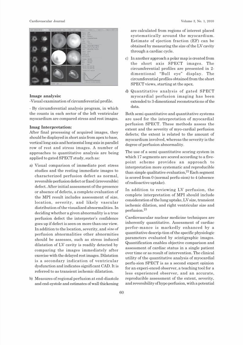
C. A patient example in which low- pass filter
(Butterworth filter) is applied to transaxialimages of the heart is shown. The unfiltered
images (top) are of poor quality and very noisy.
The background noise is eliminated in the
properly filtered images (center). However,
over filtering results in distortion of the
myocardial contour (bottom).
Fig.-2: Normal gated SPECT image.
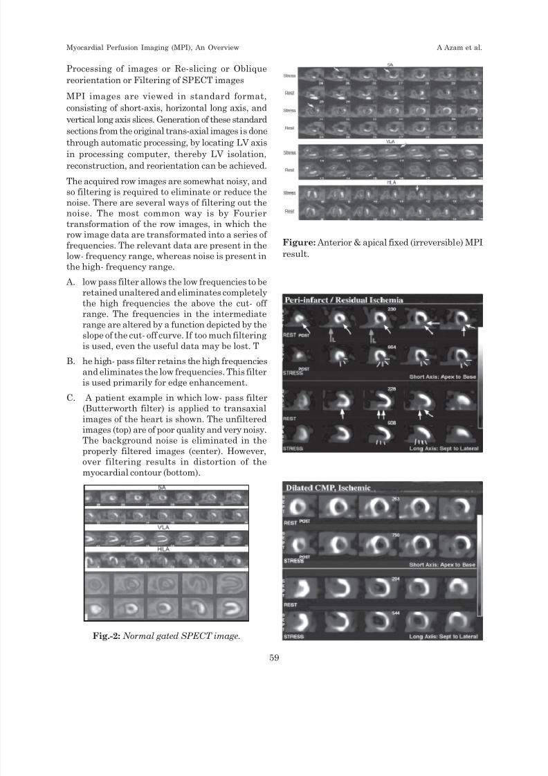
Figure: Anterior & apical fixed (irreversible) MPI
result.
Myocardial Perfusion Imaging (MPI), An Overview A Azam et al.
59
Page 6
8/12/2019 Myocardial Perfusion Imaging (MPI),
http://slidepdf.com/reader/full/myocardial-perfusion-imaging-mpi 6/11
Image analysis:
-Visual examination of circumferential profile.
- By circumferential analysis program, in which
the counts in each sector of the left ventricular
myocardium are compared stress and rest images.
Imag Interpretation:
After final processing of acquired images, they
should be displayed in short axis from apex to base,
vertical long axis and horizontal long axis in parallel
row of rest and stress images. A number of
approaches to quantitative analysis are being
applied to gated SPECT study, such as:
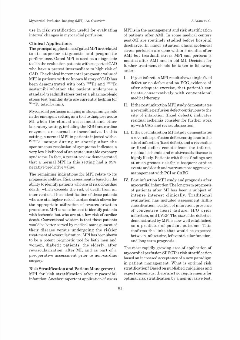
a) Visual comparison of immediate post stress
studies and the resting immediate images to
characterized perfusion defect as normal,
reversible perfusion defect or fixed (irreversible)defect. After initial assessment of the presence
or absence of defects, a complete evaluation of
the MPI result includes assessment of size,
location, severity, and likely vascular
distribution of the visualized abnormalities. In
deciding whether a given abnormality is a true
perfusion defect the interpreter’s confidence
goes up if defect is seen on more than one view.
In addition to the location, severity, and size of
perfusion abnormalities other abnormities
should be assesses, such as stress induced
dilatation of LV cavity is readily detected by
comparing the images immediately after
exercise with the delayed rest images. Dilatation
is a secondary indication of ventricular
dysfunction and indicates significant CAD. It is
referred to as transient ischemic dilatation.
b) Measures of regional perfusion at end-diastole
and end-systole and estimates of wall thickening
are calculated from regions of interest placed
systematically around the myocardium.
Estimate of ejection fraction (EF) can be
obtained by measuring the size of the LV cavity
through a cardiac cycle.
c) In another approach a polar map is created from
the short axis SPECT images. The
circumferential profiles are presented in 2-
dimentional “Bull eye” display. The
circumferential profiles obtained from the short
SPECT views, starting at the apex.
d) Quantitative analysis of gated SPECT
myocardial perfusion imaging has been
extended to 3-dimentional reconstructions of the
data.
Both semi quantitative and quantitative systems
are used for the interpretation of myocardialperfusion SPECT. These methods assess the
extent and the severity of myo-cardial perfusion
defects; the extent is related to the amount of
myocardium involved, whereas the severity is the
degree of perfusion abnormality.
The use of a semi quantitative scoring system in
which 17 segments are scored according to a five-
point scheme provides an approach to
interpretation more systematic and reproducible
than simple qualitative evaluation,22 Each segment
is scored from 0 (normal perfu-sion) to 4 (absence
of radioactive uptake).
In addition to reviewing LV perfusion, the
complete interpretation of MPI should include
consideration of the lung uptake, LV size, transient
ischemic dilation, and right ventricular size and
perfusion.23
Cardiovascular nuclear medicine techniques are
inherently quantitative. Assessment of cardiac
perfor-mance is markedly enhanced by a
quantitative descrip-tion of the specific physiologic
parameters evaluated by scintigraphic images.
Quantification enables objective comparison andassessment of cardiac status in a single patient
over time or as result of intervention. The clinical
utility of the quantitative analysis of myocardial
perfu-sion SPECT is as a second expert opinion
for an experi-enced observer, a teaching tool for a
less experienced observer, and an accurate,
reproducible assessment of the extent, severity,
and reversibility of hypo perfusion, with a potential
Cardiovascular Journal Volume 3, No. 1, 2010
60
Page 7
8/12/2019 Myocardial Perfusion Imaging (MPI),
http://slidepdf.com/reader/full/myocardial-perfusion-imaging-mpi 7/11
use in risk stratification useful for evaluating
interval changes in myocardial perfusion.
Clinical Applications
The principal applications of gated MPI are related
to its superior diagnostic and prognostic
performance. Gated MPI is used as a diagnostictool in the evaluation patients with suspected CAD
who have a pretest intermediate to high risk of
CAD. The clinical incremental prognostic value of
MPI in patients with no known history of CAD has
been demonstrated with both 201T1 and 99mTc
sestamibi whether the patient undergoes a
standard treadmill stress test or a pharmacologic
stress test (similar data are currently lacking for99mTc tetrofosmin).
Myocardial perfusion imaging is also gaining a role
in the emergent setting as a tool to diagnose acute
MI when the clinical assessment and other
laboratory testing, including the EGG and cardiac
enzymes, are normal or inconclusive. In this
setting, a normal MPI in patients injected with a99mTc isotope during or shortly after the
spontaneous resolution of symptoms indicates a
very low likelihood of an acute unstable coronary
syndrome. In fact, a recent review demonstrated
that a normal MPI in this setting had a 99%
negative predictive value.
The remaining indications for MPI relate to its
prognostic abilities. Risk assessment is based on theability to identify patients who are at risk of cardiac
death, which exceeds the risk of death from an
inter-vention. Thus, identification of those patients
who are at a higher risk of cardiac death allows for
the appropriate utilization of revascularization
procedures. MPI can also be used to identify patients
with ischemia but who are at a low risk of cardiac
death. Conventional wisdom is that these patients
would be better served by medical manage-ment of
their disease versus undergoing the riskier
treat-ment of revascularization. MPI has been shown
to be a potent prognostic tool for both men and
women, diabetic patients, the elderly, after
revascularization, after MI, and as part of a
preoperative assessment prior to non-cardiac
surgery.
Risk Stratification and Patient Management
MPI for risk stratification after myocardial
infarction: Another important application of stress
MPI is in the management and risk stratification
of patients after AMI. In some medical centers
post-MI are routinely studied before hospital
discharge. In major situation pharmacological
stress perfusion are done within 3 months after
AMI but treadmill stress MPI can perform 3
months after AMI and in old MI. Decision forfurther treatment should be taken in following
order:
I. If post infarction MPI result shows single fixed
defect or no defect and no ECG evidence of
after adequate exercise, that patient/s can
treats conservatively with conventional
medical therapy.
II. If the post infarction MPI study demonstrates
a reversible perfusion defect contiguous to the
site of infarction (fixed defect), indicates
residual ischemia consider for further workup with CAG and revascularization.
III. If the post infarction MPI study demonstrates
a reversible perfusion defect contiguous to the
site of infarction (fixed defect), and a reversible
or fixed defect remote from the infarct,
residual ischemia and multivessels disease is
highly likely. Patients with these findings are
at much greater risk for subsequent cardiac
events and death and warrant more aggressive
management with PCI or CABG.
IV. Post infarction MPI study and prognosis after
myocardial infarction:The long term prognosis
of patients after MI has been a subject of
intense interest clinically. Traditional
evaluation has included assessment Killip
classification, location of infarction, presence
of congestive heart failure, H/O prior
infarction, and LVEF. The size of the defect as
demonstrated by MPI is now well established
as a predictor of patient outcome. This
confirms the links that would be expected
between infarct size, left ventricular function,
and long term prognosis.
The most rapidly growing area of application of
myocardial perfusion SPECT is risk stratification
based on increased acceptance of a new paradigm
in patient management. What is optimal risk
stratification? Based on published guidelines and
expert consensus, there are two requirements for
optimal risk stratification by a non-invasive test.
Myocardial Perfusion Imaging (MPI), An Overview A Azam et al.
61
Page 8
8/12/2019 Myocardial Perfusion Imaging (MPI),
http://slidepdf.com/reader/full/myocardial-perfusion-imaging-mpi 8/11
First/ a negative study should be associated with a
very low risk of adverse outcomes, as measured
by the observed event rate in the population with
normal studies. The event rate should be less than
1%. To this end, a pooled analysis of 20,963 patients,
from 16 studies/ with a normal myocardial
perfusion had an event rate (death and MI) of 0.7%per year. Second, the event rate associated with
an abnormal scan should not only be greater than
that associated with a normal scan, but the relative
risk and its associated confidence interval with an
abnormal scan relative to a normal scan should be
greater than 1. Finally, the majority of the events
(> 80%-90%) should occur in those patients with
an abnormal study. MPS has been shown to
enhance risk stratification in a population already
stratified by pronuclear testing data, including
stratification by clinical data, exercise treadmill
testing (ETT) data, and clinical plus ETT data. In
all case MPS enhanced risk stratification with
respect to risk of cardiac death or nonfatal
infarction.24
In a series of 5183 patients undergoing myocardial
perfusion SPECT, Hachamovitch et al25 examined
risk stratification with regard to the risks of cardiac
death and nonfatal MI. Although significant
increases in cardiac death and MI occurred as a
function of worsening scan results, patients with
mildly abnormal myocardial perfusion SPECT were
at intermediate risk of MI (2.7% per year) but atlow risk of cardiac death (0.8%). This suggests that
a mildly abnormal myocardial perfusion study may
identify patients with significant CAD that can be
treated with medical management and aggressive
risk factor modi-fication. On the other hand, a
patient with a more severely abnormal myocardial
perfusion study is at high risk of MI (2.9% per year)
and cardiac death (4.2% per year). Thus, these
patients should be considered for catheterization
with consideration of revascularization.
Assessment of result and patency after CABGor PCI by follow up MPI test:
Follow-up stress MPI after CABG or PCI provide
an objective assessment of therapeutic effect on
the coronary circulation. Successful surgery or
angioplasty result in elimination of reversible
defects caused by exercise induced ischemia. CABG
and PCI have no effect on scarred areas, and fixed
defect should appear unchanged.
If a patient has an infarction as a result of the
therapeutic intervention, a previously reversible
defect may be converted into a fixed defect or an
entirely new defect may occur as a result of the
injury. Imaging should be delayed 6 weeks or more
because some preintervention defects may persists
if the scan is done too soon.
When symptoms recur, as they do in a significant
percentage of patients, the early post therapy study
serves as a useful baseline. The development of
new or recurrent disease is readily detected on
repeat stress imaging.
Assessment of thrombolytic therapy by MPI:
In patients undergoing thrombolysis, myocardial
perfusion studies are performed under resting
condition. After recovery, stress imaging is useful
to determine outcome and detect any areas of
residual exercise or stress induced ischemia.
An initial dose of Tc99m tetrofosmin may be given
the time of patient arrives at the hospital, but
imaging can be delayed until the patient is
stabilized or even until after thrombolytic therapy
is given. The initial dose is then used to document
the amount of myocardium at risk. A second dose
is then used to determine the effectiveness of
therapy. Reduction in defect size correlates with
vessel patency and better prognosis after MI.
Gated Myocardial Perfusion Single PhotonEmission Computed Tomography
With the advent of ECC-gated myocardial perfusion
SPECT, LV function is now routinely assessed in
patients undergoing stress perfusion imaging. A
gated study is acquired by temporally organizing
the image data acquired from the patient.
Specifically, the R-R interval is divided into equal
frames, typically eight or 16, and image data are
binned according to the frame in which they were
acquired. The data are then processed and can be
displayed in cinematic format.
The gated portion of a myocardial perfusion SPECT
is acquired simultaneously with the acquisition of
perfusion. In addition to allowing for the visual
assessment of LV function, gated SPECT’ allows
for the evaluation of LV wall motion and
thickening.
Furthermore, quantitative methods have been
devel-oped to automatically measure LV ejection
Cardiovascular Journal Volume 3, No. 1, 2010
62
Page 9
8/12/2019 Myocardial Perfusion Imaging (MPI),
http://slidepdf.com/reader/full/myocardial-perfusion-imaging-mpi 9/11
fraction (LVEF) and volumes. The most recent
versions of the systems use a three-dimensional
approach to map the endocardial and epicardial
surfaces.26,27 These methods have been shown to
be both accurate and precise. Accuracy has been
assessed by comparing the results of gated SPECT
with first-pass left ventriculography, two-dimensional echo-cardiography, magnetic
resonance imaging, thermodilution, contrast
ventriculography, and electron beam CT, using a
variety of different radiopharmaceuticals. The
Pearson r-value for LVEF and volume ranges from
0.7 to 0.99.26,28 Precision has been assessed by
repeating the measurement of LV function over
time with very strong correlation (r-values range
from 0.86-0.98).29-31
In addition to validity of the measurements,
quantita-tive gated SPECT is an objective process;in general, the LV surfaces are automatically
defined without guidance from the operator. Figure
2 is an example of one such approach, from the
Quantitative Gated SPECT program (General
Electric Medical Systems, Waukesha, WI).
The addition of functional information to
myocardial perfusion SPECT adds clinical value
in diagnosis, risk stratification as well as the
assessment of myocardial via-bility. From a
diagnostic perspective, gated SPECT has proven
most useful in differentiating infarction from
artifact. Specifically, when there is a defect on both
rest and stress perfusion, the presence of normal
LV wall motion and thickening would be indicative
of this defect being an artifact (as infarcted
myocardium would not have normal wall motion
and thickening). This results in fewer false-positive
studies and higher overall test specificity without
comprising the sensitivity of the study. This was
shown very nicely by Taillefer et al32 who found
that the speci-ficity for detecting significant
coronary artery stenosis was 68% for 201T1 SPECT
and 92% with99m
Tc sestamibi-gated SPECT (P =0.0004), whereas the sensitivity was 84% for 201T1
and 80% for 99mTc-sestamibi.
In addition, evaluation of post-stress-gated SPECT
can offer insight into the severity of coronary artery
stenosis in patients with stress-induced ischemia.
It has been shown that the development of new
wall motion abnormalities on post-stress-gated
SPECT (either not present on a rest-gated SPECT
or in a region of normal resting perfusion, with
presumed normal function) is indicative of severe
(> 90%) stenosis in the artery subtending the
ischemic myocardium; this phenomenon is due to
persistent post-stress stunning. Furthermore,
ShariretaJ33 have shown such that new wall
motion abnormalities on post-stress-gated SPECTare a better predictor of the severity of coronary
artery stenosis than the severity of the stress-
induced perfusion defect.
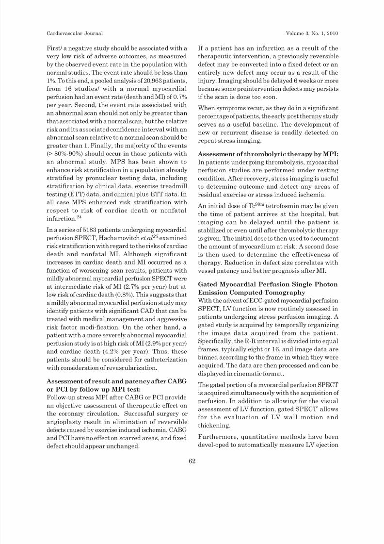
Gated SPECT can also offer insight as to the
etiology of a patient’s LV dysfunction. Specifically,
the combination of perfusion and function can
broadly differentiate ischemic from non ischemic
cardiomyopathy. Thus, patients with normal
perfusion and reduced LV systolic function can be
recognized as having a non ischemic
cardiomyopathy and be worked up accordingly,whereas those with significant perfusion
abnormalities and LV systolic dysfunction can be
categorized as having an ischemic cardiomyopathy.
More over, patients with relatively small perfusion
abnormalities with disproportionate LV systolic
dysfunction can be categorized as having a mixed
cardiomyopathy. Finally, in patients with clinical
congestive heart failure who have normal perfusion
and normal LV systolic function by gated
myocardial perfusion SPECT, the diagnosis of
diastolic dysfunction should be considered.
From a prognostic standpoint, it has long been
estab-lished that IV systolic function is a potent
tool for assess-ing the likelihood of cardiac
mortality. This has been best demonstrated in the
post-MI patient group. The Thrombolysis In
Myocardial Infarction II34 and Mullicenter Post
Myocardial Infarction Research Group35 studies
have both shown that LV systolic function is a very
impor-tant prognostic index and that mortality is
inversely related to LVEF.
Similar findings have been made for LVEFobtained by gated SPECT. A recent study
examining the outcomes of 2686 patients
undergoing myocardial perfusion-gated SPECT,
found that LVEF by post-stress-gated SPECT was
the best predicator of cardiac death, even after
adjusting for perfusion.36 Furthermore, they
showed the same inverse, exponential relationship
between EF and mortality.
Myocardial Perfusion Imaging (MPI), An Overview A Azam et al.
63
Page 10
8/12/2019 Myocardial Perfusion Imaging (MPI),
http://slidepdf.com/reader/full/myocardial-perfusion-imaging-mpi 10/11
Gated SPECT has also been evaluated as a tool for
assessing myocardial viability; in this application
the combination of perfusion and LV function is used
to assess the viability. There have been several
reports utilizing this technique for assessing
myocardial viability with conflict-ing results. Two
of these studies have shown that this techniqueunderestimates the extent of myocardial viabil-ity
compared with rest/redistribution 201T1
SPECT.37,38 However, there have been three
studies that have demon-strated equivalent results
for gated 99mTc and rest/redistri-bution 201T1
SPECT.39,40 Most recently, Duncan et al.40
evaluated 30 patients scheduled to undergo
coronary artery bypass surgery with pre- and post
revascularization-gated 99mTc and rest/
redistribution 201T1 SPECT. They found no
difference in the sensitivity, specificity positive or
negative predictive value, or predictive accuracy for
the detection of viability between these two methods.
Conclusions:
Over the past two decades gated myocardial
perfusion SPECT has developed into a widely
accepted noninvasive tool for evaluating patients
with known or suspected coronary artery disease.
It offers superior diagnostic accuracy in virtually
all patient populations. In addition, there is vast
literature supporting its ability to risk stratify
patients into low- and high-risk groups.
Furthermore, recent data support a prognosticapproach to the management of patients with
coronary artery disease based on the results of
gated myocardial perfusion SPECT imaging. The
assess-ment of LV function adds further to the
diagnostic and prognostic significant of this imaging
modality. The future development of
enhancements, such as attenuation correc-tion,
stand to further foster the clinical utility of gated
myocardial perfusion SPECT.41
References:1. Garcia HV, Nichols K, Gait}, et al.: Instrumentation,
quality assurance and performance. In imagingguidelines for nuclear cardiology procedures, part 1. J
Nucl Cardiol 1996; 3:G5-G10.
2. Berman DS, Germano G: Myocardial single photon
approaches in Imaging in Cardiovascular Disease,
Edited by Pohost GE. Philadelphia: Lippincott Williams
&Wilkins; 2002: 159-194.
3. Dilsizian V, Rocco TP, Freedman NM, et al.: Enhanced
detec-tion of ischemic but viable myocardium by the
reinfection of thallium after stress-redistribution
imaging. N Engl J Med 1990; 323:141-146.
4. Dilsizian V, SmeltzerWR, Freedman NM, etal: Thallium
reinjection after stress-redistribution imaging. Does 24-
hour delayed imaging after reinjection enhance
detection of viable myocardium? Circulation 1991;
83:1247-1255.
5. Ba.su S, senior R, Raval U, et al.: Superiority of nitrate-
enhanced 201 TI over conventional redistribution 201
TI imaging for prognostic evaluation after myocardial
infarc-tion and thrombolysis. Circulation 1997;
96:2932-2937.
6. Ragosta M, Beller GA: The noninvasive assessment of
myocardial viability. Clin Cardiol 1993; 16:531-538.
7. N Berman DS, Hayes SW, Gerrnano G: In Cardiac
Spect Imagmg. Edited by De Puey EG, Garcia EV,
Berman DS. Philadelphia: / Lippincott Williams &
Wilkins; 2000:179-210.
8. Heo J, Keget J, Iskandrian AS, et al: Comparison of
same-day protocol using technetium-99-m-sestambi
myocardial imaging, J Nucl Med 1992;33:186-191.
9. Taillefer R, Gagnon A Laflamrne L, et al: Same day
injections ofTc-99-m-methoxy isobutyl isonitrile
(hexamibi) for myocardial tomographic imaging:
comparison between rest-stress and stress-rest injection
sequences. Bur J Nucl Med 1989; 15:113-117.
10. Berrnan DS, Kiat H, Friedman JD, et al: Separate
acquisition of rest thallium- 201/ stress technetium-99-
m-sestamibi dual-isotope myocardial single photon
emission computed tomography: a clinical validation
study. J Am Coll Cardiol 1993; 22:1455-1464.
11. Kiat H, Gerrnano G, Friedman JD, et al: Comparative
feasi-bility of separate or simultaneous rest thaIHum-
201/stress technetium-99-m-sestamibi dual-isotope
myocardial SPECT. J Nucl Med 1994; 35:542-548.
12. Weiner DA, Me Cabe CH, Ryan TJ: Prognostic
assessment of patients with coronary artery disease by
exercise testing. Am Heart J 1983; 105:749-755.
13. Mc Neer F, Margolis JR, Lee KL, et al: the role of
exercise test-ing in the evaluation of patients for
ischemic heart disease. Circulation 1978; 57:64-70.
14. Gould LK, Westcott R, Albro P, et al: Noninvasive
assessment • of coronary artery stenosis by myocardial
imaging during pharmacoiogic coronary vasodilation.
II. Clinical method-ology and feasibility. Am J Cardiol
1978; 41:279.
15. ChanSY, Brunken RC, Czernin J, etal.: Comparison of
myo-cardial blood flow during adenosine infusion with
that of intravenous dipyridamole in normal men. J Am
Coll Cardiol 1992;20:979-985.
16. Thomas GS, Prill NV, Majmundar H, et al.: Treadmill
exercise during adenosine infusion is safe, results in
fewer adverse reactions, and improves myocardialperfusion image quality. J Nucl Cardiol 2000; 7:439-446.
17. Gupta NC, Esterbrooks DJ, Hilleman DE, et al.:
Comparison of adenosine and exercise thallium-201
single-photon emission computed tomography (SPECT)
myocardial perfusion imag-ing. J Am Coll Cardiol 1992;
19:265-275.
18. I lachamovitch R, Berrnan DS, Kiat H, et al.: incremental
prog-nostic value of adenosine stress myocardial
perfusion single photon emission computed
tomography and impact on subsequent management
Cardiovascular Journal Volume 3, No. 1, 2010
64
Page 11
8/12/2019 Myocardial Perfusion Imaging (MPI),
http://slidepdf.com/reader/full/myocardial-perfusion-imaging-mpi 11/11
in patients with or suspected of having myocardial
ischemia. J Am Cardiol 1997; 80:426-433.
19. Hays JT, Mahmarian JJ, Cochran AJ, etal: Dobutamine
thal-lium-201 tomography for evaluating patients with
suspected coronary artery disease unable to undergo
exercise or pharmacological stress testing. J Am Coll
Cardiol 1993; 21:1583-1590.
20. Kiat H, Iskandrian AS, ViJlegas BJ, et al: Arbutaminestress lhallium-201 single photon emission computed
tomography using a computerized closed- loop delivery
system: multi-center trial for evaluation of safety and
diagnostic accuracy. J Am Coll Cardiol 1995; 26:1159-
1167.
21. Geleijnse ML, Elhendy A, Fioretti PM, Roelandt }R:
Dobutamine ilress myocardiai perfusion imaging. J Am
Coll Cardiol 2000;
22. Cerqueira MD, Weissman NJ, Dilsizian V, etal:
Standardized myocardial segmentation and
nomenclature for tomographic imagmg of the heart: a
statement for healthcare profession-als from the
Cardiac Imaging Committee of the Council on linical
Cardiology of the American Heart Association. J Nucl
Cardiol 2002; 9:240-245.23. Van Train, Garcia EV, Cooke DC, Arreda JS:
Quantitative analy-sis of SPECT myocardial perfusion.
In: Cardiac SPECT’ Imaging. Edited by De Puey EG,
Garcia EV, Herman DS. Philadelphia: Lippincott
Williams & Wilkins; 2000:41-64.
24. Berrnan DS, Hachamovitch R, Kiat H, et al. :
Incremental value of prognostic testing in patients with
known or suspected ischemic heart disease: a basis for
optimal utilization of exercise technetium-99m sestamibi
myocardial perfusion single-photon emission computed
tomography. J Am Coll Cardiol 1995; 26:639-647.
25. Hachamovitch R, Herman DS, Shaw LJ, el at:
Incremental prognostic value of myocardial perfusion
photon emission computed tomography for the
prediction of cardiac death: differential stratificationfor risk of cardiac death and myocardial infarction.
Circulation 1998, 97:535-543.
26. Germano G, Kiat H, Kavanagh PB, et al.: Automatic
quantifica-tion of ejection fraction from gated
myocardial perfusion SPECT. J Nucl Med 1994; 35:1185-
1192.
27. FarberT, Cooke C, Folks R, etal: Left ventricular
function and perfusion from gated SPECT perfusion
images: an integrated method. JNucl Med 1999; 40:650-
659.
28. Vaduganathan P, He Z, Vick, et al.: Evaluation of left
ventricu-lar wall motion, volumes, and ejection fraction
by gated myocardial tomography with technetium 99m-
labeled tetrofosmin: a comparison with cine magnetic
resonance imaging. J Nucl Cardiol 1999; 6:3-10.
29. Johnson LL, Verdesca SA, Aude WY, et al.: Postischemic
stun-ning can affect left ventricular ejection fraction
and regional wall motion on post-stress gated
technetium-99m-sestamibi SPECT.J Am Coll Cardiol
1997; 30:1641-1648.
30. Berman D, Germano G, Lewin H, et al.: Comparison of
post-stress ejection fraction and relative left ventricular
volumes by automatic analysis of gated myocardial
perfusion SPECT acquired in the supine and prone
positions. J Nud Cardiol .1998; 5:40-47.
31. Lewin HC, Herman DS, Hayes SW, etal: Clinicalreproducibil-ity of post-stress gated myocardial
perfusion SPECT imaging. J Nucl Med 2000; 41:160P.
32. Taillefer R, DePuey EG, Udelson E, et al: Comparative
diag-nostic accuracy of T1201 and 99mTc-sestamibi
SPECT imag-Jng (perfusion and ECG-gated SPECT)
in detecting coronary artery disease in women.J .Am
Coll Cardiol 1997; 29:69-77.
33. Sharir T, Bacher-Stier C, Dhar S, et al.: Identification
of severe and extensive coronary artery disease by
postexercise regional wall motion abnormalities in Tc-
99m sestamibi gated single-photon emission computed
tomography. Am J Cardiol 2000; 86:1171-1175.
34. Zaret B, Wackers F, Terrin M, et al.: Value of
radionucHde rest and exercise left ventricular ejection
fraction to access sur-vival of patients following
thrombolytic therapy for acute myocardial infarction:
results of the Thrombolysis in Myo-cardiai Infarction
(TIMI) phase II study. J Am Coll Cardiol 1995; 26:73.
35. The Multicenter Post Myocardial Infarction Research
Group: Risk stratification and survival after myocardial
infarction. N Engl J Med 1983; 309:331.
36. SharirT, Germano G, Kavanagh PB, et al: Incremental
prog-nostic value of post-stress left ventricular ejection
fraction and volume by gated myocardial perfusion
single photon emission computed tomography.
Circulation 1999; 100: 1035-1042.
37. Cuocolo A, Pace L, Ricciardelli B, et al: Identification of
viable myocardium in patients with chronic coronary
artery disease: comparison of T1201 sdntigraphy withreinjection and tech-netium-99m-methoxyisobutyl
isonitrile. J Nud Med 1992; 33:505-511.
38. MarzuIIo P, Sambucceti G, Parodi O: The role of
sestamibi scintigraphy in the radioisotopic assessment
of myocardial viability. J Nucl Med 1992; 33:1925-1930.
39. Udelson JE, Coleman JH, Metherall J, et al: Predicting
recovery of severe regional ventricular dysfunction:
comparison of resting scintigraphy with T1201 and
99mTc sestamibi. Circula-tion 1994; 89:2552-2561.
40. Kauffman GJ, Boyne TS, Watson DD, et al: Comparison
of rest T1201 imaging and rest tc99m-sestamibi imaging
for assess-ment of myocardial viability in patients with
coronary artery disease and severe left ventricular
dysfunction. J Am Coll Cardiol 1996; 27:1592-1597.41. Abdullah Al Shafi Majumder. Nuclear Cardiology:a
promosing technology. Editorial. Journal of Dhaka
Medical College. April 1995Number-1.Vol:4.Page:III-IV.
Myocardial Perfusion Imaging (MPI), An Overview A Azam et al.
65