Myosin V Movement: Lessons from Molecular Dynamics Studies of IQ Peptides inthe Lever Arm†
Assaf Ganoth, Esther Nachliel, Ran Friedman, and Menachem Gutman*
Laser Laboratory for Fast Reactions in Biology, Department of Biochemistry, George S. Wise Faculty of Life Sciences,Tel AViV UniVersity, Tel AViV, Israel
ReceiVed July 9, 2007; ReVised Manuscript ReceiVed September 16, 2007
ABSTRACT: Myosin V moves along actin filaments by an arm-over-arm motion, known as the levermechanism. Each of its arms is composed of six consecutive IQ peptides that bind light chain proteins,such as calmodulin or calmodulin-like proteins. We have employed a multistage approach in order toinvestigate the mechanochemical structural basis of the movement of myosin V from the budding yeastSaccharomyces cereVisiae. For that purpose, we previously carried out molecular dynamics simulationsof the Mlc1p-IQ2 and the Mlc1p-IQ4 protein-peptide complexes, and the present study deals with thestructures of the IQ peptides when stripped from the Mlc1p protein. We have found that the crystallinestructure of the IQ2 peptide retains a stable rodlike configuration in solution, whereas that of the IQ4peptide grossly deviates from its X-ray conformation exhibiting an intrinsic tendency to curve and bend.The refolding process of the IQ4 peptide is initially driven by electrostatic interactions followed by nonpolarstabilization. Its bending appears to be affected by the ionic strength, when ionic strength higher than∼300 mM suppresses it from flexing. Considering that a poly-IQ sequence is the lever arm of myosin V,we suggest that the arm may harbor a joint, localized within the IQ4 sequence, enabling the elasticity ofthe neck of myosin V. Given that a poly-IQ sequence is present at the entire class of myosin V and thepossibility that the yeast’s myosin V molecule can exist either as a nonprocessive monomer or as aprocessive dimer depending on conditions (Krementsova, E. B., Hodges, A. R., Lu, H., and Trybus, K.M. (2006)J. Biol. Chem. 281, 6079-6086), our observations may account for a general structural featurefor the myosins’ arm embedded flexibility.
Myosin class V is one of the oldest classes of myosin,being distributed from low eukaryotes, such as yeast, tovertebrate cells. It is the most studied and the best character-ized member of the myosin super-family (reviewed in (1,2)). When bound to actin filaments, myosin V has the abilityto convert the energy released by ATP hydrolysis intomechanical work, i.e., movement (3). Myosin V is composedof two identical heavy chains that dimerize through aformation of a coiled coil structure to form a homodimer.Each monomer can be divided into an N-terminal head motordomain, an extended neck domain that contains six repeatingamino acid sequences called IQ motifs or peptides (desig-nated IQ1-IQ6), and a tail domain containing a coiled coilregion attached to a C-terminal globular region. The IQpeptides are∼25 amino acids segments, centered around theloosely conserved consensus sequence IQxxxR(K)xxxR, andconstitute the binding sites for calmodulin (CaM1) and CaM-related light chains, such as the Mlc1p protein. Thus, the
light chain-binding domain (LCBD) of myosin V is com-posed of a poly-IQ sequence and six associated light chain-binding proteins (reviewed in (4, 5)).
The current view of how myosin V moves along the actinfilament is known as the lever arm mechanism (6-8). Theessence of the proposed mechanism is that nucleotidebinding, hydrolysis and product release are all coupled tolocalized structural transitions within the myosin motor core.These motions are amplified and transmitted to the neckdomain, which functions as a lever arm and amplifies theminto a large directed translation movement of the entiremyosin molecule. Successive alternating cycles of ATPhydrolysis allow the two myosin’s motor heads to step alongthe actin filament toward its barbed end (9). Consistent withthe lever arm hypothesis is the observation that the myosin’sstroke size is proportional to the length of its lever arm (10).
The yeastSaccharomyces cereVisiae possesses two classV myosins, Myo2p (11) and Myo4p (12), and both wereshown to transport cargo toward the polarized regions of thecell. It is well-established that myosin V from vertebrates isa processive motor (7), however the processivity of class Vmyosins from yeast still remains an unsolved question.Indirect in Vitro motility assays suggest that it is a nonproc-essive motor (13), while single moleculein ViVo microscopyobservations indicate that it can exist either as a nonproc-essive monomer or as a processive dimer, depending onconditions (14).
† This research is supported by the Israel Science Foundation (GrantNo. 427/01-1) and the United States-Israel Bi-National ScienceFoundation (Grant No. 2002129). R.F. acknowledges the ColtonFoundation for its support through the Colton Scholarship.
* To whom correspondence should be addressed. E-mail: [email protected]. Phone:+972-3-6409875. Fax:+972-3-6409875.
During recent years, the characteristics of the dynamicmotion of myosin over actin filaments have been extensivelyinvestigated by various methodologies, such as singlemolecule fluorescence (15-19), dark-field (20) and electronmicroscopy (21-24), mathematical and kinetic modeling(25-29), and optical trapping (30, 31). These studies suggesta key role to the elasticity of the LCBD during the movementof myosin V. Yet, an atomic resolution description of theneck domain’s flexible location has not been exhibited,omitting the structural basis of its flexibility.
Despite the major role of myosins in a wide range ofintercellular processes, their structure-function relationshiphad been previously investigated by rather few moleculardynamics (MD) simulations studies. These simulations werelimited to the motor domain in an effort to follow themechanism of ATP binding and hydrolysis, and were carriedout either in the presence of the bound nucleotide (32-39)or in its absence (33, 35, 38-40). To our knowledge, theneck domainper se, being examined in this study, was neversubjected to MD simulations.
The whole length of the lever arm is too large for MDsimulations due to limitations of computational power. Thus,we focus our simulations on two of its discrete buildingblocks, the IQ peptides and their counterparts CaM-likeproteins. Accordingly, in previous studies (41, 42), we usedMD simulations to investigate the dynamics of the Mlc1p-IQ4 and the Mlc1p-IQ2 protein-peptide complexes fromthe budding yeastSaccharomyces cereVisiae myosin classV Myo2p. We have noticed that upon release from its crystalconstraints, the Mlc1p-IQ4 complex experienced a majorconformational rearrangement during MD simulations. TheMlc1p protein had lost its dumbbell-like extended shape andwas transformed into a collapsed form, which tightlyengulfed the IQ4 peptide. In parallel, the IQ4 peptide didnot keep its rodlike structure and became bent. Yet, the MD-derived structure of the Mlc1p-IQ2 complex was close toits X-ray one, whereas both the Mlc1p protein and the IQ2peptide retained a stable straight configuration. Comparisonof the two Mlc1p-IQ complexes revealed a higher similarityin their simulated configurations than that presented by theircrystal structure states. A progression toward a relativelycommon compact shape of the protein was observed as theconfigurations of the two protein-peptide complexes evolvedalong their MD trajectories.
To evaluate whether the tendency of the IQ4 peptide torefold is embedded in the Mlc1p-IQ4 protein-peptidecomplex or an innate feature of the peptide itself, we carriedout long duration (total of 0.47µs) MD simulations of freeIQ2 and IQ4 peptides from the budding yeastSaccharomycescereVisiaemyosin class V Myo2p in various conditions. Theresults demonstrate that the IQ2 peptide keeps an extendedelongated conformation in solution, whereas the IQ4 peptidebends. The bending of the IQ4 peptide appears to becontrolled by the salt concentration of its buffer solution,when high ionic strengths (∼300 mM and above) precludeit from flexing. The IQ4 peptide refolding process is initiallydriven by electrostatic interactions followed by nonpolarstabilization. When the IQ4 peptide is elongated by 10additional amino acids flanking each of its terminals(representing an enlarged fraction of the LCBD), its inherenttendency to curve was observed as well. Finally, by address-ing MD-derived stable conformations of two neck Mlc1p-
IQ complexes (41, 42), the kinetic energy obtained by ATPhydrolysis from the head domain (32), the possibility thatthe yeast myosin V can exist either as a nonprocessivemonomer or as a processive dimer (14), and the MDsimulations of free IQ2 and IQ4 peptides reported in thisstudy, we present a functional dynamic model of the LCBDof myosin V from the budding yeastSaccharomyces cer-eVisiae. We suggest that the intrinsic plasticity of the IQ4peptide provides the lever arm with a joint flexible elbowthat, despite its tight interaction with the Mlc1p protein,assists to the proper function of myosin V.
MATERIALS AND METHODS
MD Simulations. The IQ2 Peptide.The MD simulationswere performed using the GROMACS 3.2.1 package ofprograms (43-45), with the GROMOS96 43a1 force field(46). The coordinates for the IQ2 peptide were derived fromthe crystal structure of its complex with the Mlc1p protein(PDB file 1M45), determined by X-ray crystallography at1.65 Å (47), that was downloaded from the Protein DataBank (48). The protein-peptide complex was embedded ina box containing the SPC water molecules (49), that extendedto at least 12 Å between the peptide’s structure and the edgeof the box. Assuming normal charge states of ionizablegroups corresponding to pH 7, the net charge of the IQ2structure is+2e. Hence, 13 sodium and 15 chloride ionswere added to the simulation box at random positions, toneutralize the system at a physiological salt concentrationof ∼100 mM. Prior to the dynamics simulation, internalconstraints were relaxed by energy minimization. Followingthe minimization, an MD equilibration run was performedunder position restraints for 40 ps. Then, an unrestrained MDrun was initiated. The first 100 ps of the run was treated asa further equilibration simulation, and the remainder 100 nswas saved and used for the analysis. During the MD run,the LINCS algorithm (50) was used in order to constrainthe lengths of all bonds; the waters were restrained usingthe SETTLE algorithm (51). The time step for the simulationwas 2 fs. The simulation was run under NPT conditions,using Berendsen’s coupling algorithm for keeping thetemperature and the pressure constant (52) (P ) 1 bar;τP )0.5 ps;τT ) 0.1 ps;T ) 300 K). Van der Waals (vdW)forces were treated using a cutoff of 12 Å. Long-rangeelectrostatic forces were treated using the PME method (53).The coordinates were saved every 1 ps.
The IQ4 Peptide.The coordinates for the IQ4 peptide werederived from the crystal structure of its complex with theMlc1p protein (PDB file 1M46), determined by X-raycrystallography at 2.1 Å (47), that was downloaded fromthe Protein Data Bank (48). The technical details of thesimulations were similar to those described for the IQ2peptide. The MD simulations were performed under variousconditions as elaborated in Table 1. The net charge of theIQ4 peptide is+6e, whereas the number of ions that wereadded at the simulations varied according to the specificconditions of each simulation.
Visual Presentations.All protein figures were createdusing the VMD computer program (54).
Lennard-Jones Interactions.Lennard-Jones (LJ) interac-tions were computed using a standard GROMACS utility.
Electrostatic Interactions.Short-range electrostatic interac-tions were computed by using a standard GROMACS utility
MD Simulation of Myosin V Lever Arm Biochemistry, Vol. 46, No. 50, 200714525
with cutoff of 12 Å. To make sure that the calculations arealso valid for longer ranges, they were repeated with cutoffsof 14, 16, 18, and 20 Å. This did not affect the trend of theresults.
Free Energy Profile.Given a system in thermodynamicequilibrium, the change in free energy upon transformationfrom a reference state, (ref), of the system to another genericstate, (i), (e.g., from folded to refolded), at constant tem-perature and constant pressure, can be evaluated as
whereR is the ideal gas constant,T is the temperature, andpi and pref are the probabilities (obtained by the MDsimulation) of finding the system in state (i) and state (ref),respectively. Any global parameter can be used to evaluatethe free energy, such as the radius of gyration, atomic RMSD,or fluctuations along principal components (55-61). In thepresent study, we use the free energy surface as a functionof the gyration radius of the IQ4 peptide.
RESULTS
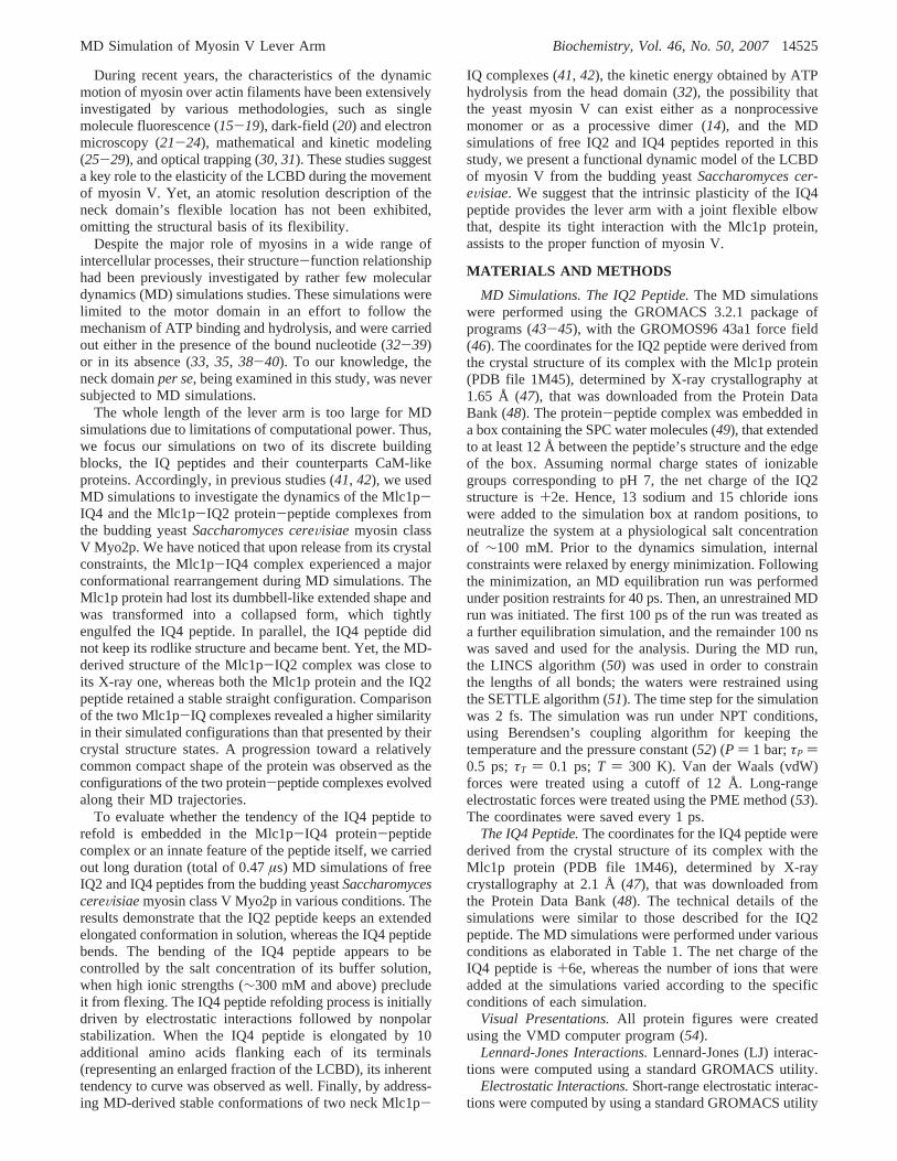
OVerall Conformational Changes during the Simulations.The simulated structures of the IQ2 peptide, as it varies fromits crystal structure with time, are presented in Figure 1 bysnapshots taken at 20 ns intervals. At its initial configuration,the IQ2 peptide is an almost linearR-helix. Within the first20 ns of simulation, both its N- and C-termini loosen, whileits mid-section retains a straight conformation. During therest of the simulation up to 100 ns, the structure of the peptidehardly changes, reflecting its stable MD-derived configura-tion. However, subtle reversible conformational modificationsmay occur, as can be seen at the 60 ns snapshot.
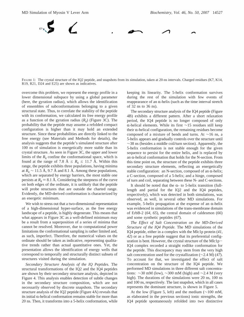
In contrast to the rigidity of the IQ2 peptide, snapshotstaken from the trajectory of the IQ4 peptide reveal a differentscenario (Figure 2A). The initial configuration of the IQ4peptide, as extracted from the Mlc1p-IQ4 protein-peptidecomplex X-ray structure, is a linearR-helix. After only 20ns of simulation, its conformation grossly deviates from itsinitial one. Its C-section loses the straight conformation, andmodulates into turn and bent structures. The conformationof the IQ4 peptide continues to explore the configurationalspace till reaching a new configuration, represented by the
following snapshots. At this newly gained conformation, theIQ4 peptide is refolded in a manner that its N- and C-terminiare relatively close to each other, having the shape of twohelices separated by a coiled hinge. Since the IQ4 peptidebears eight positive and two negative residues, it seems likelythat its refolded conformation is stabilized by internal saltbridges. Hence, we followed the minimal distance betweenpairs of its positive and negative residues as a function oftime, and present these that formed intrapeptide salt bridges(Figure 2B). As shown in Figure 2B, at the beginning of thesimulation, residues K11-E25 (black) and R4-E25 (gray)are apart from each other. For both cases, att ∼ 65 ns, thedistance between the residues sharply drops from>1 Å to adistance in which their atoms almost reached to a contact oftheir vdW radii (d ) 2.5 Å). The approach of these residuestoward each other is a relatively slow process, ranging tensof nanoseconds, that seems to be stable till the simulationwas terminated. Interestingly, the refolding of the peptideinvolves an increase of the distance between positive residueslocated on its mid-section (Figure 2C). The K12-K15(black) and the R14-K18 (gray) distances increase as thesimulation proceeds. The distribution of the positivelycharged residues along the peptide causes an electrostaticrepulsion that rejects them from each other, and consequentlythe peptide refolds and loses its extended helical conforma-tion. The role of the electrostatic potential of the peptide isfurther evaluated below.
To validate that the structure of the IQ4 peptide at 100 nsreflects a representative stable conformation, we repeatedthe simulation of the IQ4 peptide at a high temperature (400K) for 30 ns, and the last snapshot of that simulation is shownin Figure 2A (bottom, right). Inspection of the peptide’sstructure at the end of the 400 K simulation reveals a highsimilarity to the structure gained at 300 K, presenting aninnate capacity to flex. The refolding mechanism of thepeptide, at both temperatures, was similar to that observedby MD simulations of the Mlc1p-IQ4 protein-peptidecomplex at different temperatures (41).
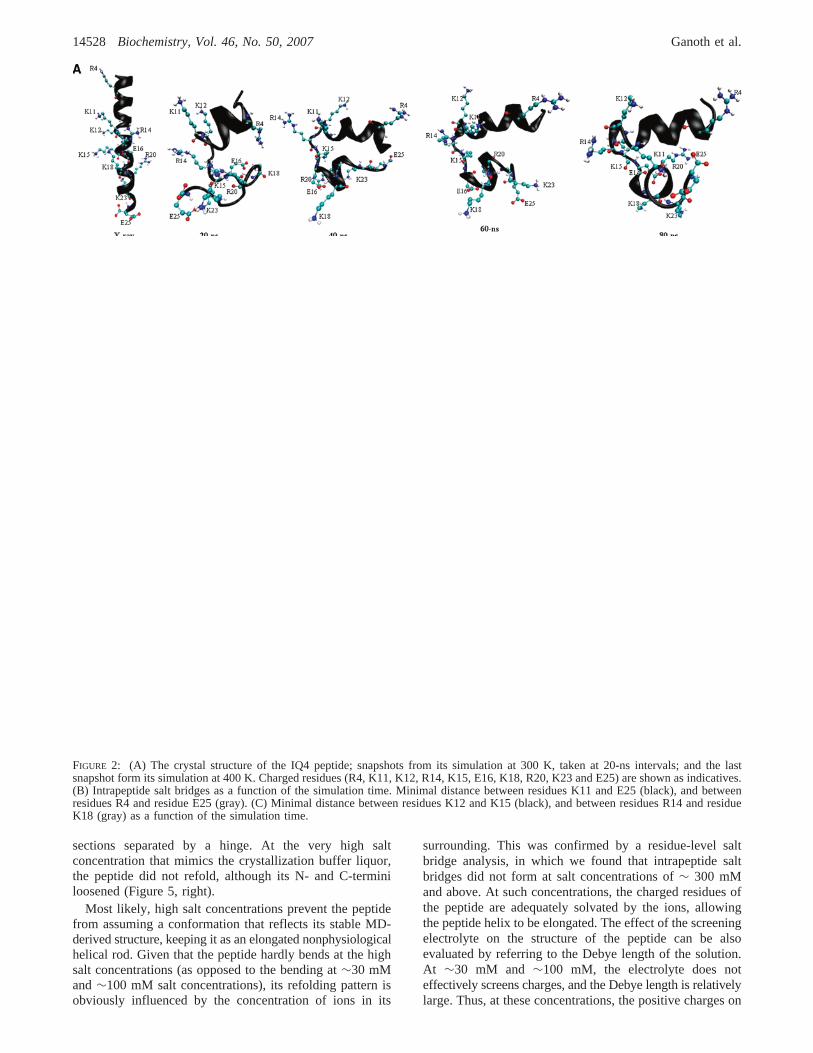
To evaluate the energy changes involved with the structuralmodulation process undergone by the IQ4 peptide, wecalculated its nonpolar, i.e., Lennard-Jones (LJ), and short-range Coulomb electrostatic energies (Figure 3A and 3B,respectively). Both terms decreased, though in differentmanners, as a function of the simulation time. The stabilitygenerated by the nonpolar interactions is∼50 kJ/mol, takingplace at a discrete time point, after which its value ispractically constant. The short-range Coulombic energycontributes∼200 kJ/mol, but appears to gradually evolveover most of the simulation time. It is of interest to pointout that the initial deformation of the peptide’sR-helicalstructure is driven by electrostatic interactions of the chargesresidues, either among themselves or with the solvent.
The energy of a protein is a function of the topologicalarrangement of itsN atoms and their interactions with thesolvent. In exact terms, it is described by a hyper-surface inthe 3N-dimensional configurational space, with a very largenumber of minima separated by activation barriers and saddlepoints. Each valley in this hyper-surface pertains to aparticular conformation of the protein (62, 63). Due to thehigh dimensionality of the configurational space of a protein,it is impossible to give a representative picture of its freeenergy landscape as a function of the entire phase space. To
Table 1: Summary of the MD Simulations of the IQ4 Peptide
duration(ns)
concnof salt
no. ofadded
Na+ ions
no. ofadded
Cl- ions
IQ4 peptide 100 ∼100 mM 12 18IQ4 peptidea 30 ∼100 mM 12 18extended IQ4 peptideb 20 ∼100 mM 10 17IQ4 peptidec 20 ∼30 mM 4 10IQ4 peptided 100 ∼300 mM 36 42IQ4 peptidee 100 ∼2.4 M 288 294
a High temperature (400 K).b An IQ4 peptide containing 10 ad-ditional amino acids flanking each of its terminals was built using SwissPDB Viewer (78). These added 20 residues belong to the IQ3 and IQ5peptides. Thus, the 45 amino acid elongated IQ4 peptide constitutes aportion of the poly-IQ sequence as present at the neck of the myosin.c Low salt concentration.d High salt concentration.e Very high saltconcentration (mimicking the salt concentration in which the Mlc1p-IQ4 protein-peptide complex was crystallized (47)).
∆Greffi ) -RT lnpi
pref(1)
14526 Biochemistry, Vol. 46, No. 50, 2007 Ganoth et al.
overcome this problem, we represent the energy profile in alower dimensional subspace by using a global parameter(here, the gyration radius), which allows the identificationof ensembles of subconformations belonging to a givenstructural state. Thus, to correlate the stability of the peptidewith its conformation, we calculated its free energy profileas a function of the gyration radius (Rg) (Figure 3C). Theprobability that the peptide may assume a refolded compactconfiguration is higher than it may hold an extendedstructure. Since these probabilities are directly linked to thefree energy (see Materials and Methods for details), theanalysis suggests that the peptide’s simulated structure after100 ns of simulation is energetically more stable than itscrystal structure. As seen in Figure 3C, the upper and lowerlimits of the Rg confine the conformational space, which isfound at the range of 7.8 Åe Rg e 11.7 Å. Within thisrange, the peptide exhibits three populations, having minimaat Rg ∼ 11.5 Å, 9.7 Å and 8.1 Å. Among these populations,which are separated by energy barriers, the most stable onepersists atRg ) 8.1 Å. Considering the steepness of the curveon both edges of the ordinate, it is unlikely that the peptidewill probe structures that are outside the charted range.Evidently, the MD-derived conformation is characterized byan energetic minimum.
We wish to stress out that a two-dimensional representationof a high-dimensional hyper-surface, as the free energylandscape of a peptide, is highly degenerate. This means thatwhat appears in Figure 3C as a well-defined minimum maybe a result from a superposition of a series of minima thatcannot be resolved. Moreover, due to computational powerlimitations the conformational sampling is rather limited and,as such, imperfect. Therefore, the numerical values on theordinate should be taken as indicative, representing qualita-tive trends rather than actual quantitative ones. Yet, thepresentation allows the identification of energy wells thatcorrespond to temporally and structurally distinct subsets ofstructures visited during the simulation.
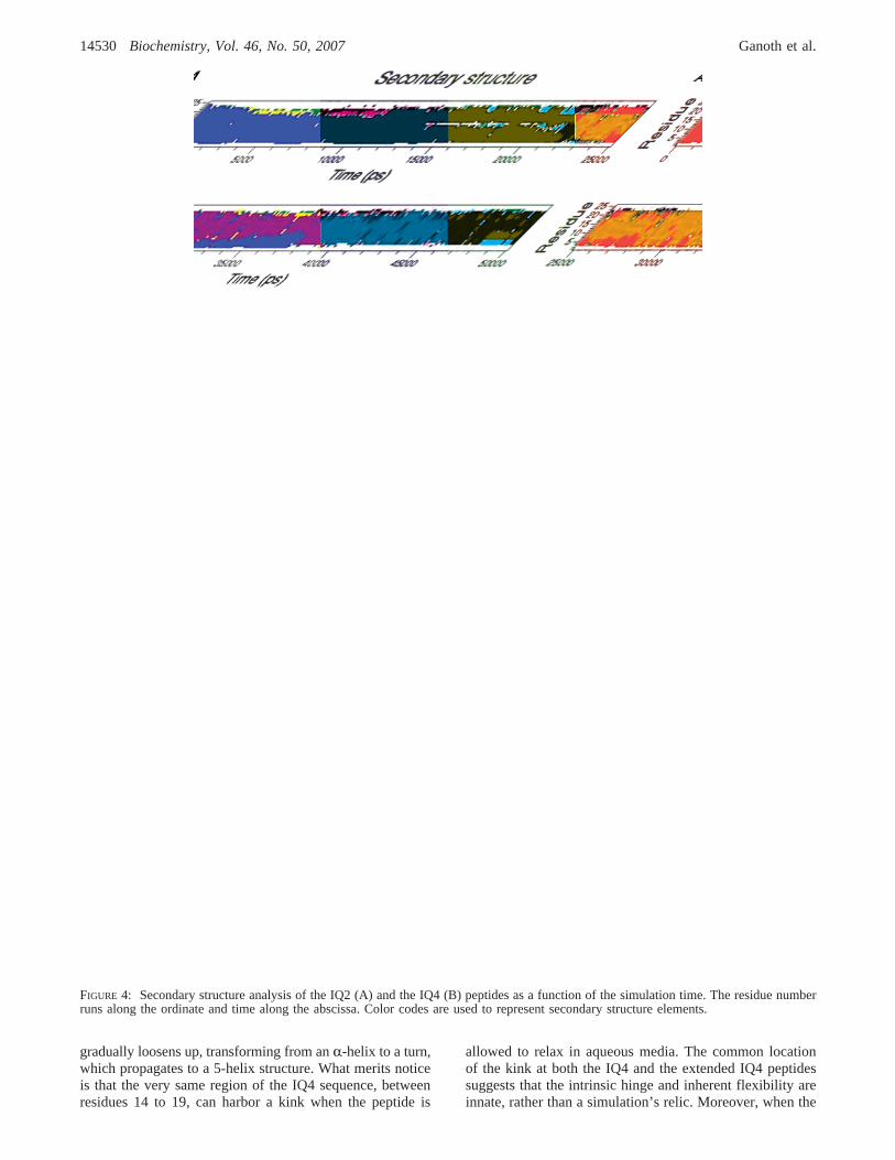
Secondary Structure Analysis of the IQ Peptides.Thestructural transformations of the IQ2 and the IQ4 peptidesare shown by their secondary structure analysis, depicted inFigure 4. This analysis enables detection of subtle changesin the secondary structure composition, which are notnecessarily observed by discrete snapshots. The secondarystructure analysis of the IQ2 peptide (Figure 4A) reveals thatits initial R-helical conformation remains stable for more than20 ns. Then, it transforms into a 5-helix conformation, while
keeping its linearity. The 5-helix conformation survivesduring the rest of the simulation with few events ofreappearance of anR-helix (such as the time interval stretchof 32 ns to 36 ns).
The secondary structure analysis of the IQ4 peptide (Figure4B) exhibits a different pattern. After a short relaxationperiod, the IQ4 peptide is no longer composed of onlyR-helical elements. While its first∼15 residues still keeptheirR-helical configuration, the remaining residues becomecomposed of a mixture of bends and turns. At∼16 ns, a5-helix appears and gradually controls over the structure until∼38 ns (besides a middle coil/turn section). Apparently, the5-helix conformation is not stable enough for the givensequence to persist for the entire helix, and is replaced byanR-helical conformation that holds for the N-section. Fromthis time point on, the structure of the peptide exhibits threesecondary structure elements, reflecting an energeticallystable configuration: an N-section, composed of anR-helix;a C-section, composed of a 5-helix; and a hinge, composedof turn and coil, separating between these N- and C-sections.
It should be noted that theR- to 5-helix transition (full-length and partial for the IQ2 and the IQ4 peptides,respectively), which was detected in both simulations, wasobserved, as well, in several other MD simulations. Forexample, 5-helix propagation at the expense of anR-helixwas evidenced in simulations of the trans-membrane domainof ErbB-2 (64, 65), the central domain of caldesmon (66)and some synthetic peptides (67).
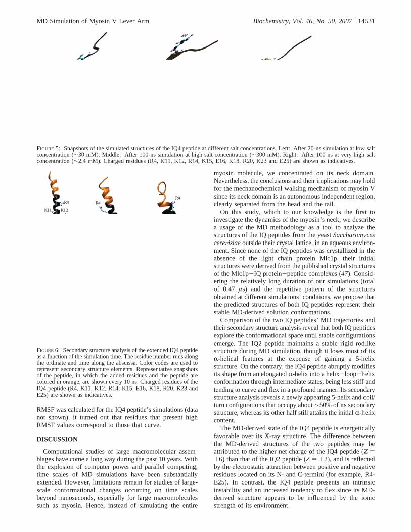
The Effect of Salt Concentration on the MD-DeriVedStructure of the IQ4 Peptide.The MD simulations of theIQ4 peptide, either in a complex with the Mlc1p protein (41,42) or as a free peptide suggest that its preferential config-uration is bent. However, the crystal structure of the Mlc1p-IQ4 complex recorded a straight rodlike conformation forthe peptide. This discrepancy may stem from the very highsalt concentration used for the crystallization (∼2.4 M) (47).To account for that, we investigated the effect of saltconcentration on the structure of the IQ4 peptide. Weperformed MD simulations in three different salt concentra-tions: ∼30 mM (low),∼300 mM (high) and∼2.4 M (veryhigh). The durations of the simulations were 20 ns, 100 nsand 100 ns, respectively. The last snapshot, which in all casesrepresents the dominant structure, is shown in Figure 5.
At the low (Figure 5, left) and the medium (∼100 mM,as elaborated in the previous sections) ionic strengths, theIQ4 peptide spontaneously refolded into two distinctive
FIGURE 1: The crystal structure of the IQ2 peptide, and snapshots from its simulation, taken at 20-ns intervals. Charged residues (K7, K14,R19, R21, D24 and E25) are shown as indicatives.
MD Simulation of Myosin V Lever Arm Biochemistry, Vol. 46, No. 50, 200714527
sections separated by a hinge. At the very high saltconcentration that mimics the crystallization buffer liquor,the peptide did not refold, although its N- and C-terminiloosened (Figure 5, right).
Most likely, high salt concentrations prevent the peptidefrom assuming a conformation that reflects its stable MD-derived structure, keeping it as an elongated nonphysiologicalhelical rod. Given that the peptide hardly bends at the highsalt concentrations (as opposed to the bending at∼30 mMand∼100 mM salt concentrations), its refolding pattern isobviously influenced by the concentration of ions in its
surrounding. This was confirmed by a residue-level saltbridge analysis, in which we found that intrapeptide saltbridges did not form at salt concentrations of∼ 300 mMand above. At such concentrations, the charged residues ofthe peptide are adequately solvated by the ions, allowingthe peptide helix to be elongated. The effect of the screeningelectrolyte on the structure of the peptide can be alsoevaluated by referring to the Debye length of the solution.At ∼30 mM and ∼100 mM, the electrolyte does noteffectively screens charges, and the Debye length is relativelylarge. Thus, at these concentrations, the positive charges on
FIGURE 2: (A) The crystal structure of the IQ4 peptide; snapshots from its simulation at 300 K, taken at 20-ns intervals; and the lastsnapshot form its simulation at 400 K. Charged residues (R4, K11, K12, R14, K15, E16, K18, R20, K23 and E25) are shown as indicatives.(B) Intrapeptide salt bridges as a function of the simulation time. Minimal distance between residues K11 and E25 (black), and betweenresidues R4 and residue E25 (gray). (C) Minimal distance between residues K12 and K15 (black), and between residues R14 and residueK18 (gray) as a function of the simulation time.
14528 Biochemistry, Vol. 46, No. 50, 2007 Ganoth et al.
the peptide effectively repel each other, and its refolding maytake place (see Figure 2C). At 300 mM and above, thescreening length is short-ranged, and hence the positivelycharged residues can tolerate the presence of other similarlycharged residues, suppressing the peptide’s refolding.
The conformation of the peptide at∼2.4 M resembles theone obtained by the simulation at the high salt concentration(Figure 5, middle) because already at∼300 mM NaCl thescreening effect caused by the ions is extensive. Accordingto our observation, the 8-fold increase at the NaCl concentra-tion did not significantly enhance the screening effect, andthus the results of the high and the very high salt concentra-tions’ simulations are similar.
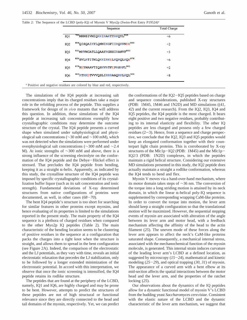
Structure and Dynamics of an Extended IQ4 Peptide.Inorder to verify that the kink presented by the IQ4 peptide
(Figure 2A) is not an artifact caused by its exposed N- andC-termini, we built an extended IQ4 peptide that was thenstudied by a 20 ns MD simulation. The sequence of thisextended peptide consisted of that of the IQ4 peptide, aspresent at the crystal structure of the Mlc1p-IQ4 complex,supplemented by additional twenty residues; ten of theseresidues flank its N-terminus and are part of the IQ3 peptidesequence, while the other ten reside after its C-terminus andbelong to the IQ5 peptide sequence. Hence, the 45 aminoacid long sequence represents a fair section (∼30%) of thefull-length poly-IQ (IQ1-IQ6) neck of myosin V.
The secondary structure analysis of the extended IQ4peptide (Figure 6) demonstrates that it experiences a similarrefolding pattern as observed for the IQ4 peptide. ItsN-section retains a rigid structure; whereas its C-section
FIGURE 3: (A) The Lennard-Jones component of the potential energy among the residues of the IQ4 peptide (gray), and its averaged valuesevery 250 ps (black). (B) The short-range electrostatic interactions among the residues of the IQ4 peptide (gray), and averaged values every250 ps (black). (C) Free energy projection over the gyration radius (Rg) of the IQ4 peptide.
MD Simulation of Myosin V Lever Arm Biochemistry, Vol. 46, No. 50, 200714529
gradually loosens up, transforming from anR-helix to a turn,which propagates to a 5-helix structure. What merits noticeis that the very same region of the IQ4 sequence, betweenresidues 14 to 19, can harbor a kink when the peptide is
allowed to relax in aqueous media. The common locationof the kink at both the IQ4 and the extended IQ4 peptidessuggests that the intrinsic hinge and inherent flexibility areinnate, rather than a simulation’s relic. Moreover, when the
FIGURE 4: Secondary structure analysis of the IQ2 (A) and the IQ4 (B) peptides as a function of the simulation time. The residue numberruns along the ordinate and time along the abscissa. Color codes are used to represent secondary structure elements.
14530 Biochemistry, Vol. 46, No. 50, 2007 Ganoth et al.
RMSF was calculated for the IQ4 peptide’s simulations (datanot shown), it turned out that residues that present highRMSF values correspond to those that curve.
DISCUSSION
Computational studies of large macromolecular assem-blages have come a long way during the past 10 years. Withthe explosion of computer power and parallel computing,time scales of MD simulations have been substantiallyextended. However, limitations remain for studies of large-scale conformational changes occurring on time scalesbeyond nanoseconds, especially for large macromoleculessuch as myosin. Hence, instead of simulating the entire
myosin molecule, we concentrated on its neck domain.Nevertheless, the conclusions and their implications may holdfor the mechanochemical walking mechanism of myosin Vsince its neck domain is an autonomous independent region,clearly separated from the head and the tail.
On this study, which to our knowledge is the first toinvestigate the dynamics of the myosin’s neck, we describea usage of the MD methodology as a tool to analyze thestructures of the IQ peptides from the yeastSaccharomycescereVisiaeoutside their crystal lattice, in an aqueous environ-ment. Since none of the IQ peptides was crystallized in theabsence of the light chain protein Mlc1p, their initialstructures were derived from the published crystal structuresof the Mlc1p-IQ protein-peptide complexes (47). Consid-ering the relatively long duration of our simulations (totalof 0.47 µs) and the repetitive pattern of the structuresobtained at different simulations’ conditions, we propose thatthe predicted structures of both IQ peptides represent theirstable MD-derived solution conformations.
Comparison of the two IQ peptides’ MD trajectories andtheir secondary structure analysis reveal that both IQ peptidesexplore the conformational space until stable configurationsemerge. The IQ2 peptide maintains a stable rigid rodlikestructure during MD simulation, though it loses most of itsR-helical features at the expense of gaining a 5-helixstructure. On the contrary, the IQ4 peptide abruptly modifiesits shape from an elongatedR-helix into a helix-loop-helixconformation through intermediate states, being less stiff andtending to curve and flex in a profound manner. Its secondarystructure analysis reveals a newly appearing 5-helix and coil/turn configurations that occupy about∼50% of its secondarystructure, whereas its other half still attains the initialR-helixcontent.
The MD-derived state of the IQ4 peptide is energeticallyfavorable over its X-ray structure. The difference betweenthe MD-derived structures of the two peptides may beattributed to the higher net charge of the IQ4 peptide (Z )+6) than that of the IQ2 peptide (Z ) +2), and is reflectedby the electrostatic attraction between positive and negativeresidues located on its N- and C-termini (for example, R4-E25). In contrast, the IQ4 peptide presents an intrinsicinstability and an increased tendency to flex since its MD-derived structure appears to be influenced by the ionicstrength of its environment.
FIGURE 5: Snapshots of the simulated structures of the IQ4 peptide at different salt concentrations. Left: After 20-ns simulation at low saltconcentration (∼30 mM). Middle: After 100-ns simulation at high salt concentration (∼300 mM). Right: After 100 ns at very high saltconcentration (∼2.4 mM). Charged residues (R4, K11, K12, R14, K15, E16, K18, R20, K23 and E25) are shown as indicatives.
FIGURE 6: Secondary structure analysis of the extended IQ4 peptideas a function of the simulation time. The residue number runs alongthe ordinate and time along the abscissa. Color codes are used torepresent secondary structure elements. Representative snapshotsof the peptide, in which the added residues and the peptide arecolored in orange, are shown every 10 ns. Charged residues of theIQ4 peptide (R4, K11, K12, R14, K15, E16, K18, R20, K23 andE25) are shown as indicatives.
MD Simulation of Myosin V Lever Arm Biochemistry, Vol. 46, No. 50, 200714531
The simulations of the IQ4 peptide at increasing saltconcentrations imply that its charged residues take a majorrole in the refolding process of the peptide. This supplies aframework for design ofin ViVo mutants that will addressthis question. In addition, these simulations of the IQ4peptide at increasing salt concentrations exemplify howcrystallographic conditions may determine the outcomestructure of the crystal. The IQ4 peptide presents a curvedshape when simulated under subphysiological and physi-ological salt concentrations (∼30 mM and∼100 mM), whichwas not detected when the simulations were performed underoverphysiological salt concentrations (∼300 mM and∼2.4M). At ionic strengths of∼300 mM and above, there is astrong influence of the screening electrolyte on the confor-mation of the IQ4 peptide and the Debye-Huckel effect isstressed. That precludes the IQ4 peptide from bending,keeping it as a straightR-helix. Apparently, as indicated bythis study, the crystalline structure of the IQ4 peptide wasimposed by specific crystallographic conditions of its crystal-lization buffer liquor (such as its salt concentration and ionicstrength). Fundamental deviations of X-ray determinedstructures from native configurations of proteins weredocumented, as well, in other cases (68-70).
The bent IQ4 peptide’s structure is too short for searchingfor similar features in other proteins except myosins, andhence evaluating of its properties is limited to the simulationsreported in the present study. The main property of the IQ4sequence is a plethora of charged residues when comparedto the other Myo2p IQ peptides (Table 2). The maincharacteristic of the bending location seems to be clusteringof positive residues in the sequence at a configuration thatpacks the charges into a tight knot when the structure isstraight, and allows them to spread in the bent configuration(see Figure 2A). Indeed, the comparison of the electrostaticand the LJ potentials, as they vary with time, reveals an initialelectrostatic relaxation that precedes the LJ stabilization, onlyto be followed by a longer extended minimization of theelectrostatic potential. In accord with this interpretation, weobserve that once the ionic screening is intensified, the IQ4peptide retains its rodlike structure.
The peptides that are found at the periphery of the LCBD,namely, IQ1 and IQ6, are highly charged and may be proneto be bent. However, attempts to predict the structures ofthese peptides are not of high structural or functionalrelevance since they are directly connected to the head andtail domains of the myosin, respectively. Yet, we can predict
the conformations of the IQ2-IQ5 peptides based on chargeand sequence considerations, published X-ray structures(PDB: 1M45, 1M46 and 1N2D) and MD simulations ((41,42) and the current research). From the IQ2, IQ3, IQ4 andIQ5 peptides, the IQ4 peptide is the most charged. It bearseight positive and two negative residues, probably contribut-ing to its internal elasticity and flexibility. The other IQpeptides are less charged and possess only a few chargedresidues (2-3). Hence, from a sequence and charge perspec-tive, we conclude that the IQ2, IQ3 and IQ5 peptides wouldkeep an elongated conformation together with their coun-terpart light chain proteins. This is corroborated by X-raystructures of the Mlc1p-IQ2 (PDB: 1M45) and the Mlc1p-IQ2/3 (PDB: 1N2D) complexes, in which the peptidesmaintain a rigid helical structure. Considering our extensiveMD simulations presented in this study, the IQ2 peptide doesactually maintain a straight a rodlike conformation, whereasthe IQ4 tends to bend and flex.
Myosin V moves via a hand-over-hand mechanism, whereits motor domain takes steps of∼36 nm. The conversion ofthe torque into a long striding motion is attained by its neckdomain, in which the linearR-helical poly-IQ sequence issupplemented by corresponding wrapping CaM-like proteins.In order to convert the torque into motion, the lever armshould keep a straight configuration so that the translationalmotion will be maximized. However, the sequential steppingevents of myosin are associated with alteration of the anglebetween its lever arm and motor head, with a feedbackmechanism affecting the affinity of the head to the actinfilament (25). The uneven mode of these forces along thelever arm appears to affect the neck’s CaM-like proteinssaturated shape. Consequently, a mechanical internal stress,associated with the mechanochemical function of the myosinmolecule, is generated. This internal strain induces curvatureof the leading lever arm’s LCBD at a defined location, assuggested by microscopy (15-24), mathematical and kineticmodeling (25-29), and optical trapping (30, 31) of myosin.The appearance of a curved arm with a bent elbow at itsmid-section affects the spatial interactions between the motorhead and the lever arm, and the properties of the ratchetlocking (25).
Our observations about the dynamics of the IQ peptidesallow for a dynamic functional model of myosin V’s LCBDfrom the budding yeastSaccharomyces cereVisiae. Consistentwith the elastic nature of the LCBD and the dynamiccharacteristic of the lever arm mechanism, we suggest that
Table 2: The Sequence of the LCBD (poly-IQ) of Myosin V Myo2p (Swiss-Prot Entry P19524)a
a Positive and negative residues are colored by blue and red, respectively.
14532 Biochemistry, Vol. 46, No. 50, 2007 Ganoth et al.
the banana-shaped conformation formed by myosin’s armis generated through the IQ4 peptide that presents an intrinsicinner characteristic to flex and curve in solution. This is inaccordance with the secondary structure analysis of the IQ2and the IQ4 peptides, in which the former peptide completelytransforms into a 5-helix conformation whereas the latterpeptide harbors a coil between anR-helix and 5-helixconformations. Thus, the secondary structure of the IQ2peptide prevents it from bending, while that of the Myo2pIQ4 peptide allows it to bend and curl. The hinge shown atthe neck’s IQ4 peptide’s conformation, observed in all oursimulations under physiological salt conditions (and also forthe extended IQ4 peptide), may correspond to the bentsolution state of the lever arm of myosin. Our proposedmodel is supported by previous studies (41, 42), in whichwe found that, in the presence of the Mlc1p protein, the IQ4peptide loses its rodlike shape while the IQ2 peptide keepsit. In this fashion, the Mlc1p protein does not hinder thecapacity of the IQ4 peptide to bend in the very same locationwhen simulated in its absence. Interestingly, further sup-porting data was supplied by fluorescence and near- and far-UV CD measurements, where it was found that a murine
myosin IQ3/4 double-length sequence cannot exist as acontinuous straight helix (71), even in the presence of CaM.
As indicated by the MD simulations of the IQ4 peptide atdifferent salt concentrations, at a physiological salt concen-tration (∼150 mM) it should be able to naturally switch froma bent to a rodlike configuration. This suggests that relativelyminor forces are needed in order to induce itsin ViVoalternation between the two conformation states. Therefore,the poly-IQ sequence may be bent at the IQ4 peptide regionthat functions as a joint since its conformational process ofrefolding is crucial to the myosin’s walking over the actinfilament. Apparently, the bended elbow conformation of theLCBD’s lead arm, which is suggested to be caused by thecurved IQ4 peptide, is a fundamental element in the myosin’smechanochemical movement mechanism.
The uniqueness of the IQ4 peptide is not restricted to theyeast Myo2p, but rather is a common feature for class Vmyosins from different species (Table 3). Among the poly-IQ sequence of yeast, mouse rat and human, the IQ4 peptidecarries the highest number of charged residues (7-10) ascompared to the other IQ peptides. Evidently, the highnumber of charged residues is evolutionarily conserved, and
Table 3: Sequences of the LCBD of Class V Myosins from Yeast (Myo2p•Sc and Myo4p•Sc), Mouse (MyoVa•Mo), Rat (MyoVa•Ra) andHuman (MyoVa•Hu)a
a The peptides (IQ1-IQ6) are colored by red, blue, green, black, pink and turquoise, respectively. Swiss-Prot accession numbers are Myo2p•Sc(P19524), Myo4p•Sc (P32492), MyoVa•Mo (Q99104), MyoVa•Ra (Q9QYF3) and MyoVa•Hu (Q9Y4I1). Charged residues are underlined.
MD Simulation of Myosin V Lever Arm Biochemistry, Vol. 46, No. 50, 200714533
this may indicate that our suggestion about the intrinsiccapacity of the IQ4 peptide to bend can be valid for class Vmyosins from vertebrates as well.
An essential point for mechanical force generation inmyosin systems is how the energy released by ATP hydroly-sis in the myosin motor domain gives rise to the movementof the myosin head along the actin filament. By performingMD simulations of the motor domain of myosin S1, Minamiand co-workers (32) suggest that the energy released by theATP hydrolysis is transformed into kinetic energy of atomslocated around the nucleotide-binding site pocket found inthe motor domain. Their analysis indicates that the structuraldeformation, which is caused by the ATP hydrolysis, extendsover the motor domain core. It spreads and induces varyingcollective motions of atoms at the actin-binding site, andsimultaneously, at the junction with the neck. Our simulationsmay complement this study by suggesting that the influenceof the torque produced by ATP hydrolysis may not cease atthe nucleotide-binding site located at the motor domain, butrather continues to move and be transferred along the myosinmolecule through its neck at the direction of the tail.Evidently, this motion induces bending of the IQ4 peptide,which consequently enables the myosin’s neck to flex.
The functional model of myosin V’s LCBD from thebudding yeastSaccharomyces cereVisiae, as presented here,may be valid only whether the yeast’s myosin V motor isprocessive. This issue is under current dispute and no clearproof, so far, conclusively solved the puzzle. Mooseker andco-workers (13) claim that the yeast’s class V myosinsMyo2p and Myo4p are nonprocessivein Vitro. Their conclu-sion is based uponin Vitro indirect assays, in which themyosin’s velocity and landing rate on the actin filament, asthey varied with the myosin’s motor domain density, werefollowed. However, the authors did not exclude the pos-sibility of in ViVo processivity. An explanation for theabsence of processivity in their study may be that the yeastclass V myosins are processive due to a regulatory eventand in its absence are nonprocessive. It was shown thatbinding of the dynactin multi-subunit protein complex to themicrotubule motors dynein (72, 73) and kinesin (74)increases their processivity. Since myosin, dynein and kinesinare all molecular motors, it is likely the myosin’s functionis regulated by dynactin or another regulating factor.Furthermore, Trybus and co-workers (14) dealt with theyeast’s myosin V lack of processivity in a recent publication,suggesting that the yeast Myo4p molecule can exist eitheras a nonprocessive monomer or a processive dimer, depend-ing on the conditions. A high identity (34%) and similarity(58%) between the LCBDs of Myo2p and Myo4p, ascalculated by BLAST (75, 76), lead to the conclusion thatmost probably both share similar features. It should be notedthat a monomer-dimer equilibrium that impacts on proces-sivity has been proposed to exist at class VI myosins (for areview, see (77)), and it is possible that this may be a generalmechanism for regulatingin ViVo processivity. Additionally,the research of Trybus and co-workers proposes that nativeyeast Myo4p is nonprocessive (or weakly processive) at ahigh ionic strength and more processive at a physiologicalionic strength. Our observations are in favor of the findingsraised by Trybus and co-workers, suggesting that at aphysiological salt concentration the IQ4 peptide is able tobend but high salt conditions prevent it from flexing. In other
words, when the conditions enable, the myosin V moleculedimerizes and becomes processive; andViceVersa, when theconditions are unfavorable, the molecule cannot dimerize andhence may present monomeric nonprocessive characteristics.
The interaction between the IQ peptides and their corre-sponding CaM-like protein along the LCBD is a complexfunction, reflecting the local protein-peptide interactions andthe interactions between the light-chain proteins, one withthe others. It is likely that the relative motion of the motordomain, with respect to the lever arm, imposes a stress notonly on the LCBD but also on the CaM-like proteins thatare threaded along it. The double path of the stress alongtheR-helix and wrapping proteins may provide a mechanismthat regulates the flexing point along the lever arm, facilitat-ing the transfer of the local torque into effective translationalmotion of the whole, double-headed protein. Thus, by carefulanalysis of data combining (i) MD simulations of the motor(32) and the neck ((41, 42) and this study) domains of myosinand (ii) single molecule experiments of chimeric class Vmyosins, we offer an additional piece to the mechanochem-ical walking mechanism puzzle of myosin V. Obviously,additional experimental and theoretical work is required tofully elucidate the mechanism of myosin V motions.
REFERENCES
1. Reck-Peterson, S. L., Provance, D. W., Jr., Mooseker, M. S., andMercer, J. A. (2000) Class V myosins,Biochim. Biophys. Acta1496, 36-51.
2. Kalhammer, G., and Bahler, M. (2000) Unconventional myosins,Essays Biochem. 35, 33-42.
3. Nascimento, A. A., Cheney, R. E., Tauhata, S. B., Larson, R. E.,and Mooseker, M. S. (1996) Enzymatic characterization andfunctional domain mapping of brain myosin-V,J. Biol. Chem.271, 17561-17569.
4. Langford, G. M. (2002) Myosin-V, a versatile motor for short-range vesicle transport,Traffic 3, 859-865.
5. Sellers, J. R., and Veigel, C. (2006) Walking with myosin V,Curr.Opin. Cell Biol. 18, 68-73.
6. Rayment, I., Holden, H. M., Whittaker, M., Yohn, C. B., Lorenz,M., Holmes, K. C., and Milligan, R. A. (1993) Structure of theactin-myosin complex and its implications for muscle contraction,Science 261, 58-65.
7. Mehta, A. D., Rock, R. S., Rief, M., Spudich, J. A., Mooseker,M. S., and Cheney, R. E. (1999) Myosin-V is a processive actin-based motor,Nature 400, 590-593.
8. Geeves, M. A., and Holmes, K. C. (1999) Structural mechanismof muscle contraction,Annu. ReV. Biochem. 68, 687-728.
9. Langford, G. M., Kuznetsov, S. A., Johnson, D., Cohen, D. L.,and Weiss, D. G. (1994) Movement of axoplasmic organelles onactin filaments assembled on acrosomal processes: evidence fora barbed-end-directed organelle motor,J. Cell Sci. 107(Part 8),2291-2298.
10. Sakamoto, T., Wang, F., Schmitz, S., Xu, Y., Xu, Q., Molloy, J.E., Veigel, C., and Sellers, J. R. (2003) Neck length andprocessivity of myosin V,J. Biol. Chem. 278, 29201-29207.
11. Brockerhoff, S. E., Stevens, R. C., and Davis, T. N. (1994) Theunconventional myosin, Myo2p, is a calmodulin target at sites ofcell growth in Saccharomyces cerevisiae,J. Cell Biol. 124, 315-323.
12. Haarer, B. K., Petzold, A., Lillie, S. H., and Brown, S. S. (1994)Identification of MYO4, a second class V myosin gene in yeast,J. Cell Sci. 107(Part 4), 1055-1064.
13. Reck-Peterson, S. L., Tyska, M. J., Novick, P. J., and Mooseker,M. S. (2001) The yeast class V myosins, Myo2p and Myo4p, arenonprocessive actin-based motors,J. Cell Biol. 153, 1121-1126.
14. Krementsova, E. B., Hodges, A. R., Lu, H., and Trybus, K. M.(2006) Processivity of chimeric class V myosins,J. Biol. Chem.281, 6079-6086.
15. Nguyen, H., and Higuchi, H. (2005) Motility of myosin V regulatedby the dissociation of single calmodulin,Nat. Struct. Mol. Biol.12, 127-132.
14534 Biochemistry, Vol. 46, No. 50, 2007 Ganoth et al.
16. Toprak, E., Enderlein, J., Syed, S., McKinney, S. A., Petschek,R. G., Ha, T., Goldman, Y. E., and Selvin, P. R. (2006) Defocusedorientation and position imaging (DOPI) of myosin V,Proc. Natl.Acad. Sci. U.S.A. 103, 6495-6499.
17. Syed, S., Snyder, G. E., Franzini-Armstrong, C., Selvin, P. R.,and Goldman, Y. E. (2006) Adaptability of myosin V studied bysimultaneous detection of position and orientation,EMBO J. 25,1795-1803.
18. Sakamoto, T., Yildez, A., Selvin, P. R., and Sellers, J. R. (2005)Step-size is determined by neck length in myosin V,Biochemistry44, 16203-16210.
19. Lu, H., Krementsova, E. B., and Trybus, K. M. (2006) Regulationof myosin V processivity by calcium at the single molecule level,J. Biol. Chem. 281, 31987-31994.
20. Dunn, A. R., and Spudich, J. A. (2007) Dynamics of the unboundhead during myosin V processive translocation,Nat. Struct. Mol.Biol. 14, 246-248.
21. Burgess, S., Walker, M., Wang, F., Sellers, J. R., White, H. D.,Knight, P. J., and Trinick, J. (2002) The prepower strokeconformation of myosin V,J. Cell Biol. 159, 983-991.
22. Walker, M. L., Burgess, S. A., Sellers, J. R., Wang, F., Hammer,J. A., 3rd, Trinick, J., and Knight, P. J. (2000) Two-headed bindingof a processive myosin to F-actin,Nature 405, 804-807.
23. Volkmann, N., Liu, H., Hazelwood, L., Krementsova, E. B.,Lowey, S., Trybus, K. M., and Hanein, D. (2005) The structuralbasis of myosin V processive movement as revealed by electroncryomicroscopy,Mol. Cell 19, 595-605.
24. Yildiz, A., Forkey, J. N., McKinney, S. A., Ha, T., Goldman, Y.E., and Selvin, P. R. (2003) Myosin V walks hand-over-hand:single fluorophore imaging with 1.5-nm localization,Science 300,2061-2065.
25. Lan, G., and Sun, S. X. (2005) Dynamics of myosin-V processivity,Biophys. J. 88, 999-1008.
26. Lan, G., and Sun, S. X. (2006) Flexible Light-Chain and HelicalStructure of F-Actin Explain the Movement and Step Size ofMyosin-VI, Biophys. J. 91, 4002-4013.
27. Skau, K. I., Hoyle, R. B., and Turner, M. S. (2006) A kineticmodel describing the processivity of myosin-V,Biophys. J. 91,2475-2489.
28. Smith, D., and Sleep, J. (2006) Strain-dependent kinetics of theMyosin working stroke, and how they could be probed withoptical-trap experiments,Biophys. J. 91, 3359-3369.
29. Smith, D. A., and Sleep, J. (2004) Mechanokinetics of rapid tensionrecovery in muscle: the Myosin working stroke is followed by aslower release of phosphate,Biophys. J. 87, 442-456.
30. Purcell, T. J., Sweeney, H. L., and Spudich, J. A. (2005) A force-dependent state controls the coordination of processive myosinV, Proc. Natl. Acad. Sci. U.S.A. 102, 13873-13878.
31. Veigel, C., Schmitz, S., Wang, F., and Sellers, J. R. (2005) Load-dependent kinetics of myosin-V can explain its high processivity,Nat. Cell Biol. 7, 861-869.
32. Kawakubo, T., Okada, O., and Minami, T. (2005) Moleculardynamics simulations of evolved collective motions of atoms inthe myosin motor domain upon perturbation of the ATPase pocket,Biophys. Chem. 115, 77-85.
33. Koppole, S., Smith, J. C., and Fischer, S. (2006) Simulations ofthe myosin II motor reveal a nucleotide-state sensing element thatcontrols the recovery stroke,J. Mol. Biol. 361, 604-616.
34. Lawson, J. D., Pate, E., Rayment, I., and Yount, R. G. (2004)Molecular dynamics analysis of structural factors influencing backdoor pi release in myosin,Biophys. J. 86, 3794-3803.
35. Mesentean, S., Koppole, S., Smith, J. C., and Fischer, S. (2007)The principal motions involved in the coupling mechanism of therecovery stroke of the myosin motor,J. Mol. Biol. 367, 591-602.
36. Okimoto, N., Yamanaka, K., Ueno, J., Hata, M., Hoshino, T., andTsuda, M. (2001) Theoretical studies of the ATP hydrolysismechanism of myosin,Biophys. J. 81, 2786-2794.
37. Woo, H. J. (2007) Exploration of the conformational space ofmyosin recovery stroke via molecular dynamics,Biophys. Chem.125, 127-137.
38. Yu, H., Ma, L., Yang, Y., and Cui, Q. (2007) MechanochemicalCoupling in the Myosin Motor Domain. I. Insights from Equi-librium Active-Site Simulations,PLoS Comput. Biol. 3, e21.
39. Yu, H., Ma, L., Yang, Y., and Cui, Q. (2007) MechanochemicalCoupling in the Myosin Motor Domain. II. Analysis of CriticalResidues,PLoS Comput. Biol. 3, e23.
40. Liu, Y., Scolari, M., Im, W., and Woo, H. J. (2006) Protein-proteininteractions in actin-myosin binding and structural effects ofR405Q mutation: a molecular dynamics study,Proteins 64, 156-166.
41. Ganoth, A., Nachliel, E., Friedman, R., and Gutman, M. (2006)Molecular dynamics study of a calmodulin-like protein with anIQ peptide: Spontaneous refolding of the protein around thepeptide,Proteins 64, 133-146.
42. Ganoth, A., Friedman, R., Nachliel, E., and Gutman, M. (2006)A molecular dynamics study and free energy analysis of complexesbetween the Mlc1p protein and two IQ motif peptides,Biophys.J. 91, 2436-2450.
43. Berendsen, H. J. C., van der Spoel, D., and van Drunen, R. (1995)GROMACS: A message-passing parallel molecular dynamicsimplementation,Comput. Phys. Commun. 91, 43-56.
44. Lindahl, E., Hess, B., and van der Spoel, D. (2001) Gromacs 3.0:A package for molecular simulation and trajectory analysis,J.Mol. Model. 7, 306-317.
45. van Der Spoel, D., Lindahl, E., Hess, B., van Buuren, A. R., Apol,E., Meulenhoff, P. J., Tieleman, D. P., Sijbers, A. L. T. M.,Feenstra, A. K., van Drunen, R., and Berendsen, H. J. C. (2004)Groningen Machine for Molecular Simulations. BIOSON Re-search Institute, Groningen, The Netherlands.
46. van Gunsteren, W. F., Billeter, S. R., Eising, A. A., Hunenberger,P. H., Kruger, P., Mark, A. E., Scott, W. R. P., and Tironi, I. G.(1996)Biomolecular Simulation: The GROMOS96 Manual andUser Guide, Vdf Hochschulverlag AG an der ETH Zurich, Zurich,pp 1-1024.
47. Terrak, M., Otterbein, L. R., Wu, G., Palecanda, L. A., Lu, R. C.,and Dominguez, R. (2002) Crystallization, X-ray characterizationand selenomethionine phasing of Mlc1p bound to IQ motifs frommyosin V,Acta Crystallogr, Sect. D: Biol. Crystallogr. 58, 1882-1885.
48. Berman, H. M., Battistuz, T., Bhat, T. N., Bluhm, W. F., Bourne,P. E., Burkhardt, K., Feng, Z., Gilliland, G. L., Iype, L., Jain, S.,Fagan, P., Marvin, J., Padilla, D., Ravichandran, V., Schneider,B., Thanki, N., Weissig, H., Westbrook, J. D., and Zardecki, C.(2002) The Protein Data Bank,Acta Crystallogr., Sect. D: Biol.Crystallogr. 58, 899-907.
49. Berendsen, H. J. C., Postma, J. P. M., van Gunsteren, W. F., andHermans, J. (1969) Interaction Models for Water in Relation toProtein Hydration,Nature 224, 175-177.
50. Hess, B., Bekker, H., Berendsen, H. J. C., and Fraaije, J. G. E.M. (1997) LINCS: A linear constraint solver for molecularsimulations,J. Comput. Chem. 18, 1463-1472.
51. Miyamoto, S., and Kollman, P. A. (1992) SETTLE: An AnalyticalVersion of the SHAKE and RATTLE Algorithms for Rigid watermodels,J. Comput. Chem. 13, 952-962.
52. Berendsen, H. J. C., Postma, J. P. M., DiNola, A., and Haak, J.R. (1984) Molecular dynamics with coupling to an external bath,J. Chem. Phys. 81, 3684-3690.
53. Essman, U., Perela, L., Berkowitz, M. L., Darden, T., Lee, H.,and Pederse, L. G. (1995) A smooth particle mesh Ewald method,J. Chem. Phys. 103, 8577-8592.
54. Humphrey, W., Dalke, A., and Schulten, K. (1996) VMD: visualmolecular dynamics,J. Mol. Graphics 14, 33 38, 27 38.
55. Daidone, I., Amadei, A., and Di Nola, A. (2005) Thermodynamicand kinetic characterization of a beta-hairpin peptide in solution:an extended phase space sampling by molecular dynamicssimulations in explicit water,Proteins 59, 510-518.
56. Tavernelli, I., Cotesta, S., and Di Iorio, E. E. (2003) Proteindynamics, thermal stability, and free-energy landscapes: a mo-lecular dynamics investigation,Biophys. J. 85, 2641-2649.
57. Amadei, A., Linssen, A. B., de Groot, B. L., van Aalten, D. M.,and Berendsen, H. J. (1996) An efficient method for samplingthe essential subspace of proteins,J. Biomol. Struct. Dyn. 13, 615-625.
58. Cecchini, M., Curcio, R., Pappalardo, M., Melki, R., and Caflisch,A. (2006) A molecular dynamics approach to the structuralcharacterization of amyloid aggregation,J. Mol. Biol. 357, 1306-1321.
59. Daidone, I., D’Abramo, M., Di Nola, A., and Amadei, A. (2005)Theoretical characterization of alpha-helix and beta-hairpin foldingkinetics,J. Am. Chem. Soc. 127, 14825-14832.
60. D’Abramo, M., Rinaldi, A. C., Bozzi, A., Amadei, A., Mignogna,G., Di Nola, A., and Aschi, M. (2006) Conformational behaviorof temporin A and temporin L in aqueous solution: a computational/experimental study,Biopolymers 81, 215-224.
MD Simulation of Myosin V Lever Arm Biochemistry, Vol. 46, No. 50, 200714535
61. Cecchini, M., Rao, F., Seeber, M., and Caflisch, A. (2004) Replicaexchange molecular dynamics simulations of amyloid peptideaggregation,J. Chem. Phys. 121, 10748-10756.
62. Onuchic, J. N., Luthey-Schulten, Z., and Wolynes, P. G. (1997)Theory of protein folding: the energy landscape perspective,Annu.ReV. Phys. Chem. 48, 545-600.
63. Wales, D. G. (2003)Energy Landscapes, Cambridge UniversityPress, Cambridge.
64. Duneau, J. P., Garnier, N., and Genest, M. (1997) Insight intosignal transduction: structural alterations in transmembrane helicesprobed by multi-1 ns molecular dynamics simulations,J. Biomol.Struct. Dyn. 15, 555-572.
65. Duneau, J. P., Crouzy, S., Chapron, Y., and Genest, M. (1999)Dynamics of the transmembrane domain of the ErbB-2 receptor,Theor. Chem. Acc. 101, 87-91.
66. Shepherd, C. M., van der Spoel, D., and Vogel, H. J. (2004)Molecular dynamics simulations of peptides from the centraldomain of smooth muscle caldesmon,J. Biomol. Struct. Dyn. 21,555-566.
67. Lee, K. H., Benson, D. R., and Kuczera, K. (2000) Transitionsfrom alpha to pi helix observed in molecular dynamics simulationsof synthetic peptides,Biochemistry 39, 13737-13747.
68. Eyal, E., Gerzon, S., Potapov, V., Edelman, M., and Sobolev, V.(2005) The limit of accuracy of protein modeling: influence ofcrystal packing on protein structure,J. Mol. Biol. 351, 431-442.
69. DePristo, M. A., de Bakker, P. I., and Blundell, T. L. (2004)Heterogeneity and inaccuracy in protein structures solved by X-raycrystallography,Structure 12, 831-838.
70. Steuber, H., Zentgraf, M., Gerlach, C., Sotriffer, C. A., Heine,A., and Klebe, G. (2006) Expect the Unexpected or Caveat forDrug Designers: Multiple Structure Determinations Using Aldose
Reductase Crystals Treated under Varying Soaking and Co-crystallisation Conditions,J. Mol. Biol. 363, 174-187.
71. Martin, S. R., and Bayley, P. M. (2002) Regulatory implicationsof a novel mode of interaction of calmodulin with a double IQ-motif target sequence from murine dilute myosin V,Protein Sci.11, 2909-2923.
72. Culver-Hanlon, T. L., Lex, S. A., Stephens, A. D., Quintyne, N.J., and King, S. J. (2006) A microtubule-binding domain indynactin increases dynein processivity by skating along micro-tubules,Nat. Cell Biol. 8, 264-270.
73. King, S. J., and Schroer, T. A. (2000) Dynactin increases theprocessivity of the cytoplasmic dynein motor,Nat. Cell Biol. 2,20-24.
74. Berezuk, M. A., and Schroer, T. A. (2007) Dynactin enhancesthe processivity of kinesin-2,Traffic 8, 124-129.
75. Altschul, S. F., Madden, T. L., Schaffer, A. A., Zhang, J., Zhang,Z., Miller, W., and Lipman, D. J. (1997) Gapped BLAST andPSI-BLAST: a new generation of protein database searchprograms,Nucleic Acids Res. 25, 3389-3402.
76. Schaffer, A. A., Aravind, L., Madden, T. L., Shavirin, S., Spouge,J. L., Wolf, Y. I., Koonin, E. V., and Altschul, S. F. (2001)Improving the accuracy of PSI-BLAST protein database searcheswith composition-based statistics and other refinements,NucleicAcids Res. 29, 2994-3005.
77. Buss, F., Spudich, G., and Kendrick-Jones, J. (2004) Myosin VI:cellular functions and motor properties,Annu. ReV. Cell DeV. Biol.20, 649-676.
78. Guex, N., and Peitsch, M. C. (1997) SWISS-MODEL and theSwiss-PdbViewer: an environment for comparative protein mod-eling, Electrophoresis 18, 2714-2723.
BI701342Y
14536 Biochemistry, Vol. 46, No. 50, 2007 Ganoth et al.