Myristylation of Poliovirus Capsid Precursor P1 Is Required forAssembly of Subviral Particles
DAVID C. ANSARDI, DONNA C. PORTER, AND CASEY D. MORROW*
Department of Microbiology, University ofAlabama at Birningham, Birmingham, Alabama 35294
Received 10 January 1992/Accepted 30 March 1992
The poliovirus capsid precursor polyprotein, P1, is cotranslationally modified by the addition of myristicacid. We have examined the importance of myristylation of the P1 capsid precursor during the poliovirusassembly process by using a recently described recombinant vaccinia virus expression system which allows theindependent production of the poliovirus P1 protein and the poliovirus 3CD proteinase (D. C. Ansardi, D. C.Porter, and C. D. Morrow, J. Virol. 65:2088-2092, 1991). We constructed a site-directed mutation in thepoliovirus cDNA encoding an alanine at the second amino acid position of P1 in place of the glycine residuerequired for the myristic acid addition and isolated a recombinant vaccinia virus (VVPlmyr-) that expresseda nonmyristylated form of the P1 capsid precursor. The 3CD proteinase expressed by a coinfecting vacciniavirus, VVP3, proteolytically processed the nonmyristylated precursor P1 expressed by VVPlmyr-. However,the processed capsid proteins, VPO, VP3, and VP1, did not assemble into 14S or 75S subviral particles, incontrast to the VPO, VP3, and VP1 proteins derived from the myristylated P1 precursor. When cells werecoinfected with VVPlmyr- and poliovirus type 1, the nonmyristylated P1 precursor expressed by VVPlmyr-was processed by 3CD expressed by poliovirus, and the nonmyristylated VPO-VP3-VP1 (VPO-3-1) protomerswere incorporated into capsid particles and virions which sedimented through a 30% sucrose cushion. Thus,the nonmyristylated P1 precursor and VPO-3-1 protomers were not excluded from sites of virion assembly, andthe assembly defects observed for the nonmyristylated protomers were overcome in the presence ofmyristylated capsid protomers expressed by poliovirus. We conclude that myristylation of the poliovirus P1capsid precursor plays an important role during poliovirus assembly by facilitating the appropriate interactionsrequired between 5S protomer subunits to form stable 14S pentamers. The results of these studies demonstratethat the independent expression of the poliovirus P1 and 3CD proteins by using recombinant vaccinia virusesprovides a unique experimental tool for analyzing the dynamics of the poliovirus assembly process.
Poliovirus is a member of the family Picornaviridae, a
group of small, nonenveloped, icosahedral viruses whichencapsidate a single molecule of their positive-sense RNAgenomes. The initial translation product of the poliovirusgenome is a polyprotein, and virus-encoded proteinasesrelease the individual proteins from the precursor by proteo-lytic cleavages (9, 32). The viral proteinase 2A releases thecapsid precursor polyprotein, P1, from the genomic polypro-tein by an intramolecular cleavage (32). The viral proteinase3CD cleaves the P1 precursor into three individual capsidproteins, VPO, VP3, and VP1 (34). These three capsidproteins do not exist in a free form in the cytoplasm butremain associated together as a 5S capsid protomer (theVP0-3-1 protomer) (4), the smallest identical subunit of theicosahedral capsid. The 5S protomers are the building blocksfor two well-characterized subviral particles present in po-liovirus-infected cells: 14S pentamers, which consist of fivecopies each of VP0, VP3, and VP1, and 75S empty capsidsor procapsids, which contain 60 copies each of VP0, VP3,and VP1 (24, 27). Upon encapsidation of the RNA genome,the proteolytic cleavage of VP0 produces VP2 and the smallinternal capsid protein VP4 (2, 11).The amino-terminal glycine residue of the P1 polyprotein
is covalently linked to a single molecule of the 14-carbonfatty acid myristic acid (n-tetradecanoic acid) (8, 21). Thehost cell enzyme N-myristoyltransferase cotranslationallymodifies N-myristylproteins by the addition of a myristicacid molecule to the ox-amino group of a glycine residue at
* Corresponding author.
the amino terminus (30, 33). The myristylated glycine resi-due of P1 is subsequently the amino terminus of VPO andVP4, which are products of the proteolytic cleavages of P1.The three-dimensional structure of poliovirus revealed thatthe myristate moieties in the mature virion are clustered near
the fivefold vertices on the capsid interior and are intimatelyassociated with a twisted beta tube structure formed by fiveintertwining VP3 amino termini (8). This location suggeststhat the myristate hydrocarbon chains participate in stabiliz-ing interactions between the five protomer subunits thatform a pentamer. For many other N-myristylproteins, thepresence of the myristate moiety is important for theirtargeting to and/or their association with cellular membranes(5, 6, 10, 13, 25, 28, 29, 31); however, this association is notcharacteristic of all myristylproteins, since several are foundprimarily in the cytosol (28, 31).
Previous studies have examined the role of myristylationof P1 and its cleavage products in the replication of thepoliovirus RNA genome and the assembly of virions (14,17-20). Myristylation of the poliovirus capsid precursor, P1,is required for virus infectivity (14, 17) and has been sug-gested to be important at different points during the assem-
bly process, including the proteolytic processing of P1 by3CD and the subsequent formation of subviral particles andvirions. The poliovirus proteinase 3CD, provided by anextract of poliovirus-infected cells, processed in vitro-trans-lated nonmyristylated P1 less efficiently than myristylatedP1, especially at the cleavage site between VPO and VP3 (14,17). Further investigations of the role of myristylation of P1and VPO during the assembly of subviral intermediates andvirions made use of mutant poliovirus genomes encoding P1
4556
Vol. 66, No. 7
Dow
nloa
ded
from
http
s://j
ourn
als.
asm
.org
/jour
nal/j
vi o
n 23
Nov
embe
r 20
21 b
y 11
8.41
.171
.128
.
NOTES 4557
proteins with altered myristylation signals that were createdby site-directed mutagenesis of poliovirus cDNA (18-20).The P1 capsid precursors produced by these mutants wereeither not myristylated or myristylated less efficiently, de-pending on which amino acids within the myristylation signalwere altered. Studies of the kinetics of virus assembly usingthe capsid proteins expressed upon transfection of mutantRNA genomes transcribed in vitro (18, 19) or from virusesgenerated by expression of mutant transcripts encoding aless efficiently myristylated P1 (20) concluded that myristy-lation of the capsid subunits was important for efficientpoliovirus assembly at multiple stages. Those studies re-ported a less efficient assembly of all capsid protein struc-tures, including 14S pentamers. Although replication of themutant RNA genomes within the transfected or infectedcells occurred normally, infectious virions did not assemblewhen the capsid proteins were unmyristylated.A drawback of the previous studies was their dependence
upon replication of the RNA genome for expression of theviral proteins required for assembly. In many cases, repli-cation of viral genomes results in the reversion of mutations,which can complicate interpretation of experimental results(18, 19). We have overcome this limitation by using arecombinant vaccinia virus expression system for the pro-duction of authentic poliovirus capsid proteins that arecompetent for assembly into 75S empty capsids (1). Thissystem makes use of two recombinant vaccinia viruses, onewhich expresses the P1 capsid protein precursor (VVP1) anda second which expresses the poliovirus --oteinase 3CD(VVP3) (23). Upon coinfection of cells with both of theserecombinant vaccinia viruses, 3CD cleaves the P1 polypro-tein, producing VPO, VP3, and VP1; subsequently, theprocessed proteins assemble into readily detectable 75Sempty capsids. In this report, we describe the use of thisrecombinant vaccinia virus expression system to furtherdefine the importance of the myristylation of P1 and VPO forprocessing by the 3CD proteinase and the assembly ofVPO-3-1 protomers into subviral particles.
Previous studies demonstrated that the glycine residue atposition 2 of the P1 capsid precursor polyprotein is requiredfor the cotranslational addition of myristic acid, and analanine residue in place of glycine at this position abolishedP1 myristylation (14, 18). We mutated P1-encoding DNAsequences by oligonucleotide site-directed mutagenesis sothat an alanine residue was encoded at position 2 andsubcloned the mutated sequences into vaccinia virus recom-bination plasmid pSC11-SalI, creating plasmid pSC11-Plmyr- (Fig. 1) (1, 7). Transfection of plasmid pSC11-Plmyr- into wild-type vaccinia virus-infected cells allowedthe generation of recombinant vaccinia viruses by homolo-gous recombination. The plaque-purified virus preparationswere screened for expression of the P1 polyprotein byimmunoprecipitation of metabolically radiolabeled proteinspresent in extracts of the infected cells using an antiserum topoliovirus type 1 (1), and several recombinant vacciniaviruses that expressed a protein which comigrated on so-dium dodecyl sulfate (SDS)-polyacrylamide gels with P1synthesized in poliovirus-infected cells were isolated. One ofthese purified virus stocks was designated VVPlmyr-, andthe P1 protein expressed by this virus was further analyzedto confirm that it was not modified by the addition of myristicacid. When HeLa cells infected with wild-type vacciniavirus, VVP1, VVPlmyr-, or poliovirus type 1 were meta-bolically radiolabeled with [3 S]Translabel (Met-Cys), wereadily detected P1 by immunoprecipitation from lysates ofeither VVP1- or VVPlmyr--infected cells but not from
FIG. 1. Poliovirus cDNA sequences in plasmid pSC11-Plmyr-.Plasmid pSC1l-Pl contains the poliovirus cDNA sequences fromnucleotides (nt.) 629 to 3382, encoding the complete P1 region,followed by an engineered termination codon and has been previ-ously described (1). Plasmid pSC11-Plmyr- was created by oligo-nucleotide-directed mutagenesis of cDNA encoding the amino-terminal portion of P1 which changed the glycine codon at position2 to an alanine codon. The mutated sequences were subcloned intothe vaccinia virus recombination plasmid pSC11-SalI (1, 7), gener-ating pSC11-Plmyr-. Prom., promoter.
lysates of wild-type vaccinia virus-infected cells (Fig. 2A).As expected, when [3H]myristic acid was used as a radiola-bel, the P1 protein was detected by immunoprecipitationfrom lysates of VVP1-infected cells but not fromVVPlmyr--infected cells (Fig. 2B). When poliovirus-in-fected cells were labeled with [3H]myristic acid, the P1precursor protein, as well as VPO, 1ABC (uncleaved VPO-3),and VP4, the products of capsid protein proteolytic process-ing that contain the amino-terminal myristylated glycineresidue, was detected by immunoprecipitation. These resultsdemonstrated the expression of a nonmyristylated form ofthe P1 polyprotein by using the recombinant vaccinia virussystem.
Previously, other laboratories reported that 3CD protein-ase provided by an extract of poliovirus-infected cells didnot process in vitro translated nonmyristylated P1 as effi-ciently as myristylated P1, especially at the cleavage sitebetween VPO and VP3 (14, 17). Since we had previouslyobserved that the 3CD proteinase processed P1 to VPO, VP3,
A 35S- METYS2 3 4
B 3H - MYRISTIC ACID
1 2 3 4:
- P1
- 1ABC
P1
_w IABC
a- VPO- vPo-- vP1
VP2
---VP34_ -VP4
FIG. 2. Expression of myristylated and nonmyristylated P1 byrecombinant vaccinia viruses VVP1 and VVPlmyr-. Fluorogramsof SDS-polyacrylamide gel separation of radiolabeled proteins im-munoprecipitated from lysates of infected HeLa cells with anantiserum to poliovirus type 1 are shown. Infected HeLa cells wereradiolabeled with either [3 S]Translabel (Met-Cys) (A) or [3H]myris-tic acid (B). The samples are from cells infected as follows: lanes 1,20 PFU of wild-type vaccinia virus per cell; lanes 2, 20 PFU ofVVP1 per cell; lanes 3, 20 PFU of VVPlmyr- per cell; lanes 4, 30PFU of poliovirus type 1 per cell. The relevant viral proteins are
indicated.
VOL. 66, 1992
Dow
nloa
ded
from
http
s://j
ourn
als.
asm
.org
/jour
nal/j
vi o
n 23
Nov
embe
r 20
21 b
y 11
8.41
.171
.128
.
4558 NOTES
A Ivvp1 VVP1 +VVP3 --PV
h 2 3: 4 1 5 6 -(7P1_-- m ..-
B yr- VVP1myr- +VVP3 |- PV
1 2 3 4 1 6 - 7Pt1 i -- ---
.P.... -VPO- vP1
- VP3
FIG. 3. Processing of myristylated and nonmyristylated P1 expressed by VVP1 or VVPlmyr- by proteinase 3CD expressed by VVP3.Fluorograms of SDS-polyacrylamide gel separation of radiolabeled proteins immunoprecipitated from lysates of infected HeLa cells with anantiserum to poliovirus type 1 are shown. (A) HeLa cell monolayers were infected with 10 PFU of VVP1 (lane 1) per cell or 10 PFU eachof VWP1 and VVP3 per cell (lanes 2 to 6). (B) HeLa cell monolayers were infected with 10 PFU of VVPlmyr- per cell (lane 1) or 10 PFUeach of VVPlmyr- and VVP3 per cell (lanes 2 to 6). In both experimental sets, the infected cells were incubated with [35S]Translabel(Met-Cys) for 1 h beginning 4 h postinfection. Cells singly infected with VVP1 alone or VVPlmyr- alone (lanes 1) were further incubatedin complete medium for 3 h after being labeled. After being labeled, one set of the coinfected cells was immediately harvested (0-h chase [lanes2]), and the remaining sets of infected cells were further incubated in complete medium for chase times of 0.5 h (lanes 3), 1 h (lanes 4), 2 h(lanes 5), or 3 h (lanes 6). The poliovirus (PV) marker proteins (lanes 7) were immunoprecipitated from lysates of poliovirus-infected cells thathad been incubated with [35S]Translabel. The relevant viral proteins are indicated. Lanes -, empty lanes on the gels.
and VP1 in cells coinfected with VVP1 and VVP3 (1), weused a similar coinfection strategy to determine whether3CD would process nonmyristylated P1 expressed byVVPlmyr-. We infected one set of HeLa cells with VVP1or VVPlmyr- alone and several sets with either VVP1 andVVP3 or VVPlmyr- and VVP3 and metabolically labeledthe infected cells with [35S]Translabel for 1 h beginning 4 hpostinfection. After the labeling period, one set each of thecoinfected cells was harvested (0-h chase) in radioimmuno-precipitation assay (RIPA) buffer (25 mM Tris-HCl [pH 8.0],150 mM NaCI, 1% Triton X-100, 1% sodium deoxycholate,0.1% SDS), while the remaining sets of infected cells werefurther incubated in complete medium for a chase time of 30min, or 1, 2, or 3 h. The cells infected with either VVP1 aloneor VVPlmyr- alone were incubated for the full chase timeof 3 h. The poliovirus capsid-related proteins were thenimmunoprecipitated by poliovirus type 1 antiserum from thecell lysates and analyzed on SDS-polyacrylamide gels. Un-processed P1 was the only poliovirus capsid-related proteinexpressed in cells infected with either VVP1 alone orVVPlmyr- alone (Fig. 3). The P1 proteins expressed byVVP1 or VVPlmyr- were stable for the entire 3-h chaseperiod. Two other immunoprecipitated proteins, one whichmigrated faster and one which migrated more slowly thanP1, were unidentified vaccinia virus proteins that reactedwith the poliovirus type 1 antiserum. The individual capsidproteins VPO, VP3, and VP1 were immunoprecipitated fromlysates of VVP1- and VVP3-coinfected cells, indicating thatthe 3CD proteinase expressed by VVP3 correctly proteolyt-ically processed the P1 precursor expressed by VVP1 (Fig.3A). The 3CD proteinase expressed by VVP3 also processedthe nonmyristylated P1 precursor in cells coinfected withVVPlmyr- and VVP3 (Fig. 31B). Although the nonmyristy-lated P1 precursor was processed to VPO, VP3, and VP1, theprocessed proteins did not appear to accumulate during theextended chase periods. In fact, after longer chase periods,we detected less VPO, VP3, and VP1 than after shorter chaseperiods, suggesting that the processed proteins were unsta-ble. In contrast, the VPO, VP3, and VP1 proteins producedin VVP1- and VVP3-coinfected cells appeared to accumulate
as chase times were extended. Interestingly, more VP1 thanVPO or VP3 was immunoprecipitated from the lysates ofcells coinfected with VVPlmyr- and VVP3. We do notbelieve this observation indicates that the P1 protein wascleaved more efficiently at the VP3-VP1 site, since we didnot observe a coordinate appearance of an unprocessedVPO-VP3 protein (1ABC).
Previously we demonstrated that the processed capsidproteins VPO, VP3, and VP1 produced in cells coinfectedwith VVP1 and VVP3 assembled into particles that cosedi-mented on sucrose density gradients with 75S procapsids orempty capsids produced in poliovirus-infected cells (1). Todetermine whether myristylation of the poliovirus capsidsubunit protomers is required for the assembly of emptycapsids, the capacity of nonmyristylated VPO-3-1 protomersto assemble into empty capsids was determined by sucrosedensity gradient fractionation of extracts made from radio-labeled infected cells. HeLa cells were coinfected with eitherVVP1 and VVP3 or VVPlmyr- and VVP3 (20 PFU of eachvirus per cell) and incubated with [35S]methionine-cysteine(0.25 mCi/ml in methionine- and cysteine-free Dulbeccomodified Eagle medium) for 2 h, beginning 4 h postinfection.The cells were incubated in complete medium for an addi-tional 2 h after being labeled, and cell extracts were thenprepared in RSBK buffer (10 mM Tris-HCl [pH 6.8], 10 mMKCl, 1.5 mM MgCl2) as previously described (1). Aftercentrifugation of the extracts through linear, continuous 10to 30% sucrose-RSBK gradients at 35,000 rpm for 3.5 h at4°C in an SW41 rotor, fractions were collected from thebottom of the tube, 4x RIPA buffer was added to a finalconcentration of 1 x, and the poliovirus capsid-related pro-teins present in the individual odd-numbered fractions wereimmunoprecipitated with a poliovirus type 1 antiserum. Theimmunoprecipitated proteins were then separated on SDS-polyacrylamide gels (Fig. 4). From this analysis, we foundthat significant portions of the processed capsid proteinsVPO, VP3, and VP1 produced in VVP1- and VVP3-coin-fected cells sedimented deeply into the 10 to 30% sucrose-RSBK gradient and were immunoprecipitated from fractions9 and 11 (Fig. 4B). These fractions corresponded to those
.- . VPO-vP1
- VP3
J . VlIROL.
Dow
nloa
ded
from
http
s://j
ourn
als.
asm
.org
/jour
nal/j
vi o
n 23
Nov
embe
r 20
21 b
y 11
8.41
.171
.128
.
VOL. 66, 1992
Po ovirus 1 O o 300 Gradient B VVP1 + VVP3
Fract!on 1 3 5 - 9 1 1 13 15 v 2, IF
_ _-VP-O
-,--VP,
- "4 - -VP3Top
C VVP1 myr- + VVP3
Fraction 1 3 5 7 9 11 13 15 17 19 21 UF;-
Bottom
ON - VP1
__ VP3
Top
D VVP1myr- + VVP31Long Exposure a
Fraction 3 5 7 9 11 13 15 17 19 21 UF
_w_
- v,P1
_*- wfP3
BottorrmFIG. 4. Sucrose density gradient (10 to 30%) analysis of infected cell extracts. Infected HeLa cells were metabolically radiolabeled with
[35S]Translabel (Met-Cys) for 2 h beginning 4 h postinfection and then further incubated in complete medium for 2 h. Extracts were made inRSBK and fractionated on linear continuous 10 to 30% sucrose density gradients. Poliovirus capsid-related proteins were immunoprecipitatedfrom the odd-numbered fractions with antiserum to poliovirus type 1 and separated on SDS-polyacrylamide gels. (A) To standardize the 10to 30% sucrose density gradients for the 75S procapsid or empty capsid fractions, an RSBK extract of radiolabeled poliovirus-infected cellswas fractionated on a 10 to 30% sucrose gradient, fractions were collected from the bottom of the tube, and the radiolabeled trichloroaceticacid-precipitable material from equal portions of each fraction was quantitated with a scintillation counter. The fractions containing the 75Sprocapsids are indicated. (B) Cells were coinfected with VVP1 and VVP3 (20 PFU of each virus per cell). The lanes of the gel contain proteinsimmunoprecipitated from gradient fractions from the bottom (left, fraction 1) to the top (right, fraction 21) of the gradient. (C) Cells werecoinfected with VVPlmyr- and VVP3 and analyzed in the same manner as in panel B. (D) Fluorogram exposed six times as long as thefluorogram in panel C to the same gel. Lane UF in panels B, C, and D contains immunoprecipitated proteins from an aliquot of unfractionatedRSBK extract.
that contained radiolabeled 75S procapsids when we per-formed an identical sucrose gradient sedimentation analysison extracts from poliovirus-infected cells (Fig. 4A). Frac-tionation of extracts from radiolabeled cells coinfected withVVPlmyr- and VVP3, however, revealed that VPO, VP3,and VP1 remained in the upper fractions of the gradient (Fig.4C). Even upon exposure of the gel to X-ray film for a periodsix times as long as the first exposure (Fig. 4D), we could notdetect VPO, VP3, and VP1 in the 75S fractions. On the longerexposure of the gel, though, we did detect three faint proteinbands in fractions 5 and 7 which did not comigrate exactlywith VPO, VP3, and VP1; we also detected similar proteinbands in fractions 5 and 7 on the longer exposure of the gelfrom the gradient fractionation of the VVP1- and VVP3-coinfected-cell extract (data not shown). In addition, weanalyzed fractions 8 and 10 from the VVPlmyr-/VVP3gradient by immunoprecipitation to ensure that nonmyristy-lated 75S capsid proteins had not sedimented primarily intoone fraction of the gradient, but those fractions were alsodevoid of VPO, VP3, and VP1 (data not shown). Theseresults demonstrate that VPO, VP3, and VP1 derived fromnonmyristylated P1 do not assemble into empty capsidsunder conditions in which VPO, VP3, and VP1 generatedfrom myristylated P1 readily form these subviral particles.Empty capsids are preceded in the assembly pathway by a
14S pentamer subviral intermediate (24, 27). The 14S penta-
mer consists of five copies each of VPO, VP3, and VP1 andforms when five 5S protomers associate together. In order todetermine whether myristylation of VPO is required for theassembly of 14S pentamers from 5S protomers, we coin-fected cells and metabolically radiolabeled them as de-scribed for the previous experiment, except that we har-vested the cells immediately after the 2-h radiolabelingperiod. We then fractionated RSBK extracts of the metabol-ically 35S-labeled coinfected cells on 5 to 20% sucrose-RSBK density gradients by centrifugation at 35,000 rpm for12.5 h at 4°C in an SW41 rotor. The VPO, VP3, and VP1proteins present in the extracts from VVP1- and VVP3-coinfected cells sedimented deeply into the 5 to 20% gradient(Fig. 5B). This position was consistent with the peak ofradioactivity detected for 14S pentamers from poliovirus-infected cells analyzed under identical centrifugation condi-tions (Fig. 5A). In fact, the intensity of the capsid proteinbands present in the gel lanes corresponding to the 14Sfractions was much greater than the intensity of the bandspresent in lanes containing proteins immunoprecipitatedfrom the top fractions of the gradient, indicating that VPO,VP3, and VP1 derived from myristylated P1 readily assem-bled into 14S pentamers. In contrast, VPO, VP3, and VP1present in extracts from cells coinfected with VVPlmyr-and VVP3 remained in the top fractions of the gradient (Fig.5C). These fractions corresponded to those in which 5S VPO,
FIG. 5. Sucrose density gradient (5 to 20%) fractionation of infected cell extracts. Infected HeLa cells were metabolically radiolabeledwith [35S]Translabel (Met-Cys) for two hours beginning at 4 h postinfection. Extracts were prepared in RSBK and fractionated on linear,continuous 5 to 20% sucrose density gradients. Fractions were collected from the bottom of the gradient, and poliovirus capsid-relatedproteins were immunoprecipitated with antiserum to poliovirus type 1. (A) Radioactivity profile of radiolabeled proteins precipitated withtrichloroacetic acid from fractions collected from the bottom (left) to top (right) of a 5 to 20% sucrose gradient separation of an RSBK extractof poliovirus-infected cells. The fractions of the gradient containing 14S and 5S poliovirus capsid proteins are indicated. (B) Fluorogram ofproteins immunoprecipitated from fractions collected from bottom (left, fraction 1) to top (right, fraction 21) of a 5 to 20% sucrose gradientfractionation of an RSBK extract of VVP1- and VVP3-coinfected cells. (C) Fluorogram of proteins immunoprecipitated from an identicalfractionation of an RSBK extract of cells coinfected with VVPlmyr- and VVP3. (D) Fluorogram exposed eight times as long as thefluorogram in panel C to the same gel. In each panel, lane UF contains immunoprecipitated proteins from an aliquot of the unfractionatedRSBK extract, and lane PV contains proteins immunoprecipitated with an antiserum to poliovirus type 1 from 35S-labeled poliovirus-infectedcells.
VP3, and VP1 generated in cells coinfected with VVP1 andVVP3 sedimented, indicating that myristylation of VPO isprobably not necessary for VPO, VP3, and VP1 to remainassociated together as a 5S protomer. We found no VPO,VP3, or VP1 in the 14S fractions of the VVPlmyr-/VVP3gradient even upon exposure of the gel to X-ray film for a
period eight times as long as the first exposure (Fig. 5D). Weagain noted that more VP1 than VPO or VP3 was immuno-precipitated by the poliovirus type 1 antiserum from extractsof the cells coinfected with VVPlmyr- and VVP3. Interest-ingly, more VP1 than VPO or VP3 was also immunoprecip-itated from the 5S fractions of the 5 to 20% sucrose gradientof VVP1- and VVP3-coinfected cell extracts. Thus, underthe experimental conditions used, the poliovirus type 1antiserum may have immunoprecipitated VP1 from 5S ma-
terial more efficiently than VPO or VP3.It was possible that the nonmyristylated 5S protomers
assembled into 14S pentamers more slowly than the myris-tylated protomers. We tested this possibility by conducting a
new coinfection experiment during which we metabolicallyradiolabeled cells coinfected with VVPlmyr- and VVP3from 4 to 6 h postinfection and then allowed them to incubatefor an additional 6 h in complete medium. Upon subsequentanalysis of extracts of the coinfected cells by sedimentationthrough 5 to 20% sucrose density gradients, however, westill did not detect VPO, VP3, and VP1 in the 14S fractions
(data not shown). In fact, we detected less VPO, VP3, andVP1 in the 5S fractions of the gradient than during theprevious experiment, confirming our observation that VPO,VP3, and VP1 derived from nonmyristylated P1 were unsta-ble. These results demonstrate that assembly of 14S penta-mers from 5S VPO-3-1 protomer subunits is prevented whenVPO is unmyristylated.To further explore the requirement for myristylation of P1
and VPO-3-1 protomers during poliovirus assembly, we
determined whether the nonmyristylated P1 precursor or
VPO-3-1 protomers were excluded from poliovirus assemblycomplexes in cells coinfected with VVPlmyr- and poliovi-rus type 1. For these experiments, we relied on a previousstudy which reported that when cells were coinfected at thesame time with a recombinant vaccinia virus and poliovirustype 1, the proteins labeled with [35S]methionine provided inthe growth medium early in infection (before 3 h postinfec-tion) were those expressed by the recombinant vacciniavirus, while the proteins synthesized by poliovirus were notdetectable by radiolabeling until 3 h postinfection and there-after (12). Therefore, using cells coinfected with VVP1 or
VVPlmyr- and poliovirus at the same time, we were able toselectively label P1 proteins synthesized by the recombinantvaccinia virus by incubating the cells with [35S]Translabelbefore 3 h postinfection. Upon removal of the radiolabelfrom the medium, the labeled proteins were chased in the
C
Fraction 1 3 5
Bottoiri
J. VIROL.
Dow
nloa
ded
from
http
s://j
ourn
als.
asm
.org
/jour
nal/j
vi o
n 23
Nov
embe
r 20
21 b
y 11
8.41
.171
.128
.
NOTES 4561
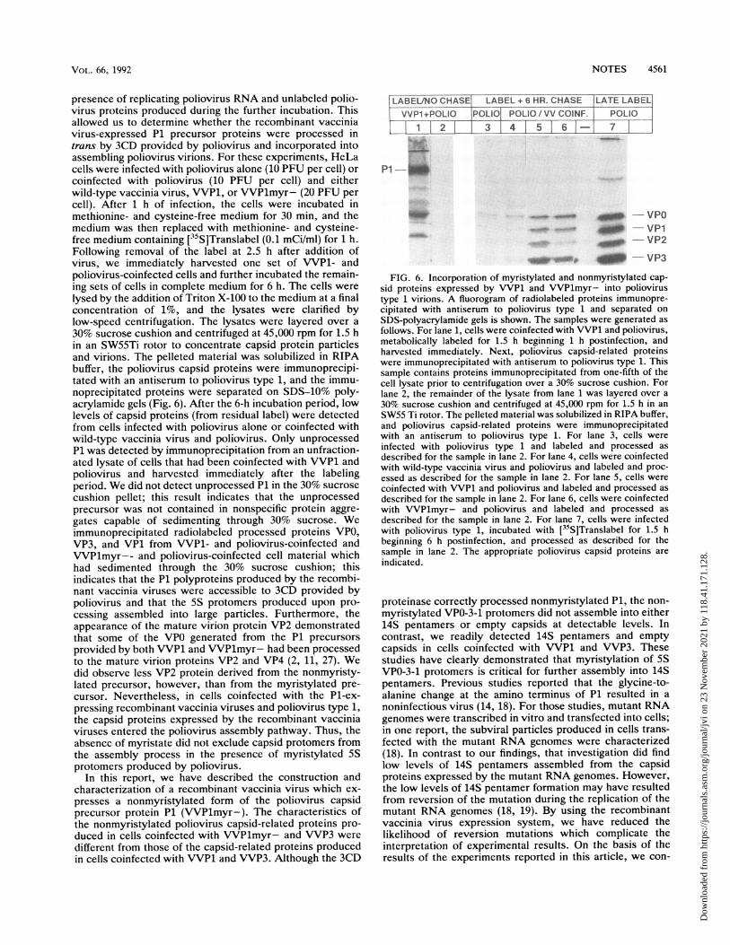
presence of replicating poliovirus RNA and unlabeled polio-virus proteins produced during the further incubation. Thisallowed us to determine whether the recombinant vacciniavirus-expressed P1 precursor proteins were processed intrans by 3CD provided by poliovirus and incorporated intoassembling poliovirus virions. For these experiments, HeLacells were infected with poliovirus alone (10 PFU per cell) orcoinfected with poliovirus (10 PFU per cell) and eitherwild-type vaccinia virus, VVP1, or VVPlmyr- (20 PFU percell). After 1 h of infection, the cells were incubated inmethionine- and cysteine-free medium for 30 min, and themedium was then replaced with methionine- and cysteine-free medium containing [35S]Translabel (0.1 mCi/ml) for 1 h.Following removal of the label at 2.5 h after addition ofvirus, we immediately harvested one set of VVP1- andpoliovirus-coinfected cells and further incubated the remain-ing sets of cells in complete medium for 6 h. The cells werelysed by the addition of Triton X-100 to the medium at a finalconcentration of 1%, and the lysates were clarified bylow-speed centrifugation. The lysates were layered over a30% sucrose cushion and centrifuged at 45,000 rpm for 1.5 hin an SW55Ti rotor to concentrate capsid protein particlesand virions. The pelleted material was solubilized in RIPAbuffer, the poliovirus capsid proteins were immunoprecipi-tated with an antiserum to poliovirus type 1, and the immu-noprecipitated proteins were separated on SDS-10% poly-acrylamide gels (Fig. 6). After the 6-h incubation period, lowlevels of capsid proteins (from residual label) were detectedfrom cells infected with poliovirus alone or coinfected withwild-type vaccinia virus and poliovirus. Only unprocessedP1 was detected by immunoprecipitation from an unfraction-ated lysate of cells that had been coinfected with VVP1 andpoliovirus and harvested immediately after the labelingperiod. We did not detect unprocessed P1 in the 30% sucrosecushion pellet; this result indicates that the unprocessedprecursor was not contained in nonspecific protein aggre-gates capable of sedimenting through 30% sucrose. Weimmunoprecipitated radiolabeled processed proteins VPO,VP3, and VP1 from VVP1- and poliovirus-coinfected andVVPlmyr-- and poliovirus-coinfected cell material whichhad sedimented through the 30% sucrose cushion; thisindicates that the P1 polyproteins produced by the recombi-nant vaccinia viruses were accessible to 3CD provided bypoliovirus and that the 5S protomers produced upon pro-cessing assembled into large particles. Furthermore, theappearance of the mature virion protein VP2 demonstratedthat some of the VPO generated from the P1 precursorsprovided by both VVP1 and VVPlmyr- had been processedto the mature virion proteins VP2 and VP4 (2, 11, 27). Wedid observe less VP2 protein derived from the nonmyristy-lated precursor, however, than from the myristylated pre-cursor. Nevertheless, in cells coinfected with the P1-ex-pressing recombinant vaccinia viruses and poliovirus type 1,the capsid proteins expressed by the recombinant vacciniaviruses entered the poliovirus assembly pathway. Thus, theabsence of myristate did not exclude capsid protomers fromthe assembly process in the presence of myristylated 5Sprotomers produced by poliovirus.
In this report, we have described the construction andcharacterization of a recombinant vaccinia virus which ex-presses a nonmyristylated form of the poliovirus capsidprecursor protein P1 (VVPlmyr-). The characteristics ofthe nonmyristylated poliovirus capsid-related proteins pro-duced in cells coinfected with VVPlmyr- and VVP3 were
different from those of the capsid-related proteins producedin cells coinfected with VVP1 and VVP3. Although the 3CD
FIG. 6. Incorporation of myristylated and nonmyristylated cap-sid proteins expressed by VVP1 and VVPlmyr- into poliovirustype 1 virions. A fluorogram of radiolabeled proteins immunopre-cipitated with antiserum to poliovirus type 1 and separated on
SDS-polyacrylamide gels is shown. The samples were generated as
follows. For lane 1, cells were coinfected with VVP1 and poliovirus,metabolically labeled for 1.5 h beginning 1 h postinfection, andharvested immediately. Next, poliovirus capsid-related proteinswere immunoprecipitated with antiserum to poliovirus type 1. Thissample contains proteins immunoprecipitated from one-fifth of thecell lysate prior to centrifugation over a 30% sucrose cushion. Forlane 2, the remainder of the lysate from lane 1 was layered over a
30% sucrose cushion and centrifuged at 45,000 rpm for 1.5 h in an
SW55 Ti rotor. The pelleted material was solubilized in RIPA buffer,and poliovirus capsid-related proteins were immunoprecipitatedwith an antiserum to poliovirus type 1. For lane 3, cells were
infected with poliovirus type 1 and labeled and processed as
described for the sample in lane 2. For lane 4, cells were coinfectedwith wild-type vaccinia virus and poliovirus and labeled and proc-essed as described for the sample in lane 2. For lane 5, cells were
coinfected with VVP1 and poliovirus and labeled and processed as
described for the sample in lane 2. For lane 6, cells were coinfectedwith VVPlmyr- and poliovirus and labeled and processed as
described for the sample in lane 2. For lane 7, cells were infectedwith poliovirus type 1, incubated with [35S]Translabel for 1.5 hbeginning 6 h postinfection, and processed as described for thesample in lane 2. The appropriate poliovirus capsid proteins are
indicated.
proteinase correctly processed nonmyristylated P1, the non-
myristylated VPO-3-1 protomers did not assemble into either14S pentamers or empty capsids at detectable levels. Incontrast, we readily detected 14S pentamers and emptycapsids in cells coinfected with VVP1 and VVP3. Thesestudies have clearly demonstrated that myristylation of 5SVPO-3-1 protomers is critical for further assembly into 14Spentamers. Previous studies reported that the glycine-to-alanine change at the amino terminus of P1 resulted in a
noninfectious virus (14, 18). For those studies, mutant RNAgenomes were transcribed in vitro and transfected into cells;in one report, the subviral particles produced in cells trans-fected with the mutant RNA genomes were characterized(18). In contrast to our findings, that investigation did findlow levels of 14S pentamers assembled from the capsidproteins expressed by the mutant RNA genomes. However,the low levels of 14S pentamer formation may have resultedfrom reversion of the mutation during the replication of themutant RNA genomes (18, 19). By using the recombinantvaccinia virus expression system, we have reduced thelikelihood of reversion mutations which complicate theinterpretation of experimental results. On the basis of theresults of the experiments reported in this article, we con-
VOL. 66, 1992
Dow
nloa
ded
from
http
s://j
ourn
als.
asm
.org
/jour
nal/j
vi o
n 23
Nov
embe
r 20
21 b
y 11
8.41
.171
.128
.
4562 NOTES
clude that myristylation of VPO-3-1 capsid protomers isrequired for stable assembly of 14S pentamers.
In cells coinfected with VVP3 and VVPlmyr-, the 3CDproteinase clearly processed the nonmyristylated P1 precur-
sor to VPO, VP3, and VP1 (Fig. 3). This result contrastedwith earlier observations that myristylation of the P1 precur-
sor was required for efficient proteolytic processing in vitro,especially at the VPO-3 cleavage site (14, 17). We believe thatthis discrepancy can be explained in part by the differencesbetween the in vitro translation and recombinant vacciniavirus expression systems used for the production of the viralcapsid proteins. The recombinant vaccinia virus systemprovides the advantage of high-level expression of P1 and3CD in the appropriate intracellular environment requiredfor the complete proteolytic processing of the nonmyristy-lated P1 protein.We noted during several experiments that VPO, VP3, and
VP1 generated from nonmyristylated P1 were more unstablein cells than VPO, VP3, and VP1 generated from myristy-lated P1. We do not believe that this instability explains thelack of assembly of nonmyristylated 5S capsid protomersinto 14S pentamers and empty capsids. During the sucrose
gradient experiments, we immunoprecipitated VPO, VP3,and VP1 from aliquots of unfractionated extracts of cellscoinfected with VVPlmyr- and VVP3 as well as from theupper fractions of the gradients, suggesting that sufficientpools of nonmyristylated 5S protomers were available forassembly into subviral particles (Fig. 4 and 5). The majorityof the processed proteins generated in VVP1- and VVP3-coinfected cells over a 2-h metabolic labeling period sedi-mented at a rate of 14S, indicating that the assembly of VPO,VP3, and VP1 derived from myristylated P1 occurred rapidly(Fig. 5B). The instability of the nonmyristylated P1 productsmay be a result of their inability to assemble. In support ofthis idea, Macadam et. al. have recently reported that theassembly-defective 5S protomers synthesized by a temper-
ature-sensitive mutant of poliovirus type 3 were unstablewhen not assembled into subviral particles at the nonpermis-sive temperature (16). These results suggest that the assem-
bly-defective 5S protomers are targeted for degradation.Interestingly, emerging concepts about cellular chaperoneproteins suggest that cells have mechanisms for recognizingproteins that are not yet maturely folded (3, 15, 22, 26). Sinceprocessed but unassembled poliovirus capsid proteins are
not mature structurally, those that cannot assemble or reachtheir final conformational state may be targeted into a
cellular degradation pathway. Further experiments are un-
derway to explore this possibility.The independent expression of P1 by recombinant vac-
cinia viruses provided the opportunity to study the interac-tion of nonmyristylated P1 with the poliovirus assemblycomplexes in cells coinfected with VVPlmyr- and poliovi-rus. Since we had observed that unmyristylated 5S pro-
tomers did not assemble into 14S pentamers in cells coin-fected with VVPlmyr- and VVP3, we were surprised to findthat, in cells coinfected with VVPlmyr- and poliovirus, thenonmyristylated protomers were present in large particlesthat sedimented through 30% sucrose cushions. This resultsuggested that the nonmyristylated capsid protomers were
still capable of making the protomer-protomer contacts
required for assembly when the stabilizing forces of myris-tate were provided by other myristylated protomers within a
common pentamer subunit. Furthermore, the nonmyristy-lated P1 precursor and nonmyristylated VPO-3-1 protomers
were not excluded from sites of virion assembly. We didnote that less of the nonmyristylated precursor P1 protein
than the myristylated P1 protein was chased into the maturevirion protein VP2. This observation might reflect an ineffi-cient incorporation of the nonmyristylated VPO-3-1 pro-tomers into the assembly pathway or a selection against theinclusion of capsid intermediates composed of a mixture ofmyristylated and nonmyristylated subunits during the finalstages of virion formation. The second explanation is inagreement with previous studies which reported an enrich-ment for myristylated capsid subunits during the formationof mature poliovirus virions in cells infected with mutantviruses that expressed a mixture of myristylated and nonmy-ristylated capsid subunits (20). Another possibility might bethat nonmyristylated VPO, and thus subsequently VP2 andVP4 upon proteolytic cleavage of VPO after RNA encapsi-dation, was selectively excluded from entering the assemblypathway, whereas VP1 and VP3 generated from the pro-cessing of nonmyristylated P1 were still included. Thispossibility seems unlikely, however, given that VPO, VP3,and VP1 remain tightly associated as a 5S protomer capsidsubunit (4). The reduced appearance of VP2 might also bethe result of an inefficient maturation cleavage of nonmy-ristylated VPO to VP2 and VP4. Further studies are requiredto address these possibilities.
In summary, the experimental results discussed in thisreport demonstrate that myristylation of the poliovirus cap-sid precursor protein P1 and the individual capsid proteinVPO is critical for the efficient assembly of poliovirus subvi-ral particles. Furthermore, the experiments presented dem-onstrate the utility of the recombinant vaccinia virus expres-sion system for studying the poliovirus assembly process.Although the first steps in the poliovirus assembly pathwayinvolving the proteolytic processing of P1 to VPO, VP3, andVP1 by the 3CD proteinase occur when P1 is unmyristy-lated, the subsequent assembly of these processed proteinsinto 14S pentamers or 75S empty capsids is prevented.Furthermore, myristylation is not required for the intracel-lular localization of 5S capsid protomers to sites of virusassembly, and the conformation of nonmyristylated 5S cap-sid protomers appears to be compatible with assembly in thepresence of excess myristylated protomers expressed bypoliovirus. Since nonmyristylated capsid protomers, whennot in the presence of myristylated protomers, do notefficiently associate into 14S pentamers, myristylation islikely to be critical for stabilization of the 14S pentamer. Astabilization function for the myristate moieties, which areclustered near the contact area between the five protomersubunits that form a pentamer, is completely compatiblewith information given by the crystal structure of the virus(8).
We thank L. Andrew Ball for reading the manuscript and forhelpful comments.We acknowledge the support of the UAB Comprehensive Cancer
Center core services for the synthesis of DNA oligonucleotides.D.C.A. is supported by NIH training grant AI 07150. This study wassupported by Public Health Services grant Al 25005 from theNational Institutes of Health to C.D.M.
REFERENCES1. Ansardi, D. C., D. C. Porter, and C. D. Morrow. 1991. Coin-
fection with recombinant vaccinia viruses expressing poliovirusP1 and P3 proteins results in polyprotein processing and forma-tion of empty capsid structures. J. Virol. 65:2088-2092.
2. Arnold, E., M. Luo, G. Vriend, M. G. Rossman, A. C. Palmen-berg, G. D. Parks, M. J. H. Nicklin, and E. Wimmer. 1987.Implications of the picornavirus capsid structure for polyproteinprocessing. Proc. Natl. Acad. Sci. USA 84:21-25.
3. Beckmann, R. P., L. A. Mizzen, and W. J. Welch. 1990.
J. VIROL.
Dow
nloa
ded
from
http
s://j
ourn
als.
asm
.org
/jour
nal/j
vi o
n 23
Nov
embe
r 20
21 b
y 11
8.41
.171
.128
.
NOTES 4563
Interaction of hsp70 with newly synthesized proteins: implica-tions for protein folding and assembly. Science 248:850-854.
4. Bruneau, P., B. Blondel, R. Crainic, F. Horodniceanu, and M.Girard. 1983. Poliovirus type 1 capsid polypeptides: absence ofa free form in the cytoplasm of infected HeLa cells. Ann. Virol.Inst. Pasteur 134E:151-164.
5. Bryant, M., and L. Ratner. 1990. Myristoylation-dependentreplication and assembly of human immunodeficiency virus 1.Proc. Natl. Acad. Sci. USA 87:523-527.
6. Buss, J. E., P. A. Solski, J. P. Schaeffer, M. J. MacDonald, andC. J. Der. 1989. Activation of the cellular proto-oncogeneproduct p2lras by addition of a myristylation signal. Science243:1600-1603.
7. Chakrabarti, S., K. Brechling, and B. Moss. 1985. Vaccinia virusexpression vector: coexpression of I-galactosidase providesvisual screening of recombinant virus plaques. Mol. Cell. Biol.5:3403-3409.
8. Chow, M., J. F. E. Newman, D. J. Filman, J. M. Hogle, D. J.Rowlands, and F. Brown. 1987. Myristylation of picornaviruscapsid protein VP4 and its structural significance. Nature (Lon-don) 327:482-486.
9. Hanecak, R., B. L. Semler, C. W. Anderson, and E. Wimmer.1982. Proteolytic processing of poliovirus polypeptides: anti-bodies to polypeptide P3-7c inhibit cleavage of glutamine-glycine pairs. Proc. Natl. Acad. Sci. USA 79:3973-3977.
10. Heuckeroth, R. O., and J. I. Gordon. 1989. Altered membraneassociation of p60 v-src and a murine 63-kDa N-myristoylpro-tein after incorporation of an oxygen-substituted analog ofmyristic acid. Proc. Natl. Acad. Sci. USA 86:5262-5266.
11. Jacobsen, M. F., J. Asso, and D. Baltimore. 1970. Furtherevidence on the formation of poliovirus proteins. J. Mol. Biol.49:657-669.
12. Jewell, J. E., L. A. Ball, and R. Rueckert. 1990. Limitedexpression of poliovirus by vaccinia virus recombinants due toinhibition of the vector by proteinase 2A. J. Virol. 64:1388-1393.
13. Johnson, D. R., A. D. Cox, P. A. Solski, B. Devadas, S. P.Adams, R. M. Leimgruber, R. 0. Heuckeroth, J. E. Buss, andJ. I. Gordon. 1990. Functional analysis of protein N-myristoyl-ation: metabolic labeling studies using three oxygen-substitutedanalogs of myristic acid and cultured mammalian cells provideevidence for protein-sequence-specific incorporation and ana-log-specific redistribution. Proc. Natl. Acad. Sci. USA 87:8511-8515.
14. Krausslich, H.-G., C. Holscher, Q. Reuer, J. Harber, and E.Wimmer. 1990. Myristoylation of the poliovirus polyprotein isrequired for proteolytic processing of the capsid and for viralinfectivity. J. Virol. 64:2433-2436.
15. Lindquist, S., and E. A. Craig. 1988. The heat-shock proteins.Annu. Rev. Genet. 22:631-677.
16. Macadam, A. J., G. Ferguson, C. Arnold, and P. D. Minor. 1991.An assembly defect as a result of an attenuating mutation in thecapsid proteins of the poliovirus type 3 vaccine strain. J. Virol.65:5225-5231.
17. Marc, D., G. Drugeon, A. L. Haenni, M. Girard, and S. van der
Werf. 1989. Role of myristoylation of poliovirus capsid proteinVP4 as determined by site directed mutagenesis of its N-termi-nal sequence. EMBO J. 8:2661-2668.
18. Marc, D., M. Girard, and S. van der Werf. 1991. A Gly' to Alasubstitution in poliovirus capsid protein VPO blocks its myris-toylation and prevents viral assembly. J. Gen. Virol. 72:1151-1157.
19. Marc, D., G. Masson, M. Girard, and S. van der Werf. 1990.Lack of myristoylation of poliovirus capsid polypeptide VPOprevents the formation of virions or results in the assembly ofnoninfectious virus particles. J. Virol. 64:4099-4107.
20. Moscufo, N., J. Simons, and M. Chow. 1991. Myristoylation isimportant at multiple stages in poliovirus assembly. J. Virol.65:2372-2380.
21. Paul, A. V., A. Schultz, S. E. Pincus, S. Oroszlan, and E.Wimmer. 1987. Capsid protein VP4 of poliovirus is N-myristoyl-ated. Proc. Natl. Acad. Sci. USA 84:7827-7831.
22. Pelham, H. 1988. Heat shock proteins: coming in from the cold.Nature (London) 332:776-777.
23. Porter, D. C., M. Lentz, and C. D. Morrow. Unpublished data.24. Putnak, J. R., and B. A. Phillips. 1981. Picornaviral structure
and assembly. Microbiol. Rev. 45:287-315.25. Rhee, S. S., and E. Hunter. 1987. Myristylation is required for
intracellular transport but not for assembly of D-type retroviruscapsids. J. Virol. 61:1045-1053.
26. Rothman, J. E. 1989. Polypeptide chain binding proteins: cata-lysts of protein folding and related processes in cells. Cell59:591-601.
27. Rueckert, R. R. 1990. Picornaviridae and their replication, p.507-548. In B. N. Fields et al. (ed.), Virology, 2nd ed. RavenPress, New York.
28. Schultz, A. M., L. E. Henderson, and S. Oroszlan. 1988. Fattyacylation of proteins. Annu. Rev. Cell Biol. 4:611-647.
29. Schultz, A. M., and A. Rein. 1989. Unmyristylated Moloneymurine leukemia virus Pr65Iag is excluded from virus assemblyand maturation events. J. Virol. 63:2370-2373.
30. Towler, D. A., S. P. Adams, S. R. Eubanks, D. S. Towery, E.Jackson-Machelky, L. Glaser, and J. I. Gordon. 1987. Purifica-tion and characterization of yeast myristoyl CoA:proteinN-myristoyltransferase. Proc. Natl. Acad. Sci. USA 84:2708-2712.
31. Towler, D. A., J. I. Gordon, S. P. Adams, and L. Glaser. 1988.The biology and enzymology of eukaryotic protein acylation.Annu. Rev. Biochem. 57:69-99.
32. Toyoda, H., M. J. H. Nicklin, M. G. Murray, C. W. Anderson,J. J. Dunn, F. W. Studier, and E. Wimmer. 1986. A secondvirus-encoded proteinase involved in proteolytic processing ofpoliovirus polyprotein. Cell 45:761-770.
33. Wilcox, C., J. S. Hu, and E. N. Olson. 1987. Acylation ofproteins with myristic acid occurs cotranslationally. Science238:1275-1278.
34. Ypma-Wong, M. F., P. G. Dewalt, V. H. Johnson, J. G. Lamb,and B. L. Semler. 1988. Protein 3CD is the major poliovirusproteinase responsible for cleavage of the P1 capsid precursor.Virology 166:265-270.