20
New tools for improved mass spectrometry results Sample preparation, protein quantitation, and instrument calibration for proteomics research
New tools for improved mass spectrometry resultsSample preparation, protein quantitation, and instrument calibration for proteomics research
We offer a complete portfolio of sample preparation, protein
quantitation, and instrument calibration reagents designed for
better mass spectrometry (MS) analysis. This portfolio has been
developed around the context of biology, and the newest products
include improvements to sample preparation (abundant protein
depletion spin columns, MS-cleavable crosslinkers, peptide
desalting spin columns) and higher multiplexing capabilities for
protein quantitation (Thermo Scientific™ NeuCode™ stable isotope
labeling with amino acids in cell culture (SILAC™) amino acids
and Tandem Mass Tag 11-plex (TMT11plex™) isobaric labeling
reagents). In addition, we have developed a new Thermo Scientific™
Pierce™ Intact Protein Standard Mixture as a tool for top-down
proteomics applications.
We recognize the need to provide complete solutions and technical
support for proteomics research and analytical analysis using
MS instrumentation. These new reagents have been verified to
assist the biologist and the mass spectrometrist to succeed in
their research. Robust, integrated workflows provide consistent
results between labs and eliminate time wasted on troubleshooting
experimental methods and results.
Introduction

Introduction 2
Workflows 4
Sample preparation 5High-Select HSA/Immunoglobulin and Top14 5 Abundant Protein Depletion Spin Columns and Resin
Thermo Scientific MS-cleavable crosslinkers 7
Pierce Peptide Desalting Spin Columns 9
Protein quantitation 11SILAC metabolic labeling kits and 11 NeuCode amino acids
Isobaric amine-reactive tandem mass tag 14 labeling reagents
Instrument calibration 17Pierce Intact Protein Standard Mix 17
Ordering information 19
Contents

4
Cells
Abundant proteindepletion
Lysis andprotein extraction
Metabolic labeling
Protein cleanup
Immunoprecipitation, crosslinking, or active-site labeling and/or enrichment
Protein quantitationassays
Gel separation
In-gel digestionPeptide or
glycan labeling
Peptide enrichmentand/or fractionation
HeavyPeptideAQUA standards
Peptide cleanup
Peptide quantitationassays
Reagents for MS instrumentcalibration, QC, and analysis
HPLC instrumentation and columns
MS instrumentation Software
In-solution digestion
TissuesSerum, plasma, and bio�uids
Workflows

5
Deplete abundant plasma proteins quickly and economically
Thermo Scientific™ High-Select™ Abundant Protein Depletion Spin Columns and resins are optimized to decrease the abundant albumin and antibody components of human plasma samples in preparation for MS. Thermo Scientific™ High-Select HSA/Immunoglobulin Depletion Resin uses highly specific, immobilized antibodies to remove HSA (Human Serum Albumin) and all major subclasses of immunoglobulin from serum and plasma. Similarly, the Thermo Scientific™ High-Select Top14 Abundant Protein Depletion Spin Columns are designed to remove HSA, IgG, and 12 other high-abundance proteins from human serum or plasma (Table 1). The resins are provided in two different convenient spin column formats or as bulk resin.
Highlights• Optimized—resin is scaled and optimized for treating
human plasma samples for MS and/or 1D and2D electrophoresis
• Effic ent—resins are designed to remove >90% of IgGand >95% of albumin, plus other abundant proteins(up to 12)
• Fast—spin columns process samples in 5–10 min
• Economical—cost-effective spin columns are pricedfor single use and provide convenience or flexibility withbulk resins
• Consistent—one-time use prevents abundant proteincarryover and experimental variability
• Flexible—choose the system appropriate for your need:HSA/immunoglobulin-specifi or top 14 abundant proteindepletion columns
Analysis of human serum is hindered by the presence of high concentrations of albumin and IgG that can account for more than 70% of the total protein present in the sample. Removal of these and other high-abundance proteins is often essential for the study of low-abundance proteins of biological interest by MS or 1D and 2D gel electrophoresis. Traditionally, researchers have depleted abundant proteins from samples using lengthy and tedious chromatography methods involving multiple purification, often resulting in low protein yields and poor reproducibility.Table 1. Proteins removed by High-Select Abundant Protein
Depletion Spin Columns. Binding and removal of proteins is achieved via specific antibodies, which are immobilized on the affinity support.
HSA/Immunoglobulin Top14 columns
• Albumin • Albumin • α1-Acid glycoprotein
• IgG • IgG • Fibrinogen
• IgA • IgA • Haptoglobin
• IgM • IgM • α1-Antitrypsin
• IgD • IgD • α2-Macroglobulin
• IgE • IgE • Transferrin
• IgG (light chains) • IgG (light chains) • Apolipoprotein A-I
Sample preparationHigh-Select HSA/Immunoglobulin and Top14 Abundant Protein Depletion Spin Columns and resin

6
Figure 3. Abundant protein depletion improves identifi ation of unique proteins. Protein group identification profiles for normal human plasma samples which were (a) not depleted, or depleted using the HSA/Immunoglobulin (b) and Top14 (c) depletion resins, are shown. All samples were reduced/alkylated and digested with trypsin. Samples were analyzed by LC-MS on an Orbitrap Fusion™ Tribrid™ mass spectrometer over a 60 minute gradient using 750 ng of sample per injection (performed in triplicate). Raw files were searched against a human protein database using Thermo Scientific™ Proteome Discoverer™ 1.4 software. Non-redundant protein group identification numbers are reported for each sample type.
Figure 2. Performance of High-Select Abundant Protein Depletion Spin Columns. Human serum (10–20 µL, Cat. No. PI31876) was loaded onto each resin and processed according to protocols. Total protein in the depleted fractions was estimated using the Pierce BCA Protein Assay Kit (Fisher Scientific, Cat. No. PI23225). The total amount of albumin in the depleted fractions was determined using the AssayMax™ Human Albumin ELISA Kit (Assaypro, Cat. No. EA2201-1). FT = flow-through (i.e., depleted sample); E = eluate (i.e., proteins that were bound by the resin, plus stripped affinity antibodies of the column). With the top 14 proteins removed, low-abundance proteins are now visible in each depleted sample lane (FT).
The High-Select HSA/Immunoglobulin Depletion Columns can deplete >95% of albumin and immunoglobulins from human serum, while the High-Select Top14 Abundant Protein Depletion Columns remove >95% of the 14 most abundant proteins (Figures 1 and 2). The removal of these high-adundance proteins enables better detection of unique proteins (Figure 3). Each prefilled depletion column can process 10 µL (mini) or 100 µL (midi) of human serum in 5–10 min using a convenient spin format compatible with low-speed centrifugation.
Learn more at thermofisher.com/msdepletion
Figure 1. Protein removal achieved using High-Select Abundant Protein Depletion Spin Columns. (A) Values were determined by targeted MS. (B) The abundant protein depletion percentage was confirmed by ELISA and was in agreement with >99% removal. Immunoglobulins include IgG (KappaXL and Lambda), IgA, IgM, IgD, and IgE.
A
B
Percent depletion
Protein HSA/Immunoglobulin
Albumin 99.30
Immunoglobulins 99.62
80
60
40
20
0
100
100
100
100
100
99 100
100
96
100
100
100
99 99 99 99100
100
100
Albumin
Imm
unog
lobuli
ns
Acid g
lycop
rote
in
Antitr
ypsin
Mac
roglo
bulin
Apolipop
rote
in A1
Fibrin
ogen
Hapto
globin
Trans
ferrin
Dep
letio
n (%
)
ELISA data
MS analysisHigh Select Top14 Abundant Protein Depletion Resin
High SelectHSA/Immunoglobulin
M FT ESer
um
Ser
um
FT E
High SelectTop14
Additional proteins identi�ed after Top14 depletion
Proteins identi�ed after HSA/Immunoglobulin depletion
Proteins common with undepleted (abundant proteins)
0
50
100
150
200
250
300
Undep
leted
HSA/Imm
unoglo
bulin
Top1
4

7
High-quality reagents in convenient packaging for protein characterization studies
Thermo Scientific™ DSSO (disuccinimidyl sulfoxide) and DSBU (disuccinimidyl dibutyric urea, also known as BuUrBu [Note: BuUrBu stands for dibutyric urea]) are high-quality MS-cleavable crosslinkers that contain an amine-reactive N-hydroxysuccinimide (NHS) ester at each end of a 7-atom and 11-atom spacer arm (Figure 4). These products are offered in convenient single-use packaging (10 x 1 mg).
Chemical crosslinking in combination with mass spectrometry is a powerful method to characterize proteins. This method has been applied to recombinant and native protein complexes and, more recently, to whole-cell lysates or intact unicellular organisms in efforts to identify protein–protein interactions on a global scale.
Highlights• High quality—products manufactured in
ISO 9001–certified facilities
• Convenience—reagents available in Thermo Scientific™
Pierce™ No-Weigh™ format
• Cleavable—enable distinctive fragmentation patterns forMS identifi ation
• MS-verified—tested utilizing different types offragmentation patterns on Thermo Scientific™
mass spectrometers
DSSO (disuccinimidyl sulfoxide)Molecular weight: 388.35Spacer arm: 10.3 Å
DSBU (disuccinimidyl dibutyric urea)Molecular weight: 426.38Spacer arm: 12.5 Å
A B
Figure 4. Chemical structures of crosslinkers: (A) DSSO and (B) DSBU.
MS-cleavable crosslinkers

8
Learn more at thermofisher.com/mscrosslinkers
Figure 5. Spectra of BSA-crosslinked peptide identified by MS2/MS3 method and XLinkX software* using (A) DSSO and (B) DSBU crosslinkers. XLinkX software uses unique fragment patterns of MS-cleavable crosslinkers (purple annotation) to detect and filter crosslinked peptides for a database search.
* Licensed from the Heck group, University of Utrecht, The Netherlands.
A BDSSO DSBU
Both traditional noncleavable and MS-cleavable crosslinkers provide insight into the identifi ation of protein–protein interaction sites, but MS-cleavable crosslinkers are advantageous due to their ability to be cleaved using different types of gas-phase fragmentation methods (e.g., CID, HCD, ETD, and EtHCD) and levels of tandem mass spectrometry (MS2 and MS3), improving identification of protein–protein interaction sites (Figure 5).
Features of DSSO and DSBU• Amine-reactive NHS ester (at both ends) reacts rapidly
with any molecule containing a primary amine
• Membrane-permeant, allowing forintracellular crosslinking
• High-purity crystalline reagent can be used to createhigh-purity conjugates
• MS-cleavable
• Water-insoluble (dissolve first in DMF or DMSO)
9
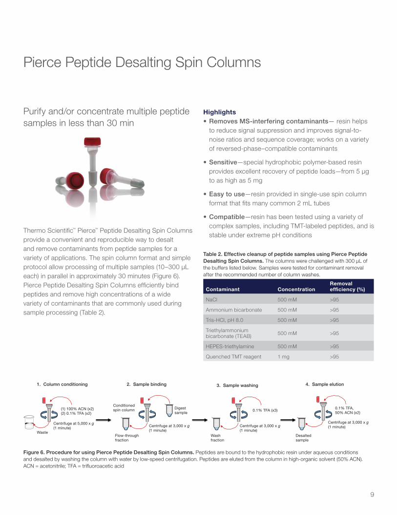
Purify and/or concentrate multiple peptide samples in less than 30 min
Thermo Scientific™ Pierce™ Peptide Desalting Spin Columns provide a convenient and reproducible way to desalt and remove contaminants from peptide samples for a variety of applications. The spin column format and simple protocol allow processing of multiple samples (10–300 µL each) in parallel in approximately 30 minutes (Figure 6). Pierce Peptide Desalting Spin Columns efficiently bind peptides and remove high concentrations of a wide variety of contaminants that are commonly used during sample processing (Table 2).
Highlights• Removes MS-interfering contaminants— resin helps
to reduce signal suppression and improves signal-to-noise ratios and sequence coverage; works on a varietyof reversed-phase–compatible contaminants
• Sensitive—special hydrophobic polymer-based resinprovides excellent recovery of peptide loads—from 5 µgto as high as 5 mg
• Easy to use—resin provided in single-use spin columnformat that fits many common 2 mL tubes
• Compatible—resin has been tested using a variety ofcomplex samples, including TMT-labeled peptides, and isstable under extreme pH conditions
Pierce Peptide Desalting Spin Columns
1. Column conditioning
(1) 100% ACN (x2)(2) 0.1% TFA (x2)
Conditioned spin column Digest
sample0.1% TFA (x3) 0.1% TFA,
50% ACN (x2)
Centrifuge at 5,000 x g(1 minute) Centrifuge at 3,000 x g
(1 minute)Centrifuge at 3,000 x g(1 minute)
Centrifuge at 3,000 x g(1 minute)
Flow-through fraction
Wash fraction
Desalted sample
Waste
2. Sample binding 3. Sample washing 4. Sample elution
Figure 6. Procedure for using Pierce Peptide Desalting Spin Columns. Peptides are bound to the hydrophobic resin under aqueous conditions and desalted by washing the column with water by low-speed centrifugation. Peptides are eluted from the column in high-organic solvent (50% ACN). ACN = acetonitrile; TFA = trifluoroacetic acid
Table 2. Effective cleanup of peptide samples using Pierce Peptide Desalting Spin Columns. The columns were challenged with 300 µL of the buffers listed below. Samples were tested for contaminant removal after the recommended number of column washes.
Contaminant ConcentrationRemoval efficiency (%)
NaCl 500 mM >95
Ammonium bicarbonate 500 mM >95
Tris-HCl, pH 8.0 500 mM >95
Triethylammonium bicarbonate (TEAB) 500 mM >95
HEPES-triethylamine 500 mM >95
Quenched TMT reagent 1 mg >95

10
Traditionally, after isolation of peptides, salts and buffers are removed using reversed-phase (RP) resins, which capture the hydrophobic peptides. The peptides bind to RP columns in high-aqueous mobile phase; salts and buffers are washed off; and the peptides are eluted using a high-organic mobile phase. Pipette tips containing C18 material are the commonly used format for salt removal. This tip design requires many repetitive pipette steps, and sample volumes are limited to 10–20 µL with binding capacities up to 5 µg. The Pierce Peptide Desalting Spin Columns contain an alternative resin and provide a convenient and reproducible way to desalt and remove contaminants from peptide samples up to 300 µL and up to 5 mg of peptides. The peptide spin columns perform similar to comparable C18 columns but with greater flexibility (Figure 7). Each spin column can bind 5 µg to 5 mg of native and/or TMT-labeled peptides, providing greater flexibility for larger peptide samples (Figures 8 and 9).
Pierce Peptide Desalting Spin Column
Harvard Apparatus, C18 Macro SpinColumn
Peptide recovery (%)
CV
100806040200
4.8
7.9
Maximum load of 5 mg
Maximum load of 300 µg
Figure 7. Pierce Peptide Desalting Spin Columns enable efficient recovery of desalted peptide samples. The polymer-based hydrophobic resin contained in each column provides excellent binding and recovery characteristics for peptide samples in preparation for MS and other methods, compared to equivalent products from other suppliers. Digested HeLa extract (300 µg or 5 mg) was loaded onto each column and processed according to the supplier’s protocol, using the maximum recommended load capacity (Harvard Apparatus, Cat. No. 74-4107). Total desalted peptide recovered was estimated using the Thermo Scientific™ Pierce™ Quantitative Colorimetric Peptide Assay (Fisher Scientific, Cat. No. PI23275).
100
80
60
40
20
0Native peptides TMT-labeled peptides
Pep
tid
e re
cove
ry (%
)
Figure 9. Peptide recovery of native and TMT-labeled peptides. Pierce Peptide Desalting Spin Columns provide excellent binding and recovery characteristics for native and TMT-labeled peptide samples in preparation for MS. The columns effectively remove excess unreactive TMT label along with contaminating salts. Digested HeLa extract (500 µg) was labeled with TMT isobaric tags and loaded onto a spin column and processed according to the protocol. Total desalted peptide recovered was estimated using the Pierce Quantitative Colorimetric Peptide Assay (Fisher Scientific, Cat. No. PI23275).
Figure 8. Peptide recovery of native peptides at various loads using Pierce Peptide Desalting Spin Columns. Digested HeLa extracts (5–5,000 µg) were loaded onto Pierce Peptide Desalting Spin Columns and processed according to the protocol. Total recovered desalted peptides were estimated using the Pierce Quantitative Colorimetric Peptide Assay (Fisher Scientific, Cat. No. PI23275).
100
80
60
40
20
05
Pep
tid
e re
cove
ry (%
)50
Peptide load (µg)
500 5,000
Learn more at thermofisher.com/peptidecleanup

11
Complete kits for stable isotope labeling with amino acids in cell culture (SILAC)
SILAC is a powerful method to identify and quantify relative differential changes in complex protein samples. The SILAC method uses metabolic incorporation of “heavy” 13C- or 15N-labeled amino acids into proteins followed by MS analysis for accelerated, comprehensive identification, characterization, and quantitation of proteins (Figure 10).
Highlights• Effic ent—100% label incorporation into proteins of
living cells
• Reproducible—minimizes intra-experiment variability caused by differential sample preparation
• Compatible—label proteins expressed in a wide variety of mammalian cell lines, including HeLa, 293T, COS-7, U2OS, A549, NIH 3T3, Jurkat, and others
• High-quality supplements—heavy amino acids with >98% isotope purity; dialyzed FBS tested to help ensure that it is sterile, endotoxin-free, and cell culture compatible
General applications• Quantitative analysis of relative changes in protein
abundance from different cell treatments
• Quantitative analysis of proteins for which there are no antibodies available
• Protein expression profiling of normal cells vs. abnormal cells
• Identifi ation and quantifi ation of hundreds to thousands of proteins in a single experiment
• Simultaneous immunoprecipitation of labeled, native proteins and protein complexes from multiple conditions
Sample 1
Sample 2
Sample 3 Harvest,lyse,
quantitateprotein
Digest,fractionate,
clean up
Excise bands,digest, clean up
Perform LC-MS/MSanalysis
Mix lysates
Load gel
In-gel workflow
In-solution workflow
SDS-PAGE
Sample 4
Ratio determination
m/zLight Heavy
Rel
ativ
e in
ten
sity
Figure 10. Procedure summary for MS experiments using Thermo Scientifi ™ SILAC™ reagents. Different cell populations are grown in SILAC cell culture media containing either light (control) or heavy (experimental) amino acids. Normalized protein extracts isolated from cells are combined, reduced, alkylated, and digested overnight. For the in-gel workflow, samples are run on an SDS-PAGE gel, excised, digested, and cleaned up; for the in-solution workflow, samples are digested, fractionated, and cleaned up. Samples are then analyzed by high-resolution Thermo Scientific™ Orbitrap™ LC-MS/MS.
Protein quantitationSILAC metabolic labeling kits and NeuCode amino acids

12
SILAC requires growing mammalian cells in specialized media supplemented with light or heavy forms of essential amino acids, e.g., 12C6 and 13C6 L-lysine, respectively. A typical experiment involves growing one cell population in a medium containing light amino acids (control), while the other population is grown in the presence of heavy amino acids (experimental). The heavy and light amino acids are incorporated into proteins through natural cellular protein synthesis. After alteration of the proteome in one sample through chemical treatment or genetic manipulation, equal amounts of protein from both cell populations are then combined, separated by SDS-PAGE, and digested with trypsin before MS analysis. Because peptides labeled with heavy and light amino acids are chemically identical, they coelute during reversed-phase column prefractionation and are detected simultaneously during MS analysis. The relative peak intensities of multiple, isotopically distinct peptides from each protein are then used to determine the average change in protein abundance in the treated sample.
Multiple SILAC kits are available, providing media that are compatible with different mammalian cell lines. Each kit includes all necessary reagents to isotopically label cells, including media, heavy and light amino acid pairs, and dialyzed serum. A wide range of isotopes of lysine and arginine are available separately (Table 3), enabling multiplex experiments and analysis. Dialyzed FBS and other stand-alone media are also available for additional mammalian cell lines. When combined with Thermo Scientific™ protein or peptide enrichment kits, the Thermo Scientific™ SILAC™ Protein Quantitation Kits enable MS analysis of low-abundance proteins such as cell surface proteins, organelle-specific proteins, and proteins with posttranslational modifications such as phosphorylation or glycosylation.
NeuCode amino acids
Thermo Scientific™ NeuCode™ amino acids augment the level of multiplexing achievable in metabolic labeling of proteins for MS analysis. NeuCode metabolic labeling is similar to SILAC but differs in that the labeling only utilizes heavy amino acids. The increased multiplexing capability of NeuCode amino acids is possible through the use of mass defects from extra neutrons in the stable isotopes (Tables 3 and 4). These small mass differences may be resolved on high-resolution mass spectrometers (Thermo Scientific™ Orbitrap Elite™, Orbitrap Fusion™ Tribrid™, and Orbitrap Fusion™ Lumos™ Tribrid™ mass spectrometers). Use of only heavy amino acids eliminates the need for 100% incorporation of amino acids used for SILAC (both heavy and light) and may be especially useful for studies with primary cells.
NeuCode amino acids have the same nominal mass and structure but are labeled with different combinations of 2H, 13C, and 15N stable isotopes (Figure 11), which can be resolved using high-resolution MS (Figure 12).
Highlights• Labeling effic ency—100% label incorporation into
proteins of living cells, without toxicity
• Compatibility—may be multiplexed with existing SILAC amino acids
• Time savings—not necessary to label to 100% incorporation if only using heavy amino acids
• High-quality supplements—heavy amino acids with >98% isotope purity

13
Isotopic clusters Example 480K spectra
K602 K080
K602 K440 K521
K602 K341 K080
K080+8
+8
+8
4-plex
3-plex
2-plex
Table 3. SILAC isotopes of amino acids available to enable multiplex experiments and analysis.
Amino acid Light D413C6 D8
13C6 15N2
13C6 15N4
Mass shift 0 Da +4 Da +6 Da +8 Da +8 Da +10 Da
Cat. No.
L-Arginine-HCl PI89989 (50 mg) PI88427 (500 mg) N/A PI88210 (50 mg)
PI88433 (500 mg) N/A N/A PI89990 (50 mg) PI88434 (500 mg)
L-Leucine PI88428 (500 mg) N/A PI88435 (50 mg) PI88436 (500 mg) N/A N/A
L-Lysine-2HCl PI89987 (50 mg) PI88429 (500 mg)
PI88437 (50 mg) PI88438 (500 mg)
PI89988 (50 mg) PI88431 (500 mg)
N/A
PI88209 (50 mg) PI88432 (500 mg) N/A
L-Proline PI88211 (115 mg) PI88430 (500 mg) N/A N/A N/A N/A N/A
Table 4. Thermo Scientific™ NeuCode™ isotopes of L-lysine-2HCl available to enable multiplex experiments and analysis.
+4 Da +8 Da
Amino acid K202 K040 K080 K521 K341 K440 K602
Mass shift 4.00078 4.02511 8.01420 8.02637 8.03221 8.03853 8.05021
Cat. No. PIA36754 (25 mg) PI88438 (500 mg)PIA36750 (25 mg) PIA33613 (50 mg) PIA33614 (500 mg)
PIA36753 (25 mg)PIA36751 (25 mg) PI88209 (50 mg) PI88432 (500 mg)
H 2 NC C
O HH 2 C
O
C H 2H 2 C
H
C H 2N H 2
K 2 0 2 1 3 C 2 1 5 N 2 L - L y s i n e
*
1 5 0 . 1 0 6 3 D a*
*
*
2 H 4 L - L y s i n e K 0 4 0
H 2 NC
CO H
H 2 C
O
C H 2 H 2 C
H
C H 2N H 2
*
1 5 0 . 1 3 0 6 D a
* **
K 0 8 0 2 H 8 L - L y s i n e
**
H 2 NC
CO H
H 2 C
O
C H 2 H 2 C
H
C H 2 N H 2
*
1 5 4 . 1 5 5 7 D a
* **
* *
K 6 0 2 1 3 C 6 1 5 N 2 L - L y s i n e
H 2 NC C
O HH 2 C
O
C H 2H 2 C
H
C H 2N H 2
*
1 5 4 . 1 1 9 7 D a*
*
**
*
*
*
K 3 4 1 1 3 C 3 2 H 4 1 5 N L - L y s i n e
H 2 NC C
O HH 2 C
O
C H 2H 2 C
H
C H 2 N H 2
*
1 5 4 . 1 3 7 7 D a*
**
**
* *
1 3 C 5 2 H 2 1 5 N 1 L - L y s i n e K 5 2 1
H 2 NC
CO H
H 2 C
O
C H 2H 2 C
H
C H 2 N H 2
1 5 4 . 1 3 1 9 D a*
** *
*
*
**
K 4 4 0 1 3 C 4 2 H 4 L - L y s i n e
**
H 2 NC
CO H
H 2 C
O
C H 2H 2 C
H
C H 2 N H 2
*
1 5 4 . 1 4 4 1 D a
* * *
**
1
Figure 11. Structures and masses of NeuCode amino acids. Stable isotope labels are indicated by red asterisks.
Figure 12. Different combination of lysine isotopologs can be used to increase multiplexing from 2-plex to 4-plex at high resolution.
Learn more at thermofisher.com/silac
PIA33613 (50 mg)PIA33614 (500 mg)
PIA36851 (25 mg) PIA36752 (25 mg)PI88437 (50 mg)

14
Higher multiplex quantitation of up to 11 samples
Thermo Scientific™ Tandem Mass Tag™ (TMT) labeling kits and reagents enable multiplex relative quantitation by MS. All of the mass tagging reagents within a set have the same nominal mass (i.e., isobaric) and chemical structure composed of an amine-reactive NHS ester group, a spacer arm, and a mass reporter (Figure 13). The reagent set can be used to label up to 11 different peptide samples prepared from cells or tissues. For each sample, a unique reporter mass (i.e., 126–131 Da) in the low-mass region of the MS/MS spectrum is used to measure relative protein expression levels during peptide fragmentation.
Previously, we expanded isobaric TMT-labeled multiplexing from 6-plex to 10-plex using high-resolution MS (>50K at m/z 200) to separate 15N and 13C stable isotope variants. Using the same principle, we synthesized the full 13C isotope variant of the TMT-131 reporter, called TMT11-131C. This tag increases isobaric tag multiplex quantitation to 11 samples in a single liquid chromatography (LC)-MS analysis without any changes in reagent structure or LC-MS analysis (Figure 14). The procedure using the TMT11plex reagents is described in Figure 15. To demonstrate the capability of using an 11th tag for relative quantitation, TMT11-131C was used to successfully measure changes in Bacillus subtilis during a protein A/G expression time course showing differential expression of key metabolic proteins over time (Figure 16).
Highlights• Powerful—concurrent MS analysis of multiple samples
increases sample throughput and enables relative quantitation of up to 11 different samples derived from cells, tissues, or biological fluids
• Consistent—identical reagent structure and performance among TMTzero™, TMTduplex™, TMTsixplex™, TMT10plex™, and TMT11plex™ reagents allow efficient transition from method development to multiplex quantitation
• Robust—increased multiplex capability results in fewer missing quantitative values
• Effic ent—amine-reactive, NHS ester–activated reagents enable efficient labeling of all peptides regardless of protein sequence or proteolytic enzyme specificity
• Compatible—optimized for use with high-resolution MS/MS platforms such as Thermo Scientific™ Orbitrap Fusion Lumos, Velos Pro™, Orbitrap Elite™, and Q Exactive™ instruments, with data analysis fully supported by Proteome Discoverer 2.1 software
Applications• Protein identification and quantitation from multiple
samples of cells, tissues, or biological fluids
• Protein expression profiling of normal vs. abnormal states, or control vs. treated cells
• Quantitative analysis of proteins for which no antibodies are available
• Identifi ation and quantifi ation of hundreds to thousands of proteins in a single experiment
NNH
ON
O O
O
O
Massreporter
Massnormalizer
Amine-reactivegroup
HCD
ETD126 Da
Figure 13. Functional regions of the TMT reagent’s chemical structure, including MS/MS sites of fragmentation by HCD and ETD.
Isobaric amine-reactive tandem mass tag labeling reagents

15
Figure 14. Chemical structures of TMT11plex reagents with 13C and 15N heavy-isotope positions (asterisks).
Reporter ion mass
127.1247610
128.1281158
129.1314706
130.1348254
131.1381802
127.1310809
126.1277261
128.1344357
129.1377905
130.1411453
131.144999
TMT10-126
TMT10-127N
TMT10-128N
TMT10-129N
TMT10-130N
Label
TMT10-127C
TMT10-128C
TMT10-129C
TMT10
10
-130C
TMT -131N
11TMT -131C
Sample 1Treat
samplesand isolateproteins Fractionate,
clean up, andperform
LC-MS/MSanalysis
Normalize usingpeptide quantitationassay and combine
Sample 2
Sample 3
Sample 4
Sample 5
Sample 6
Sample 7
Sample 8
Sample 9
Sample 10
Sample 11
400 500 600 700 800 900 1000 1100 1200 1300 1400 1500 1600
m/z
0
5
10
15
20
25
30
35
40
45
50
55
60
65
70
75
80
85
90
95
100631.89 761.41
473.82
525.64 595.38
466.83
637.86
546.84
445.27 875.46
933.49866.69
645.39
701.86 817.95432.28
771.40
1010.81
735.93
1058.50 1168.50 1212.97 1446.861396.78 1542.31
126 127 128 129 130 1310
10
20
30
40
50
60
70
80
90
100
m/z
N C
N C N C
N C
N CMS selection MS/MS quantitation Figure 15. Procedure summary for MS experiments with
TMT11plex isobaric mass tagging reagents. Protein extracts isolated from cells or tissues are reduced, alkylated, and digested overnight. Samples are labeled with the TMT reagents, and then mixed before sample fractionation and cleanup. Labeled samples are analyzed on a high-resolution Orbitrap LC-MS/MS mass spectrometer before data analysis to identify peptides and quantify relative abundance of reporter ions.

16
125.0 125.5 126.0 126.5 127.0 127.5 128.0 128.5 129.0 129.5 130.0 130.5 131.0 131.5 132.0 132.5 133.0
m/z
0
5
10
15
20
25
30
35
40
45
50
55
60
65
70
75
80
85
90
95
100
105
110
Rel
ativ
e ab
und
ance
127.1308
129.1312 128.1279 126.1275 130.1345 131.1379
131.09 131.10 131.11 131.12 131.13 131.14 131.15 131.16 131.17 131.18 131.19 131.20 131.21 0 5
10 15 20 25 30 35 40 45 50 55 60 65 70 75 80 85 90 95
100 105 110 131.1441
R=85802 131.1379 R=85402
Rel
ativ
e ab
und
ance
m/z
Figure 17. Representative TMT reporter ions in the low-mass region of a MS2 spectra from an equimolar (1:1) peptide sample mix. Zoom shows high-resolution separation (60K at m/z 200) of N and C variants of TMT-131 reporter ions.
Learn more at thermofisher.com/tmtreagents
Figure 16. Differential expression of key proteins in Bacillus subtilis expressing recombinant protein A/G over time, as determined by TMT reporter ion quantitation using the Orbitrap Fusion Tribrid mass spectrometer.
126 127N 127C 128N 128C 129N 129C 130N 130C 131N 131C
Quantitation channels
0
500
1000
1500
2000
2500
3000
1079
1475
858
334
995
1909
1434
932
308
956
2085
O34764: Sulfate adenylyltransferase
Ab
un
dan
ce [a
.u.]
Ab
un
dan
ce [a
.u.]
Ab
un
dan
ce [a
.u.]
126 127N 127C 128N 128C 129N 129C 130N 130C 131N 131C
Quantitation channels
0
1000
2000
3000
4000
5000
6000
2352
4179
2576 1758
12841951
4062
2865
1707 1165
1979
P09339: Aconitate hydratase A
126 127N 127C 128N 128C 129N 129C 130N 130C 131N 131C
Quantitation channels
0
500
1000
1500
2000
2500
3000
3500
4000
994 429
554891
2824
348 477
604 849
2680
339
O31645: PTS system, mannose-speci�c EIIBCA component

17
Optimized mixture of recombinant proteins for top-down proteomics applications
The Thermo Scientific™ Pierce™ Intact Protein Standard Mix is an LC-MS–verified, lyophilized mixture of six recombinant proteins that can be used for mass measurement of intact proteins and top-down method development.
Highlights• Wide coverage—mixture of six recombinant proteins
with wide m/z distribution (600–1,500 m/z) and mass range (10–70 kDa)
• Flexible—for use in LC, LC-MS, and direct infusion methods
• Verified sequences—protein sequences confirmed by MS with the average and monoisotopic masses corresponding to each provided sequence
• Recombinant—proteins expressed in E. coli and B. subtilis; no bovine sources
• Stable—provided in a lyophilized format that is stable for 2 years at –20°C
The Pierce Intact Protein Standard Mix is a lyophilized mixture of intact proteins that can be used for qualitative LC, LC-MS, or direct-infusion mass spectrometry experiments. The mixture is specifically formulated for direct-infusion MS experiments and does not contain salts or detergents.
Using the intact protein standard mixture routinely before the analysis of samples makes it possible to monitor and normalize LC-MS performance between samples and over time.
Instrument calibrationPierce Intact Protein Standard Mix

18
Find out more at thermofisher.com/ms-standards
Table 5. Pierce Intact Protein Standard Mix. The theoretical masses include known sequence variants and disulfide bonds. UniProt™ database accession numbers correspond to the original protein sequences.
Protein name Protein accession number(s)
Theoretical average mass (Da)
Theoretical monoisotopic mass (Da)
Human IGF-I LR3* P05019 (49–119) 9,111.47 9,105.34872
Human thioredoxin Q99757 (60–166) 11,865.52 11,858.04393
Streptococcus dysgalactiae protein G P06654 (221–413) 21,442.61 21,429.75915
Bovine carbonic anhydrase II* P00921 28,981.29 28,963.6881
Streptococcus protein AG (chimeric) P02976, P19909 50,459.74 50,429.84641
Escherichia coli Klenow fragment P00582 (324–928) 68,001.15 67,959.42515* Proteins may undergo partial deamidation under acidic conditions.
600 800 1000 1200 1400 1600 m/z
0
20
40
60
80
100
Rel
ativ
e ab
und
ance
872.8 971.3 840.5
990.4 810.5 1010.2
782.6
756.5
1052.2 1122.3
1147.8 748.3
1174.4 709.2
1262.4 1319.4 1442.6 702.0
1577.8 1644.3
895.8
600 800 1000 1200 1400 1600 1800 m/z
0
20
40
60
80
100
Rel
ativ
e ab
und
ance
913.6990 z=13 989.7568
z=12 1139.9210
z=8 848.4353
z=14
1187.5072 z=10 1319.3408
z=9 792.0066 z=15
1484.1324 z=8 766.7810
z=28 1519.5592 z=6 1696.0089
z=7
1013.3748 z=9
A B
C
Mass spectrum17.5K resolution
Mass spectrum140K resolution
Deconvoluted spectrum17.5K resolution
Deconvoluted spectrum140K resolution
D
Figure 18. Representative ESI-MS spectra of Pierce Intact Protein Standard Mix (0.38 µg/µL) reconstituted in a 1:1 (v/v) mixture of 0.1% formic acid in 50% acetonitrile and LC-MS–grade water. The sample was analyzed using a Thermo Scientific™ Q Exactive™ Plus mass spectrometer at 17.5K resolution at m/z 200 (A, B) and 140K resolution at m/z 200 (C, D) in protein mode. Deconvoluted spectra at each resolution were obtained using Thermo Scientific™ BioPharma Finder™ 2.0 software.

19
Ordering information
Product Quantity Cat. No.
Abundant protein depletion
High-Select HSA/Immunoglobulin Depletion Mini Spin Columns6 columns PIA36365
High-Select HSA/Immunoglobulin Depletion Midi Spin Columns
24 columns PIA36366
10 columns PIA36367
High-Select HSA/Immunoglobulin Depletion Resin 50 mL PIA36368
High-Select Top14 Abundant Protein Depletion Mini Spin Columns6 columns PIA36369
High-Select Top14 Abundant Protein Depletion Midi Spin Columns
24 columns PIA36370
10 columns PIA36371
High-Select Top14 Abundant Protein Depletion Resin 50 mL PIA36372
MS-cleavable crosslinkers
DSSO (disuccinimidyl sulfoxide) 10 x 1 mg PIA33545
DSBU (disuccinimidyl dibutyric urea) 10 x 1 mg PIA35459
Peptide cleanup
Pierce Peptide Desalting Spin Columns
Pierce Peptide Desalting Spin Columns
25 columns PI8985250 columns PI89851
Protein quantitation reagents—SILAC
Pierce SILAC Protein Quantitation Kit (Lys-C)—RPMI 1640 1 kit PIA33971
Pierce SILAC Protein Quantitation Kit (Lys-C)—DMEM 1 kit PIA33969
Pierce SILAC Protein Quantitation Kit (Lys-C)—DMEM:F-12 1 kit PIA33970
NeuCode Lysine-080 (3,3,4,4,5,5,6,6-D8 L-Lysine-2HCl) 25 mg PIA36750
NeuCode Lysine-602 (13C6 15N2 L-lysine-2HCl) 25 mg PIA36751
NeuCode Lysine-440 (L-Lysine:2HCl (3,4,5,6-13C4, 5,5,6,6-D4, 98%) 25 mg PIA36752
NeuCode Lysine-521 (L-Lysine:2HCl) 25 mg PIA36753
NeuCode Lysine-341 (13C3 2H4
15N1 L-Lysine-2HCl) 25 mg PIA36851
NeuCode Lysine-202 (13C2 15N2 L-Lysine-2HCl) 25 mg PIA36754
NeuCode 4-plex Bundle (NeuCode Lysine-080, NeuCode Lysine-602, NeuCode Lysine-440, NeuCode Lysine-521) 1 x 25 mg PIA36755
RPMI 1640 Medium for SILAC500 mL PI88365
6 x 500 mL PIA33823
DMEM for SILAC500 mL PI88364
6 x 500 mL PIA33822
DMEM:F-12 (1:1) for SILAC 500 mL PI88370
MEM for SILAC 500 mL PI88368
IMDM for SILAC 500 mL PI88367
Protein quantitation reagents—amine-reactive mass tag reagents
TMT10plex Isobaric Label Reagent Set plus TMT11-131C Label Reagent 1 x 5 mg/tag PIA34808
TMT11-131C Label Reagent 1 x 5 mg PIA34807
Calibration solutions and standards
Pierce Intact Protein Standard Mix 1 x 76 µg PIA33526
5 x 76 µg PIA33527

Protein sample preparation and quantitation for mass spectrometryReagents, consumables, instrumentation, and software for proteomics research
Mass spec rewards program 2017Save money, fi nd out about new products, and improve sample preparation and quantitation
SAVE UP TO
40%
Mass spectrometry digital resources
Mass spec rewards program
Save up to 40% on a single order through the Thermo Scientific™ Mass Spec Rewards Program.* Save even more if you purchase one or more Thermo Scientific™ mass spectrometers in 2017. Learn more on how to make a purchase through this program, obtain a list of eligible products, or register to be contacted by your sales representative.
Learn more at fishersci.com/msrewards
Download the free Thermo Scientific™ Protein Sample Preparation and Quantitation for Mass Spectrometry Handbook and other relevant literature to learn more about tools and techniques for more robust and reproducible outcomes. Access helpful white papers and late-breaking posters for specific applications like subcellular fractionation, peptide fractionation, isobaric labeling, and more. Increase your knowledge of sample preparation and protein quantitation through our free, on-demand webinars.
Register now at thermofisher.com/msresources
* Discount will apply to qualifying orders received by Fisher Scientific channel no later than 12/31/2017. Customer can use the discount only once. Discount only applies to a maximum of 1 order per customer. Discount applies to the list price in effect at the time the order is received by Fisher Scientific channel. Cannot be combined with otherdiscounts or promotions. Offer void where prohibited, licensed, or restricted by federal, state, provincial, or local laws or regulation or agency/institutional policy. Other restrictions may apply.
For Research Use Only. Not for use in diagnostic procedures. © 2017 Thermo Fisher Scientific Inc. All rights reserved. All trademarks are the property of Thermo Fisher Scientific and its subsidiaries unless otherwise specified. TMTzero, TMTduplex, TMTsixplex, TMT11plex, and TMT10plex are trademarks of Proteome Sciences. Tandem Mass Tag is a trademark of Electrophoretics Limited. NeuCode is a trademark of WARF. UniProt is a trademark of the European Molecular Biology Laboratory. BN20170440 0917
Find out more at thermofisher.com/msreagents
In the United States:For customer service, call 1-800-766-7000To fax an order, use 1-800-926-1166To order online: fishersci.com
In Canada:For customer service, call 1-800-234-7437To fax an order, use 1-800-463-2996To order online: fishersci.ca


![1.Set up 110 µl mix for each primer/DNA combo on ice! 1.1.1 µl 100x F primer (1 pMol/µl = 1µM final []) 2.1.1 µl 100x R primer 3.11 µl 10x PCR buffer 4.2.2.](https://static.documents.pub/doc/80x56/56649ce05503460f949aa81d/1set-up-110-l-mix-for-each-primerdna-combo-on-ice-111-l-100x-f-primer.jpg)

