Page 1
N-glycosylation promotes the cell surface expression ofKv1.3 potassium channelsJing Zhu, Jenny Yan* and William B. Thornhill
Department of Biological Sciences and Center for Cancer, Genetic Diseases and Gene Regulation, Fordham University, Bronx, NY, USA
Keywords
cell surface expression; monosaccharide
supplementation; N-glycosylation; potassium
channel; trafficking
Correspondence
W. B. Thornhill, Department of Biological
Sciences, Fordham University, Bronx, NY
10458, USA
Fax: +718 817 3645
Tel: +718 817 3688
E-mail: [email protected]
*Present address
College of Arts and Science, New York
University, New York, NY 10012, USA
(Received 28 February 2012, revised 27
April 2012, accepted 15 May 2012)
doi:10.1111/j.1742-4658.2012.08642.x
The voltage-gated potassium channel Kv1.3 plays an essential role in mod-
ulating membrane excitability in many cell types. Kv1.3 is a heavily gly-
cosylated membrane protein. Two successive N-glycosylation consensus
sites, N228NS and N229ST, are present on the S1–S2 linker of rat Kv1.3.
Our data suggest that Kv1.3 contains only one N-glycan and it is predomi-
nantly attached to N229 in the S1–S2 extracellular linker. Preventing
N-glycosylation of Kv1.3 significantly decreased its surface protein level
and surface conductance density level, which were � 49% and � 46%
respectively of the level of wild type. Supplementation of N-acetylglucos-
amine (GlcNAc), L-fucose or N-acetylneuraminic acid to the culture med-
ium promoted Kv1.3 surface protein expression, whereas supplementation
of D-glucose, D-mannose or D-galactose did not. Among the three effective
monosaccharides ⁄derivatives, adding GlcNAc appeared to reduce sialic
acid content and increase the degree of branching in the N-glycan of
Kv1.3, suggesting that the N-glycan structure and composition had chan-
ged. Furthermore, the cell surface half-life of the Kv1.3 surface protein was
increased upon GlcNAc supplementation, indicating that it had decreased
internalization. The GlcNAc effect appears to apply mainly to membrane
proteins containing complex type N-glycans. Thus, N-glycosylation pro-
motes Kv1.3 cell surface expression; supplementation of GlcNAc increased
Kv1.3 surface protein level and decreased its internalization, presumably
by a combined effect of decreased branch size and increased branching of
the N-glycan.
Introduction
The voltage-gated potassium (Kv) channel Kv1.3 is a
key regulator of various cellular functions in many cell
types. In neurons, Kv1.3 helps set the resting membrane
potential, modulates action potential firing and influ-
ences neurotransmission [1–3]. In T blood cells, Kv1.3
is the major Kv channel regulating the resting mem-
brane potential and calcium signaling [4,5]. Blockage of
Kv1.3 channels prevented T cell activation and attenu-
ated the immune response [6–8]. Kv1.3 has also been
localized to the inner mitochondrial membrane of
lymphocytes, mediating apoptosis [9]. In addition,
Kv1.3 expression was correlated with the proliferation
and apoptosis of colon, prostate and breast cancers
[10–12]. Kv1.3 is becoming a molecular target for treat-
ment of T-cell-mediated autoimmune diseases as well as
different cancers. Thus, understanding the mechanisms
regarding the cellular regulation of Kv1.3 expression
and function is of biological and clinical importance.
Kv1.3 is a heavily glycosylated tetrameric protein [13].
The topology of a subunit reveals six transmembrane
Abbreviations
CAD, cath-a-differentiated; CHO, Chinese hamster ovary; Endo H, endo-b-N-acetylglucosaminidase H; GlcNAc, N-acetylglucosamine;
GLUT, glucose transporter; Kv, voltage-gated potassium; Neu5Ac, N-acetylneuraminic acid; NXT ⁄ NXS, N-glycosylation consensus site;
PNGase F, peptide N-glycosidase F; TbR, transforming growth factor b receptor.
FEBS Journal (2012) ª 2012 The Authors Journal compilation ª 2012 FEBS 1
Page 2
domains, S1–S6, linkers that connect the domains, the
pore region, and cytoplasmic N- and C-termini. An
N-glycan(s) is attached to the S1–S2 extracellular lin-
ker of the channel protein. N-glycosylation is a com-
mon and highly diverse co- or post-translational
modification to proteins. N-glycosylation begins in the
rough endoplasmic reticulum with the addition of an
N-glycan precursor (Glc3Man9GlcNAc2) to the aspara-
gines of the N-glycosylation consensus sequence, or
sequon, NXS ⁄T (where X represents any amino acid
but proline) on the nascent polypeptide. The occu-
pancy rate of an extracellular sequon is � 67% based
on data from 749 well characterized glycoproteins [14].
The N-glycans are then trimmed and modified when
transported through the Golgi apparatus. N-glycans
have diverse compositions and structures due to the
inherent inefficiency of pathway enzymes and avail-
ability of N-glycan biosynthesis substrates. N-glycosyl-
ation can affect not only protein folding, stability,
trafficking and localization, but also functional proper-
ties such as ligand binding, signal recognition and elec-
trophysiological parameters of a variety of proteins
[15–21]. Furthermore, defects in glycosylation of pro-
teins appear to be involved in a group of congenital
multisystemic diseases, also called congenital disorders
of glycosylation, which often cause severe psychomo-
tor retardation [22].
The aim of this paper is to investigate the surface
expression of Kv1.3 by (a) identifying the N-glyco-
sylation site(s), (b) assessing the effect of N-glyco-
sylation on surface protein level and conductance
density level, and (c) examining the effects of mono-
saccharide ⁄derivative concentration on its surface
protein level, protein banding pattern and protein
internalization.
Results
Kv1.3 channels have heterogeneous
N-glycosylation
To assess the N-glycosylation state of Kv1.3
(Fig. 1A,B), the cDNA of rat Kv1.3 was transiently
transfected into Chinese hamster ovary (CHO) cells.
CHO cells are a fibroblast-like line, which do not
express endogenous Kv channels and are an ideal system
for exogenous Kv channel expression [23]. Immunoblot
analysis revealed that Kv1.3 consists of two major
forms, a diffuse upper band with a molecular mass of
� 80 kDa and a sharp lower band with a molecular
mass of � 60 kDa (Fig. 1C, lane 1). The predicted
molecular mass from the amino acid sequence of rat
Kv1.3 is � 58 kDa. The apparent molecular mass of
Kv1.3 protein bands is greater than their predicted
mass, suggesting that these molecules are post-transla-
tionally modified. Glycosidase gel shift analysis was
employed to verify the presence of N-glycans. Endo-b-N-acetylglucosaminidase H (Endo H) removes only
high-mannose type and some hybrid type glycans while
peptide-N-glycosidase F (PNGase F) removes high-
mannose, hybrid and complex type N-glycans. The
� 80 kDa bands of Kv1.3 were not sensitive to Endo
H but were sensitive to PNGase F, suggesting that these
bands contained complex type N-glycans, whereas the
� 60 kDa band of Kv1.3 was sensitive to both Endo H
and PNGase F, indicating that this band contained
high-mannose or hybrid type N-glycans (Fig. 1D, lanes
1–3). Therefore, Kv1.3 has heterogeneous N-glycosyla-
tion.
Kv1.3 has only one N-glycan attached to
its S1–S2 linker
Two successive N-glycosylation consensus sites, NNS
and NST, are present on the S1–S2 linker of rat Kv1.3
(Fig. 1B). To determine whether one or both sites are
N-glycosylated, single mutants N228Q and N229Q,
with one site mutated, and the double mutant
N228Q ⁄N229Q, with both sites mutated, were con-
structed and transiently transfected into CHO cells.
Immunoblot analysis showed that there was little or
no difference in the total protein profiles between wild
type Kv1.3 and single N-linked mutants – both formed
two bands, a diffuse upper band at � 80 kDa and a
sharp lower band at � 60 kDa. However, the double
mutant only formed one band, and its gel mobility
was significantly increased compared with the
� 60 kDa band of wild type and single mutants (Rf
� 51.9% versus � 51.4%) (Fig. 1C). This finding
implies that both single mutants are N-glycosylated,
N228Q to asparagine at position 229 and N229Q to
asparagine at position 228 (Fig. 1B). Glycosidase treat-
ment showed that the � 80 kDa bands of single
mutants were sensitive to PNGase F but resistant to
Endo H, suggesting they contained complex type
N-glycans, whereas the � 60 kDa band was sensitive
to both enzymes, suggesting it had a high-mannose or
hybrid type N-glycan (Fig. 1D, lanes 4–9). The double
mutant was insensitive to both glycosidases, indicating
that it did not contain any N-glycans (data not
shown). Therefore, Kv1.3 and two single mutants
appear to have a similar molecular mass and a similar
N-glycosylation pattern. Since each single mutant con-
tains one N-glycan, Kv1.3 should also contain one
N-glycan; otherwise, the wild type would be expected
to show higher molecular mass than the single
An N-glycan promotes Kv1.3 surface expression J. Zhu et al.
2 FEBS Journal (2012) ª 2012 The Authors Journal compilation ª 2012 FEBS
Page 3
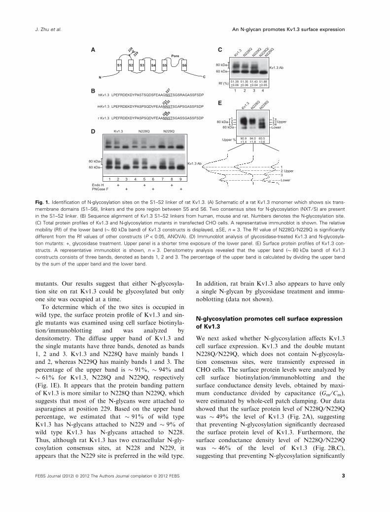
mutants. Our results suggest that either N-glycosyla-
tion site on rat Kv1.3 could be glycosylated but only
one site was occupied at a time.
To determine which of the two sites is occupied in
wild type, the surface protein profile of Kv1.3 and sin-
gle mutants was examined using cell surface biotinyla-
tion ⁄ immunoblotting and was analyzed by
densitometry. The diffuse upper band of Kv1.3 and
the single mutants have three bands, denoted as bands
1, 2 and 3. Kv1.3 and N228Q have mainly bands 1
and 2, whereas N229Q has mainly bands 1 and 3. The
percentage of the upper band is � 91%, � 94% and
� 61% for Kv1.3, N228Q and N229Q, respectively
(Fig. 1E). It appears that the protein banding pattern
of Kv1.3 is more similar to N228Q than N229Q, which
suggests that most of the N-glycans were attached to
asparagines at position 229. Based on the upper band
percentage, we estimated that � 91% of wild type
Kv1.3 has N-glycans attached to N229 and � 9% of
wild type Kv1.3 has N-glycans attached to N228.
Thus, although rat Kv1.3 has two extracellular N-gly-
cosylation consensus sites, at N228 and N229, it
appears that the N229 site is preferred in the wild type.
In addition, rat brain Kv1.3 also appears to have only
a single N-glycan by glycosidase treatment and immu-
noblotting (data not shown).
N-glycosylation promotes cell surface expression
of Kv1.3
We next asked whether N-glycosylation affects Kv1.3
cell surface expression. Kv1.3 and the double mutant
N228Q ⁄N229Q, which does not contain N-glycosyla-
tion consensus sites, were transiently expressed in
CHO cells. The surface protein levels were analyzed by
cell surface biotinylation ⁄ immunoblotting and the
surface conductance density levels, obtained by maxi-
mum conductance divided by capacitance (Gm ⁄Cm),
were estimated by whole-cell patch clamping. Our data
showed that the surface protein level of N228Q ⁄N229Q
was � 49% the level of Kv1.3 (Fig. 2A), suggesting
that preventing N-glycosylation significantly decreased
the surface protein level of Kv1.3. Furthermore, the
surface conductance density level of N228Q ⁄N229Q
was � 46% of the level of Kv1.3 (Fig. 2B,C),
suggesting that preventing N-glycosylation significantly
N C
228
229
Pore
S1 S2 S3 S4 S5 S6
LPEFRDEKDYPASTSQDSFEAAGNSTSGSRAGASSFSDP
LPEFRDEKDYPASPSQDVFEAANNSTSGAPSGASSFSDP
LPEFRDEKDYPASPSQDVFEAANNSTSGASSGASSFSDP
mKv1.3
hKv1.3
r Kv1.3228229
231232
227
Kv1.3 N228Q N229Q
Endo HPNGase F
+ + ++ ++
Kv1.3 Ab60 kDa--
80 kDa-
1 2 3 4 5 6 7 8 9
Kv1.3
N228Q
N229Q
N228Q
/
N229Q
Kv1.3 Ab
Rf (%) 51.38+0.06
51.88+0.05
51.43+0.04
51.35+0.06
60 kDa--
80 kDa-
1 2 3 4
123
--LowerUpper
123
Kv1.3
N228Q
N229Q
90.9+1.4
60.5+3.6
94.0+1.8Upper %
123Lower
Upper
60 kDa--80 kDa-
A
B
D
C
E
Fig. 1. Identification of N-glycosylation sites on the S1–S2 linker of rat Kv1.3. (A) Schematic of a rat Kv1.3 monomer which shows six trans-
membrane domains (S1–S6), linkers and the pore region between S5 and S6. Two consensus sites for N-glycosylation (NXT ⁄ S) are present
in the S1–S2 linker. (B) Sequence alignment of Kv1.3 S1–S2 linkers from human, mouse and rat. Numbers denotes the N-glycosylation site.
(C) Total protein profiles of Kv1.3 and N-glycosylation mutants in transfected CHO cells. A representative immunoblot is shown. The relative
mobility (Rf) of the lower band (� 60 kDa band) of Kv1.3 constructs is displayed, ±SE, n = 3. The Rf value of N228Q ⁄ N229Q is significantly
different from the Rf values of other constructs (P < 0.05, ANOVA). (D) Immunoblot analysis of glycosidase-treated Kv1.3 and N-glycosyla-
tion mutants: +, glycosidase treatment. Upper panel is a shorter time exposure of the lower panel. (E) Surface protein profiles of Kv1.3 con-
structs. A representative immunoblot is shown, n = 3. Densitometry analysis revealed that the upper band (� 80 kDa band) of Kv1.3
constructs consists of three bands, denoted as bands 1, 2 and 3. The percentage of the upper band is calculated by dividing the upper band
by the sum of the upper band and the lower band.
J. Zhu et al. An N-glycan promotes Kv1.3 surface expression
FEBS Journal (2012) ª 2012 The Authors Journal compilation ª 2012 FEBS 3
Page 4
decreased the surface conductance density level of
Kv1.3. Thus, these results suggest that N-glycosylation
promotes cell surface expression of Kv1.3, although it
is not required for the functional expression of Kv1.3.
To assess whether the effect of N-glycosylation on
Kv1.3 is cell line specific, Kv1.3 and N228Q ⁄N229Q
were transiently expressed in cath-a-differentiated
(CAD) cells. CAD cells are a mouse CNS cell line and
exhibit biochemical and morphological characteristics
of primary neurons but do not express endogenous
Kv1 channels [24]. Immunoblot analysis suggests that
the surface protein level of N228Q ⁄N229Q was � 66%
of the level of Kv1.3 (Fig. 2D). It appears that pre-
venting N-glycosylation decreased the surface protein
level of Kv1.3 in two different cell lines, although to
differing degrees. This finding could be due to some
differences in the intracellular glyco-processing
machinery between the two lines.
Supplementation of GlcNAc, L-fucose and
Neu5Ac increases Kv1.3 cell surface expression
whereas D-mannose, D-galactose and D-glucose
have no effect
Three types of N-glycans (high-mannose, hybrid and
complex) are produced during Golgi processing of gly-
coproteins in mammalian cells, and the component
monosaccharides and their derivatives include d-man-
nose, d-glucose, N-acetylglucosamine (GlcNAc),
l-fucose, d-galactose and N-acetylneuraminic acid
(Neu5Ac), the most common member of sialic acid
(Fig. 3A). N-glycans differ not only in the nature of
their constituents but also in the length of their chains
and in the number of branches. To investigate whether
the monosaccharide ⁄derivative level affects Kv1.3 sur-
face expression, CHO cells were exposed to increased
concentrations of different monosaccharides or deriva-
tives. Kv1.3 transfected cells without monosaccharide
supplemented were used as a control. The surface pro-
tein level of Kv1.3 was measured by cell surface bioti-
nylation and immunoblotting. Our data showed that
supplementation of d-mannose, d-glucose and d-galac-
tose had little or no effect on cell surface expression
of Kv1.3 (Fig. 3B,C,F), whereas supplementation of
GlcNAc, l-fucose and Neu5Ac caused a significant
increase in Kv1.3 surface levels. GlcNAc enhanced
Kv1.3 surface expression in a dose dependent manner
– the surface protein level of Kv1.3 increased 2, 2.5
or 3 times when GlcNAc was at 25, 50 or 100 mm
(Fig. 3D). l-Fucose also increased the Kv1.3 surface
protein level but only when its concentration reached
50 mm (Fig. 3E). Supplementation of Neu5Ac
increased Kv1.3 surface protein level when its concen-
tration reached 10 mm (Fig. 3G). Thus, addition of
GlcNAc, l-fucose or Neu5Ac to the culture medium
significantly increased the cell surface expression of
Kv1.3, presumably through altering N-glycosylation.
We speculate that the altered N-glycosylation might
further affect protein internalization or stability
resulting in a higher surface expression level of
Kv1.3.
GlcNAc supplementation reduced the branch size
of complex type N-glycans of Kv1.3, whereas
L-fucose and Neu5Ac did not
To assess whether the increased surface level of Kv1.3
upon supplementation of monosaccharides was due to
a change in N-glycan structure, the surface protein
profile of Kv1.3 was analyzed by immunoblotting and
densitometry. The gel mobility of the � 80 kDa bands
of Kv1.3 was increased significantly when GlcNAc was
Sur
face
pro
tein
(nor
mal
ized
)
Sur
face
pro
tein
(nor
mal
ized
)
0
50
100
*
Kv1.3N228Q/
N229Q
CHO transfected
Kv1.3
0
5
10
15
20
25
Gm
/Cm
(nS
/pF)
*
CHO transfected
N228Q/N229Q
Kv1.3
N228Q/N229Q
10 nA
10 ms
0
50
100
*
Kv1.3N228Q/
N229Q
CAD transfected
A
C D
B
Fig. 2. Effect of N-glycosylation on the cell surface expression of
Kv1.3. (A) Bar graphs of surface protein levels of Kv1.3 and
N228Q ⁄ N229Q in transfected CHO cells. The mean value of
N228Q ⁄ N229Q was normalized to the mean value of Kv1.3, which
was taken to be 100. Error bars represent ±SE of triplicate values.
An asterisk indicates significant difference from control (P < 0.05,
unpaired t test). (B) Bar graphs of conductance densities of Kv1.3
and N228Q ⁄ N229Q in transfected CHO cells. Gm ⁄ Cm, maximum
conductance divided by capacitance. Error bars represent ± SE of
11–13 values. An asterisk indicates a significant difference from
control (P < 0.05, unpaired t-test). (C) Whole cell current traces of
Kv1.3 and N228Q ⁄ N229Q. (D) Bar graphs of surface protein levels
of Kv1.3 and N228Q ⁄ N229Q in transfected CAD cells. The mean
value of N228Q ⁄ N229Q was normalized to the mean value of
Kv1.3, which was taken to be 100. Error bars represent ± SE of
triplicate values. An asterisk indicates significant difference from
control (P < 0.05, unpaired t-test).
An N-glycan promotes Kv1.3 surface expression J. Zhu et al.
4 FEBS Journal (2012) ª 2012 The Authors Journal compilation ª 2012 FEBS
Page 5
used, whereas the gel mobility of the � 60 kDa band
of Kv1.3 did not change with the treatment (Fig. 4A).
Glycosidase treatment showed that the upper bands
of Kv1.3 ⁄GlcNAc were sensitive to PNGase F but
resistant to Endo H, suggesting that the bands
contained complex type N-glycans, whereas the
� 60 kDa band of Kv1.3 ⁄GlcNAc was sensitive to
both PNGase F and Endo H, indicating that it con-
tained a high-mannose or hybrid type N-glycan
(Fig. 4B). As shown in Fig. 1, the � 80 kDa bands of
Kv1.3 contain complex type N-glycans and the
� 60 kDa band of Kv1.3 contains a high-mannose or
hybrid type N-glycan. These results indicate that the
type of N-glycosylation did not change upon addition
of GlcNAc; the difference in gel mobility of the
� 80 kDa bands in Kv1.3 and Kv1.3 ⁄GlcNAc sug-
gests that there was a change in structure and ⁄orcomposition of the complex type N-glycan. Previous stu-
dies showed that the gel mobility of the Na,K-ATPase
b1 subunit was increased when the length of the
branches or the number of branches decreased in com-
plex type N-glycans [25]. The increased gel mobility of
the � 80 kDa bands of Kv1.3 suggests that the branch
size and ⁄or branching of the complex type N-glycan
must be decreased.
Mature mammalian N-glycans are commonly termi-
nated with sialic acids [26]. To assess whether supple-
mentation of GlcNAc altered sialic acid content,
0
25
50
75
100
125
150
D-galactose (mM)
0
50
200
150
250
100
0 10 205 15Neu5Ac (mM)
*
050100150200250300350
GlcNAc (mM)
**
*
D-glucose (mM)
0
25
50
75
100
125
150
L-fucose (mM)
0255075100125150175 *
*
0
25
50
75
100
125
150
A
B C
D E
F G
0 50 1007525
0 50 1007525
0 50 1007525
0 50 1007525
0 50 1007525
D-mannose (mM)
Sur
face
Kv1
.3 p
rote
in(n
orm
aliz
ed)
Sur
face
Kv1
.3 p
rote
in(n
orm
aliz
ed)
Sur
face
Kv1
.3 p
rote
in(n
orm
aliz
ed)
Sur
face
Kv1
.3 p
rote
in(n
orm
aliz
ed)
Sur
face
Kv1
.3 p
rote
in(n
orm
aliz
ed)
Sur
face
Kv1
.3 p
rote
in(n
orm
aliz
ed)
GlcNAc
Galactose Neu5Ac
Glucose
Fucose
Mannose
Asn Asn Asn Asn
ER Golgi
GlycanPrecursor
High-MannoseGlycan
Hybrid typeGlycan
Complex typeGlycan
Fig. 3. Effect of the monosaccha-
rides ⁄ derivatives on the surface protein
level of Kv1.3. (A) Schematics of N-glycan
precursor, high-mannose, hybrid and com-
plex type N-glycans. (B)–(G) Surface protein
levels of Kv1.3 in transfected CHO cells
maintained in the culture medium supple-
mented with different concentrations of
monosaccharides ⁄ derivatives, including
D-mannose (B), D-glucose (C), GlcNAc (D),
L-fucose (E), D-galactose (F) and Neu5Ac (G).
Mean values of Kv1.3 in the presence of
monosaccharides ⁄ derivatives were normal-
ized to the mean value of Kv1.3 in the
absence of monosaccharides ⁄ derivatives,
which was set to 100. Error bars represent
± SE of triplicate values. *differences are
significant (P < 0.05, ANOVA).
J. Zhu et al. An N-glycan promotes Kv1.3 surface expression
FEBS Journal (2012) ª 2012 The Authors Journal compilation ª 2012 FEBS 5
Page 6
surface Kv1.3 was treated with sialidase, which
removes terminal sialic acid residues. The gel mobility
of both upper and lower bands of Kv1.3 was
increased after sialidase treatment, indicating that
both of them contained sialic acids (Fig. 4C). Since
the lower band was also sensitive to Endo H
(Fig. 1D), it appeared to contain a hybrid type N-gly-
can. In contrast, after supplementation of GlcNAc,
the upper bands of Kv1.3 were resistant to sialidase,
indicating that they did not contain detectable sialic
acids, whereas the lower band of Kv1.3 was sensitive
to sialidase, indicating that it contained sialic acids
(Fig. 4D). It appears that supplementation of GlcNAc
decreased sialic acid content in the complex type
N-glycan of Kv1.3. These results suggest that the length
of branches in complex type N-glycans decreased when
GlcNAc was added, but whether or not the number of
branches also changed was not clear. Thus, the
increased surface protein level of Kv1.3 upon supple-
mentation of GlcNAc appears to be due to decreased
branch size and ⁄or altered branching of complex type
N-glycans.
The addition of l-fucose or Neu5Ac caused little or
no change in gel mobility of the � 80 kDa bands of
Kv1.3 (Fig. 4E,F), indicating no dramatic changes in
the branch size and ⁄or branching of the complex type
N-glycans; however, whether or not the carbohydrate
composition changed was unclear. Therefore, the
increase of Kv1.3 surface expression upon supplemen-
tation of l-fucose or Neu5Ac might be due to different
mechanisms compared with GlcNAc. We speculate
that increasing the substrate concentration would
improve the efficiency of fucosylation or sialylation of
N-glycans and therefore stabilize the structure of
Kv1.3.
Supplementation of GlcNAc stabilized the cell
surface Kv1.3 population
We next asked whether GlcNAc promotes the surface
protein level of Kv1.3 by affecting its internalization.
To address this question, Kv1.3 transfected cells were
maintained in culture medium with or without GlcNAc
added, and then surface biotinylated and chased for
various times. The cell surface signals of Kv1.3 were
analyzed using a two-phase exponential decay equa-
tion. Kv1.3 had half-lives of 0.6 and 9.2 h (Fig. 5A),
whereas Kv1.3 ⁄GlcNAc had half-lives of 1.0 and
14.9 h (Fig. 5B). It appears that surface Kv1.3 was a
mixture of two components, each with its own inter-
nalization half-life, which suggests that two internali-
zation temporal pathways might exist. The half-lives
of both components of Kv1.3 were increased upon
supplementation of GlcNAc, indicating that they
exhibited decreased internalization. These results sug-
gest that supplementation of GlcNAc eventually sta-
bilized the Kv1.3 surface population and inhibited its
internalization, presumably due to decreased branch
size and ⁄or altered branching in complex type N-gly-
cans of Kv1.3.
Sialidase
60 kDa--80 kDa-- Surface
Kv1.3
+
60 kDa--70 kDa--
Sialidase
GlcNAc (25 mM)
SurfaceKv1.3
+
Neu5Ac (10 mM)– +
SurfaceKv1.321
12
80 kDa-
L-fucose (50 mM)– +
21SurfaceKv1.3
12
80 kDa-
GlcNAc (25 mM)– +
12
21 Surface
Kv1.380 kDa-
A
B
C D
E F
GlcNAc (25 mM)– +
SurfaceKv1.360 kDa--
60 kDa band
80 kDa-
Over-exposed
Endo HPNGase F
++
GlcNAc (25 mM)
60 kDa--80 kDa-- Total
Kv1.3
Fig. 4. Effect of GlcNAc, L-fucose and Neu5Ac on the protein band-
ing pattern of Kv1.3. (A) Surface protein banding patterns of Kv1.3
in CHO cells with GlcNAc supplemented: ), control, without
GlcNAc added; +, GlcNAc added. A densitometry analysis of Kv1.3
proteins is displayed in the lower panel showing the position of
two major bands. The right panel is the longer time exposure of
the left panel. (B) Immunoblot analysis of glycosidase-treated Kv1.3
with GlcNAc supplemented: +, glycosidase treatment. (C) Immuno-
blot analysis of sialidase-treated Kv1.3: +, sialidase treatment. (D)
Immunoblot analysis of sialidase-treated Kv1.3 with GlcNAc supple-
mented: +, sialidase treatment. (E), (F) Surface protein banding pat-
terns of Kv1.3 in CHO cells with L-fucose (E) and Neu5Ac (F)
supplemented: ), control, without monosaccharide added; +,
monosaccharide added. A densitometry analysis of Kv1.3 proteins
is displayed in the lower panel showing the position of two major
bands.
An N-glycan promotes Kv1.3 surface expression J. Zhu et al.
6 FEBS Journal (2012) ª 2012 The Authors Journal compilation ª 2012 FEBS
Page 7
Supplementation of GlcNAc affects only
glycoproteins and has no effect on
non-glycosylated proteins
To test whether the effect of GlcNAc supplementation
is specific to Kv1.3, glucose transporter 1 (GLUT1),
which is a heavily glycosylated membrane protein
endogenously expressed in CHO cells, was also used.
The GLUT1 on the cell surface was detected as a
broad protein band of � 70–80 kDa on SDS gels.
With GlcNAc supplementation, the gel mobility of
GLUT1 was increased significantly to � 45–75 kDa,
suggesting that there was a decrease in N-glycan
branch size and ⁄or a change in branching. GLUT1
contains one N-glycan (Fig. 6A). The deglycosylated
GLUT1 had a molecular mass of � 40 kDa on SDS
gels [27], although the predicted molecular mass of
GLUT1 was 54 kDa, suggesting that GLUT1 had
other post-translational modifications in addition to
N-glycosylation. Furthermore, the surface protein level
of GLUT1 was increased � 1.5-fold upon supplemen-
tation of GlcNAc (Fig. 6B). These results are similar
to those observed with Kv1.3. Thus, addition of
GlcNAc in cell culture also has an effect on endoge-
nous GLUT1.
To determine whether supplementation of GlcNAc
affects non-glycoproteins, Kv2.1 and N228Q ⁄N229Q
were transiently expressed in CHO cells. Kv2.1, a
potassium channel from the Kv2 subfamily, does not
contain an N-glycosylation site that is glycosylated,
and N228Q ⁄N229Q is the Kv1.3 mutant with both
N-glycosylation sites mutated. Our data showed that
the surface protein level and the protein banding pat-
tern of Kv2.1 or Kv1.3N228Q ⁄N229Q were similar in
the presence or absence of GlcNAc in the culture med-
ium (Fig. 6C–F). Actin is a non-glycosylated cytoplas-
mic protein endogenously expressed in CHO cells. Our
data showed that addition of GlcNAc in the culture
medium did not affect the total protein level or the
banding pattern of actin (Fig. 6G). Thus, supplementa-
tion of GlcNAc appears to affect mainly glycoproteins.
These results further suggest that supplementation of
GlcNAc does not affect protein synthesis, but it affects
N-glycan Golgi processing or endocytosis.
Discussion
Mouse and rat Kv1.3 have two successive N-glycosyla-
tion consensus sites, NNS and NST, on their S1–S2
linker, while human Kv1.3 has only one, NST. The
S1–S2 linker of Kv1.3 from the three species has the
same length, and mouse and rat share 97.4% sequence
identity whereas human shares 84.6% with the rodents.
Our results showed that either of the two sites on rat
Kv1.3 could be glycosylated if one is mutated, but that
only one site is occupied at a time. We are not aware
of any examples in the literature of neighboring N-gly-
can consensus site asparagines on membrane proteins
being glycosylated. Fly Kv1.1 (Shaker) has two N-gly-
cosylation sites at positions 259 and 263 on its S1–S2
linker, which are three amino acids apart; both sites
are occupied with N-glycans [28]. Rat Kv1.2 has one
N-glycosylation site at the position 207 on its S1–S2
linker. A Kv1.2 construct was engineered to contain
two additional sites at positions 199 and 203, for a
total of three NST sites, each being three amino acids
apart; all of them were N-glycosylated [20]. Human
Kv4.1 has two N-glycosylation sites at positions 352
and 355 on its S5-P loop, which are two amino acids
Chase time (h)
Chase time (h)
Nor
mal
ized
sig
nal
Nor
mal
ized
sig
nal
HL1: 0.6 hHL2: 9.2 h
Kv1.3 surface protein half-life (HL)
25
75
50
100
A
B
0
25
75
50
100
0
86420 10
86420 10
HL1: 1.0 hHL2: 14.9 h
Kv1.3/GlcNAc surface protein half-life (HL)
Fig. 5. The surface protein half-life of Kv1.3. Cell surface protein
half-lives of Kv1.3 in transiently transfected CHO cells in the
absence (A) and presence (B) of GlcNAc were estimated by surface
biotinylation ⁄ immunoblotting and chasing in nonbiotinylation media
as described in Experimental procedures. Cell surface signals of
Kv1.3 were normalized to time zero of chase, which was taken as
100. Symbols (squares and circles) represent the normalized
mean ± SE of triplicate values at each time point. Lines represent
the two-phase exponential decay curves.
J. Zhu et al. An N-glycan promotes Kv1.3 surface expression
FEBS Journal (2012) ª 2012 The Authors Journal compilation ª 2012 FEBS 7
Page 8
apart. Neither site was N-glycosylated [29]. Presumably
these two sites are too close to the predicted S5 trans-
membrane domain – they are five and eight amino
acids respectively away from the S5. In general, an N-
glycosylation site that is occupied is supposed to be 10
amino acids from a transmembrane domain [30]. Our
data showed that two successive N-glycosylation sites,
with no amino acids in between, could not be occupied
at the same time. We speculate that this finding is due
to steric hindrance from the close proximity of these
two sites preventing two N-glycans being added.
While both sites (NNS and NST) on the S1–S2 lin-
ker were capable of being N-glycosylated, the N229
site (NST) was preferred over the N228 site (NNS) in
wild type Kv1.3. It appears that 91% of Kv1.3 has
an N-glycan attached to N229 and 9% of Kv1.3 has
an N-glycan attached to N228. This finding might be
due to the sequon or the position of the site. The
serine-containing sequon (NXS) is less frequently
glycosylated and more affected by the nature of the X
amino acid compared with the threonine-containing se-
quon (NXT) [31,32]. Furthermore, the position of
N229 (NST) on the S1–S2 linker of rat Kv1.3 is equiv-
alent to the position of N227 (NST) in human. Our
previous study showed that the N-glycosylation con-
sensus site of Kv1 channels appears to be located in a
similar relative position in the S1–S2 linker and the
strict relative position of N-glycosylation consensus
sites on these linkers may be correlated with the func-
tional effect of N-glycans on some Kv1 potassium
channels [33]. Thus Kv1.3 from either rat or human
has only one N-glycan attached its S1–S2 linker.
The effect of N-glycosylation on surface expression
varies between different glycoproteins even within the
Kv1 subfamily. Removal of the N-glycosylation con-
sensus site had little effect on Kv1.1 protein levels, but
it considerably reduced surface protein levels of Kv1.2
and Kv1.4, which were � 60% and � 15% of the wild
0
50
150
100
*S
urfa
ce p
rote
in(n
orm
aliz
ed)
Sur
face
pro
tein
(nor
mal
ized
)
Sur
face
pro
tein
(nor
mal
ized
)
GLUT1
GLUT1/
GlcNAc
GLUT1 (1 N-site)Surface protein
51 kDa--
75 kDa--
45 kDa--
Kv2.1
Kv2.1/
GlcNAc
0
50
100
Kv2.1 (0 N-site)Surface protein
110 kDa--
GlcNAc
Actin Ab
+
Actin (0 N-site)CHO Lysates
45 kDa--
–
0
100
50
N228Q
/N22
9Q
N228Q
/N22
9Q/
GlcNAc
N228Q/N229Q (0 N-site)Surface protein
58 kDa--
A
C
D
E
FB
G
Fig. 6. Supplementation of GlcNAc increases surface protein levels of other membrane glycoproteins but it does not affect non-glycopro-
teins. (A) Surface protein profiles of endogenously expressed GLUT1 in CHO cells with or without monosaccharides added. A representative
immunoblot is shown. (B) Bar graphs of GLUT1 surface protein levels. (C) Surface protein profiles of Kv2.1 (contains no N-glycans) in trans-
fected CHO cells with or without monosaccharides added. A representative immunoblot is shown. (D) Bar graphs of Kv2.1 surface protein
levels. (E) Surface protein profiles of N228Q ⁄ N229Q (contains no N-glycan) in transfected CHO cells with or without monosaccharides
added. A representative immunoblot is shown. (F) Bar graphs of N228Q ⁄ N229Q surface protein levels. The mean values of proteins tested
were normalized to the mean value of the control, which was set to 100. Error bars represent ± SE of triplicate values. *differences are
significant (P < 0.05, unpaired t-test). (G) Total protein profiles of actin (cytoplasmic protein) in CHO cells: +, monosaccharide added. A
representative immunoblot is shown, n = 3.
An N-glycan promotes Kv1.3 surface expression J. Zhu et al.
8 FEBS Journal (2012) ª 2012 The Authors Journal compilation ª 2012 FEBS
Page 9
type, respectively [17,20]. Our data showed that
N228Q ⁄N229Q was � 49% of the level of Kv1.3 in
CHO cells. The differential effect of N-glycosylation
on closely related Kv channels presumably reflects the
complexity of the N-glycan structures.
Increasing the GlcNAc level has been shown to
enhance cell surface expression of some receptors and
transporters including transforming growth factor breceptor (TbR) and glucose transporter 4 (GLUT4)
[34]. The TbR surface levels were increased 1.5- and
2-fold in Mgat5) ⁄ ) tumor cells supplemented for 48 h
with 25 and 50 mm GlcNAc, respectively, quantified
by 125I-TGF-b1 binding and galectin-3 immunoprecipi-
tation; GLUT4 surface levels were increased 2-fold in
HEK293T cells supplemented for 48 h with 30 mm
GlcNAc, quantified by immunostaining and confocal
microscopy [34]. We found that the surface levels of
Kv1.3 were increased 2- and 2.5-fold in CHO cells sup-
plemented for 24 h with 25 and 50 mm GlcNAc,
respectively, quantified by cell surface biotinylation
and immunoblotting. It appears that GlcNAc supple-
mentation had similar effects on the surface expression
of Kv1.3, TbR and GLUT4, although to differing
degrees. These differences could be from varied
GlcNAc concentrations, incubation times, cell lines, as
well as techniques used to measure surface protein
levels. Furthermore, the internalization of Kv1.3 was
slowed upon GlcNAc supplementation, suggesting that
the increased surface protein level was due to
decreased endocytotic processing.
Supplementation of GlcNAc increased the degree of
N-glycan branching of TbR I and TbR II in Mgat5) ⁄ )
tumor cells and GLUT4 in HEK293T cells, which in
turn enhanced surface protein expression [34]. GlcNAc
supplementation also increased the number of
branched N-glycans in CHO cells [34], and therefore it
is predicted that the degree of N-glycan branching of
Kv1.3 in CHO cells was also increased. The branching
or branch size of an N-glycan was directly correlated
with the protein gel mobility. A previous report
showed that the Na,K-ATPase b1 subunit exhibited
increased gel mobility when the degree of N-glycan
branching was reduced with swainsonine treatment
and decreased gel mobility when the degree of N-gly-
can branching was increased using small interfering
RNA against the stop-branching enzyme, presumably
due to altered steric hindrance [25]. Our results showed
that the gel mobility of Kv1.3 was increased upon
GlcNAc supplementation. Similar results were
obtained with GLUT1 (Fig. 5), TbR I and TbR II
[34]; they all showed increased gel mobility when
GlcNAc was added. Since the degree of N-glycan
branching is generally increased with GlcNAc supple-
mentation, we speculate that the size of branches must
be decreased. Indeed, the sialic acid content of the
N-glycan in Kv1.3 was reduced when GlcNAc was
added. The increased gel mobility of Kv1.3 upon Glc-
NAc supplementation appeared to result from a com-
bined effect of decreased branch size and increased
branching.
Externally applied GlcNAc goes into the salvage
pathways in cells to form UDP-GlcNAc, which is
transported into the Golgi apparatus as a substrate in
the synthesis of N-glycoconjugates [35–37]. GlcNAc is
linked by b1,2 to the terminal mannose residue of the
Man (a1,3) branch or at the free Man (a1,6) arm
yielding the precursor for mono- or bi-antennary struc-
tures, or by b1,4 at the Man (a1,3) or b1,6 at the Man
(a1,6) branches yielding the precursor for tri- or tetra-
antennary structures [38]. The N-acetyl-d-glucos-
aminyltransferases I, II, IV and V, encoded by the
genes Mgat1, 2, 4a ⁄b and 5, act sequentially to cata-
lyze these reactions, respectively. The concentration of
Golgi UDP-GlcNAc is proportional to the degree of
GlcNAc branching in N-glycans [39]. The branched
N-glycan was crosslinked with extracellular galectins, a
family of N-acetyllactosamine binding animal lectins,
to form a molecular lattice which restricted endocyto-
sis of surface glycoproteins. b1,6 GlcNAc branching
by Mgat5 is preferentially extended by poly-N-acetyl-
lactosamine, further enhancing avidity for galectins
[38]. The desialylation of N-glycans would expose
underlying N-acetyllactosamine and enhance the
galectin binding.
The enhancement of surface proteins by GlcNAc
supplementation appeared to be dependent on complex
type N-glycans. The unglycosylated proteins, e.g.
Kv2.1 and Kv1.3N228Q ⁄N229Q (Fig. 5), proteins with
hybrid type N-glycans, e.g. the � 60 kDa band of
Kv1.3 (Fig. 4), and proteins with high-mannose type
N-glycans, e.g. in Lec1 [34], were not affected. There-
fore, GlcNAc supplementation might have a global
effect on membrane proteins containing complex type
N-glycans. However, in certain cells that highly express
Mgat5, this effect might be reduced.
In mammalian tissues, l-fucose is usually linked by
a1,2 to terminal d-galactose, by a1,3 or a1,4 to subter-
minal GlcNAc residues of the antennae and by a1,6 to
the innermost GlcNAc [40,41]. It has been shown that
the a1,6-linked fucose modification promoted protein
stability and cell surface expression of polysialic-acid-
carrying glycoproteins [42]. The a1,6-linked fucosyla-
tion was found to reduce the conformational flexibility
of the antennae of a bi-antennary N-glycan, which
might further stabilize the protein structure [43]. Our
results showed that supplementation of l-fucose
J. Zhu et al. An N-glycan promotes Kv1.3 surface expression
FEBS Journal (2012) ª 2012 The Authors Journal compilation ª 2012 FEBS 9
Page 10
increased cell surface expression of Kv1.3 channels.
We speculate that increasing l-fucose concentration
increases the number of fucosylated glycans, which in
turn increases protein stability and surface expression
of Kv1.3. It is possible that fucosylation increased
expression of other glycoproteins that affected Kv1.3
expression.
Sialic acids are commonly linked a2,3 or a2,6 to the
termini of complex glycans or are polymerized in the
form of an a2,8 or an a2,9 linkage [44,45]. The influence
of sialic acids on the expression and stability of glyco-
proteins varies. Sialylation modification is crucial for
stabilization of stem cell marker CD133 proteins, and
removal of sialic acids accelerated CD133 degradation
via a lysosome-dependent pathway [46]. Sialic acids also
contributed to the maintenance of SRIF receptors in a
high affinity biologically active conformation [47] and
improved and prolonged cell surface expression of thy-
rotropin receptor [48]. In contrast, removal of terminal
sialic acid stabilized TRPV5 channels and enhanced
their level in plasma membrane by restricting endocyto-
sis [49]. Our data suggest that supplementation of
Neu5Ac, the most common form of sialic acids, pro-
moted surface expression of Kv1.3. We speculate that
increasing Neu5Ac concentration enhanced sialylation
of Kv1.3, which in turn increased its protein stability
and cell surface expression. The differential effect of
sialylation on glycoproteins is presumably due to the
difference in sialic acid structures and protein structures.
In summary, we have presented evidence that an
N-glycan is predominantly attached to N229 on the
S1–S2 linker of rat Kv1.3 and found that N-glycosyla-
tion promoted cell surface expression of Kv1.3 chan-
nels. Furthermore, supplementation of GlcNAc
increased the surface protein level and decreased the
protein internalization of Kv1.3, presumably by a com-
bined effect of a decreased branch size and increased
degree of branching in N-glycans. The GlcNAc effect
appears to apply to mainly membrane proteins with
complex type N-glycans.
Experimental procedures
Cell lines and culture conditions
CHO pro5 cells were obtained from the American Type
Culture Collection. CAD neuronal-like cell line was a gift
from J. Wang at the Department of Neuroscience, Tufts
University School of Medicine. CHO cells were maintained
in MEM a, supplemented with 10% fetal bovine serum,
penicillin (100 unitsÆmL)1), streptomycin (100 lgÆmL)1) and
100 mm glutamine. CAD cells were maintained in Ham’s
F-12 medium, supplemented with 10% fetal bovine serum,
penicillin (100 unitsÆmL)1), streptomycin (100 lgÆmL)1),
100 mm glutamine and proline. All cells were cultured at
37 �C under 5% CO2.
PCR and quick change mutagenesis
Rat Kv1.3 cDNAs were engineered using PCR to contain a
5¢ Kozak enhanced ribosomal binding sequence (CCACC)
before the start methionine and to remove endogenous
5¢- and 3¢-untranslated regions. Mutants N228Q or N229Q,
which had one N-linked site mutated from Kv1.3, and the
double mutant N228Q ⁄N229Q, which had two N-linked
sites mutated from Kv1.3, were generated by quick-change
mutagenesis following the manufacturer’s protocol (Strata-
gene, La Jolla, CA, USA). cDNAs generated were subcl-
oned into pcDNA3, a mammalian expression vector
(Invitrogen, Carlsbad, CA, USA). cDNAs were sequenced
(Cornell University, Ithaca, NY, USA) to confirm that the
mutation was introduced without other unwanted changes.
Transient transfection
CHO or CAD cells were transiently transfected with 0.5 lgcDNA of Kv1.3 constructs in 35 mm culture dishes using
Lipofectamine Plus following the manufacturer’s protocol
(Invitrogen, Carlsbad, CA, USA). Cells were cultured for
20 h post-transfection before they were processed.
Monosaccharide supplementation
Monosaccharides used include d-mannose, d-glucose,
GlcNAc, l-fucose, d-galactose and Neu5Ac. After trans-
fection, monosaccharides were diluted in pre-warmed cul-
ture medium to different concentrations and added for
20 h to cells. Upon confluency, cells were subjected to
membrane isolation, surface protein biotinylation or other
processes.
Membrane isolation
Once cells were confluent, an ice-cold hypotonic solution
containing protease inhibitors was added. Cells were har-
vested by pipetting up and down and then homogenized.
The homogenate was centrifuged at 5000 g at 4 �C for
5 min to pellet the nuclei. The supernatant was centrifuged
again at 16 000 g at 4 �C for 1 h to pellet the membranes,
which include rough endoplasmic reticulum, Golgi appara-
tus and plasma membranes. The membranes were collected
and analyzed by SDS ⁄PAGE and immunoblotting.
SDS/PAGE and immunoblotting
Protein samples obtained from different processes were
each mixed with Laemmli sample buffer and heated.
An N-glycan promotes Kv1.3 surface expression J. Zhu et al.
10 FEBS Journal (2012) ª 2012 The Authors Journal compilation ª 2012 FEBS
Page 11
Similar amounts of proteins were used and fractionated
on a 9% SDS ⁄PAGE at 90 V for � 1.5 h. Proteins were
then transferred to polyvinylidene difluoride (PVDF)
membranes by using the HEB 2020 semi-dry blotter at
12 V for 1 h. The PVDF membranes were blocked with
5% non-fat milk in NaCl ⁄Pi plus 0.1% Tween 20 for
30 min and shaken overnight in primary antibodies
[Kv1.3 monoclonal antibodies (UC Davis ⁄NINDS ⁄NIMH
NeuroMab Facility, Davis, CA, USA), GLUT1 poly-
clonal antibodies (Abcam, Cambridge, MA, USA) or
actin monoclonal antibodies (Sigma-Aldrich, St. Louis,
MO, USA)]. Then horseradish-peroxidase-linked anti-rab-
bit or anti-mouse secondary IgG was added. The specific
proteins were detected on blots using an enhanced chemi-
luminescence detection kit (Amersham, Piscataway, NJ,
USA) and X-ray film AR5 (Eastman Kodak, Rochester,
NY, USA).
Cell surface biotinylation
Cell surface expression levels of Kv1.3 wild type and
mutants in transfected cells were analyzed by biotinylation
using hydrazide-LC-biotin, which is specific for carbohy-
drates, or EZ-link Sulfo-NHS–SS-Biotin, which is specific
to primary amines, following the manufacturer’s protocol
(Pierce, Rockford, IL, USA). Cells were collected in 1 mL
of ice-cold lysis buffer and shaken for 30 min at 4 �C.Aliquots were routinely taken from the solubilized cells to
estimate actin amounts by immunoblotting to control for
any difference in cell density between wells (data not
shown). The remaining lysates were then centrifuged to
remove the nuclei and other insoluble materials. The
supernatant was incubated with 70 lL of streptavidin-aga-
rose beads (Pierce, Rockford, IL, USA) overnight at 4 �C.The beads were washed and eluted with Laemmli sample
buffer and samples were run on SDS ⁄PAGE for immuno-
blotting.
Surface protein half-life estimation
Intact transfected cells were biotinylated as described above
and washed to remove the biotinylation reagent, and a
sample for time zero was taken and processed for streptavi-
din precipitation and immunoblotting analysis as described
above. Other cells in dishes were returned to complete med-
ium and incubated at 37 �C for increasing ‘chase’ times, in
the absence of biotinylation reagent, before streptavidin
precipitation and immunoblotting. Signals were normalized
to the value at time zero (start of chase) and plotted as a
function of time. Protein half-life values were estimated
from a curve fitted to a decaying two-phase exponential
time course, mean ± SE. A decrease in the cell surface
Kv1.3 signal was indicative that biotinylated Kv1.3 proteins
were internalized and degraded via the endosomal ⁄ lysosmal
pathway. However, if a fraction of the internalized biotiny-
lated Kv1.3 proteins were recycled intact back to the
surface, then the surface protein half-life of Kv1.3 would be
overestimated.
Glycosidase treatment
Endo H, PNGase F and sialidase (Sigma-Aldrich, St. Louis,
MO, USA) were used for detecting the presence and types of
N-glycans on Kv1.3 channels. Rat brain membranes (3 lL)or cell lysates (10 lL) were incubated with 0.3 units of Endo
H for 30 min at 37 �C or with 0.05 units of sialidase or 3
units of PNGase F overnight at 37 �C. These incubation
times were sufficient to give the maximum effect. Reactions
were terminated by adding Laemmli sample buffer, and
detected by SDS ⁄PAGE and immunoblotting.
Patch clamping
Cells were co-transfected with 0.1 lg Kv1.3 cDNA con-
structs and 0.1 lg green fluorescence protein cDNA con-
structs to allow visualization of transfected cells by
fluorescence microscopy. Whole cell currents were recorded
at room temperature (23–25 �C) with an Axopatch 200B
amplifier (Axon Instruments, Union City, CA, USA). The
bath solution contained 150 mm NaCl, 5 mm KCl, 1 mm
MgCl2, 2 mm CaCl2, 5 mm glucose and 10 mm HEPES
(pH 7.3). The intracellular solution contained 70 mm KCl,
65 mm KF, 5 mm NaCl, 1 mm MgCl2, 10 mm EGTA,
5 mm glucose and 10 mm HEPES (pH 7.3). Patch pipettes
were fashioned from 8161 Corning glass (Warner Instru-
ments, Hamden, CT, USA) to have tip resistances of 1.2–
2.0 mX when filled with the intracellular solution.
Activation protocols involved evoking currents by depolar-
izing voltage pulses (80 ms) from a holding potential of
)80 mV to levels ranging from )70 to 50 mV in 10 mV
increments. Leak and capacitance currents were subtracted
by a standard P ⁄ n procedure, and series resistance
was compensated to 80%. Maximum peak conductance
values (G) were measured from the mean value of the
leak-subtracted peak current (I) using Ohm’s law
[G = I ⁄ (Vp ) EK)]. Vp stands for the test voltage value,
and the predicted Nernst K+ equilibrium potential (EK) is
)83 mV.
References
1 Doczi MA, Morielli AD & Damon DH (2008) Kv1.3
channels in postganglionic sympathetic neurons: expres-
sion, function, and modulation. Am J Physiol Regul
Integr Comp Physiol 295, R733–R740.
2 Fadool DA, Tucker K, Perkins R, Fasciani G, Thomp-
son RN, Parsons AD, Overton JM, Koni PA, Flavell
RA & Kaczmarek LK (2004) Kv1.3 channel gene-tar-
geted deletion produces ‘Super-Smeller Mice’ with
J. Zhu et al. An N-glycan promotes Kv1.3 surface expression
FEBS Journal (2012) ª 2012 The Authors Journal compilation ª 2012 FEBS 11
Page 12
altered glomeruli, interacting scaffolding proteins, and
biophysics. Neuron 41, 389–404.
3 Kupper J, Prinz AA & Fromherz P (2002) Recombinant
Kv13. potassium channels stabilize tonic firing of
cultured rat hippocampal neurons. Pflugers Arch 443,
541–547.
4 Beeton C, Wulff H, Standifer NE, Azam P, Mullen
KM, Pennington MW, Kolski-Andreaco A, Wei E,
Grino A, Counts DR et al. (2006) Kv1.3 channels are a
therapeutic target for T cell-mediated autoimmune dis-
eases. PNAS 103, 17414–17419.
5 Lin CS, Boltz RC, Blake JT, Nguyen M, Talento A,
Fischer PA, Springer MS, Sigal NH, Slaughter RS,
Garcia ML et al. (1993) Voltage-gated potassium chan-
nels regulate calcium-dependent pathways involved in
human T lymphocyte activation. J Exp Med 177, 637–
645.
6 Beeton C, Wulff H, Barbaria J, Clot-Faybesse O,
Pennington M, Bernard D, Cahalan MD, Chandy KG
& Beraud E (2001) Selective blockade of T lymphocyte
K+ channels ameliorates experimental autoimmune
encephalomyelitis, a model for multiple sclerosis.
PNAS 98, 13942–13947.
7 Koo GC, Blake JT, Shah K, Staruch MJ, Dumont F,
Wunderler D, Sanchez M, McManus OB, Sirotina-
Meisher A, Fischer P et al. (1999) Correolide and
derivatives are novel immunosuppressants blocking the
lymphocyte Kv1.3 potassium channels. Cell Immunol
197, 99–107.
8 Schmitz A, Sankaranarayanan A, Azam P,
Schmidt-Lassen K, Homerick D, Hansel W & Wulff H
(2005) Design of PAP-1, a selective small molecule
Kv1.3 blocker, for the suppression of effector memory
T cells in autoimmune diseases. Mol Pharmacol 68,
1254–1270.
9 Szabo I, Bock J, Grassme H, Soddemann M, Wilker B,
Lang F, Zoratti M & Gulbins E (2008) Mitochondrial
potassium channel Kv1.3 mediates Bax-induced apopto-
sis in lymphocytes. Proc Natl Acad Sci USA 105,
14861–14866.
10 Abdul M & Hoosein N (2006) Reduced Kv1.3
potassium channel expression in human prostate cancer.
J Membr Biol 214, 99–102.
11 Jang SH, Kang KS, Ryu PD & Lee SY (2009) Kv1.3
voltage-gated K+ channel subunit as a potential diag-
nostic marker and therapeutic target for breast cancer.
Biochem Mol Biol Rep 42, 535–539.
12 Ousingsawat J, Spitzner M, Puntheeranurak S, Terrac-
ciano L, Tornillo L, Bubendorf L, Kunzelmann K &
Schreiber R (2007) Expression of voltage-gated potas-
sium channels in human and mouse colonic carcinoma.
Clin Cancer Res 13, 824–831.
13 Spencer RH, Sokolov Y, Li H, Takenaka B, Milicii AJ,
Aiyar J, Nguyen A, Park H, Jap BK, Hall JE et al.
(1997) Purification, visualization, and biophysical
characterization of Kv1.3 tetramers. J Biol Chem 272,
2389–2395.
14 Apweiler R, Hermjakob H & Sharon N (1999) On the
frequency of protein glycosylation, as deduced from
analysis of the SWISS-PROT database. Biochim Biophys
Acta 1473, 4–8.
15 Molinari M (2007) N-glycan structure dictates extension
of protein folding or onset of disposal. Nat Chem Biol
3, 313–320.
16 Napp J, Monje F, Stuhmer W & Pardo L (2005) Glyco-
sylation of Eag1 (Kv10.1) potassium channels: intracel-
lular trafficking and functional consequences. J Biol
Chem 280, 29506–29512.
17 Watanabe I, Zhu J, Recio-Pinto E & Thornhill WB
(2004) Glycosylation affects the protein stability and
cell surface expression of Kv1.4 but not Kv1.1 potas-
sium channels. A pore region determinant dictates the
effect of glycosylation on trafficking. J Biol Chem 279,
8879–8885.
18 Roy S, Perron B & Gallo-Payet N (2010) Role of aspar-
agine-linked glycosylation in cell surface expression and
function of the human adrenocorticotropin receptor
(melanocortin 2 receptor) in 293 ⁄FRT cells.
Endocrinology 151, 660–670.
19 Thornhill WB, Wu MB, Jiang X, Wu X, Morgan P &
Margiiotta J (1996) Expression of Kv1.1 delayed-recti-
fier potassium channels in Lec mutant CHO cell lines
reveals a role for sialylation in channel function. J Biol
Chem 271, 19093–19098.
20 Watanabe I, Zhu J, Sutachan JJ, Gottschalk A, Recio-
Pinto E & Thornhill WB (2007) The glycosylation state
of Kv1.2 potassium channels affects trafficking, gating,
and simulated action potentials. Brain Res 1144, 1–18.
21 Zhao Y, Takahash M, Gu J, Miyoshi E, Matsumoto A,
Kitazume S & Taniguchi N (2008) Functional roles of
N-glycans in cell signaling and cell adhesion in cancer.
Cancer Sci 99, 1304–1310.
22 Schachter H (2001) Congenital disorders involving
defective N-glycosylation of proteins. Cell Mol Life Sci
58, 1085–1104.
23 Yu SP & Kerchner GA (1998) Endogenous voltage-
gated potassium channels in human embryonic kidney
(HEK293) cells. J Neurosci Res 52, 612–617.
24 Qi Y, Wang JKT, McMillian M & Chikaraishi DM
(1997) Characterization of a CNS cell line, CAD, in
which morphological differentiation is initiated by
serum deprivation. J Neurosci 17, 1217–1225.
25 Vagin O, Tokhtaeva E, Yakubov I, Shevchenko E &
Sachs G (2008) Inverse correlation between the extent
of N-glycan branching and intercellular adhesion in epi-
thelia: contribution of the Na,K-ATPase b1 subunit.
J Biol Chem 283, 2192–2202.
26 Lowe JB & Marth JD (2003) A genetic approach to
Mammalian glycan function. Annu Rev Biochem 72,
643–691.
An N-glycan promotes Kv1.3 surface expression J. Zhu et al.
12 FEBS Journal (2012) ª 2012 The Authors Journal compilation ª 2012 FEBS
Page 13
27 Hirsch B & Rosen P (1999) Diabetes mellitus induces
long lasting changes in the glucose transporter of rat
heart endothelial cells. Horm Metab Res 31, 645–652.
28 Santacruz-Toloza L, Huang Y, John SA & Papazian
DM (1994) Glycosylation of shaker potassium channel
protein in insect cell culture and in Xenopus oocytes.
Biochemistry 33, 5607–5613.
29 Birnbaum SG, Varga AW, Yuan LL, Anderson AE,
Sweatt JD & Schrader LA (2004) Structure and
function of Kv4-family transient potassium channels.
Physiol Rev 84, 803–833.
30 Landolt-Marticorena C & Reithmeier RA (1994)
Asparagine-linked oligosaccharides are localized to
single extracytosolic segments in multi-span membrane
glycoproteins. Biochem J 302, 253–260.
31 Kaplan HA, Welply JK & Lennarz WJ (1987)
Oligosaccharyl transferase: the central enzyme in the
pathway of glycoprotein assembly. Biochim Biophys
Acta 906, 161–173.
32 Kasturi L, Eshleman JR, Wunner WH & Shakin-Eshl-
eman SH (1995) The hydroxy amino acid in an Asn-X-
Ser ⁄Thr sequon can influence N-linked core glycosylation
efficiency and the level of expression of a cell surface
glycoprotein. J Biol Chem 270, 14756–14761.
33 Zhu J, Watanabe I, Poholek A, Koss M, Gomez B,
Yan C, Recio-Pinto E & Thornhill WB (2003) Allowed
N-glycosylation sites on the Kv1.2 potassium channel
S1–S2 linker: implications for linker secondary structure
and the glycosylation effect on channel function.
Biochem J 375, 769–775.
34 Lau KS, Partridge EA, Grigorian A, Silvescu CI, Rein-
hold VN, Demetriou M & Dennis JW (2007) Complex
N-glycan number and degree of branching cooperate to
regulate cell proliferation and differentiation. Cell 129,
123–134.
35 Breidenbach MA, Gallagher JEG, King DS, Smart BP,
Wu P & Bertozzi CR (2010) Targeted metabolic label-
ing of yeast N-glycans with unnatural sugars. PNAS
107, 3988–3993.
36 Chigorno V, Tettamanti G & Sonnino S (1996) Meta-
bolic processing of gangliosides by normal and salla
human fibroblasts in culture. J Biol Chem 271,
21738–21744.
37 Niittymaki J, Mattila P, Roos C, Huopaniemi L, Sjob-
lom S & Renkonen R (2004) Cloning and expression of
murine enzymes involved in the salvage pathway of
GDP-L-fucose L-fucokinase and GDP-L-fucose pyro-
phosphorylase. Eur J Biochem 271, 78–86.
38 Partridge EA, Le Roy C, Di Guglielmo GM, Pawling J,
Cheung P, Granovsky M, Nabi IR, Wrana JL & Dennis
JW (2004) Regulation of cytokine receptors by Golgi
N-glycan processing and endocytosis. Science 306, 120–124.
39 Sasai K, Ikeda Y, Fujii T, Tsuda T & Taniguchi N
(2002) UDP-GlcNAc concentration is an important fac-
tor in the biosynthesis of b1,6-branched oligosaccharides:
regulation based on the kinetic properties of N-acetylglu-
cosaminyltransferase V. Glycobiology 12, 119–127.
40 Becker DJ & Lowe JB (2003) Fucose: biosynthesis
and biological function in mammals. Glycobiology 13,
41R–53R.
41 Staudacher E, Altmann F, Wilson IBH & Marz L
(1999) Fucose in N-glycans: from plant to man. Biochim
Biophys Acta 1473, 216–236.
42 Kojima N, Tachida Y & Tsuji S (1998) Alpha
1,6-linked fucose affects the expression and stability of
polysialic acid-carrying glycoproteins in Chinese
hamster ovary cells. J Biochem 124, 726–737.
43 Stubbs HJ, Lih JJ, Gustafson TL & Rice KG (1996)
Influence of core fucosylation on the flexibility of a
biantennary N-linked oligosaccharide. Biochemistry 35,
937–947.
44 Schauer R (2009) Sialic acids as regulators of molecular
and cellular interactions. Curr Opin Struct Biol 19,
507–514.
45 Stuart AD & Brown TDK (2007) a2,6-Linked sialic
acid acts as a receptor for Feline calicivirus. J Gen Virol
88, 177–186.
46 Zhou F, Cui C, Ge Y, Chen H, Li Q, Yang Z, Wu G,
Sun S, Chen K, Gu J et al. (2010) a2,3-sialylation regu-
lates the stability of stem cell marker CD133. J Biochem
148, 273–280.
47 Rens-Domiano S & Reisine T (1991) Structural analysis
and functional role of the carbohydrate component of
somatostatin receptors. J Biol Chem 266, 20094–20102.
48 Frenzel R, Krohn K, Eszlinger M, Tonjes A & Paschke
R (2005) Sialylation of human thyrotropin receptor
improves and prolongs its cell-surface expression. Mol
Pharmacol 68, 1106–1113.
49 Cha S, Ortega B, Kurosu H, Rosenblatt KP, Kuro-o M
& Huang C (2008) Removal of sialic acid involving
Klotho causes cell-surface retention of TRPV5 channel
via binding to galectin-1. PNAS 105, 9805–9810.
J. Zhu et al. An N-glycan promotes Kv1.3 surface expression
FEBS Journal (2012) ª 2012 The Authors Journal compilation ª 2012 FEBS 13