N-Linked Glycosylation Supports Cross-Talk between Receptor Tyrosine Kinases and Androgen Receptor Harri M. Itkonen 1 , Ian G. Mills 1,2 * 1 Prostate Cancer Research Group, Centre for Molecular Medicine Norway, Nordic European Molecular Biology Laboratory (EMBL) Partnership, University of Oslo and Oslo University Hospital, Oslo, Norway, 2 Department of Cancer Prevention and Department of Urology, Oslo University Hospitals, Oslo, Norway Abstract Prostate cancer is the second most common cause of cancer-associated deaths in men and signalling via a transcription factor called androgen receptor (AR) is an important driver of the disease. Androgen treatment is known to affect the expression and activity of other oncogenes including receptor tyrosine kinases (RTKs). In this study we report that AR- positive prostate cancer cell-lines express 50% higher levels of enzymes in the hexosamine biosynthesis pathway (HBP) than AR-negative prostate cell-lines. HBP produces hexosamines that are used by endoplasmic reticulum and golgi enzymes to glycosylate proteins targeted to plasma-membrane and secretion. Inhibition of O-linked glycosylation by ST045849 or N- linked glycosylation with tunicamycin decreased cell viability by 20%. In addition, tunicamycin inhibited the androgen- induced expression of AR target genes KLK3 and CaMKK2 by 50%. RTKs have been shown to enhance AR activity and we used an antibody array to identify changes in the phosphorylation status of RTKs in response to androgen stimulation. Hormone treatment increased the activity of Insulin like Growth Factor 1-Receptor (IGF-1R) ten-fold and this was associated with a concomitant increase in the N-linked glycosylation of the receptor, analyzed by lectin enrichment experiments. Glycosylation is known to be important for the processing and stability of RTKs. Inhibition of N-linked glycosylation resulted in accumulation of IGF-1R pro-receptor with altered mobility as shown by immunoprecipitation. Confocal imaging revealed that androgen induced plasma-membrane localization of IGF-1R was blocked by tunicamycin. In conclusion we have established that the glycosylation of IGF-1R is necessary for the full activation of the receptor in response to androgen treatment and that perturbing this process can break the feedback loop between AR and IGF-1R activation in prostate cells. Achieving similar results selectively in a clinical setting will be an important challenge in the future. Citation: Itkonen HM, Mills IG (2013) N-Linked Glycosylation Supports Cross-Talk between Receptor Tyrosine Kinases and Androgen Receptor. PLoS ONE 8(5): e65016. doi:10.1371/journal.pone.0065016 Editor: Irina U. Agoulnik, Florida International University, United States of America Received October 27, 2012; Accepted April 21, 2013; Published May 28, 2013 Copyright: ß 2013 Itkonen, Mills. This is an open-access article distributed under the terms of the Creative Commons Attribution License, which permits unrestricted use, distribution, and reproduction in any medium, provided the original author and source are credited. Funding: HMI is funded by an Early Stage Researcher fellowship as part of the EU FP7 Marie Curie Integrated Training Network, PRO-NEST (Prostate Research Organizations – Network Early Stage Training). IGM is supported by funding from the Norwegian Research Council, Helse Sor-Ost and the University of Oslo through the Centre for Molecular Medicine (Norway), which is part of the Nordic EMBL (European Molecular Biology Laboratory) partnership. IGM is also supported by the Norwegian Cancer Society and by EU FP7 funding. The funders had no role in study design, data collection and analysis, decision to publish, or preparation of the manuscript. Competing Interests: The authors have declared that no competing interests exist. * E-mail: [email protected]Introduction Prostate cancer is the second most common cause of cancer associated deaths in men. Androgen receptor (AR) has been identified as a key driver of localised and metastatic prostate cancer and a principal therapeutic target [1,2]. The challenge in the treatment is the development of a castration resistant disease, which still expresses AR and retains AR activity [3,4]. AR belongs to the nuclear receptor super family and it is activated by steroid hormones, predominantly testosterone and di-hydrotestosterone [5,6]. Ligand binding triggers nuclear translocation of the AR and consequent AR-driven gene expression. AR target genes have been established as candidate oncogenes and biomarkers in prostate cancer and in recent years chromatin immunoprecipita- tion coupled to high-throughputsequencing (ChIP-seq) and expression profiling has enabled an unbiased identification of AR-driven genenetworks. Pathway analysis of these networks has implicated the AR in the regulation of metabolism [7–9] and endoplasmic reticulum (ER) stress response [10] in prostate cancer cells. Changes in the expression of certain AR target genes can help to sustain AR transcriptional activity [11,12]. As an example, Insulin like Growth Factor 1-Receptor (IGF-1R) forms a regulatory feed-back loop with AR. AR itself can activate IGF- 1R expression [13] and IGF-1R stimulates AR activity in prostate cancer cells [14–16]. Receptor tyrosine kinases (RTK) form an especially interesting group of proteins as their aberrant activation is frequently documented in other cancers, which has enabled development of targeted therapies [17–19]. RTKs act as receptor kinases to activate complex down-stream signalling networks. The activity of RTKs can be regulated at the transcriptional and translational levels [17,20]. However, plasma-membrane retention time determines how long a given receptor activates signalling and is therefore a critical determinant of RTK activity [17,21–23]. Plasma-membrane retention is regulated by negative feedback via mTOR [23] but also by the amount of N-linked glycosylation, occurring in the late ER and Golgi [24,25]. The enzymes catalyzing N-linked glycosylation are sensitive to the levels of hexosamines. Hexosamine biosynthetic pathway (HBP) in turn requires glucose and glutamine, which makes this pathway capable PLOS ONE | www.plosone.org 1 May 2013 | Volume 8 | Issue 5 | e65016
Transcript
N-Linked Glycosylation Supports Cross-Talk betweenReceptor Tyrosine Kinases and Androgen ReceptorHarri M. Itkonen1, Ian G. Mills1,2*
1 Prostate Cancer Research Group, Centre for Molecular Medicine Norway, Nordic European Molecular Biology Laboratory (EMBL) Partnership, University of Oslo and Oslo
University Hospital, Oslo, Norway, 2 Department of Cancer Prevention and Department of Urology, Oslo University Hospitals, Oslo, Norway
Abstract
Prostate cancer is the second most common cause of cancer-associated deaths in men and signalling via a transcriptionfactor called androgen receptor (AR) is an important driver of the disease. Androgen treatment is known to affect theexpression and activity of other oncogenes including receptor tyrosine kinases (RTKs). In this study we report that AR-positive prostate cancer cell-lines express 50% higher levels of enzymes in the hexosamine biosynthesis pathway (HBP) thanAR-negative prostate cell-lines. HBP produces hexosamines that are used by endoplasmic reticulum and golgi enzymes toglycosylate proteins targeted to plasma-membrane and secretion. Inhibition of O-linked glycosylation by ST045849 or N-linked glycosylation with tunicamycin decreased cell viability by 20%. In addition, tunicamycin inhibited the androgen-induced expression of AR target genes KLK3 and CaMKK2 by 50%. RTKs have been shown to enhance AR activity and weused an antibody array to identify changes in the phosphorylation status of RTKs in response to androgen stimulation.Hormone treatment increased the activity of Insulin like Growth Factor 1-Receptor (IGF-1R) ten-fold and this was associatedwith a concomitant increase in the N-linked glycosylation of the receptor, analyzed by lectin enrichment experiments.Glycosylation is known to be important for the processing and stability of RTKs. Inhibition of N-linked glycosylation resultedin accumulation of IGF-1R pro-receptor with altered mobility as shown by immunoprecipitation. Confocal imaging revealedthat androgen induced plasma-membrane localization of IGF-1R was blocked by tunicamycin. In conclusion we haveestablished that the glycosylation of IGF-1R is necessary for the full activation of the receptor in response to androgentreatment and that perturbing this process can break the feedback loop between AR and IGF-1R activation in prostate cells.Achieving similar results selectively in a clinical setting will be an important challenge in the future.
Citation: Itkonen HM, Mills IG (2013) N-Linked Glycosylation Supports Cross-Talk between Receptor Tyrosine Kinases and Androgen Receptor. PLoS ONE 8(5):e65016. doi:10.1371/journal.pone.0065016
Editor: Irina U. Agoulnik, Florida International University, United States of America
Received October 27, 2012; Accepted April 21, 2013; Published May 28, 2013
Copyright: � 2013 Itkonen, Mills. This is an open-access article distributed under the terms of the Creative Commons Attribution License, which permitsunrestricted use, distribution, and reproduction in any medium, provided the original author and source are credited.
Funding: HMI is funded by an Early Stage Researcher fellowship as part of the EU FP7 Marie Curie Integrated Training Network, PRO-NEST (Prostate ResearchOrganizations – Network Early Stage Training). IGM is supported by funding from the Norwegian Research Council, Helse Sor-Ost and the University of Oslothrough the Centre for Molecular Medicine (Norway), which is part of the Nordic EMBL (European Molecular Biology Laboratory) partnership. IGM is alsosupported by the Norwegian Cancer Society and by EU FP7 funding. The funders had no role in study design, data collection and analysis, decision to publish, orpreparation of the manuscript.
Competing Interests: The authors have declared that no competing interests exist.
Identification of putative AR and Pol2 binding sitesWe took an in silico approach to find if AR and RNA polymerase
II (RNA polII) associate with specific genomic loci of interest. We
utilized two publicly available ChIP-seq datasets and visualized the
data using the UCSC Genome Browser (AR, accession number
GSE14092 [7] and RNA polII, accession number GSE28126 [8]).
ImmunofluorescenceLNCaP cells were plated on cover slips and allowed to attach for
48 hours. At this point cells were treated as described above for
R1881 stimulation. Cells were fixed with ice-cold methanol and
Glycosylation Affects Androgen Receptor Activity
PLOS ONE | www.plosone.org 2 May 2013 | Volume 8 | Issue 5 | e65016
placed in 220uC overnight. On the next day, cells were washed
twice with PBS and once with 5% BSA in PBS. Subsequently, the
cells were blocked with 5% BSA in PBS for an hour, followed by
an incubation with a primary antibody (1:50) against IGF-1R for
an hour. Coverslips were washed three times with 5% BSA in PBS
and stained with a secondary antibody (A-11010, Invitrogen) and
DAPI staining for an hour. After this, cover slips were washed
three times with PBS and mounted with fluorescent mounting
media (S3023, DAKO). Imaging was performed with Zeiss LSM
510 confocal microscope.
Results
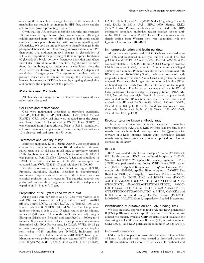
Hexosamine biosynthetic pathway is up-regulated inprostate cancer cell lines
The rate-limiting enzyme in the hexosamine biosynthetic
pathway (HBP) is glutamine-fructose-6-phosphate transaminase 1
(GFPT1), which is also the first enzyme in the pathway [27]. UDP-
N- acetylglucosamine pyrophosphorylase 1 (UAP1) is the final
enzyme in the pathway producing UDP-N-Acetylglucosamine
(UDP-GlcNAc). Interestingly, the expression of both GFPT1 and
UAP1 was increased by ,50% in AR-positive prostate cancer cell
lines LNCaP and VCaP compared to normal prostate cells PNT2
and RWPE-1 (Fig. 1A). UDP-GlcNAc is utilized by O-GlcNAc
transferase [28], to modify a multitude of target proteins by O-
linked glycosylation [27]. In addition, UDP-GlcNAc is utilized by
the ER and Golgi resident enzymes to modify proteins targeted to
the plasma membrane [25].
In order to test the importance of glycosylation for prostate
cancer cells, we utilized small molecule inhibitors against O-linked
glycosylation (ST045849) and N-linked glycosylation (tunicamy-
cin). ST045849 inhibits OGT [29], while tunicamycin inhibits the
enzymes catalyzing N-linked glycosylation, and it is also known to
induce ER stress [30]. ST045849 caused a concentration-
dependent decrease in the viability of LNCaP cells and the
highest concentration (20 mM) decreased viability by 20%
compared to the vehicle treated cells used as control (Fig. 1B).
Tunicamycin also caused reduction in viability of maximally 20%
versus the vehicle treated cells (Fig. 1C). O-linked glycosylation
was recently shown to be elevated in prostate cancer and OGT
itself was shown to regulate invasion and angiogenesis [31].
However, the role of N-linked glycosylation in prostate cancer has
been less investigated.
Inhibition of N-linked glycosylation impairs theexpression of androgen receptor target genes
Tunicamycin dose-dependently inhibited the viability of
LNCaP prostate cancer cells. AR activity has been shown to
induce ER stress response [10] and N-linked glycosylation occurs
in the endoplasmic reticulum and Golgi. In order to determine
whether N-linked glycosylation alters AR activity, we evaluated
the effect of tunicamycin on the expression of Kallikrein 3 (KLK3),
which is a direct AR target protein, and also a well-characterized
biomarker for prostate cancer and known glycoprotein [32].
Tunicamycin treatment reduced KLK3 protein by 50% in
LNCaP and VCaP cells at 12 hours after addition of tunicamycin
(Fig. 1D). The predominant effect of tunicamycin treatment was to
alter the ratio of high and low-molecular weight KLK3 over time,
which was seen more drastically in VCaP cells. Tunicamycin
induced accumulation of an ER stress marker, 78 kDa glucose-
regulated protein (BiP) [33] from eight hours onwards, following
the accumulation of un-processed KLK3 which was detected from
four hours onwards (Fig. 1D).
We noted that inhibition of N-linked glycosylation led to the
significant accumulation of a shorter, un-glycosylated form of
KLK3 [34,35] in VCaP cells (90%), while in LNCaP cells the
initial accumulation of the un-glycosylated form was followed by
60% decrease of the total KLK3 (Fig. 1D). This suggests that
tunicamycin mainly exerts its effects on processing in VCaP cells,
while in LNCaP cells it has effects not only on processing but also
on transcription. In order to test this, we stimulated LNCaP cells
with a synthetic androgen in the presence or absence of
tunicamycin and found that tunicamycin decreased the andro-
gen-dependent expression of both KLK3 and another AR target
gene, CAMKK2 [8], mRNAs by over 50% (Fig. S1).
In order to test more directly whether N-linked glycosylation
affects AR activity, we stimulated LNCaP and VCaP cells with a
synthetic androgen in the presence and absence of tunicamycin for
24 hours. We observed significant a 50% decrease in the
expression of KLK3 and CaMKK2, upon androgen treatment
in both cell-lines (Fig. 2A and 2B). This in spite of the fact that
CaMKK2, unlike KLK3 is not a known target for N-linked
glycosylation and the protein resides in the cytoplasm. This data
suggested that tunicamycin can act not only by affecting
glycosylation/post-translational processing but also through a
feedback loop that affects the transcriptional activity of the AR.
Insulin like Growth Factor 1-Receptor is activated by ARSignalling cross-talk between kinases and the AR have been
reported as one general mechanism for sustaining AR activity. In
addition it has previously been reported that RTKs are extensively
modified via N-linked glycosylation in the Golgi network prior to
the insertion into the plasma membrane [24] and signalling cross-
talk between RTKs and the AR has been reported to contribute to
metastatic disease [36]. Furthermore RTKs, such as insulin
growth factor 1-receptor (IGF-1R) are expressed in response to
androgen treatment [13]. Together this establishes the hypothesis
that the androgen-dependent expression and glycosylation of
RTKs may help to maintain AR activity and the expression of AR
target genes.
To identify candidate RTKs as mediators of this effect we used
an antibody array covering a panel of RTKs. LNCaP cells were
stimulated with a synthetic androgen for 48 hours to allow
changes in AR-dependent proteome and cell lysates were analyzed
with the RTK array to assess RTK activity (Fig. 2C). Epidermal
growth factor receptor (EGFR) phosphorylation was detectable
and unchanged both in the presence and absence of the hormone
(Fig. 2C and 2D). By contrast, androgen stimulation led to a 3-fold
decrease in the activity of ErbB2 (Her2), while signalling via IGF-
1R was increased by 10-fold. Based on this approach, the RTKs
most likely to have a feedback effect on AR activity are IGF-1R
and ErbB2, given the reciprocal increase in the activity of the
former and reduction in the activity of the latter during androgen
stimulation. We confirmed that androgen treatment did indeed
cause over 50% increase of IGF-1R in both LNCaP and VCaP
cells at both the transcript (Fig. 3B and Fig. 3D) and protein level
(Fig. 3A and Fig. 3C). This corroborates previous reports that
IGF-1R is upregulated in response to androgen treatment[13]. In
addition EGFR has been shown to be androgen-regulated at the
protein and mRNA levels and we were able to confirm this as a
control in the same experiment (Fig. 3)[37]. We also reanalysed
chromatin immunoprecipitation and sequencing (ChIP-seq) data
for AR [7] and RNA polII [8] from these cell-lines and found AR
binding sites associated with the gene body and promoter regions
of EGFR and IGF-1R in both cell lines (Fig. S2 and S3) and also
hormone-dependent recruitment of RNA polII in LNCaP cells
(Fig. S2A and S2B), further supporting androgen regulation of
Glycosylation Affects Androgen Receptor Activity
PLOS ONE | www.plosone.org 3 May 2013 | Volume 8 | Issue 5 | e65016
Figure 1. Hexosamine biosynthetic pathway is up-regulated in prostate cancer cell lines. A) Cytosolic fractions were collected andanalyzed by Western blot. Expression levels of the hexosamine biosynthetic pathway (HBP) proteins in cell lines representing prostate cancer (lane1:PC3, lane 2:LNCaP and lane 3:VCaP) and benign prostate epithelial cells (lane 4:PNT2 and lane 5:RWPE-1). The intensity of the WB signal wasdetermined by densitometry, normalized to actin and the amount in RWPE-1 cells was set to one. This experiment was repeated twice. LNCaP cellswere treated either with a concentration gradient of OGT inhibitor, ST045849 (B) or an inhibitor of N-linked glycosylation, tunicamycin (C). Viabilitywas assessed after 48 hours using an MTS assay and values were normalized to sample without any treatment. Assays were repeated three times,each time six technical replicates and SEM of the biological replicates is shown. Statistical analysis was performed with Student’s t-test, (*,0.05,**,0.01, ***,0,001). D) LNCaP and VCaP cells were treated with tunicamycin (5 mg/ml) for the indicated time and protein lysates were harvested. Theintensities of KLK3 and BiP signals were determined with densitometry, normalized to actin and the protein amount in normal condition was set toone. The relationship between the high and low mobility KLK3 is shown. This experiment was repeated twice.doi:10.1371/journal.pone.0065016.g001
Glycosylation Affects Androgen Receptor Activity
PLOS ONE | www.plosone.org 4 May 2013 | Volume 8 | Issue 5 | e65016
these genes. Strikingly IGF1-R is therefore an RTK which is
androgen dependent at three levels: in its transcript expression, in
its protein expression and in its activity. This multilevel androgen
dependency made this the RTK that we focussed on for the rest of
the study.
The activity of RTKs can be stimulated by increased N-linked
glycosylation [24,25]. In order to test whether this is the case in
prostate cancer cells, we stimulated LNCaP cells with androgen
and used a lectin-based approach to enrich proteins modified via
ed activation of IGF-1R was accompanied by 3-4-fold enhanced
glycosylation of the receptor. In contrast, the activity of EGFR was
un-altered by the androgen treatment (Fig. 2D) and we did not
observe significant changes in the androgen induced glycosylation
either (Fig. 4A). Finally, the activity of ErbB2 was decreased by
androgen stimulation (Fig. 2D), which was associated with
concomitant decrease in the glycosylation of the receptor
(Fig. 4A). This suggests that AR has effects not only on
Figure 2. Receptor tyrosine kinases are regulated by the androgen receptor. Cells were deprived of androgens for 72 hours prior tostimulation with a synthetic androgen (10 nM R1881) for24 hours. LNCaP (A) and VCaP (B) cells were stimulated with R1881 in the presence andabsence of tunicamycin (5 mg/ml) for 24 hours and protein lysates were harvested. The intensities of KLK3 and CaMKK2 were determined withdensitometry, normalized to actin (LNCaP) or CoxIV (VCAP) and the protein amount in the vehicle treated cells was set to one. This experiment wasrepeated twice. C) LNCaP cells were stimulated with R1881 for 48 hours. Cell lysates were harvested and analyzed with a human phospho-RTK array.D) The signals arising from the array were determined with densitometry and normalized to the signals arising from the negative controls printed onthe array. The RTK array data was obtained from a single experiment. Phosphorylated receptors are colour-coded in the figure.doi:10.1371/journal.pone.0065016.g002
Glycosylation Affects Androgen Receptor Activity
PLOS ONE | www.plosone.org 5 May 2013 | Volume 8 | Issue 5 | e65016
transcription and translation but also on the processing of the
RTKs.
N-linked glycosylation is required for the correctprocessing and localization of IGF-1R
IGF-1R is modified via N-linked glycosylation in prostate
cancer cells and this strongly associates with the signalling activity
of the receptor (Figs. 2D and 4A). IGF-1R is translated in a pro-
receptor form that is further processed to a and b subunits [38].
We hypothesized that modification of IGF-1R would be important
for its processing and stability. In order to test the importance of
N-linked glycosylation for AR-dependent induction of IGF-1R, we
treated LNCaP cells with androgens either in the presence or
absence of tunicamycin, and found that induction of IGF-1R was
blocked by 80% by tunicamycin (Fig. 4B). This suggested that N-
linked glycosylation of the receptor might be essential for its
stabilization. In order to get further insights into how N-linked
glycosylation regulates IGF-1R, we stimulated LNCaP cells with
R1881 for 48 hours and added tunicamycin for the last 18 hours.
This sequential treatment with R1881 and subsequently tunica-
mycin meant that the overall reduction in IGF-1R expression was
only 30% (Fig. 4C) in contrast to 80% reduction achieved with
simultaneous treatment (Fig. 4B). In order to visualize different
Figure 3. Androgen receptor regulates expression of receptor tyrosine kinases in LNCaP and VCaP cells. Cells were deprived ofandrogens for 72 hours prior to stimulation with a synthetic androgen (R1881) for the indicated time. A) LNCaP cells were treated with aconcentration gradient of R1881, protein lysates were harvested at 24 and 48 hours and samples analysed by WB. The intensity of IGF-1R and EGFRbands were determined with densitometry, normalized to actin and the protein amount in the vehicle treated sample was set to one. Experiment wasrepeated twice. B) LNCaP cells were stimulated with either 10 nM R1881 or vehicle and total mRNAs were collected after 18 hours. Values were firstnormalized to TBP and then to the vehicle treated sample. The data shown represents values obtained from a biological replicate and standard errorof mean shown. C) VCaP cells were treated with 10 nM R1881 and protein lysates were harvested at the indicated time points. The intensity of thebands was determined with densitometry, normalized to GAPDH and the Western blot signal at the zero hours timepoint was set to one. D) VCaPcells were treated with 10 nM R1881 and mRNA samples were harvested 18 hours after the stimulation. The data shown represents values obtainedfrom a biological replicate and standard standard error of mean is shown.doi:10.1371/journal.pone.0065016.g003
Glycosylation Affects Androgen Receptor Activity
PLOS ONE | www.plosone.org 6 May 2013 | Volume 8 | Issue 5 | e65016
Figure 4. N-linked glycosylation is required for the processing and localization of Insulin like Growth Factor 1-Receptor. A) LNCaPcells were deprived of androgens for 72 hours, stimulated with 10 nM R1881 and protein lysates were harvested after 48 hours of the treatment.Phaseolus Vulgaris Leucoagglutinin lectin was used to enrich proteins modified via N-linked glycosylation from androgen stimulated cells. Theseenriched fractions were then analyzed by the means of Western blotting and blotted for IGF-1R, EGFR and ErbB2. Two different exposures of thesame experiment are shown. The intensity of each band was determined with densitometry, normalized to the background and the input sampletreated with vehicle was set to one. This experiment was repeated twice. B) LNCaP cells were deprived of androgens for 72 hours, stimulated with10 nM R1881 for 24 hours either in the presence or absence of tunicamycin (5 mg/ml) and protein lysates were harvested. The intensity of the bandswere determined with densitometry, normalized to actin and the vehicle treated sample was set to one. This experiment was repeated twice. C)LNCaP cells were deprived of androgens for 72 hours, stimulated with 10 nM R1881 for 48 hours and tunicamycin (5 mg/ml) was added for the last18 hours where indicated. The intensity of the bands were determined with densitometry, normalized to actin and the vehicle treated sample was setto one. This experiment was repeated twice. D) LNCaP cells were treated as in C, protein lysates were harvested and IGF-1R antibody was used toimmunoprecipitate (IP) the receptor. Membranes were probed with an antibody against IGF-1Rb. The bands corresponding to IGF-1R pro-receptor,IGF-1Rb subunit and IgG heavy chain are depicted. The density of the total pro-receptor and the longer forms were determined with densitometryand vehicle treated condition was set to one. This experiment was repeated twice. E) LNCaP cells were treated as in C, harvested forimmunofluorescence by methanol fixation and stained for IGF-1R. Images were obtained with a confocal microscope with the same microscopesettings for each condition. This experiment was repeated twice and representative images are shown.doi:10.1371/journal.pone.0065016.g004
Glycosylation Affects Androgen Receptor Activity
PLOS ONE | www.plosone.org 7 May 2013 | Volume 8 | Issue 5 | e65016
forms of IGF-1R in prostate cancer cells, we immunoprecipitated
IGF-1R from cells stimulated with R1881 for 48 hours and
probed the membrane with an antibody against IGF-1Rb, and
observed both a long form representing pro-receptor and also a
band representing the IGF-1Rb subunit (Fig. 4D). Addition of
tunicamycin for the last 18 hours enhanced the mobility of pro-
receptor, which suggests that processing of the receptor is altered
due this treatment. The long form of the pro-receptor was
decreased over 6-fold, compared to the R1881 treated condition.
IGF-1R functions as a plasma-membrane receptor, which is
achieved by correct processing of the pro-receptor [39].
In order to understand the importance of this altered motility,
we looked at the sub-cellular localization of IGF-1R by the means
of immunofluorescence. As previously reported, we detected IGF-
1R at the plasma membrane and also in the cytoplasm and the
nucleus [40] (Fig. 4E). Androgen stimulation led to the accumu-
lation of IGF-1R to the plasma membrane, reflecting the increased
phosphorylation of the receptor observed using the RTK array
(Fig. 2D). Inhibition of N-linked glycosylation with tunicamycin
impaired the accumulation of IGF-1R at the plasma membrane.
By contrast we did not observe any significant changes in the
staining pattern and intensity in the cytoplasm or nucleus. By
taking this sequential treatment approach we were able to separate
the effects on distribution and processing from the effects on
expression that we observed with co-treatments.
Discussion
In this paper we have studied the importance of glycosylation in
prostate cancer cells. AR, the key transcription factor in prostate
cancer regulates glycolytic metabolism and lipid, amino acid and
(HBP) requires glucose, glutamine, co-enzyme A and UTP to form
UDP-GlcNAc and functions as a metabolic integration point
[24,27]. We found that two enzymes of the HBP, namely GFPT1
and UAP1 are up-regulated in prostate cancer cell lines (Fig. 1A).
GFPT1 is the rate-limiting enzyme in the HBP and it has been
identified as an important contributor to Kras-driven pancreatic
ductal adenocarcinoma (PDAC) [41]. In PDAC, activation of
Kras supports anabolic glucose metabolism and tumor growth in
mice depends on the expression of GFPT1. Interestingly, AR was
recently reported to activate glycolytic metabolism [8], suggesting
that AR might have similar effects on metabolic control in prostate
cancer to those exerted by Kras does in PDAC.
HBP provides substrates for O- and N-linked glycosylation and
we found that prostate cancer cells are sensitive to inhibitors
targeting both of these processes (Fig. 1B and C). This is supported
by the fact that the enzyme catalyzing O-linked glycosylation is
up-regulated in breast and prostate cancers and inhibition of its
expression with shRNA decreases tumor growth in animal models
of these cancers [31,42].
UDP-GlcNAc can also be utilized by ER and Golgi glycosyl
transferases to modify proteins destined to the plasma-membrane
and secretion [24]. Increased N-linked glycosylation enhances the
binding of galectins to RTKs, which in turn enhances cell-surface
expression [25,43]. In essence, HBP acts as a metabolic integration
point to regulate growth promoting pathways according to the
availability of energy. RTKs can support activation of AR, and
antibodies targeting IGF-1R have been shown to inhibit AR
activity in clinical setting [44]. We found that inhibition of N-
linked glycosylation with tunicamycin inhibited the expression
KLK3, a direct target of AR and biomarker used for blood-based
test of prostate cancer [32]. KLK3 is glycosylated in the ER and
we used other AR target proteins to see if tunicamycin affected
these as well. Interestingly, we found that tunicamycin completely
blocked androgen induced up-regulation of CaMKK2 and IGF-
1R (Figs. 2A, 2B and 4B).
We next wanted to identify RTKs that are activated by
androgen stimulation in prostate cancer cells. An antibody array
recognizing phosphorylated RTKs showed that IGF-1R is
activated by 10-fold upon androgen stimulation, ErbB2 activity
is decreased by 3-fold while EGFR activity remains the same
(Figs. 2C and 2D). Interestingly, androgen treatment induced a
prominent switch in the RTK activity. Chen et al. used a prostate
cancer mouse model to show that androgen deprivation sensitizes
prostate cancer cells to the dual inhibition of EGFR and ErbB2
[45]. This is further supported by our data, since in the absence of
androgens LNCaP cells largely rely on signalling via EGFR and
ErbB2 (Fig. 2D). IGF-1R isup-regulated in prostate cancer [46,47]
and is knownto be up-regulated atthe mRNA and protein levels by
androgen stimulation [13]. Here we show that IGF-1R is the RTK
that is most activated by androgen stimulation (Fig. 2D).
The dwell-time of a given RTK on the plasma-membrane can be
regulated by N-linked glycosylation, and increased glycosylation
triggers galectin binding to RTKs, which stabilizes them [25,43].
Based on lectin enrichment we found that IGF-1R glycosylation was
increased by hormone stimulation 4-fold, whilst glycosylation of
EGFR was only modestly increased (Fig. 4A). We used tunicamycin
to inhibit N-linked glycosylation and assess the effects on IGF-1R
distribution. Simultaneous treatment with androgen and tunicamy-
cin blocked IGF-1R induction (Fig. 4B). We therefore added
tunicamycin only in the end of the hormone stimulation, which
ensured that IGF-1R protein was induced (Fig. 4C). In order to
assess the effects on processing, we immunoprecipitated IGF-1R
and found that tunicamycin treatment altered the motility of IGF-
1R pro-receptor (Fig. 4D). A glycosylation site has been mapped on
IGF-1R, asparagines at position 913, which when mutated blocks
the trafficking of the receptor to the plasma membrane [48]. This
led us to hypothesise that the tunicamycin treatment, which resulted
in a modest shift in the mobility of IGF-1R in the gel through a
reduction in glycosylation, might also disrupt membrane localisa-
tion of the receptor. We found that tunicamycin did indeed block
androgen-induced translocation of IGF-1R to the plasma-mem-
brane (Fig. 4E). An additional example of a requirement for N-
linked glycosylation in the membrane targeting of RTKs comes
from a recent study by Chen et al.,(2012). In this study they used
tunicamycin to inhibit N-linked glycosylation and found that this to
blocked the plasma-membrane localization of c-Met in hepatocel-
lular carcinoma cells [49].
In conclusion we have established that N-linked glycosylation of
IGF-1R is necessary for the full activation of the receptor in
response to androgen treatment and that perturbing this process
can break the feedback loop between AR and IGF-1R activation
in prostate cells. Achieving similar results selectively in a clinical
setting will be an important challenge for future studies.
Supporting Information
Figure S1 Inhibition of N-linked glycosylation affectstranscription of KLK3 and CaMKK2. LNCaP cells were
deprived of androgens for 72 hours and stimulated with 10 nM
R1881 either in the presence or absence of tunicamycin (5 mg/ml)
for 24 hours and total mRNA was collected. KLK3, CaMKK2
and TBP (TATA-binding protein) were detected with TaqMan
assays. Values were first normalized to TBP and then to the
vehicle treated sample. The data shown represents values obtained
from biological replicate and standard error of mean is shown.
(EPS)
Glycosylation Affects Androgen Receptor Activity
PLOS ONE | www.plosone.org 8 May 2013 | Volume 8 | Issue 5 | e65016
Figure S2 Androgen receptor and RNA pol II arerecruited to the genomic regions of EGFR and IGF-1Rin LNCaP cells. The data for AR was obtained from Yu et al.
(2010) and for RNA polII from Massie et al. (2011) [8] and is
depicted as screenshots from the UCSC genome browser having
been uploaded and viewed in the hg18 build for (A) EGFR and (B)
IGF-1R.
(EPS)
Figure S3 Androgen receptor is recruited to the geno-mic regions of EGFR and IGF-1R in VCaP cells. The data
for AR was obtained from Yu et al. (2010) [7] and is depicted as
screenshots from the UCSC genome browser having been
uploaded and viewed in the hg18 build for (A) EGFR and (B)
IGF-1R.
(EPS)
Acknowledgments
We thank Dr. Alfonso Urbanucci and Stefan Barfeld for the critical
comments on the manuscript.
Author Contributions
Conceived and designed the experiments: HMI IGM. Performed the
experiments: HMI. Analyzed the data: HMI IGM. Contributed reagents/
materials/analysis tools: IGM. Wrote the paper: HMI IGM.
References
1. Pagliarulo V, Bracarda S, Eisenberger MA, Mottet N, Schroder FH, et al. (2012)
Contemporary role of androgen deprivation therapy for prostate cancer. EurUrol 61: 11–25.
2. Hammerer P, Madersbacher S (2012) Landmarks in hormonal therapy for
prostate cancer. BJU Int 110 Suppl 1: 23–29.
3. Bluemn EG, Nelson PS (2012) The androgen/androgen receptor axis in prostate
cancer. Curr Opin Oncol 24: 251–257.
4. Waltering KK, Urbanucci A, Visakorpi T (2012) Androgen receptor (AR)aberrations in castration-resistant prostate cancer. Mol Cell Endocrinol 360: 38–
43.
5. Itkonen H, Mills IG (2012) Chromatin binding by the androgen receptor inprostate cancer. Mol Cell Endocrinol 360: 44–51.
6. Evans RM (1988) The steroid and thyroid hormone receptor superfamily.Science 240: 889–895.
7. Yu J, Mani RS, Cao Q, Brenner CJ, Cao X, et al. (2010) An integrated network
of androgen receptor, polycomb, and TMPRSS2-ERG gene fusions in prostatecancer progression. Cancer Cell 17: 443–454.
8. Massie CE, Lynch A, Ramos-Montoya A, Boren J, Stark R, et al. (2011) Theandrogen receptor fuels prostate cancer by regulating central metabolism and
biosynthesis. EMBO J 30: 2719–2733.
9. Moon JS, Jin WJ, Kwak JH, Kim HJ, Yun MJ, et al. (2011) Androgen stimulatesglycolysis for de novo lipid synthesis by increasing the activities of hexokinase 2
and 6–phosphofructo-2-kinase/fructose-2,6-bisphosphatase 2 in prostate cancer
cells. Biochem J 433: 225–233.
10. Segawa T, Nau ME, Xu LL, Chilukuri RN, Makarem M, et al. (2002)Androgen-induced expression of endoplasmic reticulum (ER) stress response
genes in prostate cancer cells. Oncogene 21: 8749–8758.
11. Carver BS, Chapinski C, Wongvipat J, Hieronymus H, Chen Y, et al. (2011)
Reciprocal feedback regulation of PI3K and androgen receptor signaling inPTEN-deficient prostate cancer. Cancer Cell 19: 575–586.
12. Karacosta LG, Foster BA, Azabdaftari G, Feliciano DM, Edelman AM (2012) A
regulatory feedback loop between Ca2+/calmodulin-dependent protein kinasekinase 2 (CaMKK2) and the androgen receptor in prostate cancer progression.
J Biol Chem 287: 24832–24843.
13. Pandini G, Mineo R, Frasca F, Roberts CT Jr, Marcelli M, et al. (2005)
Androgens up-regulate the insulin-like growth factor-I receptor in prostatecancer cells. Cancer Res 65: 1849–1857.
14. Sayeed A, Alam N, Trerotola M, Languino LR (2012) Insulin-like growth factor
15. Plymate SR, Tennant MK, Culp SH, Woodke L, Marcelli M, et al. (2004)Androgen receptor (AR) expression in AR-negative prostate cancer cells results
in differential effects of DHT and IGF-I on proliferation and AR activity
between localized and metastatic tumors. Prostate 61: 276–290.
16. Wu JD, Haugk K, Woodke L, Nelson P, Coleman I, et al. (2006) Interaction ofIGF signaling and the androgen receptor in prostate cancer progression. J Cell
Biochem 99: 392–401.
17. Yarden Y, Pines G (2012) The ERBB network: at last, cancer therapy meetssystems biology. Nat Rev Cancer 12: 553–563.
18. Krause DS, Van Etten RA (2005) Tyrosine kinases as targets for cancer therapy.N Engl J Med 353: 172–187.
20. Fiorini M, Alimandi M, Fiorentino L, Sala G, Segatto O (2001) Negative
regulation of receptor tyrosine kinase signals. FEBS Lett 490: 132–141.
21. Stuible M, Tremblay ML (2010) In control at the ER: PTP1B and the down–regulation of RTKs by dephosphorylation and endocytosis. Trends Cell Biol 20:
672–679.
22. Jura N, Zhang X, Endres NF, Seeliger MA, Schindler T, et al. (2011) Catalytic
control in the EGF receptor and its connection to general kinase regulatorymechanisms. Mol Cell 42: 9–22.
M, et al. (2011) mTOR kinase inhibition causes feedback-dependent biphasicregulation of AKT signaling. Cancer Discov 1: 248–259.
24. Dennis JW, Lau KS, Demetriou M, Nabi IR (2009) Adaptive regulation at the
cell surface by N-glycosylation. Traffic 10: 1569–1578.
25. Lau KS, Partridge EA, Grigorian A, Silvescu CI, Reinhold VN, et al. (2007)Complex N–glycan number and degree of branching cooperate to regulate cell
proliferation and differentiation. Cell 129: 123–134.
26. Zaitlen N, Lindstrom S, Pasaniuc B, Cornelis M, Genovese G, et al. (2012)
Informed conditioning on clinical covariates increases power in case-controlassociation studies. PLoS Genet 8: e1003032.
link between diabetes and cancer? Trends Biochem Sci 35: 547–555.
28. Radtke C, Akiyama Y, Lankford KL, Vogt PM, Krause DS, et al. (2005)Integration of engrafted Schwann cells into injured peripheral nerve: axonal
association and nodal formation on regenerated axons. Neurosci Lett 387: 85–
89.
29. Filhoulaud G, Guillemain G, Scharfmann R (2009) The hexosaminebiosynthesis pathway is essential for pancreatic beta cell development. The
Journal of biological chemistry 284: 24583–24594.
30. Bull VH, Thiede B (2012) Proteome analysis of tunicamycin-induced ER stress.
Electrophoresis 33: 1814–1823.
31. Lynch TP, Ferrer CM, Jackson SR, Shahriari KS, Vosseller K, et al. (2012)Critical role of O-GlcNAc transferase in prostate cancer invasion, angiogenesis
and metastasis. J Biol Chem.
32. Chu TM (1994) Prostate-specific antigen in screening of prostate cancer. J Clin
Lab Anal 8: 323–326.
33. Li J, Lee AS (2006) Stress induction of GRP78/BiP and its role in cancer. CurrMol Med 6: 45–54.
34. Belanger A, van Halbeek H, Graves HC, Grandbois K, Stamey TA, et al. (1995)
Molecular mass and carbohydrate structure of prostate specific antigen: studies
for establishment of an international PSA standard. Prostate 27: 187–197.
35. Peracaula R, Tabares G, Royle L, Harvey DJ, Dwek RA, et al. (2003) Alteredglycosylation pattern allows the distinction between prostate-specific antigen
(PSA) from normal and tumor origins. Glycobiology 13: 457–470.
Androgen receptor controls EGFR and ERBB2 gene expression at differentlevels in prostate cancer cell lines. Cancer Res 69: 2941–2949.
38. LeRoith D, Werner H, Beitner-Johnson D, Roberts CT Jr (1995) Molecular andcellular aspects of the insulin-like growth factor I receptor. Endocr Rev 16: 143–
163.
39. Adams TE, Epa VC, Garrett TP, Ward CW (2000) Structure and function ofthe type 1 insulin-like growth factor receptor. Cell Mol Life Sci 57: 1050–1093.
40. Sehat B, Tofigh A, Lin Y, Trocme E, Liljedahl U, et al. (2010) SUMOylationmediates the nuclear translocation and signaling of the IGF-1 receptor. Sci
Signal 3: ra10.
41. Ying H, Kimmelman AC, Lyssiotis CA, Hua S, Chu GC, et al. (2012)Oncogenic Kras maintains pancreatic tumors through regulation of anabolic
glucose metabolism. Cell 149: 656–670.
42. Caldwell SA, Jackson SR, Shahriari KS, Lynch TP, Sethi G, et al. (2010)
Nutrient sensor O-GlcNAc transferase regulates breast cancer tumorigenesisthrough targeting of the oncogenic transcription factor FoxM1. Oncogene 29:
2831–2842.
43. Boscher C, Dennis JW, Nabi IR (2011) Glycosylation, galectins and cellular
signaling. Curr Opin Cell Biol 23: 383–392.
44. Chi KN, Gleave ME, Fazli L, Goldenberg SL, So A, et al. (2012) A phase IIpharmacodynamic study of preoperative figitumumab in patients with localized
prostate cancer. Clin Cancer Res 18: 3407–3413.
45. Chen L, Mooso BA, Jathal MK, Madhav A, Johnson SD, et al. (2011) Dual
EGFR/HER2 inhibition sensitizes prostate cancer cells to androgen withdrawalby suppressing ErbB3. Clin Cancer Res 17: 6218–6228.
46. Turney BW, Turner GD, Brewster SF, Macaulay VM (2011) Serial analysis of
resected prostate cancer suggests up-regulation of type 1 IGF receptor withdisease progression. BJU Int 107: 1488–1499.
Glycosylation Affects Androgen Receptor Activity
PLOS ONE | www.plosone.org 9 May 2013 | Volume 8 | Issue 5 | e65016
47. Cardillo MR, Monti S, Di Silverio F, Gentile V, Sciarra F, et al. (2003) Insulin-
like growth factor (IGF)-I, IGF-II and IGF type I receptor (IGFR-I) expression in
prostatic cancer. Anticancer Res 23: 3825–3835.
48. Kim JG, Kang MJ, Yoon YK, Kim HP, Park J, et al. (2012) Heterodimerization
of glycosylated insulin-like growth factor-1 receptors and insulin receptors incancer cells sensitive to anti-IGF1R antibody. PLoS One 7: e33322.
49. Chen R, Li J, Feng C, Chen S, Liu Y, et al. (2012) c-Met function requires N-
linked glycosylation modification of pro-Met. J Cell Biochem.
Glycosylation Affects Androgen Receptor Activity
PLOS ONE | www.plosone.org 10 May 2013 | Volume 8 | Issue 5 | e65016