Experimental and Toxicologic Pathology 59 (2008) 409–414 N-nitrosodiethylamine-induced toxicity in relation to oxidative stress and development of atherosclerosis in hypercholesterolemic diet-fed rabbits Gaurav Mittal a, , Apminder Pal Singh Brar b , Giridhar Soni a a Department of Biochemistry and Chemistry, Punjab Agricultural University, Ludhiana 141 004, India b Department of Veterinary Pathology, Punjab Agricultural University, Ludhiana 141 004, India Received 3 May 2007; accepted 31 October 2007 Abstract N-nitrosodiethylamine (NDEA) is an important carcinogenic nitrosamine frequently present in human environment, besides being a part of the human food chain by virtue of its reported presence in various foodstuffs and beverages. This study was planned to investigate the toxicity of NDEA in relation to the development of atherosclerosis in experimental rabbits. Oral administration of NDEA at 50 mg per day along with hypercholesterolemic diet to rabbits resulted in significant increase in osmotic fragility of erythrocytes as well as increased in vitro lipid peroxidation (LPO) of erythrocytes. The plasma total lipids, cholesterol and glycerides continued to increase during the feeding of hypercholesterolemic diet with or without NDEA. However, after the cessation of hypercholesterolemic diet, decrease in the lipid fractions was relatively less in the experimental group receiving NDEA. Administration of NDEA in the hypercholesterolemic diet did not affect the total lipid content in the liver, although it marginally increased the hepatic cholesterol levels. Histopathological changes in different tissues (heart, aorta and liver) were relatively more severe in experimental rabbits receiving NDEA treatment as compared to the control ones. Our study therefore indicates that oral administration of NDEA results in increased LPO of blood and decreased lipid clearance, which may in turn result in increased degree of atherosclerosis. r 2007 Elsevier GmbH. All rights reserved. Keywords: N-nitrosodiethylamine (NDEA); Hypercholesterolemia; Oxidative stress; Atherosclerosis; Rabbit Introduction N-nitroso compounds are important chemical carci- nogens that pose a significant human health hazard (Aiub et al., 2003; Knekt et al., 1999). Presence of nitroso compounds like N-nitrosodiethylamine, N-nitrosodimethy- lamine, N-nitrosopyrrolidine and N-nitrosopiperidine has been widely reported in various foodstuffs such as milk products, meat products, soft drinks and alcoholic beverages (Levallois et al., 2000; Prasad and Krishnas- wamy, 1994; Tricker et al., 1991) along with their reported presence in tobacco smoke that accounts for one of the biggest causes for individual exposure to these nitrosamines (Wu et al., 2005). The cellular and molecular changes induced by some nitroso compounds in animals have been shown to be very similar to those in human tissues (Bartsch, 1991; Bansal and Bhatnagar, 1998). The nitroso compounds such as N-nitrosodiethy- lamine (NDEA) have been suggested to cause oxidative ARTICLE IN PRESS www.elsevier.de/etp 0940-2993/$ - see front matter r 2007 Elsevier GmbH. All rights reserved. doi:10.1016/j.etp.2007.10.009 Corresponding author at: Institute of Nuclear Medicine and Allied Sciences, Brig. S.K. Mazumdar Marg, Delhi 110 054, India. Tel.: +91 11 23905125; fax: +91 11 23919509. E-mail address: [email protected] (G. Mittal).
Transcript
ARTICLE IN PRESS
0940-2993/$ - se
doi:10.1016/j.et
�CorrespondSciences, Brig.
Tel.: +9111 23
E-mail addr
Experimental and Toxicologic Pathology 59 (2008) 409–414
www.elsevier.de/etp
N-nitrosodiethylamine-induced toxicity in relation to oxidative stress and
development of atherosclerosis in hypercholesterolemic diet-fed rabbits
Gaurav Mittala,�, Apminder Pal Singh Brarb, Giridhar Sonia
aDepartment of Biochemistry and Chemistry, Punjab Agricultural University, Ludhiana 141 004, IndiabDepartment of Veterinary Pathology, Punjab Agricultural University, Ludhiana 141 004, India
Received 3 May 2007; accepted 31 October 2007
Abstract
N-nitrosodiethylamine (NDEA) is an important carcinogenic nitrosamine frequently present in human environment,besides being a part of the human food chain by virtue of its reported presence in various foodstuffs and beverages.This study was planned to investigate the toxicity of NDEA in relation to the development of atherosclerosis inexperimental rabbits. Oral administration of NDEA at 50mg per day along with hypercholesterolemic diet to rabbitsresulted in significant increase in osmotic fragility of erythrocytes as well as increased in vitro lipid peroxidation (LPO)of erythrocytes. The plasma total lipids, cholesterol and glycerides continued to increase during the feeding ofhypercholesterolemic diet with or without NDEA. However, after the cessation of hypercholesterolemic diet, decreasein the lipid fractions was relatively less in the experimental group receiving NDEA. Administration of NDEA in thehypercholesterolemic diet did not affect the total lipid content in the liver, although it marginally increased the hepaticcholesterol levels. Histopathological changes in different tissues (heart, aorta and liver) were relatively more severe inexperimental rabbits receiving NDEA treatment as compared to the control ones. Our study therefore indicates thatoral administration of NDEA results in increased LPO of blood and decreased lipid clearance, which may in turnresult in increased degree of atherosclerosis.r 2007 Elsevier GmbH. All rights reserved.
N-nitroso compounds are important chemical carci-nogens that pose a significant human health hazard(Aiub et al., 2003; Knekt et al., 1999). Presence of nitrosocompounds like N-nitrosodiethylamine, N-nitrosodimethy-lamine, N-nitrosopyrrolidine and N-nitrosopiperidine has
e front matter r 2007 Elsevier GmbH. All rights reserved.
p.2007.10.009
ing author at: Institute of Nuclear Medicine and Allied
been widely reported in various foodstuffs such as milkproducts, meat products, soft drinks and alcoholicbeverages (Levallois et al., 2000; Prasad and Krishnas-wamy, 1994; Tricker et al., 1991) along with theirreported presence in tobacco smoke that accounts forone of the biggest causes for individual exposure to thesenitrosamines (Wu et al., 2005). The cellular andmolecular changes induced by some nitroso compoundsin animals have been shown to be very similar to thosein human tissues (Bartsch, 1991; Bansal and Bhatnagar,1998). The nitroso compounds such as N-nitrosodiethy-lamine (NDEA) have been suggested to cause oxidative
ARTICLE IN PRESSG. Mittal et al. / Experimental and Toxicologic Pathology 59 (2008) 409–414410
stress and cellular injury due to the involvement of freeradicals (Bartsch et al., 1989; Masuda et al., 2000).In vitro studies in human and rat erythrocytes have alsoshown that NDEA exposure increases lipid peroxidation(LPO) and decreases the activity of antioxygenicenzymes (Mittal et al., 2006; Bansal et al., 1996).
Atherosclerosis disease remains a leading cause ofdeath in the world, and reactive oxygen species (ROS)play a pivotal role in atherogenesis (Cai and Harrison,2000; Chisolm and Steinberg, 2000). There is now aconsensus that atherosclerosis represents a state ofheightened oxidative stress characterized by lipid andprotein oxidation in the vascular wall (Stocker andKeaney, 2004). Oxidative stress results from excessivegeneration of ROS that outstrips the antioxidant system(Yung et al., 2006). Thus, the present investigation wasaimed at studying the NDEA-induced toxicity andoxidative stress in relation to development of athero-sclerosis in rabbits.
Materials and methods
All the chemicals used in the present study were ofanalytical grade. NDEA was purchased from SigmaChemical Company, St. Louis, MO, USA.
The Social Justice and Empowerment Committee forthe purpose of Control and Supervision of Experimentson Animals, Ministry of Government of India, NewDelhi approved the animal experiments. Disease-freemale rabbits (3–4 months) were obtained from theVeterinary College of Punjab Agricultural University,Ludhiana. The rabbits were divided into two groups of6 rabbits each. All the rabbits were housed individuallyand were given free access to water and normal pelletdiet meant for laboratory animals. In addition, animalswere given cholesterol at 500mg/day orally, mixed with
Table 1. Effect of NDEA on Hb, osmotic fragility and in vitro LP
hypercholesterolemia in rabbits
Feeding
period (days)
Hb (g/dL) Osmotic frag
C E C
Development phase
0 12.8070.19 12.6570.36 32.1471.86
15 12.6670.76 12.7070.45 34.6771.99
30 12.4570.36 12.6170.06 39.0271.52
60 11.9170.13 11.9470.12 46.2071.72
Reversal phase
90 12.5970.28 12.4570.07 45.1273.60
120 12.7070.32 12.5470.03 55.7270.69
150 12.3270.35 12.2170.21 65.9070.33
Values are mean7S.D., n ¼ 6. C ¼ Control group. E ¼ Experimental groupaPo0.01.bPo0.05 with respect to the control group.
50 g of decorted gram soaked overnight, for 2 months toinduce hypercholesterolemia and hence atherosclerosis.Group II rabbits were also given NDEA at 50mg/dayalong with hypercholesterolemic diet. The dose ofNDEA was chosen on the basis of previous studiesinvolving experimental rats reported from our lab(Kaushal et al., 2003). After 2 months, the cholesterolwas withdrawn from the diets of both the groups whileNDEA treatment continued for another 3 months in theNDEA-fed group. Blood samples were drawn at 15, 30,60, 90, 120 and 150 days of the experimental period. Allthe rabbits were sacrificed at the end of the experimentalperiod. Liver, heart and aorta were collected forbiochemical and histopathological analysis. The aortawas cut open longitudinally, fixed for 24–48 h in neutralbuffered formalin and stained in bulk with Sudan IV(Holman et al., 1958). Percentage of total intimalsurface involved by atherosclerotic lesions was calcu-lated and lesions were graded according to therecommendations of the WHO study group on athero-sclerosis (1958). Blood was analyzed for hemoglobin(Hb) and osmotic fragility (Dacie and Lewis, 1968).In vitro LPO of erythrocytes was determined by themethod of Stocks and Dormandy (1971). Plasma andtissue (liver and heart) were analyzed for lipid profile bythe methods previously described from our lab (Vadheraet al., 2003). All data were expressed as mean7standarddeviation, and Student’s t-test was used for comparativeanalysis. The results were considered significant if thep-value was 0.05 or less.
Results
Supplementation of NDEA in hypercholesterolemicdiet did not affect the Hb content of experimentalrabbits as compared to control (Table 1). However,
O in erythrocytes during development and reversal phases of
ility (% hemolysis) LPO (nmol of MDA formed/g/Hb/h)
E C E
34.3372.93 613.3078.79 623.92712.97
39.7271.94b 620.25714.18 647.48718.80a
44.9674.15a 654.00712.93 710.60715.77a
56.4071.28a 753.96718.08 888.32719.38a
63.7272.93a 778.86714.18 957.48728.87a
76.5272.10a 821.98719.81 988.10718.80a
79.6470.42a 854.67720.59 1047.32728.66a
(NDEA treated).
ARTICLE IN PRESS
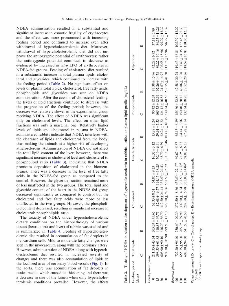
Table
2.
ToxicityofNDEA
inrelationto
developmentofhypercholesterolemia
inrabbits–effect
onplasm
alipid
profile
(mg/dL)
Feedingperiod
(days)
Totallipids
Cholesterol
Freefattyacids
Phospholipids
Glycerides
CE
CE
CE
CE
CE
Dev
elo
pm
ent
ph
ase
0211.35743.24
205.24745.65
62.3378.51
64.8779.23
16.3273.12
15.8873.24
90.42712.24
87.38715.96
42.2876.72
37.1175.89
15
398.33761.83
410.83769.75
182.68716.77
194.13718.25
30.1076.41
29.9175.53
105.33713.59
102.26714.78
80.22710.16
84.53711.29
30
608.90790.10
616.70786.70
312.50724.43
337.50724.43
65.7076.39
62.2475.22
124.12716.88
121.67714.97
106.58715.96
95.29713.37
60
974.307108.90
999.507117.30
522.50778.66
567.50775.29
107.18716.46
103.16716.36
230.84740.56
232.55741.30
113.78715.96
96.29715.37
Rev
ersa
lp
ha
se
90
722.20795.40
750.60790.30
377.50718.86
437.50727.17a
69.6775.87
65.1376.29b
155.60722.80
142.58720.24
119.43716.81
105.39713.27
120
655.55795.60
705.95789.90
302.50714.84
367.50717.52a
71.2576.37
64.1476.56
150.40720.12
146.86717.28
131.40724.35
127.45725.33
150
607.30794.30
641.50790.50
287.50715.98
335.00714.04a
68.8776.35
67.9277.14
132.34718.96
128.52720.26
118.59716.81
110.06712.18
Values
are
mean7S.D
.,n¼
6.C¼
Controlgroup.E¼
Experim
entalgroup(N
DEA
treated).
aPo0.01.
bPo0.05withrespectto
controlgroup.
G. Mittal et al. / Experimental and Toxicologic Pathology 59 (2008) 409–414 411
NDEA administration resulted in a substantial andsignificant increase in osmotic fragility of erythrocytesand the effect was more pronounced with increasingfeeding period and continued to increase even afterwithdrawal of hypercholesterolemic diet. Moreover,withdrawal of hypercholesterolemic diet did not im-prove the antioxygenic potential of erythrocytes; ratherthe antioxygenic potential continued to decrease asevidenced by increased in vitro LPO of erythrocytes inNDEA-fed groups. Feeding of cholesterol diet resultedin a substantial increase in total plasma lipids, choles-terol and glycerides, which continued to increase withthe feeding period (Table 2). No significant effect onlevels of plasma total lipids, cholesterol, free fatty acids,phospholipids and glycerides was seen on NDEAadministration. After the cession of cholesterol feeding,the levels of lipid fractions continued to decrease withthe progression of the feeding period; however, thedecrease was relatively slower in the experimental groupreceiving NDEA. The effect of NDEA was significantonly on cholesterol levels. The effect on other lipidfractions was only a marginal one. Relatively higherlevels of lipids and cholesterol in plasma in NDEA-administered rabbits indicate that NDEA interferes withthe clearance of lipids and cholesterol from the body,thus making the animals at a higher risk of developingatherosclerosis. Administration of NDEA did not affectthe total lipid content of the liver; however, there wassignificant increase in cholesterol level and cholesterol tophospholipid ratio (Table 3), indicating that NDEApromotes deposition of cholesterol in the biomem-branes. There was a decrease in the level of free fattyacids in the NDEA-fed group as compared to thecontrol. However, the glyceride fraction remained moreor less unaffected in the two groups. The total lipid andglyceride content of the heart in the NDEA-fed groupdecreased significantly as compared to control but thecholesterol and free fatty acids were more or lessunaffected in the two groups. However, the phospholi-pid content decreased, resulting in significant increase incholesterol: phospholipids ratio.
The toxicity of NDEA under hypercholesterolemicdietary conditions on the histopathology of varioustissues (heart, aorta and liver) of rabbits was studied andis summarized in Table 4. Feeding of hypercholester-olemic diet resulted in accumulation of fat droplets inmyocardium cells. Mild to moderate fatty changes wereseen in the myocardium along with the coronary artery.However, administration of NDEA along with hyperch-olesterolemic diet resulted in increased severity ofchanges and there was also accumulation of lipids inthe localized area of coronary blood vessels (Fig. 1). Inthe aorta, there was accumulation of fat droplets intunica media, which caused its thickening and there wasa decrease in size of the lumen when only hypercholes-terolemic conditions prevailed. However, the effects
ARTICLE IN PRESS
Table 3. Effect of NDEA on hepatic and cardiac lipid profile (mg/g tissue)
Parameter Liver Heart
C E C E
Total lipids 76.2571.87 77.5071.73 Total lipids 57.5073.88 46.2573.44a
Values are mean7S.D., n ¼ 6. C ¼ Control group. E ¼ Experimental group (NDEA treated).aPo0.01.bPo0.05 with respect to the control group.
Table 4. Oral toxicity of NDEA in relation to hypercholes-
terolemia in rabbits – effect on histopathology of various
tissues
Tissue/pathological changes Treatment/effect
C E
Heart
Accumulation of cholesterol in tunica
intima of coronary blood vessel
++ +++
Accumulation of lipids in
myocardium
++ +++
Aorta
Accumulation of lipids ++ +++
Liver
Accumulation of fat/lipid in
hepatocytes
+++ +++
Infiltration of mononuclear cells + +++
Fibrosis – +++
Regeneration of hepatocytes + ++
C ¼ Control group. E ¼ Experimental group (NDEA treated).
� Normal.
+Mild.
++Moderate.
+++Severe.
Fig. 1. Section of heart showing accumulation of lipids in the
localized area of coronary blood vessel (arrow) in the presence
of NDEA (H.E.� 20 magnification).
Fig. 2. Section of aorta showing increased thickness of tunica
media (arrow) due to accumulation of fat droplets in the
presence of NDEA (H.E.� 40 magnification).
G. Mittal et al. / Experimental and Toxicologic Pathology 59 (2008) 409–414412
were more severe in the presence of NDEA (Fig. 2).Surface area of aorta involved in atherosclerosis(sudanophilic lesions) was relatively higher in NDEA-fed groups (25.4071.81%, n ¼ 5) as compared to thatof the control (15.2071.48%, n ¼ 5), indicating thatNDEA induces increased development of atherosclero-sis. In case of liver, there were severe granular changes inthe hepatocytes along with infiltration of mononuclearcells and accumulation of lipids (Figs. 3 and 4).Cholesterol overload is already known to induce liverfibrosis in rabbits (Buyssens et al., 1996). The effectswere, however, more profound in animals of experi-mental group receiving NDEA in their diet (Fig. 5).Fibrosis and other hepatic degenerative changes can be
attributed to the increased LPO of hepatocytes uponNDEA administration, indicating peroxidative damageleading to histopathological degenerative changes.
ARTICLE IN PRESS
Fig. 3. Section of liver showing granular changes in hepato-
cytes (lower arrow) and lymphocyte aggregation (upper arrow)
in the absence of NDEA (H.E.� 40 magnification) (middle
arrow is an artifact).
Fig. 4. Section of liver showing proliferation of connective
tissue indicating fibrosis (arrows) in the absence of NDEA
(H.E.� 40 magnification).
Fig. 5. Section of liver showing accumulation of large-sized fat
droplets (arrows) and fibrosis in the presence of NDEA
(H.E.� 40 magnification).
G. Mittal et al. / Experimental and Toxicologic Pathology 59 (2008) 409–414 413
Discussion
Nitroso compounds such as NDEA have beensuggested to cause oxidative stress and cellular injurydue to the involvement of free radicals (Bartsch et al.,1989). Exposure to NDEA and other nitrosamines hasbeen shown to increase LPO and alter the antioxidantstatus of experimental animals (Mittal et al., 2006,2007). It is well established now that ROS play a pivotalrole in the development and progression of athero-sclerosis, a disease which is the major source ofmorbidity and mortality in the world, and is character-ized by the accumulation of cholesterol deposits in large-and medium-sized arteries (Stocker and Keaney, 2004;Ross, 1993). Keeping this in mind, it was thought thatexposure to nitrosamines such as NDEA through food,beverage or smoking, as already mentioned earlier, maycontribute to an increased risk of developing athero-sclerosis in a vast majority of population that consumesa high-fat diet.
In the present study, oral administration of NDEAhas been shown to result in increased LPO of blood anddecreased lipid clearance in experimental rabbits. Theincreased osmotic fragility in experimental rabbits canbe due to the increased LPO of erythrocytes. Moreover,withdrawal of hypercholesterolemic diet did not im-prove the antioxygenic potential of erythrocytes; ratherthe antioxygenic potential continued to decrease asevidenced by increased in vitro LPO of erythrocytes inNDEA-fed groups. This could be due to the continuedprevalence of hypercholesterolemia/hyperlipidemia ob-served. NDEA administration has already been shownto increase in vitro LPO of erythrocytes in normal aswell as hypercholesterolemic rats (Kaushal et al., 2003;Mittal et al., 2006).
Relatively higher levels of lipids and cholesterol inplasma in NDEA-administered rabbits indicate thatNDEA interferes with the clearance of lipids andcholesterol from the body, thus making the animals ata higher risk of developing atherosclerosis. Moreover, asignificant increase in cholesterol: phospholipids ratio inthe heart and liver tissues of NDEA-fed rabbits indicatethat NDEA promotes deposition of cholesterol in thebiomembranes. The decreased lipid clearance observedon NDEA administration can be due to the alteredfunctions of liver and extra-hepatic tissues resultingfrom the oxidative stress caused by NDEA, as it hasbeen shown to increase LPO of hepatic and other tissuesby interfering with their antioxidant system (Mittalet al., 2006; Bansal et al., 2000).
The effect of NDEA on the histopathology of hearttissue of hypercholesterolemic rabbits showed anincreased accumulation of fat droplets in myocardiumcells and accumulation of lipid in the localized area ofcoronary blood vessels as compared to control. Thisagrees well with the observed increase in cholesterolto phospholipid ratio in heart. The relatively highersurface area of the aorta involved in atherosclerosis
ARTICLE IN PRESSG. Mittal et al. / Experimental and Toxicologic Pathology 59 (2008) 409–414414
(sudanophilic lesions) in NDEA-fed groups (nearly 25%)as compared to that of the control (nearly 15%) indicatesthat NDEA induces increased development of athero-sclerosis. Hypercholesterolemia is obviously known toincrease atherosclerotic changes in the aorta (Das et al.,2006; Mahfouz et al., 1997). Early events of atherogenesishave been reported to be mediated by generation of ROSand oxidation of lipids and proteins in the vascular wall(Stocker and Keaney, 2004; Cai and Harrison, 2000). AsNDEA has been shown to cause oxidative stress andcellular injury due to involvement of free radicals (Bansalet al., 1996; Kaushal et al., 2003), the higher degree ofatherosclerotic plaque observed is obvious.
Thus it appears that oral administration of NDEAresults in increased LPO of blood and decreased lipidclearance, which in turn may result in an increaseddegree of atherosclerosis. The underlying molecularmechanisms, however, need to be further investigated.
Acknowledgment
The authors are grateful to the Head, Department ofBiochemistry and Chemistry for providing necessaryfacilities for the study, and to Mr. Avtar Singh fortechnical help.
References
Aiub CA, Pinto LF, Felzenszwalb I. N-Nitrosodiethylamine
mutagenicity at low concentrations. Toxicol Lett 2003;145:
36–45.
Bansal AK, Bhatnagar D. In vitro effect of N-nitrosodiethy-
lamine on lipid peroxidation and antioxidant enzymes in rat
erythrocytes. Fresenius Environ Bull 1998;7:264–8.
Bansal AK, Bhatnagar D, Soni GL. In vitro effect of
N-nitrosodiethylamine on lipid peroxidation and antiox-
idant system in human erythrocytes. Toxicol In Vitro
1996;10:649–53.
Bansal AK, Trivedi R, Soni GL, Bhatnagar D. Hepatic and
renal oxidative stress in acute toxicity of N-nitrosodiethy-
lamine in rats. Indian J Exp Biol 2000;38:916–20.
Bartsch H. N-nitroso compounds and human cancer, where do
we stand? IARC Sci Publ 1991;105:1–10.
Bartsch H, Hietanen E, Malaveille C. Carcinogenic nitrosa-
mines: free radical aspects of their action. Free Rad Biol
Med 1989;7:637–44.
Buyssens N, Kockx MM, Herman AG, Lazou JM, Van den
Berg K, Wisse E, et al. Centrolobular liver fibrosis in the