The Journal of Neuroscience, March 1992, 72(3): 1101-I 114 /n situ Histochemical Localization of Type I Interleukin-1 Receptor Messenger RNA in the Central Nervous System, Pituitary, and Adrenal Gland of the Mouse Emmett T. Cunningham, Jr., lea Etsuko Wada,* Donald B. Carter,3 Daniel E. Tracey,4 James F. Battey,* and Errol B. De Souzal ‘Neurobiology Laboratory, National Institute on Drug Abuse, Addiction Research Center, Baltimore, Maryland 21224, *Laboratory of Neurochemistry, National Institute of Neurological Disorders and Stroke, National Institutes of Health, Bethesda, Maryland 20892, and 3Molecular Biology and 4Hypersensitivity Diseases Research, The Upjohn Company, Kalamazoo, Michigan 49007 The cytokine interleukin-1 (IL-l) has a number of biologic activities, including pronounced effects on the nervous and neuroendocrine systems. In this study, in situ histochemical techniques were used to investigate the distribution of cells expressing type I IL-1 receptor mRNA in the CNS, pituitary, and adrenal gland of the mouse. Hybridization of %-labeled antisense cRNA probes derived from a murine T-cell IL-1 receptor cDNA revealed a distinct regional distribution of the type I IL-1 receptor, both in brain and in the pituitary gland. In the brain, an intense signal was observed over the granule cell layer of the dentate gyrus, over the entire midline raphe system, over the choroid plexus, and over endothelial cells of postcapillary venules throughout the neuraxis. A weak to moderate signal was observed over the pyramidal ceil layer of the hilus and CA3 region of the hippocampus, over the anterodorsal thalamic nucleus, over Purkinje cells of the cer- ebellar cortex, and in scattered clusters over the external- most layer of the median eminence. In the pituitary gland, a dense and homogeneously distributed signal was observed over the entire anterior lobe. No autoradiographic signal above background was observed over the posterior and in- termediate lobes of the pituitary, or over the adrenal gland. This study therefore provides evidence for discrete receptor substrates subserving the central effects of IL-l, thus sup- porting the notion that IL-1 acts as a neurotransmitter/neu- romodulator in brain. It also supports studies suggesting that IL- 1 -mediated activation of the hypothalamic-pituitary-ad- renal axis occurs primarily at the level of the brain and/or pituitary gland. Interleukin-1 (IL-l) is one of a growing number of cytokines that mediate important regulatory interactions between lym- phocytes and many other cell types (see Dinarello, 1989). IL-l has also been identified as a key mediator in the acute-phase Received July 10, 1991; revised Oct. 15, 1991; accepted Oct. 23, 1991. We thank Ms. Melanie Holden, Ms. Jean Gonnella, and Mr. Mark Fleming for their superb photographic assistance, and Mrs. Deborah Gorman for help with preparation of the manuscript. Correspondence should be addressed to Dr. Errol B. De Souza, The Du Pont Merck Pharmaceutical Company, Experimental Station, E40014352, Wilmington, DE 19880-0400. a Present address: The Salk Institute for Biological Studies, La Jolla, CA 92037. Copyright 0 1992 Society for Neuroscience 0270-6474/92/121 lOl-14$05.00/O response, a generalizedhost reaction to microbial invasion, an- tigenic challenge, or large-scale tissue injury (see Dinarello, 1984). As its name suggests, the acute-phase response is rapid in onset and is characterized by marked alterations in metabolic, en- docrinologic, immunologic, and neurologic functions (seeDi- narello, 1984). Neurologic changes include the induction of fever (see Kluger, 199 l), increased duration of slow-wave sleep (Opp et al., 1991) decreased appetite (McCarthy et al., 1985; Mro- sovsky et al., 1989) activation of the hypothalamic-pituitary- adrenal (HPA) axis (Woloski et al., 1985; Besedovsky et al., 1986; Berkenboschet al., 1987; Bernton et al., 1987; Sapolsky et al., 1987; seealso Lumpkin, 1987; Bateman et al., 1989; Dunn, 1990), and inhibition of the hypothalamic-pituitary%o- nadal axis (Rivier and Vale, 1989; Kalra et al., 1990), all of which are thought to be mediated by IL-1 (see Blatteis, 1988). In addition, centrally administered IL-l has been found to el- evate circulating levels of growth hormone and prolactin, while inhibiting the release of thyroid-stimulating hormone (Rettori et al., 1987; Dubuis et al., 1988). More recently, intracerebro- ventricular IL-l has also been observed to inhibit both gastric acid (Ishikawa et al., 1990; Saperas et al., 1990; Uehara et al., 1990; Okumura et al., 199 1; Shibasaki et al., 1991) and pepsin (Okumura et al., 1990, 1991) secretion, and to have profound analgesic properties (Nakamura et al., 1988). A number of recent radioligand binding studies have used lZSI-labeled forms of IL-l to localize receptors that might me- diate the above-mentioned effects of the cytokine in the rodent CNS (Farrar et al., 1987; Katsuura et al., 1988; De Souzaet al., 1989; Haour et al., 1990; Takao et al., 1990; Ban et al., 1991). These studieshave, however, produced somewhatdifferent re- sults, with earlier studies in rat brain reporting a fairly wide- spreaddistribution (Farrar et al., 1987; Katsuura et al., 1988), and more recent studies in mouse brain (Haour et al., 1990; Takao et al., 1990; Ban et al., 1991) describing a relatively restricted localization particularly in the dentate gyrus of the hippocampusand choroid plexus. These latter studies also de- scribeddiffuse binding over the anterior pituitary (Haour et al., 1990; Takao et al., 1990; Ban et al., 1991). In addition, marked species differences have been demonstrated in the pattern of central binding using human recombinant human IL-la in mouse, rat, and guinea pig (Takao et al., 1990). Finally, it is well known that such binding studies provide limited insight into the site of synthesis of putative receptors,sinceligands may
Transcript
The Journal of Neuroscience, March 1992, 72(3): 1101-I 114
/n situ Histochemical Localization of Type I Interleukin-1 Receptor Messenger RNA in the Central Nervous System, Pituitary, and Adrenal Gland of the Mouse
Emmett T. Cunningham, Jr., lea Etsuko Wada,* Donald B. Carter,3 Daniel E. Tracey,4 James F. Battey,* and Errol B. De Souzal
‘Neurobiology Laboratory, National Institute on Drug Abuse, Addiction Research Center, Baltimore, Maryland 21224, *Laboratory of Neurochemistry, National Institute of Neurological Disorders and Stroke, National Institutes of Health, Bethesda, Maryland 20892, and 3Molecular Biology and 4Hypersensitivity Diseases Research, The Upjohn Company, Kalamazoo, Michigan 49007
The cytokine interleukin-1 (IL-l) has a number of biologic activities, including pronounced effects on the nervous and neuroendocrine systems. In this study, in situ histochemical techniques were used to investigate the distribution of cells expressing type I IL-1 receptor mRNA in the CNS, pituitary, and adrenal gland of the mouse. Hybridization of %-labeled antisense cRNA probes derived from a murine T-cell IL-1 receptor cDNA revealed a distinct regional distribution of the type I IL-1 receptor, both in brain and in the pituitary gland. In the brain, an intense signal was observed over the granule cell layer of the dentate gyrus, over the entire midline raphe system, over the choroid plexus, and over endothelial cells of postcapillary venules throughout the neuraxis. A weak to moderate signal was observed over the pyramidal ceil layer of the hilus and CA3 region of the hippocampus, over the anterodorsal thalamic nucleus, over Purkinje cells of the cer- ebellar cortex, and in scattered clusters over the external- most layer of the median eminence. In the pituitary gland, a dense and homogeneously distributed signal was observed over the entire anterior lobe. No autoradiographic signal above background was observed over the posterior and in- termediate lobes of the pituitary, or over the adrenal gland. This study therefore provides evidence for discrete receptor substrates subserving the central effects of IL-l, thus sup- porting the notion that IL-1 acts as a neurotransmitter/neu- romodulator in brain. It also supports studies suggesting that IL- 1 -mediated activation of the hypothalamic-pituitary-ad- renal axis occurs primarily at the level of the brain and/or pituitary gland.
Interleukin-1 (IL-l) is one of a growing number of cytokines that mediate important regulatory interactions between lym- phocytes and many other cell types (see Dinarello, 1989). IL-l has also been identified as a key mediator in the acute-phase
Received July 10, 1991; revised Oct. 15, 1991; accepted Oct. 23, 1991. We thank Ms. Melanie Holden, Ms. Jean Gonnella, and Mr. Mark Fleming for
their superb photographic assistance, and Mrs. Deborah Gorman for help with preparation of the manuscript.
Correspondence should be addressed to Dr. Errol B. De Souza, The Du Pont Merck Pharmaceutical Company, Experimental Station, E40014352, Wilmington, DE 19880-0400.
a Present address: The Salk Institute for Biological Studies, La Jolla, CA 92037.
Copyright 0 1992 Society for Neuroscience 0270-6474/92/121 lOl-14$05.00/O
response, a generalized host reaction to microbial invasion, an- tigenic challenge, or large-scale tissue injury (see Dinarello, 1984). As its name suggests, the acute-phase response is rapid in onset and is characterized by marked alterations in metabolic, en- docrinologic, immunologic, and neurologic functions (see Di- narello, 1984). Neurologic changes include the induction of fever (see Kluger, 199 l), increased duration of slow-wave sleep (Opp et al., 1991) decreased appetite (McCarthy et al., 1985; Mro- sovsky et al., 1989) activation of the hypothalamic-pituitary- adrenal (HPA) axis (Woloski et al., 1985; Besedovsky et al., 1986; Berkenbosch et al., 1987; Bernton et al., 1987; Sapolsky et al., 1987; see also Lumpkin, 1987; Bateman et al., 1989; Dunn, 1990), and inhibition of the hypothalamic-pituitary%o- nadal axis (Rivier and Vale, 1989; Kalra et al., 1990), all of which are thought to be mediated by IL-1 (see Blatteis, 1988). In addition, centrally administered IL-l has been found to el- evate circulating levels of growth hormone and prolactin, while inhibiting the release of thyroid-stimulating hormone (Rettori et al., 1987; Dubuis et al., 1988). More recently, intracerebro- ventricular IL-l has also been observed to inhibit both gastric acid (Ishikawa et al., 1990; Saperas et al., 1990; Uehara et al., 1990; Okumura et al., 199 1; Shibasaki et al., 199 1) and pepsin (Okumura et al., 1990, 1991) secretion, and to have profound analgesic properties (Nakamura et al., 1988).
A number of recent radioligand binding studies have used lZSI-labeled forms of IL-l to localize receptors that might me- diate the above-mentioned effects of the cytokine in the rodent CNS (Farrar et al., 1987; Katsuura et al., 1988; De Souza et al., 1989; Haour et al., 1990; Takao et al., 1990; Ban et al., 1991). These studies have, however, produced somewhat different re- sults, with earlier studies in rat brain reporting a fairly wide- spread distribution (Farrar et al., 1987; Katsuura et al., 1988), and more recent studies in mouse brain (Haour et al., 1990; Takao et al., 1990; Ban et al., 1991) describing a relatively restricted localization particularly in the dentate gyrus of the hippocampus and choroid plexus. These latter studies also de- scribed diffuse binding over the anterior pituitary (Haour et al., 1990; Takao et al., 1990; Ban et al., 199 1). In addition, marked species differences have been demonstrated in the pattern of central binding using human recombinant human IL-la in mouse, rat, and guinea pig (Takao et al., 1990). Finally, it is well known that such binding studies provide limited insight into the site of synthesis of putative receptors, since ligands may
1102 Cunningham et al. * IL-l Receptor mRNA in Brain, Pituitary, and Adrenal Gland
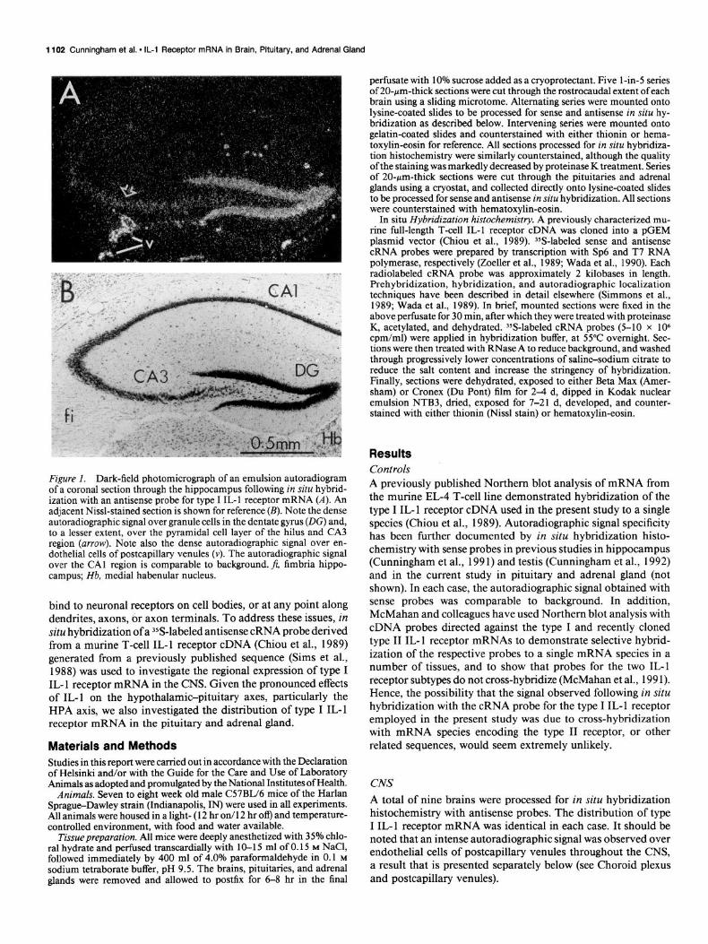
Figure I. Dark-field photomicrograph of an emulsion autoradiogram of a coronal section through the hippocampus following in situ hybrid- ization with an antisense probe for type I IL-l receptor mRNA (A). An adjacent Nissl-stained section is shown for reference (B). Note the dense autoradiographic signal over granule cells in the dentate gyrus (DC) and, to a lesser extent, over the pyramidal cell layer of the hilus and CA3 region (arrow). Note also the dense autoradiographic signal over en- dothelial cells of postcapillary venules (v). The autoradiographic signal over the CA1 region is comparable to background. fi, fimbria hippo- campus; Hb, medial habenular nucleus.
bind to neuronal receptors on cell bodies, or at any point along dendrites, axons, or axon terminals. To address these issues, in situ hybridization of a 35S-labeled antisense cRNA probe derived from a murine T-cell IL-l receptor cDNA (Chiou et al., 1989) generated from a previously published sequence (Sims et al., 1988) was used to investigate the regional expression of type I IL- 1 receptor mRNA in the CNS. Given the pronounced effects of IL-l on the hypothalamic-pituitary axes, particularly the HPA axis, we also investigated the distribution of type I IL-1 receptor mRNA in the pituitary and adrenal gland.
Materials and Methods Studies in this report were carried out in accordance with the Declaration of Helsinki and/or with the Guide for the Care and Use of Laboratory Animals as adopted and promulgated by the National Institutes of Health.
Animals. Seven to eight week old male C57BIJ6 mice of the Harlan Spragu+Dawley strain (Indianapolis, IN) were used in all experiments. All animals were housed in a light- ( 12 hr on/l 2 hr ofl) and temperature- controlled environment, with food and water available.
Tissue preparation. All mice were deeply anesthetized with 35% chlo- ral hydrate and perfused transcardially with 1 O- 15 ml of 0.15 M NaCl, followed immediatelv bv 400 ml of 4.0% paraformaldehyde in 0.1 M sodium tetraborate buffer, pH 9.5. The brains, pituitaries; and adrenal glands were removed and allowed to postfix for 6-8 hr in the final
pet&sate with 10% sucrose added as a cryoprotectant. Five l-in-5 series of 20-pm-thick sections were cut through the rostrocaudal extent of each brain using a sliding microtome. Alternating series were mounted onto lysine-coated slides to be processed for sense and antisense in situ hy- bridization as described below. Intervening series were mounted onto gelatin-coated slides and counterstained with either thionin or hema- toxylin-eosin for reference. All sections processed for in situ hybridiza- tion histochemistry were similarly counterstained, although the quality ofthe staining was markedly decreased by proteinase K treatment. Series of 20-pm-thick sections were cut through the pituitaries and adrenal glands using a cryostat, and collected directly onto lysine-coated slides to be processed for sense and antisense in situ hybridization. All sections were counterstained with hematoxylin-eosin.
In situ Hybridization histochemistry. A previously characterized mu- tine full-length T-cell IL-l receptor cDNA was cloned into a pGEM plasmid vector (Chiou et al., 1989). YS-labeled sense and antisense cRNA probes were prepared by transcription with Sp6 and T7 RNA polymerase, respectively (Zoeller et al., 1989; Wada et al., 1990). Each radiolabeled cRNA probe was approximately 2 kilobases in length. Prehybridization, hybridization, and autoradiographic localization techniques have been described in detail elsewhere (Simmons et al., 1989; Wada et al., 1989). In brief, mounted sections were fixed in the above perfusate for 30 min, after which they were treated with proteinase K, acetylated, and dehydrated. YS-labeled cRNA probes (5-10 x lo6 cpm/ml) were applied in hybridization buffer, at 55°C overnight. Sec- tions were then treated with RNase A to reduce background, and washed through progressively lower concentrations of salin+sodium citrate to reduce the salt content and increase the stringency of hybridization. Finally, sections were dehydrated, exposed to either Beta Max (Amer- sham) or Cronex (Du Pont) film for 2-4 d, dipped in Kodak nuclear emulsion NTB3, dried, exposed for 7-21 d, developed, and counter- stained with either thionin (Nissl stain) or hematoxylin-eosin.
Results Controls A previously published Northern blot analysis of mRNA from the murine EL-4 T-cell line demonstrated hybridization of the type I IL- 1 receptor cDNA used in the present study to a single species (Chiou et al., 1989). Autoradiographic signal specificity has been further documented by in situ hybridization histo- chemistry with sense probes in previous studies in hippocampus (Cunningham et al., 1991) and testis (Cunningham et al., 1992) and in the current study in pituitary and adrenal gland (not shown). In each case, the autoradiographic signal obtained with sense probes was comparable to background. In addition, McMahan and colleagues have used Northern blot analysis with cDNA probes directed against the type I and recently cloned type II IL- 1 receptor mRNAs to demonstrate selective hybrid- ization of the respective probes to a single mRNA species in a number of tissues, and to show that probes for the two IL-l receptor subtypes do not cross-hybridize (McMahan et al., 199 1). Hence, the possibility that the signal observed following in situ hybridization with the cRNA probe for the type I IL- 1 receptor employed in the present study was due to cross-hybridization with mRNA species encoding the type II receptor, or other related sequences, would seem extremely unlikely.
CNS
A total of nine brains were processed for in situ hybridization histochemistry with antisense probes. The distribution of type I IL- 1 receptor mRNA was identical in each case. It should be noted that an intense autoradiographic signal was observed over endothelial cells of postcapillary venules throughout the CNS, a result that is presented separately below (see Choroid plexus and postcapillary venules).
The Journal of Neuroscience, March 1992, E’(3) 1103
Forebrain. The strongest autoradiographic signal in the fore- brain was found in the hippocampal formation (Cunningham et al., 199 l), where an intense autoradiographic signal was ob- served over the granule cell layer of the dentate gyrus, and a weak to moderate autoradiographic signal was observed over the pyramidal cell layer of the hilus and the CA3 region (Fig. 1). The autoradiographic signal over other aspects of the hip- pocampal formation, including the CA2 and CA1 regions, the subiculum, and the entorhinal area, was comparable to back- ground. A similar pattern was found at all rostrocaudal levels.
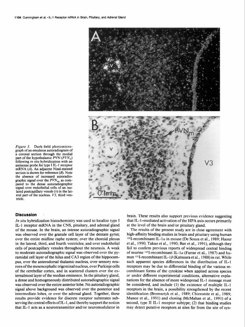
A weak to moderate autoradiographic signal was also ob- served over the anterodorsal nucleus of the thalamus (Fig. 2). The autoradiographic signal over other aspects of the forebrain, including the hypothalamic paraventricular nucleus (PVN, Fig. 3), was comparable to background. While the autoradiographic signal over most aspects of the median eminence was also com- parable to background, scattered clusters of autoradiographic signal were consistently observed over its externalmost layer (Fig. 4). The quality of counterstaining in the median eminence following proteinase K treatment of frozen sections did not, however, allow for precise localization to either neurons or the small hypothalamic-hypophyseal portal vessels known to oc- cupy this layer.
Brainstem. An intense autoradiographic signal was observed over all aspects of the midline raphe system (see Tork, 1985), including all subdivisions of the dorsal raphe (Fig. 5), the median raphe, the raphe pontis, the raphe magnus, the raphe obscurus, and the raphe pallidus (not shown). An autoradiographic signal above background was also observed over those portions of the pontine and medullary reticular formation known to contain extra-midline serotonergic neurons, but this signal was almost invariably associated with postcapillary venules. A moderate to dense autoradiographic signal was observed over sensory neu- rons of the mesencephalic trigeminal nucleus (Fig. 6). A weak to moderate autoradiographic signal was also found over the Purkinje cell layer of the entire cerebellar cortex (Figs. 6,7). The autoradiographic signal over other aspects of the brainstem was comparable to background.
Spinal cord. Although a comprehensive analysis of all spinal levels was not performed, serial sections were examined through cervical level 3 or 4 in all animals. No autoradiographic signal above background was observed over any portion of the cervical spinal cord.
Choroid plexus and postcapillary venules. A dense autoradio- graphic signal was observed over the choroid plexus in the lat- eral, third, and fourth ventricles. While the intensity of the autoradiographic signal did not allow for localization to a spe- cific cell type of the choroid plexus, densely labeled fronds of choroid plexus were often observed originating from venules, the endothelium of which was also densely labeled (Fig. 8).
An intense autoradiographic signal was found over endothe- lial cells of postcapillary venules throughout the CNS, both in the parenchyma and at the pial surface (Fig. 9). Such venules were readily recognized by their size (50-400 Mm) and the ab- sence of a muscular wall. An autoradiographic signal over ar- terioles at the pial surface (Fig. 9) and over capillaries through- out the CNS, was comparable to background.
Pituitary A total of 12 pituitaries were processed for in situ hybridization histochemistry with antisense probes. A dense and homoge-
Figure 2. Dark-field photomicrograph of an emulsion autoradiogram of a coronal section through the dorsal thalamus following in situ hy- bridization with an antisense probe for type I IL- 1 receptor mRNA (A). An adjacent Nissl-stained section is shown for reference (B). Note the moderate autoradiographic signal over the anterodorsal nucleus (AD), as compared to the dense autoradiographic signal over granule cells in the dentate gyrus (DC), and over endothelial cells of postcapillary ve- nules (v). Hb, medial habenular nucleus; sm, stria medullat-is.
neously distributed autoradiographic signal was observed over the entire anterior lobe in each case. The autoradiographic signal over the posterior and intermediate lobes was comparable to background (Fig. 10). As with the median eminence, the quality of counterstaining following proteinase K treatment of frozen sections did not allow for localization to specific cell types within the anterior lobe.
Adrenal gland A total of 16 adrenal glands from 12 animals were processed for in situ hybridization histochemistry with antisense probes. In each case, the autoradiographic signal over the medulla and all cortical layers, including the zona glomerulosa, the zona fasciculata, and the zona reticularis, was comparable to back- ground (Fig. 11).
1104 Cunningham et al. l IL-1 Receptor mRNA in Brain, Pituitary, and Adrenal Gland
Figure 3. Dark-field photomicro- graph of an emulsion autoradiogram of a coronal section through the medial part of the hypothalamic PVN (PI’N,) following in situ hybridization with an antisense probe for type I IL- 1 receptor mRNA (A). An adjacent Nissl-stained section is shown for reference (B). Note the absence of increased autoradio- graphic signal over the Pm,,,, as com- pared to the dense automdiographic sigd aver endathehal cells af an isa- lated postcapillary venule (v) in the lat- eral part of the nucleus. V3, third ven- tricle.
Discussion In situ hybridization histochemistry was used to localize type I IL-I receptor mRNA in the CNS, pituitary, and adrenal gland of the mouse. In the brain, an intense autoradiographic signal was observed over the granule cell layer of the dentate gyrus; over the entire midline raphe system; over the choroid plexus in the lateral, third, and fourth ventricles; and over endothelial cells of postcapillary venules throughout the neuraxis. A weak to moderate autoradiographic signal was observed over the py- ramidal cell layer of the hilus and CA3 region of the hippocam- pus, over the anterodorsal thalamic nucleus, over sensory neu- rons of the mesencephalic trigeminal nucleus, over Purkinje cells of the cerebellar cortex, and in scattered clusters over the ex- ternalmost layer of the median eminence. In the pituitary gland, a dense and homogeneously distributed autoradiographic signal was observed over the entire anterior lobe. No autoradiographic signal above background was observed over the posterior and intermediate lobes, or over the adrenal gland. Together, these results provide evidence for discrete receptor substrates sub- serving the central effects of IL- 1, and thereby support the notion that IL- 1 acts as a neurotransmitter and/or neuromodulator in
brain. These results also support previous evidence suggesting that IL- l-mediated activation of the HPA axis occurs primarily at the level of the brain and/or pituitary gland.
The results of the present study are in close agreement with high-affinity binding studies in brain and pituitary using human 1251-recombinant IL- 1 (Y in mouse (De Souza et al., 1989; Haour et al., 1990; Takao et al., 1990; Ban et al., 199 l), although they fail to confirm previous reports of widespread central binding of murine 1*51-recombinant IL-la (Farrar et al., 1987) and hu- man 1251-recombinant IL- l/3 (Katsuura et al., 1988) in rat. While such apparent species differences in the distribution of IL-I receptors may be due to differential binding of the various re- combinant forms of the cytokine when applied across species or under different experimental conditions, alternative expla- nations for the absence of more widespread IL- 1 message must be considered, and include (1) the existence of multiple IL- 1 receptors in the brain, a possibility strengthened by the recent identification (Bromsztyk et al., 1989; Chizzonite et al., 1989; Munoz et al., 1991) and cloning (McMahan et al., 1991) of a second, type II IL-1 receptor subtype; (2) that binding studies may detect putative receptors at sites far from the site of syn-
The Journal of Neuroscience, March 1992, 12(3) 1105
Figure 4. Dark-field photomicrograph of an emulsion autoradiogram of a coronal section through the median eminence following in situ hybrid- ization with an antisense probe for type I IL-1 receptor mRNA (A). The same section counterstained with hematoxylin-eosin is shown for reference (A’). Note the absence of increased autoradiographic signal over most of the median eminence, with the exception of scattered clusters of autora- diographic signal over its externalmost layer (arrows), a layer known to contain the small hypothalamiohypophyseal portal vessels. V3, third ventricle; Ar, arcuate nucleus.
thesis, such as at axon terminals; and (3) that some areas that synthesize type I IL-l receptor may do so at such a low level as to avoid detection with current in situ techniques.
The restricted localization of autoradiographic signal for type I IL-l receptor mRNA to well-recognized cytoarchitectonic boundaries in the hippocampus, the thalamus, the raphe, the mesencephalic trigeminal nucleus, and the cerebellar cortex strongly suggests significant neuronal synthesis of the receptor. Additional support for neuronal expression of type I IL-1 re- ceptor,also comes from at least four lines of evidence. First, the most intense autoradiographic signal in the hippocampus was observed over the granule cell layer of the dentate gyrus, a layer virtually devoid of glial cells (Rickmann et al., 1987). Second, IL- 1 binding in the hippocampus virtually disappears following application of the selective neurotoxins to this region (Takao et al., 1990; Ban et al., 199 1). Third, IL- 1 increases synaptic in- hibition in rat hippocampal neurons in vitro (Zeise et al., 199 1). And fourth, hippocampal neurons respond to IL- 1 by increasing production of NGF, both in vitro (Friedman et al., 199 1; Spran- ger et al., 1990), and in vivo (Spranger et al., 1990). It should be noted, however, that astrocytes cultured from both hippo- campus (Friedman et al., 199 1) and cortex (Gradient et al., 1990) have also been found to respond to IL- 1 with an increase in the production of NGF.
Given the above-mentioned response of hippocampal neu- rons and glial cells to IL-l in vitro and in viva, it is perhaps not surprising that the cytokine has been suggested to promote neu- ronal survival and/or repair in this region, a notion fostered by the recent description of increased IL-l production in hippo- campal astrocytes following perferant path deafferentation, con-
current with the onset of cholinergic sprouting (Fagan and Gage, 1990). However, the hippocampus has also been suggested to be involved in thermoregulation, since it has been shown that hippocampal stimulation may directly influence thermorespon- sive neurons in the preoptic area (Boulant and Demieville, 1977; Hori et al., 1982). Moreover, IL-l might exert at least some of its central effects on the hypothalamic-pituitary axes at the level of the hippocampus, as has been postulated for glucocorticoids and regulation of the HPA axis (see Keller-Wood and Dallman, 1984; Jacobson and Sapolsky, 199 1). This latter possibility seems particularly appealing given the relative absence of type I IL- 1 receptor mRNA in either the PVN or median eminence (Fig. 12). It should be noted, however, that IL-1 has been shown to stimulate release of corticotropin-releasing factor (CRF) from perfused rat hypothalami (Tsagarakis et al., 1989; Navarra et al., 199 l), and to produce an increase in plasma ACTH following microinjection into the median eminence in vivo (Sharp et al., 1989). One possible explanation for such apparently discrepant results may lie in the presence of a second IL- 1 receptor subtype in the hypothalamus and/or median eminence, as discussed above (Bromsztyk et al., 1989; Chizzonite et al., 1989; Mc- Mahan et al., 199 1; Munoz et al., 199 1). A second possibility is that IL-1 receptors are synthesized outside the hypothalamus and transported to axon terminals in this region. Lastly, the type I IL-l receptors observed in endothelial cells of postcapillary venules in the PVN (Fig. 3), or scattered in external layer of the median eminence (Fig. 4), possibly in endothelial cells of portal vessels, might somehow affect CRF and/or ACTH release.
This study represents the first suggestion of IL- 1 receptors in the midline raphe system, an intriguing finding for a number of
Figu
re
5.
Dar
k-fie
ld
phot
omic
rogr
aphs
of
em
ulsi
on
auto
radi
ogra
ms
of e
qual
ly sp
aced
(ap
prox
imat
ely
1.5
mm
) co
rona
l se
ctio
ns
thro
ugh
the
dors
omed
ial
mid
brai
n fo
llowi
ng
in s
itu h
ybrid
izat
ion
with
an
ant
isen
se
prob
e fo
r ty
pe I
IL-l
rece
ptor
m
RN
A (A
-C).
Adja
cent
N
issl
-sta
ined
se
ctio
ns a
re s
how
n fo
r re
fere
nce
(A’-C
’).
Not
e th
e de
nse
auto
radi
ogra
phic
signa
l ov
er
all
aspe
cts
of t
he
dors
al
raph
e nu
cleu
s (O
R).
Aq,
cere
bral
aq
uedu
ct;
CG
. ce
ntra
l gr
ay;
mlf;
m
edia
n lo
ngitu
dina
l fa
scic
ulus
.
The Journal of Neuroscience, March 1992, 12(3) 1107
Figure 6. Dark-field photomicro- graph of an emulsion autoradiogram of a coronal section through the dorsal pons following in situ hybridization with an antisense probe for type I IL-l re- ceptor mRNA (A). An adjacent Nissl- stained section is shown for reference (B). Note the moderate to dense auto- radiographic signal over sensory neu- rons of the mesencephalic trigeminal nucleus (Mev). Note also the weak to moderate autoradiographic signal over Purkinje cells of the cerebellar cortex (arrows; see also Fig. 7). LC, locus co- eruleus; bc, brachium conjunctivum; V4, fourth ventricle.
reasons. First, the raphe system, particularly the rostra1 raphe, has long been implicated in the control of sleep and wakefulness, with increased activity having been correlated with synchro- nization of the electroencephalogram and enhancement of slow- wave sleep, presumably via projections to the cerebral cortex (see Wauquier and Dugovic, 1990). Second, 5-HT is well known to suppress appetite, most probably via hypothalamic projec- tions (see Curson, 1990). Third, a number of lines of evidence have implicated the raphe and 5-HT in thermoregulation, in- cluding (1) the presence ofthermosensitive neurons in the rostra1 raphe (Cronin and Baker, 1976; Hori and Harada, 1976); (2) enhanced firing of neurons in the preoptic area of the hypo- thalamus following rostra1 raphe stimulation (Bruck and Hinck- el, 1980; Werner and Bienek, 1985) and (3) abolition of the thermoresponsiveness of preoptic neurons following lesions of the rostra1 raphe (Taylor, 1982; Werner and Bienek, 1985). In- terestingly, the fever induced by administering bacterial pyro- gens may be attenuated by raphe lesions (Kadlecova et al., 1977) or by depleting (Harvey and Milton, 1974; Kandasamy, 1977; Matuszek and Ishikawa, 198 1) or antagonizing (Kandasamy,
1977) central 5-HT (see Myers, 1980). Fourth, 5-HT is known to activate the HPA axis (see Cowen et al., 1990) a finding of great interest given the description of direct serotonergic pro- jections from the rostra1 and caudal raphe nuclei to the medial part of the PVN (Sawchenko et al., 1983), that part of the PVN known to contain the majority of CRF-containing neurosecre- tory neurons (Swanson et al., 1983). Moreover, it has recently been shown that electrical stimulation of the rostra1 raphe excites as many as 50% of PVN neurons (Saphier and Feldman, 1989), that chemical stimulation of the caudal raphe elicits a rise in circulating ACTH levels (Bereiter and Gann, 1990), and that elevated levels of ACTH following central IL- 1 administration depend in part on intact afferent inputs (Ovadia et al., 1989). The serotonergic input from the raphe to the PVN might, there- fore, provide an additional route for IL-l-mediated HPA ac- tivation. Rostra1 raphe stimulation has also been found to in- fluence the secretion of hypothalamic growth hormone (Koibuchi et al., 1988) and pituitary luteinizing hormone (Morello and Taleisnik, 1985). Fifth, stimulation of the raphe, which sends dense serotonergic projections to the trigeminal nuclei and to
Figu
re
7.
Low-
(A
) an
d hi
gh-p
ower
(B
) da
rk-fi
eld
phot
omic
rogr
aphs
of
an
emul
sion
au
tora
diog
ram
of
a c
oron
al
sect
ion
thro
ugh
the
cere
bella
r co
rtex
follo
wing
in
situ
hyb
ridiz
atio
n w
ith
an a
ntis
ense
pr
obe
for
type
I I
L- 1
rece
ptor
m
RN
A.
Adja
cent
N
issl
-sta
ined
se
ctio
ns a
re s
how
n fo
r re
fere
nce
(A’,
B’);
boxe
d re
gion
in
A’
show
s ar
ea e
nlar
ged
in B
and
B’
). N
ote
the
wea
k to
mod
erat
e au
tora
diog
raph
ic sig
nal
over
Pu
rkin
je
cells
(ar
row
s),
as c
ompa
red
to t
he d
ense
aut
orad
iogr
aphi
c sig
nal
over
en
doth
elia
l ce
lls o
f po
stca
pilla
ry
venu
les
(v).
Not
e al
so t
hat
the
auto
radi
ogra
phic
signa
l ov
er
othe
r as
pect
s of
the
cer
ebel
lar
corte
x is
com
para
ble
to
back
grou
nd.
The Journal of Neuroscience, March 1992, 12(3) 1109
the spinal cord (see Tork, 1985) elicits profound behavioral analgesia (Fields and Basbaum, 1978; Zieglgansberger, 1986) of the type reported to follow central IL-l administration (Naka- mura et al., 1988). And sixth, the raphe densely innervates the dorsal vagal complex (see Tork, 1985) thus providing an in- direct route for IL-l-mediated inhibition of gastric acid and pepsin secretion. In fact, recent studies have demonstrated a direct effect on gastric motility following stimulation of med- ullary raphe nuclei (McCann et al., 1989; Hornby et al., 1990). The presence of IL- 1 receptors on raphe neurons would seem, therefore, to offer a particularly parsimonious explanation for many of the central effects of IL- 1.
The physiological role of type I IL- 1 receptors in the antero- dorsal thalamic nucleus, a region best known as a limbic relay between the mammillary complex in the posterior hypothala- mus and the cingulate cortex (Cruce, 1975; Beckstead, 1976), is at present unclear. Similarly, the effect of IL-l on sensory neurons of the mesencephalic trigeminal nucleus, and on Pur- kinje cells of the cerebellar cortex, has yet to be addressed. With regard to localization of IL- 1 receptor to sensory neurons, how- ever, it should be noted that IL-l-like immunoreactivity has been described in unspecialized, perhaps sensory, nerve fibers in a number of peripheral tissues (Shultzberg et al., 1989).
Figure 8. Dark-field photomicro- graph of an emulsion autoradiogram of a coronal section through the dorsal third ventricle following in situ hybrid- ization with an antisense probe for type I IL-l receptor mRNA (A). The same section counterstained with hematox- ylin-eosin is shown for reference (A’). Note the dense autoradiographic signal over the choroid plexus (CP), and over endothelial cells of its supplying ve- nules (v). Note also that the autoradio- graphic signal over the fimbria hippo- campus vi), the stria medullaris (sm), and the subfomical organ (SF@ is comparable to background.
The intense autoradiographic signal observed over the cho- roid plexus confirms the findings of the more recent high-affinity IL- 1 binding studies in mouse (De Souza et al., 1989; Haour et al., 1990; Takao et al., 1990; Ban et al., 199 1). However, given the intensity of the signal over the choroid plexus, it was not possible to determine whether the observed autoradiographic signal represented synthesis of type I IL-1 receptor by choroid plexus epithelial cells, postcapillary endothelial cells, or adher- ent macrophages. The finding of an autoradiographic signal over endothelial cells in supplying postcapillary venules (Fig. 8) might be taken, however, as a point in favor of endothelial expression in the choroid plexus.
The presence of type I IL-l receptor mRNA signal over en- dothelial cells of postcapillary venules might, perhaps, have been predicted, particularly given the purported role of the cy- tokine in mediating the vascular changes that accompany in- fectious meningitis (Saez-Llorens et al., 1990), and its pivotal role in mediating inflammatory changes at postcapillary venules throughout the body (see Pober and Cotran, 1990). Interestingly, IL- 1 has recently been reported to stimulate synthesis ofamyloid B-protein precursor mRNA in peripheral endothelial cells (Goldgaber et al., 1989), a finding accented by a report of dif- ferential regulation of amyloid P-protein mRNA expression in
Figu
re
9.
High
-pow
er
dark
-fiel
d ph
otom
icro
grap
hs
of e
mul
sion
au
tora
diog
ram
s of
sec
tions
thr
ough
po
stca
pilla
ry
venu
les
(v)
in t
he p
aren
chym
a (A
) an
d at
the
pial
su
rface
(B)
of
the
bra
in
follo
wing
in
situ
hyb
ridiz
atio
n w
ith
an a
ntis
ense
pr
obe
for
type
IL-
l re
cept
or
mR
NA.
Th
e sa
me
sect
ions
co
unte
rsta
ined
w
ith
hem
atox
ylin
-eos
in
are
show
n fo
r re
fere
nce
(A‘,
B’).
Not
e th
e de
nse
auto
radi
ogra
phic
signa
l ov
er
endo
thel
ial
cells
(ar
rowh
eads
). N
ote
also
th
at
the
auto
radi
ogra
phic
signa
l ov
er
an
adja
cent
ar
terio
le
(a)
is
com
para
ble
to b
ackg
roun
d.
The Journal of Neuroscience, March 1992, 12(3) 1111
Figure 10. Low-power dark-field photomicrograph of an emulsion autoradiogram of a coronal section through the pituitary following in situ hybridization with an antisense probe for type I IL-l receptor mRNA (A). The same section counterstained with hematoxylin-eosin is shown for reference (B). Note the dense autoradiographic signal over the entire anterior lobe (A). Note also that the autoradiographic signal over the posterior (P) and intermediate (I) lobes is comparable to background.
Figure I I. Low- (A) and high-power (B) dark-field photomicrographs of an emulsion autoradiogram of a section through a whole adrenal gland following in situ hybridization with an antisense probe for type I IL- 1 receptor mRNA. The same section counterstained with hematoxylin-eosin is shown for reference (A ‘, B’; the boxed region in A ’ shows area enlarged in B and B?. Note that the autoradiographic signal over the entire adrenal gland is comparable to background, including the capsular layer (c), the zona glomerulosa (zg), the zona fasciculata (zf), the zona reticularis (zr), and the medulla (m).
1112 Cunningham et al. l IL-1 Receptor mRNA in Brain, Pituitary, and Adrenal Gland
CA1
Hlppccampus
\ 2 0 Y
Adrenal
?
Cortex
Corticosteroids
Figure 12. A schematic summary of the relationship between the hip- pocampus and the HPA axis illustrating the possible sites of action of interleukin- 1 (shading). Only the hippocampus and the anterior pitui- tary were found in the present study to express high levels of type I IL- 1 receptor (*). For details of HPA regulation, see Keller-Wood and Dall- man (1984) and Jacobson and Sapolsky (199 1). DG, dentate gyrus.
hippocampal neurons in the brains of patients with Alzheimer’s disease (Higgins et al., 1988), and more recently of increased IL- l-like immunoreactivity in the brains of patients with Alz- heimer’s disease and Down’s syndrome (Griffin et al., 1989). It could be, therefore, that the characteristic buildup of amyloid in these disorders is the result of chronic or repeated IL- 1 stim- ulation (see Vandenabeel and Fiers, 199 1). While the origin of centrally acting IL- 1 is at present unclear, both glia (Fontana et al., 1982; Nieto-Sampedro and Berman, 1987; Giulian et al., 1988; Fagan and Gage, 1990) and neuronal (Breder et al., 1988) sources have been suggested. In addition, it should be noted that IL-1 has been found to cross the endothelial blood-brain barrier much faster, on average, than may be accounted for by leakage alone (Banks et al., 1989; Banks and Kastin, 1991), suggesting carrier-mediating active transport, and possibly pro- viding circulating IL- 1 increased access to central receptors.
The presence of dense and homogeneously distributed type I IL-1 receptor mRNA over the anterior pituitary confirms pre- vious IL-l high-affinity binding studies using whole mouse pi- tuitary glands (De Souza et al., 1989; Haour et al., 1990; Ban et al., 199 1) and AtT-20 cells (De Souza et al., 1989). In addition, a recent study employed Northern blot analysis to demonstrate that AtT-20 cells express low levels of type I IL-l receptor
mRNA (Bristulf et al., 199 1). Taken together, these studies lend support to the numerous reports of HPA activation occurring at the level of the anterior pituitary (Fig. 12; Woloski et al., 1985; Bernton et al., 1987; Fagarason et al., 1989, 1990; Fukata et al., 1989). It should be noted, however, that some groups have been unable to observe a direct IL-l-induced release of ACTH (Sapolsky et al., 1987; Uehara et al., 1987; see Lumpkin, 1987; Bateman et al., 1989; Dunn, 1990).
Lastly, we did not observe type I IL-l mRNA in mouse ad- renal glands, a finding at odds with some reports describing a direct effect of IL-l on adrenal cortical cells (Roh et al., 1987; Whitcomb et al., 1988; Andreis et al., 1991). However, two recent studies have failed to find any acute stimulatory effect of IL- 1 on glucocorticoid secretion at the level of the adrenal gland (Naito et al., 1990; Harlin and Parker, 199 1). While the apparent discrepancy may again be due to use of various forms of the cytokine under different experimental conditions, and our in- ability to demonstrate IL- 1 receptor mRNA may be due to either the presence of a second IL-l receptor subtype or inadequate sensitivity, the results of the present experiment would tend to support the currently popular contention that IL- l-mediated HPA activation occurs primarily at the level of the brain and/ or pituitary (Fig. 12; see Lumpkin, 1987; Bateman et al., 1989; Dunn, 1990).
GG (199 1) Interleukin- 18 enhances corticosterone secretion by act- ing directly on the rat adrenal gland. Endocrinology 12953-57.
Ban E, Milon G, Prudhomme N, Fillion G, Haour F (199 1) Receptors for interleukin-1 ((Y and 8) in mouse brain: mapping and neuronal localization in hippocampus. Neuroscience 43:21-30.
Banks WA, Kastin AJ (199 1) Blood to brain transport of interleukin links the immune and central nervous systems. Life Sci 48:PL117- PL121.
Banks WA, Kastin AJ, Durham DA (1989) Bidirectional transport of interleukin- 1 alpha across the blood-brain barrier. Brain Res Bull 23: 433-437.
Bateman A, Singh A, Kral T, Solomon S (1989) The immune-hypo- thalamic-pituitary-adrenal axis. Endocr Rev 10:92-98.
Beckstead RM (1976) Convergent thalamic and mesencephalic pro- jections to the anterior medial cortex in the rat. J Comp Neural 166: 403-416.
Bereiter DA, Gann DS (1990) Comparison of the influence of rostra1 and caudal raphe neurons on the adrenal secretion of catecholamines and on the release of adrenocorticotropin in the cat. Pain 42:8 1-9 1.
Berkenbosch F, Van Oers J, Del Rey A, Tilders F, Besedovsky H (1987) Corticotropin-releasing factor-producing neurons in the rat activated bv interleukin-1. Science 238:524-526.
Be&on EW, Beach JE, Holaday JW, Smallridge RC, Fein HG (1987) Release of multiple hormones by a direct action of interleukin-1 on pituitary cells. Science 238:5 19-52 1.
Besedovsky H, Del Rey A, Sorkin E, Dinarello CA (1986) Immunoreg- ulatory feedback between interleukin- 1 and glucocorticoid hormones. Science 230~652655.
Blatteis CM (1988) Neural mechanisms in the pyrogenic and acute- phase responses to interleukin-1. Int J Neurosci 38:223-232.
Boulant JA, Demieville HN (1977) Responses of thermosensitive preoptic and septal neurons to hippocampal and brain stem stimu- lation. J Neurophysiol40: 1356-1368.
Breder CD, Dinarello CA, Saper CB (1988) Interleukin-1 immuno- reactivity innervation of the human hypothalamus. Science 240:32 l- 323.
Bristulf J, Simoncsits A, Bartfai T (199 1) Characterization of a neu- ronal interleukin-1 receptor and the corresponding mRNA in the mouse anterior pituitary cell line AtT-20. Neurosci Lett 128:173- 176.
Bromsztyk K, Sims JE, Stanton TH, Slack J, McMahan CJ, Valentine MA, Dower SK (1989) Evidence for different interleukin 1 receptors
The Journal of Neuroscience, March 1992, 12(3) 1113
in murine B- and T-cell lines. Proc Nat1 Acad Sci USA 86:8034- 8038.
Bruck K, Hinckel P (1980) Thermoregulatory noradrenergic and se- rotonergic pathways to hypothalamic units. J Physiol (Lond) 304: 193-202.
Chizzonite R, Truitt T, Kilian PL, Stern AS, Nunes P, Parker KP, KalIka KL, Chua AO, Lugg DK, Gubler U (1989) Two high-affinity inter- leukin 1 receptors represent separate gene products. Proc Natl Acad Sci USA 86:8029-8033.
Cowen PJ, Anderson IM, Gartside SE (1990) Endocrinological re- sponses to 5-HT. Ann NY Acad Sci 600:250-259.
Cronin MJ, Baker MA (1976) Heat sensitive midbrain raphe neurons in the anesthetized cat. Brain Res 110: 175-l 8 1.
Cruce JAF (1975) An autoradiographic study of the projections of the mammillothalamic tract in the rat. Brain Res 85:2 1 l-2 19.
Cunningham ET Jr, Wada E, Carter DB, Tracey DE, Battey JF, De Souza EB (199 1) Localization of interleukin- 1 receptor messenger RNA in murine hippocampus. Endocrinology 128:2666-2668.
Cunningham ET Jr, Wada E, Carter DB, Tracey DE, Battey JF, De Souza EB (1992) Distribution of type I interleukin- 1 receptor mes- senger RNA in testis: an in situ histochemical study in the mouse. Neuroendocrinology, in press.
Curson G (1990) Serotonin and appetite. Ann NY Acad Sci 600:52 l- 530.
De Souza EB, Webster EL, Grigoriadis DE, Tracey DE (1989) Cor- ticotropin-releasing factor (CRF) and interleukin-1 (IL-l) receptors in the brain-pituitary-immune axis. Psychopharmacol Bull 25:299- 304.
Dinarello CA (1984) Interleukin-1. Rev Infect Dis 6:5 l-95. Dinarello CA (1989) Interleukin- 1 and its biologically related cyto-
kines. Adv Immuno144: 153-205. Dubuis J-M, Dayer J-M, Siegrist-Kaiser CA, Burger AG (1988) Hu-
man recombinant interleukin-18 decreases plasma thyroid hormone and thyroid stimulating hormone levels in rats. Endocrinology 123: 2175-2181.
Dunn AJ (1990) Interleukin- 1 as a stimulator of hormone secretion. Prog Neuroendoimmunol3:26-34.
Fagan AM, Gage FH (1990) Cholinergic sprouting in the hippocam- pus: a proposed role for IL- 1. Exp Neurol 110: 105-l 20.
Fagarasan MO, Eskay R, Axelrod J (1989) Interleukin-1 potentiates the secretion of B-endorphin induced by secretagogues in a mouse pituitary cell line (AtT-20). Proc Natl Acad Sci USA 86:2070-2073.
Fagarasan MO, Aiello F, Muegge K, Durum S, Axelrod J (1990) In- terleukin-1 induces p-endorphin secretion via Fos and Jun in AtT- 20 pituitary cells. Proc Nat1 Acad Sci USA 87:7871-7874.
Farrar WL, Kilian PL, Ruff MR, Hill JM, Pert CB (1987) Visualization and characterization of interleukin 1 receptors in brain. J Immunol 139:459463.:
Fields HL, Basbaum AI (1978) Brainstem control of spinal pain trans- mission neurons. Annu Rev Physiol 40:2 17-248.
Fontana A, Kristensen F, Dubs R, Gemsa D, Webe E (1982) Pro- duction of prostaglandin E and an interleukin- 1 -like factor by cultured astrocytes and C6 glioma cells. J Immunol 129:24 13-24 19.
Friedman WJ. Larkfors L. Aver-LeLievre C. Ebendal T. Olson L. Pers- son H (199 1) Regulation of B-nerve growth factor expression by inflammatory mediators in hippocampal cultures. J Neurosci Res 27: 374-388.
Fukata J, Usui T, Naito Y, Nakai Y, Imura H (1989) Effects of re- combinant human interleukin- 1 alpha - 1 beta 2 and 6 on ACTH syn- thesis and release in the mouse pituitary tumour cell line AtT-20. J Endocrinol 122:33-39.
Giulian D, Young DG, Woodward J, Brown DC, Lachman LB (1988) Interleukin-1 is an astroglial growth factor in developing brain. J Neurosci 8:709-714.
Goldgaber D, Harris HW, Hla T, Maciag T, Donnelly RJ, Jacobsen JS, Vitek MP, Gajdusek DC (1989) Interleukin 1 regulates synthesis of amyloid B-protein precursor mRNA in human endothelial cells. Proc Nat1 Acad Sci USA 86:7606-76 10.
Gradient RA, Cron KC, Otten U (1990) Interleukin-lp and tumor necrosis factor-a synergistically stimulate nerve growth factor (NGF) release from cultured rat astrocvtes. Neurosci L&t 117:335-340.
Griffin WST, Stanley LC, Ling C, White L, MacLeod V, Perot LJ, Wite
CL III, Araoz C (1989) Brain interleukin 1 and S-100 immuno- reactivity are elevated in Down syndrome and Alzheimer disease. Proc Nat1 Acad Sci USA 86:76 1 l-76 15.
Haour F, Ban E, Milon G, Baman D, Fillion G ( 1990) Brain interleukin 1 receptors: characterization and modulation after lipopolysaccharide iniection. Pron Neuroendoimmunol 3: 196-204.
Ha&n CA, Parker CR Jr (1991) Investigation of the effect of inter- let&in-l beta on steroidogenesis in human fetal adrenal gland, Ste- roids 56172-76.
Harvey CA, Milton AS (1974) The effect of parachlorophenyalanine on the response of conscious cat to intravenous and intraventricular bacterial pyrogen and to intraventricular prostaglandin El. J Physiol (Lond) 236:14P-15P.
Higgins GA, Lewis DA, Bahmanyar S, Goldgaber D, Gajdusek DC, Young WG, Morrison JH, Wilson MC (1988) Differential regulation of amyloid-&protein mRNA expression within hiDDocamna1 neu- ronal subpopulations in Alzheimer’s disease. Proc Nati AcadSci USA 85:1297-1301.
Hori T, Harada Y (1976) Responses of midbrain raphe neurons to local temperature. Pfluegers Arch 364:205-207.
Hori T, Osaka T, Kiyohara T, Shibata M, Nakashima T (1982) Hip- pocampal input to preoptic thermosensitive neurons in the rat. Neu- rosci Lett 32:155-l 58.
Homby PJ, Rossiter CD, White RL, Norman WP, Kuhn DH, Gillis RA (1990) Medullary raphe: a new site for vagally mediated stim- ulation of gastric motility in cats. Am J Physiol 258:G637&647.
Ishikawa T, Nagata S, Ago Y, Takahashi K, Karibe M (1990) The central inhibitory effect of interleukin-1 on gastric acid secretion. Neurosci Lett 119:114-l 17.
Jacobson L, Sapolsky L ( 199 1) The role of the hippocampus in feed- back regulation of the hypothalamic-pituitary-adrenocortical axis. Endocr Rev, in press.
Kadlecova 0, Masek K, Petrovicky P (1977) A possible site of action of bacterial peptidoglycan in the CNS. Neuropharmacology 16:699- 702.
Kalra PS, Sahu A, Kalra SP ( 1990) Interleukin- 1 inhibits the ovarian steroid-induced luteinizing hormone surge and release of hypotha- lamic luteinizing hormone-releasing hormone in rats. Endocrinology 126:2145-2152.
Kandasamy SB (1977) Central effect of 5,8,11,14-eicosatetraenoic acid (arachidonic acid) on the temperature in the conscious rabbits. Ex- perientia 33:16261627.
Katsuura G, Gottschall PE, Arimura A (1988) Identification of a high- affinity receptor for interleukin- 1 beta in rat brain. Biochem Biophys Res Commun 156:6 l-67.
Keller-Wood M, Dallman M (1984) Corticosteroid inhibition of ACTH secretion. Endocr Rev 5: l-24.
Kluger MJ (199 1) Fever: role of pyrogens and cryogens. Physiol Rev 71:93-127.
Koibuchi N, Kato M, Kakegawa T, Suzuki M (1988) Suppression of human growth hormone (GH)-releasing hormon+induced GH se- cretion in pentobarbital-anesthetized rats after electrical stimulation of the midbrain central gray and several raphe nuclei. Endocrinology 122:659-664.
Lumpkin MD (1987) The regulation of ACTH secretion by IL-l. Science 238:452456.
Matuszek M, Ishikawa Y (1981) Effects of 5,7-dihydroxytryptamine and 6-hydroxydopamine on fever response in conscious rats. Pol J Pharmacol Pharm 33:305-3 12.
McCann MJ, Hermann GE, Rogers RC ( 1989) Nucleus raphe obscurus (nR0) influences vagal control of gastric motility in rats. Brain Res 486:181-184.
McCarthy DO, KIuger MJ, Vander AJ (1985) Suppression of food intake during infection: is interleukin-1 involved? Am J Clin Nutr 42:1179-l 182.
McMahan CJ, Slack JL, Mosley B, Cosman D, Lupton SD, Brunton LL, Grubin CE, Wignall JM, Jenkins NA, Brannon CI, Copeland NG, Huebner K, Croce CM, Cannizzarro LA, Benjamin D, Dower SK Spriggs MK, Sims JE (199 1) A novel IL- 1 receptor, cloned from B cellsby mammalian expression, is expressed in many cell types. EMBG J 10:2821-2832.
Morello H, Taleisnik S (1985) Changes on the release of luteinizing hormone (LH) on the day of proestrus after lesions or stimulation of the raphe nuclei in rats. Brain Res 360:3 1 l-3 17.
Mrosovsky N, Molony LA, Conn CA, Kluger MJ (1989) Anorexic
1114 Cunningham et al. * IL-1 Receptor mRNA in Brain, Pituitary, and Adrenal Gland
effects of interleukin 1 in the rat. Am J Physiol 257:R 13 15-R 132 1. Munoz E, Zubiaba AM, Sims JE, Huber BT (199 1) IL- 1 signal trans-
duction pathways. I. Two functional IL- 1 receptors are expressed in T cells. J Immunol 146:136-143.
Myers RD (1980) Hypothalamic control of thermoregulation: Neu- rochemical mechanisms. In: Handbook of the hypothalamus, Vol 3 (Moraane PJ. Pankseeu J. eds), pp 83-210. New York: Dekker.
N&to f, Fuk& J, Nakaishi S,‘N%cai Y, Hirai Y, Tamai S, Mori K, Imura’ H ( 1990) Chronic effects of interleukin- 1 on hypothalamus oituitarv and adrenal dands in rat. Neuroendocrinologv 5 1:637-64 1.
Nakamura H, NakanishiK, Kita A, Kadokawa T (1988)-Interleukin- 1 induces analgesia in mice by a central action. Eur J Pharmacol 159: 49-54.
Navarra P, Tsagarakis S, Faria MS, Rees LH, Besser M, Grossman AB (199 1) Interleukins- 1 and -6 stimulate the release of corticotropin- releasing hormone-4 1 from rat hypothalamus in vitro via the eicosa- noid cyclooxygenase pathway. Endocrinology 128~3744.
Nieto-Sampedro M, Berman MA ( 1987) Interleukin- 1 -like activity in rat brain: sources, targets and effect of injury. J Neurosci Res 17:2 14- 219.
Okumura T, Uehara A, Okamura K, Takasugi Y, Namiki M (1990) Inhibition of gastric pepsin secretion by peripherally or centrally in- jected interleukin- 1 in rats. Biochem Biophys Res Commun 167:956- 961.
Okumura T, Uehara A, Kitamori S, Okamura K, Takasugi Y, Namiki M (199 1) Prevention by interleukin- 1 of intracisternally injected thyrotropin-releasing hormone (TRH)-induced gastric mucosal le- sions in rats. Neurosci Lett 125:31-33.
Opp MR, Obal F’ Jr, Krueger JM (199 1) Interleukin 1 alters rat sleep: temooral and dose-related effects. Am J Phvsiol 260:R52-R58.
Ovadia H, Abramsky 0, Barak V, Conforti N, Saphier D, Weidenfeld J (1989) Effect of interleukin-1 on adrenocortical activity in intact and hypothalamic deafferentated male rats. Exp Brain Res 76:246- 249.
Rettori V, Jurcovicova J, McCann SM (1987) Central action of in- terleukin- 1 in altering the release of TSH growth hormone and pro- lactin in the male rat. J Neurosci Res 18: 179-l 83.
Rickmann M, Amaral DG, Cowan WM (1987) Organization of radial glial cells during the development of the rat dentate gyrus. J Comp Neurol 264:449-479.
Rivier C, Vale W ( 1989) In the rat interleukin- 1 alpha acts at the level of the brain and the gonads to interfere with gonadotropin and sex steroid secretion. Endocrinology 124:2 105-2 109.
Roh MS, Drazenovich KA, Barbose JJ, Dinarello CA, Cobb CF (1987) Direct stimulation of the adrenal cortex by interleukin- 1. Surgery 102: 140-146.
Saez-Llorens X, Ramilo 0, Mustafa MM, Mertsola J, McCracken GH (1990) Molecular Dathophvsiologv of bacterial meningitis: current concepts and therapeutic~implic&ns. J Pediatr 116:671-684.
Saperas SE, Yang H, Rivier C, Tache Y (1990) Central action of recombinant interleukin-1 to inhibit acid secretion in rats. Gastro- enterology 99: 1599-1606.
Saphier D, Feldman S (1989) Paraventricular nucleus neuronal re- sponses following electrical stimulation of the midbrain dorsal raphe: evidence for cotransmission. Exp Brain Res 78:4074 14.
Sapolsky R, Rivier C, Yamamoto G, Plotsky P, Vale W (1987) In- terleukin- 1 stimulates the secretion of hypothalamic corticotropin- releasing factor. Science 238~522-524.
Sawchenko PE, Swanson LW, Steinbusch HWM, Verhofstad AAJ (1983) The distribution and cells of origin of serotonergic inputs to the oaraventricular and suuraoptic nuclei of the rat. Brain Res 277: 3551360.
_ -
Schultzberg M, Andersson C, Unden A, Troye-Blomberg M, Svenson SB, Bartfai T (1989) Interleukin- 1 like immunoreactivity in pe- ripheral tissues. J Neurosci Res 18: 184-189.
Sharp BM, Matta SG, Peterson PK, Newton R, Chao C, McAllen K (1989) Tumor necrosis factor-alpha is a potent ACTH secretagogue: comoarison to interleukin-lbeta. Endocrinology 124:3131-3135.
Shibasaki T, Yamauchi N, Hotta M, Imaki T, O& T, Ling N, Demura
H (199 1) Interleukin- 1 inhibits stress-induced gastric erosion in rats. Life Sci 48:2267-2273.
Simmons DM, Arriza JL, Swanson LW (1989) A complete protocol for in situ hybridization of messenger RNAs in brain and other tissues with radio-labeled single-stranded RNA probes. J Histotechnol 12: 169-181.
Sims JE, March CJ, Cosman D, Widmer MB, MacDonald HR, McMahan CJ, Grubin CE, Wignall JM, Jackson JL, Call SM, Friend D, Alpert AR, Gillis S, Urdal DL, Dower SK (1988) cDNA expression cloning of the IL- 1 receptor, a member of the immunoglobulin superfamily. Science 241:585-589.
Spranger M, Lindholm D, Bandlow C, Heumann R, Grahn H, Naher- Noe M. Thoenen H ( 1990) Rerrulation of nerve arowth factor (NGD synthesis in the rat central ne&ms system: comparison between the effects of interleukin- 1 and various growth factors in astrocyte cultures and in vivo. Eur J Neurosci 2:69-76.
Swanson LW, Sawchenko PE, Rivier J, Vale W (1983) Organization of ovine corticotropin-releasing factor immunoreactive cells and fi- bers in the rat brain: an immunohistochemical study. Neuroendo- crinology 36:165-186.
Takao T, Tracey DE, De Souza EB (1990) Interleukin-1 receptors in mouse brain: characterization and autoradiographic localization. En- docrinology 127:3070-3078.
Taylor DCM (1982) The effects of nucleus raphe magnus lesions on an ascending thermal pathway in the rat. J Physiol (Lond) 326:309- 318.
Tork I (1985) Raphe nuclei and serotonin containing systems. In: The rat nervous system, Vol 2, Hindbrain and spinal cord (Pazinos G, ed), pp 43-78. New York: Academic.
Tsagarakis S, Gillies G, Rees LH, Besser M, Grossman A (1989) In- terleukin- 1 directly stimulates the release of corticotrophin releasing factor from rat hypothalamus. Neuroendocrinology 49:98-101.
Uehara A. Gottschall PE. Dahl RR. Arimura A (1987) Interleukin-1 stimulates ACTH release by an indirect action which requires en- dogenous corticotropin releasing factor. Endocrinology 12 1: 1580-l 582.
Uehara A. Okumura T. Kitamori S. Takasuai Y. Namiki M (1990) Interleukin- 1: a cytokme that has potent at&secretory and anti-ulcer actions via the central nervous system. Biochem Biophys Res Com- mun 173:585-590.
Vandenabeele P, Fiers W (199 1) Is amyloidogensis during Alzheimer’s disease due to an IL I-/IG6-mediated ‘acute phase response’ in brain? Immunol Today 12:2 17-2 19.
Wada E, Wada K, Boulter J, Deneris E, Heinemann S, Patrick J, Swan- son LW (1989) Distribution of alpha2 alpha3 alpha4 and beta2 neuronal nicotinic receptor subunit mRNAs in the central nervous system: a hybridization histochemical study in the rat. J Comp Neurol 284:314-325.
Wada E, Way J, Lebacq-Verheyden AM, Battey JF (1990) Neuro- medin fi and gastrin-releasing peptide mRNAs are differentially dis- tributed in the rat nervous system. J Neurosci 10:2917-2930.
Wauquier A, Dugovic C (1990) Serotonin and sleep-wakefulness. Ann NY Acad Sci 600:447459.
Werner J, Bienek A (1985) The significance of nucleus raphe dorsalis and centralis for thermoafferent signal transmission to the preoptic area of the rat. Exp Brain Res 591543-547.
Whitcomb RW, Linehan WM, Lahn LM, Knazek RA (1988) Mono- cytes stimulate cortisol production by cultured human adrenocortical cells. J Clin Endocrinol Metab 66:33-38.
Woloski BMRNJ, Smith EM, Meyer WJ III, Fuller GM, Blalock JE (1985) Corticotropin-releasing activity of monokines. Science 230: 1035-1037.
Zeise MI, Madamba SG, Siggins GR (1991) Interleukin-8 (IL-IS) increases synaptic inhibition in rat hippocampal pyramidal neurons (HPNs) in vitro. Sot Neurosi Abstr 17:9 14.
Zieglgansberger W (1986) Central control of nociception. In: The ner- vous system: intrinsic regulatory systems of the brain (Bloom FE, ed), pp 581-645. Bethesda, MD: American Physiological Society.
Zoeller RT, Lebacq-Verheyden AM, Battey J (1989) Distribution of two distinct messenger ribonucleic acids encoding gastrin-releasing peptide in rat brain. Peptides 10:4 15-422.