pubs.acs.org/Biochemistry Published on Web 08/14/2009 r 2009 American Chemical Society Biochemistry 2009, 48, 8559–8567 8559 DOI: 10.1021/bi900534r N-Terminal Aliphatic Residues Dictate the Structure, Stability, Assembly, and Small Molecule Binding of the Coiled-Coil Region of Cartilage Oligomeric Matrix Protein † Susheel K. Gunasekar, ‡ Mukta Asnani, ‡ Chandani Limbad, ‡ Jennifer S. Haghpanah, ‡ Wendy Hom, ‡ Hanna Barra, ‡ Soumya Nanda, ‡ Min Lu, § and Jin Kim Montclare* ,‡, ) ‡ Department of Chemical and Biological Sciences, Polytechnic Institute of New York University, Brooklyn, New York 11201, § Department of Biochemistry, Weill Medical College of Cornell University, New York, New York 10021, and ) Department of Biochemistry, SUNY-Downstate Medical Center, Brooklyn, New York 11203 Received March 29, 2009; Revised Manuscript Received July 19, 2009 ABSTRACT: The coiled-coil domain of cartilage oligomeric matrix protein (COMPcc) assembles into a homopentamer that naturally recognizes the small molecule 1,25-dihydroxyvitamin D 3 (vit D). To identify the residues critical for the structure, stability, oligomerization, and binding to vit D as well as two other small molecules, all-trans-retinol (ATR) and curcumin (CCM), here we perform an alanine scanning mutagenesis study. Ten residues lining the hydrophobic pocket of COMPcc were mutated into alanine; of the mutated residues, the N-terminal aliphatic residues L37, L44, V47, and L51 are responsible for maintaining the structure and function. Furthermore, two polar residues, T40 and Q54, within the N-terminal region when converted into alanine improve the R-helical structure, stability, and self-assembly behavior. Helical stability, oligomerization, and binding appear to be linked in a manner in which mutations that abolish helical structure and assembly bind poorly to vit D, ATR, and CCM. These results provide not only insight into COMPcc and its functional role but also useful guidelines for the design of stable, pentameric coiled-coils capable of selectively storing and delivering various small molecules. Cartilage oligomeric matrix protein (COMP) is a noncolla- genic glycoprotein present in cartilage, tendons, ligaments, and osteoblasts (1-4). Belonging to the family of thromospondins (TSPs), 1 COMP has a pentameric boquetlike structure with a molecular mass of 524 kDa (1, 5). It is composed of an N- terminal globular domain followed by an epidermal growth factor (EGF) type 2 repeat domain, a calcium binding type 3 repeat domain, and a C-terminal globular domain (5, 6). The C- terminal globular domain of COMP interacts with collagen I and II and induces the formation of collagen fibrils (7, 8). Mutations in COMP result in genetic disorders, including pseudoachon- droplasia and multiple epiphyseal dysplasia in humans charac- terized by short stature and other vertebral abnormalities (9-14). Although COMP is comprised of various domains, it is assembled into a homopentamer via an N-terminal coiled-coil domain (COMPcc). COMPcc possesses a hydrophobic pore that is 7.3 nm long with a diameter of 0.2-0.6 nm (15-18) (Figure 1). Recent structural studies reveal that the hormone 1,25-dihydrox- yvitamin D 3 (vit D) can bind in the pore (17). This, in addition to other biochemical studies, suggests a putative role of the penta- meric coiled coil in storing vit D for signaling events during morphogenesis and repair of cartilage and bone (19). Further- more, COMPcc has the ability to bind hydrophobic molecules like all-trans-retinol (ATR), retinoic acid, elaidic acid, cyclohex- ane, and benzene as demonstrated by an increased thermal stability (17). This ability to house a variety of small molecules in the pore of the protein represents an important feature for storage and delivery. To date, mutagenesis of the COMPcc domain has been centered on the Q54 residue since on the basis of crystallographic studies, it appears to separate the hydrophobic pore into two compartments (17, 20). Mutation of Q54 into leucine resulted in higher stability (>120 °C) compared to that of COMPcc (108 °C) (20). A slightly different Q54I mutant bound ATR with a dissociation equilibrium constant (K D ) of 0.8 mM, exhibiting an affinity similar to that of the COMPcc (K D = 0.6 mM) (17). Although these studies in addition to the structure provide insight into the significance of the single Q54 residue, there are a set of residues that line the pocket of COMPcc that may contribute to its overall structure and function that have not yet been explored. Here we perform a single-alanine mutagenesis study to identify the a and d residues within the hydrophobic pore of COMPcc critical for the structure, stability, oligomerization, and binding to the target vit D as well as ATR and curcumin (CCM) (Figure 1). Residues within the N-terminal region extending to Q54 play a significant role in the structure and self-assembly of † This work was supported by the Air Force Office of Scientific Research YIP (FA-9550-07-1-0060) and DURIP (FA-9550-08-1-0266) (J.K.M.), in part by the National Science Foundation MRSEC Program through Grant DMR-0820341 and GK-12 Fellows Grant 0741714 (J.S. H.), by National Institutes of Health Grant AI42382 (M.L.), by a Wechsler Award (J.K.M.), by Unilever (J.K.M.), and by the Othmer Institute for Interdisciplinary Studies (J.K.M.). *To whom correspondence should be addressed: Department of Chemical and Biological Sciences, Polytechnic Institute of New York University, Brooklyn, NY 11201. Telephone: (718) 260-3679. Fax: (718) 260-3676. E-mail: [email protected]. 1 Abbreviations: COMPcc, cartilage oligomeric matrix protein coiled coil; TSP, thromospondin; EGF, epidermal growth factor; vit D, 1,25- dihydroxyvitamin D 3 ; ATR, all-trans-retinol; CCM, curcumin; LB, Luria broth; IPTG, isopropyl thio-β-galactopyranoside; BSA, bovine serum albumin; PBS, phosphate-buffered saline; BS 3 , bis- (sulfosuccinimidyl) suberate; TFA, trifluoroacetic acid; DMSO, di- methyl sulfoxide; MALDI, matrix-assisted laser desorption ionization; CD, circular dichroism; SDS-PAGE, sodium dodecyl sulfate-polyacrylamide gel electrophoresis.
Transcript
pubs.acs.org/BiochemistryPublished on Web 08/14/2009r 2009 American Chemical Society
Biochemistry 2009, 48, 8559–8567 8559
DOI: 10.1021/bi900534r
N-Terminal Aliphatic Residues Dictate the Structure, Stability, Assembly, and SmallMolecule Binding of the Coiled-Coil Region of Cartilage Oligomeric Matrix Protein†
Susheel K. Gunasekar,‡ Mukta Asnani,‡ Chandani Limbad,‡ Jennifer S. Haghpanah,‡ Wendy Hom,‡ Hanna Barra,‡
Soumya Nanda,‡ Min Lu,§ and Jin Kim Montclare*,‡, )
‡Department of Chemical and Biological Sciences, Polytechnic Institute of New York University, Brooklyn, New York 11201,§Department of Biochemistry, Weill Medical College of Cornell University, New York, New York 10021, and )Department of
Biochemistry, SUNY-Downstate Medical Center, Brooklyn, New York 11203
Received March 29, 2009; Revised Manuscript Received July 19, 2009
ABSTRACT: The coiled-coil domain of cartilage oligomeric matrix protein (COMPcc) assembles into ahomopentamer that naturally recognizes the smallmolecule 1,25-dihydroxyvitaminD3 (vit D). To identify theresidues critical for the structure, stability, oligomerization, and binding to vit D as well as two other smallmolecules, all-trans-retinol (ATR) and curcumin (CCM), here we perform an alanine scanning mutagenesisstudy. Ten residues lining the hydrophobic pocket of COMPcc were mutated into alanine; of the mutatedresidues, the N-terminal aliphatic residues L37, L44, V47, and L51 are responsible for maintaining thestructure and function. Furthermore, two polar residues, T40 and Q54, within the N-terminal region whenconverted into alanine improve the R-helical structure, stability, and self-assembly behavior. Helical stability,oligomerization, and binding appear to be linked in amanner in whichmutations that abolish helical structureand assembly bind poorly to vit D, ATR, and CCM. These results provide not only insight into COMPcc andits functional role but also useful guidelines for the design of stable, pentameric coiled-coils capable ofselectively storing and delivering various small molecules.
Cartilage oligomeric matrix protein (COMP) is a noncolla-genic glycoprotein present in cartilage, tendons, ligaments, andosteoblasts (1-4). Belonging to the family of thromospondins(TSPs),1 COMP has a pentameric boquetlike structure with amolecular mass of 524 kDa (1, 5). It is composed of an N-terminal globular domain followed by an epidermal growthfactor (EGF) type 2 repeat domain, a calcium binding type 3repeat domain, and a C-terminal globular domain (5, 6). The C-terminal globular domain of COMP interacts with collagen I andII and induces the formation of collagen fibrils (7, 8). Mutationsin COMP result in genetic disorders, including pseudoachon-droplasia and multiple epiphyseal dysplasia in humans charac-terized by short stature and other vertebral abnormalities (9-14).
Although COMP is comprised of various domains, it isassembled into a homopentamer via an N-terminal coiled-coil
domain (COMPcc). COMPcc possesses a hydrophobic pore thatis 7.3 nm long with a diameter of 0.2-0.6 nm (15-18) (Figure 1).Recent structural studies reveal that the hormone 1,25-dihydrox-yvitamin D3 (vit D) can bind in the pore (17). This, in addition toother biochemical studies, suggests a putative role of the penta-meric coiled coil in storing vit D for signaling events duringmorphogenesis and repair of cartilage and bone (19). Further-more, COMPcc has the ability to bind hydrophobic moleculeslike all-trans-retinol (ATR), retinoic acid, elaidic acid, cyclohex-ane, and benzene as demonstrated by an increased thermalstability (17). This ability to house a variety of small moleculesin the pore of the protein represents an important feature forstorage and delivery.
To date, mutagenesis of the COMPcc domain has beencentered on the Q54 residue since on the basis of crystallographicstudies, it appears to separate the hydrophobic pore into twocompartments (17, 20). Mutation of Q54 into leucine resultedin higher stability (>120 �C) compared to that of COMPcc(108 �C) (20). A slightly different Q54I mutant bound ATR witha dissociation equilibrium constant (KD) of 0.8 mM, exhibitingan affinity similar to that of the COMPcc (KD = 0.6 mM) (17).Although these studies in addition to the structure provide insightinto the significance of the single Q54 residue, there are a set ofresidues that line the pocket of COMPcc that may contribute toits overall structure and function that have not yet been explored.
Herewe perform a single-alaninemutagenesis study to identifythe a and d residues within the hydrophobic pore of COMPcccritical for the structure, stability, oligomerization, and bindingto the target vit D as well as ATR and curcumin (CCM)(Figure 1). Residues within the N-terminal region extending toQ54 play a significant role in the structure and self-assembly of
†This work was supported by the Air Force Office of ScientificResearch YIP (FA-9550-07-1-0060) and DURIP (FA-9550-08-1-0266)(J.K.M.), in part by theNational Science FoundationMRSECProgramthrough Grant DMR-0820341 and GK-12 Fellows Grant 0741714 (J.S.H.), by National Institutes of Health Grant AI42382 (M.L.), by aWechsler Award (J.K.M.), by Unilever (J.K.M.), and by the OthmerInstitute for Interdisciplinary Studies (J.K.M.).*To whom correspondence should be addressed: Department of
Chemical and Biological Sciences, Polytechnic Institute of New YorkUniversity, Brooklyn, NY 11201. Telephone: (718) 260-3679. Fax: (718)260-3676. E-mail: [email protected].
8560 Biochemistry, Vol. 48, No. 36, 2009 Gunasekar et al.
COMPcc. Specifically, our studies demonstrate that the fouraliphatic residues (L37, L44, V47, and L51) are necessary formaintaining helical content, stability, pentamer structure, andsmall molecule recognition. By contrast, the two polar residues(T40 and Q54) when mutated into alanine facilitate R-helixformation, thermal stability, and oligomerization state, whilemaintaining vit D and ATR binding. These results bear interest-ing implications on what dictates COMPcc oligomerization andprovide guidelines for the design of highly helical and stablepentamers capable of binding specific small molecules (21-23).
EXPERIMENTAL PROCEDURES
Materials. The oligonucleotides were purchased from Sigma.The pfu Ultra enzyme for performing the site-directed mutagen-esis was from Stratagene.DpnI restriction enzyme was purchasedfrom Roche Diagnostics. Sodium phosphate (monobasic anddibasic), Trizma base, Ni-NTA resins, acetonitrile, trifluoroace-tic acid (TFA), dimethyl sulfoxide (DMSO), and R-cyano-4-hydrocinnamic acid were from Sigma-Aldrich. Isopropyl β-D-thiogalactopyranoside (IPTG), ampicillin, tryptone, sodiumchloride, and urea were purchased from Fisher. Methanol, yeastextract, all-trans-retinol (ATR), and curcumin (CCM) were fromAcros. 1,25-Dihydroxyvitamin D3 (vit D) was purchased fromAlfa Aesar. The MicroBCA kit was from Pierce, and bis-(sulfosuccinimidyl) suberate (BS3) was purchased from ThermoScientific.Site-DirectedMutagenesis.TheCOMPcc gene in the pQE9
vector (gift from K. Zhang) was used as the template for site-directed mutagenesis. Residues L37, T40, L44, V47, L51, Q54,I58, L61, V65, and S68 at the a and d positions were mutated toalanine using the following primers and their complements:L37A, 50-GCTGCGTGAAGCGCAGGAAACCAACGCGG-30;T40A, 50-GAACTGCAGGAAGCGAACGCGGCGCTGC-30;L44A, 50-CCAACGCGGCGGCGCAGGACGTCGTG-30;V47A, 50-GCGCTGCAGGACGCGCGTGAACTGCTGC-30;L51A, 50-GGACGTTCGTGAACTGGCGCGTCAGCAGG-30;Q54A, 50-CGTGAACTGCTGCGTCAGGCGGTTAAAGA-AATCACC-30; I58A, 50-CTGCGTCAGCAGGTTAAAGA-AGCGACCTTCCTGAAAAACACC-30; L61A, 50-GAA-ATCACCTTCGCGAAAAACACCGTTATGGAATGTGA-CG-30; V65A, 50-CTGAAAAACACCGCGATGGAATGT-GACGCGTGTGG-30; S68A, 50-CTGAAAAACACCGTTA-TGGAAGCGGACGCGTGTGGTAAGC-30. The mutationswere confirmed by DNA sequencing.
Expression and Purification. Escherichia coli strain XL1Blue was used as the host strain for the mutants. Starter cultureswere grown at 37 �C and 250 rpm in Luria broth bearing 1 mMampicillin (LB-amp). An approximately 1:500 dilution of thestarter cultures was used for large-scale expression in LB-ampand allowed growth at 37 �C and 250 rpm. When an opticaldensity of 1.0 at 600 nm had been reached, proteins were inducedby addition of 1 mM isopropyl β-D-thiogalactopyranoside(IPTG) and allowed to incubate at 37 �C and 250 rpm for 3 h.Cells were harvested and stored at -80 �C. The cell pellet wasthawed and resuspended in equilibration buffer [0.1 M sodiumphosphate monobasic, 10 mM Trizma base, and 8 M urea (pH8.0)]. Purification was conducted under denaturing conditionsusingNi-NTAaffinity resin, and the proteinswere eluted at lowerpH values of 5.47, 5.12, and 4.44. The denatured protein wasrefolded by dialysis containing 6 and 4 M urea in 100 mMphosphate buffer (pH 8.0) each for 3 h at 4 �C. The last step ofdialysis is repeated three times containing 100 mM phosphatebuffer (pH 8.0) each for 3 h at 4 �C. The purified proteinconcentration was estimated by the MicroBCA kit using bovineserum abumin (BSA) as a standard. To confirm the molecularmasses of the proteins, matrix-assisted laser desorption ioniza-tion (MALDI) mass spectrometry was performed using anOmniflexBrukerDaltonics Instrument andanalyzedwithXMas-sOmniflex Analyzer. The calculated molecular masses of all theproteins were in the range of the expected molecular mass of 6.9kDa (Table S1 of the Supporting Information).Circular Dichroism. Circular dichroism (CD) spectroscopy
was conducted on a Jasco J-815 CD spectrometer in which allproteins were at a concentration of 10 μM. The wavelengthspectrum was measured over a range from 190 to 250 nm with astep size of 1 nm. Temperature scans of each protein wereperformed over a range of 25-85 �C with a temperature stepof 0.1 �C/min. This temperature rangewas selected on the basis ofprevious COMPcc CD work from others (17) and from our lab(data not shown). The starting temperature of 25 �C was chosensince the wavelength scans at this temperature revealed stablehelical structures for COMPccs (vida infra). All scans were madein duplicate. The ellipticity value (Θ) was converted to meanmolar residue ellipticity (MRE) using the following conver-sion (24)
ΘMRE ¼Θ=ð10cplÞ ð1Þwhere c is the molar concentration of the protein, p is the pathlength in centimeters, and l is the number of amino acids in theprotein.
The fractionhelicity (f) was calculated using the expression (25)
The fraction folded (F) was estimated using the conversion (26)
F ¼ ðΘA -ΘUÞ=ðΘN -ΘUÞ ð3Þwhere ΘA is the observed MRE, ΘU is the MRE value at theunfolded state, and ΘN is the MRE at the native state. Themelting temperature (Tm) of each protein was calculated byplotting the first derivative of fraction folded versus the tempera-ture.
The thermodynamic parameters were estimated from thethermal denaturation curves assuming a two-state model forthose proteins that exhibited a reversible melting curve (27). The
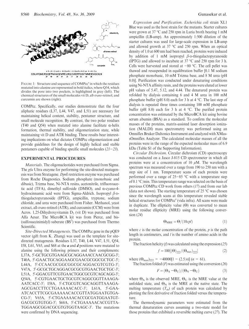
FIGURE 1: Structure and sequence of COMPccs in which the residuesmutated into alanine are represented inbold italics,whereQ54,whichdivides the pore into two pockets, is highlighted in gray (left). Thechemical structures of the smallmolecules vitD, all-trans-retinol, andcurcumin are shown (right).
Article Biochemistry, Vol. 48, No. 36, 2009 8561
self-association and dissociation of COMPcc (pentamer) followthe equilibrium reaction (28, 29)
5UTN5 ð4ÞThe equilibrium constant (k) for reaction 4 is expressed in
terms of fraction folded (F) by
k¼F=5CT4ð1-FÞ5 ð5Þ
where CT = [U] + 5[N] represents the total peptide concentra-tion. The Gibbs free energy of folding (ΔG�) is given by
ΔG�¼ -RT ln k¼ΔH�-TΔS� ð6Þwhere ΔH� and ΔS� signify the standard enthalpy and entropychange, respectively. The van’t Hoff enthalpy (ΔH�) was calcu-lated at the thermal midpoint of transition (Tm) of the meltingcurve using the following equation
ΔH�¼ARTm2ðdF=dTÞT ¼Tm
ð7ÞwhereA=12 for a pentamer. Under equilibrium,ΔS�=(ΔH�)/Tmwhich allows for the calculation of the change in entropy. TheΔG� at 25 �C (298 K) was calculated using eq 6.
When T = Tm and F = 0.5, it follows from eq 5 that
Tm ¼ΔH�=ΔS� þ R lnð0:31CT4Þ ð8Þ
Solving for ΔS� from eq 8 and substituting in eq 6 result in thefollowing expression
k¼ exp½ΔH�=Rð1=Tm -1=TÞ-lnð0:31CT4Þ� ð9Þ
The ΔH� was calculated using eq 7, which was then used torecalculate the equilibrium constant (k) using eq 9. The k valueobtainedwas used to recalculateF from eq 5. The resulting valuesof F were plotted and employed to determine the ΔH� value.
CD studies in the presence of vitDwere conducted by allowingthe protein to bind vit D overnight at 4 �C. The protein wascentrifuged to remove any excess vit D, and wavelength andthermal scans were conducted for most of the samples. VariantsQ54A, L61A,V65A, and S68Awere heated to their respectiveTm
values, and vit D was added to facilitate binding. They weregradually cooled to room temperature and allowed to bindovernight at 4 �C.Chemical Cross-Linking. The oligomerization states of the
proteins were determined by chemical cross-linking using bis-(sulfosuccinimidyl) suberate (BS3) ester with a spacer arm of11.4 A (30). Proteins were incubated with 1.5mMBS3 in 100mMphosphate buffer (pH. 8.0) for 1 h at room temperature. Thereaction was quenched via addition of 25 mM Trizma base.Sodium dodecyl sulfate (12%)-polyacrylamide gel electrophore-sis (SDS-PAGE) was used to identify the cross-linked states ofthe proteins. Similarly, vit D was bound overnight to the proteinsat 4 �C, and cross-linking studies were conducted.Sedimentation Equilibrium Analysis. Analytical ultracen-
trifugation measurements were taken on a Beckman XL-A(Beckman Coulter) analytical ultracentrifuge equipped with anAn-60Ti rotor (BeckmanCoulter) at 20 �C.Protein samples weredialyzed overnight against PBS [100 mM sodium phosphate and100 mM NaCl (pH 8.0)], loaded at initial concentrations of50 and 200 μM, and analyzed at rotor speeds of 17000 and20000 rpm. Data were acquired at two wavelengths per rotorspeed setting and processed simultaneously with a nonlinearleast-squares fitting routine (31). The solvent density and protein
partial specific volume were calculated according to solvent andprotein composition, respectively (32).Fluorescence. Fluorescence measurements of the pro-
tein 3ATR and protein 3CCM complexes were performed with9.5 μM protein and 40 μM ATR or 50 μM CCM in100 mM phosphate buffer (pH 8.0) with 0.4% DMSO and0.5% methanol, respectively. After the samples had been incu-bated for 30 min at room temperature, fluorescence was mea-sured using a SpectraMax PlusM2 instrument with an excitationwavelength of 330 nm and an emission wavelength from 360 to600 nm in the case of ATR (17). For CCM binding, fluorescencewas monitored with an excitation wavelength of 420 nm and anemission wavelength from 450 to 600 nm (33, 34). For both ATRand CCM, the maximal peak was identified from the emissionspectrum. Most proteins demonstrated an emission maximumnear 460 and 490 nm for ATR andCCM, respectively. For CCMbinding, two variants, I58A and L61A, possessed maxima near460 nm. A time course study was conducted to identify theoptimal equilibration time for both small molecules. Controls ofbuffer, protein, ATR, and CCM alone did not demonstrate anincrease in fluorescence. All fluorescence measurements weretaken in triplicate. Error bars denote the standard deviation.
RESULTS
COMPccs and Alanine Variants. The original COMPccsequence bears two cysteines (C68 and C71) near the C-terminusthat form disulfide bridges which stabilize the pentamer (17). Toinvestigate the behavior of the proteins without the complicationof oxidation, the cysteines were mutated into serines and usedas our template (COMPccs) for single-alanine mutagenesis(Figure 1). The 10 residues in the a and d positions lining thepore of COMPccs were individually mutated to alanine via site-directed mutagenesis. Each alanine variant in addition to theCOMPccs was characterized for structure, stability, and oligo-merization state. Binding of the variants to the hydrophobicsmall molecules vit D, ATR, and CCM was also assessed.Impact of Variants on R-Helicity. Far-UV circular dichro-
ism (CD) was employed to determine the secondary structure ofCOMPccs and variants at room temperature (35). A wavelengthscan of COMPccs demonstrated double minima at 208 and222 nm indicative of an R-helix with a calculated fractionalhelicity of 70.1% as previously reported (20) (Figure 2a andTable 1). This confirmed that mutation of the cysteines to serineswithin the COMPcc sequence did not significantly alter thestructure of the protein. Variants L37A, L44A, V47A, L51A,and I58A exhibited a substantial loss of R-helical structure(Figure 2a and Table 1). Each of these variants expressed afractional helicity with less than 50% and a Θ222/Θ208 of <1,indicating that these residues are critical for the maintenance ofR-helix. Variants L61A, V65A, and S68A exhibited a modestdecrease or no change in helical content (Figure 2a and Table 1).These variants possessed a 58-69% fractional helicity andretained a Θ222/Θ208 of g1. Finally, the two variants T40A andQ54A illustrated an increase in R-helicity as demonstrated by the>80% fractional helicity values and the Θ222/Θ208 of >1.
Of the total residues that line the N-terminal pocket divided byQ54, all the aliphatic residues (L37, L44, V47, andL51) appear toplay a significant role inmaintaining the helix (Figure 1). ResidueI58 is the only residue located within the second C-terminalpocket that yielded a significant loss in helical content whenmutated to alanine. The remaining residues (L61, V65, and S68)
8562 Biochemistry, Vol. 48, No. 36, 2009 Gunasekar et al.
within the C-terminal pocket bear modest implications on theR-helix. The two polar residues, T40 andQ54, improve the helicalcontent when mutated to alanines in the N-terminal pocket.Together, this suggests that the residues within the N-terminalpocket are important in establishing and maintaining the helicalstructure, while the C-terminal residues do not substantiallyinfluence the R-helix.Impact of Variants on Thermostability. Temperature
scans at 222 nm were performed to determine the thermosta-bilities of all the proteins under identical concentrations of10 μM (36). The COMPccs presented a cooperative thermaltransition temperature (Tm) of 41 �C, similar to previouslyreported values (17). Of the six variants that produced adiscerniblemelting curve, five demonstrated a significant increasein thermostability relative to that of COMPccs (Figure 2b andTable 1). Variant S68A displayed a 39 �C enhancement inTm, thelargest increase observed for any variant. This was followed bythe values ofV65AandQ54A,which showed improvements of 35and 33 �C, respectively, while variants L61A andT40A presentedincreases of 26 and 21 �C relative to that of COMPccs, respec-tively. By contrast, variant I58A displayed a 3 �C decrease in Tm,when compared to that of COMPccs.
The reversibility and monophasic behavior of the melts enableus to assume a two-state mechanism of unfolding (27-29). As aresult, we can calculate the thermodynamic parameters of theaforementioned variants and COMPccs based on the fractionfolded (28, 29, 37). The stability of COMPccs arises from a strongenthalpic component with a van’t Hoff enthalpy (ΔH�) of-85.2 kcal/mol (Table 2). This leads to a calculated Gibbs freeenergy (ΔG�) of -4.3 kcal/mol within the range of the othercoiled-coil systems, including human cartilage oligomeric matrixprotein (29, 30), tetramers (30), and dimers (38). In general, forthe T40A and Q54A variants that exhibited enhanced helicalcontent and stability, increases in free energy of 8.4 and 13.4 kcal/mol are observed relative to that of COMPccs, respectively(Table 2). The remaining C-terminal residues (L61, V65, andS68) demonstrated 11.3, 19.4, and 22.7 kcal/mol increases instability, respectively, compared to that of COMPccs whenmutated to alanine (Table 2). By contrast, I58A revealed aminimal change in stability relative to that of COMPccs of0.2 kcal/mol (Table 2).
The majority of the residues that impact the thermostabilityare located within the C-terminal pocket starting from thedivision point of Q54 (Figure 1). C-Terminal residues L61,
FIGURE 2: CDspectra ofCOMPccs and single-alanine variants in the absence (a andb) andpresence (c andd) of vitD. (a and c)Wavelength scans atroom temperature (25 �C) and (b andd) temperature scans at 222 nmofCOMPccs (black), L37A (red), T40A (gray), L44A (orange), V47A (yellow),L51A (pink), Q54A (purple), I58A (blue), L61A (green), V65A (light blue), and S68A (light green). All scans represent an average of two trials.
Table 1: Summary of Structure and Stability Comparison from Circular Dichroism Studies
without 1,25-dihydroxyvitamin D3 with 1,25-dihydroxyvitamin D3
V65, and S68 stabilize the protein whenmutated to alanine, whilealanine at position 58 does not significantly impact stability.Although most of residues that influence stability are localizedon the C-terminal end, T40 and Q54 within the first pocketalso affect the thermostability when converted to alanine. Theenhancement in stability observed by these two variants appearsto be linked to the improved helical content described above.Impact of Variants on Oligomerization State. To deter-
mine the oligomerization states of COMPccs and variants, cross-linking studies were performed using bis(sulfosuccinimidyl)suberate (BS3) at the same concentrations used for CD andanalyzed via SDS-PAGE (30). The COMPccs demonstrated thepresence of five equally intense bands indicating the presence ofpentamers, in addition to tetra-, tri-, di-, and monomers(Figure 3). In contrast, variants L37A, L44A, V47A, and L51Aexhibited predominantly monomer bands. Both L44A and L51Ashowed evidence for dimer in addition to monomer. V47Adisplayed the existence of di-, tri-, and tetramers, while L37Ademonstrated the presence of di-, tri-, tetra-, and pentamers withless intensity than the monomer band. Variants T40A, Q54A,I58A, L61A, V65A, and S68A illustrated an enhanced oligomer-ization in which the monomer band disappeared, while retainingthe di-, tri-, tetra-, and pentamer bands (Figure 3). In general,residues L37, L44, V47, and L51 are important for maintainingthe oligomeric state of the protein and are positioned in theN-terminal pocket region since mutation to alanine abolishes thepresence of the pentamer bands.
Furthermore, sedimentation equilibrium experiments indicatethat the apparent molecular masses of COMPccs and the T40A,Q54A, I58A, L61A, V65A, and S68A variants were within 10%of those calculated for a pentamer, with no systematic deviationof the residuals (Figure 4 and Table 3). Hence, residues T40, Q54,I58, L61, V65, and S68 do not appear to be critical forpentamerization. In fact, mutation to alanine facilitates oli-gomerization. These residues are mostly distributed at theC-terminal end with the exception of two polar residues, T40and Q54, that reside in the N-terminal region.Influence of 1,25-dihydroxyvitamin D3. One of the key
features of COMPcc is its ability to bind vit D as demonstrated bystructural and biochemical studies (17). vit D is an importanthormone involved in promoting cellular differentiation and pro-liferation in terms of cartilage and bone tissue in addition to themaintenance of calcium and phosphate homeostasis (39, 40). Toinvestigate the impact of the presence of vit D, we performed CDand cross-linking studies after incubating COMPccs and variantswith the small molecule.
The proteins before and after binding vit D were compared toevaluate whether there is any effect of vit D on helicity. Variants
L37A, L44A, V47A, L51A, I58A, L61A, V65A, and S68Aexhibiting large and modest losses in R-helical content revealedsimilar decreases, while variants T40A and Q54A demonstratingenhanced helicity maintained an increase with respect to that ofCOMPccs in the presence of vitD (Figure 2c andTable 1). Nearlyall the variants, including COMPccs, exhibited a slight loss ofhelical content upon incubation with vit D relative to theunbound proteins, except T40A, L44A, and I58A.
As described for the variants in the absence of vit D, similarincreases and decreases in Tm were observed relative to that ofCOMPccs (Figure 2d and Table 1). Variants Q54A, V65A, S68A,L61A, and T40A presented increases inTm ranging from 36 to 18�C, while I58A showed a decrease in Tm of 4 �C when comparedto that of COMPccs. Previously, COMPcc binding to vit D wasdetected by a shift in the Tm (17). COMPccs demonstrated anincrease in the Tm of 4 �C upon incubation with vit D, affirmingearlier data (17) (Figure 2d and Table 1). Variants I58A andQ54A each exhibited a 3 �C increase, followed by S68A whichrevealed a 1 �Cenhancement.However, variantL61Adidnot affectthe Tm, while V65A illustrated a decrease in Tm by 1 �C (Figure 2dand Table 1). Variants L37A, L44A, V47A, and L51A wereunable to bind vit D because of their inability to form R-helices.
To study the impact of vit D on oligomerization, cross-linkingstudies were performed after incubation with vit D. A similartrend was observed in which L37A, L44A, V47A, and L61Ademonstrated mostly monomers or a lack of pentamer forma-tion, while the remaining T40A, Q54A, I58A, L61A, V65A, andS68A variants exhibited strong pentamer bands (Figure S1 of theSupporting Information).Binding to all-trans-Retinol and Curcumin. Guo and co-
workers demonstrated that COMPcc is capable of bindinghydrophobic small molecules such as ATR in addition to thetarget vit D (17). ATR, also known as vitamin A, is critical fororgan tissue and limb growth in both embryonic cells and adulttissue (41, 42). Because of its unique fluorescence behavior, thebinding of ATR to COMPcc can be readily monitored spectro-photometrically (17). To explore the effect of the mutations onATR recognition, we investigated binding via fluorescence.
Variants L37A, L44A, V47A, L51A, I58A, and L61A demon-strated dramatic losses in their levels of binding, indicating thatthese residues may be critical for ATR recognition (Figure 5).The N-terminal residues L37, L44, V47, and L51 responsiblefor the maintenance of helical content and oligomerization statebound poorly to ATR when substituted with alanine. By con-trast, the Q54A variant that previously illustrated enhancedhelical content and oligomerization revealed an increase influorescence, indicative of improved binding. Interestingly,S68A previously shown to display modest effects on the
Table 2: Summary of the Calculated Thermodynamic Parameters
avan’t Hoff enthalpy calculated as described in Experimental Procedures. bAt equilibrium,ΔG�=0.Hence, the change in entropy (ΔS�)=ΔHo/Tm.cFree
energy of folding at 25 �C calculated by the expression ΔG� = ΔH� - TΔS�.
8564 Biochemistry, Vol. 48, No. 36, 2009 Gunasekar et al.
coiled-coil structure and self-assembly also bound slightly betterto ATR.
Because of the predominant hydrophobicity of the COMPccpore, it has been suggested that the protein may potentially bindother retinoids and hydrophobic compounds (17). Curcumin(CCM) is a polyphenol isolated from turmeric bearing uniquepharmacological activity, including anticarcinogenic (43, 44),antioxidant (45), and antihypertrophy (46, 47) properties. LikeATR, it can be monitored for binding to the variants because ofits fluorescence behavior (33, 34). We sought to investigate the
binding of the variants to CCM in part because of its structuraldifference from the retinoids and seco steroid vit D.
Variants L37A, L44A, V47A, L51A, and Q54A demonstratedsignificant reductions in their levels of binding (Figure 6). Thefour residues (L37, L44, V47, and L51) positioned in theN-terminal pocket that were previously noted to be essentialfor helicity and oligomerization also bound CCM poorly whensubstituted with alanine, which is similar to the ATR studies.Notably, I58A and L61A revealed an increase in fluorescencerelative to that of COMPccs (Figure 6). Comparison of ATR andCCMbinding studies reveals a consistency in the binding abilitiesof the variants for both substrates; however, variants Q54A,I58A, and L61A demonstrate a clear preference for either ATRor CCM. These results suggest that the residues that line thepocket play an important role in selectively binding varioushydrophobic small molecules.
DISCUSSION
Coiled-coil proteins have been extensively studied because oftheir significance in biological systems (48-54). They are dis-tinguished by heptad repeats (abcdefg) in which the residues inthe a and d positions play a significant role in its overallstructure (48). COMPcc represents a coiled coil that assemblesinto a pentamer of parallel R-helices. Of the 10 residues in the aand d positions of COMPccs, L37, L44, V47, and L51 areresponsible for maintaining the structure (Figure 1). Thesealiphatic residues when mutated to alanine result in a loss ofhelical structure, stability, and oligomerization state. By contrast,
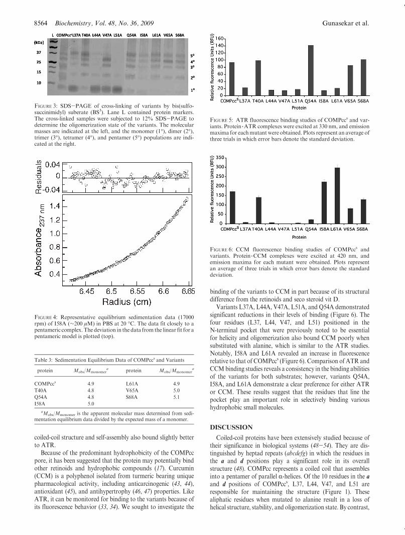
FIGURE 3: SDS-PAGE of cross-linking of variants by bis(sulfo-succinimidyl) suberate (BS3). Lane L contained protein markers.The cross-linked samples were subjected to 12% SDS-PAGE todetermine the oligomerization state of the variants. The molecularmasses are indicated at the left, and the monomer (1�), dimer (2�),trimer (3�), tetramer (4�), and pentamer (5�) populations are indi-cated at the right.
FIGURE 4: Representative equilibrium sedimentation data (17000rpm) of I58A (∼200 μM) in PBS at 20 �C. The data fit closely to apentameric complex.The deviation in the data from the linear fit for apentameric model is plotted (top).
Table 3: Sedimentation Equilibrium Data of COMPccs and Variants
protein Mobs/Mmonomera protein Mobs/Mmonomer
a
COMPccs 4.9 L61A 4.9
T40A 4.8 V65A 5.0
Q54A 4.8 S68A 5.1
I58A 5.0
aMobs/Mmonomer is the apparent molecular mass determined from sedi-mentation equilibrium data divided by the expected mass of a monomer.
FIGURE 5: ATR fluorescence binding studies of COMPccs and var-iants. Protein 3ATR complexes were excited at 330 nm, and emissionmaxima for eachmutantwere obtained. Plots represent an average ofthree trials in which error bars denote the standard deviation.
FIGURE 6: CCM fluorescence binding studies of COMPccs andvariants. Protein 3CCM complexes were excited at 420 nm, andemission maxima for each mutant were obtained. Plots representan average of three trials in which error bars denote the standarddeviation.
Article Biochemistry, Vol. 48, No. 36, 2009 8565
polar residues T40 and Q54 within the N-terminal region whenconverted to alanine improve the R-helical structure, stability,and self-assembly behavior.Interplay of Helicity, Stability, and Oligomerization.
Residues L37, L44, V47, and L51 that exhibited a substantialloss of helicity when substituted with alanine were not sufficientlystable to determinemelts, while I58Awhich showed amodest lossof R-helical structure demonstrated a loss in Tm (Table 1). Theseresults suggest that the residues critical for maintaining helicalcontent are important for preserving stability. Moreoever, resi-dues L37, L44, V47, and L51 that no longer displayed helicalstructure and discernible stability when mutated to alaninerevealed predominantly monomeric states (Figures 2 and 3).These data are consistent with the previous work by Miura andco-workers in which alanine mutation of synthetic peptides thatled to an abolishment in R-helix also prevented the formation offibrous assembly (55).
The residues important for structure, stability, and pentamerformation are located in the N-terminal pocket. On the basis ofthe heptad pattern, L37, L44, and L51 occupy the a positionwhile V47 occupies the d position (Figure 1). Thus, the stability ofthe pentameric assembly is largely mediated by the interactionsbetween three leucines from one helix with a valine from theadjacent helix. Another pentameric coiled-coil, phospholamban,has been demonstrated to be governed by the packing interac-tions among three N-terminal leucines, L37, L44, and L51, in thea positionwith two isoleucines, I40 and I47, in the d position froma neighboring helix (56, 57). This suggests that an N-terminalrepeat of three leucines in the a site along with a valine or set ofisoleucines in the d site of the adjacent helix is indispensible for theformation of pentamers.
The two polar residues, T40 and Q54, that improved theR-helical content when mutated to alanine also demonstrated asubstantial increase in Tm (Table 1). Alanine substitution ofeither residue in this N-terminal region resulted in pentamerformation and enhanced oligomerization (Figures 3 and 4 andTable 3). In this case, increased helicity and stability facilitateoligomer formation.
The remainder of residues L61, V65, and S68 located in theC-terminal regionwhen substitutedwith alanine yielded amodestloss of helical content (within 15%), while exhibiting an enhancedstability (Table 1). Interestingly, alanine substitution of theseresidues maintains pentamer formation and improves the oligo-merization state where the monomer population essentiallydisappears (Figures 3 and 4 and Table 3). Here, the C-terminalresidues maintain the link between stability and oligomerizationas long as a threshold level of R-helical structure is maintained.Influence of vit D on Helicity, Stability, and Oligomer-
ization. No significant change in helical content, stability, oroligomerization was observed in the presence of vit D for theCOMPccs and alanine variants relative to the unincubatedproteins (Table 1). The major discernible differences can beidentified from shifts in the Tm. Variants T40A, Q54A, I58A,L61A ,and S68A in addition to COMPccs exhibited an increaseor no change in theirTm values, indicating binding of vitD. Thesefive span both pockets with a majority concentrated in the C-terminus and do not play a significant role in vit D recognition.Variant V65A exhibits a decrease inTm, suggesting that it may beimportant for vit D binding. However, residues L37, L44, V47,and L51 represent the most crucial in vit D recognition asabsolutely no binding can even occur because of the inabilityto oligomerize as described above.
Variants That Exhibit a Specific Preference for vit D,ATR, or CCM. From the vit D, ATR, and CCM studies,variants Q54A, I58A, and L61A demonstrated selective binding.Variant Q54A exhibited an enhanced affinity for ATR whileillustrating poor binding toCCMand vitD (Figures 2d, 5, and 6).Remarkably, two variants, I58A an L61A, bound specifically toCCM, while weakly binding ATR and vit D (Figures 2d, 5,and 6). Thus, by altering the sequence within the binding pocket,one may be able to control the specificity for small moleculebinding.Structural Data Add Molecular Detail to Interactions
with SmallMolecules.On the basis of the structural analysis ofCOMPcc 3 vit D (19) and COMPcc 3ATR (17) complexes, Q54separated the core into two distinct cavities. The vit D binds bothpockets, while ATR is occupied exclusively in the N-terminalcavity. Both small molecules are poised between the T40 andQ54residues. T40 recognizes the dimethyl group of vit D (19) and theβ-ionine ring of ATR (17). Q54 mediates a H-bond contact tothe hydroxyl group ofATRwhile providing separationwithin thecavity for both ATR and vit D (17, 20). While both residuesappear to make contacts with both small molecules, they are notneeded as demonstrated by previous biochemical studies and ourmutagenesis data (17, 20).
Both cocrystal structures reveal that residues L44, V47, andL51 lining the N-terminal cavity can accommodate vit D andATR even though both small molecules differ structurally (17,20). For the COMPcc 3 vit D complex, V47 is able to accommo-date the seco B ring system, imposing a planar 6-s-trans con-formation of vit D, while residue L44 interacts with the methylC18 atom of the C ring on the β face (19). In the case of theCOMPcc 3ATR complex, L44 contacts the C16, C17, and C18methyl groups of the β-ionine of ATR, while the cavities betweenL44-V47 and V47-L51 interact with the C19 and C20 methylmoieties of the isoprene group (17). Our data confirm thatmutation of any of these three residues results in poor bindingto either compound.Moreover, these residues are also importantfor CCMbinding, indicating their significance for small moleculerecognition.
CONCLUSION
Here we show that the helicity, stability, oligomerization, andsmall molecule binding of COMPcc are predominantly con-trolled in positive and negative ways by the residues within theN-terminal region. Four N-terminal aliphatic residues are neces-sary for maintaining the structure and self-assembly, while thetwo polar residues are dispensable. Alanine substitution of thetwo polar residues renders variants that are more helical andthermostable and facilitate pentamer formation. These findingshave interesting implications onhowCOMPcc is able tomaintainits structure and function. Moreover, the lessons learned fromthese studies can aid in the design of exceptionally stable artificialoligomeric coiled coils (21-23, 58) capable of binding varioussmall molecules with high selectivity (17). Future studies employ-ing COMPcc variants in the context of artificial peptides orproteins for specific binding and delivery of small molecules aspotential biomaterials are underway (50, 59-63).
ACKNOWLEDGMENT
We thank Neville Kallenbach and anonymous reviewersfor assistance and critical insight into the experiments andmanuscript.
8566 Biochemistry, Vol. 48, No. 36, 2009 Gunasekar et al.
SUPPORTING INFORMATION AVAILABLE
The MALDI data to confirm the molecular weights of theproteins and the cross-linking studies of the proteins in thepresence of vit D. This material is available free of charge viathe Internet at “http://pubs.acs.org”.
REFERENCES
1. Hedbom, E., Antonsson, P., Hjerpe, A., Aeschlimann, D., Paulsson,M., Rosapimentel, E., Sommarin, Y., Wendel, M., Oldberg, A., andHeinegard, D. (1992) Cartilage Matrix Proteins: An Acidic Oligo-meric Protein (Comp) Detected Only in Cartilage. J. Biol. Chem. 267,6132–6136.
2. Dicesare, P., Hauser, N., Lehman, D., Pasumarti, S., and Paulsson,M. (1994) Cartilage Oligomeric Matrix Protein (Comp) Is an Abun-dant Component of Tendon. FEBS Lett. 354, 237–240.
3. Muller, G., Michele, A., and Altenburg, E. (1998) COMP (cartilageoligomeric matrix protein) is synthesized in ligament tendon, menis-cus and articular cartilage. Connect. Tissue Res. 39, 233–244.
4. Di Cesare, P. E., Fang, C., Leslie, M. P., Tulli, H., Perris, R., andCarlson, C. S. (2000) Expression of cartilage oligomeric matrixprotein (COMP) by embryonic and adult osteoblasts. J. Orthop.Res. 18, 713–720.
5. Oldberg, A., Antonsson, P., Lindblom, K., and Heinegard, D. (1992)Comp (Cartilage Oligomeric Matrix Protein) Is Structurally Relatedto the Thrombospondins. J. Biol. Chem. 267, 22346–22350.
6. Efimov, V. P., Lustig, A., and Engel, J. (1994) The Thrombospondin-Like Chains of Cartilage Oligomeric Matrix Protein Are Assembledby a 5-Stranded R-Helical Bundle between Residue-20 and Residue-83. FEBS Lett. 341, 54–58.
7. Rosenberg, K., Olsson, H., Morgelin, M., and Heinegard, D. (1998)Cartilage oligomeric matrix protein shows high affinity zinc-dependent interaction with triple helical collagen. J. Biol. Chem.273, 20397–20403.
8. Halasz, K., Kassner, A., Morgelin, M., and Heinegard, D. (2007)COMP acts as a catalyst in collagen fibrillogenesis. J. Biol. Chem. 282,31166–31173.
9. Hecht, J. T., Nelson, L. D., Crowder, E., Wang, Y., Elder, F. F. B.,Harrison, W. R., Francomano, C. A., Prange, C. K., Lennon, G. G.,Deere, M., and Lawler, J. (1995) Mutations in Exon 17b of CartilageOligomeric Matrix Protein (Comp) Cause Pseudoachondroplasia.Nat. Genet. 10, 325–329.
10. Briggs, M. D., Hoffman, S. M. G., King, L. M., Olsen, A. S.,Mohrenweiser, H., Leroy, J. G., Mortier, G. R., Rimoin, D. L.,Lachman, R. S., Gaines, E. S., Cekleniak, J. A., Knowlton, R.G., andCohn, D. H. (1995) Pseudoachondroplasia and Multiple EpiphysealDysplasia Due to Mutations in the Cartilage Oligomeric MatrixProtein Gene. Nat. Genet. 10, 330–336.
11. Spitznagel, L., Nitsche, D. P., Paulsson, M., Maurer, P., and Zaucke,F. (2004) Characterization of a pseudoachondroplasia-associatedmutation (His(587) f Arg) in the C-terminal, collagen-bindingdomain of cartilage oligomeric matrix protein (COMP). Biochem. J.377, 479–487.
12. Hecht, J. T., Hayes, E., Haynes, R., and Cole, W. G. (2005) COMPmutations, chondrocyte function and cartilage matrix. Matrix Biol.23, 525–533.
13. Kennedy, J., Jackson, G., Ramsden, S., Taylor, J., Newman, W.,Wright,M. J., Donnai, D., Elles, R., and Briggs, M. D. (2005) COMPmutation screening as an aid for the clinical diagnosis and counsellingof patients with a suspected diagnosis of pseudoachondroplasia ormultiple epiphyseal dysplasia. Eur. J. Hum. Genet. 13, 547–555.
14. Chen, T. L. L., Posey, K. L., Hecht, J. T., and Vertel, B. M. (2008)COMP mutations: Domain-dependent relationship between abnor-mal chondrocyte trafficking and clinical PSACH and MED pheno-types. J. Cell. Biochem. 103, 778–787.
15. Efimov, V. P., Engel, J., and Malashkevich, V. N. (1996) Crystal-lization and preliminary crystallographic study of the pentamerizingdomain from cartilage oligomeric matrix protein: A five-strandedR-helical bundle. Proteins: Struct., Funct., Genet. 24, 259–262.
16. Malashkevich, V. N., Kammerer, R. A., Efimov, V. P., Schulthess, T.,and Engel, J. (1996) The crystal structure of a five-stranded coiled coilin COMP: A prototype ion channel? Science 274, 761–765.
17. Guo, Y., Bozic, D., Malashkevich, V. N., Kammerer, R. A.,Schulthess, T., and Enger, J. (1998) All-trans retinol, vitamin D andother hydrophobic compounds bind in the axial pore of the five-stranded coiled-coil domain of cartilage oligomeric matrix protein.EMBO J. 17, 5265–5272.
18. Guo, Y., Kammerer, R. A., and Engel, J. (2000) The unusually stablecoiled-coil domain of COMP exhibits cold and heat denaturation in4-6 M guanidinium chloride. Biophys. Chem. 85, 179–186.
19. Ozbek, S., Engel, J., and Stetefeld, J. (2002) Storage function ofcartilage oligomericmatrix protein: The crystal structure of the coiled-coil domain in complex with vitamin D-3. EMBO J. 21, 5960–5968.
20. Terskikh, A. V., Potekhin, S. A., Melnik, T. N., and Kajava, A. V.(1997) Mutation Gln(54)Leu of the conserved polar residue in theinterfacial coiled coil position (d) results in significant stabilization ofthe original structure of the COMP pentamerization domain. Lett.Pept. Sci. 4, 297–304.
21. Slovic, A.M., Summa, C.M., Lear, J. D., andDeGrado,W. F. (2003)Computational design of a water-soluble analog of phospholamban.Protein Sci. 12, 337–348.
22. Slovic, A.M., Lear, J. D., andDeGrado,W. F. (2005)De novo designof a pentameric coiled-coil: Decoding the motif for tetramer versuspentamer formation in water-soluble phospholamban. J. Pept. Res.65, 312–321.
23. Liu, J., Yong, W., Deng, Y. Q., Kallenbach, N. R., and Lu, M. (2004)Atomic structure of a tryptophan-zipper pentamer. Proc. Natl. Acad.Sci. U.S.A. 101, 16156–16161.
24. Lee, D. L., Mant, C. T., and Hodges, R. S. (2003) A novel method tomeasure self-association of small amphipathicmolecules: temperatureprofiling in reversed-phase chromatography. J. Biol. Chem. 278,22918–22927.
25. Flaugh, S. L.,Mills, I. A., andKing, J. (2006) Glutamine deamidationdestabilizes human γD-crystallin and lowers the kinetic barrier tounfolding. J. Biol. Chem. 281, 30782–30793.
26. Forood, B., Feliciano, E. J., and Nambiar, K. P. (1993) Stabilizationof R-helical structures in short peptides via end capping. Proc. Natl.Acad. Sci. U.S.A. 90, 838–842.
27. Pace, C. N. (1990)Measuring and Increasing Protein Stability.TrendsBiotechnol. 8, 93–98.
28. Marky, L. A., and Breslauer, K. J. (1987) Calculating thermodynamicdata for transitions of any molecularity from equilibrium meltingcurves. Biopolymers 26, 1601–1620.
29. Beck, K., Gambee, J. E., Bohan, C. A., and Bachinger, H. P. (1996)The C-terminal domain of cartilage matrix protein assembles intoa triple-stranded R-helical coiled-coil structure. J. Mol. Biol. 256,909–923.
30. Beck, K., Gambee, J. E., Kamawal, A., and Bachinger, H. P. (1997) Asingle amino acid can switch the oligomerization state of the R-helicalcoiled-coil domain of cartilage matrix protein. EMBO J. 16, 3767–3777.
31. Johnson, M. L., Correia, J. J., Yphantis, D. A., and Halvorson, H. R.(1981) Analysis of Data from the Analytical Ultra-Centrifuge byNon-Linear Least-Squares Techniques. Biophys. J. 36, 575–588.
32. Laue, T. M., Shah, B. D., Ridgeway, T. M., and Pelletier, S. L. (1992)Computer-aided interpretation of analytical sedimentation data forproteins. In Analytical Ultracentrifugation in Biochemistry andPolymer Science (Harding, S. E., Rowe, A. J., and Horton, J. C., Eds.)pp 90-125, Royal Society of Chemistry, Cambridge, U.K.
33. Barik, A., Priyadarsini, K. I., and Mohan, H. (2003) Photophysicalstudies on binding curcumin to bovine serum albumin. Photochem.Photobiol. 77, 597–603.
34. Khopde, S. M., Priyadarsini, K. I., Palit, D. K., and Mukherjee, T.(2000) Effect of solvent on the excited-state photophysical propertiesof curcumin. Photochem. Photobiol. 72, 626–631.
35. Greenfield, N. J. (2006) Using circular dichroism spectra to estimateprotein secondary structure. Nat. Protoc. 1, 2876–2890.
36. Greenfield, N. J. (2006) Using circular dichroism collected as afunction of temperature to determine the thermodynamics of proteinunfolding and binding interactions. Nat. Protoc. 1, 2527–2535.
37. Pace, C. N., Laurents, D. V., and Thomson, J. A. (1990) pHDependence of theUrea andGuanidine-HydrochlorideDenaturationof Ribonuclease-a andRibonuclease-T1.Biochemistry 29, 2564–2572.
38. Krylov, D., Barchi, J., andVinson, C. (1998) Inter-helical interactionsin the leucine zipper coiled coil dimer: pH and salt dependenceof coupling energy between charged amino acids. J. Mol. Biol. 279,959–972.
39. Walters, M. R. (1992) Newly ientified actions of the vitamin Dendocrine system. Endocrinol. Rev. 13, 719–764.
40. Bouillon, R., Okamura, W. H., and Norman, A. W. (1995) Structure-function relationships in the vitamin D endocrine system. Endocrinol.Rev. 16, 200–257.
41. Means, A. L., and Gudas, L. J. (1995) The roles of retinoids unvertebrate development. Annu. Rev. Biochem. 64, 201–233.
42. Marshall, H., Morrison, A., Studer, M., Properl, H., and Krumlauf,R. (1996) Retinoids and Hox genes. FASEB J. 10, 969–978.
Article Biochemistry, Vol. 48, No. 36, 2009 8567
43. Mehta, K., Pantazis, P., McQueen, T., and Agarwal, B. (1997)Antiproliferative effect of curcumin against human breast tumor celllines. Anticancer Drugs 8, 471–480.
44. Cheng, A. L., Hsu, C. H., Lin, J. K., Hsu, M. M., Ho, Y. F., Shen, T.S., Ko, J. Y., Lin, J. T., Lin, B. R., Wu, M. S., Yu, H. S., Jee, S. H.,Chen, G. S., Chen, T. M., Chen, C. A., Lai, M. K., Pu, Y. S., Pan, M.H., Wang, Y. J., Tsai, C. C., and Hsieh, C. Y. (2001) Phase 1 clinicaltrials of curcumin: A chemopreventive agent in patients with high riskor pre-malignant lesions. Anticancer Res. 21, 2895–2900.
45. Barclay, L. R. C., Vinqvist, M. R., Mukai, K., Goto, H., Hasimoto,Y., Tokunaga, A., andUno, H. (2000) On the antioxidant mechanismof curucmin: Classical methods are needed to determine antioxidantmechanism and activity. Org. Lett. 2, 2841–2843.
46. Balasubramanyam, K., Varier, R. A., Altaf, M., Swaminathan, V.,Siddappa, N. B., Ranga, U., and Kundu, T. K. (2004) Curcumin, anovel p300/CREB-binding protein-specific inhibitor of acetyltrans-ferase, represses the acetylation of histone/nonhistone proteins andhistone acetyltransferase-dependent chromatin transcription. J. Biol.Chem. 279, 51163–51171.
47. Morimoto, T., Sunagawa, Y., Kawamura, T., Takaya, T., Wada, H.,Nagasawa, A., Komeda, M., Fujita, M., Shimatsu, A., Kita, T., andHasegawa, K. (2008) The dietary compound curcumin inhibits p300histone acetyltransferase activity and prevents heart failure in rats.J. Clin. Invest. 118, 868–878.
49. Landschulz, W. H., Johnson, P. F., and McKnight, S. L. (1988) TheLeucine Zipper: AHypothetical Structure Common to aNewClass ofDNA-Binding Proteins. Science 240, 1759–1764.
50. Gunasekar, S. K., Haghpanah, J. S., and Montclare, J. K. (2008)Assembly of bioinspired helical protein fibers. Polym. Adv. Technol.19, 454–468.
51. Harbury, P. B., Zhang, T., Kim, P. S., and Alber, T. (1993) A Switchbetween 2-Stranded, 3-Stranded and 4-Stranded Coiled Coils in Gcn4Leucine-Zipper Mutants. Science 262, 1401–1407.
52. Montclare, J. K., Sloan, L. S., and Schepartz, A. (2001) Electrostaticcontrol of half-site spacing preferences by the cyclic AMP responseelement-binding protein CREB. Nucleic Acids Res. 29, 3311–3319.
53. Diss, M. L., and Kennan, A. J. (2008) Orthogonal recognition indimeric coiled coils via buried polar-group modulation. J. Am. Chem.Soc. 130, 1321–1327.
54. Deng, Y. Q., Liu, J., Zheng, Q., Li, Q. N., Kallenbach, N. R., and Lu,M. (2008) A Heterospecific Leucine Zipper Tetramer. Chem. Biol. 15,908–919.
55. Takei, T., Okonogi, A., Tateno, K., Kimura, A., Kojima, S., Yazaki,K., and Miura, K. (2006) The effects of the side chains of hydro-phobic aliphatic amino acid residues in an amphipathic polypeptideon the formation of a helix and its association. J. Biochem. 139,271–278.
56. Simmerman, H. K. B., Kobayashi, Y. M., Autry, J. M., and Jones, L.R. (1996) A leucine zipper stabilizes the pentameric membranedomain of phospholamban and forms a coiled-coil pore structure.J. Biol. Chem. 271, 5941–5946.
57. Slovic, A. M., Stayrook, S. E., North, B., and DeGrado,W. F. (2005)X-ray structure of a water-soluble analog of the membrane pro-tein phospholamban: Sequence determinants defining the topo-logy of tetrameric and pentameric coiled coils. J. Mol. Biol. 348,777–787.
58. Liu, J., Zheng, Q., Deng, Y. Q., Cheng, C. S., Kallenbach, N. R., andLu, M. (2006) A seven-helix coiled coil. Proc. Natl. Acad. Sci. U.S.A.103, 15457–15462.
59. Shen, W., Zhang, K. C., Kornfield, J. A., and Tirrell, D. A. (2006)Tuning the erosion rate of artificial protein hydrogels through controlof network topology. Nat. Mater. 5, 153–158.
60. Papapostolou, D., Smith, A. M., Atkins, E. D. T., Oliver, S. J.,Ryadnov, M. G., Serpell, L. C., and Woolfson, D. N. (2007)Engineering nanoscale order into a designed protein fiber. Proc. Natl.Acad. Sci. U.S.A. 104, 10853–10858.
61. Woolfson, D. N., and Ryadnov, M. G. (2006) Peptide-based fibrousbiomaterials: Some things old, new and borrowed. Curr. Opin. Chem.Biol. 10, 559–567.
62. Woolfson, D., Pandya, M., Ryadnov, M., and Smith, A. (2005)Designing nano-to-micron scale peptide-based self-assembling sys-tems from the bottom up. Biopolymers 80, 493–493.
63. Fairman, R., and Ankerfedt, K. S. (2005) Peptides as novel smartmaterials. Curr. Opin. Struct. Biol. 15, 453–463.