N-WASP Is Required for Structural Integrity of the Blood-Testis Barrier The Harvard community has made this article openly available. Please share how this access benefits you. Your story matters Citation Xiao, X., D. D. Mruk, E. I. Tang, R. Massarwa, K. W. Mok, N. Li, C. K. C. Wong, et al. 2014. “N-WASP Is Required for Structural Integrity of the Blood-Testis Barrier.” PLoS Genetics 10 (6): e1004447. doi:10.1371/journal.pgen.1004447. http://dx.doi.org/10.1371/ journal.pgen.1004447. Published Version doi:10.1371/journal.pgen.1004447 Citable link http://nrs.harvard.edu/urn-3:HUL.InstRepos:12717588 Terms of Use This article was downloaded from Harvard University’s DASH repository, and is made available under the terms and conditions applicable to Other Posted Material, as set forth at http:// nrs.harvard.edu/urn-3:HUL.InstRepos:dash.current.terms-of- use#LAA
Transcript
N-WASP Is Required for StructuralIntegrity of the Blood-Testis Barrier
The Harvard community has made thisarticle openly available. Please share howthis access benefits you. Your story matters
Citation Xiao, X., D. D. Mruk, E. I. Tang, R. Massarwa, K. W. Mok, N. Li, C. K.C. Wong, et al. 2014. “N-WASP Is Required for Structural Integrityof the Blood-Testis Barrier.” PLoS Genetics 10 (6): e1004447.doi:10.1371/journal.pgen.1004447. http://dx.doi.org/10.1371/journal.pgen.1004447.
Published Version doi:10.1371/journal.pgen.1004447
Citable link http://nrs.harvard.edu/urn-3:HUL.InstRepos:12717588
Terms of Use This article was downloaded from Harvard University’s DASHrepository, and is made available under the terms and conditionsapplicable to Other Posted Material, as set forth at http://nrs.harvard.edu/urn-3:HUL.InstRepos:dash.current.terms-of-use#LAA
N-WASP Is Required for Structural Integrity of the Blood-Testis BarrierXiang Xiao1., Dolores D. Mruk1., Elizabeth I. Tang1., R’ada Massarwa2., Ka Wai Mok1, Nan Li1,
Chris K. C. Wong3, Will M. Lee4, Scott B. Snapper5, Ben-Zion Shilo2, Eyal D. Schejter2*, C. Yan Cheng1*
1 The Mary M. Wohlford Laboratory for Male Contraceptive Research, Center for Biomedical Research, Population Council, New York, New York, United States of America,
2 Department of Molecular Genetics, The Weizmann Institute of Science, Rehovot, Israel, 3 Department of Biology, Hong Kong Baptist University, Hong Kong, China,
4 School of Biological Sciences, University of Hong Kong, Hong Kong, China, 5 Harvard Medical School, Boston, Massachusetts, United States of America
Abstract
During spermatogenesis, the blood-testis barrier (BTB) segregates the adluminal (apical) and basal compartments in theseminiferous epithelium, thereby creating a privileged adluminal environment that allows post-meiotic spermatiddevelopment to proceed without interference of the host immune system. A key feature of the BTB is its continuousremodeling within the Sertoli cells, the major somatic component of the seminiferous epithelium. This remodeling isnecessary to allow the transport of germ cells towards the seminiferous tubule interior, while maintaining intact barrierproperties. Here we demonstrate that the actin nucleation promoting factor Neuronal Wiskott-Aldrich Syndrome Protein (N-WASP) provides an essential function necessary for BTB restructuring, and for maintaining spermatogenesis. Our datasuggests that the N-WASP-Arp2/3 actin polymerization machinery generates branched-actin arrays at an advanced stage ofBTB remodeling. These arrays are proposed to mediate the restructuring process through endocytic recycling of BTBcomponents. Disruption of N-WASP in Sertoli cells results in major structural abnormalities to the BTB, including mis-localization of critical junctional and cytoskeletal elements, and leads to disruption of barrier function. These impairmentsresult in a complete arrest of spermatogenesis, underscoring the critical involvement of the somatic compartment of theseminiferous tubules in germ cell maturation.
Citation: Xiao X, Mruk DD, Tang EI, Massarwa R, Mok KW, et al. (2014) N-WASP Is Required for Structural Integrity of the Blood-Testis Barrier. PLoS Genet 10(6):e1004447. doi:10.1371/journal.pgen.1004447
Editor: Wei Yan, University of Nevada School of Medicine, United States of America
Received October 9, 2013; Accepted May 2, 2014; Published June 26, 2014
Copyright: � 2014 Xiao et al. This is an open-access article distributed under the terms of the Creative Commons Attribution License, which permits unrestricteduse, distribution, and reproduction in any medium, provided the original author and source are credited.
Funding: This work was supported by grants from the National Institutes of Health (NICHD, U54 HD029990, Project 5 to CYC.; R01 HD056034 to CYC.), The HongKong General Research Fund (HKU771513 to WML, HKBU261812 to CKCW) and by an Administral Anslat Foundation grant to BZS, who is an incumbent of theHilda and Cecil Lewis Chair in Molecular Genetics. The funders had no role in study design, data collection and analysis, decision to publish, or preparation of themanuscript.
Competing Interests: The authors have declared that no competing interests exist.
in testes of these N-WASPSC-cKO males were strongly reduced to
,5% of age-matched controls (Fig. 1A), as monitored using
specific antibodies (Table 1). The residual N-WASP protein is
likely to represent germ cell expression [10], which is not affected
in this setting. Disrupting N-WASP function in the somatic
support cells had a dramatic effect on spermatogenesis. Testes of
8-week-old N-WASPSC-cKO mice weighed ,20% of age-matched
controls. Seminiferous tubule diameter was shrunk by ,30% and
the tubules were virtually devoid of advanced stage germ cells
(Fig. 1B). Round and elongating spermatids could only be
detected in less than 3% of tubules from 8-wk old mice (n = 4 mice,
a total of 200 randomly selected tubules scored), and more mature
(step 13–16) elongating/elongated spermatids were completely
absent (Fig. 1B), indicating that while meiosis could take place,
spermiogenesis failed to go to completion. The histological analysis
was further supported and complemented by the absence of
laminin-c3, a specific marker of late spermatids in mouse [20] and
rat [21] testes (Fig. 1B). It is noteworthy that the spermatogonia/
spermatogonial stem-cell (SSC) population is decreased in N-
WASPSC-cKO tubules, as assessed using the spermatogonia/SSC
marker Utf1 [22,23] (,0.2 Utf-1-positive cells/section in mutant
testes versus ,1.1 cell/section in wild-type controls), a feature that
we ascribe to the overall smaller size of the Sertoli cell population
(described below).
Taken together, these observations suggest a complete failure of
spermiogenesis in N-WASPSC-cKO mice, beyond early meiotic
phases. To verify this assertion, and rule out the possibility that the
observed phenotypes reflect secondary consequences of spermato-
genic arrest, we compared the morphologies of seminiferous
tubules from younger (21- and 35-day-old) control and mutant
mice, closer to the onset of sexual maturity (Fig. 1C). A substantial
amount of round spermatids were present in N-WASPSC-cKO
tubules in 21-day-old mice when meiosis is known to occur,
implying that germ cells in these tubules were capable of entering
meiosis, and properly initiated spermiogenesis. However, degen-
eration at both spermatid and spermatocyte stages is readily
apparent, such as the presence multi-nucleated round spermatids
and multi-nucleated spermatocytes by 21 and 35 dpp versus 8-wk
old mutants (Fig. 1C vs. 1B), implying a block to maturation at
these stages of spermiogenesis.
Disorganization of the Sertoli-cell actin cytoskeleton andfunctional impairment of the BTB in N-WASPSC-cKO
tubulesWe next turned to a detailed analysis of Sertoli cell features in
N-WASPSC-cKO mice in order to understand the basis for the
dramatic impact on spermiogenesis. We had previously demon-
strated that N-WASPSC-cKO mice possess an outwardly intact
Sertoli cell epithelium (15). This was now supported by assessing
the size of the Sertoli cell population in sectioned tubules using
GATA-1, a nuclear Sertoli cell marker [24]. The total number of
Sertoli cells within the epithelium is somewhat smaller in mutant
tubules (,4 Sertoli cells/cross-section) in comparison to wild-type
controls (,8 Sertoli cells/cross-section) due to shrinkage in tubule
diameter. Similar population-size values were obtained, however,
regardless of age, consistent with a generally healthy epithelium
Author Summary
Mammalian spermatogenesis takes place within a shel-tered environment, whereby somatic Sertoli cells protectand guide germ cells as they mature and differentiate. Akey structure generated by the protective Sertoli cellepithelium is the blood-testis barrier (BTB), a composite ofjunctional and cytoskeletal elements, which preventsexposure of post-meiotic spermatids to the immunesystem. The BTB is a highly dynamic structure, whichneeds to be dismantled and rapidly rebuilt, in order toallow passage of maturing preleptotene spermatocytes,without compromising their isolation. Here we show thatN-WASP, a conserved facilitator of formation of branchedactin microfilament arrays, provides a function that isessential for maintenance of an intact BTB. Geneticdisruption of N-WASP in mouse Sertoli cells leads to lossof BTB impermeability, resulting in a complete arrest ofspermatogenesis at early and post-meiotic stages. Basedon the localization patterns of key elements, we proposethat branched-actin filaments participate in recycling ofBTB materials to ensure the dynamic and efficientmaintenance of this structure, one of a series of blood-tissue barriers that preserve privileged organ environ-ments.
Figure 1. Spermatogenetic arrest and abnormal actin microfilament organization in the seminiferous epithelium of N-WASPSC-cKO
mouse testes. (A) Lysates (,50 mg protein) from 8-wk-old testes of N-WASPSC-cKO mice (Dhh-Cre; N-WASPflox/N-WASP2) and age-matched wild-type(WT) control mice (N-WASPflox/NWASP2) were used for immunoblotting, to assess changes in the expression of N-WASP, with GAPDH serving as aprotein loading control. Histograms in this and subsequent figures are composites of quantified immunoblot data (mean 6 SD) from n = 4 mice,normalized for the loading control. WT protein levels in these graphs were arbitrarily set at 1, against which statistical comparison was performed. **,P,0.01. (B) (Left panels) Hematoxylin and eosin (H&E) staining of paraffin sections, illustrating that specific disruption of N-WASP in Sertoli cells ledto major defects in spermiogenesis. N-WASPSC-cKO tubules (bottom) were shrunk in diameter and lacked the normal spermatid-filled seminiferousepithelium seen in control (WT) mouse testes (top). Magnified images of mutant tubules (bottom center and right panels) demonstrate the presenceof meiotic round spermatids (red arrowheads) and rare step 11 or 12 spermatids (green arrowhead). Scale bars: 240 mm (left column), and 60 mm(magnified columns). (Right panels) Laminin-c3 chain (green fluorescence), a specific marker of step 13–16 spermatids at the apical ES [20,21], wasnot detected in the seminiferous epithelium of N-WASPSC-cKO mouse testes. Absence of advanced-stage spermatids is indicative of a full failure ofspermiogenesis in these mutant mice. Scale bar: 50 mm, which applies to other micrographs. (C) Histological analysis of age-matched control (WT)and N-WASPSC-cKO tubules from 21- and 35-D (day)-old mice. At 21D and 35D, round spermatids (annotated by blue arrowheads) were found in sometubules from N-WASPSC-cKO testes, illustrating the occurrence of meiosis. However, abnormal multinucleated spermatocytes (annotated by yellowarrowheads) derived from normal spermatocytes (green arrows), were found in 21D N-WASPSC-cKO-testes, presumably giving rise to the degeneratedspermatocytes (red arrow). Degenerating round spermatids were also noted (blue arrow) in 21D N-WASPSC-cKO-testes. In 35D N-WASPSC-cKO-testes,elongating spermatids were occasionally found (red arrowhead) and also abnormal multinucleated round spermatids as annotated by bluearrowheads in the yellow boxed area. The lack of elongating spermatids at different stages supports the notion of a full failure of spermiogenesis inN-WASPSC-cKO-testes. Scale bars: 120 mm, and 40 mm in insets.doi:10.1371/journal.pgen.1004447.g001
Abbreviations used: IB, immunoblotting; IHC, immunohistochemistry; IF, immunofluorescence analysis.*, the number bracketed represents the corresponding Tyr residue of p-FAK in the rat testis.doi:10.1371/journal.pgen.1004447.t001
Figure 2. Sertoli cell-specific knockout of N-WASP leads to disorganization of F-actin in the seminiferous epithelium. (A)Immunofluorescence analysis of sectioned seminiferous tubules from 8W (week)-old control (WT) and N-WASPSC-cKO testes, showing the expressionand localization patterns of N-WASP (green fluorescence, left) and F-actin (FITC-phalloidin, green fluorescence, right). Yellow arrowheads in thecontrol panels point to the localization of either N-WASP or F-actin at the site of the BTB near the basement membrane of the tunica propria, which isannotated by a broken white-line. Cell nuclei were visualized by DAPI staining. Scale bars: 80 mm in the first two rows, and 240 mm in the third row.(B) A similar analysis was performed on testes from younger, 35D (day)-old mice. Actin microfilament organization in N-WASPSC-cKO tubules wasstrongly disrupted at this stage as well. Scale bar: 80 mm.doi:10.1371/journal.pgen.1004447.g002
structural and functional constituent of Sertoli cell gap junctions
[27,28], while occludin, an integral member of intercellular TJs,
including the TJ portion of the BTB, acts to stabilize TJs and to
optimize their function as barriers [29–31]. Remarkably, both
connexin-43 and occludin were virtually undetectable in N-
WASPSC-cKO tubules (Fig. 4A,C), implying a major disruption of
the tight and gap junction components of the N-WASPSC-cKO
BTB.
Given the prominent effects observed, we extended our analysis
to include two additional transmembrane BTB elements, the basal
ES component N-cadherin, an integral constituent of the BTB,
and the TJ integral membrane protein coxsackievirus and
adenovirus receptor (CAR) [32,33]. In contrast to the tight basal
localization normally observed for these proteins, their distribution
in the seminiferous epithelium of N-WASPSC-cKO tubules was no
longer restricted, and they were detected throughout the Sertoli
cell epithelium (Fig. 4A,C), further strengthening the notion of an
impaired BTB, supporting the functional data shown in Fig. 3.
Both the general disruption of microfilament organization and
the relatively early arrest in the progress of spermatogenesis suggest
that the second Sertoli cell ES structure, the apical ES, would not
form properly - if at all - in the N-WASPSC-cKO seminiferous
epithelium. We examined this issue directly by assessing the
localization patterns of representative apical ES components.
Nectin-3, a spermatid-specific adhesion molecule that mediates
Sertoli cell-spermatid attachment at the apical ES [34], was
undetectable in the mutant tubules (Fig. 5A,B), consistent with
the arrest of spermiogenesis at immature, early spermatid stages.
Furthermore, both b1-integrin, a Sertoli cell-specific adhesion
protein [35,36], and ICAM-2, an adhesion protein normally found
in both Sertoli cells and spermatids [37], were mis-localized in N-
WASPSC-cKO Sertoli cells, and no longer restricted to the adluminal
compartment (Fig. 5A,B). Taken together, these observations
confirm the expectation of a severely disrupted apical ES.
BTB restructuring in N-WASPSC-cKO seminiferous tubulesarrests at the phase requiring branched-actin nucleation
In contrast to the widespread disturbances to junctional and
cytoskeletal BTB components in the N-WASPSC-cKO seminiferous
epithelium, we found that two elements of the branched-actin
nucleation machinery, namely Arp3 and drebrin E, were properly
localized and robustly expressed (Fig. 6A, B). In particular, the
Arp3 subunit of the Arp2/3 complex, which normally localizes to
the BTB vicinity during the restructuring phase of stage VIII,
displayed a tightly restricted localization to the basal ES in the
seminiferous epithelium of mutant mice, analogous to the age-
matched control mouse testis (Fig. 6A, B). We have recently
demonstrated that drebrin E, the non-neuronal isoform of the
actin-binding element drebrin [38], acts to recruit Arp3 to the
BTB [39]. Examination of N-WASPSC-cKO tubules revealed that,
like Arp3, drebrin E retained its basally restricted expression
pattern, similar to WT control mouse testes (Fig. 6B). Taken
together, the defects in BTB structure, coupled with the observed
basal localization of Arp3 and its recruiting partner drebrin E,
Figure 3. BTB integrity is compromised in testes of N-WASPSC-cKO mice. Images show localization within seminiferoustubules of FITC-inulin (green), a small fluorescent molecule injectedinto the blood system of control (WT) and N-WASPSC-cKO mice (seeMaterials and Methods for detailed experimental protocol). In controlmouse testes, the functional BTB blocked FITC-inulin from entering intothe apical (adluminal) compartment, and the distance traveled by themarker was limited to the basal compartment (annotated by a whitebracket). The lower magnification inset shows four seminiferous tubules(white asterisks) that are devoid of green fluorescence inside the
epithelium. In N-WASPSC-cKO mouse tubules, the marker penetrateddeep inside the seminiferous epithelium (yellow arrows), reaching thetubule lumen (annotated by yellow bracket). The lower magnificationinset shows five such tubules (yellow asterisks), demonstrating thatdisruption of BTB integrity is a common feature of N-WASPSC-cKO mousetestes. Scale bars: 50 mm (magnified image), and 200 mm in insets. Datafrom BTB integrity assays were semi-quantified and are shown in thebar graph, which displays the distance traveled by FITC-inulin vs. thetubule radius for n = 5 mice (8–14 weeks old) in each group. *, P,0.01.doi:10.1371/journal.pgen.1004447.g003
suggest a molecular scenario in which dismantling of the
BTB is properly initiated in the seminiferous epithelium of N-
WASPSC-cKO mouse testes during the epithelial cycle, but is
arrested at a relatively late stage of the process, when the Arp2/3
actin-nucleation promoting activity of N-WASP must come into
play, in order to enable recycling of ‘‘old’’ BTB components and
formation of a ‘‘new’’ barrier.
Disruption of spatiotemporal expression of p-FAK-Tyr438
and -Tyr614 in the seminiferous epithelium ofN-WASPSC-cKO testes
Studies in the rat testis have shown that the non-receptor
protein tyrosine kinase FAK, a regulator of cell adhesion in a wide
variety of cellular contexts [40], functions to coordinate events at
the basal and apical ES. Key features of FAK activity in the
Figure 4. BTB junctional and basal ES proteins are absent or abnormally localized in the seminiferous epithelium of N-WASPSC-cKO
mouse testes. (A) Immunoblotting data using lysates of testes from N-WASPSC-cKO and age-matched control mice, to examine changes in theexpression of integral membrane proteins of GJs (connexin 43), TJs (occludin, CAR), and the basal ES (N-cadherin). b-Actin served as a protein loadingcontrol. Histogram generated as in Figure 1. **, P,0.01. N.D., not detectable. (B) Immunohistochemical localization of connexin 43 in theseminiferous epithelium of mouse testes. In WT testes, connexin 43 appears as a reddish brown precipitate, prominently expressed near the BTB, aswell as at the apical ES (near lumen) at early stage VIII. Connexin 43 was virtually undetectable in the N-WASPSC-cKO epithelium, consistent with theimmunoblot analysis shown in (A). Scale bars, 80 mm in the first two rows; 240 mm in the third row. (C) Immunofluorescence analysis of occludin (redfluorescence, left), N-cadherin (red fluorescence, middle) and CAR (red fluorescence, right) in the seminiferous epithelium of N-WASPSC-cKO and WTcontrol mice. Yellow arrowheads indicate the localization of occludin, N-cadherin or CAR at the BTB, which is located near the basement membrane atthe tunica propria, annotated by a white broken-line. In the micrographs illustrating localization of CAR (right), the boxed area was magnified andshown in insets, illustrating that CAR was also associated with the apical ES at the elongating/elongated spermatid/Sertoli cell interface. Cell nucleiwere visualized by DAPI staining. Scale bars: 80 mm in the first two rows, 240 mm in the third row, and 15 mm in insets.doi:10.1371/journal.pgen.1004447.g004
seminiferous epithelium are the restricted spatiotemporal patterns
of the phosphorylated forms p-FAK-Tyr407 (i.e., p-FAK-Tyr438 in
mouse testes) [41] and p-FAK-Tyr576 (i.e., p-FAK-Tyr614) [42].
The implication is that p-FAK-Tyr407 serves as a molecular switch
regulating BTB adhesion capacities, via changes in actin
polymerization kinetics [41,43]. We therefore used general and
form specific antibodies to assess FAK (Table 1) expression and
localization patterns in the tubules of N-WASPSC-cKO mice.
Expression levels of the p-FAK-Tyr614 form were significantly
lower, while the key phosphorylated form p-FAK-Tyr438 was
practically undetectable via either immunoblot or immunofluo-
rescence microscopy (Fig. 7A–C). Absence of Tyr-phosphorylated
FAK proteins from the BTB microenvironment thus considerably
impaired the adhesive capacity of this key ES structure, and this
notion was also supported by data shown in Fig. 3.
Discussion
Blood-tissue barriers are designed to protect sensitive organ
environments from infiltration by harmful substances. Efficient
barrier properties need to be coupled, however, with a degree of
permeability, and a capacity for selective passage of factors
essential for tissue differentiation and function. The dynamic
behavior of the BTB, which governs the initial phases of
spermatogenesis in the seminiferous tubules of the testis, provides
a striking example of such situations. In this instance, the BTB is
required to act as an efficient barrier, while enabling passage not
only of molecular elements, but also of entire cells, namely, the
differentiating preleptotene spermatocytes. This is made possible
by a tissue remodeling process, involving near simultaneous
dismantling and construction of the barrier at positions ahead and
in the wake of transiting spermatocytes, making their way towards
the adluminal compartment of the seminiferous epithelium.
In the current study, we demonstrate that the nucleation
promoting factor N-WASP is required in murine Sertoli cells for
maintenance of a properly structured BTB. Sertoli cell-specific
disruption of N-WASP results in abnormal spatiotemporal
expression patterns of key BTB elements, and in loss of barrier
impermeability, suggesting major impairment of BTB structure
and function. Key constituents of both the tight junction (occludin,
Figure 5. Abnormal localization and expression patterns of apical ES proteins in the seminiferous epithelium of N-WASPSC-cKO
mouse testes. (A) Immunoblotting data using lysates of testes from N-WASPSC-cKO and age-matched control mice, to examine changes in theexpression of the apical ES integral membrane proteins nectin-3, b1-integrin, and ICAM-2. b-Actin served as a protein loading control. Histogramgenerated as in Figure 1. **, P,0.01. N.D., not detectable. (B) Immunofluorescence analysis of nectin-3 (green fluorescence, left), b1-integrin (greenfluorescence, middle) and ICAM-2 (green fluorescence, right) in the seminiferous epithelium of control (WT) and N-WASPSC-cKO mice. Boxed areas aremagnified and shown in insets. Nectin-3, b1-integrin and ICAM-2 were associated with apical ES in control (WT) testes. In N-WASPSC-cKO mouse testes,nectin-3 was considerably diminished to a level almost undetectable in the seminiferous epithelium, consistent with the immunoblotting data shownin (A). While b1-integrin and ICAM-2 were detectable in the mutant testes, they were mis-localized and were both found near the disrupted BTB (seeyellow arrowheads that annotate ICAM-2 in the mutant testes). Relative location of the basement membrane is annotated by a white broken-line).Cell nuclei were visualized by DAPI staining. Scale bars: 80 mm in the first two rows, 240 mm in the third row, and 15 mm in insets.doi:10.1371/journal.pgen.1004447.g005
CAR) and gap junction (connexin-43) components of the BTB are
either significantly mis-localized or absent altogether. Importantly,
major structural abnormalities are apparent in organization of the
basal ES, the unique, hallmark component of the BTB. In
particular, the tightly packed arrangement of basal ES microfil-
aments is disrupted, and this key cytoskeletal structure becomes
dispersed in the Sertoli cell cytoplasm. Similar dispersal is observed
for N-cadherin, an adherens junction component that normally
displays tight association with the basal ES. Impairment of the
adhesive properties of the BTB are further implied by the marked
reduction in levels of phospho-FAK proteins, key regulators of ES-
based adhesion. Taken together, these data present a substantial
list of defects in BTB structure and functional properties,
demonstrating that disruption of N-WASP in Sertoli cells results
in significant deterioration of BTB barrier capabilities.
The generally disorganized spatial distribution of junctional and
cytoskeletal BTB components in N-WASPSC-cKO tubules is in
marked contrast to elements of the branched-actin nucleation
system, which retain their proper spatiotemporal localization
patterns. Both Arp3, a subunit of the Arp2/3 complex, and
drebrin E, a factor that mediates Arp3 localization in Sertoli cells,
preserve their tight basal localization during stage VIII of the
seminiferous epithelial cycle in the mutant tubules. These
observations are significant for several reasons. First, they argue
that abnormal localization patterns do not result from a general
disruption of cytoplasmic organization in N-WASPSC-cKO tubules,
Figure 6. The branched microfilament regulators Arp3 and drebrin E are properly localized to the BTB in N-WASPSC-cKO tubules. (A)Immunoblotting data using lysates of testes from N-WASPSC-cKO and age-matched control mice demonstrate up-regulation in Arp3 and drebrin Eexpression in mutant mouse testes vs. the age-matched control testes with b-actin served as a protein loading control. Histogram generated as inFigure 1. **, P,0.01. (B) Immunofluorescence analysis of Arp3 (green fluorescence, left) and drebrin E (green fluorescence, right) in the seminiferousepithelium of control (WT) and N-WASPSC-cKO mice. Yellow arrowheads mark the localization of either Arp3 or drebrin E at the BTB, which is near thebasement membrane of the tunica propria (annotated by a broken white-line). Unlike basal ES proteins (see Figure 4) and apical ES proteins (seeFigure 5), which were mis-localized in the seminiferous epithelium of mutant testes, these branched actin regulatory proteins remained properlylocalized to the damaged BTB. Cell nuclei were visualized by DAPI staining. Scale bars: 80 mm in the first two rows, and 240 mm in the third row.doi:10.1371/journal.pgen.1004447.g006
but are likely to arise from specific defects caused by the absence of
actin nucleation. This assertion is also supported by the morpho-
logical similarities between N-WASPSC-cKO tubules at different
ages, implying that mutant phenotypes are a primary consequence
of disrupting N-WASP function, and not a result of tissue
degeneration over time. Second, our observations identify a
particular juncture during which lack of N-WASP function becomes
apparent. A timeline consistent with the various observations
suggests proper localization of the Arp2/3 complex (mediated in
part by drebrin E) to the basal aspect of Sertoli cells, at the stage
when BTB restructuring is required. Under normal circumstances,
N-WASP then acts to stimulate Arp2/3 and promote nucleation of
branched actin arrays necessary for BTB restructuring. The actual
involvement of the N-WASP-Arp2/3 machinery is likely to take
place after restructuring is underway, and when many of the BTB
components are transiently displaced, to allow the dynamic events
of the process to unfold. Arrest of restructuring in the N-WASPSC-
cKO mutant tubules occurs therefore at a critical phase, when the
BTB has been dismantled, but is now incapable of forming a new
intact barrier, such as at the rear end of the preleptotene
spermatocytes under transport at the immunological barrier.
Identification of the juncture at which the branched actin
polymerization system contributes to BTB remodeling allows
for informed speculation regarding the underlying molecular
Figure 7. Modifications in the localization and expression patterns of the ES regulatory protein FAK and its phosphorylated formsin N-WASPSC-cKO tubules. (A) Immunoblotting data using lysates of testes from N-WASPSC-cKO and age-matched control mice, to examine changesin the expression of the non-receptor protein tyrosine kinase FAK and its phosphorylated/activated forms p-FAK-Tyr438 and -Tyr614. b-Actin served asa protein loading control. Histogram generated as in Figure 1. **, P,0.01. N.D., not detectable. (B) Immunofluorescence analysis of p-FAK-Tyr438
(green fluorescence, top) and p-FAK-Tyr614 (green fluorescence, bottom), in the seminiferous epithelium of mouse testes in both animal groups.Yellow arrowheads annotate p-FAK-Tyr438 that was normally localized to the BTB, which was located near the basement membrane at the tunicapropria (annotated by white broken-line). Unlike p-FAK-Tyr438 which was localized both at the BTB and the apical ES, p-FAK-Tyr614 was restrictivelyexpressed at the apical ES, analogous to p-FAK-Tyr576 in the rat testis [42]. Boxed areas were magnified and shown in insets, illustrating that p-FAK-Tyr438 and -Tyr614 were normally associated with the apical ES at the elongating/elongated spermatid/Sertoli cell interface. The expression of p-FAK-Tyr438 was considerably diminished in the seminiferous epithelium of N-WASPSC-cKO mouse testes, consistent with the immunoblot data shown in (A).While the expression p-FAK-Tyr614 was considerably down-regulated, yet it remained detectable in the seminiferous epithelium of N-WASPSC-cKO
mouse testes, and its spatiotemporal expression was considerably altered. For instance, p-FAK-Tyr614 was no longer restricted to the apical ES at theelongating/elongated spermatids but associated with round spermatids and spermatocytes and also at the damaged BTB. Cell nuclei were visualizedby DAPI staining. Scale bars: 80 mm, and 15 mm in insets.doi:10.1371/journal.pgen.1004447.g007
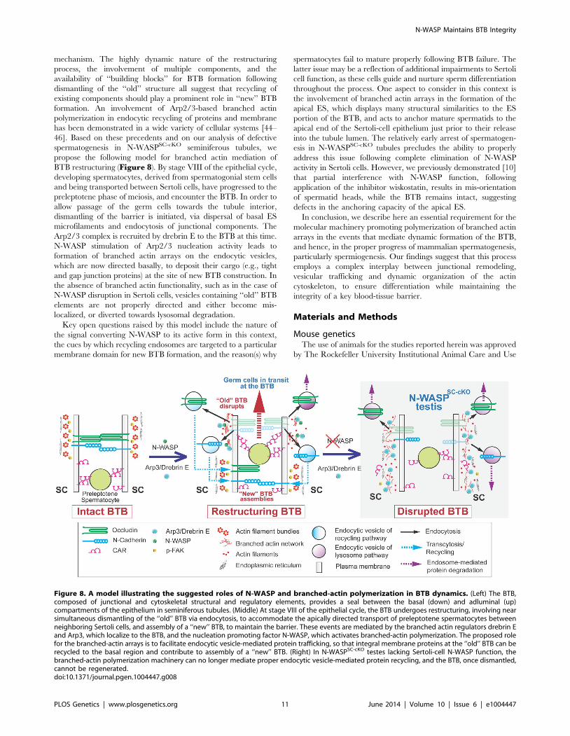
mechanism. The highly dynamic nature of the restructuring
process, the involvement of multiple components, and the
availability of ‘‘building blocks’’ for BTB formation following
dismantling of the ‘‘old’’ structure all suggest that recycling of
existing components should play a prominent role in ‘‘new’’ BTB
formation. An involvement of Arp2/3-based branched actin
polymerization in endocytic recycling of proteins and membrane
has been demonstrated in a wide variety of cellular systems [44–
46]. Based on these precedents and on our analysis of defective
spermatogenesis in N-WASPSC-cKO seminiferous tubules, we
propose the following model for branched actin mediation of
BTB restructuring (Figure 8). By stage VIII of the epithelial cycle,
developing spermatocytes, derived from spermatogonial stem cells
and being transported between Sertoli cells, have progressed to the
preleptotene phase of meiosis, and encounter the BTB. In order to
allow passage of the germ cells towards the tubule interior,
dismantling of the barrier is initiated, via dispersal of basal ES
microfilaments and endocytosis of junctional components. The
Arp2/3 complex is recruited by drebrin E to the BTB at this time.
N-WASP stimulation of Arp2/3 nucleation activity leads to
formation of branched actin arrays on the endocytic vesicles,
which are now directed basally, to deposit their cargo (e.g., tight
and gap junction proteins) at the site of new BTB construction. In
the absence of branched actin functionality, such as in the case of
N-WASP disruption in Sertoli cells, vesicles containing ‘‘old’’ BTB
elements are not properly directed and either become mis-
localized, or diverted towards lysosomal degradation.
Key open questions raised by this model include the nature of
the signal converting N-WASP to its active form in this context,
the cues by which recycling endosomes are targeted to a particular
membrane domain for new BTB formation, and the reason(s) why
spermatocytes fail to mature properly following BTB failure. The
latter issue may be a reflection of additional impairments to Sertoli
cell function, as these cells guide and nurture sperm differentiation
throughout the process. One aspect to consider in this context is
the involvement of branched actin arrays in the formation of the
apical ES, which displays many structural similarities to the ES
portion of the BTB, and acts to anchor mature spermatids to the
apical end of the Sertoli-cell epithelium just prior to their release
into the tubule lumen. The relatively early arrest of spermatogen-
esis in N-WASPSC-cKO tubules precludes the ability to properly
address this issue following complete elimination of N-WASP
activity in Sertoli cells. However, we previously demonstrated [10]
that partial interference with N-WASP function, following
application of the inhibitor wiskostatin, results in mis-orientation
of spermatid heads, while the BTB remains intact, suggesting
defects in the anchoring capacity of the apical ES.
In conclusion, we describe here an essential requirement for the
molecular machinery promoting polymerization of branched actin
arrays in the events that mediate dynamic formation of the BTB,
and hence, in the proper progress of mammalian spermatogenesis,
particularly spermiogenesis. Our findings suggest that this process
employs a complex interplay between junctional remodeling,
vesicular trafficking and dynamic organization of the actin
cytoskeleton, to ensure differentiation while maintaining the
integrity of a key blood-tissue barrier.
Materials and Methods
Mouse geneticsThe use of animals for the studies reported herein was approved
by The Rockefeller University Institutional Animal Care and Use
Figure 8. A model illustrating the suggested roles of N-WASP and branched-actin polymerization in BTB dynamics. (Left) The BTB,composed of junctional and cytoskeletal structural and regulatory elements, provides a seal between the basal (down) and adluminal (up)compartments of the epithelium in seminiferous tubules. (Middle) At stage VIII of the epithelial cycle, the BTB undergoes restructuring, involving nearsimultaneous dismantling of the ‘‘old’’ BTB via endocytosis, to accommodate the apically directed transport of preleptotene spermatocytes betweenneighboring Sertoli cells, and assembly of a ‘‘new’’ BTB, to maintain the barrier. These events are mediated by the branched actin regulators drebrin Eand Arp3, which localize to the BTB, and the nucleation promoting factor N-WASP, which activates branched-actin polymerization. The proposed rolefor the branched-actin arrays is to facilitate endocytic vesicle-mediated protein trafficking, so that integral membrane proteins at the ‘‘old’’ BTB can berecycled to the basal region and contribute to assembly of a ‘‘new’’ BTB. (Right) In N-WASPSC-cKO testes lacking Sertoli-cell N-WASP function, thebranched-actin polymerization machinery can no longer mediate proper endocytic vesicle-mediated protein recycling, and the BTB, once dismantled,cannot be regenerated.doi:10.1371/journal.pgen.1004447.g008
(VWR International). For antigen/epitope retrieval, slides were
microwave-heated twice (5 min each) in 10 mM sodium citrate
(pH 6.0 at 22uC). Thereafter, sections were immersed sequentially
in 3% hydrogen peroxide (v/v) for 20 min and Triton X-100
(0.1%, v/v) and normal goat serum (10%, v/v) in PBS, which were
used as penetration enhancer and blocking solution, respectively.
After overnight incubation with anti-connexin 43 antibody
(Table 1), a biotin-conjugated F(ab9)2 fragment of goat anti-
rabbit IgG and HRP-streptavidin conjugate (Invitrogen) were
added to the sections in succession, and a color reaction was
obtained using 3-amino-9-ethylcarbazole (AEC, red precipitate,
Invitrogen) as the chromogenic substrate.
Hematoxylin and eosin (H&E) stainingBouin’s solution-fixed 4-mm-thick paraffin sections were used for
H&E staining as described [52]. In brief, sections were rehydrated
through successive xylene and ethanol, tap and deionized water.
Cell nuclei were stained with Hematoxylin 7211 (Richard-Allan
Scientific) for 4–5 min to a blue-purple coloration, followed by
immersing in Clarifier and Scott’s solution for 1 min, respectively.
The cytoplasm was then stained using Eosin-Y for 30 sec to obtain
a pink-red color. Sections were dehydrated and cleared with 100%
ethanol and xylene, sealed in PolyMount (Polysciences), and
examined microscopically.
Statistical analysisGB-STAT (V7.0, Dynamic Microsystems) was used for
statistical analysis. Student’s t-test was used to compare changes
in protein levels between N-WASPSC-cKO and age-matched
control mice, in which the relative protein level was arbitrarily
set at 1. For immunofluorescence analysis, each time point
represented at least n = 3 mice. For immunoblotting data, each bar
is the mean6SD of 4 mice including control group. P,0.05 was
interpreted as statistically significant.
Author Contributions
Conceived and designed the experiments: CYC. Performed the experi-
ments: XX DDM EIT RM KWM NL CYC. Analyzed the data: XX CYC.
Contributed reagents/materials/analysis tools: CKCW WML SBS BZS
EDS CYC. Wrote the paper: CYC EDS.
References
1. de Kretser DM, Kerr JB (1988) The cytology of the testis. In: Knobil E, Neill JB,
Ewing LL, Greenwald GS, Markert CL, et al., editors. The Physiology of
Reproduction Vol 1. New York: Raven Press. pp. 837–932.
2. Cheng CY, Mruk DD (2012) The blood-testis barrier and its implication in male
contraception. Pharmacol Rev 64: 16–64.
3. Franca LR, Auharek SA, Hess RA, Dufour JM, Hinton BT (2012) Blood-tissuebarriers: Morphofunctional and immunological aspects of the blood-testis and
blood-epididymal barriers. Adv Exp Med Biol 763: 237–259.
4. Vogl AW, Vaid KS, Guttman JA (2008) The Sertoli cell cytoskeleton. Adv ExpMed Biol 636: 186–211.
5. Russell LD, Peterson RN (1985) Sertoli cell junctions: morphological and
functional correlates. Int Rev Cytol 94: 177–211.
6. Cheng CY, Mruk DD (2010) A local autocrine axis in the testes that regulatesspermatogenesis. Nature Rev Endocrinol 6: 380–395.
7. Wang CQF, Cheng CY (2007) A seamless trespass: germ cell migration across
the seminiferous epithelium during spermatogenesis. J Cell Biol 178: 549–556.
8. Russell LD (1977) Movement of spermatocytes from the basal to the adluminal
compartment of the rat testis. Am J Anat 148: 313–328.
9. Cheng CY, Lie PPY, Mok KW, Cheng YH, Wong EWP, et al. (2011)Interactions of laminin b3 fragment with b1-integrin receptor: a revisit of the
apical ES-BTB-hemidesmosome functional axis in the testis. Spermatogenesis 1:
40. Arold ST (2011) How focal adhesion kinase achieves regulation by linking ligand
binding, localization and action. Curr Opin Struct Biol 21: 808–813.41. Lie PPY, Mruk DD, Mok KW, Su L, Lee WM, et al. (2012) Focal adhesion
kinase-Tyr407 and -Tyr397 exhibit antagonistic effects on blood-testis barrier
dynamics in the rat. Proc Natl Acad Sci USA 109: 12562–12567.42. Siu MKY, Mruk DD, Lee WM, Cheng CY (2003) Adhering junction dynamics
in the testis are regulated by an interplay of b1-integrin and focal adhesioncomplex (FAC)-associated proteins. Endocrinology 144: 2141–2163.
43. Cheng CY, Lie PPY, Wong EWP, Mruk DD (2013) Focal adhesion kinase and
actin regulatory/binding proteins that modulate F-actin organization at thetissue barrier. Lession from the testis. Tissue Barriers 1:e24252 (DOI:10.4161/
tisb.24252).44. Monastyrska I, He C, Geng J, Hoppe AD, Li Z, et al. (2008) Arp2 links
autophagic machinery with the actin cytoskeleton. Mol Biol Cell 19: 1962–1975.45. Shivas JM, Skop AR (2012) Arp2/3 mediates early endosome dynamics
necessary for the maintenance of PAR asymmetry in Caenorhabditis elegans. Mol
Biol Cell 23: 1917–1927.46. Yamamoto T, Mochida J, Kadota J, Takeda MB, E., Tanaka K (2010) Initial
polarized bud growth by endocytic recycling in the absence of actin cable-dependent vesicle transport in yeast. Mol Biol Cell 21: 1237–1252.
47. Jaegle M, Ghazvini M, Mandemakers W, Piirsoo M, Driegen S, et al. (2003) The
POU proteins Brn-2 and Oct-6 share important functions in Schwann cell
development. Genes Dev 17: 1380–1391.
48. Snapper SB, Takeshima F, Anton I, Liu CH, Thomas SM, et al. (2001) N-
WASP deficiency reveals distinct pathways for cell surface projections and