International Journal of Oral Health and Medical Research | ISSN 2395-7387 | NOVEMBER-DECEMBER 2017 | VOL 4 | ISSUE 4 36 CASE REPORT Mathen RE et al.: Non-Syndromic Multiple Hyperdontia Correspondence to: Dr. Rashmi Elizabeth Mathen, Department of Oral Medicine and Radiology, Mar Baselios Dental College, India. Contact Us: www.ijohmr.com Non-Syndromic Multiple Hyperdontia: Report of 4 Cases Rashmi Elizabeth Mathen 1 , Sheeba Padiyath 2 , Anuja S 3 , Shamji Shajahan 4 , Gigi Roy 5 Hyperdontia is the presence of extra teeth in relation to the normal primary or permanent dentition and the extra teeth is supernumerary teeth. Presence of multiple supernumerary teeth without an associated syndrome (non-syndromic) is rare. Four cases with multiple supernumerary teeth were reported in our Department, and the cases were found to be non-syndromic due to the negative family history, extraoral findings, and pathology. Here we are including discussion, review of literature, and the syndromes associated with multiple supernumerary teeth with the four different non- syndromic Hyperdontia cases. KEYWORDS: Hyperdontia, Multiple Teeth, Multiple Supernumerary Teeth, Nonsyndromic, Supernumerary Teeth AA Hyperdontia is the development of an increased number of teeth in either primary or permanent dentition, and the additional teeth are termed Supernumerary teeth (ST). Supernumerary teeth may be single or multiple, and they vary in size, shape, and locations. Presence of supernumerary teeth can lead to complications such as retained teeth, delayed eruption of permanent teeth, alterations in neighbouring teeth, dental malposition, ectopic eruption, occlusal problems, diastema, and rotation. 1 The occurrence of multiple supernumerary teeth without any abnormal extraoral finding or syndrome (non- syndromic) is rare. This report presents four cases of multiple supernumerary teeth without any syndrome or developmental anomaly.All the cases were confirmed non-syndromic after eliciting family history and after consulting a general physician. Case 1 A 16-year-old male patient came to our dental college with a chief complaint of spacing between the upper front tooth region since 5 years and wished to do orthodontic treatment.On examination, midline diastema with high maxillary frenal attachment and crowding was noted on both arches with buccally erupted 23 (Fig.1) and partially erupted 35 (Fig 2). Panoramic radiograph showed the presence of a missing tooth 48 with four impacted supernumerary teeth, one resembling premolar between 44 and 45; one resembling canine in the second quadrant. displacing 23 buccally; two supernumeraries between 33 and 35, with the distal supernumerary impeding the eruption of 35 (Fig.3). After orthodontic and surgical How to cite this article: RE Mathen, Padiyath S, Anuja S, Shajahan S, Roy G. Non-Syndromic Multiple Hyperdontia: Report of 4 Cases. Int J Oral Health Med Res 2017;4(4):36- 39. INTRODUCTION 1,3,4,5-Postgraduate Student, Department of Oral Medicine and Radiology, Mar Baselios Dental College. 2-Professor, Department of Oral Medicine and Radiology, Mar Baselios Dental College, India. ABSTRACT CASE REPORT Fig.1: Intraoral photograph showing midline diastema, buccally erupted 23 Fig.2 Intraoral photograph showing partially erupted 35. Fig 3: Panoramic view showing impacted Supernumeraries, in 23, 34,35, and 45 region, with distal supernumerary in 35 region preventing eruption of 45.
Transcript
International Journal of Oral Health and Medical Research | ISSN 2395-7387 | NOVEMBER-DECEMBER 2017 | VOL 4 | ISSUE 4 36
CASE REPORT Mathen RE et al.: Non-Syndromic Multiple Hyperdontia
Correspondence to: Dr. Rashmi Elizabeth Mathen, Department of Oral Medicine and
Radiology, Mar Baselios Dental College, India. Contact Us: www.ijohmr.com
Non-Syndromic Multiple Hyperdontia: Report
of 4 Cases Rashmi Elizabeth Mathen1, Sheeba Padiyath2, Anuja S3, Shamji Shajahan4, Gigi Roy5
Hyperdontia is the presence of extra teeth in relation to the normal primary or permanent dentition and the extra teeth is
supernumerary teeth. Presence of multiple supernumerary teeth without an associated syndrome (non-syndromic) is
rare. Four cases with multiple supernumerary teeth were reported in our Department, and the cases were found to be
non-syndromic due to the negative family history, extraoral findings, and pathology. Here we are including discussion,
review of literature, and the syndromes associated with multiple supernumerary teeth with the four different non-
AA aaaasasasss Hyperdontia is the development of an increased number
of teeth in either primary or permanent dentition, and the
additional teeth are termed Supernumerary teeth (ST).
Supernumerary teeth may be single or multiple, and they
vary in size, shape, and locations. Presence of
supernumerary teeth can lead to complications such as
retained teeth, delayed eruption of permanent teeth,
alterations in neighbouring teeth, dental malposition,
ectopic eruption, occlusal problems, diastema, and
rotation.1
The occurrence of multiple supernumerary teeth without
any abnormal extraoral finding or syndrome (non-
syndromic) is rare. This report presents four cases of
multiple supernumerary teeth without any syndrome or
developmental anomaly.All the cases were confirmed
non-syndromic after eliciting family history and after
consulting a general physician.
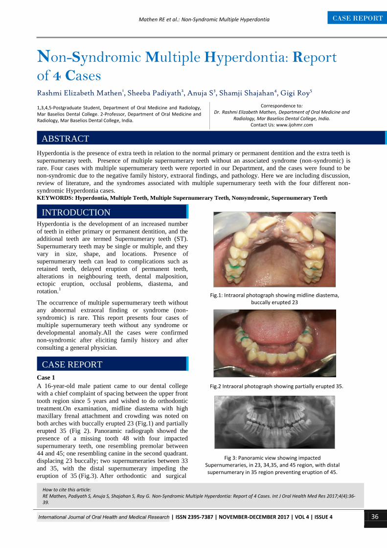
Case 1
A 16-year-old male patient came to our dental college
with a chief complaint of spacing between the upper front
tooth region since 5 years and wished to do orthodontic
treatment.On examination, midline diastema with high
maxillary frenal attachment and crowding was noted on
both arches with buccally erupted 23 (Fig.1) and partially
erupted 35 (Fig 2). Panoramic radiograph showed the
presence of a missing tooth 48 with four impacted
supernumerary teeth, one resembling premolar between
44 and 45; one resembling canine in the second quadrant.
displacing 23 buccally; two supernumeraries between 33
and 35, with the distal supernumerary impeding the
eruption of 35 (Fig.3). After orthodontic and surgical
How to cite this article: RE Mathen, Padiyath S, Anuja S, Shajahan S, Roy G. Non-Syndromic Multiple Hyperdontia: Report of 4 Cases. Int J Oral Health Med Res 2017;4(4):36-39.
INTRODUCTION
1,3,4,5-Postgraduate Student, Department of Oral Medicine and Radiology, Mar Baselios Dental College. 2-Professor, Department of Oral Medicine and Radiology, Mar Baselios Dental College, India.


![[XLS] Details/UnpaidDivided-FY... · Web viewJETHALAL HIRABEN PANNA DOSHI HANSABEN DILIP KANTILAL 360002 KETAN TANNA MAGANLAL SHAMJIBHAI BHAI VARSHA NANDLAL 360005 RATILAL RAWAL SHAMJI](https://static.documents.pub/doc/80x56/5aa8ec927f8b9a81188c1ec0/xls-detailsunpaiddivided-fyweb-viewjethalal-hiraben-panna-doshi-hansaben-dilip.jpg)


