N7 '9| EFFECT OF AMINO ACIDS ON GROWTH AND CAROTENOGENESIS IN CORYNEBACTERIUM SPECIES STRAIN 7EIC THESIS Presented to the Graduate Council of the North Texas State University in Partial Fulfillment of the Requirements For the Degree of MASTER OF SCIENCE By Carolyn S. Coughran, B. S. Denton, Texas May, 1977
Transcript
N7
'9|
EFFECT OF AMINO ACIDS ON GROWTH AND CAROTENOGENESIS
IN CORYNEBACTERIUM SPECIES STRAIN 7EIC
THESIS
Presented to the Graduate Council of the
North Texas State University in Partial
Fulfillment of the Requirements
For the Degree of
MASTER OF SCIENCE
By
Carolyn S. Coughran, B. S.
Denton, Texas
May, 1977
Coughran, Carolyn S., Effect of Amino Acids on Growth
and Carotenogenesis of Corynbeacterium Species Strain 7ElC.
Master of Science (Biology), May, 1977, 34 pp., 7 tables,
2 illustrations, bibliography, 32 titles.
Studies were evaluated on the effects of known growth
factors on the growth and carotenogenesis of Corynebacterium
species strain 7ElC.
The complex medium, Tryptic Soy Broth,was found to
stimulate growth and production of more pigment in the light
and in the dark than did a mineral salts-glucose medium. A
complete amino acid mixture added to LSG enhanced caroteno-
genesis in the dark in Corynebacterium 7ElC, while B-vitamins
retarded carotenogenesis. No absolute requirement for one or
more amino acids was found,indicating a multiple amino acid
requirement. The fewest amino acids found to stimulate caro-
tenogenesis in the dark were a combination of those in the
Serine and Histidine families which include serine, glycine,
cysteine, and histidine.
TABLE OF CONTENTS
LIST OF TABLES ....................... ....... *..
LIST OF ILLUSTRATIONS 0 . .. 4. .9 .0.0.. . .
Chapter
I. INTRODUCTION.... ..... ........
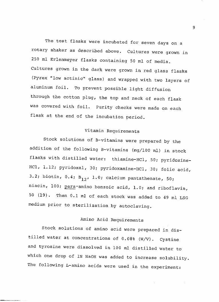
II. MATERIALS AND METHODS ................
Organism and InoculumVitamin RequirementsAmino Acid RequirementsCell Harvest and Assay of Total PigmentOptical Density vs Dry Weight
III. RESULTS.......*......................
IV. DISCUSSION
BIBLIOGRAPHY... ..... ......
iii
Page
. . iv
. . 1
. . 8
. . 13
. . 28
1 0 . 32
LIST OF TABLES
Table Page
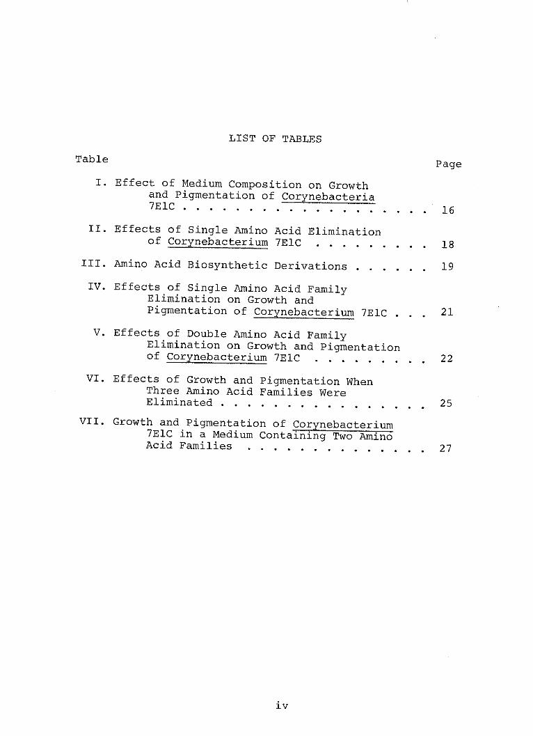
I. Effect of Medium Composition on Growthand Pigmentation of Corynebacteria7ElC. . ....... 16
II. Effects of Single Amino Acid Eliminationof Corynebacterium 7ElC................18
III. Amino Acid Biosynthetic Derivations.,.........19
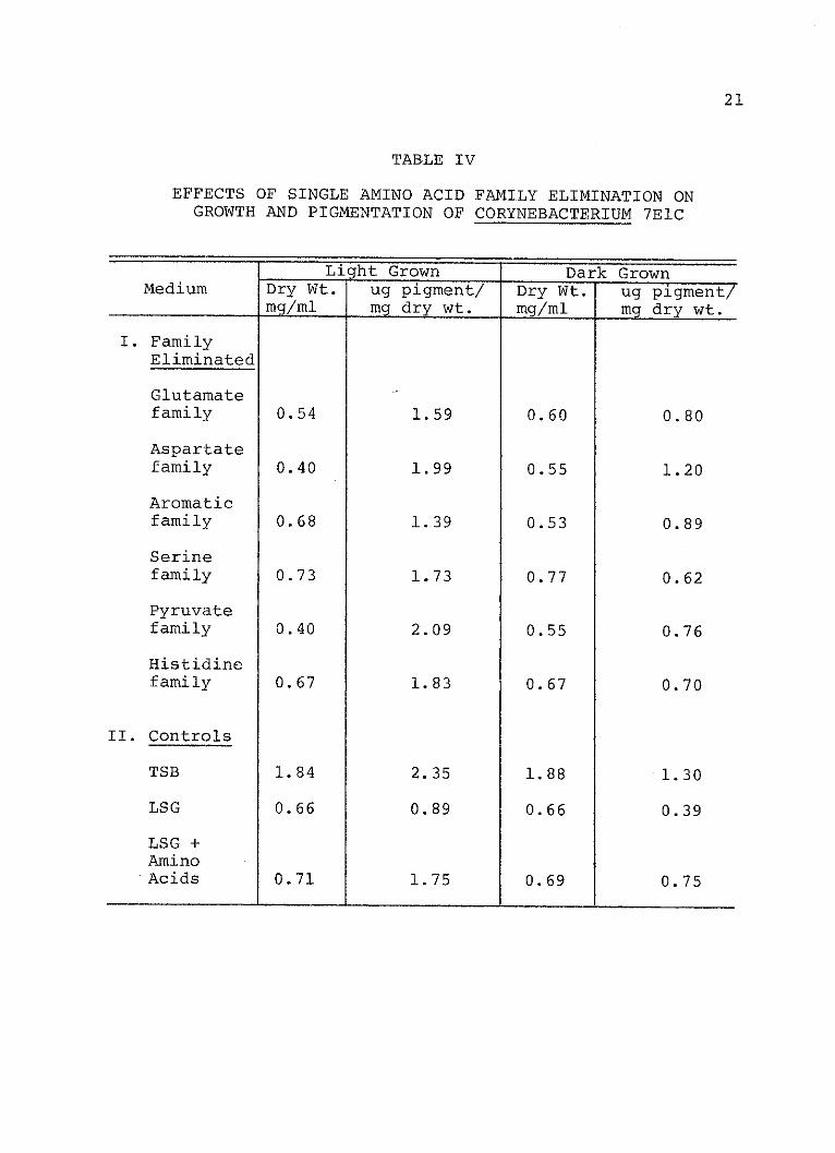
IV. Effects of Single Amino Acid FamilyElimination on Growth andPigmentation of Corynebacterium 7ElC . .. 21
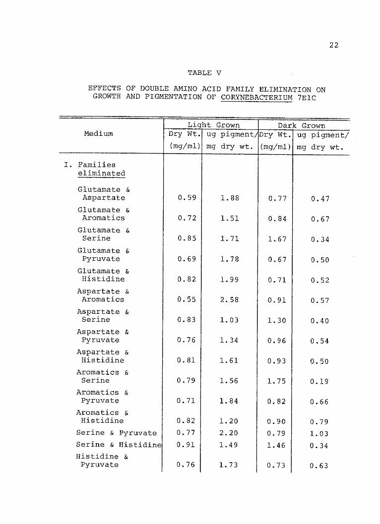
V. Effects of Double Amino Acid FamilyElimination on Growth and Pigmentationof Corynebacterium 7ElC........ ........ 22
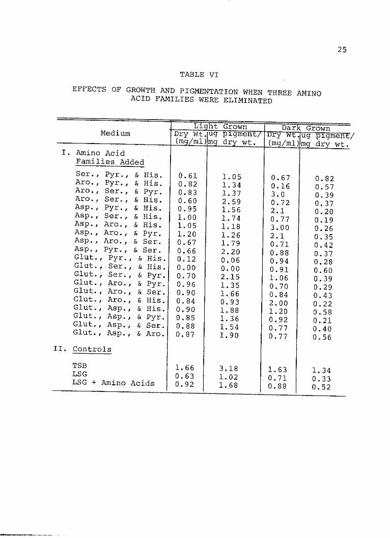
VI. Effects of Growth and Pigmentation WhenThree Amino Acid Families WereEliminated .......................... 25
VII. Growth and Pigmentation of Corynebacterium7ElC in a Medium Containing Two AminoAcid Families.-.-.-.................27
iv
LIST OF ILLUSTRATIONS
Figure Page
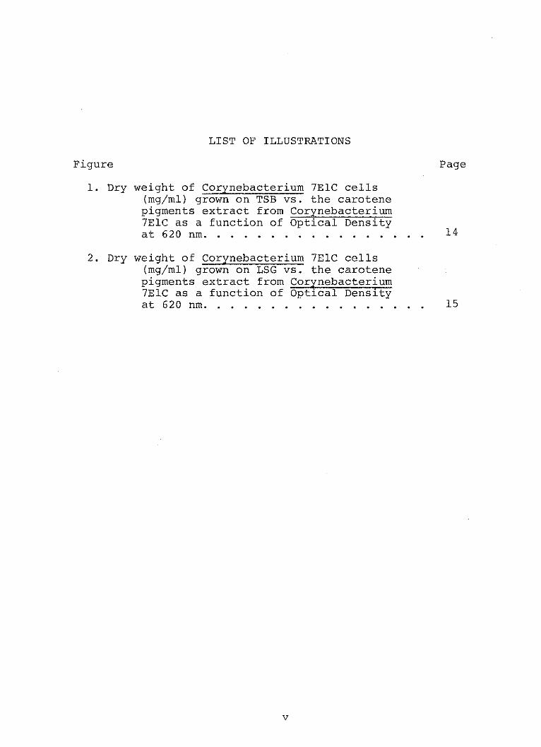
1. Dry weight of Corynebacterium 7ElC cells(mg/ml) grown on TSB vs. the carotenepigments extract from Corynebacterium7ElC as a function of Optical Densityat 620 nm......... ..................... 14
2. Dry weight of Corynebacterium 7ElC cells(mg/ml) grown on LSG vs. the carotenepigments extract from Corynebacterium7ElC as a function of Optical Densityat 620 nm............................ 15
V
CHAPTER I
INTRODUCTION
Carotenoids form a class of pigments, hydrocarbons and
their oxygenated derivatives, red to orange to yellow in
color, which are widely distributed in nature. As defined
by the International Union of Pure and Applied Chemistry
(15), carotenoids are "a class of hydrocarbons (carotenes)
and their oxygenated derivatives (xanthophylls) consisting
of eight isoprenoid units joined in such a manner that the
arrangement of isoprenoid units is reversed at the center
of the molecule."
Depending on the organism, available nutrients and other
environmental factors affect the production of carotenoid
pigments and the type of pigment produced. A number of
studies have shown that light, carbon/nitrogen ratio, carbon
sources, amino acids, and B-vitamins affect pigmentation.
The effect of light on the quantity of carotenoids was
investigated with Mycobacterium lacticolum, strain 35,by
Nikitina (21). He found that illumination with a flux of
1000 lux stimulated carotenogenesis. In a later study
using Mycobacterium lacticolum and Mycobacterium flavum,
Nikitina (23) found that carotenoid pigments were several
times greater in cells recovered from cultures exposed to
1
2
light than from cells grown in the dark (dark-grown cells).
The blue-violet portion of the visible light spectrum was
found to be most effective in stimulating carotenoid syn-
thesis in these organisms. The pigment content of Myco-
Fig. 1--Dry weight of Corynebacterium 7ElC cells(mg/ml) grown on TSB vs.' the carotene pigments extract fromCorynebacterium 7ElC as a function of Optical Density at620 nm.
15
LSG
2N -
10-P
4...2
-HP
04
03
2--
Dry Weight (mg/ml)
Fig. 2--Dry weight of Corynebacterium 7ElC cells(mg/ml) grown on LSG vs. the carotene pigments extract fromCorynebacterium 7ElC as a function of Optical Density at620 nm.
16
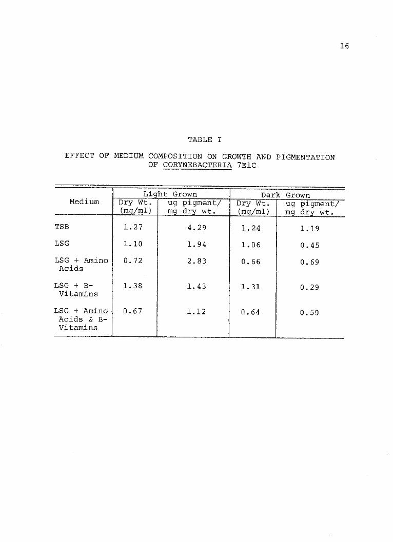
TABLE I
EFFECT OF MEDIUM COMPOSITION ON GROWTH AND PIGMENTATIONOF CORYNEBACTERIA 7ElC
Light Grown Dark GrownMedium Dry Wt. ug pigment/ Dry Wt. ug pigment/
_(mg/ml) mg dry wt. (mg/ml) mg dry wt.
TSB 1.27 4.29 1.24 1.19
LSG 1.10 1.94 1.06 0.45
LSG + Amino 0.72 2.83 0.66 0.69Acids
LSG + B- 1.38 1.43 1.31 0.29Vitamins
LSG + Amino 0.67 1.12 0.64 0.50Acids & B-Vitamins
17
pigment synthesis. Amino acids, although effective in LSG,
were not as effective as TSB. The amino acids give a 36%
increase in LSG when cells were grown in the light and a 63%
increase when grown in the dark. Cell growth was inhibited
somewhat by the amino acid mixture but not by the vitamins.
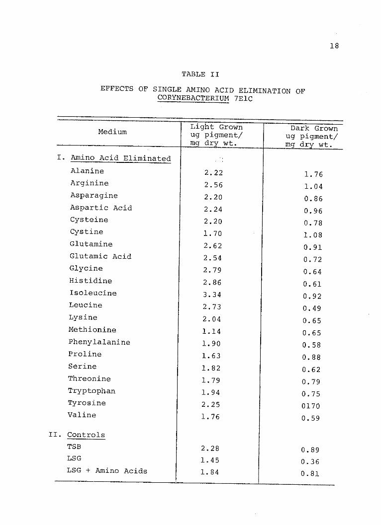
Effects of single amino acid exclusion on the pigmen-
tation of Corynebacterium 7ElC are shown in Table II. The
light-grown cells produced more pigment than the dark grown
cells. These data show that when the organism was grown in
the light the elimination of any one amino acid, except
isoleucine, did not markedly affect pigmentation any more
than the control containing the complete amino acid mixture.
Elimination of isoleucine, however, led to enhanced pigment
production. In the dark-grown cells when alanine, arginine,
and cystine were eliminated pigment levels increased.
Elimination of glutamine and isoleucine also resulted in a
modest increase in pigment. In no case did the removal of
a single amino acid from the mixture result in pigment
levels equivalent to that found in the LSG control indica-
ting that the effect is due to a multiple amino acid re-
quirement.
In order to determine what combination of amino acids
were required, further studies were conducted with amino
acids grouped on the basis of their biosynthetic derivation
are shown on Table III. Since some of the amino acids could
18
TABLE II
EFFECTS OF SINGLE AMINO ACID ELIMINATION OFCORYNEBACTERIUM 7ElC
Light Grown Dark Grownug pigment/ ug pigment/mrg dry wt. mg dry wt.
Table VII indicates the growth and pigmentation of Coryne-
bacterium 7ElC in an LSG medium which contains two amino
acid families. The data indicate that both the Histidine-
Serine, and Aromatic-Serine family-combinations allow for
pigment production equivalent to that of the complete amino
acid mixture, but do not allow for pigment production equal
to that of the cells grown in TSB. This table also shows
that glucose is not necessary for pigmentation. When the
complete amino acid mixture served as the sole carbon source,
pigment yields equivalent to those of the amino acids and
glucose were obtained.
27
TABLE VII
GROWTH AND PIGMENTATION OF CORYNEBACTERIUM 7ElC INA MEDIUM CONTAINING TWO AMINO ACID FAMILIES
Light Grown Dark GrownMedium Dry Wt. ug pigment! Dry Wt. ug pigment
(mg/mi) mg dry wt. (mg/m) mg dry wt.
I. Amino AcidFamiliesAdded
His. & Ser. 0.44 0.90 0.42 0.54
Aro. & Ser. 0.19 1.98 0.35 0.58
II. Controls
TSB 1.67 2.88 1.50 1.12
LSG 0.49 0.96 0.75 0.32
LSG + AminoAcids 0.93 1.50 0.85 0.55
LS + AminoAcidsWithoutGlucose 0.26 1.36 0.26 0.58
CHAPTER IV
DISCUSSION
When the effect of medium composition on pigment
production in Corynebacterium 7ElC was tested, the results
indicated that more pigment was produced in a complex
medium, in both the light grown cells and the dark grown
cells than in a minimal medium containing only one carbon
source (LSG). When known growth factors, such as amino
acids and B-vitamins, were tested, it was found that the
amino acids enhanced and the B-vitamins retarded pigment
synthesis. However, the response to amino acids was not as
great as that obtained with TSB. One explanation of this
response may be that the concentration of amino acids was
not optimal. The results might also reflect the necessity
of another factor or factors in maximum pigment yield.
A third reason for this response might be that some of the
amino acids stimulate pigment synthesis and others retard
it. The levels of the individual amino acids in TSB are
not known although they were all added in essentially the
same concentration in the LSG medium. Some evidence for
inhibition of pigment production by certain amino acids is
shown in Table II. These data indicate that the omission
29
of either alanine, arginine, or cystine from the amino
acid mixture results in increased pigment production.
The amino acids and the complex medium TSB stimulated
pigmentation in the light as well as in the dark. However,
in no case did the amount of pigment produced in the dark
equal the amount produced in the light. Light-grown cells
normally had four times the amount of pigment as dark-grown
cells. The implication of this is that two mechanisms are
operative; one light dependent, the other substrate
dependent.
That the amino acid effect is not due to an absolute
requirement for one or more amino acids is shown in Table II.
If the requirement was an absolute one, then the pigment
produced when it was omitted should drop to the level found
in the LSG control. Since the drop in level of pigment
production did not occur, the results indicate a multiple
requirement.
When combinations of amino acid families were elimi-
nated from the media, the Serine family was found important
in stimulating carotenogenesis in the dark since its
elimination with nearly all other families resulted in a
reduction of pigment synthesis. However, the requirement
for the Serine family was not absolute. As indicated in
Table VI, the combination of the Glutamate-Aspartate-
Histidine families and the aromatic-Pyruvate-histidine
families also stimulated pigment production. Apparently
30
the organism has the ability to interconvert certain amino
acids so that no absolute requirement exists.
The amino acid families tested also effected growth,
although growth stimulation did not necessarily correlate
with stimulation of pigment synthesis. When the Pyruvate
family and the Serine family were eliminated together, there
was an increase in pigment and a decrease in growth in dark
grown cells. When the Aromatic and Serine families were
eliminated together there was an increase in growth and a
decrease in pigment production. The explanation for this
is as yet unknown.
The minimum number of amino acids found so far which
will stimulate pigment production in the dark are a combin-
ation of those in the Serine and Histidine families which
include four amino acids: serine, glycine, cysteine, and
histidine. Which of the three amino acids in the Serine
family is important has yet to be determined.
The results reported here show that amino acids exert
a stimulatory effect on carotenogenesis in the dark in
Corynebacterium 7ElC. Furthermore, this response is not due
to an absolute requirement for one or more amino acids. A
combination of as few as four amino acids has been found tostimulate carotenogenesis. In addition, some amino acid
combinations have been found to stimulate growth but not
pigment synthesis. The optimum concentration of amino acid
31
combinations and the mechanisms by which they exert their
effects await further research.
BIBLIOGRAPHY
1. Batra, P. P., R. M. Gleason, Jr., and J. Jenkins, 1969.Mechanism of Photo-induced and Antimycin A-inducedCarotenoid Synthesis in the Mycobacterium marinum.Requirements for Carotenogenesis and Further Evidencefor Protein Synthesis Following Induction. Biochem.Biophys. Acta. 177: 124-135.
2. Codner, R. C. and B. C. P6latt. 1959. Light-InducedProduction of Carotenoid Pigments by Cephalosporia.Nature 184: 741-742.
3. Cooney, J. J. and 0. C. Thierry. 1966. A definedMedium for Growth and Pigment Synthesis of Micrococcusroseus. Can. J. Microbiol. 12: 83-89.
4. Davies, B. H. 1965. Analysis of Carotenoid Pigments,p. 489-531. In T. W. Goodwin, Chemistry and Bio-chemistry of Plant Pigments. Academic Press. NewYork.
5. Friend, J. and T. W. Goodwin. 1954. The Effect ofTemperature and Thiamine Concentration on Caroteno-genesis by Phycomyces blakesleeanus. Biochem. J.57: 434-437.
6. Garton, G. A., T. W. Goodwin, and W. Lijinsky. 1951.General Conditions Governing B-Carotene Synthesis bythe Fungus Phycomyces blakesleeanus burgeff. Biochem.J. 48:154-1630
7. Goodwin, T. W., M. Jamikorn, and J. S. Willmer. 1953.Further Observations Concerning the Action of Diphenyl-amine in Inhibiting the Synthesis of B-Carotene inPhycomyces blakesleeanus. Biochem. J. 53: 532-540.
8. Gordon, R. E. 1965. Some strains in search of a genus-Corynebacterium, Mycobacterium, Nocardia, or what?J. Gen. Microbiol. 43: 329-343.
9. Grechushkina, N. N., M. B. Yakovleva, and I. L. Rabotnova.1968. Carotenoid Biosynthesis by Mycobacterium lacti-colum (Strain 105) in Medium Containing n-Hexadecane,Mikrobiol. 37: (5), 799-80.3.
32
33
10. Gribanovski-Sassu, 0. and F. H. Foppen. 1968. Lightand Temperature Effect on Epicoceum nigrum- Phytochem.7: 1605-1612.
11. Hammond, R. D. and D. C. White. 1970. CarotenoidFormation by Staphylococcus sureus. J. of Bacteriol.103: (1) 191-198.
12. Howard, M. D. 1974. Effect of light and other environ-mental factors on growth and carotenogenesis ofCerynebacterium species strain 7ElC, unpublishedMaster's thesis, Department of Biology, North TexasState University, Denton, Texas.
13. Ingraham, M. A. and H. Steenbock. 1935. The Relationof Microorganisms to Carotenoids and Vitamin A. Bio-chem. J. 29: 2553-2562.
14. Isakova, D. M. and H. S. Yelisyeyeva. 1974. Effects ofthiamine on pigment formation in Mycobacterium luteumand Nocardia corallina. Mikrobiol. 34(2): 160-164.
15. IUPAC commission on the nomenclature of organic chemistryand IUPAC-IUB commission on biochemical nomenclature:Tentative rules for the nomenclature of carotenoids.1971. Biochem. 10: 4827-4837.
16. Kester, A. S. and J. W. Fester. 1962. Studies on theOxidation of Hydrocarbons by Microorganisms. J.Bacteriol. 85: 850-869.
17. Leadbetter, E. R. and J. W. Foster. 1958. Studies onsome methane-utilizing bacteria. Arch. Microbiol.30: 69-82.
18. Mathews, M. M. 1963. Studies on the Localization,Function, and Formation of the Carotenoid Pigments ofa Strain of Mycobacterium marinum. Photochem. andPhotobiol. 2: 1-8.
19. Mayfield, D. 1972. A comparative Study of Two Vitre-oscilla species, unpublished doctor's dissertation,Department of Biology, North Texas State University,Denton, Texas.
20. Nakayama, T., G. Mackinney and H. J. Phaff. 1954.Carotenoids in Asperogeneus Yeasts. Antonie VanLeeuwenhoek; J. Microbiol. 9: 601-611.
34
21. Nikitina, K. A. 1966. Pigment Formation by Myco-bacterium lacticolum strain 35. Mikrobiol. 35(2):253-257.
22. Nikitina, K. A. 1967. Influence of Light on PigmentSynthesis by Saprophytic Mycobacterium. Mikrobiol.36(4): 608-613.
23. Kikitina, K. A. 1968. Influence of Light on CarotenoidBiosynthesis by Mycobacterium flavum var. methaniumB and Mycobacterium lacticolum, 35. Mikrobiol.37(1): 48-56.
24. Seviour, R. J. and R. C. Codner. 1973. Effect of lighton Carotenoid and Riboflavin Production by the FungusCephalosporium diospyri. J. Gen. Microbiol. 77:403-415.
25. Stanier, R. Y. 1970. Biosynthetic derivations of theamino acids, p. 241. In R. Y. Stanier, The MicrobialWorld, Fourth Edition, Prentice-Hall Inc., New Jersey.
26. Starr, M. P. and S. Saperstein. 1953. Thiamine and theCarotenoid Pigments of Corynebacterium poinsettiae.Arch. Biochem. Biophys. 43: 157-168.
27. Sutter, R. P. 1970. Effect of Light on B-CaroteneAccumulation in Blakeslea trispora. J. Gen. Microbiol.64: 215-221.
28. Thierry, 0. C. and J. J. Cooney. 1966. PhysiochemicalFactors Influencing Growth and Pigment Synthesis byMicrococcus reseus. Can. J. Microbiol. 12: 691-698.
29. Ghirkell, D. 1969. Growth and pigmentation of Micro-coccus radiodurans. J. Gen. Microbiol. 55: 337-340.
30. Weeks, 0. B. and R. J. Garner. 1967. Biosynthesis ofCarotenoids in Flavobacterium dehydrogenes. Arch.Biochem. and Biophys. 121: 35-49.
31. Wilson, T. E. and A. G. Nunez. 1965. Factors Influ-encing Pigment Production by Staphylococcus aureus.Texas Reports on Biology and Medicine. 23: 147.
32. Zalekar, M. 1957. Variations in the Production ofCarotenoids in Neurospora. Arch. Biochem. andBiophys. 70: 561-567.