Beyond platinum: synthesis, characterization,and in vitro toxicity of Cu(II)-releasing polymernanoparticles for potential use as a drugdelivery vectorAlesha N Harris, Barbara R Hinojosa, Montaleé D Chavious and Robby A Petros*
Abstract
The field of drug delivery focuses primarily on delivering small organic molecules or DNA/RNA as therapeutics andhas largely ignored the potential for delivering catalytically active transition metal ions and complexes. The deliveryof a variety of transition metals has potential for inducing apoptosis in targeted cells. The chief aims of this workwere the development of a suitable delivery vector for a prototypical transition metal, Cu2+, and demonstration ofthe ability to impact cancer cell viability via exposure to such a Cu-loaded vector. Carboxylate-functionalizednanoparticles were synthesized by free radical polymerization and were subsequently loaded with Cu2+ via bindingto particle-bound carboxylate functional groups. Cu loading and release were characterized via ICP MS, EDX, XPS,and elemental analysis. Results demonstrated that Cu could be loaded in high weight percent (up to 16 wt.%) andthat Cu was released from the particles in a pH-dependent manner. Metal release was a function of both pH andthe presence of competing ligands. The toxicity of the particles was measured in HeLa cells where reductions incell viability greater than 95% were observed at high Cu loading. The combined pH sensitivity and significanttoxicity make this copper delivery vector an excellent candidate for the targeted killing of disease cells whencombined with an effective cellular targeting strategy.
Keywords: copper, polymer nanoparticles, copper ion release, drug delivery, oxidative stress, HeLa cells
IntroductionThe field of drug delivery focuses primarily on deliver-ing small organic molecules or DNA/RNA as therapeu-tics and has largely ignored the potential for deliveringcatalytically active transition metal ions and complexes[1-3]. Some success has been realized in the case ofcisplatin [4-7]; however, vectors designed to deliverother metal species are rare [8-11]. Thus, a significantopportunity exists for examining the impact of selec-tively delivering a variety of metal ions and complexesto cells. Rational design of a vector capable of sequester-ing and releasing metals is therefore needed. Nanoparti-cles based on nanoscale metal/organic frameworks andinfinite coordination polymers are being pursuedactively as drug delivery vectors; however, the metal is
used as a structural component of the particle, and ingeneral is not the therapeutically active moiety [12,13].We have developed a prototypical approach that allowsus to accomplish reversible metal binding to polymericnanoparticles that are stable in aqueous solutions andthat are capable of releasing bound metal in a pH-dependent manner. We also postulate that release couldbe triggered by a change in reduction potential. Sensitiv-ity to pH allows one to capitalize on the drop in pHknown to occur along the endosomal/lysosomal pathwayfor endocytosis to facilitate release, while sensitivity to areducing environment could stimulate release inresponse to the reducing nature of cytosol [1].If targeted delivery can be achieved, transition metal
species would be expected to display a range of activitiesinside the cell ranging from redox catalysis to the tar-geted binding of biomolecules [14-17]. Recent findings[18-26] indicate that many types of nanoparticles are
* Correspondence: [email protected] of Chemistry, University of North Texas, 1155 Union Circle,CB#305070, Denton, TX, 76203-5017, USA
Harris et al. Nanoscale Research Letters 2011, 6:445http://www.nanoscalereslett.com/content/6/1/445
capable of inducing oxidative stress, which is of greatconcern in terms of the nanotoxicology of particlesbeing pursued for a variety of consumer products.Furthermore, some colloidal metal particles have beenshown to be particularly effective at generating reactiveoxygen species (ROS) presumably through the slowleaching of metal ions from the particle core [19-21,25].Increased ROS production is capable of inducing biolo-gical damage and has been linked to a variety of diseasestates including cancer, cardiovascular disease, arthritis,diabetes, Alzheimer’s disease, and Parkinson’s disease[27]. Cancer cells use ROS to suppress apoptosis, accel-erate proliferation, induce metastasis and angiogenesis,and promote genetic instability through DNA damage[27-32]. However, the inherent toxicity of increasedROS production represents an opportunity if it can beharnessed by selectively targeting ROS-generating parti-cles to diseased cells [28,30]. In this case, it would bedesirable to release large amounts of metal ions in ashort period of time, which is opposite to what isobserved for the slow leaching of metal ions from colloi-dal metal particles. Increased ROS production has thepotential to induce cell death by altering the expressionof apoptosis-related genes, such as Fas, c-fos, c-jun, p53,and Bcl-2 [22,24,33,34]. It is important to note thatmost chemotherapeutics display high levels of toxicity,and that their maximum tolerated dose is often dictatedby the maximum tolerable off-target toxicity. Transitionmetal complexes also routinely exhibit high levels oftoxicity; however, such toxicity does not limit theirpotential for treating disease [17]. For example, a seriesof Cu2+ -containing compounds that exhibit high levelsof cytotoxicity and genotoxicity are being actively pur-sued as cancer chemotherapeutics [35,36].We have therefore designed a carboxylate-functiona-
lized, polymer-based nanoparticle capable of sequester-ing a prototypical metal, Cu2+, for the ultimate goal ofdelivering Cu2+ to cancer cells to facilitate apoptosis.The particles described here represent a single exampleof a multitude containing other metal/ligand combina-tions that can be envisaged [37]. Here, we report thesynthesis, characterization, and metal binding propertiesof our Cu-binding particles, as well as preliminaryin vitro toxicity in cancer cells
ResultsSynthesis and characterization of Cu-loaded polymericnanoparticlesCarboxylate-functionalized, acrylate-based nanoparticleswere synthesized via standard microwave-assisted, freeradical polymerization techniques [38]. Nanoparticlesused for all experiments described in this work wereprepared from an aqueous pre-polymer solutioncontaining 50 wt.% of an acrylic acid monomer. Nano-particles were synthesized in aqueous solution andremained well dispersed over the course of severalweeks. Excess unreacted monomer was removed via dia-lysis and nanoparticle concentration was determined bylyophilizing a sample of purified particles and weighingthe resultant solid.Cu2+ loading to form Cu-loaded carboxylate-functio-
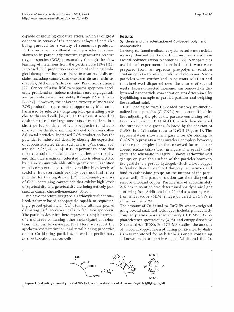
nalized nanoparticles (CuCNPs) was accomplished byfirst adjusting the pH of the particle-containing solu-tion to 7.0 using 1.0 M NaOH, which deprotonatedthe carboxylic acid groups, followed by the addition ofCuSO4 in a 1:1 molar ratio to NaOH (Figure 1). Therepresentation shown in Figure 1 for Cu binding toCuCNPs represents a mononuclear complex; however,a dinuclear complex like that observed for molecularcopper acetate (also shown in Figure 1) is equally likely(note: the schematic in Figure 1 shows carboxylic acidgroups only on the surface of the particle; however,the particle is a porous hydrogel, which allows copperto freely diffuse throughout the polymer network andbind to carboxylate groups on the interior of the parti-cle as well). The particle solution was then dialyzed toremove unbound copper. Particle size of approximately215 nm in solution was determined via dynamic lightscattering (see Additional file 1) and a scanning elec-tron microscope (SEM) image of dried CuCNPs isshown in Figure 2A.The amount of Cu bound to CuCNPs was investigatedusing several analytical techniques including: inductivelycoupled plasma mass spectrometry (ICP MS), X-rayphotoelectron spectroscopy (XPS), and energy-dispersiveX-ray analysis (EDX). For ICP MS studies, the amountof unbound copper released during purification by dialy-sis was monitored for 48 h from a sample containinga known mass of particles (see Additional file 2).
Figure 1 Cu-loading chemistry for CuCNPs (left) and the structure of dinuclear Cu2(OAc)4(H2O)2 (right).
Harris et al. Nanoscale Research Letters 2011, 6:445http://www.nanoscalereslett.com/content/6/1/445
Page 2 of 10
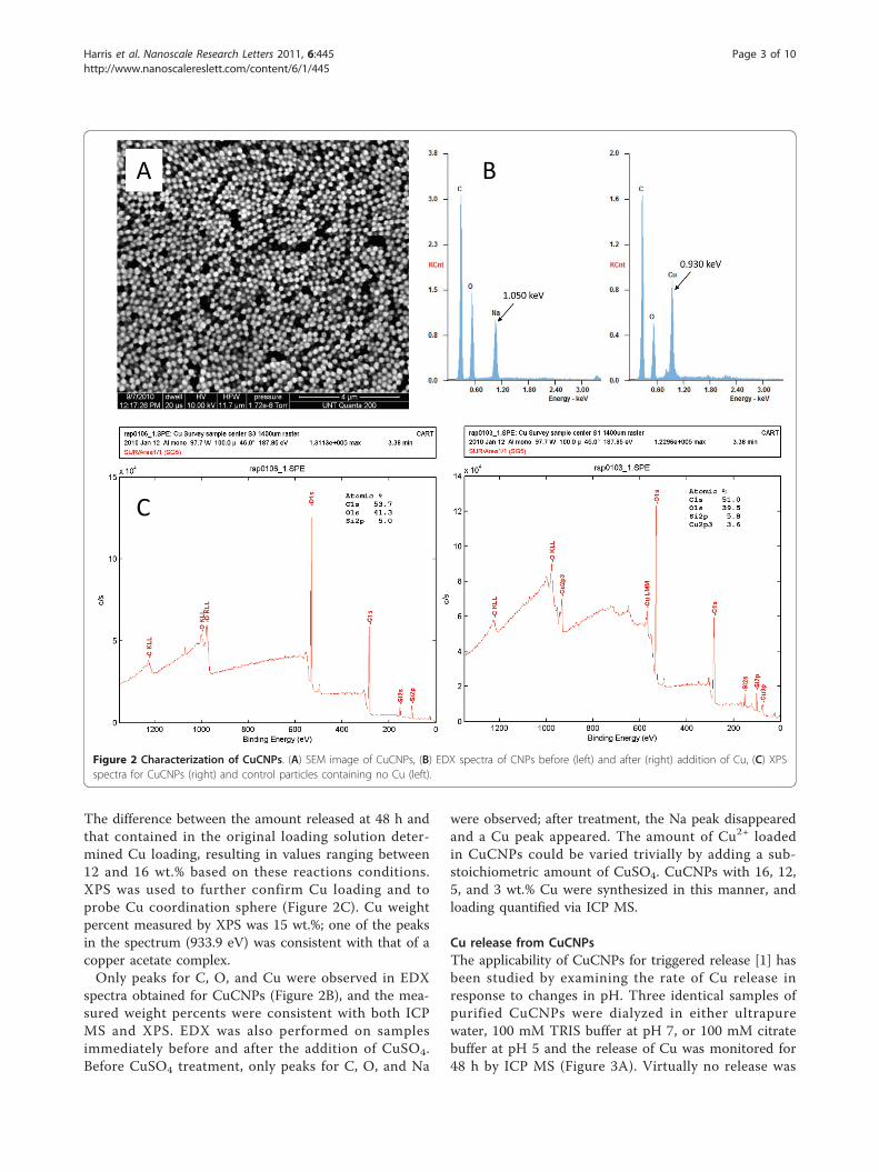
The difference between the amount released at 48 h andthat contained in the original loading solution deter-mined Cu loading, resulting in values ranging between12 and 16 wt.% based on these reactions conditions.XPS was used to further confirm Cu loading and toprobe Cu coordination sphere (Figure 2C). Cu weightpercent measured by XPS was 15 wt.%; one of the peaksin the spectrum (933.9 eV) was consistent with that of acopper acetate complex.Only peaks for C, O, and Cu were observed in EDX
spectra obtained for CuCNPs (Figure 2B), and the mea-sured weight percents were consistent with both ICPMS and XPS. EDX was also performed on samplesimmediately before and after the addition of CuSO4.Before CuSO4 treatment, only peaks for C, O, and Na
were observed; after treatment, the Na peak disappearedand a Cu peak appeared. The amount of Cu2+ loadedin CuCNPs could be varied trivially by adding a sub-stoichiometric amount of CuSO4. CuCNPs with 16, 12,5, and 3 wt.% Cu were synthesized in this manner, andloading quantified via ICP MS.
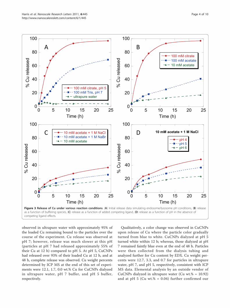
Cu release from CuCNPsThe applicability of CuCNPs for triggered release [1] hasbeen studied by examining the rate of Cu release inresponse to changes in pH. Three identical samples ofpurified CuCNPs were dialyzed in either ultrapurewater, 100 mM TRIS buffer at pH 7, or 100 mM citratebuffer at pH 5 and the release of Cu was monitored for48 h by ICP MS (Figure 3A). Virtually no release was
BA
C
Figure 2 Characterization of CuCNPs. (A) SEM image of CuCNPs, (B) EDX spectra of CNPs before (left) and after (right) addition of Cu, (C) XPSspectra for CuCNPs (right) and control particles containing no Cu (left).
Harris et al. Nanoscale Research Letters 2011, 6:445http://www.nanoscalereslett.com/content/6/1/445
Page 3 of 10
observed in ultrapure water with approximately 95% ofthe loaded Cu remaining bound to the particles over thecourse of the experiment. Cu release was observed atpH 7; however, release was much slower at this pH(particles at pH 7 had released approximately 55% oftheir Cu at 12 h) compared to pH 5. At pH 5, CuCNPshad released over 93% of their loaded Cu at 12 h, and at48 h, complete release was observed. Cu weight percentsdetermined by ICP MS at the end of this set of experi-ments were 12.1, 1.7, 0.0 wt.% Cu for CuCNPs dialyzedin ultrapure water, pH 7 buffer, and pH 5 buffer,respectively.
Qualitatively, a color change was observed in CuCNPsupon release of Cu where the particle color graduallyturned from blue to white. CuCNPs dialyzed at pH 5turned white within 12 h; whereas, those dialyzed at pH7 remained faintly blue even at the end of 48 h. Particleswere then collected from the dialysis tubing andanalyzed further for Cu content by EDX. Cu weight per-cents were 12.7, 3.3, and 0.7 for particles in ultrapurewater, pH 7, and pH 5, respectively, consistent with ICPMS data. Elemental analysis by an outside vendor ofCuCNPs dialyzed in ultrapure water (Cu wt.% = 10.92)and at pH 5 (Cu wt.% = 0.04) further confirmed our
0
20
40
60
80
100
0 5 10 15 20 25
10 mM acetate + 1 M NaCl
pH 4pH 5pH 6
% C
u re
leas
ed
Time (h)
0
20
40
60
80
100
0 5 10 15 20 25
100 mM citrate, pH 5100 mM Tris, pH 7ultrapure water
% C
u re
leas
ed
Time (h)
0
20
40
60
80
100
0 5 10 15 20 25
100 mM citrate100 mM acetate10 mM acetate
% C
u re
leas
edTime (h)
A B
C D
0
20
40
60
80
100
0 5 10 15 20 25
10 mM acetate + 1 M NaCl10 mM acetate + 1 M NaBr10 mM acetate
% C
u re
leas
ed
Time (h)Figure 3 Release of Cu under various reaction conditions. (A) Initial release data simulating endosome/lysosome pH conditions, (B) releaseas a function of buffering species, (C) release as a function of added competing ligand, (D) release as a function of pH in the absence ofcompeting ligand effects.
Harris et al. Nanoscale Research Letters 2011, 6:445http://www.nanoscalereslett.com/content/6/1/445
Page 4 of 10
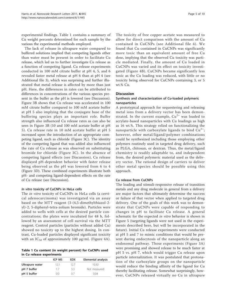
experimental findings. Table 1 contains a summary ofCu weight percents determined for each sample by thevarious the experimental methods employed.The lack of release in ultrapure water compared to
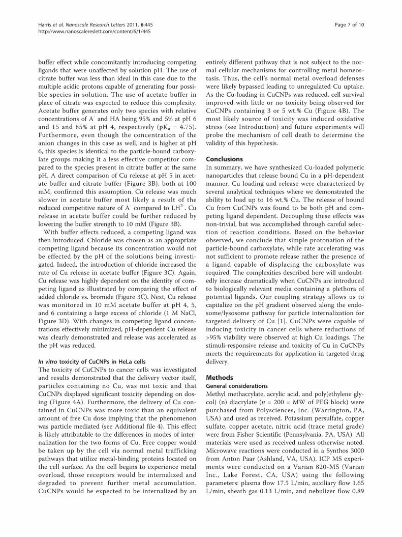
buffered solutions implied that competing ligands otherthan water must be present in order to facilitate Curelease, which led us to further investigate Cu release asa function of competing ligand. Cu release experimentsconducted in 100 mM citrate buffer at pH 4, 5, and 6revealed faster metal release at pH 6 than at pH 4 (seeAdditional file 3), which was surprising and further illu-strated that metal release is affected by more than justpH. Here, the differences in rates can be attributed todifferences in concentrations of the various species pre-sent in the buffer as the pH is lowered (see Discussion).Figure 3B shows that Cu release was accelerated in 100mM citrate buffer compared to 100 mM acetate bufferat pH 5 also implying that the conjugate base of thebuffering species plays an important role. Bufferstrength also influenced Cu release rates as can also beseen in Figure 3B (10 and 100 mM acetate buffer at pH5). Cu release rate in 10 mM acetate buffer at pH 5increased upon the introduction of an appropriate com-peting ligand, such as chloride (Figure 3C). The identityof the competing ligand that was added also influencedthe rate of Cu release as was observed on substitutingbromide for chloride (Figure 3C). In the absence ofcompeting ligand effects (see Discussion), Cu releasedisplayed pH-dependent behavior with faster releasebeing observed as the pH was lowered from 6 to 4(Figure 3D). These combined experiments illustrate bothpH- and competing ligand-dependent effects on the rateof Cu release (see Discussion).
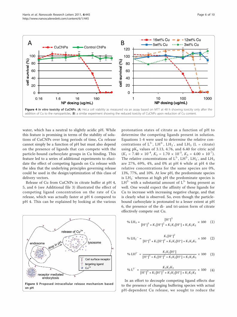
In vitro toxicity of CuCNPs in HeLa cellsThe in vitro toxicity of CuCNPs in HeLa cells (a cervi-cal adenocarcinoma) was investigated via an assaybased on the MTT reagent (3-(4,5-dimethylthiazol-2-yl)-2, 5-diphenyl-tetra-zolium bromide). Particles wereadded to wells with cells at the desired particle con-centrations; the plates were incubated for 48 h, fol-lowed by an assessment of cell survival via the MTTreagent. Control particles (particles without added Cu)showed no toxicity up to the highest dosing. In con-trast, Cu-loaded particles displayed significant toxicitywith an IC50 of approximately 100 μg/mL (Figure 4A).
The toxicity of free copper acetate was measured toallow for direct comparison with the amount of Cucontained in CuCNPs (see Additional file 4). Wefound that Cu contained in CuCNPs was significantlymore toxic than an equivalent amount of free Cudose, implying that the observed Cu toxicity was parti-cle mediated. Finally, the amount of Cu loaded inCuCNPs was varied and its effect on toxicity investi-gated (Figure 4B). CuCNPs became significantly lesstoxic as the Cu loading was reduced, with little or notoxicity being observed for CuCNPs containing 3, or 5wt.% Cu.
DiscussionSynthesis and characterization of Cu-loaded polymericnanoparticlesA prototypical approach for sequestering and releasingmetal ions from a delivery vector has been demon-strated. In the current example, Cu2+ was loaded toacrylate-based nanoparticles with Cu loadings as highas 16 wt.%. This strategy relied on functionalizing thenanoparticle with carboxylate ligands to bind Cu2+;however, other metal/ligand/polymer combinationscould be synthesized including those employing otherpolymers routinely used in targeted drug delivery, suchas PLGA, chitosan, or dextran. Thus, the metal/ligandchemistry is readily adaptable to, and independentfrom, the desired polymeric material used as the deliv-ery vector. The rational design of carriers to deliverother metal species should be possible using thisapproach.
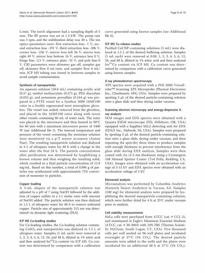
Cu release from CuCNPsThe loading and stimuli-responsive release of transitionmetals and any drug molecule in general from a deliveryare major factors that ultimately determine the successor failure of that vector when applied to targeted drugdelivery. One of the goals of this work was to demon-strate that CuCNPs were capable of responding tochanges in pH to facilitate Cu release. A generalschematic for the expected in vitro behavior is shown inFigure 5 (targeting ligands were not used in the experi-ments described here, but will be incorporated in thefuture). Initial Cu release experiments were conductedat pH 5 and 7 to mimic conditions that would be pre-sent during endocytosis of the nanoparticle along anendosomal pathway. Those experiments (Figure 3A)were promising and showed release to be much faster atpH 5 vs. pH 7, which would trigger Cu release uponparticle internalization. It was postulated that protona-tion of the carboxylate groups on the nanoparticlewould reduce the binding affinity of the ligand for Cuthereby facilitating release. Somewhat surprisingly, how-ever, CuCNPs released virtually no Cu in ultrapure
Table 1 Cu content (in weight percent) for CuCNPs usedin Cu release experiments
ICP MS EDX Elemental analysis
Ultrapure water 12.1 12.7 10.92
pH 7 buffer 1.7 3.3 Not measured
pH 5 buffer 0.0 0.7 0.04
Harris et al. Nanoscale Research Letters 2011, 6:445http://www.nanoscalereslett.com/content/6/1/445
Page 5 of 10
water, which has a neutral to slightly acidic pH. Whilethis feature is promising in terms of the stability of solu-tions of CuCNPs over long periods of time, Cu releasecannot simply be a function of pH but must also dependon the presence of ligands that can compete with theparticle-bound carboxylate groups in Cu binding. Thisfeature led to a series of additional experiments to eluci-date the effect of competing ligands on Cu release withthe idea that the underlying principles governing releasecould be used in the design/optimization of this class ofdelivery vectors.Release of Cu from CuCNPs in citrate buffer at pH 4,
5, and 6 (see Additional file 3) illustrated the effect ofcompeting ligand concentration on the rate of Curelease, which was actually faster at pH 6 compared topH 4. This can be explained by looking at the various
protonation states of citrate as a function of pH todetermine the competing ligands present in solution.Equations 1-4 were used to determine the relative con-centrations of L3-, LH2-, LH2
-, and LH3 (L = citrate)using pKa values of 3.13, 4.76, and 6.40 for citric acid(K1 = 7.40 × 10⁻⁴, K2 = 1.70 × 10⁻⁵, K3 = 4.00 × 10⁻⁷).The relative concentrations of L3-, LH2-, LH2
-, and LH3
are 27%, 69%, 4%, and 0% at pH 6 while at pH 4 therelative concentrations for the same species are 0%,13%, 77%, and 10%. At low pH, the predominate speciesis LH2
- whereas at high pH the predominate species isLH2- with a substantial amount of L3- being present aswell. One would expect the affinity of these ligands forCu to increase with increasing negative charge, and thatis clearly what is observed. So, even though the particle-bound carboxylate is protonated to a lesser extent at pH6, the presence of the di- and tri-anion form of citrateeffectively compete out Cu.
% LH3 =[H+]3
[H+]3 + K1[H+]2 + K1K2[H+] + K1K2K3× 100 (1)
% LH2− =
K1[H+]2
[H+]3 + K1[H+]2 + K1K2[H+] + K1K2K3× 100 (2)
% LH2− =K1K2[H+]
[H+]3 + K1[H+]2 + K1K2[H+] + K1K2K3× 100 (3)
% L3−=
K1K2K3
[H+]3 + K1[H+]2 + K1K2[H+] + K1K2K3× 100 (4)
In an effort to decouple competing ligand effects dueto the presence of changing buffering species with actualpH-dependent Cu release, we sought to reduce the
A B
Figure 4 In vitro toxicity of CuCNPs. (A) HeLa cell viability as measured via an assay based on MTT at 48 h showing toxicity only after theaddition of Cu to the nanoparticles, (B) a similar experiment showing the reduced toxicity of CuCNPs upon reduction of Cu content.
Harris et al. Nanoscale Research Letters 2011, 6:445http://www.nanoscalereslett.com/content/6/1/445
Page 6 of 10
buffer effect while concomitantly introducing competingligands that were unaffected by solution pH. The use ofcitrate buffer was less than ideal in this case due to themultiple acidic protons capable of generating four possi-ble species in solution. The use of acetate buffer inplace of citrate was expected to reduce this complexity.Acetate buffer generates only two species with relativeconcentrations of A- and HA being 95% and 5% at pH 6and 15 and 85% at pH 4, respectively (pKa = 4.75).Furthermore, even though the concentration of theanion changes in this case as well, and is higher at pH6, this species is identical to the particle-bound carboxy-late groups making it a less effective competitor com-pared to the species present in citrate buffer at the samepH. A direct comparison of Cu release at pH 5 in acet-ate buffer and citrate buffer (Figure 3B), both at 100mM, confirmed this assumption. Cu release was muchslower in acetate buffer most likely a result of thereduced competitive nature of A- compared to LH2-. Curelease in acetate buffer could be further reduced bylowering the buffer strength to 10 mM (Figure 3B).With buffer effects reduced, a competing ligand was
then introduced. Chloride was chosen as an appropriatecompeting ligand because its concentration would notbe effected by the pH of the solutions being investi-gated. Indeed, the introduction of chloride increased therate of Cu release in acetate buffer (Figure 3C). Again,Cu release was highly dependent on the identity of com-peting ligand as illustrated by comparing the effect ofadded chloride vs. bromide (Figure 3C). Next, Cu releasewas monitored in 10 mM acetate buffer at pH 4, 5,and 6 containing a large excess of chloride (1 M NaCl,Figure 3D). With changes in competing ligand concen-trations effectively minimized, pH-dependent Cu releasewas clearly demonstrated and release was accelerated asthe pH was reduced.
In vitro toxicity of CuCNPs in HeLa cellsThe toxicity of CuCNPs to cancer cells was investigatedand results demonstrated that the delivery vector itself,particles containing no Cu, was not toxic and thatCuCNPs displayed significant toxicity depending on dos-ing (Figure 4A). Furthermore, the delivery of Cu con-tained in CuCNPs was more toxic than an equivalentamount of free Cu dose implying that the phenomenonwas particle mediated (see Additional file 4). This effectis likely attributable to the differences in modes of inter-nalization for the two forms of Cu. Free copper wouldbe taken up by the cell via normal metal traffickingpathways that utilize metal-binding proteins located onthe cell surface. As the cell begins to experience metaloverload, those receptors would be internalized anddegraded to prevent further metal accumulation.CuCNPs would be expected to be internalized by an
entirely different pathway that is not subject to the nor-mal cellular mechanisms for controlling metal homeos-tasis. Thus, the cell’s normal metal overload defenseswere likely bypassed leading to unregulated Cu uptake.As the Cu-loading in CuCNPs was reduced, cell survivalimproved with little or no toxicity being observed forCuCNPs containing 3 or 5 wt.% Cu (Figure 4B). Themost likely source of toxicity was induced oxidativestress (see Introduction) and future experiments willprobe the mechanism of cell death to determine thevalidity of this hypothesis.
ConclusionsIn summary, we have synthesized Cu-loaded polymericnanoparticles that release bound Cu in a pH-dependentmanner. Cu loading and release were characterized byseveral analytical techniques where we demonstrated theability to load up to 16 wt.% Cu. The release of boundCu from CuCNPs was found to be both pH and com-peting ligand dependent. Decoupling these effects wasnon-trivial, but was accomplished through careful selec-tion of reaction conditions. Based on the behaviorobserved, we conclude that simple protonation of theparticle-bound carboxylate, while rate accelerating wasnot sufficient to promote release rather the presence ofa ligand capable of displacing the carboxylate wasrequired. The complexities described here will undoubt-edly increase dramatically when CuCNPs are introducedto biologically relevant media containing a plethora ofpotential ligands. Our coupling strategy allows us tocapitalize on the pH gradient observed along the endo-some/lysosome pathway for particle internalization fortargeted delivery of Cu [1]. CuCNPs were capable ofinducing toxicity in cancer cells where reductions of>95% viability were observed at high Cu loadings. Thestimuli-responsive release and toxicity of Cu in CuCNPsmeets the requirements for application in targeted drugdelivery.
MethodsGeneral considerationsMethyl methacrylate, acrylic acid, and poly(ethylene gly-col) (n) diacrylate (n = 200 = MW of PEG block) werepurchased from Polysciences, Inc. (Warrington, PA,USA) and used as received. Potassium persulfate, coppersulfate, copper acetate, nitric acid (trace metal grade)were from Fisher Scientific (Pennsylvania, PA, USA). Allmaterials were used as received unless otherwise noted.Microwave reactions were conducted in a Synthos 3000from Anton Paar (Ashland, VA, USA). ICP MS experi-ments were conducted on a Varian 820-MS (VarianInc., Lake Forest, CA, USA) using the followingparameters: plasma flow 17.5 L/min, auxiliary flow 1.65L/min, sheath gas 0.13 L/min, and nebulizer flow 0.89
Harris et al. Nanoscale Research Letters 2011, 6:445http://www.nanoscalereslett.com/content/6/1/445
Page 7 of 10
L/min. The torch alignment had a sampling depth of 5mm. The RF power was set at 1.3 kW. The pump ratewas 3 rpm, and the stabilization delay was 30 s. The ionoptics parameters were: first extraction lens -1 V, sec-ond extraction lens -191 V, third extraction lens -206 V,corner lens -236 V, mirror lens left 56 V, mirror lensright 49 V, mirror lens bottom 16 V, entrance lens 0 V,fringe bias -2.5 V, entrance plate -31 V, and pole bias 0V. CRI parameters were skimmer gas off, sampler gasoff, skimmer flow 0 mL/min, and sampler flow 0 mL/min. ICP MS tubing was rinsed in between samples toavoid sample contamination.
Synthesis of nanoparticlesAn aqueous solution (58.8 mL) containing acrylic acid(0.57 g), methyl methacrylate (0.575 g), PEG diacrylate(0.053 g), and potassium persulfate (0.164 g) was pre-pared in a PTFE vessel for a Synthos 3000 16MF100rotor in a freshly regenerated inert atmosphere glove-box. The vessel was sealed, removed from the glovebox,and placed in the 16MF100 rotor along with sevenother vessels containing 60 mL of water each. The rotorwas placed in the microwave and then heated to 90°Cfor 60 min with a maximum microwave power of 1400W (see Additional file 5). The internal temperature andpressure of the vessel containing the monomer solutionwere monitored via a p/T sensor accessory (AntonPaar). The resulting nanoparticle solution was dialyzedin 4 L of ultrapure water for 48 h with a change in thewater after the first 24 h. The particle concentrationafter purification was determined by lyophilizing aknown volume and then weighing the resulting solid,which resulted in a final particle concentration of 12.8mg/mL. Based on this number, a total of 0.896 g of par-ticles was synthesized with approximately 75% conver-sion of monomer to particles.
Copper loadingA 3-mL aliquot of the nanoparticle solution wasadjusted to a pH of 7 using NaOH followed by the addi-tion of copper sulfate in a 1:1 molar ratio with amountof NaOH added. The particle solution was then dialyzedin 1.5 L of ultrapure water for 48 h to remove unboundcopper. Particle size of approximately 215 nm was deter-mined via dynamic light scattering (DLS).
ICP MS Cu-loading studiesFor Cu-loading studies, the Cu-loading solution contain-ing CuSO4 and nanoparticles was dialyzed in 1.5 L ofultrapure water. Samples (1 mL each) were removed at1, 2, 3, 4, 5, 6, 12, 24, and 48 h, diluted in 1% nitric acidand then analyzed for63Cu content via ICP MS. Cu con-tent was determined by comparison with a calibration
curve generated using known samples (see Additionalfile 6).
ICP MS Cu release studiesPurified CuCNP-containing solutions (3 mL) were dia-lyzed in 1.5 L of the desired buffering solution. Samples(1 mL each) were removed at 0.08, 1, 2, 3, 4, 5, 6, 12,24, and 48 h, diluted in 1% nitric acid and then analyzedfor63Cu content via ICP MS. Cu content was deter-mined by comparison with a calibration curve generatedusing known samples.
X-ray photoelectron spectroscopyXPS spectra were acquired with a PHI 5000 VersaP-robe™ Scanning XPS Microprobe (Physical ElectronicsInc., Chanhassen, MN, USA). Samples were prepared byspotting 5 μL of the desired particle-containing solutiononto a glass slide and then drying under vacuum.
Scanning electron microscopy and energy-dispersive X-ray analysisSEM images and EDX spectra were obtained with aQuanta ESEM microscope (FEI, Hillsboro, OR, USA)equipped with a Sapphire Si(Li) detecting unit for EDX(EDAX Inc., Mahwah, NJ, USA). Samples were preparedby spotting 5 μL of the desired particle-containing solu-tion onto a glass slide, drying under vacuum, and thenrepeating the spot/dry three times to produce sampleswith enough thickness to prevent interference from theglass slide during EDX analysis. Samples were thencoated with Au (2-5 nm thickness) using a Cressington108 Manual Sputter Coater (Ted Pella, Redding, CA,USA). Images were obtained with an acceleration vol-tage of 5-15 kV and EDX spectra were obtained with anacceleration voltage of 5 kV.
Elemental analysisMicroanalysis was performed by Columbia Analytics(formerly Desert Analytics) in Tucson, AZ. Samples(100 mg) for elemental analysis were prepared by lyo-philizing the desired nanoparticle-containing solution,which were further dried for 4 h at 25°C under vacuumprior to analysis.
Cell viability measurementsHeLa cells were purchased from ATCC (cat. # CCL-2),and maintained in Eagle’s Minimum Essential Medium(ATCC, cat. # 30-2003) with 10% FBS (Thermo Scienti-fic HyClone, South Logan, UT, USA). Five thousandcells per well seeded on 96-well plates and incubatedovernight at 37°C (5% CO2). The desired particleamounts were added to the wells and the plates wereincubated for an additional 48 h at 37°C (5% CO2).
Harris et al. Nanoscale Research Letters 2011, 6:445http://www.nanoscalereslett.com/content/6/1/445
Page 8 of 10
After the incubation, cell viability was evaluated with theMTT reagent. Media was removed each well andreplaced with fresh media containing 1 mg/mL MTT.The cells were incubated for 4 h at 37°C (5% CO2) afterwhich time the media was removed and replaced withDMSO. Light absorption was measured on a Synergy 2multi-mode microplate reader (BioTek, Winooski, VT,USA). The viability of the cells exposed to particles wasexpressed as a percentage of the viability of cells grownin the absence of particles on the same plate.
Additional material
Additional file 1: DLS results for purified CuCNPs. graph showingparticle size as determined by Dynamic Light Scattering.
Additional file 2: Release of unbound Cu over time duringpurification of CuCNPs as monitored by ICP MS. graph showing allcopper that is not bound to the particle is removed by dialysis for 48 h.
Additional file 3: Release of Cu from purified CuCNPs over time in100 mM citrate buffer at pH 4, 5, and 6. graph showing that Curelease is actually slower as the pH is lowered due to competing ligandeffects.
Additional file 4: In vitro toxicity for comparison of Cu in CuCNPsversus similar dosing of free Cu(OAc)2. graph showing coppercontained in nanoparticles was more toxic than an equivalent amount ofcopper dosed as a free complex.
Additional file 5: Graph of reaction time vs. temperature, pressure,and microwave power during nanoparticle synthesis. graphsshowing microwave conditions used for nanoparticle synthesis.
Additional file 6: Typical calibration curve used for determining theCu concentration in unknown samples. calibration curve generatedfrom samples containing a known amount of copper.
AcknowledgementsWe thank Guido Verbeck and William Hoffmann for their help with ICP MSstudies, Nancy Bunce, David Diercks, and David Garrett for aid with EDX,XPS, and SEM analysis. Portions of this work were conducted at the UNTLaboratory of Imaging and Mass Spectrometry and the UNT Center forAdvanced Research and Technology. MC was an NSF-REU scholar (grantCHE-1004878) at the University of North Texas.
Authors’ contributionsAH carried out Cu loading and release studies via ICP MS, participated in thedesign and coordination of ICP MS studies, and helped draft the manuscript.BH optimized microwave conditions for the free radical polymerizationreaction used in the synthesis of polymeric nanoparticles. MC carried outparticle synthesis, and metal loading. RP conceived of the study, andparticipated in its design and coordination, carried out in vitro toxicitystudies, SEM and EDX analysis and drafted the manuscript.
Competing interestsThe authors declare that they have no competing interests.
Received: 7 January 2011 Accepted: 11 July 2011Published: 11 July 2011
References1. Petros RA, DeSimone JM: Strategies in the design of nanoparticles for
therapeutic applications. Nat Rev Drug Discov 2010, 9:615-627.2. Davis ME, Chen Z, Shin DM: Nanoparticle therapeutics: an emerging
treatment modality for cancer. Nat Rev Drug Discov 2008, 7:771-782.
3. Zhang L, Gu FX, Chan JM, Wang AZ, Langer RS, Farokhzad OC:Nanoparticles in medicine: Therapeutic applications and developments.Clin Pharmacol Ther 2008, 83:761-769.
4. Dhar S, Daniel WL, Giljohann DA, Mirkin CA, Lippard SJ: PolyvalentOligonucleotide Gold Nanoparticle Conjugates as Delivery Vehicles forPlatinum(IV) Warheads. J Am Chem Soc 2009, 131:14652-14653.
5. Dhar S, Gu FX, Langer R, Farokhzad OC, Lippard SJ: Targeted delivery ofcisplatin to prostate cancer cells by aptamer functionalized Pt(IV)prodrug-PLGA-PEG nanoparticles. Proc Natl Acad Sci USA 2008,105:17356-17361.
6. Rieter WJ, Pott KM, Taylor KML, Lin WB: Nanoscale coordination polymersfor platinum-based anticancer drug delivery. J Am Chem Soc 2008,130:11584-11585.
7. Haxton KJ, Burt HM: Polymeric Drug Delivery of Platinum-BasedAnticancer Agents. J Pharm Sci 2009, 98:2299-2316.
8. Treiber C, Quadir MA, Voigt P, Radowski M, Xu SJ, Munter LM, Bayer TA,Schaefer M, Haag R, Multhaup G: Cellular Copper Import by NanocarrierSystems, Intracellular Availability, and Effects on Amyloid beta PeptideSecretion. Biochemistry 2009, 48:4273-4284.
9. Withey ABJ, Chen G, Nguyen TLU, Stenzel MH: Macromolecular CobaltCarbonyl Complexes Encapsulated in a Click-Cross-Linked MicelleStructure as a Nanoparticle To Deliver Cobalt Pharmaceuticals.Biomacromolecules 2009, 10:3215-3226.
10. Chen H, Ahn R, Van den Bossche J, Thompson DH, O’Halloran TV: Folate-mediated intracellular drug delivery increases the anticancer efficacy ofnanoparticulate formulation of arsenic trioxide. Mol Cancer Ther 2009,8:1955-1963.
11. Neuse E: Macromolecular Ferrocene Compounds as Cancer Drug Models.J Inorg Organomet Polym Mater 2005, 15:3-31.
12. Spokoyny AM, Kim D, Sumrein A, Mirkin CA: Infinite coordination polymernano- and microparticle structures. Chem Soc Rev 2009, 38:1218-1227.
13. Della Rocca J, Lin WB: Nanoscale Metal-Organic Frameworks: MagneticResonance Imaging Contrast Agents and Beyond. Eur J Inorg Chem 2010,3725-3734.
14. Gianferrara T, Bratsos I, Alessio E: A categorization of metal anticancercompounds based on their mode of action. J Chem Soc, Dalton Trans2009, 7588-7598.
15. Jung YW, Lippard SJ: Direct cellular responses to platinum-induced DNAdamage. Chem Rev 2007, 107:1387-1407.
16. Messori L, Casini A, Gabbiani C, Sorace L, Muniz-Miranda M, Zatta P:Unravelling the chemical nature of copper cuprizone. J Chem Soc DaltonTrans 2007, 2112-2114.
17. Gasser G, Ott I, Metzler-Nolte N: Organometallic Anticancer Compounds. JMed Chem 2011, 54:3-25.
18. Thubagere A, Reinhard BM: Nanoparticle-Induced Apoptosis Propagatesthrough Hydrogen-Peroxide-Mediated Bystander Killing: Insights from aHuman Intestinal Epithelium In Vitro Model. ACS Nano 2010, 4:3611-3622.
19. George S, Pokhrel S, Xia T, Gilbert B, Ji Z, Schowalter M, Rosenauer A,Damoiseaux R, Bradley KA, Mädler L, Nel AE: Use of a Rapid CytotoxicityScreening Approach To Engineer a Safer Zinc Oxide Nanoparticlethrough Iron Doping. ACS Nano 2010, 4:15-29.
20. AshaRani PV, Low Kah Mun G, Hande MP, Valiyaveettil S: Cytotoxicity andGenotoxicity of Silver Nanoparticles in Human Cells. ACS Nano 2009,3:279-290.
21. Xia T, Kovochich M, Liong M, Mädler L, Gilbert B, Shi H, Yeh JI, Zink JI,Nel AE: Comparison of the Mechanism of Toxicity of Zinc Oxide andCerium Oxide Nanoparticles Based on Dissolution and Oxidative StressProperties. ACS Nano 2008, 2:2121-2134.
22. Ahamed M, Akhtar MJ, Siddiqui MA, Ahmad J, Musarrat J, Al-Khedhairy AA,AlSalhi MS, Alrokayan SA: Oxidative stress mediated apoptosis induced bynickel ferrite nanoparticles in cultured A549 cells. Toxicology 2011,283:101-108.
23. Park E-J, Yi J, Chung K-H, Ryu D-Y, Choi J, Park K: Oxidative stress andapoptosis induced by titanium dioxide nanoparticles in cultured BEAS-2B cells. Toxicol Lett 2008, 180:222-229.
24. Lunov O, Syrovets T, Büchele B, Jiang X, Röcker C, Tron K, Nienhaus GU,Walther P, Mailänder V, Landfester K, Simmet T: The effect ofcarboxydextran-coated superparamagnetic iron oxide nanoparticles onc-Jun N-terminal kinase-mediated apoptosis in human macrophages.Biomaterials 2010, 31:5063-5071.
Harris et al. Nanoscale Research Letters 2011, 6:445http://www.nanoscalereslett.com/content/6/1/445
25. Ahamed M: Toxic response of nickel nanoparticles in human lungepithelial A549 cells. Toxicol in Vitro 2011, 25:930-936.
26. Ahamed M, Siddiqui MA, Akhtar MJ, Ahmad I, Pant AB, Alhadlaq HA:Genotoxic potential of copper oxide nanoparticles in human lungepithelial cells. Biochem Bioph Res Co 2010, 396:578-583.
27. Valko M, Leibfritz D, Moncol J, Cronin MTD, Mazur M, Telser J: Free radicalsand antioxidants in normal physiological functions and human disease.Int J Biochem Cell Biol 2007, 39:44-84.
28. Wondrak GT: Redox-Directed Cancer Therapeutics: MolecularMechanisms and Opportunities. Antioxid Redox Sign 2009, 11:3013-3069.
29. Federico A, Morgillo F, Tuccillo C, Ciardiello F, Loguercio C: Chronicinflammation and oxidative stress in human carcinogenesis. Int J Cancer2007, 121:2381-2386.
30. Pelicano H, Carney D, Huang P: ROS stress in cancer cells and therapeuticimplications. Drug Resist Update 2004, 7:97-110.
31. Halliwell B: Oxidative stress and cancer: have we moved forward?Biochem J 2007, 401:1-11.
32. Erez A, Shchelochkov Oleg A, Plon Sharon E, Scaglia F, Lee B: Insights intothe Pathogenesis and Treatment of Cancer from Inborn Errors ofMetabolism. Am J Hum Genet 2011, 88:402-421.
33. Mao XW, Green LM, Mekonnen T, Lindsey N, Gridley DS: Gene ExpressionAnalysis of Oxidative Stress and Apoptosis in Proton-irradiated RatRetina. In Vivo 2010, 24:425-430.
35. Alemón-Medina R, Bravo-Gómez ME, Gracia-Mora MI, Ruiz-Azuara L:Comparison between the antiproliferative effect and intracellularglutathione depletion induced by Casiopeína IIgly and cisplatin inmurine melanoma B16 cells. Toxicol in Vitro 2011, 25:868-873.
36. Alemón-Medina R, Breña-Valle M, Muñoz-Sánchez J, Gracia-Mora M, Ruiz-Azuara L: Induction of oxidative damage by copper-based antineoplasticdrugs (Casiopeínas). Cancer Chemoth Pharm 2007, 60:219-228.
37. Petros RA: Synthesis and Use of Metal Ion-Containing Polymeric Particles.US Patent Appl 61/370,682 2010.
38. An Z, Tang W, Hawker CJ, Stucky GD: One-Step Microwave Preparation ofWell-Defined and Functionalized Polymeric Nanoparticles. J Am ChemSoc 2006, 128:15054-15055.
doi:10.1186/1556-276X-6-445Cite this article as: Harris et al.: Beyond platinum: synthesis,characterization, and in vitro toxicity of Cu(II)-releasing polymernanoparticles for potential use as a drug delivery vector. NanoscaleResearch Letters 2011 6:445.
Submit your manuscript to a journal and benefi t from:
7 Convenient online submission
7 Rigorous peer review
7 Immediate publication on acceptance
7 Open access: articles freely available online
7 High visibility within the fi eld
7 Retaining the copyright to your article
Submit your next manuscript at 7 springeropen.com
Harris et al. Nanoscale Research Letters 2011, 6:445http://www.nanoscalereslett.com/content/6/1/445