j ourna l homepage: www.e lsev ie r.com/ locate / jconre l
Review
“Nanoantibiotics”: A new paradigm for treating infectious diseases usingnanomaterials in the antibiotics resistant era
Ae Jung Huh a,b, Young Jik Kwon a,c,d,⁎a Department of Pharmaceutical Sciences, University of California, Irvine, CA 92697, United Statesb Division of Infectious Disease, Department of Internal Medicine, National Health Insurance Corporation Ilsan Hospital, 1232 Baekseok-dong, Ilsandong-gu, Goyang-si, Gyeonggi-do 411-719,Republic of Koreac Department of Chemical Engineering and Materials Science, University of California, Irvine, CA 92697, United Statesd Department of Biomedical Engineering, University of California, Irvine, CA 92697, United States
Article history:Received 3 January 2011Accepted 29 June 2011Available online 6 July 2011
Keywords:Infectious diseaseAntibiotics resistanceNanoparticlesAntimicrobial drug deliveryNanoantibiotics
Despite the fact that we live in an era of advanced and innovative technologies for elucidating underlyingmechanisms of diseases and molecularly designing new drugs, infectious diseases continue to be one of thegreatest health challenges worldwide. The main drawbacks for conventional antimicrobial agents are thedevelopment of multiple drug resistance and adverse side effects. Drug resistance enforces high doseadministration of antibiotics, often generating intolerable toxicity, development of new antibiotics, and requestsfor significant economic, labor, and time investments. Recently, nontraditional antibiotic agents have been oftremendous interest in overcoming resistance that is developed by several pathogenic microorganisms againstmost of the commonly used antibiotics. Especially, several classes of antimicrobial nanoparticles (NPs) andnanosized carriers for antibiotics delivery have proven their effectiveness for treating infectious diseases,including antibiotics resistant ones, in vitro aswell as in animalmodels. This review summarizes emerging effortsin combating against infectious diseases, particularly using antimicrobial NPs and antibiotics delivery systems asnew tools to tackle the current challenges in treating infectious diseases.
num oxide; AMs, alveolar macrophages; Apt, aptamers; Acans, Candida albicans; CdS, cadmium sulfide; Chol, chocopper oxide; DC-Chol, dimethylammonium ethane carbrol; E. coli, Escherichia coli; E. faecium, Enterococcus faecirylate; GPS, Glycerol palmitostearate; GSH, glutathione;cytogenes, Listeria monocytogenes; L. pneumophila, Legioneconcentration; MPS, mononuclear phagocytic system; MNGF, nerve growth factor; N. gonorrheae, Neisseria gonorrnas aeruginosa; PAMAM, polyamidoamine; PC, phosphatiPEG-DSPE, 1-2-disteroyl-sn-glycero-3-phosphoethanolamphosphatidyl choline; PIHCA, polyisohexylcyanoacrylate;y(trimethylene carbonate); PVA, polyvinyl alcohol; PVP,oxygen species; SA, stearic acid; S. aureus, StaphylococcSLNPs, solid lipid nanoparticles; SMZ, sulfamethoxazolealled nanotubes; TBGC, teicoplanin-loaded borate bioactivin-resistant Enterococcus; VRSA, vancomycin-resistant Starvine, CA 92697-3905, United States. Tel.: +1 949 824 8
At the beginning of the 20th century, infectious diseases were theleading cause of death worldwide [1]. The decreases in morbidity andmortality from infectious diseases over the last century wereattributed mainly to an introduction of antimicrobial agents. Nowa-days, however, resistance to antibiotics has been reaching a criticallevel, invalidatingmajor antimicrobial drugs that are currently used inthe clinic [2,3]. The bacterial resistance to antimicrobial drugs hasbeen attempted to be resolved by discovering new antibiotics andchemically modifying existing antimicrobial drugs. Unfortunately,there is no assurance that the development of new antimicrobialdrugs can catch up to the microbial pathogen's fast and frequentdevelopment of resistance in a timely manner. For example, drug-resistant infections in hospitals and in the communities caused byboth Gram-positive and Gram-negative bacterial pathogens aregrowing [4], and the continued evolution of antimicrobial resistancethreatens human health by seriously compromising our ability to treatserious infections [5]. This challenging and dynamic pattern ofinfectious diseases and the emergence of bacterial strains resistantto many currently used antibiotics demand for longer-term solutionsto this ever-growing and foreseeable problem [6].
One of the recent efforts in addressing this challenge lies in exploringantimicrobial nanomaterials, to which microbial pathogens may not beable to develop resistance, and novel nanosized platforms for efficientantibiotics delivery. For example, it has been suggested in recent studiesthat some metal nanoconstructs are known to possess antimicrobialactivities, which is utilized in controlling infectious diseases [7–9].Antimicrobial nanoparticles (NPs) offer many distinctive advantages inreducing acute toxicity, overcoming resistance, and lowering cost,whencompared to conventional antibiotics [10,11]. Various nanosized drugcarriers are also available to efficiently administer antibiotics byimproving pharmacokinetics and accumulation, while reducing theadverse effects of antibiotics. Theoretically, NPs are retained muchlonger in the body than small molecule antibiotics, which could bebeneficial for achieving sustained therapeutic effects. On the other hand,the safety profiles of NPs and nanosized antibiotics drug carriers,particularly upon long-term exposure, could be an overriding safetyfactor andmust be consideredwith therapeutic effects [12]. This review
introduces employingnanotechnology as a newparadigm in controllinginfectious diseases, especially in overcoming antimicrobial drugresistance, in the context of research and clinical prospectives of thisnovel and promising strategy.
2. Challenges and use of nanotechnology in treatinginfectious diseases
2.1. Resistant ‘superbugs’ create needs for breakthrough
Use of antibiotics began with commercial production of penicillin inthe late 1940s and claimed to be a great success until the 1970–1980swhennewer and even stronger antibioticswere additionally developed.In the ongoing race of the development of antimicrobial agents,however, microbes appear to be the winner, and the pipeline for newdrugs is verging on empty (Fig. 1) [13]. Development of antimicrobialdrugs gives a low returnon investment, contributing to the current crisisin fighting against drug-resistant pathogens [3]. Despite extensiveefforts in research and enormous investment of resources, the pace ofdrug development has not kept up with the development of resistance(Fig. 2). Increasing rates of bacterial resistance also invalidate the utilityof even the most potent antibiotics, resulting in mortality due to failurein infection control and high health care costs [14].
Changes in societal activities, progress of technology, and evolvingmicroorganisms themselves are cooperatively contributing to theescalation of emerging and re-emerging infectious diseases, and to thedevelopment of antimicrobial resistance. A combination of increasedpressure of antibiotic selections and a decline in the development ofnew antibiotics has raised the specter that once treatable infectionsbecome untreatable [1,5]. The first serious clinical threat in treatinginfectious diseases using antibioticswas the emergence of vancomycin-resistant Enterococcus (VRE); which has intrinsic resistance to severalcommonly used antibiotics and, perhaps more importantly, a capabilityof acquiring resistance to all currently available antibiotics [2]. Morethan 40% of Staphylococcus aureus strains collected from hospitals wereresistant to methicillin (methicillin-resistant S. aureus, MRSA) [1] andsome of them were found to be resistant to vancomycin. Treatingvancomycin-resistant S. aureus (VRSA) strains is a global and dauntingmedical challenge for the twenty-first century because vancomycin is
the latest generation of antibiotics and is assumed at the moment to bethe most effective for S. aureus infection [15,16]. Multi-drug resistancehas been increasing particularly among nosocomial Gram-negativebacteria that are capable of developing many different mechanisms forantimicrobial resistance, often rendering them multiplication-resistant[2]. This also explains the increasing antimicrobial resistance ofnosocomial Gram-negative bacteria to extended-spectrum cephalospo-rin, a class of the newest andmost powerful antibiotics [4]. Resistance toantimicrobial drugs becomes a threatening problem not only inhospitals but also in communities, resulting in fewer effective drugsavailable to control infections by “old” established bacteria such asStreptococcus pneumoniae (S. pneumoniae) [17]. Drug-resistantNeisseriagonorrheae (N. gonorrheae) and Haemophilus influenzae (H. influenzae)were already recognized worldwide in the 1970s [18]. Recently, morethan 1500 people in Germany have been infected by a new virulentstrains of Escherichia coli (E. coli),whichwas not previously known to beinvolved in any outbreaks but turned out to be highly infectious andtoxic, leading to a number of fatal cases. Moreover, bioinformaticsanalysis revealed that this deadly bacterial strain carries severalantibiotic resistance genes, including resistance to aminoglycosides,macrolides, and β-lactam antibiotics. This implicates that treating thisvirulent bacteria with antibiotics is extremely difficult [19–21]. Thespread of resistance tomany currently used antimicrobial agents amongfungi, viruses, andparasites is a high level alert tofindparadigm-shiftingapproaches for treating microbial infections as a top priority in
Fig. 2. History of antimicrobial agent development vs. subs
medicine. Therefore, design, discovery, and delivery of antimicrobialdrugs with improved efficacy and avoidance of resistance are highlydemanded [22]. There have been considerable efforts in searching fornew natural product-derived antibiotics to control infections by VRE,VRSA, MRSA, and other multidrug-resistant microorganisms [23]. Anattractive alternative to using antibiotics is utilizing agents that causephysical damages to antimicrobial resistant strains. For example,selective photothermal therapy for in vivo antimicrobial treatmentusing pulsed laser was reported [24].
Another challenge in antimicrobial therapy is the treatment ofchronically infected conditions such as cystic fibrosis and other chronicobstructive pulmonary diseases. Treating such conditions necessitatesfrequent intravenous administrations of high-dose antibiotics, whichcause serious adverse effects from a high concentration of antibiotics inserum. Evenwith aggressive antibiotic treatment, complete eradicationof infection under such conditions is hard to achieve because ofbacteria's ability to formbiofilms [25]. The persistent infection also leadsto the rise of bacterial strains that possess elevated tolerance to currentantibiotics. Therefore, a newantimicrobial therapy employing a cutting-edge technology that is more effective and safer than the currentlyavailable ones is desperately desired [26].
2.2. Potential impact of nanomedicine on the control of infectiousdiseases
Use of nanotechnology in immunization, design and delivery ofantimicrobial drugs, and diagnosis and control of cross-infections, inparticular in overcoming antibiotics-resistant pathogens, has beenexplored as a promising alternative to the current antibiotics-basedapproaches [6,12]. This section introduces various challenges incontrolling infectious diseases, encompassing diagnosis of bacterialresistance, delivery of antimicrobial agents, and vaccination, usingnanotechnology.
2.2.1. Nanotechnology-assisted detection of antimicrobial infection andresistance
Despite their high sensitivity and reproducibility, conventionaldiagnostic methods for a microbial infection require cumbersomesample preparation and long readout times [27]. Unique electrical,magnetic, luminescent, and catalytic properties of nanomaterialsenable fast, sensitive, and cost-effective diagnosis as well as rapiddetermination of the susceptibility and resistance of anti-bacterialdrugs [28,29]. Antibody-conjugated NPs amplify the signals forbioanalysis and enumeration of highly pathogenic bacteria such asE. coli O157:H7, resulting in highly selective, convenient, and rapid
equent acquaintance of resistance by microorganisms.
detection of single bacterium within 20 min [30]. A quick method fordetecting infections in the urinary tract have also been developedusing gold nano wire arrays (GNWA) in conjunction with a linker armattached to specific E. coli antibodies [31]. It was shown that NPs withspecific Raman spectroscopic fingerprints can distinguish antibiotic-resistant bacteria, such as MRSA from non-resistant strains, bydetecting single-nucleotide polymorphisms in microarray-basedsystems [32]. The use of magnetic NPs also could be a very sensitiveand rapid strategy to detect microbial infection. For example, dextran-coated supermagnetic iron oxide NPs were clustered by Con-Atreatment, or equipped with Con A-conjugated nanosensors [33].These NPs were able to assess the microbial metabolic activity anddetermine antimicrobial susceptibility in blood, by rapidly quantifyingpolysaccharides. Supermagnetic iron oxide nanoprobes greatly assistedthe identification of Mycobacterium avium spp. paratuberculosis (MAP)as well as the quick quantification of MAP in milk and blood with highsensitivity [34]. The broad absorption spectra (i.e., excitation at a widerange of wavelengths) of quantum dots (QDs) can be exploited tosimultaneously excite QDs emitting different colors using a singlewavelength [35]. These characters suggest that QDs are a promisingmodality for the analysis of complex samples for histology, pathologyand cytology, and can facilitate double or even triple immunostaining ofbacterial cells (e.g., Listeria monocytogenes [L. monocytogenes]) [36]. Agreat deal of efforts have been invested in developing affordable, robust,and reproducible nanodiagnostic assays to be globally accessible andapplicable even in rural areas of developing countries [27,37].Furthermore, recent studies using nanotechnology have demonstratedthe feasibility of achieving fast and reliable pharmaceutical assays formicrobial infections in opaque media (e.g., whole blood and milk),without any sample preparations [38,39].
2.2.2. Emerging roles of nanotechnology in antimicrobial actions andtreatment of infectious diseases
Metal and metal oxide NPs produce reactive oxygen species (ROS)under UV light and find their increasing uses in antimicrobialformulations and dressings [12]. In particular, nanosized silver, zinc,and their compounds have been reported to be effective in inactivatingvariousmicroorganisms [40,41]. The high reactivity of titaniumand zincdioxide has been extensively utilized in the bactericidal substances thatare used in filters and coatings on catheters [41–43].
Recently, antibiotics formulated in polymeric NPs have demon-strated enhanced antimicrobial activities and anti-MRSA activities,compared with nonpolymerized forms of penicillin and N-methylthioβ-lactams [22,44]. The studies illustrated facile one-step preparationsof antibiotics-conjugated polyacrylate network in aqueous media andincorporation of water-insoluble drugs directly onto the polymericnetworkwithout post-synthesis modifications of the NPs. Vancomycin-capped gold (Au) NPs has also exhibited enhanced antimicrobialactivities against VRE strains and E. coli strains [45].
Selective killing of target bacteria by irradiating Au NP-attachedbacterial surface with a laser has been recently demonstrated [46].This technology leads to effective and irreparable damage to thebacterium bound with Au NPs of different sizes conjugated with anti-protein A antibodies. A new photothermal approach to antimicrobialnanotherapy and pathogen detection at a single bacterium level hasalso been developed using carbon nanotubes (CNTs) [47]. This studyrevealed CNTs' threefold roles: high near-infrared (NIR) contrastagents, high affinity and clustering agents for bacteria, and highlyefficient converter of optical energy into thermal energy, especially inliquid environment [24].
A wide range of antimicrobial agents can be effectively adminis-tered using various NPs. Many types of lipophilic and water-solubleantibiotics can be conjugated inside or on the surface of NPs, or carriedvia encapsulation [48]. Key pharmacokinetic characteristics ofantibiotics, including improved solubility, controlled release, and
specific site-targeted delivery, can be achieved by employingappropriate nanocarriers [12].
2.2.3. Nanotechnology for vaccination and prevention of infectiousdiseases
Use of NPs as novel adjuvants and colloidal vaccine carriers forimmunization has been explored [12]. Particulate systems provideseveral benefits for vaccine delivery [49] in such that micro- andnanoparticles are approximately the sizes of bacteria and viruses thatthe immune system recognizes. The size, chemical composition,charge, and surface properties of particulate vaccine carriers can alsobe tuned for enhanced uptake by mononuclear phagocytic system(MPS), stimulation of antigen presenting cells (APs), and immunepresentation of antigens [50,51]. For example, amphipathically coatedNPs efficiently deliver antigens to dendritic cells (DCs) that concom-itantly orchestrate cellular and humoral immunity [51,52]. In in vivostudies, mixtures of nanoemulsions with either whole viruses (e.g.,influenza A virus) or proteins (e.g., recombinant Bacillus anthracis[B. anthracis] protective antigens) have been explored as potentialvaccines [53,54]. This type of vaccine does not require cold storageand can be administered via mucosal routes, which would beparticularly desirable for vaccination in developing countries [12].
3. Nanoantibiotics: Nanomaterials for infection control
Nanomaterials, which either show antimicrobial activity bythemselves [43] or elevate the effectiveness and safety of antibioticsadministration [7,48,55], are called “nanoantibiotics” and theircapability of controlling infections in vitro and in vivo has beenexplored and demonstrated. Unlike many antimicrobial agentscurrently being used in the clinic, antimicrobial NPs may not posedirect and acute adverse effects, although potential toxicity uponlong-term exposure is questionable. Most importantly, antimicrobialNPs tackle multiple biological pathways found in broad species ofmicrobes (Fig. 3) and many concurrent mutations would have tooccur in order to develop resistance against NPs' antimicrobialactivities. Preparation of antimicrobial NPs could be cost-effective,compared with antibiotics synthesis, and they are quite stable enoughfor long-term storage with a prolonged shelf-life [11]. In addition,some NPs can withstand harsh conditions, such as high temperaturesterilization, under which conventional antibiotics are inactivated.Antibiotics delivery using nanomaterials offer multiple advantages: 1)controllable and relatively uniform distribution in the target tissue, 2)improved solubility, 3) sustained and controlled release, 4) improvedpatient-compliance, 5) minimized side effects, and 6) enhancedcellular internalization [56–58].
3.1. Antimicrobial nanomaterials
Antibacterial NPs consist of metals and metal oxides, naturallyoccurring antibacterial substances, carbon-based nanomaterials, andsurfactant-based nanoemulsions [43]. High surface area to volumeratios and unique chemico-physical properties of various nanomaterialsare believed to contribute to effective antimicrobial activities [11]. Arecent study also demonstrated that naturally occurring bacteria do notdevelop antimicrobial resistance to metal NPs [40]. Antimicrobialmechanisms of nanomaterials include: 1) photocatalytic production ofreactive oxygen species (ROS) that damage cellular and viral compo-nents, 2) compromising the bacterial cell wall/membrane, 3) interrup-tion of energy transduction, and 4) inhibition of enzyme activity andDNA synthesis [10,11,41–43,55,59] (Fig. 3). Table 1 also summarizesnanomaterials with their antimicrobial mechanisms, and potentialclinical and industrial uses. Recently, intracellular or extracellularsynthesis of metallic NPs (cadmium sulfide [CdS], gold [Au], and silver[Ag]) using microbial cells or enzymes has also been explored as novelbiological and environment-friendly NP preparation [60–64].
Fig. 3. Various antimicrobial mechanisms of nanomaterials.
3.1.1. Silver (Ag) NPsThe antibacterial property of silver has been noticed since ancient
times. Silver has been used for burn wound treatment, dental work,catheters, and bacterial infection control, in the forms of metallicsilver, silver nitrate, and silver sulfadiazine [65]. Using silver to treatbacterial infections became unpopular after penicillin was introducedin the 1940s [66]. The recent emergence of antibiotics-resistantbacteria and the limited effectiveness of antibiotics revived the clinicaluse of silver (e.g., wound dressings) [13]. Among the many differenttypes of metallic and metal oxide NPs, Ag NPs have proven to be themost effective against bacteria, viruses, and other eukaryoticmicroorganisms [67–70]. Ag NPs attack the respiratory chain andcell division that finally lead to cell death, while concomitantlyreleasing silver ions that enhance bactericidal activity [71]. Theantimicrobial activity of Ag NPs is inversely dependent on size [72,73]and shape [10]. Combined use of Ag NPs with antibiotics, such aspenicillin G, amoxicillin, erythromycin, and vancomycin, resulted inenhanced and synergistic antimicrobial effects against Gram-positiveand Gram-negative bacteria (e.g., E. coli and S. aureus) [7,74,75].Diverse applications of Ag NPs include wound dressings, coating for
Table 1Antimicrobial nanomaterials.
Nanomaterial Antimicrobial mechanism Cl
Ag NPs Release of Ag+ ions; disruption of cell membrane andelectron transport; DNA damage
Dde
ZnO NPs Intracellular accumulation of NPs; cell membrane damage;H2O2 production; release of Zn2+ ions
Anm
TiO2 NPs Production of ROS; cell membrane and wall damage Antr
Au NPs Interaction with cell membranes; strong electrostatic attraction Phaf
Chitosan Increased permeability and rupture of membrane;chelation of trace metals; enzyme inactivation
Dbi
Fullerenes Destruction of cell membrane integrity; enhancing activity ofinfiltrating neutrophil
Po
CNTs Cell membrane damage by ROS; oxidation of cell membraneproteins and lipids
Ansu
NO-releasingNPs
NO release and production of ROS In
Nanoemulsion Membrane disruption; disruption of the spore coat Ande
medical devices and surgical masks, impregnated textile fabrics,nanogels, and nanolotions [76–79].
Prolonged exposure to soluble silver-containing compounds mayproduce an irreversible pigmentation in the skin (argyria) and theeyes (argyrosis), in addition to other toxic effects, including organdamages (e.g., liver and kidney), irritation (e.g., eyes, skin, respiratory,and intestinal tract), and changes in blood cell counts [80]. On thecontrary, metallic silver appears to pose a minimal risk to health andAg NPs are suggested to be non-toxic in some studies [81,82], butsome studies reported concentration-dependent adverse effects of AgNPs on the mitochondrial activity [83,84]. The advent of Ag NPs aspromising antimicrobial nanomaterials, therefore, requires clear andfull elucidations of their potential toxicity.
3.1.2. Zinc oxide (ZnO) NPsSome NPs made of metal oxides are stable under harsh processing
conditions and have selective toxicity to bacteria but also exhibit aminimal effect on human and animal cells [85–87]. For example, ZnONPs, which are nontoxic and biocompatible, have been utilized as drugcarriers, cosmetics ingredients, and medical filling materials [88,89].Recently, ZnO NPs were found to have antibacterial activity againstimportant food borne pathogens, such as E. coli O157:H7 andenterotoxigenic E. coli [67,85,90]. These studies suggested that theapplication of ZnO NPs may be effective for preserving agriculturalproducts and food. ZnONPs have advantages over Ag NPs, such as a lowproduction cost, a white appearance, and UV-blocking properties [91].The nano-ZnOmultilayer deposited on cotton fabrics showed excellentantibacterial activity against S. aureus [92]. ZnO NPs are believed todestruct lipids and proteins of thebacterial cellmembrane, resulting in aleakage of intracellular contents and eventually the death of bacterialcells [41,67]. In addition, generation of hydrogen peroxide and Zn+2
ionswere suggested tobekeyantibacterialmechanismsof ZnONPs [93].Increased membrane permeability, cellular internalization, and intra-cellular structural change of polyvinyl alcohol (PVA)-coated ZnO NPswere also reported [41].
3.1.3. Titanium dioxide (TiO2) NPsTiO2 is a commonly used semiconductor photocatalyst and TiO2
NPs are the most studied for photocatalytic antimicrobial activityamong various NPs [94]. A great amount of information accumulatedover the last 20 years [95] demonstrated the strong bactericidal
inical and industrial applications References
ressing for surgical wound and diabetic foot; coatings for medicalvices; portable water filters; antibacterial agent; antifungal agent
[10,43,71–73]
tibacterial creams; lotions and ointment; surface coating ofedical device; mouthwash
[41,91,92]
tibacterial agent; food sterilizing agent; air purifiers; watereatment systems
[42,95,96,98]
otothermal therapy with near infrared light; adjuvant treatmentter serious infections antibacterial agent; antifungal agent
[68,81]
rinking water disinfectants; bacteria immobilizer; microbiocide inomedical products
[43,59,113]
tential disinfection applications [124–128]
tibacterial agent; biofouling-resistant membranes; water filter;rface-coating
activity of TiO2 upon receiving irradiation with near-UV light and UV-A. The required concentration for killing bacteria varies in the range of100–1000 ppm, depending on the TiO2 NP size as well as the intensityand wavelength of the light source [42]. A new study reported the TiO2
NPs' antibacterial efficiency in the order of E. coliNP. aeruginosaNS. aureusNE. faeciumNC. albicans, is seemingly determined by thecomplexity and the density of the cell membrane/wall [96]. It was alsoreported that the photocatalytic antimicrobial efficiency of TiO2NPswasin the order of virusNbacterial wallNbacterial spore, depending on thethickness of microbial surface structure [96]. Growth inhibition ofEnterobacter cloacae by UVA-irradiated TiO2 NPs was less effective thanthat of E. coli and P. aeruginosa [97].
The photocatalytic antibacterial activity of TiO2 is attributed to theproduction of ROS such as free hydroxyl radicals and peroxide [98].Hydroxyl radicals generated by photocatalytic TiO2 are very potentoxidantswitha broad reactivity, and themicrobial surface is theprimarytarget of the initial oxidative attack by irradiated TiO2 NPs, in directcontact with or close to a microbe. Damaged membrane structureimpairs many crucial biological functions, such as semipermeability,respiration, and oxidative phosphorylation reactions [42]. Irradiation-independent bacterial death also indicates other unknown non-photocatalytic antimicrobial activity of TiO2 NPs [99]. An attractivefeature of disinfection by TiO2 is its potential for activation by visiblelight (e.g., sunlight). In addition, metal doping (e.g., Ag/TiO2) improvedthe light absorbanceof TiO2 and increased its photocatalytic inactivationof bacteria and viruses [95,100]. Very interestingly, Ag/(C,S)-TiO2 NPswere shown to have strong light-independent antimicrobial activitiesagainst both E. coli and B. subtilis spores, by exploiting the combinedbactericidal activity of Ag and TiO2 together [101].
TiO2 is particularly suitable for water treatment because it is stablein water, non-toxic by ingestion, and cost-effective. The photocata-lytic activity by UV-A and the potential activation by visible light,when doped with novel metals, make TiO2-mediated disinfectionespecially useful in developing countries where electricity is notavailable for sterilization [43]. According to recent studies, TiO2 alsoinactivates various microorganisms that are highly resistant todesiccation and UV radiation, which makes TiO2 a promising agentfor improving process hygiene and product safety in food industry andcosmetics [95,100]. Antibacterial effects of TiO2 on Lactobacillusacidophilus would also be used in orthodontic appliances, such as pitand fissure sealants, toothbrushes, dental implants, and screws [98].
3.1.4. Gold (Au) NPsNear-infrared (NIR) light-absorbing Au NPs, nanorods, nanoshells,
and nanocages have been employed to treat bacterial infections viairradiation with focused laser pulses of suitable wavelengths [69,102].The antimicrobial activity of Au NPs seems to be initially mediated bystrong electrostatic attractions to the negatively charged bilayer of thecell membrane [68,81], which was also supported by the observationthat cationic particles were found to be moderately toxic while anionicparticles were not [8]. Au NPs conjugated with antimicrobial agentsand antibodies have been explored to obtain selective antimicrobialeffects [103]. For example, selective killing of S. aureus by Au NPsconjugated with anti-protein A antibodies, which target the bacterialsurface, was demonstrated [40]. It was found that strong laser-inducedhyperthermic effects accompanied by bubble-formation aroundclustered Au NPs effectively damaged bacteria [40]. Many studieshave reported strong antimicrobial effects against Gram-positive andGram-negative bacteria, including antibiotic-resistant strains [69,104],by Au/drug nanocomposites (e.g., Au NPs coated with antibiotics suchas streptomycin, gentamicin, and neomycin). In a recent study,chitosan-capped Au NPs coupled with ampicillin showed a 2-foldincrease in antimicrobial activity, compared with that of freeampicillin [68]. Therefore, Au NPs are promising adjuvants forantibiotics therapy in treating serious bacterial infections at a reducedantibiotics dosage with minimal adverse effects.
3.1.5. Aluminum (Al) and copper (Cu) NPsAluminum oxide (Al2O3) NPs are known to exhibit mild inhibitory
effects on microbial growth via cell wall disruption but only at veryhigh concentrations [105] while Ag/meso-Al2O3 NPs showed broadinhibitory effects on P. aeruginosa and S. aureus in a recent study [106].Copper is a structural constituent of many enzymes in many livingmicroorganisms. However, free ionic Cu2+ at a high concentration cangenerate toxic effects by generating ROS that disrupts the amino acidsynthesis and DNA [107]. Although antibacterial activity of Ag NPs iswell established and proven to be most effective in general among themetallic NPs [55,70,108], the antibacterial activity of NPs varydepending on the microbial species [109]. For example, it wasshown that Cu NPs have greater affinity to amines and carboxylgroups at a high density on the surface of Bacillus subtilis than that ofAg NPs, hence, superior antibacterial activity [109,110]. Copper oxide(CuO) is cheaper than silver, easily miscible with polymers, andrelatively stable chemically and physically [111].
3.1.6. Antimicrobial peptides and chitosanChitosan is a partially deacetylated chitin (a long biopolymer chain
of N-acetylglucosamine) and it has a wide-spectrum of antibacterialactivity [112]. It is only recently when these materials have beenengineered into a form of NPs, that the nano-scale chitosan as well asits derivatives exhibit antimicrobial effects against bacteria, viruses,and fungi [59,112,113]. Chitosan was found to be more effective forcontrolling fungal and viral infections than bacterial ones [59], and theantimicrobial activity of chitosan has previously been consideredgreater for Gram-positive bacteria than Gram-negative ones[113,114]. A further elucidated tendency has been shown by recentstudies [115,116]. The antimicrobial effect of chitosan is stronglydependent on the molecular weight of chitosan and intrinsicdifferences in target bacterial wall structure: chitosan of a lowmolecular weight generated high antimicrobial effects on Gram-negative bacteria while the reverse is observedwith chitosan of a highmolecular weight on Gram-positive bacteria [117]. A study alsoshowed chitosan's synergistic antimicrobial activity against drugresistant P. aeruginosa when used with sulfamethoxazole [118]. Theclinical potential for such combinatory uses of chitosan in overcominguntreatable resistant infections could be immense.
Chitosan's antimicrobial mechanisms have been explained byvarious theories. One of them is the binding to the negatively chargedbacterial surface to cause agglutination, increasing the permeability ofthe microbial wall, which eventually induces a leakage of intracellularcomponents [113]. According to another proposed mechanism,chitosan chelates trace metals and thereby inhibits enzyme activitiesand the microbial growth [119]. It was also proposed that chitosanliberated from the fungal wall by host hydrolytic enzymes penetratesto the nucleus of fungi and inhibits RNA and protein syntheses [59].The limited solubility only in acidic media and precipitation in theculture medium prevent from correctly investigating the antibacterialactivity and its mechanisms [112]. Water-soluble derivatives ofchitosan showed a higher antimicrobial activity, by disrupting theouter and inner membranes of bacteria, than native chitosan [120]. Adifferent study reported a strong antibacterial activity of oleoyl-chitosan NPs with enhanced dispersion in the culture medium andreduced pH effects on solubility [121]. Even with distinctivelydifferent internalization pathways in E. coli and S. aureus, potentantimicrobial activities of oleoyl-chitosan NPs against both bacteriawere observed [121].
Utilizing chitosan is a promising, cost-effective, and technologi-cally affordable disinfection method that is particularly useful indeveloping countries. Nano-scale chitosan could be used to disinfectmicrobes in membranes, sponges, or surface coatings of water storagetanks [113]. It has several advantages over other disinfectants, such asa high antibacterial activity, a broad spectrum of activity, a highmicrobe-killing efficiency, and a low toxicity onmammalian cells [59].
3.1.7. Fullerenes (C60) and fullerene-derivativesFullerenes' antimicrobial properties are of very recent findings
based on limited knowledge [122]. Although native fullerenes arenearly aqueous-insoluble, they can be dispersed in water by theseveral recently developed methods [123]. In particular, numeroustechniques for creating stable colloidal C60 aggregates (nC60) in waterare noted for their potent and broad antibacterial activity [43,124].The debatable antibacterial mechanism for nC60 includes photocata-lytic ROS production in eukaryotic cells [125–127]. Some studiesassert that antibacterial activity of nC60 to prokaryotic cells ismediated via lipid peroxidation in the cell membrane [124,128].Lacking protein oxidation as well as light- and oxygen-independentantibacterial properties of nC60 indicate the ROS-independent toxicitymechanism [127]. Recently, new observations suggest that theobserved toxicity of nC60 in both human cells and microorganismsmay be due to solvent contaminants (e.g., tetrahydrofuran [THF] or itsoxidative by-products) that were used or generated during C60preparation [43,123,129]. The suspension of nC60 prepared via THF-independent methods was demonstrated to be nontoxic, particularlyafter γ-irradiation [123,130,131]. Moreover, nC60 prepared withoutusing any polar organic solvent resulted in no acute or subacutetoxicity in rodents, and also protected the livers from damages byfree-radicals in a dose-dependent manner [132]. The antimicrobialactivity of carboxyfullerene is mediated by insertion into the cell wall,followed by disruption of the cell membrane structure [122].Alkylated C60-bis(N,N-dimethylpyrrolidinium iodide) derivatives af-fect the respiratory chain and effectively inhibit bacteria growth,which is comparable to the antimicrobial effects of vancomycin [133].Polyhydroxylated fullerenes [C60(OH)n], known as fullerols, exhibiteda strong antimicrobial activity over a wide-range of microorganismswith lower toxicity than that of nC60 [134]. Fullerol can also be used asa drug carrier that bypasses the blood ocular barriers [135]. Althoughthe acute toxicity of fullerols is low, long retention in the body raisesconcerns about chronic toxic effects [136–138]. Aggregated form ofC60 in water (fullerene water suspensions [FWS]) has unique physico-chemical properties, including antimicrobial activity different fromthose of bulk solid C60 [43,139]. For example, FWS prepared by usingTHF as a solvent (THF/nC60), sonicating C60 dissolved in toluene withwater (son/nC60), stirring C60 powder in water (aq/nC60), and usingpolyvinylpyrrolidone (PVP/C60) as a solubilizing agent exhibitedstrong antibacterial activity [139].
3.1.8. Carbon nanotubes (CNTs)CNTs are cylindrical nanostructures made of pure carbon atoms
covalently bonded in hexagonal arrays [140], and their unique optical,electrical, mechanical, and thermal properties have been of greatinterest [141]. Single-walled nanotubes (SWNTs) are a single pipe witha diameter in the range of 1–5 nm, while multi-walled tubes (MWNTs)have several nested tubes with lengths varying from 100 nm up toseveral tens of micrometers [43]. Early studies indicate profoundcytotoxicity of CNTs in alveolar macrophage, in the order ofSWNTsNMWNTsNquartzNC60 [142,143]. A transient inflammationand lung injury after SWNTs instillation in vivo was also reported[144,145]. Although it was suggested that SWNTs have antimicrobialproperties [146], the poor aqueous dispersion of pure CNTs underminedthe promise as an antibacterial and antiviral agent [43]. Recently, it wasdemonstrated that the aqueous dispersity of CNTs can be greatlyimproved after being stabilized by surfactants or polymers (e.g., sodiumdodecyl benzene sulfate [SDBS], PVP, and Triton-X) [140].
Among various carbon-based nanomaterials, which are cytotoxic ingeneral, SWNTs exhibit the strongest antimicrobial activity[141,146,147] via combination of membrane and oxidative stress,possibly in a synergic way [146,148]. The latest work proposed detailedantimicrobial mechanisms of SWNTs in three-steps: initial SWNT-bacteria contact, membrane perturbation, and membrane oxidation inan electronic structure (i.e., metallic vs. semiconducting)-dependent
manner [149]. Biofilm formation and subsequent biofouling of surfaces(e.g., water filtration membranes) may be sufficiently prevented bySWNTs [43,150]. High chemical stability and ease of functionalizationmake SWNTs additionally attractive antimicrobial biomaterials [148].In order to exploit fully effective antimicrobial properties, the degree ofaggregation, the stabilization effects by natural organic matter, and thebioavailability of CNTsmust be considered [140]. For example, rope-likeCNT agglomerates are more cytotoxic than well-dispersed CNTs at thesame concentrations [142].
Utilizing CNTs for water purification, effective inactivation of E. coliand poliovirus, and removal of MS2 bacteriophage has beenincreasingly explored [24,147,150]. Unlike conventional filters, CNTfilters can be cleaned repeatedly to regain their full filtering efficiency[150,151]. CNTs can also be used for antimicrobial photothermaltherapy by delivering CNT nanoclusters to an infected area, followedby spontaneous bacterial adsorption to the clusters and selectivedestruction of drug-resistance microorganisms upon near infraredirradiation [21,152].
3.1.9. Nitric oxide (NO)-releasing NPsNitric oxide (NO), a diatomic free radical, is a molecular modulator
for immune responses to infection, and NO-releasing NPs can be apromising antimicrobial alternative [153]. NOand its derivatives knownas ‘reactive nitrogen species (RNS)’ generate broad antibacterial activity[154].WhileNOhas been reported to effectively act in combinationwithother agents [155], the antimicrobial efficacy of independent NOdonorswere not well-documented. Recently, both Gram-negative and positivebacteria, including MRSA, have been found to be susceptible to gaseousNO and small molecule NO donors [156].
Unfortunately, the lack of suitable vehicles for NO storage anddelivery has been a limiting factor for utilizing NO as an antibacterialagent. Treating cutaneous infections using acidified nitritewas reportedto be effective but also caused inflammation [157]. [A large quantity ofNO can be reversibly constrained within the lattice structure of zeolites[158]. Recently, spontaneous NO release under aqueous conditions atphysiological temperature and pHwas demonstrated using various NP-based scaffolds that are capable of storing large NO payloads [159–161].Gram-negative (P. aeruginosa and E. coli) and Gram-positive (S. aureusand S. epidermidis) bacteria as well as fungi (Candida albicans) withinestablished biofilmswere effectively killed by NO-releasing silica NPsthat were found to be nontoxic to mammalian cells [159,162]. NO-releasing silane hydrogen-based NPs showed antimicrobial activityagainst MRSA in vitro as well as in abscesses that frequently lead toserious complications (e.g., sepsis, tissue damage, and death)[156,163]. The physico-chemical properties of NO-releasing NPs(e.g., hydrophilicity/hydrophobicity, surface charge, and size) areeasily tunable in comparison with small molecular NO donors [161].In addition, NO-releasing NPs can be used to treat infected wounds[154,160], whereas NOdonormoleculeswere reported to be effectivein healing wounds in diabetic mice [157].
3.1.10. Surfactant-based nanoemulsionsNanoemulsions, mixed water-immiscible and aqueous phases
(oil-in-water [O/W], bicontinuous, and water-in-oil [W/O] emulsions,determined bywater to oil ratios) via high-stressmechanical extrusion,have been investigated for their antimicrobial activity [164–166]. Inrecent years, some O/Wmicro- and nanoemulsions, which were foundto be thermodynamically stable and either transparent or translucent,showed antimicrobial properties [165,167–170]. Bactericidal propertiesof soybean oil-based nanoemulsion against Gram-positive, but notagainst enteric Gram-negative species, were documented [167,171].Stable and antimicrobial O/W microemulsions with various composi-tions of Tween 80, pentanol, and ethyl oleate [169,170] were alsoobtained. These microemulsions were found to be effective in killingS. aureus and resistant P. aeruginosa [169] as well as biofilms ofP. aeruginosa [170]. BCTP, aqueous nanoemulsions of soybean oil, Triton
X-100, and tri-n-butyl phosphate [65], disrupt the membranes ofenveloped viruses and bacteria, and inactivate the spores of differentBacillus species [168]. NB-401, a mixture of BCTP and P10 liposomes(Tween 60, soybean oil, glycerol monooleate, refined soya sterols, andthe cationic cetylpyridinium chloride), is a fast-working antimicrobialagent against bacteria that are planktonically grown, as a biofilm, or inthe sputum [164,170]. A preliminary study showed that multiple dailyinhaled doses of undiluted NB-401 were well tolerated in mice with noapparent pulmonary inflammation and toxic injury identified uponpostmortem pathologic examination [164]. The nasal administration ofO/W nanoemulsion-based anthrax and hepatitis B vaccines were foundto bewell tolerated and did not induce inflammation inmice [171,172].
3.2. NPs for efficient antimicrobial drug delivery
Despite the well-established efficacy of antimicrobial drugs, asuboptimized therapeutic index and local/systemic adverse reactionsneed to be addressed in order to obtain maximized therapeutic effects[173]. In addition, intracellular infections and acquired resistance ofinfectious microbes are also key challenges for many antimicrobialdrugs [174]. Novel nanomaterials, NPs in particular, have uniquephysicochemical properties (e.g., ultrasmall and controllable size,large surface area to mass ratio, high interactions with microorgan-isms and host cells, and structural/functional versatility) and are apromising platform to overcome those limitations [173,175]. Theadvantages of NP-based antimicrobial drug delivery include improvedsolubility of poorly water-soluble drugs, prolonged drug half-life andsystemic circulation time, and sustained and stimuli-responsive drugrelease, which eventually lowers administration frequency and dose[57,175]. Moreover, minimized systemic side effects via targeteddelivery of antimicrobial drugs as well as combined, synergistic, andresistance-overcoming effects via co-delivery of multiple antimicro-bial drugs can be achieved using NP carriers [173,175].
3.2.1. Liposomes for antimicrobial drug deliveryLiposomes are nano- to micro-sized vesicles comprising of a
phospholipid bilayer with an aqueous core. Since Doxil (doxorubicin-encapsulating PEGylated liposomes) became the first liposomal drugapproved by the Food and Drug Administration (FDA) in 1995 [176],liposomes have been popularly studied as promising clinicallyacceptable delivery carriers of enzymes, proteins, and drugs fortreating many different diseases [177]. Liposomes are also the mostwidely used antimicrobial drug delivery vehicles [48,58] because theirlipid bilayer structure mimics the cell membrane and can readily fusewith infectious microbes [173]. In addition, both hydrophilic andhydrophobic antimicrobial drugs can be encapsulated and retained,without chemical modifications, in aqueous core and in thephospholipids bilayer, respectively [58,178]. A number of parameterssuch as the physico-chemical properties of lipids, drugs to be loaded,particle size and polydispersity, surface charge (zeta-potential),stability in storage (shelf-life), and reproducibility and feasibility forlarge-scale production should be considered in utilizing liposomes forantimicrobial drug delivery [173].
Upon administration, liposomes are rapidly cleared from the bloodbymononuclear phagocytic system (MPS) [57] so various strategies toextend circulation were developed in the 1980s [178,179]. Forexample, incorporation of certain glycolipids (e.g., monosialoganglio-side and phosphatidylinositol) in the liposomes resulted in prolongedcirculation time and reduced uptake by the MPS in the liver and thespleen [180–182]. Conjugating “stealth” material (e.g., polyethyleneglycol, PEG) on the surface of liposomes not only resulted in enhancedin vivo stability (i.e., long-circulation) but also enabled targeteddelivery of antimicrobial drugs after tethered with various targetingligands (e.g., antibody, antibody segments, aptamers, peptides andsmall molecule) [173,174,179]. Imaging infectious and inflammatory
foci using radioactively labeled PEGylated liposomes was alsodemonstrated [180].
Systemic administration of polymyxin B, which is effective incontrolling infections by P. aeruginosa, has been limited due to its toxicside effects (e.g., nephrotoxicity, ototoxicity, and neuromuscularblockade) [183]. Formulation of polymyxin B in liposomes dramati-cally diminished side effects of the drug, while improving itsantimicrobial activity [184]. Aminoglycosides-loaded liposomes in-teract with the outer membrane of multidrug resistant P. aeruginosa,leading to the membrane deformation, which was confirmed bytransmission electron microscopy (TEM), flow cytometry, lipidmixing assay, and immunocytochemistry [185]. A high dosage ofantimicrobial agents is immediately dumped into the bacteria when aliposome fuses with the cell membrane, potentially outriding theefflux pumps and suppressing the drug resistance of microbes[173,183]. Lauric acids loaded in liposomes can be an innate, safe,and effective formulation for treating acne vulgaris and otherPropionibacterium acnes-associated diseases [186,183]. The drugstability and antimicrobial activity against Micrococcus luteus wereshown to be greatly enhanced when ampicillin was loaded inliposomes, in comparison with free drugs [187]. Completely inhibitedgrowth of S. aureus strain by benzyl penicillin-encapsulating cationicliposomes was reported at lower drug concentrations for shorterexposure times than when free drugs were used [188]. Ciprofloxacinin liposomal formulation was found to be rapidly cleared from theblood but the drug persisted in the liver and the spleen at least 48 hafter the last administration, which suggests that liposomal cipro-floxacin can be an effective therapy for systemic salmonella infection[189].
Altered distribution in tissue and significantly extended half-life(blood 24.5 h, tissue 63–465 h) were obtained with liposomalamikacin [190]. Successful treatment of Mycobacterium avium-infected mice by liposomal streptomycin was also demonstrated[191]. Liposomal gentamicin and ceftazidime showed prolongedblood circulation and enhanced localization at the infection site[192]. Encapsulation of vancomycin and teicoplanin in liposomesresulted in significantly improved elimination of intracellular MRSAinfection [193]. Liposomal carriers for antimicrobial drug delivery aresummarized in Table 2.
3.2.2. Solid lipid (SL) NPsSLNPs offer combined advantages of traditional solid NPs and
liposomes, while avoiding some of their disadvantages [194,195].Improved bioavailability and targeted delivery of antimicrobial drugusing SLNPs have been investigated [196], via parenteral, topical,ocular, oral, and pulmonary administration routes [197–199]. Whenapplied onto the skin, SLNPs tend to adhere to the surface and form adense hydrophobic film [200] that is occlusive and affords a longresidence time on the stratum corneum [201]. In addition, increasedtransdermal diffusion of water-insoluble azole antifungal drugs (e.g.,clotrimazole, miconazole, econazole, oxiconazole, and ticonazole) wasreported by encapsulating them in SLNPs [173,201–203].
SLNPs in various formulations for oral administration (e.g., tablets,capsules, and pellets) can also be used for antimicrobial drug delivery[204]. P-glycoproteins (P-gp), an ATP-dependent efflux pump on thebrush border of small intestine actively export the drugs, resulting inpoor intestinal absorption of tobramycin [199], which was proposedto be overcome by tobramycin-loaded SLNPs [197]. High area undercurve (AUC), low amounts trapped in the kidneys, and highconcentrations in the lungs was achieved after intravenous adminis-tration of tobramycin-encapsulating SLNPs, which also markedlyimproved capability of crossingblood–brainbarriers [197]. Tobramycin-encapsulating SLNPs provided significantly higher bioavailability in theaqueous humor than standard eye drops [199] and may replace theadvantages of subconjunctival injections for pseudomonal keratitis andpreoperative prophylaxis. SLNPs are also a promising means for
Table 2Lipid-based nanocarriers for antimicrobial drug delivery.
Nanocarrier type Composition Encapsulatedantibiotics
Target microorganism Mechanism for improved therapeutic effects Ref.
Liposomes PG, PC, and Chol Streptomycin Mycobacterium avium Increased antimicrobial activity by drugencapsulation; targeted delivery to thesite of bacterial multiplication
[191]
DPPC and Chol Ciprofloxacin Salmonella dubli Decreased mortality of animals; distributionof liposomes to all areas of infection
[189]
EPC, DCP, andChol
Vancomycin orTeicoplanin
MRSA Enhanced drug uptake by macrophages;enhanced intracellular antimicrobial effect
[193]
SPC and Chol Ampicillin Micrococcus luteus andSalmonella typhimurium
DPPC and Chol Polymyxin B Pseudomonas aeruginosa Decreased bacterial colony count in lung;decreased lung injury caused by bacteria;increased bioavailability
[183]
DPPC, Chol, andDC-Chol
Benzyl penicillin Staphylococcus aureus Lower drug concentrations and shorter timeof exposure
[188]
Solid lipid NPs SA, SPC, and STC Tobramycin Pseudomonas aeruginosa Increased drug bioavailability [199]GB, and SDC Ketoconazole fungi Prolonged drug release; high physical stability [279]SA Rifampicin, isoniazid,
pyrazinamideMycobacterium tuberculosis Increased drug bioavailability and residence time;
decreased administration frequency[196]
GPS Econazole nitrate Fungi Controlled drug release profile; high encapsulationefficiency; enhanced drug penetration throughstratum corneum
prolonged ciprofloxacin release, particularly in ocular and skin in-fections via local delivery [205].
Intravenously injected colloidal drug carriers are undesirably takenup by the MPS, therefore, improving drug targeting and accumulationnecessitates an alternative administration route such as pulmonarydrug delivery [198]. Unlike liposomes and polymeric NPs, inhalableSLNPs are stable, have a high drug incorporation capability, and offer asignificantly reduced risk of retaining residual organic solvents [195].SLNPs are assumed to be phagocyted by alveolar macrophages in thelungs, and subsequently transported to the lymphoid tissues [196–198].For example, no tubercle bacilli were detected in the lungs and spleensafter rifampicin, isoniazid, and pyrazinamide-encapsulating SLNPswerenebulized to infected guinea pigs every 7 days, whereas daily oraladministrations of the free drugs for the same period were required inorder to obtain equivalent therapeutic effects [196,206]. These resultspropose a cost-effective and patient-friendly approach to obtain a highchemotherapeutic potential for tuberculosis treatment using antimi-crobial drug-loaded SLNPs. Table 2 summarizes SLNPs investigated forantimicrobial drug delivery.
3.2.3. Polymeric NPsShortly after the first polymer-based delivery of macromolecules
(e.g., albumin and peptide hormones using poly[ethylene vinylacetate] polymer) was demonstrated in 1976 [207–209], controlleddrug release using biocompatible and biodegradable polymers furtheremerged in the 1980s and has been extensively investigated in theclinic for enhanced intracellular drug delivery and reduced rapidclearance by reticuloendothelial system (RES) [210].
Antimicrobial drug delivery using polymeric NPs offers severaladvantages: 1) structural stability in biological fluids and under harshand various conditions for preparation (e.g., spray drying and ultra-
fine milling) and storage, 2) precisely tunable properties (e.g., size,zeta-potentials, and drug release profiles) by manipulating polymerlengths, surfactants, and organic solvents used for NP preparation[48,173], and 3) facile and versatile surface functionalization forconjugating drugs and targeting ligands [57]. It was demonstratedthat lectin-conjugated gliadin NPs selectively adhered to the carbo-hydrate receptors on the surface of microbes, such as Helicobacterpylori, and released antimicrobial agents into the bacteria [211].
Two major types of polymeric NPs have been explored forantimicrobial drug delivery: linear polymers (e.g., polyalkyl acrylatesand polymethyl methacrylate) and amphiphilic block copolymers.Majority of polymeric NPs prepared with linear polymers are eithernanocapsules or solid nanospheres [58]. In polymeric nanocapsules, apolymeric membrane that controls the release rate surrounds thedrugs that are solubilized in aqueous or oily solvents. In contrast,drugs are homogeneously distributed in the polymeric matrices ofvariable porosities in solid nanospheres [212,213]. Amphiphilic blockcopolymers spontaneously self-assemble micellar NPs with the drug-encapsulating hydrophobic core and the hydrophilic corona shieldingthe core from opsonization and degradation [214,215]. A library ofbiodegradable polymers, including poly(lactic acid) (PLA), poly(glycolicacid)(PGA), poly(lactide-co-glycolide)(PLGA), poly(ε-carprolactone)(PCL), and poly(cyanoacrylate)(PCA), has been used as hydrophobicsegments (formingdrug-encapsulating core for controlled drug release)of the amphilphilic copolymers, whereas PEG has beenmost commonlyused as a hydrophilic segment [173]. Often targeting ligands (e.g.,aptamers, Apt) are conjugated on the termini of PEG (e.g., PLGA-b-PEG-b-Apt) for selective delivery [216,217].
Polymeric NPs have been explored to deliver various antimicrobialagents and greatly enhanced therapeutic efficacy in treatingmany typesof infectious diseases has been reported. For example, ampicillin
encapsulated in poly(isohexyl cyanoacrylate) (PIHCA) NPs resulted in120-fold enhancedefficacy in treatingSalmonella typhimurium infectionsin mice [218]. Similarly, efficiently controlled intracellularL. monocytogenes infection in mouse peritoneal macrophages wasreported by using ampicillin-encapsulating NPs [219]. Ultrasonicautography confirmed that the NPs readily diffused through themembrane of a human cell and acted on the cell wall of intracellularbacterial parasites [220]. Therehavebeenefforts to overcome the limitedoral administration of the drugs, which are unstable or inadequatelyabsorbed in the gastrointestinal tract, using polyethylcyanoacrylate(PECA) NPs [221]. In addition, PEGylation of PECA NPs reducedphagocytosis, dramatically increased thehalf-life in serum, and renderedmucoadhesivei capabilities [222]. Penicillin incorporated in the poly-acrylate NPs, which were prepared by free radical emulsion polymer-ization in water, was able to retain its full antimicrobial activity againstMRSA even in the presence of β-lactamase at high concentrations[19,48]. N-thiolated β-lactam antibiotics covalently conjugated onto thepolymer network of polyacrylate NPs demonstrated potent antibacterialproperties against MRSA with improved bioactivity relative to the freedrug [223]. Cyanoacrylate NPs and their antimicrobial therapeuticpotentials with a variety of antibiotics are well-documented [212].Meanwhile, the cationic and hydrophilic gentamicin entrapped in thePLA/PLGA NPs showed good antimicrobial activity against intracellularBrucella infection due to their suitable size for phagocytosis [224].Polymeric NPs for antimicrobial drug delivery are summarized in Table 3.
3.2.4. DendrimersDendrimers are hyperbranched polymers with precise nanoarch-
itecture and low polydispersity, which are synthesized in a layer-by-layer fashion around a core unit, resulting in a high level control ofsize, branching points (drug conjugation capability), and surfacefunctionality [225]. Polyamidoamine (PAMAM) is the initial and mostcommonly studied dendrimer and also a variety of dendritic buildingblocks have become exponentially available [226]. The highly-branched nature of dendrimers provides enormous surface area tosize ratios that generate great reactivity to microorganisms in vivo[173]. The highly dense surface of functional groups allows the
Table 3Polymer-based nanocarriers for antimicrobial drug delivery.
synthesis of dendrimers with specific and high binding affinities to awide variety of viral and bacterial receptors [227]. Both hydrophobicand hydrophilic drugs can be loaded/conjugated/adsorbed insideempty internal cavities in the core and on the multivalent surfaces ofdendrimers, respectively [228]. In addition, the dendrimers functio-nalized with quaternary ammonium, which is known as antimicro-bials, on the surface at a high density displayed greater antibacterialactivity than free antibiotics [173,229]. Directly destroying the cellmembrane of microorganisms or disrupting multivalent bindinginteractions between microorganism and host cell are the primarymechanisms of antimicrobial action of dendrimer biocides [229].
PAMAM dendrimer is a promising drug delivery carrier but itscytotoxicity because of amine-terminated nature has been a limitingfactor for clinical use [58]. Carboxylic- or hydroxyl-terminatedPAMAM dendrimers, which appear to be more biocompatible andless toxic than unmodified ones, can be easily conjugated withantimicrobial agents via abundant functional groups [58,227].Sulfamethoxazole (SMZ)-encapsulating PAMAM dendrimers led tosustained release of the drug in vitro and 4–8 folds increasedantibacterial activity against E. coli, compared to free SMZ [230].Aqueous insoluble quinolones, which prevents their liquid formula-tions and restricts their use in topical application [225], were loadedin PAMAM dendrimers, generating not only excellent solubility butalso similar or increased antibacterial activity [228]. Solubilization andcontrolled delivery of a hydrophobic antimalarial drug, artemether,were also achieved using PEGylated lysine-based dendrimers [231].Many other antimicrobial drugs have been successfully incorporatedinto dendrimer NPs for improved solubility and, hence, therapeuticefficacy (Table 3).
4. Translation of nanoantibiotics from bench to bedside
4.1. Advantages of nanoantibiotics
The use of NPs as delivery vehicles for antimicrobial agentssuggests a new and promising paradigm in the design of effectivetherapeutics against many pathogenic bacteria [13]. Antimicrobial
Mechanism for improved therapeutic effects Ref.
Increased drug concentration in liver and spleen; increased cellulardrug uptake by macrophages; decreased mortality in animal model
[218]
Increased activity of antibiotics inside phagocytes by efficientintracellular release of antibiotics
[219]
Greater therapeutic efficacy by improved availability of the druginteracting with target membrane molecules (i.e., ergosterol)
[280]
Enhanced activity for water-insoluble drug conjugated with PAA;enhanced anti-MRSA activity by PAA NPs' anti-MRSA activity
[223]
Improved antibacterial properties against MSSA and MRSA by theprotection of the drug from enzymatic degradation and enhanceddelivery of the PAA-bound antibiotics to the bacteria
[22]
Improved bioavailability; higher therapeutic efficacy by theantibiotics-conjugated with GPAA
[213]
s High payload; prolonged circulation half -life [239]
Increased drug stability; enhanced solubility; prolonged drugcirculation half-life
[231]
Sustained drug release; increased antibacterial activity via enhancedpenetration of antibiotics through the bacterial membrane, assistedby surface amine groups at a high density
[230]
Improved water solubility with strong antimicrobial activity viaenhanced penetration of antibiotics through the bacterial membrane
[228]
glycosylated polyacrylate; PAMAM, polyamidoamine; PLCP, pegylated lysine based
NPs propose several clinical advantages. First, nanocarriers can beengineered to be activated by stimuli (e.g., chemical, magnetic field,heat, and pH) for targeted delivery as well as biological sensors[232,233]. For example, amoxicillin was freeze-dried in formulationwith chitosan and polyvinyl pyrrolidone for acid-responsive release ofantibiotics [234]. This system could be particularly useful for treatingabscess which is frequently acidic and reduces the potency ofconventional antimicrobial therapy. Generally, most molecules poorlycross the blood brain barriers (BBB), a tight barrier to protect the brainfrom the penetration of xenobiotics. However, it was also reportedthat antimicrobial NPs made of certain materials and at varyingparticle sizes (e.g., Fe2O3 280 nm; TiO2 25, 80, 155 nm;Mn2O3 30 nm;13C NPs 36 nm) were capable of efficiently targeting infectiousdiseases by overcoming anatomic barriers (e.g., BBB) [235]. Second,NPs can be molecularly tailored for versatile physico-chemicalproperties in order to minimize side effects generated upon systemicadministration of traditional antimicrobial agents (e.g., hepatotoxicityof cephalosporins, and ototoxicity and nephrotoxicity of aminoglyco-sides) [236]. Nanocarriers seem to be able to reduce the side effects byimproving the solubility and stability of antimicrobial agents[173,237]. Third, NP-based antimicrobial drug delivery is promisingin overcoming resistance to traditional antibiotics developed bymanypathogenic bacteria [13]. Fourth, administration of antimicrobialagents using NPs can improve therapeutic index, prolong drugcirculation (i.e., extended half-life), and achieve controlled drugrelease, enhancing the overall pharmacokinetics [173]. Many studiesdemonstrated greater efficacy of antimicrobial NPs than theirconstituent antibiotics alone [236]. For example, vancomycin-cappedAu NPs (Au@Van NPs) exhibited 64-fold improved efficacy againstVRE strains and Gram-negative bacteria such as E. coli overvancomycin alone [45]. For another example, mesoporous silica NPswere used as controlled release ionic liquids with proven bactericidalefficacy against E. coli K12 [238]. In addition, antimicrobial NPs can beprepared and administered in convenient and cost-effective ways viavarious routes with lowered administration frequency [11]. NP-basedantimicrobial drug delivery can achieve improved solubility andsuspension of drugs, and concurrent delivery of multiple agents forsynergistic antimicrobial therapy [173,175]. Thus, antimicrobial NPsare of great interest as they provide a number of benefits over freeantimicrobial agents (Table 4).
4.2. Disadvantages of nanoantibiotics, including nanotoxicology
Although nanoantibiotics promises significant benefits and ad-vances in addressing the key hurdles in treating infectious disease,there are foreseeable challenges in translating this exciting technol-ogy for clinical use. These include thoroughly evaluating the in-
Table 4Advantages and disadvantages of antimicrobial NPs over free antimicrobial agents.
Antimicrobial NPs
Advantage Targeted drug delivery via specific accumulationLowered side effects of chemical antimicrobialsLow antimicrobial resistanceExtended therapeutic lifetime due to slow eliminationControlled drug releaseBroad therapeutic indexImproved solubilityLow immunosuppressionLow cost
Disadvantage Accumulation of intravenously injected nanomaterials in tissues and oHigh systemic exposure to locally administrated drugsNanotoxicity (lung, kidney, liver, brain, germ cell, metabolic, etc.)Lack of characterization techniques that are not affected by NPs' prope
teractions of nanoantibiotics with cells, tissues, and organs, whichconsequently recalibrates doses and identifies proper administrationrouts to obtain desired therapeutic effects [232,239]. Profoundknowledge about the potential toxicity of nanoantibiotics is alsorequired to warrant successful clinical translation [240]. It has beenshown that intravenously injected NPs can be accumulated in colon,lung, bone marrow, liver, spleen, and lymphatics [241]. Inhaled NPsalso can enter the systemic circulation and reach lung, liver, heart,spleen, and brain [240,242], which is particularly facilitated for smallsize NPs because of efficient cellular uptake and transcytosis acrossepithelial and endothelial cells into blood and lymph circulation [59].Potential toxicity of nanoantibiotics to human health is not knownmuch at the moment although it likely shares the nanotoxicity ofvarious non-antibiotic nanomaterials [240,242]. Many recent studiessuggest the possibility of multi-organ nanotoxicity that therapeuti-cally administered antimicrobial NPs may generate. For example, freeradical-mediated oxidative stress generated by the interaction ofantimicrobial NPs with cells may result in hepatotoxicity andpulmonary toxicity [243,244]. Various metabolic changes suggestmitochondrial failure, and enhanced ketogenesis, fatty acid β-oxidation, and glycolysis, contributing to hepatotoxicity and nephro-toxicity [244]. The toxic effects of antimicrobial NPs on centralnervous system (CNS) are still unknown, and the interactions of NPswith the cells and tissues in CNS are poorly understood [235]. Besides,some classes of NPs can affect the circulatory system by altering heartrate [245] as well as reproductive system by increased detachment ofseminiferous epithelium [246] and possible spermatotoxicity[240,247]. Table 5 presents potential multiorgan nanotoxicity thathas been implicated to be generated by therapeutically usedantimicrobial NPs.
NPs exhibit size-specific properties that limit the use of currentlyavailable in vitro assays in a universal way, and there is nostandardized definition for NP dose in mass, number, surface area,and biological specimens (e.g., blood, urine, and inside organs)[241,248]. This means that there is a high demand to develop newcharacterization techniques that are not affected by NP properties aswell as biological media [243]. Toxicogenomics, coupled with otheremerging technologies (e.g., proteomics and metabonomics), couldreveal the mechanisms of NPs' toxic action at a molecular andgenomic level [242].
4.3. Treatment of drug-resistant microorganisms and biofilms
Antimicrobial resistance to classical antibiotics is attributed to thealtered bacterial growth phase, in particular the decreased suscepti-bility by halted division, genetic polymorphisms, and overexpressionof efflux pumps [174,232]. One alternative antimicrobial drug delivery
Free antimicrobial agents
Disadvantage No specific accumulationHigh side effects of chemical antimicrobialsHigh antimicrobial resistanceShort half life due to fast eliminationUsual pharmacokinetics of free drugsNarrow therapeutic indexSometimes poor solubilityImmunosuppressionHigh cost
rgans Advantage Absence of nanomaterials in the whole bodyLow systemic exposure to locally administrated drugsAbsence of nanotoxicity
diminished ability to form neuritis in response to NGF; mild cognitive impairment, edema formation[235,240]
Spermatotoxicity Sperm fragmentation; parital vacuolation of seminiferous tubules and cellular adhesion of seminiferous epithelium;suppressed proliferation of Leydig cell
[246,247]
NP-protein interactions Abnormal protein functions generated by structural and conformational changes upon adsorption to NPs;raising protein potentialfor autoimmune effects
strategy to overcome antibiotics-resistance is to incorporate morethan one antimicrobial agent in the same NPs for concurrent delivery[173]. Combining antibiotics and antimicrobial NPs (e.g., Ag NPs) isalso a promising approach to improve antimicrobial activity andpotentially overcome resistance to the current antibiotics. Forexample, the antibacterial activities of chloramphenicol, kanamycin,erythromycin, and ampicillin against Gram-positive and Gram-negative bacteria were increased in the presence of Ag NPs [249]. Itwas also reported that the antibacterial activity of cefoperazoneagainst MRSA was enhanced when it was used with colloidal silver[250]. Vancomycin-capped gold NPs (Au@Van) were demonstrated toenhance antibacterial activity against VRE in vitro [45]. A similar resulthas been reported for ciprofloxacin-coated Au NPs [251]. In additionto vancomycin- and teicoplanin-encapsulating liposomes, antibiotics-conjugated polyacrylate and carbohydrate NPs showed potentantibacterial properties against MRSA [22,193,213,223]. A folic acid-tagged chitosan NPs loaded with vancomycin were found to be aneffective drug delivery carrier for VRSA treatment [15]. It is highlyforeseeable that the use of NP-based drug delivery systems willcontinue to improve treating bacterial infections, especially by MRSA,VRE, VRSA, and multidrug-resistant P. aeruginosa.
Bacterial biofilms, a common cause of recurring infections, are notresponsive to antimicrobial drugs [188]. Benzyl penicillin-encapsulatingcationic liposomes and NO-releasing silica NPs showed antimicrobialand antibiofilm activities [156,188]. Recently, magnesium fluoride(MgF2) NPs prepared by microwave-assisted MgF2 coating on theglass surfaces prevented the formation of bacterial biofilms [252]. It wasshown that vancomycin-encapsulating cationic liposomes have strongaffinity to biofilms and efficiently penetrated into the skin layers [253].Some micro- and nanoemulsions with antimicrobial properties mightbe effective anti-biofilm agents. For example, itwas found that BCTP andTEOP (O/Wmicroemulsion of ethyl oleate, Tween 80, n-pentanol) werehighly effective in eradicating biofilms of MRSA and P. aeruginosa [254]which are common nosocomial pathogens and very difficult toeliminate, especially when forming biofilms [255]. Carvacrol andeugenol, two essential oil compounds, encapsulated in a micellarnonionic surfactant solution, also showed anti-biofilm activities againstfood pathogens such as E. coli O157:H7 and L. monocytogenes [256] Astudy reported that BCTP has higher activity against the biofilms ofS. typhimurium, E. coliO157:H7, and S. aureus than those P. aeruginosa orL. monocytogenes [254].
4.4. Targeted therapy for infections using NPs
Targeted antimicrobial drug delivery to the site of infection,especially intracellular infections, using NPs is an exciting prospect intreating infectious diseases [38,173]. Intracellularmicroorganisms, such
asM. tuberculosis, C. pneumoniae, L. pneumophila, and L. monocytogenes,are taken up by alveolar macrophages (AMs), intracellulary survive ormultiply, and are resistant to the antimicrobial agents [257]. Antibiotics-loaded NPs can enter host cells through endocytosis, followed byreleasing the payloads to eliminate intracellular microbes [174,193].Delivery of antimicrobial agents to the lung via systemic NP adminis-tration is invasive and potentially harmful upon systemic exposure tothe drugs [39]. Alternatively, various NPs displaying preferentialaccumulation in the lung and other organs have been attempted[210,258]. It was reported that intratracheally administered antibiotics-loadedNPswere able to penetrate through the alveolar-capillary barrierinto the systemic circulation and accumulate in extrapulmonary organsincluding liver, spleen, bone, and kidney [259]. Further enhancedtargeted antimicrobial drug delivery to AMs has been explored bytethering the surface of NPs to bind to mannose receptors which arehighly expressed in AMs [260]. Recently, ciprofloxacin-loaded, man-nose-conjugated liposomes led to efficient drug targeting to AMs bypulmonary administration [257]. Other in vivo studies reportedsubstantially higher AM-targeted antimicrobial drug delivery thanalveolar epithelial type II cells using mannose-coated liposomes[261,262]. In contrast, the low endocytic capacity of non-MPS cellsinterferes with targeted antimicrobial drug delivery to the cells that areintracellulary infected [174]. The delivery to infected non-MPS tissuewas improvedwithMPS-avoiding liposomes or stealth liposomes [176].
4.5. Local administration
Antibiotics are generally administered via oral or intravenousroutes in order to treat infections, often resulting in undesiredsystemic side effects by nonspecific drug distributions in manydifferent tissues and organs. Therefore, local administrations, iffeasible, are the ideal mode of antimicrobial drug administration. Forexample, local antibiotics delivery to the lung via inhalation can avoiddrug loss and alteration by metabolism and enzymes in the gut andliver, while minimizing systemic adverse effects [258,263]. In addition,both low- and high-molecular weight drugs can be selectivelydelivered to nasal epithelia and the lung using NP delivery platforms[264]. NPs can be formulated for enhanced suspension of water-insoluble drugs, better control of the drug morphology than in a drypowder form, and optimized low-density microstructure for deliveryto the peripheral lung [58,263]. However, the toxicity caused by highlocal drug concentration should be evaluated. NPs may have adverseeffects on respiratory mucosa, including drastically reduced mito-chondrial function, increased membrane leakage, necrosis/apoptosisinduction, and inflammation in the lung, cardiovascular system, andcentral nervous system, upon long-term exposure [240]. Assessing therespiratory toxicity of inhaled pharmaceuticals is greatly affected by
the nature of the administered materials [265]. Among many currentlyavailable methods (e.g., histopathology of lung sections, bronchoal-veolar lavage, sputum cytology, and evaluations on pulmonaryfunction, mucociliary clearance, immune response, and enzyme andmediators), bronchoalveolar lavage coupled with biochemical analysisis regarded as a proper way of assessment for the respiratory toxicityof NPs [133,265]. How various antimicrobial NPs and antimicrobialdrug carriers affect the respiratory function, mucociliary clearance, andimmune response needs to be fully elucidated in order to achieveefficient and safe treatment of pulmonary infections.
Antibiotics-loaded SLNPs were explored to treat ocular infections byintravitreously administering antimicrobial drugs for sustained drugrelease at a high drug concentration [199,201,205]. Antibioticsencapsulated in SLNPs produced significantly lower ocular irritationthan most antimicrobial drugs are known to cause [174]. Preliminaryclinical studies indicated that intravitreal injection of antibiotics-encapsulating liposomes and SLNPs can be more advantageous thanusing freedrugs, for treatmentof ‘resistant’pseudomonal keratitis, or forpreoperative prophylaxis against endophthalmitis [199,266]. The localdelivery of tobramycin-encapsulating liposomes to surgical woundinfections in soft tissue by P. aeruginosa reduced bacterial counts moresignificantly and for a longer period than the free antibiotics [267].
Biodegradable carriers have also recently been introduced inorthopedic surgery for prophylaxis of postoperative osteomyelitis andtreatment of chronic osteomyelitis [268]. For example, D,L-dilactideachieved sustained drug release of pefloxacin, reaching the drugconcentration peak, which is 100 times higher than the minimuminhibitory concentration (MIC) of pefloxacin for MRSA, and complete-ly eradicated the bacteria in the MRSA osteomyelitis in rabbits [269].Prolonged and controlled release of antibiotics can be achieved,without surgical removal, using several FDA-approved biodegradablepolymers, including PLA and/or PGA and their variants [268,270,271].High local therapeutic efficacy by septacin, a gentamicin sulfate-containing polyanhydride implant, was demonstrated in the rat skin-abscess, horse-joint infections, and infections in human prosthetic hipand knee joint [272]. In contrast to PLA/PGA, poly (trimethylenecarbonate) (PTMC) generate non-acidic products after uniformsurface erosion by enzymatic degradation, providing sustained andhigh antibiotics release rates [273]. Several types of bioceramics,including silicate-based bioactive glass and calcium phosphate-basedmaterials (e.g., synthetic hydroxyapatite and tricalcium phosphate),not only locally deliver antibiotics but also contribute to the boneregeneration process. For example, it was demonstrated thatteicoplanin-loaded borate bioactive glass and chitosan (TBGC)implants treated chronic osteomyelitis in rabbit, while simultaneouslyplaying roles in bone regeneration [274]. Temperature-responsivepolymers are attractive for the injectable antimicrobial drug deliverybecause of the minimally invasive administration, easy preparationwithout using harmful organic solvents, and high drug encapsulationefficiency [275,276]. An in vivo study showed treatment of osteomy-elitis in rabbits via sustained release of teicoplanin from biodegrad-able thermosensitive PEG-PLGA hydrogel NPs [277].
5. Concluding remarks
For more than a half century antibiotics have been saving anenormous number of lives from many infectious diseases. However,the emergence of resistance to antibiotics acquired by microbialvariants is a serious threat in combating against infectious diseases. Itbecomes clear that overcoming antibiotic resistance by developingmore powerful antibiotics, which has been attempted by pharma-ceutical companies, can lead to an only limited and temporary successand eventually contribute to developing greater resistance.
Because of high surface area to volume ratio and unique physico-chemical properties, nanomaterials are promising antimicrobialagents of a new class. NPs themselves have been employed as potent
antimicrobial agents for a variety of medical applications, such as NP-based dressings, NP-coated medical devices, nanogels, and nanolo-tions. Of many different approaches to overcome antimicrobialresistance, using NPs as antibiotics carriers seems to hold highestpromise. Various NPs have been investigated as efficient antibioticsdelivery vehicles which also protect antimicrobial drugs from aresistant mechanism in a target microbe (e.g., degradation by β-lactamases). Most importantly, NPs enable combining multipleindependent and potentially synergistic approaches on the sameplatform, in order to enhance antimicrobial activity and overcomeresistance to antibiotics.
The field of nanomaterial-based or assisted antibiotics (“nanoan-tibiotics”) is barely in its infancy, compared to cancer- andcardiovascular diseases-targeted nanomedicine. At the moment,there are very little data on the clinical applications and toxicity ofNPs as antibiotics themselves and carriers of antimicrobial drugs. Asbriefly introduced in this review, “nanoantibiotics” strategies todevelop efficient, safe, cost-effective, and targeted therapy forinfectious diseases in antibiotics resistant era require interdisciplinaryknowledge and tools of microbiology, immunology, biomaterials,polymers, pathology, toxicology, pharmacology, and nanotechnology.
Acknowledgment
The authors thank Kellie Komoda (UC Irvine) for proofreading themanuscript.
References
[1] M.L. Cohen, Changing patterns of infectious disease, Nature 406 (2000) 762–767.[2] H.S. Gold, R.C. Moellering, Antimicrobial-drug resistance, N. Engl. J. Med. 335
(1996) 1445–1453.[3] C. Walsh, Molecular mechanisms that confer antibacterial drug resistance,
[7] M. Rai, A. Yadav, A. Gade, Silver nanoparticles as a new generation ofantimicrobials, Biotechnol. Adv. 27 (2009) 76–83.
[8] C.M. Goodman, C.D. McCusker, T. Yilmaz, V.M. Rotello, Toxicity of goldnanoparticles functionalized with cationic and anionic side chains, Bioconjug.Chem. 15 (2004) 897–900.
[9] M. Schaller, J. Laude, H. Bodewaldt, G. Hamm, H.C. Korting, Toxicity andantimicrobial activity of a hydrocolloid dressing containing silver particles in anex vivo model of cutaneous infection, Skin Pharmacol. Physiol. 17 (2004) 31–36.
[10] S. Pal, Y.K. Tak, J.M. Song, Dose the antibacterial activity of silver nanoparticlesdepend on the shape of the nanoparticle? A study of the gram-negativebacterium Escherichia coli, Appl. Environ. Microbiol. 27 (6) (2007) 1712–1720.
[11] E. Weir, A. Lawlor, A.Whelan, F. Regan, The use of nanoparticles in anti-microbialmaterials and their characterization, Analyst 133 (2008) 835–845.
[12] R.P. Allaker, G. Ren, Potential impact of nanotechnology on the control ofinfectious disease, Trans. R. Soc. Trop. Med. Hyg. 102 (1) (2008) 1–2.
[13] G. Taubes, The bacteria fight back, Science 321 (2008) 356–361.[14] R.E. Siegel, Emerging gram-negative antibiotic resistance: daunting challenges,
declining sensitivities, and dire consequences, Respir. Care 53 (4) (2008)471–479.
[15] S.P. Chakraborty, S.K. Sahu, S.K. Mahapatra, S. Susmita, M. Bal, S. Roy, P. Pramanik,Nanoconjugatedvancomycin: newopportunities for thedevelopment of anti-VRSAagents, Nanotechnology 21 (2010) 1–9.
[16] B. Périchon, P. Courvalin, VanA-type vancomycin-resistant Staphylococcusaureus, Antimicrob. Agents Chemother. 53 (11) (2009) 4580–4587.
[22] E. Turos, G.S. Reddy, K. Greenhalgh, P. Ramaraju, S.C. Abeylath, S. Jang, S. Dickey,D.V. Lim, Penicillin-bound polyacrylate nanoparticles: restoring the activity of β-lactam antibiotics against MRSA, Bioorg. Med. Chem. Lett. 17 (2007) 3468–3472.
[23] S. Hemaiswarya, A.K. Kruthiventi, M. Doble, Synergism between naturalproducts and antibiotics against infectious diseases, Phytomedicine 15 (2008)639–652.
[24] J.W. Kim, E.V. Shashkov, E.I. Galanzha, N. Ktagiri, V.P. Zharov, Photothermalantimicrobial nanotherapy and nanodiagnostics with self-assembling carbonnanotube clusters, Lasers Surg. Med. 39 (2007) 622–634.
[25] C. Beaulac, S. Clement-Major, J. Hawari, J. Legace, Eradication of mucoidPseudomonas aeruginosa with fluid liposome-encapsulated tobramycin in ananimal model of chronic pulmonary infection, Antimicrob. Agents Chemother.40 (1996) 665–669.
[26] K.M. Hindi, A.J. Ditto, M.J. Panzner, D.A. Medvetz, D.S. Han, C.E. Hovis, J.K. Hilliard,J.B. Taylor, Y.H. Yun, C.L. Cannon, W.J. Youngs, The antimicrobial efficacy ofsustained release silver-carbene complex-loaded L-tyrosine polyphosphatenanoparticles: Characterization, in vitro and in vivo studies, Biomaterials 30(2009) 3771–3779.
[27] C. Kaittanis, S. Santra, J.M. Perez, Emerging nanotechnology-based strategies forthe identification of microbial pathogenesis, Adv. Drug Deliv. Rev. 62 (4–5)(2010) 408–423.
[28] K.K. Jain, Applications of nanobiotechnology in clinical diagnostics, Clin. Chem.53 (11) (2007) 2002–2007.
[30] M. Look, A. Bandyopadhyay, J.S. Blum, T.M. Fahmy, Application of nanotechnol-ogies for improved immune response against infectious diseases in thedeveloping world, Adv. Drug Deliv. Rev. 62 (4–5) (2010) 378–393.
[31] A. de la Escosura-Muñiz, A. Merkoçi, A nanochannel/nanoparticle-based filteringand sensing platform for direct detection of a cancer biomarker in blood, Small 7(5) (2011) 675–682.
[32] M. Li, B. Hu, J. Li, R. Chen, X. Zhang, H. Chen, Extractive electrospray ionizationmass spectrometry toward in situ analysis without sample pretreatment, Anal.Chem. 81 (18) (2009) 7724–7731.
[33] X. Zhao, L.R. Hilliard, S.J. Mechery, Y. Wang, R.P. Bagwe, S. Jin, W. Tan, A rapidbioassay for single bacterial cell quantitation using bioconjugated nanoparticles,Rroc. Natl. Acad. Sci. U. S. A. 101 (42) (2004) 15027–15032.
[34] M. Basu, S. Seggerson, J. Henshaw, J. Jiang, R. Cordna, C. Lefave, P.J. Boyle, A. Miller,M. Pugia, S. Basu, Nano-biosensor development for bacterial detection duringhuman kidney infection: use of glycoconjugate-specific antibody-bound goldnanowire arrays (GNWA), Glycoconj. J. 21 (2004) 487–496.
[35] Y.C. Cao, R. Jin, C.A. Mirkin, Nanoparticles with Raman spectroscopic fingerprintsfor DNA and RNA detection, Science 297 (2002) 1536–1540.
[36] C. Kaittanis, S. Nath, J.M. Perez, Rapid nanoparticle-mediated monitoring ofbacterial metabolic activity and assessment of antimicrobial susceptibility inblood with magnetic relaxation, PLoS One 3 (9) (2008) e3253.
[37] C. Kaittanis, S.A. Naser, J.M. Perez, One-step, nanoparticle-mediated bacterialdetection with magnetic relaxation, Nano Lett. 7 (2) (2007) 380–383.
[38] H.L. Grossman, W.R. Myers, V.J. Vreeland, R. Bruehl, M.D. Alper, C.R. Bertozzi,J. Clarke, Detection of bacteria in suspension by using a superconductingquantum interference device, Proc. Natl. Acad. Sci. 101 (1) (2004) 129–134.
[39] E. Tully, S. Hearty, P. Leonard, R. O'Kennedy, The development of rapidfluorescence-based immunoassays, using quantum dot-labelled antibodies forthe detection of Listeria monocytogenes cell surface proteins, Int. J. Biol.Macromol. 39 (1–3) (2006) 127–134.
[40] M. Mühling, A. Bradford, J.W. Readman, P.J. Somerfield, R.D. Handy, Aninvestigation into the effects of silver nanoparticles on antibiotic resistance ofnaturally occurring bacteria in an estuarine sediment, Mar. Environ. Res. 68(2009) 278–283.
[41] Z. Huang, X. Zheng, D. Yan, G. Yin, X. Liao, Y. Kang, Y. Yao, D. Huang, B. Hao,Toxicological effect of ZnO nanoparticles based on bacteria, Langmuir 24 (8)(2008) 4140–4144.
[42] P.C. Maness, S. Smolinski, D.M. Blake, Z. Huang, E.J. Wolfrum, W.A. Jacoby,Bactericidal activity of photocatalytic TiO2 reaction: toward an understanding ofits killing mechanism, Appl. Environ. Microbiol. 65 (9) (1999) 4094–4098.
[43] Q. Li, S. Mahendra, D.Y. Lyon, L. Brunet, M.V. Liga, D. Li, P.J. Alvarez, Antimicrobialnanomaterials for water disinfection and microbial control: potential applica-tions and implications, Water Res. 42 (2008) 4591–4602.
[44] E. Turos, J.Y. Shim, Y. Wang, K. Greenhalgh, G. Suresh Kumar Reddy, S. Dickey, D.V.Lim, Antibiotic-conjugated polyacrylate nanoparticles: new opportunities fordevelopment of anti-MRSA agents, Bioorg. Med. Chem. Lett. 17 (1) (2007) 53–56.
[45] H. Gu, P.L. Ho, E. Tong, L. Wang, B. Xu, Presenting vancomycin on nanoparticles toenhance antimicrobial activities, Nano Lett. 3 (2003) 1261–1263.
[46] V.P. Zharov, K.E. Mercer, E.N. Galitovskaya, M.S. Smeltzer, Photothermalnanotherapeutics and nanodiagnostics for selective killing of bacterial targetedwith gold nanoparticles, Biophys. J. 90 (2) (2006) 619–627.
[47] V.P. Zharov, E.I. Galanzha, E.V. Shashkov, J.W. Kim, N.G. Khlebtsov, V.V. Tuchin,Photoacoustic flow cytometry: principle and application for real-time detectionof circulating single nanoparticles, pathogens, and contrast dyes in vivo,J. Biomed. Opt. 12 (5) (2007) 051503.
[48] S.C. Abeylath, E. Turos, Drug delivery approaches to overcome bacterial resistanceto β-lactam antibiotics, Expert Opin. Drug Deliv. 5 (9) (2008) 931–949.
[49] L.J. Peek, C.R. Middaugh, C. Berkland, Nanotechnology in vaccine delivery, Adv.Drug Deliv. Rev. 60 (8) (2008) 915–928.
[50] M. Singh, A. Chakrapani, D. O'Hagan, Nanoparticles and microparticles asvaccine-delivery systems, Expert Rev. Vaccines 6 (5) (2007) 797–808.
[51] A.C. Rice-Ficht, A.M. Arenas-Gamboa, M.M. Kahl-McDonagh, T.A. Ficht, Polymericparticles in vaccine delivery, Curr. Opin. Microbiol. 13 (1) (2010) 106–112.
[52] P. Johansen, C. Raynaud, M. Yang, M.J. Colston, R.E. Tascon, D.B. Lowrie, Anti-mycobacterial immunity induced by a single injection of M. leprae Hsp65-encoding plasmid DNA in biodegradable microparticles, Immunol. Lett. 90 (2–3)(2003) 81–85.
[53] A. Myc, J.F. Kukowska-Latallo, A.U. Bielinska, P. Cao, P.P. Myc, K. Janczak, T.R. Sturm,M.S. Grabinski, J.J. Landers, K.S. Young, J. Chang, T. Hamouda,M.A. Olszewski, J.R. BakerJr., Development of immune response that protectsmice from viral pneumonitis aftera single intranasal immunizationwith influenzaAvirus andnanoemulsion,Vaccine21(25–26) (2003) 3801–3814.
[54] A.U. Bielinska, K.W. Janczak, J.J. Landers, P. Makidon, L.E. Sower, J.W. Peterson, J.R.Baker Jr., Mucosal immunization with a novel nanoemulsion-based recombinantanthrax protective vaccine protects against Bacillus anthracis spore challenge,Infect. Immun. 75 (8) (2007) 4020–4029.
[55] J.S. Kim, E. Kuk, K.N. Yu, J.H. Kim, S.J. Park, H.J. Lee, S.H. Kim, Y.K. Park, Y.H. Park,C.Y. Hwang, Y.K. Kim, Y.S. Lee, D.H. Jeong, M.H. Cho, Antimicrobial effects of silvernanoparticles, Nanomedicine 3 (2007) 95–101.
[57] N.S. Santos-Magalhães, V.C. Mosqueira, Nanotechnology applied to the treat-ment of malaria, Adv. Drug Deliv. Rev. 62 (2010) 560–575.
[58] A. Sosnik, A.M. Carcaboso, R.J. Glisoni, M.A. Moretton, D.A. Chiappetta, New oldchallenges in tuberculosis: potentially effective nanotechnologies in drugdelivery, Adv. Drug Deliv. Rev. 62 (2010) 547–559.
[59] E.I. Rabea, M.E. Badawy, C.V. Stevens, G. Smagghe, W. Steurbaut, Chitosan asantimicrobial agent: applications and mode of action, Biomacromolecules 4 (6)(2003) 1457–1465.
[60] A. Nanda, M. Saravanan, Biosynthesis of silver nanoparticles from Staphylococcusaureus and its antimicrobial activity against MRSA and MRSE, Nanomedicine 5(4) (2009) 452–456.
[61] D.S. Balaji, S. Basavaraja, R. Deshpande, D.B.Mahesh, B.K. Prabhakar, A. Venkataraman,Extracellular biosynthesis of functionalized silver nanoparticles by strains ofCladosporium cladosporioides fungus, Colloids Surf. B Biointerfaces 68 (2009) 88–92.
[62] S. Basavaraja, S.D. Balaji, A. Lafashetty, A.H. Rajasab, A. Venkataraman,Extracellular biosynthesis of silver nanoparticles using the fungus Fusariumsemitectum, Mater. Res. Bull. 43 (2008) 1164–1170.
[63] J.D. Holmes, P.R. Smith, R. Evans-Gowing, D.J. Richardson, D.A. Russell, J.R. Sodeau,Energy-dispersive X-ray analysis of the extracellular cadmium sulfide crystallites ofKlebsiella aerogenes, Arch. Microbiol. 163 (2) (1995) 143–147.
[64] M. Saravanan, A. Nanda, Extracellular synthesis of silver bionanoparticles fromAspergillus clavatus and its antimicrobial activity against MRSA and MRSE,Colloids Surf. B Biointerfaces 77 (2010) 214–218.
[65] P. Dibrov, J. Dzioba, K.K. Gosink, C.C. Häse, Chemiosmotic mechanism ofantimicrobial activity of Ag+ in Vibrio cholera, Antimicrob. Agents Chemother.46 (8) (2002) 2668–2670.
[66] I. Chora, The increasing use of silver-based products as antimicrobial agents: auseful development or a cause for concern? J. Antimicrob. Chemother. 59 (2007)587–590.
[67] Y. Liu, L. He, A. Mustapha, H. Li, Z.Q. Hu, M. Lin, Antibacterial activities of zincoxide nanoparticles against Escherichia coli O157:H7, J. Appl. Microbiol. 107(2009) 1193–1201.
[68] M. Chamundeeswari, S.S. Sobhana, J.P. Jacob, M.G. Kumar, M.P. Devi, T.P. Sastry,A.B. Mandal, Preparation, characterization and evaluation of a biopolymeric goldnanocomposite with antimicrobial activity, Biotechnol. Appl. Biochem. 55(2010) 29–35.
[69] W.C. Huang, P.J. Tsai, Y.C. Chen, Multifunctional Fe3O4@Au nanoeggs asphotothermal agents for selective killing of nosocomial and antibiotic-resistantbacteria, Small 5 (1) (2009) 51–56.
[70] V.K. Sharma, R.A. Yngard, Y. Liu, Silver nanoparticles: green synthesis and theirantimicrobial activities, Adv. Colloid Interface Sci. 145 (1–2) (2008) 83–96.
[71] H.J. Klasen, Historical review of the use of silver in the treatment of burns. I. Earlyuses, Burns 26 (2000) 117–130.
[72] F. Raimondi, G.G. Scherer, R. Kötz, A. Wokaun, Nanoparticles in energytechnology: examples from electrochemistry and catalysis, Angew. Chem. Int.Ed Engl. 44 (2005) 2190–2209.
[73] I. Sondi, B. Salopek-Sondi, Silver nanoparticles as antimicrobial agent: a casestudy on E. coli as a model for gram-negative bacteria, J. Colloid Interface Sci. 275(2004) 177–182.
[74] A.R. Shahverdi, A. Fakhimi, H.R. Shahverdi, S. Minaian, Synthesis and effect ofsilver nanoparticles on the antibacterial activity of different antibiotics againstStaphylococcus aureus and Escherichia coli, Nanomedicine 3 (2) (2007) 168–171.
[75] A.M. Fayaz, K. Balaji, M. Girilal, R. Yadav, M. Tech, P.T. Kalaichelvan, R. Venketesan,Biogenic synthesis of silver nanoparticles and their synergistic effect withantibiotics: a study against gram-positive and gram-negative bacteria, Nanomedi-cine 6 (2010) 103–109.
[76] F. Furno, K.S. Morley, B. Wong, B.L. Sharp, P.L. Arnold, S.M. Howdle, R. Bayston,P.D. Brown, P.D. Winship, H.J. Reid, Silver nanoparticles and polymeric medicaldevices: a new approach to prevention of infection? J. Antimicrob. Chemother.54 (2004) 1019–1024.
[77] M. Ip, S.L. Lui, V.K. Poon, I. Lung, A. Burd, Antimicrobial activities of silverdressings: an in vitro comparison, J. Med. Microbiol. 55 (2006) 59–63.
[78] D.J. Leaper, Silver dressings: their role in woundmanagement, Int.Wound J. 3 (4)(2006) 282–294.
[79] Y. Li, P. Leung, L. Yao, Q.W. Song, E. Newton, Antimicrobial effect of surgicalmasks coated with nanoparticles, J. Hosp. Infect. 62 (2006) 58–63.
[80] P.L. Drake, K.J. Hazelwood, Exposure-related health effects of silver and silvercompounds: a review, Ann. Occup. Hyg. 49 (7) (2005) 575–585.
[81] H.J. Johnston, G. Hutchison, F.M. Christensen, S. Peters, S. Hankin, V. Stone, Areview of the in vivo and in vitro toxicity of silver and gold particulates: particleattributes and biological mechanisms responsible for the observed toxicity, Crit.Rev. Toxicol. 40 (4) (2010) 328–346.
[82] G. Oberdörster, E. Oberdörster, J. Oberdörster, Nanotoxicology: an emergingdiscipline evolving from studies of ultrafine particles, Environ. Health Perspect.113 (7) (2005) 823–939.
[83] L. Braydich-Stolle, S. Hussainn, J.J. Schlager, M.C. Hofmann, In vitro cytotoxicity ofnanoparticles in mammalian germline stem cells, Toxicol. Sci. 88 (2005) 412–419.
[84] S.M. Hussain, K.L. Hess, J.M. Gearhart, K.T. Geiss, J.J. Schlager, In vitro toxicity ofnanoparticles in BRL 3A rat liver cells, Toxicol. In Vitro 19 (2005) 975–983.
[85] R. Brayner, R. Ferarri-Iliou, N. Brivois, S. Djediat, M.F. Benedetti, F. Fiévet,Toxicological impact studies based on Escherichia coli bacteria in ultrafine ZnOnanoparticles colloidal medium, Nano Lett. 6 (2006) 866–870.
[86] K.M. Reddy, K. Feris, J. Bell, D.G. Wingett, C. Hanley, A. Punnoose, Selectivetoxicity of zinc oxide nanoparticles to prokaryotic and eukaryotic systems, Appl.Phys. Lett. 90 (2007) 2139021–2139023 (213902).
[87] G. Fu, P.S. Vary, C.T. Lin, Anatase TiO2 nanocomposites for antimicrobial coatings,J. Phys. Chem. B 109 (2005) 8889–8898.
[88] P. Jiang, J.J. Zhou, H.F. Fang, C.Y. Wang, Z.L. Wang, S.S. Xie, Hierarchical shelledZnO structures made of bunched nanowire arrays, Adv. Funct. Mater. 17 (2007)1303–1310.
[89] J. Zhou, N. Xu, Z.L. Wang, Dissolving behavior and stability of ZnO wires inbiofluids: a study on biodegradability and biocompatibility of ZnO nanostruc-tures, Adv. Mater. 18 (2006) 2432–2435.
[90] M. Roselli, A. Finamore, I. Garaguso, M.S. Britti, E. Mengheri, Zinc oxide protectscultured enterocytes from the damage induced by Escherichia coli, J. Nutr. 133(12) (2003) 4077–4082.
[91] R. Dastjerdi, M. Montazer, A review on the application of inorganic nano-structured materials in the modification of textiles: focus on anti-microbialproperties, Colloids Surf. B Biointerfaces 79 (1) (2010) 5–18.
[92] S.S. Uğur, M. Sarıışık, A.H. Aktaş, M.C. Uçar, E. Erden, Modifying of cotton fabricsurface with nano-ZnO multilayer films by layer-by-layer deposition method,Nanoscale Res. Lett. 5 (2010) 1204–1210.
[93] J. Sawai, Quantitative evaluation of antibacterial activities of metallic oxidepowders (ZnO, MgO and CaO) by conductimetric assay, J. Microbiol. Methods 54(2) (2003) 177–182.
[94] S. Gelover, L.A. Gómez, K. Reyes, M.T. Leal, A practical demonstration of waterdisinfection using TiO2 films and sunlight, Water Res. 40 (2006) 3274–3280.
[95] M.P. Reddy, A. Vengopal, M. Subrahmanyam, Hydroxyapatite-supported Ag-TiO2
as Escherichia coli disinfection photocatalyst, Water Res. 41 (2007) 379–386.[96] K.P. Kühn, I.F. Cahberny, K. Massholder, M. Stickler, V.W. Benz, H. Sonntag, L.
Erdinger, Disinfection of surfaces by photocatalytic oxidation with titaniumdioxide and UVA light, Chemosphere 53 (2003) 71–77.
[97] O.J. Oppezzo, R.A. Pizarro, Sublethal effects of ultraviolet A radiation onEnterobacter cloacae, J. Photochem. Photobiol. B 62 (2001) 158–165.
[98] J.Y. Choi, K.H. Kim, K.C. Choy, K.T. Oh, K.N. Kim, Photocatalytic antibacterial effectof TiO2 film formed on Ti and TiAg exposed to Lactobacillus acidophilus, J. Biomed.Mater. Res. B Appl. Biomater. 80 (2) (2007) 353–359.
[99] L.K. Adams, D.Y. Lyon, P.J. Alvarez, Comparative eco-toxicity of nanoscale TiO2,SiO2, and ZnO water suspensions, Water Res. 40 (19) (2006) 3527–3532.
[100] P. Muranyi, C. Schraml, J. Wunderlich, Antimicrobial efficiency of titaniumdioxide-coated surfaces, J. Appl. Microbiol. 108 (6) (2010) 1966–1973.
[101] D.B. Hamal, J.A. Haggstrom, G.L. Marchin, M.A. Ikenberry, K. Hohn, K.J. Klabunde,A multifunctional biocide/sporocide and photocatalyst based on Titaniumdioxide (TiO2) codoped with silver, carbon, and sulfur, Langmuir 26 (4)(2010) 2805–2810.
[104] A.N. Grace, K. Pandian, Antibacterial efficacy of aminoglycosidic antibioticsprotected gold nanoparticles—a brief study, Colloids Surf., A 297 (2007) 63–70.
[105] I.M. Sadiq, B. Chowdhury, N. Chandrasekaran, A. Mukherjee, Antimicrobialsensitivity of Escherichia coli to alumina nanoparticles, Nanomedicine 5 (2009)282–286.
[106] J.J. Buckley, P.L. Gai, A.F. Lee, L. Olivi, K. Wilson, Silver carbonate nanoparticlesstabilized over alumina nanoneedles exhibiting potent antibacterial properties,Chem. Commun. (Camb.) 14 (34) (2008) 4013–4015.
[107] L. Esteban-Tejeda, F. Malpartida, A. Esteban-Cubillo, C. Pecharromán, J.S. Moya,Antibacterial and antifungal activity of a soda-lime glass containing coppernanoparticles, Nanotechnology 20 (50) (2009) 505701.
[108] H.L. Pape, F.S. Serena, P. Contini, C. Devillers, A. Maftah, P. Leprat, Evaluation ofthe anti-microbial properties of an activated carbon fibre supporting silver usinga dynamic method, Carbon 40 (2002) 2947–2954.
[109] J.P. Ruparelia, A.K. Chatterijee, S.P. Duttagupta, S. Mukherji, Strain specificity inantimicrobial activity of silver and copper nanoparticles, Acta Biomater. 4 (2008)707–716.
[110] K.Y. Yoon, J. Hoon Byeon, J.H. Park, J. Hwang, Susceptibility constants ofEscherichia coli and Bacillus subtilis to silver and copper nanoparticles, Sci. TotalEnviron. 373 (2–3) (2007) 572–575.
[111] G. Ren, D. Hu, E.W. Cheng, M.A. Vargas-Reus, P. Reip, R.P. Allaker, Characteri-sation of copper oxide nanoparticles for antimicrobial applications, Int. J.Antimicrob. Agents 33 (6) (2009) 587–590.
[112] Y.C. Chung, H.L. Wang, Y.M. Chen, S.L. Li, Effect of abiotic factors on theantibacterial activity of chitosan against waterborne pathogens, Bioresour.Technol. 88 (2003) 179–184.
[113] L. Qi, Z. Xu, X. Jiang, C. Hu, X. Zou, Preparation and antibacterial activity ofchitosan nanoparticles, Carbohydr. Res. 339 (2004) 2693–2700.
[115] H.K. No, N.Y. Park, S.H. Lee, S.P. Meyers, Antibacterial activity of chitosans andchitosan oligomers with different molecular weights, Int. J. Food Microbiol. 74(1) (2002) 65–72.
[116] J.C. Fernandes, F.K. Tavaria, J.C. Soares, O.S. Ramos, M. João Monteiro, M.E.Pintado, F. Xavier Malcata, Antimicrobial effects of chitosans and chitooligo-sacharides, upon Staphylococcus aureus and Escherichia coli, in food modelsystems, Food Microbiol. 25 (2008) 922–928.
[117] J.C. Fernandes, F.K. Tavaria, S.C. Fonseca, O.S. Ramos, M.E. Pintado, F.X. Malcata, Invitro screening for anti-microbial activity of chitosans and chitooligosaccharides,aiming at potential uses in functional textiles, J. Microbiol. Biotechnol. 20 (2)(2010) 311–318.
[118] S. Tin, K.R. Sakharkar, C.S. Lim,M.K. Sakharkar, Activity of chitosans in combinationwith antibiotics in Pseudomonas aeruginosa, Int. J. Biol. Sci. 5 (2) (2009) 153–160.
[119] R.G. Cureo, G. Osuji, A. Washington, N-carboxymethylchitosan inhibition ofaflatoxin production: role or zinc, Biotechnol. Lett. 13 (1991) 441–444.
[120] J.Y. Je, S.K. Kim, Chitosan derivatives killed bacteria by disrupting the outer andinner membrane, J. Agric. Food Chem. 54 (18) (2006) 6629–6633.
[121] K. Xing, X.G. Chen, C.S. Liu, D.S. Cha, H.J. Park, Oleoyl-chitosan nanoparticlesinhibits Escherichia coli and Staphylococcus aureus by damaging the cellmembrane and putative binding to extracellular or intracellular targets, Int. J.Food Microbiol. 132 (2009) 127–133.
[122] N. Tsao, T.Y. Luh, C.K. Chou, T.Y. Chang, J.J. Wu, C.C. Liu, H.Y. Lei, In vitro action ofcarboxyfullerene, J. Antimicrob. Chemother. 49 (2002) 641–649.
[123] J.A. Brant, J. Labille, J.Y. Bottero, M.R. Wiesner, Characterizing the impact ofpreparation method on fullerene cluster structure and chemistry, Langmuir 22(2006) 3878–3885.
[124] D.Y. Lyon, J.D. Fortner, C.M. Sayes, V.L. Colvin, J.B. Hughe, Bacterial cell associationand antimicrobial activity of a C60 water suspension, Environ. Toxicol. Chem. 24(11) (2005) 2757–2762.
[125] C.M. Sayes, A.M. Gobin, K.D. Ausman, J. Mendez, J.L. West, V.L. Colvin, Nano-C60cytotoxicity is due to lipid peroxidation, Biomaterials 26 (2005) 7587–7595.
[126] Z. Markovic, B. Todorovic-Markovic, D. Kleut, N. Nikolic, S. Vranjes-Djuric, M.Misirkic, L. Vucicevic, K. Janjetovic, A. Isakovic, L. Harhaji, B. Babic-Stojic, M.Dramicanin, V. Trajkovic, The mechanism of cell-damaging reactive oxygengeneration by colloidal fullerenes, Biomaterials 28 (2007) 5437–5448.
[127] D.Y. Lyon, L. Brunet, G.W. Hinkal, M.R. Wiesner, P.J. Alvarez, Antibacterial activityof fullerene water suspensions (nC60) is not due to ROS-medicated damage,Nano Lett. 8 (5) (2008) 1539–1543.
[128] J. Fang, D.Y. Lyon, M.R. Weisner, P.J. Alvarez, Effect of a fullerene watersuspension on bacterial phospholipids and membrane phase behavior, Environ.Sci. Technol. 41 (7) (2007) 2636–2642.
[129] X.R. Xia, N.A. Monteiro-Riviere, J.E. Riviere, Intrinsic biological property of colloidalfullerene nanoparticles (nC60): lack of lethality after high dose exposure to humanepidermal and bacterial cells, Toxicol. Lett. 197 (2) (2010) 128–134.
[130] A. Isakovic, Z. Markovic, N. Nikolic, B. Todorovic-Markovic, S. Vranjes-Djuric, L.Harhaji, N. Raicevic, N. Romcevic, D. Vasiljevic-Radovic, M. Dramicanin, V.Trajkovic, Inactivation of nanocrystalline C60 cytotoxicity by gamma-irradiation,Biomaterials 27 (29) (2006) 5049–5058.
[132] N. Gharbi, M. Pressac, M. Hadchouel, H. Szwarc, S.R. Wilson, F. Moussa, [60]fullerene is a powerful antioxidant in vivo with no acute or subacute toxicity,Nano Lett. 5 (12) (2005) 2578–2585.
[133] T. Mashino, D. Nishikawa, K. Takahashi, N. Usui, T. Yamori, M. Seki, T. Endo, M.Mochizuki, Antibacterial and antiproliferative activity of cationic fullerenederivatives, Bioorg. Med. Chem. Lett. 13 (24) (2003) 4395–4397.
[134] H. Aoshima, K. Kokubo, S. Shirakawa, M. Ito, S. Yamana, T. Oshima, Antimicrobialactivity of fullerenes and their hydroxylated derivatives, Biocontrol Sci. 14 (2)(2009) 69–72.
[135] Z.Q. Ji, H. Sun, H. Wang, Q. Xie, Y. Liu, Z. Wang, Biodistribution and tumor uptakeof C60(OH)x in mice, J. Nanopart. Res. 8 (2006) 53–63.
[136] A. Isakovic, Z. Markovic, B. Todorovic-Markovic, N. Nikolic, S. Vranjes-Djuric, M.Mirkovic, M. Dramicanin, L. Harhaji, N. Raicevic, Z. Nikolic, V. Trajkovic, Distinctcytotoxic mechanisms of pristine versus hydroxylated fullerene, Toxicol. Sci. 91(1) (2006) 173–183.
[137] J.E. Roberts, A.R. Wielgus, W.K. Boyes, U. Andley, C.F. Chignell, Phototoxicity andcytotoxicity of fullerol in human lens epithelial cells, Toxicol. Appl. Pharmacol.228 (1) (2007) 49–58.
[138] A.R. Wielgus, B. Zhao, C.F. Chignell, D.N. Hu, J.E. Roberts, Phototoxicity andcytotoxicity of fullerol in human retinal pigment epithelial cells, Toxicol. Appl.Pharmacol. 242 (1) (2010) 79–90.
[139] D. Lyon, L. Adams, J. Falkner, Antibacterial activity of fullerene watersuspensions: effect of preparation method and particle size, Environ. Sci.Technol. 40 (2006) 4360–4366.
[140] H. Hyung, J.D. Fortner, J.B. Hughes, J.H. Kim, Natural organic matter stabilizescarbon nanotubes in the aqueous phase, Environ. Sci. Technol. 41 (1) (2007)179–184.
[141] L.R. Arias, L. Yang, Inactivation of bacterial pathogens by carbon nanotubes insuspensions, Langmuir 25 (2009) 3003–3012.
[142] P. Wick, P. Manser, L.K. Limbach, U. Dettlaff-Weglikowska, R. Krumeich, S. Roth,W.J. Stark, A. Bruinink, The degree and kind of agglomeration affect carbonnanotube cytotoxicity, Toxicol. Lett. 168 (2007) 121–131.
[143] G. Jia, H. Wang, L. Yan, X. Wang, R. Pei, T. Yan, T. Zhao, X. Guo, Cytotoxicity ofcarbon nanomaterials: single-wall nanotube, multi-wall nanotube, and fuller-ene, Environ. Sci. Technol. 39 (5) (2005) 1378–1383.
[145] C.W. Lam, J.T. James, R. McCluskey, R.L. Hunter, Pulmonary toxicity of single-wallcarbon nanotubes in mice 7 and 90 days after intratracheal instillation, Toxicol.Sci. 77 (1) (2004) 126–134.
[146] S. Kang, M. Pinault, L.D. Pfefferle, M. Elimelech, Single-walled carbon nanotubesexhibit strong antimicrobial activity, Langmuir 23 (2007) 8670–8673.
[147] A.S. Brady-Estévez, T.H. Nguyen, L. Gutierrez, M. Elimelech, Impact of solutionchemistry on viral removal by a single-walled carbon nanotube filter, Water Res.44 (2010) 3773–3780.
[148] S. Aslan, C.Z. Loebick, S. Kang, M. Elimelech, L.D. Pfefferle, P.R. Van Tassel,Antimicrobial biomaterials based on carbon nanotubes dispersed in poly(lactic-co-glycolic acid), Nanoscale 2 (9) (2010) 1789–1794.
[149] C.D. Vecitis, K.R. Zodrow, S. Kang, M. Elimelech, Electronic-structure-dependentbacterial cytotoxicity of single-walled carbon nanotubes, ACS Nano 4 (9) (2010)5471–5479.
[150] A.S. Brady-Estévez, S. Kang, M. Elimelech, A single-walled-carbon-nanotubefilter for removal of viral and bacterial pathogens, Small 4 (4) (2008) 481–484.
[151] A. Srivastava, O.N. Srivastava, S. Talapatra, R. Vajtai, P.M. Ajayan, Carbonnanotube filters, Nat. Mater. 3 (2004) 610–614.
[152] T. Maisch, A new strategy to destroy antibiotic resistant microorganisms:antimicrobial photodynamic treatment, Mini Rev. Med. Chem. 9 (8) (2009)974–983.
[154] R.B. Weller, Nitric oxide-containing nanoparticles as an antimicrobial agents andenhancer of wound healing, J. Invest. Dermatol. 129 (2009) 2335–2337.
[155] R. Weller, R.J. Price, A.D. Ormerod, N. Benjamin, C. Leifert, Antimicrobial effect ofacidified nitrite on dermatophyte fungi, Candida and bacterial skin pathogens, J.Appl. Microbiol. 90 (2001) 648–652.
[156] L.R. Martinez, G. Han, M. Chacko, M.R. Mihu, M. Jacobson, P. Gialanella, A.J.Friedman, J.D. Nosanchuk, J.M. Friedman, Antimicrobial and healing efficacy ofsustained release nitric oxide nanoparticles against Staphylococcus aureus skininfection, J. Invest. Dermatol. 129 (2009) 2463–2469.
[157] R. Weller, M.J. Finnen, The effects of topical treatment with acidified nitrite onwound healing in normal and diabetic mice, Nitric Oxide 15 (2006) 395–399.
[158] M. Mowbray, X. Tan, P.S. Wheatley, R.E. Morris, R.B. Weller, Topically appliednitric oxide induces T-lymphocyte infiltration in human skin, but minimalinflammation, J. Invest. Dermatol. 128 (2009) 352–360.
[161] R.P. Bagwe, L.R. Hilliard, W. Tan, Surface modification of silica nanoparticles toreduce aggregation and nonspecific binding, Langmuir 22 (2006) 4357–4362.
[162] N. Barraud, D.J. Hassett, S.H. Hwang, S.A. Rice, S. Kjelleberg, J.S. Webb,Involvement of nitric oxide in biofilm dispersal of Pseudomonas aeruginosa, J.Bacteriol. 188 (2006) 7344–7353.
[163] G. Han, L.R. Martinez, M.R. Mihu, A.J. Friedman, J.M. Friedman, Nitric oxidereleasing nanoparticles are therapeutic for Staphylococcus aureus abscesses in amurine model of infection, PLoS One 4 (11) (2009) 1–7 e7804.
[164] J.J. LiPuma, S. Rathinavelu, B.K. Foster, J.C. Keoleian, P.E. Makidon, L.M. Kalikin, J.R.Baker Jr., In vitro activities of a novel nanoemulsion against Burkholderia andother multidrug-resistant cystic fibrosis-associated bacterial species, Antimi-crob. Agents Chemother. 53 (1) (2009) 249–255.
[165] M.J. Lawrencea, G.D. Reesb, Microemulsion-based media as novel drug deliverysystems, Adv. Drug Deliv. Rev. 45 (2000) 89–121.
[166] Y. Cao, J. Sheng, Microstructure of microemulsion in MEEKC, Electrophoresis 31(4) (2010) 672–678.
[167] T. Hamouda, J.R. Baker Jr., Antimicrobial mechanism of action of surfactant lipidpreparations in enteric gram-negative bacilli, J. Appl. Microbiol. 89 (2000)397–403.
[168] T. Hamouda, M.M. Hayes, Z. Cao, R. Tonda, K. Johnson, D.C. Wright, J. Brisker, J.R.Baker Jr., A novel surfactant nanoemulsion with broad-spectrum sporicidalactivity against Bacillus species, J. Infect. Dis. 180 (1999) 1939–1949.
[169] I.S. Al-Adham, E. Khalil, N.D. Al-Hmoud, M. Kierans, P.J. Collier, Microemulsionsare membrane-active, antimicrobial, self-preserving systems, J. Appl. Microbiol.89 (2000) 32–39.
[170] I.S. Al-Adham, N.D. Al-Hmoud, E. Khalil, M. Kierans, P.J. Collier, Microemulsionsare highly effective anti-biofilm agents, Lett. Appl. Microbiol. 36 (2003) 97–100.
[171] P.E. Makidon, A.U. Bielinska, S.S. Nigaverkar, K.W. Janczak, J. Knowlton, A.J. Scott,N. Mank, Z. Cao, S. Rathinavelu, M.R. Beer, J.E. Wilkinson, L.P. Blanco, J.J. Landers,J.R. Baker Jr., Pre-clinical evaluation of a novel nanoemulsion-based hepatitis Bmucosal vaccine, PLoS One 3 (8) (2008) e2954.
[172] A.U. Bielinska, K.W. Janczak, J.J. Landers, P. Makidon, L.E. Sower, J.W. Peterson, J.R.Baker Jr., Mucosal immunization with a novel nanoemulsion-based recombinant
[173] L. Zhang, D. Pornpattananangkul, C.M. Hu, C.M. Huang, Development ofnanoparticles for antimicrobial drug delivery, Curr. Med. Chem. 17 (2010)585–594.
[174] H.P. Alphandary, A. Andremont, P. Couvreur, Targeted delivery of antibioticsusing liposomes and nanoparticles: research and applications, Int. J. Antimicrob.Agents 13 (2000) 155–168.
[179] K. Maruyama, S.J. Kennel, L. Huang, Lipid composition is important for highlyefficient target binding and retention of immunoliposomes, Proc. Natl. Acad. Sci.U.S.A. 87 (1990) 5744–5748.
[180] I.A. Bakker-Woudenberg, Long-circulating sterically stabilized liposomes ascarriers of agents for treatment of infection or for imaging infectious foci, Int. J.Antimicrob. Agents 19 (2002) 299–311.
[181] M. Moonis, I. Ahmad, B.K. Bachhawat, Effect of elimination of phagocytic cells byliposomal dichloromethylene diphosphonate on aspergillosis virulence andtoxicity of liposomal amphotericin B in mice, Antimicrob. Chemother. 33 (3)(1994) 571–583.
[182] T.M. Allen, C. Hansen, J. Rutledge, Liposomes with prolonged circulation times:factors affecting uptake by reticuloendothelial and other tissues, Biochem.Biophys. Acta 981 (1) (1989) 27–35.
[183] A. Omri, Z.E. Suntres, P.N. Shek, Enhanced activity of liposomal polymyxin Bagainst Pseudomonas aeruginosa in a rat model of lung infection, Biochem.Pharmacol. 64 (2002) 1407–1413.
[184] M. Alipour, M. Halwani, A. Omri, Z.E. Suntres, Antimicrobial effectiveness ofliposomal polymyxin B against resistant gram-negative bacterial strains, Int. J.Pharm. 355 (2008) 293–298.
[185] C. Mugabe, M. Halwani, A.O. Azghani, R.M. Lafrenie, A. Omri, Mechanism ofenhanced activity of liposome-entrapped aminoglycosides against resistantstrains of Pseudomonas aeruginosa, Antimicrob. Agents Chemother. 50 (6)(2006) 2016–2022.
[186] D. Yang, D. Pornpattananangkul, T. Nakatsuji, M. Chan, D. Carson, C.M. Huang, L.Zhang, The antimicrobial activity of liposomal lauric acids against Propionibac-terium acnes, Biomaterials 30 (2009) 6035–6040.
[187] I. Schumacher, R. Margalit, Liposome-encapsulted ampicillin: physicochemicaland antibacterial properties, J. Pharm. Sci. 86 (5) (1997) 635–641.
[188] H.J. Kim, M.N. Jones, The delivery of benzyl penicillin to Staphylococcus aureus, J.Liposome Res. 14 (3–4) (2004) 123–139.
[189] M. Magallanes, J. Dijkstra, J. Fierer, Liposome-incorporated ciprofloxacin intreatment of murine salmonellosis, Antimicrob. Agents Chemother. 37 (11)(1993) 2293–2297.
[190] P.R. Gangadhanun, D.A. Ashtekar, N. Gborf, J.A. Goldstein, R.J. Debs, N.D. fizgfines,Chemotherapentic potential of free and liposome encapsulated streptomycinagainst experimental Mycobacterium avium complex infections in beige mice, J.Antimicrob. Chemother. 28 (1991) 425–435.
[191] R.M. Fielding, R.O. Lewis, L.M. McDermott, Altered tissue distribution andelimination of amikacin encapsulated in unilamellar, low-clearance liposomes(MiKasome®), Pharm. Res. 15 (11) (1998) 1775–1781.
[192] I.A. Bakker-Woudenberg, M.T. ten Kate, L.E. Stearne-Cullene, M.C. Woodle,Efficacy of gentamicin or ceftazidime entrapped in liposomes with prolongedblood circulation and enhanced localization in Klebsiella pneumoniae-infectedlung tissue, J. Infect. Dis. 171 (4) (1995) 938–947.
[193] C.O. Onyeji, C.H. Nightingale, M.N. Marangos, Enhanced killing of methicillin-resistant Staphylococcus aureus in human macrophages by liposome-entrappedvancomycin and teicoplanin, Infection 22 (5) (1994) 338–342.
[194] S.A. Wissing, R.H. Müller, Cosmetic applications for solid lipid nanoparticles(SLN), Int. J. Pharm. 254 (2003) 65–68.
[195] R.H. Müller, K. Mäder, S. Gohla, Solid lipid nanoparticles (SLN) for controlleddrug delivery—a review of the state of the art, Eur. J. Pharm. Biopharm. 50 (1)(2000) 161–177.
[196] R. Pandey, G.K. Khuller, Solid lipid particle-based inhalable sustained drug deliverysystem against experimental tuberculosis, Tuberculosis 85 (2005) 227–234.
[197] A. Bargoni, R. Cavalli, G.P. Zara, A. Fundarò, O. Caputo, M.R. Gasco, Transmucosaltransport of tobramyin incorporated in solid lipid nanoparticles (SLN) afterduodenal administration to rats. Part II—tissue distribution, Pharmacol. Res. 43(5) (2001) 497–502.
[198] M.A. Videira, M.F. Botelho, A.C. Santos, L.F. Gouveia, J.J. de Lima, A.J. Almeida,Lymphatic uptake of pulmonary delivered radiolabelled solid lipid nanoparticles,J. Drug Target. 10 (8) (2002) 607–613.
[199] R. Cavalli, M.R. Gasco, P. Chetoni, S. Burgalassi, M.F. Saettone, Solid lipidnanoparticles (SLN) as ocular delivery system for tobramycin, Int. J. Pharm. 238(2002) 241–245.
[200] E.B. Souto, R.H. Müller, Cosmetic features and applications of lipid nanoparticles(SLN, NLC), Int. J. Cosmet. Sci. 30 (3) (2008) 157–165.
[201] E.B. Souto, S.A. Wissing, C.M. Barbosa, R.H. Müller, Development of a controlledrelease formulation based on SLN and NLC for topical clotrimazole delivery, Int. J.Pharm. 278 (2004) 71–77.
[202] A.K. Gupta, E.A. Cooper, Update in antifungal therapy of dermatophytosis,Mycopathologia 166 (2008) 353–367.
[203] V. Sanna, E. Gavini, M. Cossu, G. Rassu, P. Giunchedi, Solid lipid nanoparticles(SLN) as carriers for the topical delivery of econazole nitrate: in-vitrocharacterization, ex-vivo and in-vivo studies, J. Pharm. Pharmacol. 59 (8)(2007) 1057–1064.
[204] C.W. Pouton, Lipid formulations for oral administration of drugs: non-emulsifying, self-emulsifying and ‘self-microemulsifying’ drug delivery systems,Eur. J. Pharm. Sci. 11 (Suppl. 2) (2000) S93–S98.
[205] D. Jain, R. Banerjee, Comparison of ciprofloxacin hydrochloride-loaded protein,lipid, and chitosan nanoparticles for drug delivery, J. Biomed. Mater. Res. B Appl.Biomater. 86 (1) (2008) 105–112.
[206] S. Gelperina, K. Kisich, M.D. Iseman, L. Heifets, The potential advantages ofnanoparticle drug delivery systems in chemotherapy of tuberculosis, Am. J.Respir. Crit. Care Med. 172 (12) (2005) 1487–1490.
[207] J. Murray, L. Brown, R. Langer, Controlled release of microquantities ofmacromolecules, Cancer Drug Deliv. 1 (2) (1984) 119–123.
[208] R.A. Siegel, R. Langer, Controlled release of polypeptides and other macromol-ecules, Pharm. Res. 1 (1) (1984) 2–10.
[209] R. Langer, J. Folkman, Polymers for the sustained release of proteins and othermacromolecules, Nature 263 (1976) 797–800.
[210] K.S. Soppimath, T.M. Aminabhavi, A.R. Kulkarni, W.E. Rudzinski, Biodegradablepolymeric nanoparticles as drug delivery devices, J. Control. Release 70 (2001) 1–20.
[211] R.B. Umamaheshwari, N.K. Jain, Receptor mediated targeting of lectin conjugatedgliadin nanoparticles in the treatment ofHelicobacter pylori, J. Drug Target. 11 (7)(2003) 415–424.
[212] C. Vauthier, C. Dubernet, E. Fattal, H. Pinto-Alphandary, P. Couvreur, Poly(alkylcyanoacrylates) as biodegradable materials for biomedical applications,Adv. Drug Deliv. Rev. 55 (2003) 519–548.
[213] S.C. Abeylath, E. Turos, S. Dickey, D.V. Limb, Glyconanobiotics: novel carbohy-drate nanoparticle antibiotics for MRSA and Bacillus anthracis, Bioorg. Med.Chem. 16 (5) (2008) 2412–2418.
[214] D.E. Owens 111, N.A. Peppas, Opsonization, biodistribution, and pharmacoki-netics of polymeric nanoparticles, Int. J. Pharm. 307 (2006) 93–102.
[215] S.R. Croy, G.S. Kwon, Polymeric micelles for drug delivery, Curr. Pharm. Des. 12(2006) 4669–4684.
[216] J. Cheng, B.A. Teply, I. Sherifi, J. Sung, G. Luther, F.X. Gu, E. Levy-Nissenbaum, A.F.Radovic-Moreno, R. Langer, O.C. Farokhzad, Formulation of functionalized PLGA–PEG nanoparticles for in vivo targeted drug delivery, Biomaterials 28 (2007)869–876.
[217] F. Gu, L. Zhang, B.A. Teply, N. Mann, A.Wang, A.F. Radovic-Moreno, R. Langer, O.C.Farokhzad, Precise engineering of targeted nanoparticles by using self-assembled biointegrated block copolymers, Proc. Natl. Acad. Sci. U.S.A. 105 (7)(2008) 2586–2591.
[218] E. Fettal, M. Youssef, P. Couvreur, A. Andremont, Treatment of experimentalsalmonellosis in mice with ampicillin-bound nanoparticles, Antimicrob. AgentsChemother. 33 (9) (1989) 1540–1543.
[219] F. Forestier, P. Gerrier, C. Chaumard, A.M. Quero, P. Couvreurr, C. Labarre, Effect ofnanoparticle-bound ampicillin on the survival of Listeria monocytogenes inmouse peritoneal macrophages, J. Antimicrob. Chemother. 30 (1992) 173–179.
[220] O. Balland, H. Pinto-Alphandary, A. Viron, E. Puvion, A. Andremont, P. Couvreur,Intracellular distribution of ampicillin in murine macrophages infected withSalmonella typhimurium and treated with (3H)ampicillin-loaded nanoparticles, J.Antimicrob. Chemother. 37 (1996) 105–115.
[221] G. Fontana, G. Pitarresi, V. Tomarchio, B. Carlisi, P.L. San Biagio, Preparation,characterization and in vitro antimicrobiral activity of ampicillin-loadedpolyethylcyanoacrylate nanoparticles, Biomaterials 19 (1998) 1009–1017.
[222] G. Fontana, M. Licciardi, S. Mansueto, D. Schillaci, G. Giammona, Amoxicillin-loaded polyethylcyanoacrylate nanoparticles: Influence of PEG coating on theparticle size, drug release rate and phagocytic uptake, Biomaterials 22 (2001)2857–2865.
[223] E. Turos, J.Y. Shim, Y. Wang, K. Greenhalgh, G.S. Reddy, S. Dickey, D.V. Lim,Antibiotic-conjugated polyacrylate nanoparticles: New opportunities for devel-opment of anti-MRSA agents, Bioorg. Med. Chem. Lett. 17 (1) (2007) 53–56.
[224] S. Prior, C. Gamazo, J.M. Irache, H.P. Merkle, B. Gander, Gentamicin encapsulationin PLA/PLGA microspheres in view of treating Brucella infections, Int. J. Pharm.196 (2000) 115–125.
[225] S. Svenson, Dendrimers as versatile platform in drug delivery applications, Eur. J.Pharm. Biopharm. 71 (2009) 445–462.
[226] H.K. Sajja, M.P. East, H. Mao, Y.A. Wang, S. Nie, L. Yang, Development ofmultifunctional nanoparticles for targeted drug delivery and noninvasiveimaging of therapeutic effect, Curr. Drug Discov. Technol. 6 (1) (2009) 43–51.
[227] E.R. Gillies, J.M. Fréchet, Dendrimers and dendritic polymers in drug delivery,Drug Discov. Today 10 (1) (2005) 35–43.
[228] Y. Cheng, H. Qu, M. Ma, Z. Xu, P. Xu, Y. Fang, T. Xu, Polyamidoamine (PAMAM)dendrimers as biocompatible carriers of quinolone antimicrobials: an in vitrostudy, Eur. J. Med. Chem. 42 (2007) 1032–1038.
[229] C.Z. Chen, S.L. Cooper, Interactions between dendrimer biocides and bacterialmembranes, Biomaterials 23 (16) (2002) 3359–3368.
[230] M. Ma, Y. Cheng, Z. Xu, P. Xu, H. Qu, Y. Fang, T. Xu, L. Wen, Evaluation ofPolyamidoamine (PAMAM) dendrimers as drug carriers of anti-bacterial drugsusing sulfamethoxazole (SMZ) as a model drug, Eur. J. Med. Chem. 42 (2007)93–98.
[231] D. Bhadra, S. Bhadra, N.K. Jain, Pegylated lysine based copolymeric dendriticmicelles for solubilization and delivery of artemether, J. Pharm. Pharmaceut. Sci.8 (3) (2005) 467–482.
[232] S. Sandhiya, S.A. Dkhar, A. Surendiran, Emerging trends of nanomedicine—anoverview, Fundam. Clin. Pharmacol. 23 (3) (2009) 263–269.
[233] M.H. Dufresne, D.L. Garrec, V. Sant, J.C. Leroux, M. Ranger, Preparation andcharacterization of water-soluble pH-sensitive nanocarriers for drug delivery,Int. J. Pharm. 277 (2004) 81–90.
[234] M.V. Risbud, A.A. Hardikar, S.V. Bhat, R.R. Bhonde, pH-sensitive freeze-driedchitosan-polyvinyl pyrrolidone hydrogels as controlled release system forantibiotic delivery, J. Control. Release 68 (1) (2000) 23–30.
[235] Y.L. Hu, J.Q. Gao, Potential neurotoxicity of nanoparticles, Int. J. Pharm. 394 (1–2)(2010) 115–121.
[237] S.M. Moghimi, A.C. Hunter, J.C. Murray, Nanomedicine: current status and futureprospects, FASEB J. 19 (3) (2005) 311–330.
[238] B.G. Trewyn, C.M. Whitman, V.S.Y. Lin, Morphological control of room-temperature ionic liquid template mesoporous silica nanoparticles for con-trolled release of antibacterial agents, Nano Lett. 4 (11) (2004) 2139–2143.
[239] S.S. Suri, H. Fenniri, B. Singh, Nanotechnology-based drug delivery systems, J.Occup. Med. Toxicol. 2 (2007) 16.
[240] A. El-Ansary, S. Al-Daihan, On the toxicity of therapeutically used nanoparticles:an overview, J. Toxicol. (2009) 754810.
[241] W.I. Hagens, A.G. Oomen, W.H. de Jong, F.R. Cassee, A.J. Sips, What do we (needto) know about the kinetic properties of nanoparticles in the body? Regul.Toxicol. Pharmacol. 49 (3) (2007) 217–229.
[242] A. Poma, M.L. Di Giorgio, Toxicogenomics to improve comprehension of themechanisms underlying responses of in vitro and in vivo systems to nanomater-ials: a review, Curr. Genomics 9 (2008) 571–585.
[243] W.H. De Jong, P.J. Borm, Drug delivery and nanoparticles: applications andhazards, Int. J. Nanomedicine 3 (2) (2008) 133–149.
[244] R. Lei, C. Wu, B. Yang, H. Ma, C. Shi, Q.Wang, Q.Wang, Y. Yuan, M. Liao, Integratedmetabolomic analysis of the nano-sized copper particle-induced hepatotoxicityand nephrotoxicity in rats: a rapid in vivo screening method for nanotoxicity,Toxicol. Appl. Pharmacol. 232 (2) (2008) 292–301.
[245] D.C. Chalupa, P.E. Morrow, G. Oberdörster, M.J. Utell, M.W. Frampton, Ultrafineparticle deposition in subjects with asthma, Environ. Health Perspect. 112 (8)(2004) 879–882.
[246] S. Yoshida, K. Hiyoshi, S. Oshio, H. Takano, T. Ichinose, Effects of fetal exposure tocarbon nanoparticles on reproductive function in male offspring, Fertil. Steril. 93(5) (2010) 1695–1699.
[247] T. Komatsu, M. Tabata, M. Kubo-Irie, T. Shimizu, K. Suzuki, Y. Nihei, K. Takeda,The effects of nanoparticles on mouse testis Leydig cells in vitro, Toxicol. In Vitro22 (8) (2008) 1825–1831.
[248] A. Kroll, M.H. Pillukat, D. Hahn, J. Schnekenburger, Current in vitro methods innanoparticle risk assessment: limitations and challenges, Eur. J. Pharm.Biopharm. 72 (2009) 370–377.
[249] A.M. Fayaz, K. Balaji, M. Girilal, R. Yadav, P.T. Kalaichelvan, R. Venketesan,Biogenic synthesis of silver nanoparticles and their synergistic effect withantibiotics: a study against gram-positive and gram-negative bacteria, Nano-medine 6 (2010) 103–109.
[250] A. De Souza, D. Mehta, R.W. Leavitt, Bactericidal activity of combinations ofsilver–water dispersion with 19 antibiotics against seven microbial strains, Curr.Sci. 91 (2006) 926–929.
[251] R.T. Tom, V. Suryanarayanan, P.G. Reddy, S. Baskaran, T. Pradeep, Ciprofloxacin-protected gold nanoparticles, Langmuir 20 (5) (2004) 1909–1914.
[252] J. Lellouche, E. Kahana, S. Elias, A. Gedanken, E. Banin, Antibiofilm activity ofnanosized magnesium fluoride, Biomaterials 30 (2009) 5969–5978.
[253] N.M. Sanderson, M.N. Jones, Encapsulation of vancomycin and gentamicin withincationic liposomes for inhibition of growth of Staphylococcus epidermidis, J. DrugTarget. 4 (3) (1996) 181–189.
[254] P.C. Teixeira, G.M. Leite, R.J. Domingues, J. Silva, P.A. Gibbs, J.P. Ferreira,Antimicrobial effects of a microemulsion and a nanoemulsion on enteric andother pathogens and biofilms, Int. J. Food Microbiol. 118 (1) (2007) 15–19.
[255] D.M. Livermore, Of Pseudomonas, porins, pumps and carbapenems, J. Antimicrob.Chemother. 47 (2001) 247–250.
[256] D. Pérez-Conesa, L. McLandsborough, J. Weiss, Inhibition and inactivation ofListeria monocytogenes and Escherichia coli O157:H7 colony biofilms by micellar-encapsulated eugenol and carvacrol, J. Food Prot. 69 (12) (2006) 2947–2954.
[257] S. Chono, T. Tanino, T. Seki, K. Morimoto, Efficient drug targeting to rat alveolarmacrophages by pulmonary administration of ciprofloxacin incorporated intomannosylated liposomes for treatment of respiratory intracellular parasiticinfections, J. Control. Release 127 (2008) 50–58.
[258] J.S. Patton, C.S. Fishburn, J.G. Weers, The lungs as a portal of entry for systemicdrug delivery, Proc. Am. Thorac. Soc. 1 (4) (2004) 338–344.
[259] X. He, H. Zhang, Y. Ma, W. Bai, Z. Zhang, K. Lu, Y. Ding, Y. Zhao, Z. Chai, Lungdeposition and extrapulmonary translocation of nano-ceria after intratrachealinstillation, Nanotechnology 21 (28) (2010) 285103.
[260] J.M. Irache, H.H. Salman, C. Gamazo, S. Espuelas, Mannose-targeted systems forthe delivery of therapeutics, Expert Opin. Drug Deliv. 5 (6) (2008) 703–724.
[261] W. Wijagkanalan, S. Kawakami, M. Takenaga, R. Igarashi, F. Yamashita, M.Hashida, Efficient targeting to alveolar macrophages by intratracheal adminis-tration of mannosylated liposomes in rats, J. Control. Release 125 (2) (2008)121–130.
[262] D.L. Poelma, L.J. Zimmermann, H.H. Scholten, B. Lachmann, J.F. Van Iwaarden, Invivo and in vitro uptake of surfactant lipids by alveolar type II cells andmacrophages, Am. J. Physiol. Lung Cell. Mol. Physiol. 283 (2002) L648–L654.
[264] I. Roy, N. Vij, Nanodelivery in airway diseases: challenges and therapeuticapplications, Nanomedicine 6 (2010) 237–244.
[265] A. Misra, K. Jinturkat, D. Patel, J. Lalani, M. Chougule, Recent advances inliposomal dry powder formulations: preparation and evaluation, Expert Opin.Drug Deliv. 6 (1) (2009) 71–89.
[266] G.A. Peyman, H.C. Charles, K.R. Liu, B. Khoobehi, M. Niesman, Intravitrealliposome-encapsulated drugs: a preliminary human report, Int. Ophthalmol. 12(3) (1988) 175–182.
[267] C.I. Price, J.W. Horton, C.R. Baxter, Topical liposomal delivery of antibiotics in softtissue infection, J. Surg. Res. 49 (2) (1990) 174–178.
[268] K. Kanellakopoulou, E.J. Giamarellos-Bourboulis, Carrier systems for the localdelivery of antibiotics in bone infections, Drugs 59 (6) (2000) 1223–1232.
[269] K. Kanellakopoulou, N. Galanakis, E.J. Giamarellos-Bourboulis, C. Rifiotis, K.Papakostas, A. Andreopoulos, E. Dounis, P. Karagianakos, H. Giamarellou,Treatment of experimental osteomyelitis caused by methicillin-resistantStaphylococcus aureus with a biodegradable system of lactic acid polymerreleasing pefloxacin, J. Antimicrob. Chemother. 46 (2) (2000) 311–314.
[270] J.K. Koort, E. Suokas, M. Veiranto, T.J. Mäkinen, J. Jalava, P. Törmälä, H.T. Aro, Invitro and in vivo testing of bioabsorbable antibiotic containing bone filler forosteomyelitis treatment, J. Biomed. Mater. Res. A 1:78 (3) (2006) 532–540.
[271] P.K. Naraharisetti, H.C. Guan Lee, Y.C. Fu, D.J. Lee, C.H. Wang, In vitro and in vivorelease of gentamicin from biodegradable discs, J. Biomed. Mater. Res. B Appl.Biomater. 77 (2) (2006) 329–337.
[272] L.C. Li, J. Deng, D. Stephens, Polyanhydride implant for antibiotic delivery-fromthe bench to the clinic, Adv. Drug Deliv. Rev. 54 (2002) 963–986.
[273] D. Neut, O.S. Kluin, B.J. Crielaard, H.C. van der Mei, H.J. Busscher, D.W. Grijpma, Abiodegradable antibiotic delivery system based on poly-(trimethylene carbon-ate) for the treatment of osteomyelitis, Acta Orthop. 80 (5) (2009) 514–519.
[274] W.T. Jia, X. Zhang, S.H. Luo, X. Liu, W.H. Huang, M.N. Rahaman, D.E. Day, D.Q.Zhang, Z.P. Xie, J.Q. Wang, Novel borate glass/chitosan composite as a deliveryvehicle for teicoplanin in the treatment of chronic osteomyelitis, Acta Biomater.6 (2010) 812–819.
[275] K. Nagahama, M. Hashizume, H. Yamamoto, T. Ouchi, Y. Ohya, Hydrophobicallymodified biodegradable poly(ethylene glycol) copolymers that form tempera-ture-responsive nanogels, Langmuir 25 (17) (2009) 9734–9740.
[276] M. Qiao, D. Chen, X. Ma, Y. Liu, Injectable biodegradable temperature-responsivePLGA-PEG-PLGA copolymers: synthesis and effect of copolymer composition onthe drug release from the copolymer-based hydrogels, Int. J. Pharm. 294 (2005)103–112.
[278] R. Schiffelers, G. Storm, I. Bakker-Woudenberg, Liposome encapsulated amino-glycosides in pre-clinical and clinical studies, J. Antimicrob. Chemother. 48(2001) 333–344.
[279] E.B. Souto, R.H. Müller, SLN and NLC for topical delivery of ketoconazole, J.Microencapsul. 22 (2005) 501–510.
[280] M.S. Espuelas, P. Legrand, P.M. Loiseau, C. Bories, G. Barratt, J.M. Irache, In vitroantileishmanial activity of amphotericin B loaded in poly(1-Caprolactone)nanospheres, J. Drug Target. 10 (8) (2002) 593–599.
[281] V.E. Kagan, H. Bayir, A.A. Shvedova, Nanomedicine and nanotoxicology: twosides of the same coin, Nanomedicine 1 (4) (2005) 313–316.


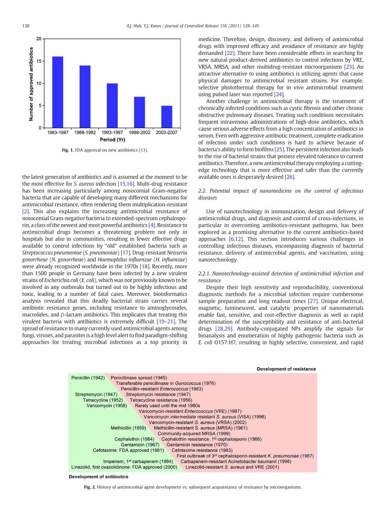
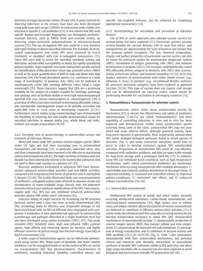


![Teaching an Old Class New Tricks: A Novel Semi-Synthetic ...tance mechanisms [4]. Aminoglycosides (AGs) are broad spectrum, bactericidal, antibiotics used for treating complicated](https://static.documents.pub/doc/80x56/5f7cadd241e288758311250f/teaching-an-old-class-new-tricks-a-novel-semi-synthetic-tance-mechanisms-4.jpg)


