Nanomaterials and nanoparticles: Sources and toxicityCristina Buzeaa�
Department of Physics, Queen’s University, Kingston, Ontario K7L 3N6, Canada
Ivan I. Pachecob�
Gastrointestinal Diseases Research Unit and Department of Physiology, Queen’s University at KingstonGeneral Hospital, 76 Stuart St., Kingston, Ontario K7L 2V7, Canada
Kevin Robbiec�
Department of Physics, Queen’s University, Kingston, Ontario K7L 3N6, Canada
�Received 13 June 2007; accepted 30 October 2007; published 28 December 2007�
MR18 Buzea, Pacheco, and Robbie: Nanomaterials and nanoparticles: Sources and toxicity MR18
Biointerphases, Vol. 2, No. 4, December 2007
1D One Dimensional2D Two Dimensional3D Three DimensionalAFM Atomic Force Microscop�e��y�AQI Air Quality IndexCNTs Carbon NanoTubesDNA DeoxyriboNucleic AcidEDS Energy Dispersive SpectrometryEPA Environmental Protection AgencyGLAD Glancing Angle DepositionHIV Human Immunodeficiency VirusMISR Multi-angle Imaging
SpectroradiometerMWCNTs Multiple-Wall Carbon NanoTubesNASA National Aeronautics and Space
Administrationnm nanometerPM Particulate MatterROS Reactive Oxygen SpeciesSEM Scanning Electron MicroscopySWCNs Single-Wall Carbon NanotubesTEM Transmission Electron Microscope
DEFINITIONS
aerosol—a material that, while not gaseous itself, remainssuspended in air for prolonged periods. Typical examplesinclude dust and fine-droplet liquid paint or hairspray.aggregate/aggregation—a material that is composed of alarge number of small components which have come to-gether as clusters, usually with branching, porous shapes.Aggregation is the process whereby the many small compo-nents form clusters, and can be driven by gravity or otherforces.Alzheimer’s disease—a progressive, irreversible, neurode-generative disease characterized by loss of function anddeath of nerve cells in several regions of the brain, leading toloss of attention, memory, and language. Its cause is un-known.antibody—a protein produced by the immune system as aresponse to a foreign substance, or antigen.antigen—a foreign substance that triggers the production ofantibodies by the immune system.apoptosis—also called “programmed cell death,” is the pro-cess of cellular suicide that can be initiated for several rea-sons: when extensive cellular damage occurs, when the cellis no longer needed within the organism, and in embryonicdevelopment, among others. Apoptosis is different from cellnecrosis �a form of traumatic cell death due to physical orbiological injuries� in its biochemical and morphological as-pects. Aberrations in apoptosis contribute to various diseases,such as cancer.atomic force microscopy—a scanning-probe form of surfacemicroscopy that can image and manipulate matter at the na-nometer scale.
autoimmune diseases—a group of disorders where overac-tive functioning of the immune system results in the immunesystem producing antibodies or autoreactive T cells �a typeof white blood cells� against its own tissue.bacteriophage—a virus that infects bacteria.cancer—disease characterized by rapid and uncontrolled celldivision.chelator—a chemical agent that binds reversibly to a metalion, forming a metallic complex.chronic disease—disease lasting a long time, which is ongo-ing or recurring, usually not caused by an infection and notcontagious.clearance—the removal of particles or substances out of anorganism, usually via urine or stool.Crohn’s disease—a chronic inflammatory disease of un-known cause that may affect any part of the gastrointestinaltract, most commonly the small bowel, as well as other or-gans. Symptoms of the disease include diarrhea, abdominalpain, and excessive weight loss.cytokine—a small protein released by cells that has a specificeffect on interactions between cells, on communications be-tween cells, or on the behavior of cells.cytoplasm—includes both the fluid �cytosol� and the or-ganelles contained within a cell.degenerative disease—disease characterized by progressivedeterioration of function or structure of tissue.DNA—a nucleic acid found within the nucleus of each cell,carrying genetic information on cell growth, division, andfunction. DNA consists of two long strands of nucleotidestwisted into a double helix and held together by hydrogenbonds. The sequence of nucleotides determines hereditarycharacteristics. Each cell contains an identical, completecopy of the organism’s DNA, with differing cell characteris-tics determined by differences in gene expression.endemic disease—disease constantly present in and limitedto people living in a certain location.endogenous—substances originating within, or synthesizedby an organism �e.g., hormones and neurotransmitters�.endoplasmic reticulum—a membrane network that extendsthroughout the cytoplasm and is involved in the synthesis,processing, secretion, and transport of proteins throughoutthe cell.endothelium—the layer of cells that line the interior surfaceof all parts of the circulatory system, including the heart, andblood vessels. Specialized endothelial cells perform impor-tant filtering functions in the kidney and at the blood-brainbarrier.enzyme—a protein that acts as a catalyst in a biochemicalreaction.epidemiology—the branch of medical sciences that studiesvarious factors influencing the incidence, distribution, andpossible control of diseases in human population.etiology—set of causes or origin of a disease.exogenous—substances originating outside an organism.fibroblast—a connective-tissue cell that secretes collagenand other components of the extracellular matrix. It migratesand proliferates during wound healing and in tissue culture.
MR19 Buzea, Pacheco, and Robbie: Nanomaterials and nanoparticles: Sources and toxicity MR19
Biointerphases, Vol. 2, No. 4, December 2007
gene—a sequence of nucleotides �DNA� that defines a pro-tein. Genes are the fundamental unit of heritability, and theircollection in an individual organism �its genome� representsa code or protocol for the growth and development of thatindividual. Genes are arranged along the length of chromo-somes, of which each species has a fixed number.genotype—the genetic constitution of an organism.granuloma—tissue resulting from aggregation ofinflammation-fighting cells unable to destroy foreign sub-stances.hydrophilic—having an affinity for water, or causing waterto adhere.hydrophobic—having no affinity for water, or repelling wa-ter.inflammation—a localized protective response, produced bytissue, injury that serves to destroy or arrest both the agentand the affected tissue. Blood vessel permeability locally in-creases, and the area becomes heavily populated with whiteblood cells. Signs of inflammation are redness, swelling,pain, and sometimes loss of function.ischemia—decrease in the blood supply to an organ, tissue,limb, or other part of a body caused by the narrowing orblockage of the blood vessels. Ischemia may lead to a short-age of oxygen �hypoxia� within the tissue and may result intissue damage or tissue death.Kaposi’s sarcoma—type of cancer that may affect the bloodand lymph vessels among others.lavage—washing out or clearance of a body cavity, organ, orsystem.lung burden—the product of exposure rate and residencytime of particulate matter inhaled into the lungs.lymph—a fluid containing white blood cells, proteins, andfats; can also carry bacteria, viruses, and cancer cells aroundthe body. Lymph is collected from the tissues and returned tothe circulatory system.lymphatic system—the network of vessels, nodes, and or-gans �spleen, thymus, and bone marrow� that produce, store,and carry lymph. The lymphatic system lacks a centralpump, such as the heart in the circulatory system, and mustrely on muscles pumping.lymphedema—a condition in which lymph nodes becomeenlarged and prevent lymph fluid from passing through them.macrophage—a phagocytic tissue cell of the reticuloendothe-lial system that is derived from the blood monocyte. Themonocyte migrates from the blood into tissues, where ittransforms into a macrophage. Macrophages are present inmost tissues. Macrophages ingest and process degeneratedcells and foreign invaders, such as viruses, bacteria, and par-ticles. The long-lived macrophages are reservoirs of HIV.mesothelioma—a rare form of cancer occurring in the liningof the lungs and chest cavity.mitochondrion—an organelle responsible for most of theoxidative metabolism in the cell. Mitochondria generate en-ergy �in the form of adenosine triphosphate �ATP�� by break-ing down glucose �a type of sugar�.monocyte—the largest form of a white blood cell, with akidney-shaped nucleus; its function is the ingestion of for-
eign invaders, such as bacteria, tissue debris, and particles.Monocytes belong to the group of phagocytes, and matureinto various macrophages in tissue.murine—pertaining to the rodent family, i.e., rats and mice.nanoparticulate matter—a collection of particles with at leastone dimension smaller than 1 �m yet larger than atoms andmolecules.neutrophil—an immune cell that ingests and degrades for-eign organisms. Neutrophils are the most abundant type ofwhite blood cells, and are the first to reach the site of aninfection to attack foreign antigens.oxidative stress—an imbalance in favor of pro-oxidant ver-sus antioxidant chemicals, potentially leading to damage tobiomolecules.Parkinson’s disease—a progressive disorder of the nervoussystem manifested by muscle tremors and rigidity, decreasedmobility, and slow voluntary movements.particulate matter—airborne particles of solids and/or liquidswith sizes ranging from several nanometers to several hun-dred microns.phagocyte—cell that ingests and kills foreign intruders viathe process called phagocytosis. Three examples are mono-cytes, macrophages, and neutrophils.PM0.1—particulate matter having a diameter smaller than0.1 �m �100 nm�.PM10—particulate matter having a diameter smaller than10 �m.PM2.5—particulate matter having a diameter smaller than2.5 �m.pneumoconiosis—lung disease due to permanent depositionof substantial amounts of particles in the lungs and by thetissue reaction to its presence. Its severity varies from rela-tively harmless forms of sclerosis to destructive fibrosis andscarring of the lungs.podoconiosis—impaired lymphatic system drainage affectingthe limbs due to clogging with nano- and microparticles.protein—molecule containing a long chain of amino acids inthe order specified by a gene’s DNA sequence. Proteins canbe, for example, enzymes, hormones, and antibodies.quantum dot—semiconductor crystals with a diameter of afew nanometers, having many properties resembling those ofatoms.receptor—A protein or large molecule on the surface of a cellthat binds selectively to specific substances �ligands�.reperfusion—restoration of blood flow.reticuloendothelial system—a part of the immune systemthat consists of phagocytic cells, including macrophages andmacrophage precursors, specialized endothelial cells liningthe sinusoids of the liver, spleen, and bone marrow, and re-ticular cells of lymphatic tissue �macrophages� and bonemarrow �fibroblasts�.rheumatoid arthritis—chronic, autoimmune, inflammatorydisorder affecting the connective tissue lining the joints.Symptoms include pain, swelling, stiffness, and deformities.It can extend to organs.
MR20 Buzea, Pacheco, and Robbie: Nanomaterials and nanoparticles: Sources and toxicity MR20
Biointerphases, Vol. 2, No. 4, December 2007
scleroderma—a degenerative, autoimmune disease of theconnective tissue, characterized by the formation of fibroustissue �collagen� which surround the joints, blood vessels,and sometimes internal organs.systemic lupus erythematosus—a chronic, autoimmune dis-order. Symptoms include fatigue, butterfly-shaped facialrash, inflammation of the joints, tendons, connective tissues,and organs: heart, lungs, blood vessels, brain, kidneys, andskin.toxicology—the branch of medical and biological sciencestudying the nature, adverse effects, detection, and treatmentof poisons on living organisms. A fundamental principle oftoxicology is that any substance is poisonous if given in alarge amount. From the study of cancer-causing substances,carcinogens, it appears that there are some materials forwhich there is no safe dose, no level of exposure belowwhich they do not cause cancer.transcription factor—a protein that binds to enhancer ele-ments in DNA to regulate the level of transcription and ex-pression of certain genes.translocation—the process of transit of particles or sub-stances within an organism.ulcerative colitis—a chronic disease of unknown cause char-acterized by inflammation of the colon producing ulcer-ations. Symptoms are abdominal pain, cramps, loose dis-charges of pus, blood, and mucus from the bowel, andweight loss.UFP—nanoparticles with size smaller than 100 nm.
I. INTRODUCTION
Every person has been exposed to nanometer-sized for-eign particles; we inhale them with every breath, and con-sume them with every drink. In truth, every organism onEarth continuously encounters nanometer-sized entities. Thevast majority causes little ill effect, and go unnoticed, butoccasionally an intruder will cause appreciable harm to theorganism. The most advanced of the toxic intruders are vi-ruses, composed as they are of nucleic-acid-based structuresthat allow them to not only interfere with biological systems,but also to parasitically exploit cellular processes to replicatethemselves. Among the more benign viruses are the onescausing the familiar human symptoms of the common coldor flu, which are the evident manifestations of biochemicalbattles occurring between these foreign intruders and our im-mune systems, whose nanometer-sized constituents �chemi-cals and proteins� usually destroy and remove the viral in-vaders. A growing number of recent studies show, however,that nano- and microorganisms may play a role in manychronic diseases where infectious pathogens have not beensuspected, diseases that were previously attributed only togenetic factors and lifestyle. Among these diseases are leu-kemia �caused by viruses from the retrovirus and herpesvirusfamilies�,1 cervical cancer �papillomavirus�,2 liver cancer�hepatitis virus�,3 gastric ulcer �Helicobacter pylori�,4 na-sopharyngeal cancer �Epstein-Barr virus�,5 kidney stones�nanobacteria�,6 severe acquired respiratory syndrome �Co-rona virus�,7 heart disease �Chlamydia pneumonia�,8 juvenile
diabetes �Coxsackie virus�,9 Alzheimer’s disease �Chlamydiapneumoniae�,10 pediatric obsessive-compulsive disorder�streptococcal bacteria�,11 psychotic disorders �Bornavirus�,12 and prion diseases such as mad cow disease �pro-teins prions�.13
One is tempted to think that nanoparticles �such as dust orash particles�, while similar in size to viruses, would be morebenign, as these materials lack the viruses’ ability to repli-cate. Nevertheless, while nonreplicating bodily intruders donot directly take control of cellular processes, some havebeen shown to sufficiently interfere with cellular function toinfluence basic process of cells, such as proliferation, me-tabolism, and death. Many diseases can be associated withdysfunction of these basic processes, the most notable beingcancer �uncontrolled cells proliferation� and neurodegenera-tive diseases �premature cell death�. In addition, several dis-eases with unknown cause, including autoimmune diseases,Crohn’s, Alzheimer’s, and Parkinson’s diseases, appear to becorrelated with nanoparticle exposure. Conversely, the toxicproperties of some nanoparticles may be beneficial, as theyare thereby able to fight disease at a cellular level, and couldbe used as a medical treatment by, for example, targeting anddestroying cancerous cells.Very small particles, so-called nanoparticles, have the
ability to enter, translocate within, and damage living organ-isms. This ability results primarily from their small size,which allows them to penetrate physiological barriers andtravel within the circulatory systems of a host. While naturalprocesses have produced nanoparticles for eons, modern sci-ence has recently learned how to synthesize a bewilderingarray of artificial materials with structure that is engineeredat the atomic scale. The smallest particles contain tens orhundreds of atoms, with dimensions at the scale of nanom-eters, hence nanoparticles. They are comparable in size toviruses, where the smallest have dimensions of tens of na-nometers �for example, a human immunodeficiency virus�HIV� particle is 100 nm in diameter�, and which in theemerging science of nanotechnology might be called “na-noorganisms.” Like viruses, some nanoparticles can pen-etrate lung or dermal �skin� barriers and enter the circulatoryand lymphatic systems of humans and animals, reachingmost bodily tissues and organs, and potentially disruptingcellular processes and causing disease. The toxicity of eachof these materials depends greatly, however, on the particulararrangement of its many atoms. Considering all the possiblevariations in shape and chemistry of even the smallest nano-particles, with only tens of atoms, yields a huge number ofdistinct materials with potentially very different physical andtoxicological properties. Asbestos is a good example of atoxic nanomaterial causing lung cancer and other diseases.Asbestos exists in several forms, with slight variations inshape and chemistry, yet significantly varying toxicity.Nanometer-sized particles are created in countless physi-
cal processes from erosion to combustion, with health risksranging from lethal to benign. Industrial nanoparticle mate-rials today constitute a tiny but significant pollution sourcethat is, so far, literally buried beneath much larger natural
MR21 Buzea, Pacheco, and Robbie: Nanomaterials and nanoparticles: Sources and toxicity MR21
Biointerphases, Vol. 2, No. 4, December 2007
sources and nanoparticle pollution incidental to other humanactivities, particularly automobile exhaust soot.The misapprehension of nanotoxicity may create a gen-
eral fear that all nanomaterials are toxic. The online14 andprinted media15 are inadvertently making no distinction be-tween nanostructured fixed structures, which are not likely tocause harm �such as computer processors�, and detachable orfree nanoparticles, which are likely to cause adverse healtheffects. While uncontained nanoparticles clearly represent aserious health threat, fixed nanostructured materials, such asthin film coatings, microchip electronics, and many otherexisting nanoengineered materials, are known to be virtuallybenign. Many synthetic nanoparticulate materials producepositive health effects, for example, functionalized fullerenechemicals that act as antioxidants. The use of nanoparticlesin medical diagnostics and treatment is driven by their safetyas well as utility.In the following pages, we outline existing sources of
nanoparticles, both natural and man-made, and the knowneffects of exposure to nanoparticles. In Sec. I, we introducebasic concepts and terminology relevant to nanoscience andnanotechnology, define concepts and terms, give examples ofnanoscale systems, and introduce the basics of nanoparticletoxicity. In Sec. II, we briefly discuss nanoparticle classifica-tions. Section III reviews natural and anthropogenic nanopar-ticle sources together with their associated health effects andtreatment. In Sec. IV, we present current opinions and re-search results related to the health implications and toxicol-ogy of nanoparticles, and we define exposure pathways andmigration or translocation mechanisms within biological sys-tems, adverse health effects, and treatment. The mechanicsand biochemistry of toxicity are discussed in Sec. V, as wellas toxicity-related risk factors, such as particle size, shape,chemistry, and surface properties. In Sec. VI, we provide anoverview of current and developing applications of nanoma-terials. Finally, Sec. VII contains conclusions and reflections.
A. Nano etymology
The prefix “nano,” derived from the Greek “nanos,” sig-nifying “dwarf,” is becoming increasingly common in scien-tific literature. Nano is now a popular label for much ofmodern science, and many nano words have recently ap-peared in dictionaries, including nanometer, nanoscale, nano-science, nanotechnology, nanostructure, nanotube, nanowire,and nanorobot. Many words that are not yet widely recog-nized are used in respected publications, such as Science andNature. These include nanoelectronics, nanocrystal, nanov-alve, nanoantenna, nanocavity, nanoscaffolds, nanofibers, na-nomagnet, nanoporous, nanoarrays, nanolithography, nano-patterning, nanoencapsulation, etc. Although the idea ofnanotechnology, i.e., producing nanoscale objects and carry-ing out nanoscale manipulations, has been around for quitesome time, the birth of the concept is usually linked to aspeech by Feynman at the December 1959 meeting of theAmerican Physical Society.16 where he asked, “What wouldhappen if we could arrange the atoms one by one the way wewant them?”
The nanometer is a metric unit of length, and denotesone-billionth of a meter or 10−9 m. Popularly, nano is alsoused as an adjective to describe objects, systems, or phenom-ena with characteristics arising from nanometer-scale struc-ture. While “micro” has come to mean anything small, nanoemphasizes the atomic granularity that produces the uniquephenomena observed in nanoscience. While there are someexceptional examples, most of the exciting properties ofnano begin to be apparent in systems smaller than 1000 nmor 1 �m. For the purpose of this review, we will describeparticles with any dimension smaller than 1 �m as “nano-particles,” and those somewhat larger as “microparticles.”Nanostructured materials did not first come into existence
with the recent emergence of the field of nanotechnology.Many existing materials are structured on the micro- andnanometer scales, and many industrial processes that havebeen used for decades �e.g., polymer and steel manufactur-ing� exploit nanoscale phenomena. The most advanced nano-technological fabrication process is microelectronic fabrica-tion, where thin film coatings and lithography are used tocreate micro- and nanosized features on computer chips. Thenatural world is replete with examples of systems withnanoscale structures, such as milk �a nanoscale colloid�, pro-teins, cells, bacteria, viruses, etc. Moreover, many materialsthat seem smooth to the naked eye have an intricate structureon the scale of nanometers �Fig. 1�. Thus, in many ways,nanomaterials are not new. Recent advances in synthesis andcharacterization tools, however, have fueled a boom in thestudy and industrial use of nanostructured materials. A newvocabulary has emerged from this research, and its importantterms and concepts are defined below.Nanomaterials are materials that have structural compo-
nents smaller than 1 �m in at least one dimension. While theatomic and molecular building blocks ��0.2 nm� of matterare considered nanomaterials, examples such as bulk crystalswith lattice spacing of nanometers but macroscopic dimen-sions overall are commonly excluded.Nanoparticles are particles with at least one dimension
smaller than 1 �m, and potentially as small as atomic andmolecular length scales ��0.2 nm�. Nanoparticles can haveamorphous or crystalline form, and their surfaces can act ascarriers for liquid droplets or gases. To some degree, nano-particulate matter should be considered a distinct state ofmatter, in addition to the solid, liquid, gaseous, and plasmastates, due to its distinct properties �large surface area andquantum size effects�. Examples of materials in crystallinenanoparticle form are fullerenes and carbon nanotubes, whiletraditional crystalline solid forms are graphite and diamond.Many authors limit the size of nanomaterials to 50 nm �Ref.17� or 100 nm,18 the choice of this upper limit being justifiedby the fact that some physical properties of nanoparticlesapproach those of the bulk when their size reaches thesevalues. However, this size threshold varies with material typeand cannot be the basis for such a classification. A legitimatedefinition extends this upper size limit to 1 �m, the submi-cron range being classified as nano.
MR22 Buzea, Pacheco, and Robbie: Nanomaterials and nanoparticles: Sources and toxicity MR22
Biointerphases, Vol. 2, No. 4, December 2007
Nanoparticulate matter refers to a collection of nanopar-ticles, emphasizing their collective behavior.Nanotechnology can be defined as the design, synthesis,
and application of materials and devices whose size andshape have been engineered at the nanoscale. It exploitsunique chemical, physical, electrical, and mechanical prop-erties that emerge when matter is structured at the nanoscale.Nanotoxicology was proposed as a new branch of toxicol-
ogy to address the adverse health effects caused bynanoparticles.19 Despite suggestions that nanotoxicologyshould only address the toxic effects of engineered nanopar-ticles and structures,20 we recommend that nanotoxicology
should also encompass the toxic effects of atmospheric par-ticles as well as the fundamentals of virology and bacteriol-ogy. While significant differences exist between the healtheffects of nonbiological particles and viruses and bacteria,there are significant common aspects of intrusion andtranslocation.The new terminology of nano has united previously seem-
ingly disparate fields, and a lexicon is needed to find andappreciate the great wealth of existing nano research, notconveniently labeled with the nano keyword.
Health sciences epidemiology terminology. In existingmedical and toxicological terminology, nanoparticles havinga diameter smaller than 100 nm are often called ultrafineparticles �UFP� or ultrafine particulate matter. Ultrafine par-ticles are labeled as a function of their size. For example,particulate matter with constituents having diameters smallerthan 10 �m is abbreviated PM10. Particulate matter having asize smaller than 100 nm is labeled as PM0.1.
Environmental sciences terminology. Ambient particulatematter is categorized into three size distributions: ultrafineparticles less than 0.1 �m in diameter �mainly resulting fromcombustion�, accumulation mode particles between 0.1 and2.5 �m in diameter �resulting from aggregation of ultrafineparticles and vapors�, and coarse-mode particles larger than2.5 �m �mostly mechanically generated�.24
Proposed terminology. It is important, and timely, to unifythe terminology used for describing particle size in nanotech-nology, health, and environmental sciences.The materials under discussion can be classified as par-
ticles, regardless of their source. The size of these particlesvaries between 1 nm and several microns, and they can,therefore, be classified as either nanoparticles �NP� �any di-mension smaller than 1 �m� or microparticles �MP� �all di-mensions larger than 1 �m�. To further specify particle size,we propose a modification of the health sciences epidemiol-ogy terminology, labeling particles by their largest dimen-sion; for example, 10 nm in diameter are labeled “NP10,”while 10 �m microparticles are labeled “MP10.”Given that microparticles and nanoparticles vary in their
conception by only their size, it can be difficult to fully ap-preciate the differences between them. To illuminate the ef-fect of the size difference, the sizes of several natural micro-and nanostructures are shown in Fig. 2, as measured fromscanning and transmission microscope images.25,26 Gener-ally, the sizes of nanomaterials are comparable to those ofviruses, DNA, and proteins, while microparticles are compa-rable to cells, organelles, and larger physiological structures�Fig. 2�. A red blood cell is approximately 7 �m wide, a hair60 �m, while lung alveoli are approximately 400 �m.
B. Main differences between nanomaterials and bulkmaterials
Two primary factors cause nanomaterials to behave sig-nificantly differently than bulk materials: surface effects�causing smooth properties scaling due to the fraction of at-oms at the surface� and quantum effects �showing discon-tinuous behavior due to quantum confinement effects in ma-
FIG. 1. SEM images showing the complexity of the world at the micro- andnanoscale: �a� the inner surface of a bird’s eggshell, �credit: Janice Carr,Sandra L. Westmoreland, courtesy Public Health Image Library �Ref. 21��;�b� the rough surface of table grape, �credit: Janice Carr, courtesy PublicHealth Image Library �Ref. 21��; �c� the textured surface of a parsley leaf,�credit Janice Carr, courtesy Public Health Image Library �Ref. 21��; �d�Kleenex paper, �courtesy of Jim Ekstrom �Ref. 22��; �e� pollen from a vari-ety of common plants, �credit Louisa Howard, Charles Daghlian, courtesyPublic Health Image Library �Ref. 21��; �f� green algae, �credit ElizabethSmith, Louisa Howard, Erin Dymek, Public Health Image Library �Ref.21��; �g� Gecko nano-adhesive system, with increasing magnification fromleft to right: gecko climbing vertical glass, adhesive surface microstructure,individual setae, nanostructure of spatular endings, �courtesy of PNAS �Ref.23��.
MR23 Buzea, Pacheco, and Robbie: Nanomaterials and nanoparticles: Sources and toxicity MR23
Biointerphases, Vol. 2, No. 4, December 2007
terials with delocalized electrons�.27 These factors affect thechemical reactivity of materials as well as their mechanical,optical, electric, and magnetic properties.The fraction of the atoms at the surface in nanoparticles is
increased compared to microparticles or bulk. Compared tomicroparticles, nanoparticles have a very large surface areaand high particle number per unit mass. For illustration, onecarbon microparticle with a diameter of 60 �m has a mass of0.3 �g and a surface area of 0.01 mm2. The same mass ofcarbon in nanoparticulate form, with each particle having adiameter of 60 nm, has a surface area of 11.3 mm2 and con-
sists of 1�109 nanoparticles �Fig. 3�. The ratio of surfacearea to volume �or mass� for a particle with a diameter of60 nm is 1000 times larger than a particle with a diameter of60 �m �Fig. 3�b��. As the material in nanoparticulate formpresents a much larger surface area for chemical reactions,reactivity is enhanced roughly 1000-fold. While chemical re-activity generally increases with decreasing particle size, sur-face coatings and other modifications can have complicatingeffects, even reducing reactivity with decreasing particle sizein some instances.The atoms situated at the surface have less neighbors than
bulk atoms, resulting in lower binding energy per atom withdecreasing particle size.27 A consequence of reduced bindingenergy per atom is a melting point reduction with particleradius, following the Gibbs-Thomson equation.27. For ex-ample, the melting temperature of 3 nm gold nanoparticles ismore than 300 degrees lower than the melting temperature ofbulk gold, as shown in Fig. 3�c�.27
An example of a class of materials that clearly exploitsquantum effects is quantum dots—synthesized nanostruc-tures with sizes as small as a few nanometers �Fig. 4�. Theelectronic behavior of quantum dots is similar to that of in-
FIG. 2. Logarithmical length scale showing size of nanomaterials comparedto biological components and definition of “nano” and “micro” sizes.
FIG. 3. �a� Schematics illustrating amicroparticle of 60 �m diameter,about the size of a human hair—shown in the left at scale �courtesyChelsea Elliott�, and the number ofnanoparticles with diameter of 600and 60 nm having the same mass asone microparticle of 60 �m diameter.�b� Surface area normalized to massversus particle diameter. �c� Goldmelting temperature as a function ofparticle diameter, according to Gibbs-Thomson equation, shown inset; thegold bulk melting temperature is1336 K �Ref. 27�.
FIG. 4. �a� TEM image of CdSe semiconductor nanoparticles, and �b� pho-tograph of CdSe nanoparticles in solution, photo-luminescent under UVillumination. Images courtesy of Graham Rodway and Harry Ruda, Univer-sity of Toronto.
MR24 Buzea, Pacheco, and Robbie: Nanomaterials and nanoparticles: Sources and toxicity MR24
Biointerphases, Vol. 2, No. 4, December 2007
dividual atoms or small molecules, and quantum dots areregarded as akin to artificial atoms.28 Notably, the confine-ment of the electrons in quantum dots in all three spatialdirections results in a quantized energy spectrum. Anotherresult of quantum confinement effect is the appearance ofmagnetic moments in nanoparticles of materials that are non-magnetic in bulk, such as gold, platinum, or palladium.27
Magnetic moments result from several unpaired electronspins in nanoparticles formed of several hundred atoms.Quantum confinement also results in quantified changes inthe ability to accept or donate electrical charge �or electronaffinity�, also reflected in the catalytic ability.27 For example,the reactivity of cationic platinum clusters in the decompo-sition of N2O is dictated by the number of atoms in thecluster, namely, 6–9, 11, 12, 15, and 20 atom-containingclusters are very reactive, while clusters with 10, 13, 14, and19 atoms have low reactivity.27
C. Nanomaterials and nanotoxicology publicationsstatistics
The number of publications on the topic of nanomaterialshas increased at an almost exponential rate since the early1990s, reaching about 40 000 in the year 2005 �Fig. 5�, asindicated by a search on the ISI Web of Knowledgedatabase.29 There is also a notable rise in the number ofpublications discussing their toxicity, particularly in the pasttwo years. The total number of papers on toxicity, however,remains low compared to the total number of publications onnanomaterials, with only around 500 publications in the year2005.The large number of publications on nanomaterials can be
explained by the fact that nanoscience and nanotechnologyencompass a wide range of fields, including chemistry, phys-ics, materials engineering, biology, medicine, and electron-ics. There are several reviews addressing nanotoxicology as-pects; however, they are intended for a narrow, specializedaudience. Several are comparatively general,18,20,30–32 whileothers address selected aspects of nanoparticle toxicology,such as health effects of air pollution;33 epidemiological re-views of exposure to particles;34 epidemiological studies ofcardiovascular effects of airborne particles;35 occupationalaspects of nanoparticles;36 particle inhalation, retention, andclearance;37 pulmonary effects of inhaled particles;38,39 inha-lation and lung cancer;40,41 toxicity of combustion-derived
particles inhalation;42 environmental factors in neurodegen-erative diseases;43 oxidative mechanisms;44–51 gastrointesti-nal uptake of particles;52 targeted drug delivery;53 particlecharacterization methods;54 screening strategies and futuredirections of research;55 and regulation of nanomaterials.56
Existing reviews are either written in jargon comprehensiveonly to specialists in a particular field, or are, if more acces-sible, very succinct.32,57 Most nanotechnology reviews writ-ten to date focus on a specific subfield, disregarding the vastamount of existing knowledge on the general theme of nano.In this review, we attempt to bring together a broader audi-ence by unifying the language and experience of scientistsworking within these diverse fields.
D. Introduction to nanoparticle toxicity
Human skin, lungs, and the gastrointestinal tract are inconstant contact with the environment. While the skin is gen-erally an effective barrier to foreign substances, the lungsand gastrointestinal tract are more vulnerable. These threeways are the most likely points of entry for natural or anthro-pogenic nanoparticles. Injections and implants are other pos-sible routes of exposure, primarily limited to engineeredmaterials.Due to their small size, nanoparticles can translocate from
these entry portals into the circulatory and lymphatic sys-tems, and ultimately to body tissues and organs. Some nano-particles, depending on their composition and size, can pro-duce irreversible damage to cells by oxidative stress and/ororganelle injury. Figure 6 illustrates the size of an examplecell and its organelles compared to nanoparticles of varioussizes, making it easy to understand why nanoparticles areable to enter cells and interact with various cell components�nucleus, mitochondria, etc.�.In Fig. 7, we summarize the possible adverse health ef-
fects associated with inhalation, ingestion, and contact withnanoparticles. We emphasize that not all nanoparticles pro-duce these adverse health effects—the toxicity of nanopar-ticles depends on various factors, including size, aggrega-
FIG. 5. Statistics on scientific articles published on �a� nanomaterials and �b�their toxicity �Ref. 29�.
FIG. 6. Comparison of rat macrophage cells size to nanoparticles size �atscale�. Human macrophages are up to two times larger than rat macroph-ages. TEM image reproduced with permission from Environmental HealthPerspectives �Ref. 238�.
MR25 Buzea, Pacheco, and Robbie: Nanomaterials and nanoparticles: Sources and toxicity MR25
Biointerphases, Vol. 2, No. 4, December 2007
tion, composition, crystallinity, surface functionalization, etc.In addition, the toxicity of any nanoparticle to an organism isdetermined by the individual’s genetic complement, whichprovides the biochemical toolbox by which it can adapt toand fight toxic substances. While these effects will be dis-cussed in detail in Secs. III and IV, we summarize below themost extreme adverse health effects produced by nanopar-ticles in order to immediately increase the awareness of po-tential toxicity of some nanoparticles. Diseases associatedwith inhaled nanoparticles are asthma, bronchitis, emphy-sema, lung cancer, and neurodegenerative diseases, such asParkinson’s and Alzheimer’s diseases. Nanoparticles in thegastrointestinal tract have been linked to Crohn’s disease andcolon cancer. Nanoparticles that enter the circulatory systemare related to occurrence of arteriosclerosis, blood clots, ar-rhythmia, heart diseases, and ultimately cardiac death. Trans-location to other organs, such as liver, spleen, etc., may leadto diseases of these organs as well. Exposure to some nano-particles is associated with the occurrence of autoimmunediseases, such as systemic lupus erythematosus, scleroderma,and rheumatoid arthritis.
II. NANOPARTICLE CLASSIFICATION
Nanoparticles are generally classified based on their di-mensionality, morphology, composition, uniformity, and ag-glomeration.
An important additional distinction should be made be-tween nanostructured thin films or other fixed nanometer-scale objects �such as the circuits within computer micropro-cessors� and free nanoparticles. The motion of freenanoparticles is not constrained, and they can easily be re-leased into the environment, leading to human exposure thatmay pose a serious health risk. In contrast are the manyobjects containing nanostructured elements that are firmlyattached to a larger object, where the fixed nanoparticlesshould pose no health risk when properly handled. An ex-ample of this important distinction is the material asbestos,which is perfectly safe in its primary state �basically a typeof solid rock�, but is a significant health hazard when minedor worked in such a way as to produce the carcinogenicnanometer-scale fibrous particles that become airborne �aero-sol� and are, therefore, readily absorbed in the lungs.It is also very important to recognize that not all nanopar-
ticles are toxic; toxicity depends on at least chemical com-position and shape in addition to simply size and particleaging. In fact, many types of nanoparticles seem to benontoxic,58,59 others can be rendered nontoxic,60 while othersappear to have beneficial health effects.61,62 An importantlesson we are in the process of learning from nanoscience isthat simple classifications of physical behavior �and, there-fore, toxicity� are overly limiting and that we must study
FIG. 7. Schematics of human body with pathways of exposure to nanoparticles, affected organs, and associated diseases from epidemiological, in vivo and invitro studies.
MR26 Buzea, Pacheco, and Robbie: Nanomaterials and nanoparticles: Sources and toxicity MR26
Biointerphases, Vol. 2, No. 4, December 2007
toxicology of each material and each morphology, in addi-tion to particle aging, to obtain accurate information to in-form policy and regulatory processes.
A. Dimensionality
As shape, or morphology, of nanoparticles plays an im-portant role in their toxicity, it is useful to classify thembased on their number of dimensions �Fig. 8�. This is a gen-eralization of the concept of aspect ratio.
One-dimensional (1D) nanomaterials. Materials with onedimension in the nanometer scale are typically thin films orsurface coatings, and include the circuitry of computer chipsand the antireflection and hard coatings on eyeglasses. Thinfilms have been developed and used for decades in variousfields, such as electronics, chemistry, and engineering. Thinfilms can be deposited by various methods,63 and can begrown controllably to be only one atom thick, a so-calledmonolayer.
Two-dimensional (2D) nanomaterials. Two-dimensionalnanomaterials have two dimensions in the nanometer scale.These include 2D nanostructured films, with nanostructuresfirmly attached to a substrate, or nanopore filters used forsmall particle separation and filtration. Free particles with alarge aspect ratio, with dimensions in the nanoscale range,are also considered 2D nanomaterials. Asbestos fibers are anexample of 2D nanoparticles.
Three-dimensional (3D) nanomaterials. Materials that arenanoscaled in all three dimensions are considered 3D nano-materials. These include thin films deposited under condi-
tions that generate atomic-scale porosity, colloids, and freenanoparticles with various morphologies.64
B. Nanoparticle morphology
Morphological characteristics to be taken into account areflatness, sphericity, and aspect ratio. A general classificationexists between high- and low-aspect-ratio particles �Fig. 8�.High-aspect-ratio nanoparticles include nanotubes and nano-wires, with various shapes, such as helices, zigzags, belts, orperhaps nanowires with diameter that varies with length.Low-aspect-ratio morphologies include spherical, oval, cu-bic, prism, helical, or pillar. Collections of many particlesexist as powders, suspension, or colloids.
C. Nanoparticle composition
Nanoparticles can be composed of a single constituentmaterial �Fig. 8� or be a composite of several materials. Thenanoparticles found in nature are often agglomerations ofmaterials with various compositions, while pure single-composition materials can be easily synthesized today by avariety of methods �see Sec. III B 6�.
D. Nanoparticle uniformity and agglomeration
Based on their chemistry and electromagnetic properties,nanoparticles can exist as dispersed aerosols, as suspensions/colloids, or in an agglomerate state �Fig. 8�. For example,magnetic nanoparticles tend to cluster, forming an agglomer-ate state, unless their surfaces are coated with a nonmagnetic
FIG. 8. Classification of nanostructured materials from the point of view of nanostructure dimensions, morphology, composition, uniformity and agglomerationstate.
MR27 Buzea, Pacheco, and Robbie: Nanomaterials and nanoparticles: Sources and toxicity MR27
Biointerphases, Vol. 2, No. 4, December 2007
material. In an agglomerate state, nanoparticles may behaveas larger particles, depending on the size of the agglomerate.Hence, it is evident that nanoparticle agglomeration and sizeand surface reactivity, along with shape and size, must betaken into account when considering health and environmen-tal regulations of new materials.
III. SOURCES OF NANOPARTICLES AND THEIRHEALTH EFFECTS
A. Natural sources of nanoparticles
Nanoparticles are abundant in nature, as they are pro-duced in many natural processes, including photochemicalreactions, volcanic eruptions, forest fires, and simple erosion,and by plants and animals, e.g., shedding of skin and hair.Though we usually associate air pollution with humanactivities—cars, industry, and charcoal burning—naturalevents such as dust storms, volcanic eruptions, and forestfires can produce such vast quantities of nanoparticulate mat-ter that they profoundly affect air quality worldwide. Theaerosols generated by human activities are estimated to beonly about 10% of the total, the remaining 90% having anatural origin.65 These large-scale phenomena are visiblefrom satellites, and produce particulate matter and airborneparticles of dust and soot ranging from micro- to nanoscales.Small particles suspended in the atmosphere, often known asaerosols, affect the entire planet’s energy balance becausethey both absorb radiation from the sun and scatter it back tospace.66 It has been estimated that the most significant com-ponents of total global atmospheric aerosols are, in decreas-ing mass abundance, mineral aerosols primarily from soildeflation �wind erosion� with a minor component ��1% �from volcanoes �16.8 Tg�, sea salt �3.6 Tg�, natural and an-thropogenic sulfates �3.3 Tg�, products of biomass burningexcluding soot �1.8 Tg�, and of industrial sources includingsoot �1.4 Tg�, natural and anthropogenic nonmethane hydro-carbons �1.3 Tg�, natural and anthropogenic nitrates �0.6 Tg�,and biological debris67 �0.5 Tg�. �Note: “Tg” here denotesterragram, equal to 1012 g.�
1. Dust storms and health effects
Terrestrial dust storms. Dust storms appear to be the larg-est single source of environmental nanoparticles. Long-rangemigration of both mineral dust and anthropogenic pollutantsfrom the major continents has recently been the subject ofintense investigation. Approximately 50% of troposphere at-mospheric aerosol particles are minerals originating from thedeserts.68 The size of particles produced during a dust stormvaries from 100 nm to several microns �Fig. 9�d��, with one-third to a half of the dust mass being smaller than2.5 �m.65,68 Particles in the range 100–200 nm can reachconcentrations of 1500 particles /cm3.69
Meteorological observations and modeling have identifiedten main sources of global dust events, shown in Fig. 9�e�:�1� the Salton Sea, �2� Patagonia, �3� the Altipläno, �4� theSahel region, �5� the Sahara Desert, �6� the Namibian desert
lands, �7� the Indus Valley, �8� the Taklimakan Desert, �9� theGobi Desert, and �10� the Lake Eyre basin.65
Satellite imagery has revealed the dynamics of large-scaledust migration across continents, and demonstrated thatnanoparticles generated by major environmental events inone part of the world can affect regions thousands of kilo-meters away, as shown in Fig. 9. For example, dust stormsoccurring every spring in the Gobi Desert strongly affect theair quality in Asia and North America.70,71 The dust routeacross the Pacific can be seen in satellite images by the yel-low color of the dust itself �Fig. 9�a��.72 The dust migrationpattern during the 1998 trans-Pacific transport is shown inFig. 9�c�, the dates representing the approximate daily loca-tion of the dust cloud.70 During this event, the dust cloudreached the west coast of North America within five to sixdays after emission, with the region affected experiencing anintense haze and elevated particles concentrations, with anaverage excess of 20–50 �g /m3 with local peaks�100 �g /m3.70,71
Extraterrestrial dust. Nanoparticles exist widely in extra-terrestrial space. Examples of dust collected from space,from the moon, and on Mars are shown in Fig. 10. Theextraterrestrial dust poses major environmental problems forastronauts as well as for equipment.73 Lunar dust is very finegrained compared to typical terrestrial dust �some of thelarger grains being shown in Fig. 10�c��, with more than 50%of particles found to be in the micron range or smaller.74 Thelunar dust contains a considerable amount of magneticnanoparticles,75 clinging to electrostatically chargedsurfaces74 such as the astronauts’ space suits �Fig. 10�b��,rendering it nearly impossible to remove. On Mars, dust ac-cumulating on the solar panels of the exploration robots haslimited the power available to them for locomotion, sensing,and communication.76 Aiming to mitigate the environmentaleffects of extraterrestrial dust on humans and machines, vari-ous research projects are directed towards the fabrication offilters or thin film coatings that repel dust.76
Health effects. Terrestrial airborne dust particles can leadto a number of health problems, especially in subjects withasthma and emphysema.65 The composition of dusts is im-portant, as dust rich in iron or other metals can generatereactive oxygen species on the lung surface that can scarlung tissue.65 In addition, viruses, bacteria, fungi, or chemi-cal contaminants hitchhiking dust particles may adverselyaffect health and the environment �Fig. 9�f��. In this regard, itis important to note that 200 types of viable bacteria andfungi have been found to survive ultraviolet light exposureduring intercontinental journeys from Africa to America.65
Extraterrestrial dust brought inside the lunar module be-came airborne, and irritated the lungs and eyes of Apolloastronauts.77 On longer missions to the moon or Mars, pro-longed exposure could increase the risk of respiratory dis-eases in the astronauts, and mechanical failures of spacesuitsand airlocks. Studies on rats have found that intratrachealadministration of small amounts of lunar material resulted inpneumoconiosis with fibrosis formation78 �lung disease andabnormal tissue growth�.
MR28 Buzea, Pacheco, and Robbie: Nanomaterials and nanoparticles: Sources and toxicity MR28
Biointerphases, Vol. 2, No. 4, December 2007
2. Forest fires and health effects
Forest fires and grass fires have long been a part ofEarth’s natural history, and are primarily caused by lightningstrikes or by human activity. Major fires can spread ash andsmoke �Fig. 11�a�� over thousands of square miles �Figs.11�b� and 11�c�� and lead to an increase of particulate matter�including nanoparticles� exceeding ambient air qualitystandards.79 Satellite maps show a unique picture of globalfire activity. Using daily global fire detection provided bymoderate-resolution imaging spectroradiometer �MODIS� onNASA’s Terra satellite, the fire activity for the entire surface
of the Earth has been mapped every day since February 2000�Fig. 11�d��.80 As noticed in this figure, numerous fires occurthroughout the world in the savannas of Africa, Australia,and Brazil, and in North America, Europe, and Asia.
Health effects. Epidemiological studies showed that dur-ing the weeks of forest fires, medical visits increase morethan 50% in the affected regions.81 Patients with preexistingcardiopulmonary conditions reported worsening symptomsduring smoke episodes. The usage of air cleaners was asso-ciated with less adverse health effects on the lower respira-tory tract.81 Around 75% of fire-related deaths are due to
FIG. 9. Sand storms visualized at macro and microscale. �a� Satellite image showing dust blowing off mainland China over the Sea of Japan and Pacific Oceanin April 2002, �credit Jacques Descloitres, MODIS Land Rapid Response Team, NASA/GSFC �Ref. 72��. �b� Beijing during a dust storm. �c� Approximatelocation of the dust cloud from a Gobi desert dust storm during April 1998, based on satellite images, after ��Ref. 70��. �d� Asia dust storm samples collectedduring the 16 March 2002 dust storm �Ref. 68�, courtesy of American Geophysical Union. �e� Ten major sources of dust in the world; �f� bacteria collectedfrom African dust that reached North America. Both �e� and �f� were reproduced with permission from Environmental Health Perspectives �Ref. 65�.
MR29 Buzea, Pacheco, and Robbie: Nanomaterials and nanoparticles: Sources and toxicity MR29
Biointerphases, Vol. 2, No. 4, December 2007
respiratory problems related to smoke inhalation and notnecessarily burns. The treatment for smoke inhalation in anemergency room is usually oxygen. Due to the fact that thesymptoms may be delayed until 24–36 h after inhalation, thepatient must be kept under observation for several days.
3. Volcanoes and health effects
When a volcano erupts, ash and gases containing particu-late matter ranging from the nanoscale to microns �Figs.12�a�–12�c�� are propelled high into the atmosphere, some-times reaching heights over 18 000 m. The quantity of par-ticles released into the atmosphere is enormous; a single vol-canic eruption can eject up to 30�106 tons of ash.65
Volcanic ash that reaches the upper troposphere and thestratosphere �the two lowest layers of the atmosphere� canspread worldwide and affect all areas of the Earth for years.A primary effect of upper atmospheric particulate debris isthe blocking and scattering of radiation from the sun. Oneparticularly harmful volcanic product is particles composedof heavy metals, as these are known to be toxic to humans.
While some effects are seen worldwide, the highest levels ofparticulate matter are found in areas within tens of kilome-ters from the volcano.82
Health effects. Short-term effects of ash on health includerespiratory effects �nose and throat irritation, and bronchiticsymptoms� and eye and skin irritation. To assess the impactof long-term exposure to volcanic particulate pollution, wecan look to the barefoot agricultural populations living inparts of the world containing volcanic soils, such as Africa,Mediterranean, and Central America. A large percentage ofthis population is affected by diseases of lympho-endothelialorigin. The diseases include podoconiosis83–85 �Fig. 12�d��and Kaposi’s sarcoma81,86 �Fig. 12�f��.Podoconiosis is a noncommunicable disease producing
lymphedema �localized fluid retention� of the lower limbs.The cause of this disease is believed to be the absorptionthrough the skin of the feet �podos� of nano- and micropar-ticles from the soil �konia�.87 Lymphedema occurs when thelymphatic system fails to properly collect and drain the in-terstitial fluid of the body, resulting in the long-term swellingof a limb or limbs �Fig. 12�d��. The lymphatic system is a
FIG. 11. �a� TEM image of smoke aggregates from a fire in Zambia �Ref. 91� �courtesy of American Geophysical Union�. NASA satellite images showingsmoke pollution from fires, indicated with red dots. �b� South California fires �credit: Jacques Descloitres, MODIS Rapid Response Team, NASA/GSFC �Ref.72��. �c� Smoke from fires of Central America spreads towards Golf of Mexico and US in May 2003, �credit Jeff Schmaltz, MODIS Rapid Response Team,NASA/GSFC�. �d� Distribution of active fires detected by Terra’s MODIS sensor across the planet on 10-19 July 2006 �courtesy of NASA/MODIS RapidResponse Team/Scientific Visualization Studio �Ref. 72��.
MR30 Buzea, Pacheco, and Robbie: Nanomaterials and nanoparticles: Sources and toxicity MR30
Biointerphases, Vol. 2, No. 4, December 2007
FIG. 10. Extraterrestrial dust. �a� SEM of interplanetary dust composed of glass, carbon and silicate mineral nanoparticles �courtesy of NASA �Ref. 346��. �b�Lunar dust on the suit of an astronaut inside the lunar module on the lunar surface. The picture was taken after the second extravehicular activity on thismission on December 12, 1972 �Image ID: AS17-145-22224, courtesy of NASA Johnson Space Center �NASA-JSC��. �c� Larger lunar dust particles returnedfrom the moon by Apollo 17 in 1973. These orange glass spheres and fragments range in size from 20 to 45 �m �courtesy of NASA-JSC �Ref. 89��. �d� GlobalMars dust storm of 2001 �courtesy NASA/JPL/Malin Space Science Systems �Ref. 90��. �e� Mars devil-streaked surface �courtesy NASA/JPL/Malin SpaceScience Systems �Ref. 90��. �c� Mars dust devil �courtesy NASA �Ref. 90��.
MR31 Buzea, Pacheco, and Robbie: Nanomaterials and nanoparticles: Sources and toxicity MR31
Biointerphases, Vol. 2, No. 4, December 2007
secondary circulatory system in the body that collects fluidfrom several sources, primarily that lost from the circulatorysystem �blood�, for example, from damaged blood vessels inan area of inflammation �e.g., after a burn or other injury�.88
The lymphatic system lacks a central pump, i.e., the equiva-lent to the heart in the circulatory system, so it relies on anetwork of vessels and nodes that pump during usual�muscle� motion of the body. If the accumulation of the in-terstitial fluid is faster than the pumping, then the tissueswells. In podoconiosis, the effect is irreversible, and affectsabout 10% of the populations in volcanic tropics. Soil par-ticles with size ranging from 400 nm up to 25 �m werefound in the dermis of the foot of individuals withpodoconiosis.83,84 These particles were found in the mac-rophages, in the cytoplasm of other cells, as well as in lymphnode biopsies, as indicated by scanning electron microscopy.Energy-dispersive x-ray analysis techniques showed compo-sitions consistent with the elements present in black lava soiland red clay soil.85 It is hypothesized that large quantities ofsmall particles and chronic exposure overwhelm the normalfunction of the lymphatic drainage system in the patientswith podoconiosis, blocking drainage of both particles andlymph fluid.83–85
Kaposi’s sarcoma is a form of cancer affecting the bloodand lymph vessels �Fig. 12�f��, and is also related to human
herpesvirus infection.81 Endemic Kaposi’s sarcoma81,86 ischaracteristic to parts of the world containing volcanicsoils.81 It was found that iron particles from the iron-richvolcanic soils �Fig. 12�e�� may be one of the cofactors in-volved in the etiology �set of causes� of Kaposi’s sarcoma.81
In chronic exposure to iron volcanic clays, ferromagneticnanoparticles penetrate the skin of barefoot agriculturalworkers, leading to impaired lymphatic drainage and localimmunity, leaving the organism prone to infections �such asherpesvirus�.
Treatment. The treatment of podoconiosis in early stagesinvolves elevation and elastic stockings, while in more ad-vanced stages, the only treatment is surgical. Treatment ofKaposi’s sarcoma involves iron withdrawal and ironchelators.81 Both of these diseases, podoconiosis and Kapo-si’s sarcoma, could be prevented by wearing shoes or boots�not sandals or shoes with open spaces� starting from earlychildhood.92
4. Ocean and water evaporation, and health effects
A large amount of sea salt aerosols are emitted from seasand oceans around the world.67 These aerosols are formed bywater evaporation and when wave-produced water drops areejected into the atmosphere �Fig. 13�a��. Their size rangesfrom 100 nm to several microns. An example of sea saltnanoparticles is shown in Fig. 13�b�. Nanoparticles can alsoform in bodies of water through precipitation, as a result oftemperature changes and evaporation. An example of thisphenomenon is Lake Michigan that rests in a limestone ba-sin, the water containing high levels of calcium carbonate.During most of the year, the calcium carbonate remains dis-solved in the cold water, but at the end of summer, the watertemperature increases, lowering the solubility of calcium car-bonate. As a result, the calcium carbonate may precipitateout of the water, forming clouds of nanometer-scale particlesthat appear as bright swirls when viewed from above, asshown in Fig. 13�c�.93
Health effects. No adverse health effects have been asso-ciated with sea salt aerosols. On the contrary, beneficialhealth effects have been suggested from the use of salt aero-sols in the restoration of the mucociliary clearance in patientswith respiratory diseases.94 The unique microclimate of saltmines is a popular way to treat asthma, particularly in East-ern Europe. However, sea salt aerosols may transport pollut-
FIG. 12. �a� The eruption plume of St. Helen volcano, in 1980 �courtesy ofNASA�. �b� Rabaul Eruption Plume, New Britain Island, 1994. The largescale of eruption can be compared to the Earth’s curvature �courtesy ofImage Science and Analysis Laboratory, NASA-JSC �Ref. 95��; �c� Scan-ning electron microscope image of volcanic ash from the first volcanic erup-tion of Mount St. Helens, Washington state, USA in 1980, �courtesy ofChuck Daghlian Louisa Howard �Ref. 96��. �d� Podoconiosis �Ref. 92; cour-tesy of Elsevier�. �e� Volcanic iron oxide rich soil in Rwanda �Ref. 172;courtesy Biomed Central�. �f� Aggressive African-endemic Kaposi’s sar-coma of the foot �Ref. 172; courtesy Biomed Central�.
FIG. 13. �a� Sea spray from ocean waves �courtesy NASA�. �b� TEM imageof mineral dust mixture with sea salt collected from the marine troposphere�Ref. 67�. �c� the pale blue swirls in Lake Michigan are probably caused bycalcium carbonate �chalk� from the lake’s limestone floor �credit: JeffSchmaltz, MODIS Rapid Response Team, NASA/GSFC �Ref. 93��.
MR32 Buzea, Pacheco, and Robbie: Nanomaterials and nanoparticles: Sources and toxicity MR32
Biointerphases, Vol. 2, No. 4, December 2007
ants and microorganisms that themselves may cause adversehealth effects.
5. Organisms and health effects
Many organisms are smaller than a few microns �Figs.14�c� and 14�d��, including viruses �10–400 nm� and somebacteria �30 nm–700 �m�. However, we should make aclear distinction between what we call “particles” �micropar-ticle or nanoparticle� and nanoorganisms or their components�including bacteria, viruses, cells, and their organelles�.Cells, bacteria, and viruses are self-organizing, self-replicating, dissipative structures with a shorter-lived struc-ture than inorganic solids. Nanoorganisms generally dissipatewhen their supply of energy is exhausted. In contrast, nano-particles are typically inorganic solids that require no supplyof energy to remain in a stable form. They interact, dissipate,or transform via chemical reactions with their environment.Many organisms, both uni- and multicellular, produce
nanoparticulate inorganic materials through intracellular andextracellular processes �Fig. 14�.97 For example, magnetitenanoparticles are synthesized by magnetotactic bacteria, andused for navigation relative to the Earth’s magnetic field
�Fig. 14�a��, siliceous materials are produced by diatoms�Fig. 14�b��, or calcium carbonate layers are produced byS-layer bacteria.97 Magnetotactic bacteria �Fig. 14�a�� orientand migrate along the geomagnetic field towards favorablehabitats using nanometer-size magnetic particles inside thecell. These bacteria are aquatic microorganisms inhabitingfreshwater and marine environments. In Fig. 14�b� are shownvarious diatom species frustules �siliceous shells�. Diatomsare unicellular algae with cell walls made of silica. They areabundant in plankton communities and sediments in marineand freshwater ecosystems, where they are an important foodsource for other marine organisms. Some may even be foundin moist soils. Diatoms are used in forensic science to con-firm drowning as a cause of death and localize the site ofdrowning, based on the observation of diatoms in lungs,blood, bone marrow, and organs.98 Nanobacterium is a na-noorganism that synthesizes a shell of calcium phosphate tocover itself, and resembles an inorganic particle �Figs. 14�e�and 14�f��. The shell ranges in size between 20 and 300 nmand, due to its porous nature, it allows the flow of a slimysubstance. This slime �presumably together with electricalcharge� promotes the adhesion to biological tissues and theformation of colonies. Nanobacteria are very resilient, beingtemperature and gamma radiation resistant.100
Health effects and treatment. Among these biologicalnanoparticles, diatoms might pose a health risk to workers ofdiatomaceous earth mining and processing;101 biogenic mag-netite is associated with neurodegenerative diseases,20 andnanobacteria shells were found in humans and animals.6,100
Nanobacteria are ubiquitous within living organisms, hu-mans and animals, being identified in blood, serum, andorgans.100 These very small bacteria are suspected of beingthe cause �at least in part� for many diseases involving cal-cifications, such as artery plaque, aortic aneurysm, heartvalves, renal stone formation, chronic prostatitis, ovarian andbreast tumors.6,100,102 They may also be the cause of rapidkidney stone formation in astronauts on space travels, ac-cording to a NASA study, probably due to the fact that theirmultiplication rate in a microgravity environment increasesfourfold compared to the rate under normal condition ofgravity �of only about three days for doubling rate103�. De-finitive mechanisms relating nanobacteria to these abovementioned diseases are unknown; however, there are specu-lations that nanobacteria colonies may act as nucleation sitesfor plaque or stone formation.104 Specific therapies, such aslaser irradiation,105 or antibiotics,104 have shown reducedplaque formation and even the regression of plaques.
B. Anthropogenic nanomaterials
Humans have created nanomaterials for millennia, as theyare by-products of simple combustion �with sizes down toseveral nanometers� and food cooking, and more recently,chemical manufacturing, welding, ore refining and smelting,combustion in vehicle and airplane engines,106 combustion oftreated pulverized sewage sludge,107 and combustion of coaland fuel oil for power generation.108 While engineered nano-particles have been on the market for some time and are
MR33 Buzea, Pacheco, and Robbie: Nanomaterials and nanoparticles: Sources and toxicity MR33
Biointerphases, Vol. 2, No. 4, December 2007
commonly used in cosmetics, sporting goods, tires, stain-resistant clothing, sunscreens, toothpaste, food additives,etc., these nanomaterials, and new more deliberately fabri-cated nanoparticles, such as carbon nanotubes, constitute asmall minority of environmental nanomaterials. The quantityof man-made nanoparticles ranges from well-establishedmultiton per year production of carbon black �for car tires� tomicrogram quantities of fluorescent quantum dots �markersin biological imaging�.
1. Diesel and engine exhaust nanoparticles andhealth effects
Diesel and automobile exhaust are the primary source ofatmospheric nano- and microparticles in urban areas.109 Mostparticles from vehicle exhaust are in the size range of20–130 nm for diesel engines and 20–60 nm for gasolineengines �Fig. 15�a��,24,110 and are typically approximatelyspherical in shape. Carbon nanotubes and fibers, already afocus of ongoing toxicological studies, were recently foundto be present in engine exhaust as a by-product of dieselcombustion111 and also in the environment near gas-combustion sources.112 The aspect ratio of these fibers iscomparable to those of lung-retained asbestos, suggestingthat strong carcinogens may exist in exhaust. Prior to therelease of this findings,111 they were thought not to exist inthe environment, and their existence was attributed exclu-sively to engineering by materials scientists. Nanoparticlesconstitute 20% of the particles’ mass but more than 90% ofthe number of diesel-generated particles.17 Due to recenthealth concerns, particle size distribution and number con-centrations studies were conducted in various cities alongdifferent continents.114
A high number concentration of nanoparticles can be lo-cated near freeways on scales of hundreds of meters, show-ing that vehicular pollution is a major source of local con-taminant particulate matter that includes nanoparticles �Fig.15�b��. The daily profile of nanoparticles matches that oflocal vehicle usage.114 High pollution episodes or proximityto high-traffic roads can increase the mass concentration ofnanoparticles by several times from typically low back-ground levels of approximately 0.5–2 �g /m3.20
Health effects. Research has shown some heterogeneity inthe magnitude of adverse health effects of engine exhaust in
different cities, probably related to the complexity and com-position of particle mixtures.50 Generally, diesel exhaust isknown to be toxic as it contains high levels of polynucleararomatic hydrocarbons including the known carcinogenbenzo-a-pyrene.115
Atmospheric particle pollution from automobile exhaustseems to have a major influence on mortality, with a strongassociation between increased cardiopulmonary mortalityand living near major roads.116,117 The findings of this epide-miological study are in concordance with measurements ofnanoparticle concentration near highways, the concentrationdecreasing exponentially over several hundred meters fromthe traffic.24 Childhood cancers were also found to bestrongly determined by prenatal or early postnatal exposureto oil-based combustion gases, primarily engine exhaust.118
Professional drivers show elevated rates of myocardial in-farction �heart attack�.119 Studies done in nonsmoking,healthy, young patrol officers have shown that nanoparticlesfrom vehicular traffic may activate one or more signalingpathways that cause proinflammatory, prothrombotic, andhemolytic �breakdown of red blood cells� responses.120 Itwas noted that heart rate variability was significantly associ-ated with measures of pollution. Epidemiological studiesconducted on diesel locomotive drivers showed a correlationbetween occupational exposures to diesel engine exhaust andincidence of lung cancer in the workers.121
These findings suggest that pollutants emitted by vehiclesharm the health of many people, and that professional driv-ers, frequent drivers, passengers, and people living near ma-jor roads are at elevated risk. Results seen in these studiessuggest that exposure to exhaust nanoparticles leads to in-creased risk of cardiovascular events over the long term.
2. Indoor pollution and health effects
Indoor air can be ten times more polluted than outdoor air,according to the Environmental Protection Agency �EPA�.122
Humans and their activities generate considerable amountsof particulate matter indoors �Fig. 16�. Nanoparticles aregenerated through common indoor activities, such as cook-ing, smoking, cleaning, and combustion �e.g., candles andfireplaces�. Examples of indoor nanoparticles are textile fi-bers, skin particles, spores, dust mite droppings, chemicals,and smoke from candles, cooking, and cigarettes. A quanti-tative determination of nanoparticle emissions from selectedindoor sources is given in Table I.123 Particles have also beenshown to enter buildings from outdoors through ventilationsystems.24 As humans generally spend much of their timeindoors �more than 80%�, indoor pollution directly affectsour health.
Health effects. Long-term exposure to indoor cookingemissions may pose adverse health effects due to particulatematter inhalation.124 During cooking, the level of particulatematter increases more than tenfold compared to noncookinghours.124 In many regions of the world, death caused fromindoor smoke from solid fuels is considerable, especially inAfrica and Asia �Fig. 16�e��. Poorly ventilated stoves usingbiomass fuels �wood, crop residue, dung, and coal� are
FIG. 15. �a� TEM showing typical engine exhaust particles consisting ofcarbon aggregates �small arrow� around a larger mineral particle �large ar-row� �Ref. 113�. �b� particle concentration decreases exponentially withdownwind distance from the freeway �particles’ diameter between 6 and220 nm� �Ref. 24�. �c� Traffic in Los Angeles �courtesy EPA�.
MR34 Buzea, Pacheco, and Robbie: Nanomaterials and nanoparticles: Sources and toxicity MR34
Biointerphases, Vol. 2, No. 4, December 2007
mainly responsible for the death of an estimated 1.6�106
people annually, from which more than a half are childrenunder the age of 5.125 The World Health Organization esti-mates that more than 50% of the world population uses solidfuels for cooking and heating, including biomass fuels. Woodburning is often disregarded as a source of nanoparticles andassumed to be benign to the environment simply becausewood is a renewable source.
3. Cigarette smoke and health effects
As a combustion product, tobacco smoke is composed ofnanoparticles with size ranging from around 10 nm up to700 nm, with a maximum located around 150 nm �Fig.17�a��.127 The environmental tobacco smoke has a very com-plex composition, with more than 100 000 chemical compo-nents and compounds.127
Health effects. Environmental tobacco smoke is known tobe toxic, both due to some of its gas phases and nanopar-ticles. A plethora of studies have investigated the adverse
health effects of environmental cigarette smoke. Substantialevidence shows that, in adults, first or second hand cigarettesmoke is associated with an increased risk of chronic respi-ratory illness �Fig. 17�b��, including lung cancer, nasal can-cer, and cardiovascular disease, as well as other malignanttumors, such as pancreatic cancer128 and geneticalterations.129 Children exposed to cigarette smoke show anincreased risk of sudden infant death syndrome, middle eardisease, lower respiratory tract illnesses, and exacerbatedasthma.128 Cigarette smokers are more likely than nonsmok-ers to develop many conditions including cancers and vascu-lar diseases.130 It was noted that the risk of myocardial inf-arction decreases substantially within two years aftersmoking cessation, proving a reversibility of inhalednanoparticle-induced vulnerability.131
4. Building demolition and health effects
Concentrations of respirable particulate matter �particleswith diameters smaller than 10 �m can rise to very highlevels when large buildings are demolished.132 Older build-ings are very likely to have been constructed with parts con-taining known toxins. Consequently, respirable asbestos fi-bers, lead, glass, wood, paper, and other toxic particles areoften found at the site of demolition.132 In addition, the dustcloud can travel tens of kilometers and affect the regionsneighboring the building site.132
Health effects of exposure to demolition particles and soot�Fig. 18� are not entirely known. Early clinical and epide-miological assessments of firefighters present at the site ofthe environmental disaster generated by the attack on theWorld Trade Center on September 11, 2001, indicatedexposure-related health effects, with prevalence of respira-tory symptoms, especially increased cough and bronchialhyperactivity.133 Long-term effects, however, remain to beseen.
5. Cosmetics and other consumer products, andhealth effects
Cosmetics. The use of nanomaterials in cosmetics is notnew. Black soot and mineral powders have been used ascosmetics thousands of years ago in ancient Egypt, and some
FIG. 16. Indoor air pollution from �a� heating, �b� cooking �courtesy of E.K.Schafhauser�, and �c� candle smoke. �d� TEM of soot particle from indoorspollution �Ref. 126; reproduced with permission from Environmental HealthPerspectives�. �e� Death from indoor smoke from solid fuels according toWorld Health Organization �Ref. 125�.
TABLE I. Measured concentrations of nanoparticles resulting from variouscommon indoor household activities �after Ref. 123�.
FIG. 17. �a� Measured environmental tobacco smoke particles concentrationversus nanoparticle diameter. Nanoparticles are generated upon smoking onecigarette �after Ref. 127�. �b� Pathology of lung showing centrilobular em-physema characteristic of smoking. The cut surface shows multiple cavitiesheavily lined by black carbon deposits �content providers Dr. Edwin P. Ew-ing, Jr., courtesy of Public Health Image Library �Ref. 21��.
MR35 Buzea, Pacheco, and Robbie: Nanomaterials and nanoparticles: Sources and toxicity MR35
Biointerphases, Vol. 2, No. 4, December 2007
of them continue to be used today. Due to the recent devel-opment of nanotechnology, engineered nanomaterials havebeen embraced by the cosmetics industry for several reasons.
�a� Because of their ability to penetrate deeper into theprotective layers of skin than any cosmetic before, they areused as delivery agents for skin nutrients, such as syntheticpeptides that instruct cells to regenerate.136
�b� Some nanoparticles have antioxidant properties,137
features that help maintain a youthful appearance of the skin.For example, functionalized fullerenes are now incorporatedinto cosmetic products, such as creams, claiming radicalscavenging properties.14
�c� Due to their small size and specific optical properties,they are thought to conceal wrinkles and small creases.14 Forexample, alumina nanopowder is used for optical reductionof fine lines.14
Many cosmetic and personal care products incorporatenanomaterials. For a compilation of websites and productinformation, see Ref. 138. They include personal care prod-ucts �deodorants, soap, toothpaste, shampoo, and hair condi-tioner�, sunscreen, and cosmetics �cream, foundation, facepowder, lipstick, blush, eye shadow, nail polish, perfume,and after-shave lotion�.
There are two trends regarding the use of engineerednanoparticles in cosmetics. First, a swift application of nano-technology advances in the cosmetic industry, in addition torelabeling of the products that already contain nanoparticles,so that they are more appealing to the consumers.139 Second,targeting of cosmetic companies that use nanoparticles. Forthe general public and uninformed journalists, there is notmuch of a difference between the various types of nanopar-ticles currently used in cosmetics, such as lipid based nano-particles, fullerenes, silicon, etc. Everything labeled “nano-particle” is considered dangerous to some. These trendsresult, at least in part, from the lack of regulations for testingof cosmetic products before they are sold to the public,56
unlike pharmaceutical products that are required to undergoseveral years of research before being considered safe. De-spite the fact that many of the cosmetic companies claimsafety related research, their results are not always disclosedto the public.
Other consumer products. Many consumer products in-corporate nano- or microparticles. A noncomprehensive listof currently available consumer products that incorporatenanotechnology can be found in Ref. 14. The authors of thislist make no distinction between nanostructured fixed struc-tures, which are not likely to cause harm �an example is theirlisting of computer processors�, and detachable or free nano-particles, which can cause adverse health effects.Titanium dioxide �TiO2� particles with diameter larger
than 100 nm are considered biologically inert in both hu-mans and animals.140 Based on this understanding, titaniumdioxide nanoparticles have been widely used in many prod-ucts, such as white pigment, food colorant, sunscreens, andcosmetic creams.19 However, adverse effects of titanium di-oxide nanoparticles have recently been uncovered.141–145
New research is exploring the potential use of nanostructuredtitanium dioxide photocatalyst materials for sterilizing equip-ment of environmental microorganisms in the health carefacility.146
Silver nanoparticles are used as antibacterial/antifungalagents in a diverse range of applications: air sanitizer sprays,socks, pillows, slippers, face masks, wet wipes, detergent,soap, shampoo, toothpaste, air filters, coatings of refrigera-tors, vacuum cleaners, washing machines, food storage con-tainers, cellular phones, and even in liquid condoms.14
Coatings of nanoparticles are widely used for modifyingfabrics to create stain- and wrinkle-free properties. In addi-tion, one can find clothes with built-in sunscreen and mois-ture management technology.14 Fabric containing bamboo-charcoal nanoparticles claims antibacterial and antifungalproperties.14 They are intended for use as face mask clothand shoe insoles. Nanocoatings are applied to wetsuits forhigher performance of athletes, or self-cleaning surfaces.Textiles with 30 nm embedded nanoparticles help preventpollen from entering gaps in the fabric.14 Nanoparticles ornanofibers are starting to be used in water-repellent, stain-resistant plush toys and stain-repellent mattresses.14 Nano-sealant sprays for fabrics or leather, and hydrophobic nano-particle solutions adhering to concrete, wood, glass, cloth,
FIG. 18. �a� Dust cloud from the World Trade Center collapse spreads toneighboring streets �courtesy EPA�. �b� Heavy dust accumulation in storeclosed to World Trade Center �Ref. 134�. Particle collected from the site ofcollapse and neighboring streets: �c� soot �Ref. 134�, �d� glass fiber �Ref.134�, �e� and �f� dust containing Ca, S, and O �Ref. 135�, �g� lead �Ref. 134�,�h� titanium particle �Ref. 135�. Images �b�–�h� courtesy EnvironmentalHealth Perspectives.
MR36 Buzea, Pacheco, and Robbie: Nanomaterials and nanoparticles: Sources and toxicity MR36
Biointerphases, Vol. 2, No. 4, December 2007
etc., allow the surfaces to deflect water.14 The most peculiarapplications of nanofibers and nanoparticles discovered inour literature review are nanofibers that hide hair loss, andliquid condoms.14
Health effects. All the health effects of the gamut of nano-particles used in consumer products are not yet known,though nanotoxicology has revealed adverse health effects ofmaterials previously considered safe. For example, silver,widely used as an antibacterial agent, proves to be toxic tohumans or animal cells when in nanoparticle form, its cyto-toxicity being higher than that of asbestos.112 Inhalation ofsilver nanoparticles leads to their migration to the olfactorybulb, where they locate in mitochondria,20 as well as trans-location to circulatory system, liver, kidneys, and heart.147
Silver nanoparticles have been found in the blood of patientswith blood diseases148 and in the colon of patients with coloncancer.149
A controversial subject is the association between the up-take of aluminum and Alzheimer’s disease. Epidemiologicalstudies researching the connection between aluminum in an-tiperspirants, antacids, or drinking water, and Alzheimer’sdisease are conflicting, some finding positive associationsand others none.150 Due to their latent evolving nature andmultipart etiology, these neurological diseases are difficult toassociate with specific factors. For example, the exposuretakes place much earlier than the disease occurrence, hencethe subjects may not recall possible exposure, their memorybeing already affected by the disease. Moreover, subjects thatsuffer from advanced neurodegenerative diseases are notlikely to participate in epidemiological studies due to theirreduced ability to communicate and remember.150 In addi-tion, multiple factors are known to contribute to Alzheimer’sdisease, such as genetics, increasing age, endocrine condi-tions, oxidative stress, inflammation, smoking, infections,pesticides, and electromagnetic fields.150
In general, several questions arise related to the safety ofnanoparticles as consumer products. Are they biocompatible?Do the nanoparticles enter the lymphatic and circulatory sys-tems? If not, do they accumulate in the skin, and what are thelong-term effects of accumulation? Do they produce inflam-mation? If they enter the lymphatic and circulatory system, isthe amount significant? What are the long-term effects of thisuptake? Related to the beneficial antioxidant properties ofsome nanomaterials, long-term effect need to be studied, inaddition to the short-term antioxidant effect. What is thelong-term fate of these nanoparticles? Are they stored in theskin? Do they enter circulation? What happens when thenanoparticles undergo chemical reactions and lose their anti-oxidant properties? The answers to some of these questionsare known, and will be presented in the section dedicated tonanoparticle toxicity; however, most of the remaining ques-tions still remain unanswered.
6. Engineered nanomaterials and health effects
The fabrication of nanomaterials is a broad and evolvingfield. Nanomaterials can be synthesized by many methodsincluding gas phase processes �flame pyrolysis, high tem-
perature evaporation, and plasma synthesis�; vapor deposi-tion synthesis �electron, thermal, and laser beam evapora-tion�; colloidal or liquid phase methods in which chemicalreactions in solvents lead to the formation of colloids; andmechanical processes including grinding, milling, and alloy-ing. A review of nanomaterial fabrication processes is givenin Ref. 151. A critical fact to consider with engineered nano-materials is that they can be synthesized in almost any shapeand size by materials scientists. Several examples are givenin Figs. 19–21. Nanostructured materials shown in Fig. 19are firmly attached to a substrate and do not pose a healthrisk as long as they do not detach from the substrate. Figure20 shows nanostructured materials where nanostructures arefree and can become airborne, consequently posing a poten-tial health risk. In Fig. 21, man-made nanoparticles engi-neered by glancing angle deposition152,153 are shown togetherwith microorganisms, such as bacteria and viruses.
Health effects. As a main focus of this paper, the adversehealth effects of engineered nanoparticles will be discussedin Sec. IV. An important initiative by the National Institutefor Occupational Safety and Health was the creation of anonline nanoparticles information library that is updated withvarious compositions of nanoparticles as well as with theknown health effects of some nanoparticles.154 As the fieldsof nanotechnology and nanotoxicology are developing soquickly, this is a great way to update the current knowledgeon nanoparticles fabrication and toxicology.
C. Environmental and occupational exposure to toxicsubstances
1. Metals and other dusts
Small quantities of many metals, including copper, mag-nesium, sodium, potassium, calcium, and iron are essentialfor proper functioning of biological systems. At higherdoses, however, metals can have toxic effects, and exposureto high levels of environmental metals causes disease inhumans.160 The metals listed below are known to be toxicupon inhalation, ingestion, or dermal exposure. Nanopar-ticles manufactured from these metals will have health ef-fects not necessarily easily predicted from previous studiesof non-nanoparticulate quantities of the same metals. As it
FIG. 21. Engineered nanoparticles �Ref. 64� together with selected microor-ganisms, shown at equal magnification.
MR37 Buzea, Pacheco, and Robbie: Nanomaterials and nanoparticles: Sources and toxicity MR37
Biointerphases, Vol. 2, No. 4, December 2007
could easily expose workers to these toxic materials, manu-facturing of metal nanoparticles should be considered a seri-ous occupational hazard.The inhalation of metallic or other dusts is known to have
negative health effects. The type of lung disease caused bydust inhalation depends on the nature of the material, expo-sure duration, and dose. The inhalation of some metal fumes�e.g., zinc and copper� may lead to metal fume fever, aninfluenzalike reaction.162 Several metal dusts �e.g., platinum,nickel, chromium, and cobalt� can lead to asthma,162 whileinhalation of other metallic dusts can cause pulmonary fibro-sis and ultimately lung cancer. The percentage of lung can-cers attributable to occupational hazards is about 15%, withexposure to metals being a major cause.162
Beryllium. Beryllium alloys are used for making electricaland electronic parts, and molds for plastic. Inhalation cancause lung damage, leading to a pneumonialike syndromecalled acute beryllium disease.163 Beryllium exposure canalso lead to hypersensitivity and allergic reaction character-ized by an inflammatory immune response to even tinyamounts of beryllium. Hypersensitivity can lead to chronicberyllium disease, where white blood cells accumulatearound absorbed beryllium particles and form granulomas,leading to anorexia, weight loss, cyanosis of the extremities,and heart enlargement.163 Long-term exposure to berylliumcauses cancer in animals and increased risk of lung cancer inhumans.164
Lead. Exposure to lead occurs through the air, householddust, food, and drinking water. Airborne lead may be presentin industrial emissions, such as those from smelters and re-fineries. Exposure to high levels of lead and its compoundscan cause serious disability. At highest risk are workers in-volved in the manufacture of batteries, metals, and paints;the printing industry; or those chronically exposed to leaddust �e.g., through sanding of surfaces coated with lead� orinsecticides. Inhaled or ingested lead circulates in the bloodand is deposited in bone and other tissue.160 Following inha-lation, about 50%–70% of lead is absorbed into the blood,allowing it to circulate to most organs. Manifestations of leadintoxication include impairment of mental functions, visual-motor performance, memory, and attention span, as well asanemia, fatigue, lack of appetite, abdominal pain, and kidneydisease, among others.160
Cobalt. Diseases associated with exposure to cobalt are—asthma, acute illness �fever, anorexia, malaise, and difficultybreathing, resembling a viral illness�, and interstitialpneumonitis.162,163
Cadmium. Cadmium is used in batteries, pigments, metalcoatings, and plastics, and is a by-product of the burning offossil fuels and cigarettes. As a result of industrial and con-sumer waste, cadmium accumulates in soil at a rate increaseof 1% per year.160 Plants and feed crops growing in contami-nated soil take up cadmium, leading to contamination of veg-etables and animals. High-dose inhalation exposure leads tosevere lung irritation, nausea, and vomiting. Long-term low-dosage exposure in humans causes lung emphysema, impair-ment of the immune system and central nervous system, and
FIG. 19. Examples of nanostructured materials in thin film form, which arenot toxic unless the nanoparticles get detached: �a� Si rugate filter �Ref.155�, �b� Si 12-layered structure �Ref. 156�, �c� MgF2 capping layered heli-cal films �Reff. 157�, �d� Ti pillars �Ref. 158�, �e� Cu pyramids �unpub-lished�, �f� Cu oblique columns �Ref. 158�, �g� ZnO nanowires �credit: Y.Lu, courtesy of National Science Foundation�, �h� porous Ag �Ref. 64�, and�i� porous Si �Ref. 159�. The scale bars represent 100 nm.
FIG. 20. Examples of free nanoparticles. �a� MWCNTs and �b� groundMWCNTs �Ref. 224� �reproduced with permission from Elsevier�. �c� Sili-con rods �Ref. 347�. �d� Carbon black. �e� Silver �f� titanium dioxide �Ref.112; reproduced with permission from Springer Science and Business Me-dia�. �g� Gold nanorods �Ref. 161; courtesy of National Academy of Sci-ences of US�. �h� Silicon zigzags �Ref. 347�. �i� Magnesium fluoride helices�Ref. 347�. The scale bar represents 100 nm.
MR38 Buzea, Pacheco, and Robbie: Nanomaterials and nanoparticles: Sources and toxicity MR38
Biointerphases, Vol. 2, No. 4, December 2007
liver damage.160 Occupational exposure to cadmium hasbeen linked to lung cancer in humans, some studies associ-ating cadmium exposure with cancer of the liver, bladder,and stomach, and possibly of pancreas.165
Aluminum. Exposure to aluminum occurs through con-sumption of food and water, as well as usage of many prod-ucts containing aluminum, including antacids and antiperspi-rants. The use of antiperspirants combined with underarmshaving has been associated with an earlier age of breastcancer diagnosis.166 Aluminum excess can lead to anemia,bone disease, and dementia.167 Exposure to high levels ofaluminum �and other metals, such as iron� is implicated inneurological disorders, such as dialysis encephalopathy, Par-kinson dementia, and especially Alzheimer’s disease.168
Studies of brain plaques associated with Alzheimer’s diseaseshow abnormally high aluminum,160 but have not shown ifthis is a cause or effect of the disease. However, it is hypoth-esized that a critical mass of metabolical errors is importantin producing Alzheimer’s disease.169 If aluminum can reachthe brain via the olfactory bulb by passing the blood-brainbarrier, or via the circulatory system, then brain metabolicalerrors resulting from accumulations of this metal in parts ofthe brain could contribute to the onset of Alzheimer’sdisease.169 Rats that received subcutaneous injection of alu-minum glutamate show pathological signs similar to thoseobserved in human Alzheimer’s disease.170 They show a sig-nificant increase of aluminum content in the brain �hippoc-ampus, occipitoparietal cortex, cerebellum, and striatum� andsymptoms that include trembling, equilibrium instabilities,and convulsions, followed by death 1 h after the injection.
Nickel and chromium. Nickel is used for the production ofstainless steel and other nickel alloys with numerous appli-cations. Occupational exposure to nickel via inhalation ofdust and fumes is associated with cancers of the lung andsinus.160 Chromium derived from smelting has also beenfound to cause cancer.
Manganese. Manganese is an essential nutrient, but is alsoknown to have neurotoxic effects.150,171 At high levels, man-ganese exposure through contaminated water or inhalationresults in neurological impairment. Occupational exposuregenerally occurs only to those involved in mining and weld-ing. An example of welding-generated nanoparticles is givenin Fig. 22�a�. There is a clear association between manganese
and neurological disease in miners exposed to MnO2 dust.171
The neurological disorder linked most closely to manganeseis Parkinson’s disease.150,171 Some welders develop Parkin-son’s disease much earlier in their life, usually in their mid40s, compared to the 60s in the general population.150 Ofconcern for public health is the risk of neurological diseasesemerging after long latencies in regions with only mildlyelevated environmental manganese levels.
Iron. Iron is incorporated into numerous enzymes in-volved in cell division, DNA replication, and cellular me-tabolism, and it is essential for oxygen transport and gasexchange. As with manganese, low doses of iron are vital forsurvival. Several observations have been made linking cellu-lar iron content to the development of cancers.172 In studiesof animals administered excessive amounts of iron, orallyand by injection, an increased risk of adenocarcinomas, col-orectal tumors, hepatomas, mammary tumors, mesothelioma,renal tubular cell carcinomas, and sarcomas was observed. Inhumans, injection of iron compounds has been shown tocause sarcomas at the sites of deposition. Patients withhemochromatosis �genetic disease characterized by increasediron absorption� have an enhanced susceptibility to liver can-cer. The accumulation of iron in brain regions with decreasedfunction, and cell loss has been observed in many neurologi-cal diseases, such as Parkinson’s disease, Alzheimer’s dis-ease, etc.173 Inhalation of iron dust causes a respiratory dis-ease called pneumoconiosis.162
Organic dust. Organic dusts originate from animals and/orplants, and contain fragments and fibers from wood, bone,fur, skin, leather, brooms, flour, grains, tobacco, carpets, pa-per, etc. Organic dust from these various sources irritates theupper respiratory system, eyes, and skin, causing bronchitis,allergic reactions, asthma, conjunctivitis, and dermatitis.160
Silica. Exposure to silica, or silicon dioxide �SiO2�, themain constituent of sand and granite, produces silicosis, adisabling pulmonary fibrosis. A controversial subject in oc-cupational medicine is the association of silicosis with lungcancer.160 In addition, exposure to silica is associated withautoimmune diseases including scleroderma, rheumatoid ar-thritis, and systemic lupus erythematosus.174
Coal and coal ash. Coal dust produces pneumoconiosis incoal miners, their lungs retaining a considerable amount ofdust, of up to 30 g �roughly two tablespoons of dust�.175
Epidemiological study on more than 500 chimney sweepsshowed an increased number of deaths due to heart and res-piratory diseases, lung, esophageal, and liver cancer.176
Asbestos. Asbestos is a naturally occurring fibrous mate-rial consisting of very long chains of silicon and oxygen�polysilicate or long chain silicate�. A scanning electron mi-croscope �SEM� image of asbestos can be seen in Fig. 22�b�.Asbestos fibers have high tensile strength, flexibility, andflame retardant and insulating properties. In ancient times,asbestos was woven and used “in fabrics such as Egyptianburial cloths and Charlemagne’s tablecloth, which accordingto legend he threw in a fire to clean.”177 Due to its desirableproperties, it was once used extensively in construction ma-terials �cement, floors, roofing, pipe insulation, and fire
FIG. 22. �a� TEM of welding nanoparticles �courtesy of Pam Drake, Na-tional Institute for Occupational Safety and Health NIOSH�. �b� Asbestosfibers �courtesy of the US Geological Survey�.
MR39 Buzea, Pacheco, and Robbie: Nanomaterials and nanoparticles: Sources and toxicity MR39
Biointerphases, Vol. 2, No. 4, December 2007
proofing� and in materials industry �brake pads�.178 Asbestosexposure occurs when its handling produces small fibers,nanoparticles, that are easily carried as a suspension in bothair and water where they are absorbed by inhalation andingestion. Studies of occupational health show that exposurecan cause lung cancer and mesothelioma �a rare cancer of themembranes lining the abdominal cavity and surrounding in-ternal organs�.160 Recent studies in a community with occu-pational and environmental exposures to asbestos showedincreased risk of autoimmune diseases, such as systemic lu-pus erythematosus, scleroderma, and rheumatoidarthritis.174,179 These diseases affect connective tissues, skin,and organs.
Polymer fumes. Humans exposed to polytetrafluoroethyl-ene �or Teflon� and other polymer fumes develop an influen-zalike syndrome �polymer fume fever�. The symptoms occurseveral hours after exposure, and include chest pain, fever,chills, sweating, nausea, and headache.180 Severe toxic ef-fects, such as pulmonary edema, pneumonitis, and death, arealso possible.181
2. Carcinogens and poorly soluble „durable… particles
It is clear that some types of particles cause cancer, but itis not known which characteristics of the particles are re-sponsible for their carcinogenicity. Some particles are inher-ently toxic, such as metal dust, welding fume, and quartzdust, while other particles have a much lower toxicity, butstill cause toxic effects under some circumstances. The lattercategory includes poorly soluble particles, biodurable par-ticles without known specific toxicity that include diesel ex-
haust particles, carbon black, coal-mine dust, titanium diox-ide, and several others listed in Table II.40 Poorly solubleparticles have been shown to cause cancer in rodents; how-ever, epidemiologic studies do not clearly indicate increasedcancer rates in humans exposed to these particles. The latestresearch on nanoparticles shows that they can exhibit morepronounced toxicity than larger microparticles, suggestingthat environmental and health regulating agencies must takemore consideration of particle size distribution, shape,and agglomeration when establishing regulatory exposureguidelines.
D. Aerosol pollution, monitoring, and health effects
1. Aerosol size and composition
Aerosol pollution is a combination of particulate matterand gaseous and liquid phases from natural and anthropo-genic sources. Ambient particulate matter is generally classi-fied according to three size distributions: nanoparticlessmaller than 100 nm in diameter �mainly resulting fromcombustion�, accumulation mode particles between 100 nmand 2.5 �m in diameter �from aggregation of smaller par-ticles and vapors�, and coarse-mode particles larger than2.5 �m �mostly mechanically generated�.24 These three par-ticle categories have distinct chemical compositions �Fig.23�, sources, and lifetime in the atmosphere. The larger par-ticles, which settle faster due to gravity, are removed fromthe atmosphere fastest. Smaller particles are transported overgreater distances and have longer lifetimes in theatmosphere.24 Nanoparticles usually form atmospheric
TABLE II. Particles with proven lung carcinogenic effects in animals and/or humans �adapted from Ref. 40�.Some poorly soluble particles are shown to be carcinogenic only in rodents, while epidemiological studies donot clearly indicate human carcinogenicity. Classification in animal studies was done as follows: “�” meanspositive in more than one animal during inhalation studies, “�” means negative or no inhalation studies, “�/�”indicates inadequate evidence in rats, and “blank” indicates not decided yet.
Coal mine dust Mining � Not classifiableRockwool Insulation � Not classifiableIron oxides Pigments, paramagnetic
diagnostics�/�
Graphite Occupational �/�Cement Constructions, buildings � Not classifiableAmorphous silica Cleaning, paints, drugs � Not classifiable
MR40 Buzea, Pacheco, and Robbie: Nanomaterials and nanoparticles: Sources and toxicity MR40
Biointerphases, Vol. 2, No. 4, December 2007
fractal-like dendritic aggregates similar to the soot in Fig.16�d�. The polydispersity �variation in particle sizes� varieswith the source, for example, primary particles in diesel ag-gregates range from 10 to 40 nm. Atmospheric measure-ments show that nanoparticles make up a small portion of theparticulate matter mass concentration compared tomicroparticles.122 However, the number concentration ofnanoparticles is significantly larger than that of micropar-ticles.Combustion-derived carbon particles, with traces of tran-
sition metals, make up about 50% of the mass of typicalurban particulate matter, while the remaining 50% includessalts, geological dust, and organic matter.50 As shown in thisstudy of particulate matter in Los Angeles �Fig. 23�,122 whensorted by size, we see that the particles vary considerably incomposition, with the smallest nanoparticles being mostlycarbon �organic and elemental�, while the larger micropar-ticles are mostly metal. In general, environmental pollutionparticles differ in their quantities of nitrates, sulfates, crustalmaterials, and carbon, with blown soil a major source inrural areas. Due to the high chemical reactivity of atmo-spheric nanoparticles �resulting from their high surface area�,they are very likely to interact with water or other chemicalsin the atmosphere to form new species. This dynamic natureof aerosol nanoparticles means that their environmental im-pact will be long and complex, as reactions create a cascadeof products with varying effects—while some particles willbe long lived, or persistent, others may experience transfor-mations to more or less damaging states.
2. Aerosol concentration: Air quality index
Nanoparticles with sizes smaller than 100 nm are presentin large numbers in typical ambient air with a level rangingbetween 5000 and 10 000 particles /ml, increasing duringpollution episodes to 3 000 000 particles /ml.50 Their concen-
tration varies from region to region, as well as from season toseason. Nanoparticles smaller than 100 nm make up about70% of the total number of ambient aerosols in urban areas,while their mass contribution is only about 1%.182 In certainparts of the world, the peak number concentration of air-borne nanoparticles was found to increase over time. Forexample, in California, the peak concentration of nanopar-ticles in January 1999 �1.45�1011 particles /m3� was foundto be three times higher than previously measured peaks.183
At the other extreme are modern cleanroom facilities whereair particles are almost eliminated through careful design ofairflow and filtering, and meticulous elimination of potentialparticle sources. A typical cleanroom, with Class 10 or ISO 3particle levels, has only several hundred 100 nm particles percubic meter.Increased awareness of the influence of particle size and
shape on health impact has led the Environment ProtectionAgency to propose new ambient standards on fine particlessmaller than 2.5 �m. The air quality index �AQI� is a stan-dard measure used by the Environmental Protection Agencyfor monitoring daily air quality.122 It quantifies air pollutionand predicts health effects of concern that may be experi-enced within a few hours or days of exposure to polluted air.The calculation of the AQI includes five major pollutants:particulate matter, ozone, carbon monoxide, sulfur dioxide,and nitrogen dioxide, all of which are regulated under theClean Air Act. The AQI has not been standardized interna-tionally, and other countries use different systems for de-scribing air quality.122,184 The AQI values for particulate mat-ter are shown in Table III.
3. Satellite monitoring of aerosol concentration andsize
Aerosols play an important role in the global atmosphere,directly influencing global climate and human health. Dust,smoke, and haze locally impair visibility and health in bothurban and rural regions. Anthropogenic aerosol nanoparticlesare especially abundant in the atmosphere, and they consti-tute a significant uncertainty factor in estimating the climaticchange resulting from human pollution.67 Satellite imagesclearly show particulate matter from both anthropogenic andnatural sources in industrialized and heavily populated partsof the world �Figs. 24�a�–24�d��.185 Atmospheric aerosols aremonitored worldwide via satellites, and several years worthof measured global aerosol maps are available from NASA’s
FIG. 23. �a� Los Angeles smog. �b� Size distribution and composition ofparticulate matter over Los Angeles during 2002-2003. NP, nanoparticles;MP, microparticles �after Ref. 122�.
TABLE III. AQI values for concentrations of particulate matter with diam-eters smaller than 2.5 �MP�2.5� and 10 �m �MP�10� �Ref. 122�.
AQIMP�2.5
��g /m3�MP�10
��g /m3� Air quality descriptor
0–50 0.0–15.4 0.54 Good51–100 15.5–40.4 55–154 Moderate101–150 40.5–65.4 155–254 Unhealthy for sensitive groups151–200 65.5–150.4 255–354 Unhealthy201–300 150.5–250.4 355–452 Very unhealthy
MR41 Buzea, Pacheco, and Robbie: Nanomaterials and nanoparticles: Sources and toxicity MR41
Biointerphases, Vol. 2, No. 4, December 2007
FIG. 24. Images of pollution over the world. While clouds appear solid-white, pollution appears as a misty semitransparent gray that masks the image’sgeographic and aquatic features. �a� Smog layer over upstate New York and North-Eastern Ontario �courtesy Earth Science and Image Analysis Laboratory,Johnson Space Center�. �b� Dust from the Sahara Desert, air pollution, and smoke lingers over the Atlantic Ocean and Great Britain in April 2003 �Ref. 72��credit Jacques Descloitres, MODIS Rapid Response Team, NASA/GSFC�. �c� Pollution in China blowing east towards the Yellow Sea, Korea and Japan.Beijing, China’s capital, lies under the densest portion of the aerosol pollution �credit NASA-GSFC �Ref. 185��. �d� Pollution over India. Haze follows thecourse of the Ganges River in northern India, flowing eastward along the Himalaya Mountains, before turning south and spreading out in the Indian Ocean�credit Jacques Descloitres, MODIS Rapid Response Team, NASA/GSFC �Ref. 185��. �e� Optical depth showing worldwide concentration of aerosols on June2005, derived from data taken by MISR, NASA’s first Earth Observing System �EOS� spacecraft, launched on December 18, 1999. The MISR instrumentorbits the Earth about 15 times each day, observes the Earth continuously from pole to pole, and every nine days views the entire globe between 82 degreesnorth and 82 degrees south latitude �Ref. 186�.
MR42 Buzea, Pacheco, and Robbie: Nanomaterials and nanoparticles: Sources and toxicity MR42
Biointerphases, Vol. 2, No. 4, December 2007
multiangle imaging spectroradiometer �MISR� �Figs.24�e��.186 Global aerosol data are measured by imaging se-quential columns through the atmosphere below the satelliteas it orbits the Earth, in each of four wavelengths �blue,green, red, and near infrared�. These measures also givesome indication of particle size and shape, from the variationin scene brightness over several different view angles andwavelengths. The MISR results distinguish desert dust frompollution and forest fire particles: desert dust particles andsea salt are usually larger than aerosols originating from theprocesses of combustion, e.g., forest fires and burning offossil fuels. MISR can help to determine ground-level pollu-tion concentrations necessary in understanding and assessinglinks between pollution exposure and human health. A fullassessment of the impact of pollution aerosol exposure willrequire records of aerosol mapping for several decades—thetypical time scale of pollution-linked disease appearance.
4. Health effects associated with air pollution
Human exposure to inhaled ambient particles can haveadverse health effects.19,116,160,187,188 Pulmonary and cardio-vascular diseases result when inhaled particles interfere withthe normal function of bodily systems.49,189,190 The healthconsequences of particle inhalation vary greatly with particlecomposition, concentration, etc., from benign candle wax tocarcinogenic asbestos or tobacco smoke.As our understanding of nanoparticles has grown, so has
our knowledge of disease resulting from their exposure. Un-til recently, it was believed that particles 10 �m or smallerwere responsible for diseases resulting from particle pollu-tion. But further study has shown that most of these diseasesare caused by particles smaller than 100 nm, similar in sizeto viruses. Nanoparticles seem to be generally more toxicthan microparticles, primarily due to their ability to penetrateliving cells, translocate within the body, and affect the func-tion of major organs.
Cardiovascular diseases. The correlation between ambi-ent particle exposure and heart disease was accepted in themid 1990’s, when it was observed that hospital admission forcardiovascular illness increased on days with high concentra-tions of particles.191 Atmospheric particle pollution from au-tomobile exhaust seems to have a major influence on mortal-ity, with a strong association between increasedcardiopulmonary mortality and living near major roads.116
The risk of myocardial infarction onset increases with el-evated concentrations of particulate matter smaller than2.5 �m on the day before onset and with volume of vehicu-lar traffic. Cardiovascular diseases and effects associatedwith particulate pollution include ischemic heart disease, hy-pertensive heart disease,192 arrhythmia, heart failure, arterio-sclerosis, brachial artery vasoconstriction, and increasedblood pressure in subjects with lung disease.116
Respiratory illnesses. Pneumonia, bronchial asthma,chronic bronchitis, emphysema, lung cancer, acute deteriora-tion of lung function, and hospital admissions for respiratoryillnesses were all found to increase with higher levels ofpollution.193,194
Malignant tumors. An epidemiological study researchingthe effects of chronic exposure to particulate matter smallerthan 10 �m in nonsmoking subjects revealed a high inci-dence of lung cancer.195 This study also showed an 8% in-crease in risk of lung cancer for each 10 �g /m3 increase inparticulate matter smaller than 2.5 �m.192 To some surprise,levels of particulate matter smaller than 2.5 �m pollutionwere also found to correlate significantly with cancers of thebreast, endometrium, and ovary,194 an effect that might beexplained by recent studies of nanoparticle translocation toorgans. Childhood cancers were also found to be stronglydetermined by prenatal or early postnatal exposure to oil-based combustion gases, primarily engine exhaust.118
Mortality and morbidity. There is compelling evidence ofcorrelation between particle pollution levels on a given day,and overall mortality the following day.116,160 Epidemiologi-cal studies have shown that the increased morbidity and mor-tality, correlated with increased particle pollution, are fre-quently the result of respiratory problems,172 but primarilydue to cardiovascular diseases.160,196 In 1998, it was esti-mated that around 4000 deaths were related to atmosphericpollution in Canada. These deaths occur mainly in heavilyindustrialized urban centers.160
Analysis of mortality statistics for approximately 500 000adults residing in the United States of America covering a16 year period of chronic exposure to air pollutants showsthat cardiovascular deaths increased by 0.69% for each10 �g /m3 increase in particulate matter.116,192 The studyfound a strong correlation between a cause of death of eithercardiopulmonary disease or lung cancer, and levels of par-ticulate matter smaller than 2.5 �m.192
Figure 25 shows the correlation of mortality rates withextreme levels of pollution during London smog episodes ofthe 1950s through the 1970s.20 The exposure-response obser-vations of daily mortality exhibit two distinct regions, with asteeper slope at lower mass concentrations and a shallowerslope at higher mass concentrations. It has been suggestedthat a high concentration of aerosol nanoparticles would pro-
FIG. 25. Correlation between daily mortality rate and urban particle concen-trations during the London smog episodes in the winters of 1958-1972 �datafrom Ref. 200�. Also shown are the regression lines for the steep and shal-low slopes together with the inflection point at �125 �g /m3 �after Ref. 20�.
MR43 Buzea, Pacheco, and Robbie: Nanomaterials and nanoparticles: Sources and toxicity MR43
Biointerphases, Vol. 2, No. 4, December 2007
mote particle aggregation.147 Aggregation of nanoparticles athigh particle concentrations reduces toxicity by decreasingthe reactive surface area and possibly limiting the transloca-tion of the particles.
Postneonatal infant mortality and birth defects. Positiveassociations between exposure to particles and selected birthdefects �such as atrial septal defects� were reported in studiesin various countries.197,198 It was found that outdoor air pol-lution above a reference level of 12.0 �g /m3 of particulatematter smaller than 10 �m contributes substantially to post-neonatal infant mortality in infants born with a normal birthweight.199
Exacerbation of preexisting diseases and other risks. Cer-tain segments of the population appear to be at greater risk tothe toxic effects of particulate pollution. Patients sufferingfrom various diseases, such as diabetes, chronic pulmonarydiseases, heart diseases, or with previous myocardial infarc-tion, are likely to suffer an increase in the severity of symp-toms on days with high levels of pollutants.49,116 In addition,the presence of inflammation may enhance the translocationof nanoparticles into circulation,30,201–204 or via blood-brainbarrier.18,205
Cumulative exposure. In addition to immediate effects,time-series studies have shown cumulative effects overweeks, associated with elevated particle concentrations.49
Further studies are needed to assess the health effects ofchronic exposure to nanoparticles.
Treatment. Ambient particles induce oxidative stress inbiological systems, either directly by introducing oxidantsubstances, or more indirectly by supplying soluble metals,including transition metals, that shift the redox balance ofcells toward oxidation. Oxidative stress is believed to be theprimary mechanism by which nanoparticles generate disease.Consequently, dietary nutrients that play a protective role inthe oxidative process are suggested as potential mitigators ofthe toxic effects of nanoparticle pollution. Antioxidant vita-mins �such as vitamin C� have a protective effect againstlung diseases, and a high intake of fresh fruit and some veg-etables appears to have a beneficial effect on overall lunghealth206 perhaps due to the reduction of the toxic effects ofenvironmental nanoparticles. Treatment of underlying healthconditions also reduces the impact of air pollution.206
IV. NANOTOXICOLOGY: TOXICOLOGY OFNANOPARTICLES
A. Respiratory tract uptake and clearance
1. Particle size dependent inhalation
After inhalation, nanoparticles deposit throughout the en-tire respiratory tract, starting from nose and pharynx, downto the lungs.38,207 Lungs consist of airways, which transportair in and out, and alveoli, which are gas exchange surfaces,as shown in Figs. 26�c� and 26�d� Human lungs have aninternal surface area between 75 and 140 m2, and about300�106 alveoli.30 Due to their large surface area, the lungis the primary entry portal for inhaled particles.
Spherically shaped solid material with particle diameterssmaller than 10 �m can reach the gas exchange surfaces�Fig. 26�d��.30,38 Larger diameter particles tend to be depos-ited further up in the respiratory tract as a result of gravita-tional settling, impaction, and interception.208 Many larger-diameter fibers are deposited at “saddle points” in thebranching respiratory tree. Smaller-diameter particles aremore affected by diffusion, and these can collect in thesmaller airways and alveoli. Fibers having a small diametermay penetrate deep into the lung, though very long-aspect-ratio fibers will remain in the upper airways.30 As shown inFig. 26�e�, the nasopharyngeal region captures mainly micro-particles and nanoparticles smaller than 10 nm, while thelungs will receive mainly nanoparticles with diameters be-tween 10 and 20 nm.38
2. Upper airway clearance: Mucociliary escalator
Pulmonary retention and clearance of particles has beenunder study for many years. The 1950s were marked by a
MR44 Buzea, Pacheco, and Robbie: Nanomaterials and nanoparticles: Sources and toxicity MR44
Biointerphases, Vol. 2, No. 4, December 2007
great interest in pneumoconiosis and studies of the effects ofinhalation of radioactive particles, while in the 1990s, studiesof occupational and environmental particles generated a con-siderable amount of knowledge regarding the adverse healtheffects of nano- and microparticles in the respiratory tract.37
The clearance of deposited particles in the respiratorytract is by physical translocation to other sites, and chemicalclearance. Chemical dissolution in the upper or lower respi-ratory tract occurs for biosoluble particles in the intracellularor extracellular fluids, and will not make the subject of fur-ther discussions in this review. Nonsoluble particles will un-dergo a different, much slower clearance mechanism that wewill discuss further in detail later. For relatively insolubleparticles, the elimination process is very slow in comparisonto soluble nanoparticles.147
In the upper airways, particle clearance is performedmainly by the mucociliary escalator.37 The first contact ofinhaled nanoparticles in the respiratory tract is with the lin-ing fluid, composed of phospholipids and proteins.209 Thiscontact leads to particle wetting and displacement towardsthe epithelium by surface forces from the liquid-airinterface.205 When in contact with esophageal epithelialcells, nanoparticles uptake by these cells is possible in thepresence of preexistent inflammation.210 The cilia of thebronchial epithelial cells �Fig. 26�a�� move the covering mu-cous layer, including particles, away from the lungs and intothe pharynx, a process generally requiring up to severalhours.211 The nanoparticles that are cleared from the lung viathe mucociliary escalator enter the gastro-intestinaltract.147,212 The clearance from the gastrointestinal tract willbe discussed in Sec. IV G. The mucus layer contains protec-tive antioxidants, which can become depleted when largenumbers of oxidative compounds are inhaled.209
Phagocytosis. Particles smaller than 10 �m can reach thelower airways.213 Particle clearance from the lung alveolioccurs primarily through macrophage phagocytosis. Mac-rophages are cells that act as vehicles for the physical re-moval of particles from alveoli to the mucociliary escalatoror across the alveolar epithelium to the lymph nodes in thelung or to those closely associated with the lungs.205 Whenthe lung is subjected to prolonged exposure, white bloodcells from the circulatory system �neutrophils� are recruitedto help.Phagocytes engulf and break down pathogenic microor-
ganisms, damaged or apoptotic cells, and inert particles.214 Inaddition to the “professional cleaners,” phagocytes �neutro-phils and monocyte/macrophages, see Fig. 26�b��, most cellsalso have some phagocytic ability.214 The main differencebetween the phagocytic ability of professional and nonpro-fessional phagocytes is related to the presence of dedicatedreceptors able to recognize molecules pertaining to patho-gens, molecules very different from those found in the hu-man body.214 Phagocytosis is a very complex mechanism dueto the diversity of receptors, its understanding requiring thor-
ough knowledge of chemical processes at molecular level.Many phagocytic receptors serve a dual function: adhesionand particle internalization.214 The phagocytosis of particlesis more effective if the particles are labeled with special mol-ecules �such as antibodies or complement molecules� able tospeed up phagocytosis, a labeling process called opsoniza-tion. Opsonins are present in the lung-lining fluid.213 Hydro-phobic particles will be readily coated by opsonins and willsubsequently be available for phagocytosis.215 Coating ofparticles with hydrophilic polymers, such as polyethyleneglycol, diminishes the opsonization of particles, conse-quently decreasing the probability of being phagocytized.215
However, unopsonized particles are, nevertheless, eventuallyphagocytized by macrophages.216
Phagocytosis takes up to several hours and involves sev-eral steps:
1. First, specific receptors on the phagocyte membrane bindwith specific molecules �ligands� localized on the surfaceof the particle.214,216 Older studies suggest that the op-sonization with complement protein 5a may be respon-sible for the chemotactic �pertaining to the movement of acell in a direction corresponding to a concentration gradi-ent of a chemical substance� signal of nanoparticles,20
while newer studies propose that the electric charge mayplay a role in activating the scavenger-type receptors for acertain type of nanoparticles �such as titanium dioxide,iron oxide, quartz�.217 For uncharged nanoparticles, suchas carbon based nanoparticles �diesel exhaust�, some au-thors suggest that toll-like receptors are responsible forthe recognition of these nanoparticles �as well as bacteria,virus, and fungi�.218
2. After the binding of the phagocyte receptor with a ligand,the cytoskeleton �a network of protein filaments� of thephagocyte rearranges, resulting in pseudopod formation,and ultimately leading to internalization of the particlewith the formation of a phagocytic vesicle�phagosome�.219
3. The phagosome fuses with a lysosome �an organelle con-taining digesting enzymes�, forming a phagolysosome.The fusion process can take from 30 min up to severalhours, depending on the chemical interaction between thesurface of the particle and the phagosome membrane.214
Lysosomes release protease �which break down proteins�and nicotinamide adenine dinucleotide phosphate�NADPH� oxidase �oxygen radicals�.219 This process as-sists in the chemical dissolution of the particle.219 De-pending on the type of receptor used in the detection ofthe particle, macrophages may also release intercellularchemical messengers alerting the immune system that aninfection is present.
4. If the particle is digested by lysosome enzymes, the resi-dues are removed by exocytosis �release of chemical sub-stances into the environment�. If not, phagocytosis is fol-lowed by gradual movement of macrophages withinternalized particles towards the mucociliary escalator, aprocess that can last up to 700 days in humans.20 If themacrophage is unable to digest the particle and the par-ticle produces damage to the phagosomal membrane dueto peroxidation, the oxidative compounds will likely in-
MR45 Buzea, Pacheco, and Robbie: Nanomaterials and nanoparticles: Sources and toxicity MR45
Biointerphases, Vol. 2, No. 4, December 2007
teract with the macrophage’s cytoskeleton and lead to re-duced cell motility, impaired phagocytosis, macrophagedeath,220 and ultimately reduced clearance of particlesfrom the lungs.221 Macrophage death can lead to the re-lease of oxidative lysosome compounds outside the cells.If particles cannot be cleared, they can kill successivemacrophages in an attempt to clear them and create asource of oxidative compounds and cause inflammationwith macrophage debris accumulation �pus�. Oxidativestress is associated with various diseases, such as cancerand neurodegenerative and cardiovascular diseases.
This mechanism of alveolar clearance is not perfect, as itallows smaller nanoparticles to penetrate the alveolar epithe-lium and reach the interstitial space.20 From the interstitialspace, nanoparticles may enter the circulatory and lymphaticsystems, and reach other sites throughout the body.24,147
Phagocytosis occurs in different areas of the body; phago-cytes present in lungs, spleen, liver, etc., have differentnames according to their location, such as alveolar macroph-ages, splenic macrophages, and Kupfer cells, respectively.88
4. Nanoparticle size dependent phagocytosis
Human alveolar macrophages measure between 14 and21 �m, while rat alveolar macrophages measure between 10and 13 �m.39 Macrophages can engulf particles of a sizecomparable to their own dimensions, but are significantlyless effective with particles that are much larger or smaller.Experimental data show that, compared with larger particles,nanoparticles smaller than 100–200 nm are more capable ofevading alveolar macrophage phagocytosis,205 entering pul-monary interstitial sites, and interacting with epithelial cellsto get access to the circulatory and lymphatic systems.20,147
There are contradictory reports related to the phagocytosisof nanoparticles smaller than 100 nm. In vitro studies showthat nanoparticles activate and are phagocytized by alveolarmacrophages.20 However, macrophage lavage recovery stud-ies show that nanoparticles smaller than 100 nm are not ef-ficiently phagocytized in comparison with particles between1 and 3 �m.20,147 A 12-week inhalation study in rats showedthat 20 nm nanoparticles of titanium dioxide are character-ized by longer retention time in the lungs and increasedtranslocation to interstitial sites than larger nanoparticles�250 nm� of the same material.222 Small nanoparticles thatevade the alveolar macrophages penetrate the alveolar epi-thelium, resulting in a slower clearance rate from the lungand possibly later translocation to the circulatory and lym-phatic systems.
5. Concentration-dependent phagocytosis
At high concentrations, nanoparticles tend to cluster,forming aggregates often larger than 100 nm. Larger nano-particles ��100 nm� can be readily phagocytized by alveolarmacrophages.147,223 Results of studies involving inhalation orintratracheal instillation of high concentrations of nanopar-ticle �silver, iron, India ink, or titanium dioxide� smaller than100 nm, which aggregate in larger particles, suggest thatmost nanoparticles are indeed stopped by alveolar
macrophages.147 Rat studies based on inhalation of low con-centrations of 15 nm diameter silver nanoparticles showedthat soon after inhalation �30 min�, nanoparticles are distrib-uted in the blood and brain, and subsequently, to organs,such as heart and kidney, while the lungs are rapidly clearedoff of the nanoparticles.147 Hence, minute concentrations ofnanoparticles with size smaller than 100 nm can have ahigher probability of translocating to the circulatory systemand organs �and produce damage� than high concentrationsof the same particles, which are likely to form aggregatesand which will be stopped from translocation by macrophagephagocytosis.
6. Lung burden
Insoluble particle burden in the lungs can induce a rangeof toxicological responses differing from those due to solubleparticles.40 Particles that are soluble or partly soluble �forexample, cement� will dissolve in the aqueous fluid liningthe epithelium �and pass into the circulatory and lymphaticsystems�, while the insoluble ones �such as carbon black�must be removed through other mechanisms such as the mu-cociliary escalator. Particles that are not soluble or degrad-able in the lungs will rapidly accumulate upon continuedexposure, as shown in Fig. 27 for carbon black, asbestos,multiwall carbon nanotubes, and grounded carbonnanotubes.224 If the macrophage clearance capacity is ex-ceeded, then the lungs defense mechanisms are over-whelmed, resulting in injury to the lung tissue.The adverse effect of inhaled nanoparticles on the lungs
depends on the lung burden �determined by the rate of par-ticle deposition and clearance� and on the residence time ofthe nanoparticles in the lungs.40,205 For example, carbonnanotubes are not eliminated or very slowly eliminated �81%found in rat lungs after 60 days� from the lungs.224 The per-sistent presence within the alveoli of inhaled particles �Fig.27�, especially those with mutagenic potential, increases therisk of lung cancer.40
7. Translocation and clearance of inhalednanoparticles
Inhaled nanoparticles are shown to reach the nervous sys-tem via the olfactory nerves,18,20,225 and/or blood-brainbarrier.18,205 Nanoparticles that reach the lungs are predomi-nantly cleared via the mucociliary escalator into the gas-trointestinal tract �and then eliminated in the feces�,212 lym-phatic system,226 and circulatory systems.20 From thelymphatic and circulatory systems, nanoparticles may be dis-tributed to organs, including kidneys from where partial ortotal clearance may occur.
8. Adverse health effects in the respiratory tract
Adverse health effects. Recent research has led to changesin terminology and brought about the realization that no par-ticles are completely inert, and that even low concentrationsof particles can have negative health effects.37 The adversehealth effects of nanoparticles depend on the residence timein the respiratory tract.205 Smaller particles have a higher
MR46 Buzea, Pacheco, and Robbie: Nanomaterials and nanoparticles: Sources and toxicity MR46
Biointerphases, Vol. 2, No. 4, December 2007
toxicity than larger particles of the same composition andcrystalline structure, and they generate a consistently higherinflammatory reaction in the lungs.222 Smaller nanoparticlesare correlated with adverse reactions such as impaired mac-rophage clearance, inflammation, accumulation of particles,and epithelial cell proliferation, followed by fibrosis, emphy-sema, and the appearance of tumors.37,40,222,227–229 Particleuptake and potential health effects may be dependent on ge-netic susceptibility and health status.212
Recent research has demonstrated that nanoparticle inha-lation can affect the immune system defense ability to com-bat infections.230 Nanoparticles of various compositions areable to modulate the intrinsic defensive function of macroph-ages, affecting their reactivity to infections. It was found thatseveral types of nanoparticles �such as ZrO2� enhance theexpression of some viral receptors, making macrophages ex-posed to nanoparticles hyper-reactive to viral infections andleading to excessive inflammation.230 On the other hand, ex-posure to other nanoparticles �SiO2 and TiO2� leads to adecrease in the expression of some other viral and bacterialreceptors, leading to lower resistance to some viruses orbacteria.
Adaptability. Organisms are capable of adapting to spe-cific environmental stresses. Recent studies suggest that pre-exposure to low concentrations of nanoparticles stimulatesthe phagocytic activity of cells, while high concentrations ofnanoparticles impair this activity.231,232 At the same time,genotype is an important factor in adaptability.233
Treatment. Treatments for inhalation of nanoparticles in-clude those that act to enhance mucociliary clearance andthose that reduce the effects of oxidation and inflammation.Mucociliary clearance can be enhanced twofold by inhala-tion of increasing concentrations of saline solutions.234 Thesaline solution acts as an osmotic agent, increasing the vol-ume of airway surface liquid. Anti-inflammatory medicine�sodium cromoglycate� was found to strongly reduce airway
inflammation caused by diesel exhaust nanoparticles.116 So-dium cromoglycate works by reducing allergic responses �in-hibits the release of mediators from mast cells—cells respon-sible for the symptoms of allergy�. Antioxidant vitamins�particularly vitamin C�,206 rosmarinic acid,47 and a high in-take of fresh fruit and some vegetables have a protectiveeffect against lung diseases.206
In order to better understand the adverse health effectsand possible treatment of inhaled nanoparticles, the next sec-tion explores the biological interaction of nanoparticles at acellular level.
B. Cellular interaction with nanoparticles
1. Cellular uptake
Like nanoorganisms �viruses�, nanoparticles are able toenter cells and interact with subcellular structures. Cellularuptake, subcellular localization, and ability to catalyze oxi-dative products depend on nanoparticle chemistry, size, andshape.235 The mechanism by which nanoparticles penetratecells without specific receptors on their outer surface is as-sumed to be a passive uptake or adhesive interaction. Thisuptake may be initiated by van der Waals forces, electrostaticcharges, steric interactions, or interfacial tension effects, anddoes not result in the formation of vesicles.205,236 �Steric in-teractions occur when nanoparticles have molecules withsize, geometries, bondings, and charges optimized for theinteraction with the receptors.� After this type of uptake, thenanoparticles are not necessarily located within a phagosome�which offers some protection to the rest of the cellular or-ganelles from the chemical interaction with the nanopar-ticle�. For example, C60 molecules enter cells and can befound along the nuclear membrane and within the nucleus.220
This type of uptake and free movement within the cell makesthem very dangerous by having direct access to cytoplasmproteins and organelles. Upon nonphagocytic uptake, nano-
FIG. 27. Rat lung lesions induced by nanoparticles of �B,G,L� carbon black, �C,H,M� asbestos, �D,I,N� multiwall carbon nanotubes, and �F,J,O� groundednanotubes compared to saline solution �A,F,K� �Ref. 224; Reproduced with permission from Elsevier�.
MR47 Buzea, Pacheco, and Robbie: Nanomaterials and nanoparticles: Sources and toxicity MR47
Biointerphases, Vol. 2, No. 4, December 2007
particles can be found in various locations inside the cell,such as the outer-cell membrane,132,237 cytoplasm,132,237
mitochondria,235,238 lipid vesicles,115,237 along the nuclearmembrane,132 or within the nucleus.235,237 Depending ontheir localization inside the cell, the nanoparticles can dam-age organelles or DNA, or ultimately cause cell death.Nanoparticles are internalized not only by professional
phagocytes such as alveolar macrophages,30,147,235 but byvarious types of cells, including endothelial cells,237 pulmo-nary epithelium,140,239–244 gastrointestinal epithelium,210 redblood cells,205,245 platelets,246 and nerve cells.247
Particle internalization location depends on nanoparticlesize. For example, environmental particles with size between2.5 and 10 �m were found to collect in large cytoplasmicvacuoles �Figs. 28�c� and 28�d��, while smaller nanoparticles��100 nm� localize in organelles, such as mitochondria�Figs. 28�e� and 28�f��, leading to disruption of mitochon-drial architecture.238 Very small nanoparticles, such as C60molecules with a diameter of 0.7 nm, are able to penetratecells via a different mechanism than phagocytosis, probablythrough ion channels or via pores in the cell membrane.220
Uptake location is likely to depend on material type; how-ever, current research does not provide sufficient informationto drawing conclusions on this subject.
2. Oxidative stress, inflammation, and genotoxicity
While the exact mechanism whereby nanoparticles induceproinflammatory effects is not known, it has been suggestedthat they create reactive oxygen species, and thereby modu-late intracellular calcium concentrations, activate transcrip-tion factors, and induce cytokine production.248 Below weoutline in a very simplified and schematic depiction the cur-rent understanding of these very complex cellularmechanisms.
Oxidative stress generation. Both in vivo and in vitrostudies have shown that nanoparticles of various composi-tions �fullerenes, carbon nanotubes, quantum dots, and auto-mobile exhaust� create reactive oxygen species.20 Reactiveoxygen species have been shown to damage cells by peroxi-dizing lipids, altering proteins, disrupting DNA, interferingwith signaling functions, and modulating genetranscription.248
Oxidative stress is a response to cell injury, and can alsooccur as an effect of cell respiration, metabolism, ischemia/reperfusion, inflammation, and metabolism of foreigncompounds.47
The oxidative stress induced by nanoparticles may haveseveral sources:47
�i� Reactive oxygen species can be generated directlyfrom the surface of particles when both oxidants andfree radicals are present on the surface of the par-ticles. Many compounds hitchhiking on the surface ofnanoparticles �usually present in ambient air� are ca-pable of inducing oxidative damage, including ozone�O3� and NO2.
�ii� Transition metal �iron, copper, chromium, vanadium,etc.� nanoparticles can generate reactive oxygen spe-cies acting as catalysts in Fenton-type reactions.47 Forexample, the reduction of hydrogen peroxide �H2O2�with ferrous iron �Fe2+�
·O2− + H2O2→
Fe
·OH + OH− + O2
results in the formation of a hydroxyl radical �·OH�that is extremely reactive, attacking biological mol-ecules situated within the diffusion range.47
�iii� Altered functions of mitochondrion. As shown in sev-eral studies, small nanoparticles are able to entermitochondria235,238 and produce physical damage,contributing to oxidative stress.24
�iv� Activation of inflammatory cells, such as alveolarmacrophages and neutrophils, which can be inducedby phagocytosis of nanoparticles, can lead to genera-tion of reactive oxygen species and reactive nitrogenspecies.47,249 Alveolar macrophages participate in theinitiation of inflammation in the lungs �see Sec.IV A 3�.
FIG. 28. TEM images showing effects of environmental particles size �P� onmurine macrophage cells RAW 264.7 treated with various size particles: ��a�and �b�� untreated, ��c� and �d�� 2.5–10 �m size particles, and ��e� and �f��particles smaller than 100 nm. M denotes mitochondria �Ref. 238; repro-duced with permission from Environmental Health Perspectives�.
MR48 Buzea, Pacheco, and Robbie: Nanomaterials and nanoparticles: Sources and toxicity MR48
Biointerphases, Vol. 2, No. 4, December 2007
Nanoparticles have been shown to generate more freeradicals and reactive oxygen species than larger particles,likely due to their higher surface area.24,239,250
Inflammation. Inflammation is the normal response of thebody to injury. When generated in moderation, inflammationstimulates the regeneration of healthy tissue; however, whenin excess, it can lead to disease.50 In vitro and in vivo experi-ments demonstrate that exposure to small nanoparticles isassociated with inflammation, with particle size and compo-sition being the most important factors.47 Inflammation iscontrolled by a complex series of intracellular and extracel-lular events. The oxidative stress results in the release ofproinflammatory mediators or cytokines—intercellularchemical messengers alerting the immune system when aninfection is present.221,249 Some nanoparticles can producecell death via mitochondrial damage withoutinflammation.235
Antioxidants. The oxidative stress also results in the re-lease of antioxidants—proteins that act to remove the oxida-tive stress.47,50 In addition to the antioxidants released as aresponse to the oxidative process, nanoparticles may interactwith metal-sequestering proteins and antioxidants �frombody fluids and intracellularly� that will likely modify thesurface properties of the nanoparticle to some extent, render-ing them less toxic.47
DNA damage. Generation of reactive oxygen species tothe point that they overwhelm the antioxidant defense system�shifting the redox balance of the cell� can result in oxida-tion, and therefore destruction, of cellular biomolecules, suchas DNA, leading to heritable mutations.47,205. For example,the chemical modification of histones �or binding proteinsthat support the supercoiled structure of DNA� opens the
coiled DNA and allows its alteration.50 Epidemiological, invitro and in vivo studies show that nanoparticles of variousmaterials �diesel, carbon black, welding fumes, and transi-tion metals� are genotoxic in humans or rats.42 OxidativeDNA damage markers showed higher levels on workdays forbus drivers from central areas compared to bus drivers fromsuburban/rural areas of Copenhagen.47 Nasal biopsies fromchildren living in Mexico City showed greater DNA damagecompared to children living on less polluted areas.47
A general schematic of the molecular events by whichnanoparticles exert their toxic effects at the cellular level isgiven in Fig. 29. In summary, nanoparticles can directly gen-erate reactive oxygen species on their surfaces or by activa-tion of macrophages.20,47,249 Overall, the generation of oxi-dative species leads to increased inflammation,221,249 andincreased antioxidant production.50 The activation of mac-rophages leads to modulation in intracellular calcium con-centration that, in turn, activates further the reactive oxygenspecies production, which, in turn, enhances further calciumsignaling by oxidation of calcium pumps in the endoplasmicreticulum, leading to calcium depletion.47,248,251 Intracellularcalcium modulation results in impaired motility and reducedmacrophage phagocytosis.47 Nonphagocytized nanoparticlesare likely to access and interact with epithelial cells,47 thusenhancing inflammation. Ultimately, the interaction of nano-particles with cells may lead to DNA modifications, cell in-jury, and disease.50
3. Adverse health effects and treatment
Nanoparticles, due to their small size, can influence basiccellular processes, such as proliferation, metabolism, anddeath. Many diseases can be associated with dysfunction of
FIG. 29. Schematics of the molecularevents by which nanoparticles exerttheir toxic effects at the cellular level.
MR49 Buzea, Pacheco, and Robbie: Nanomaterials and nanoparticles: Sources and toxicity MR49
Biointerphases, Vol. 2, No. 4, December 2007
these basic processes. For example, cancer results from un-controlled cell proliferation, while neurodegenerative dis-eases are caused in part by premature cell death.150 Oxidativestress has been implicated in many diseases, including car-diovascular and neurological diseases, pancreatitis, andcancer.47 Severe inflammation is assumed to be the initiatingstep51 in the appearance of autoimmune diseases �systemiclupus erythematosus, scleroderma, and rheumatoid arthritis�that can sometimes be associated with exposure to somenanoparticles, such as silica and asbestos.174,179
Regarding the treatment of adverse health effects causedby nanoparticle cytotoxicity, antioxidants,24,50,61,62,206 anti-inflammatory drugs,116,252 and metal chelators248,253 showpromising effects. It has been reported that rats that under-went instillation of nanoparticles into the lungs together withan antioxidant �nacystelin� showed inflammation reduced byup to 60% in comparison to those exposed to nanoparticlesalone.50 Antioxidant therapy has been found to protectagainst the development of hypertension, arteriosclerosis,cardiomyopathies, and coronary heart disease,24 providingfurther evidence of the link between the oxidative stress re-sponse and cardiovascular effects. The adverse health effectsof transition metals can be diminished by metal chelators.248
4. “Noninvasive” terminology to be questioned
The process of nanoparticle uptake by cells is clinicallyused today in targeted drug delivery and cell imaging �Fig.30�. The safety of these techniques, however, depends oncellular uptake of nanoparticles without affecting normal cel-lular function. Cellular imaging techniques are currentlynamed “noninvasive” techniques,254,255 which means non-penetrating; however, they should perhaps be relabeled as“minimally invasive,” given that the nanoparticles enter thecells and are likely to affect cellular functions. Iron oxideand other magnetic nanoparticles have been used for manyyears as magnetic resonance imaging �MRI� contrast agents.Depending on their size and coating, MRI nanoparticles canlocalize in liver, spleen, lymph nodes, etc.254 Some nanopar-ticles were found to be teratogenic �causing birth defects� inrats and rabbits.254 Minor side effects of contrasting agentsare nausea, vomiting, hives, and headache.256 More serious
adverse reactions involving life-threatening cardiovascularand respiratory reactions are possible in patients with respi-ratory disorders.256
C. Nervous system uptake of nanoparticles
The nervous system is composed of the brain, spinal cord,and nerves that connect the brain and spinal cord to the restof the body. In addition to nanoparticle uptake due to inha-lation �discussed below�, nervous system uptake may occurvia other pathways �such as dermal�. Olfactory nerves andthe blood-brain barrier are the most studied pathways.
1. Neuronal uptake via olfactory nerves
Neuronal uptake �Fig. 31�a�� of inhaled nanoparticles maytake place via the olfactory nerves18,20,225 or/and blood-brainbarrier.18,205
The nasal and tracheobronchial regions have many sen-sory nerve endings.20 As demonstrated several decades agowith polio viruses �30 nm� and silver coated gold nanopar-ticles �50 nm� in monkeys, intranasally instilled viruses andparticles migrate to the olfactory nerves and bulb with anaxonal transport velocity of about 2.5 mm /h.20 The silvercoated gold nanoparticles that reached the olfactory bulbwere preferentially located in mitochondria,20 raising a majorconcern of their toxicity. More recent studies confirm theuptake of inhaled nanoparticles from olfactory mucosa viathe olfactory nerves in the olfactory bulb.18,20,225,247 For ex-
FIG. 30. �a� Head MRI image �courtesy of United States National Library ofMedicine, National Institute of Health�. �b� MRI �courtesy of National In-stitute of Neurological Disorders and Stroke�.
MR50 Buzea, Pacheco, and Robbie: Nanomaterials and nanoparticles: Sources and toxicity MR50
Biointerphases, Vol. 2, No. 4, December 2007
ample, rat inhalation studies with 30 nm magnesium oxide207
and 20–30 nm carbon247 nanoparticles indicate that nanopar-ticles translocate to the olfactory bulb.207 If inhalation oc-curred via one nostril only, the accumulation was observedonly in the side of the open nostril.20 Experiments show thatmicroparticles with diameter larger than a micron do notcross the olfactory nerve, as expected from the geometricalrestrictions imposed by the diameter of the olfactory axonsof only 100–200 nm �Fig. 31�a��.20 Translocation of nano-particles into deeper brain structures may be possible,147 assuggested by the movement of viruses through neurons.20
2. Neuronal uptake via blood-brain barrier
The passage of nanoparticle to the nervous system is alsopossible via the blood-brain barrier �Fig. 31�a��. The blood-brain barrier is a physical barrier with negative electrostaticcharge between the blood vessels and brain,257 selectivelyrestricting the access of certain substances.18 This anionicbarrier is believed to stop most anionic molecules, while thecationic molecules increase the permeability of the blood-brain barrier by charge neutralization.18 This route has beenextensively studied for the purpose of drug delivery to thebrain.18,257 Regarding the passage of nanoparticles, theblood-brain-barrier permeability is dependent on the chargeof nanoparticles.257 It allows a larger number of cationicnanoparticles to pass compared to neutral or anionic par-ticles, due to the disruption of its integrity.257 As shown byMRI with magnetic nanoparticles, the blood-brain barrier inhealthy subjects stops some proteins and viruses present inthe brain vascular system from translocating to the brain.18
However, subjects with specific circulatory diseases �likehypertension�,18 brain inflammation,18 and respiratory tractinflammation �increased levels of cytokines that cross theblood-brain barrier and induce inflammation�205 may haveincreased blood-brain-barrier permeability, which will allownanoparticles access to the nervous system.
3. Adverse health effects of neuronal nanoparticleuptake and treatment
Experimental evidence suggests that the initiation andpromotion of neurodegenerative diseases, such as Alzhe-imer’s disease, Parkinson’s disease, and Pick’s disease, areassociated with oxidative stress and accumulation of highconcentrations of metals �such as copper, aluminum, zinc,and especially iron� in brain regions associated with functionloss and cell damage.169,253,258 Iron is necessary in many cel-lular functions, especially in the brain, where it participatesin many neuronal processes. In excess, however, iron is toxicto cells. The brain continuously accumulates iron, resultingin increased stored iron amounts with age. In order to pre-vent its toxicity, organisms developed a way to store excessiron in proteins called ferittin �Ft�. Dysfunction of ferittinresulting from excessive accumulation of iron �Fig. 31�b��may lead to oxidative stress and myelin �the electrically in-sulating coatings of axons� breakdown.173 Metal homeostasisimbalance and neuronal loss are both present in neurodegen-erative diseases. �Homeostasis is a dynamic equilibrium bal-
ancing act necessary for a proper function of a living sys-tem.� It is not known if the presence of metals in the brain ofsubjects with neurodegenerative diseases is due to nanopar-ticles themselves translocating to the brain or their solublecompounds.42
Despite the fact that the etiology of neurodegenerativediseases is unknown, environmental factors are believed toplay a crucial factor in their progress, being able to triggerproinflammatory responses in the brain tissue.43,258 Recentstudies on DNA damage in nasal and brain tissues of caninesexposed to air pollutants show evidence of chronic braininflammation, neuronal dysfunction, and similar pathologicalfindings with those of early stages of Alzheimer’sdisease.205,259 Autopsy reports on humans suggest similarresults.205 Significant oxidative damage was found in thebrain of largemouth bass after exposure to C60.
260 Rat inha-lation studies with stainless steel welding fumes showed thatmanganese accumulates in the blood, liver, and brain.42 Epi-demiological studies show a clear association between inha-lation of dust containing manganese and neurological dis-eases in miners171 and welders.150 Some welders developParkinson’s disease much earlier in their life, usually in theirmid 40s, compared to the 60s in the general population.150
Brain inflammation appears to be a cumulative process, andthe long-term health effects may not be observed fordecades.205 Currently there are 1.5�106 people sufferingfrom Alzheimer’s disease in the United States of America,205
and an estimated 18�106 worldwide.261
Treatment. Antioxidants and metal chelators are treatmentoptions for the adverse health effects caused by the neuronaluptake of nanoparticles. In the therapy of neurodegenerativediseases, metal chelators transported across the blood-brainbarrier seem to be a very promising approach.253 Functional-ized fullerenes61 and nanoparticles made of compounds hold-ing oxygen vacancies show great antioxidant properties.62
Fullerols, or polyhydroxylated fullerenes, are excellent anti-oxidants with high solubility and ability to cross the blood-brain barrier, showing promising results as neuroprotectiveagents.61 CeO2 and Y2O3 nanoparticles have strong antioxi-dant properties on rodent nervous system cells.62 Cerium ox-ide tends to be nonstoichiometric; Ce atoms having a dualoxidation state, +3 or +4, lead to oxygen vacancies. Dualoxidation state confers CeO2 and probably Y2O3 nanopar-ticle antioxidant properties that promote cell survival underconditions of oxidative stress. It appears that the antioxidantproperties depend on the structure of the particle, but theyare independent of its size within 6–1000 nm.
D. Nanoparticle translocation to the lymphaticsystems
Translocation of nanoparticles to lymph nodes is a topicof intense investigation today for drug delivery and tumorimaging.226 Progression of many cancers �lung, esophageal,mesothelioma, etc.� is seen in the spread of tumor cells tolocal lymph nodes.226 The detection and targeted drug deliv-ery to these sites are the steps involved in the therapeutictreatment of cancer. Several studies show that interstitially
MR51 Buzea, Pacheco, and Robbie: Nanomaterials and nanoparticles: Sources and toxicity MR51
Biointerphases, Vol. 2, No. 4, December 2007
injected particles pass preferentially through the lymphaticsystem and not through the circulatory system, probably dueto permeability differences.226 After entering the lymphaticsystem, they locate in the lymph nodes.226 The free nanopar-ticles reaching the lymph nodes are ingested by residentmacrophages.262 Nanoparticles that are able to enter the cir-culatory system can also gain access to the interstitium andfrom there are drained through the lymphatic system to thelymph nodes as free nanoparticles and/or insidemacrophages.226,262
The adverse health effects of nanoparticle uptake by thelymphatic system are not sufficiently explored. However, onecan hypothesize that oxidative stress created by certain typesof nanoparticles could lead to damage of lymphocytes �typeof white blood cell�, lymph nodes, and/or spleen.
E. Nanoparticle translocation to the circulatory system
Inhalation or instillation studies in healthy animals showthat metallic nanoparticles with size smaller than 30 nm passrapidly into the circulatory system,20,42,147,226,236 while non-metallic nanoparticles with size between 4 and 200 nm passvery little or not at all.201–203,263,264 In contrast, subjects suf-fering from respiratory and circulatory diseases have highercapillary permeability, allowing fast translocation of metallicor nonmetallic nanoparticles into circulation.201–203
1. Long-term translocation
Nanoparticles, unlike larger particles, are able to translo-cate across the respiratory epithelium after being depositedin the lungs.20,236 Once they have crossed the respiratoryepithelium, they may persist in the interstitium for years orthey may enter the lymphatic system226 and circulatorysystem.147 From the circulatory system, long-term transloca-tion to organs �such as the liver, heart, spleen, bladder, kid-ney, and bone marrow� is possible, depending on the dura-tion of exposure.20 Smaller particles �20 nm� are clearedfaster from the lungs than larger particles �100 nm�, probablybecause small nanoparticles are not efficiently phagocytizedby macrophages and are able to enter more rapid the circu-latory and/or lymphatic systems.147
2. Short-term translocation of metals
Evidence of rapid translocation of metal nanoparticlesfrom lungs into the circulation and to organs has been pro-vided by animal studies. These results show the location ofnanoparticles with diameters of 30 nm �Au�20 and 22 nm�TiO2� �Ref. 236� in pulmonary capillaries, and 15 nm�Ag�,147 and particles of various compositions from weldingfumes42 in the blood, liver, kidney, spleen, brain, and heart.Animal studies on rats with inhalation of titanium dioxidenanoparticles �22 nm diameter� show that they can translo-cate to the heart and can be found in the heart connectivetissue �fibroblasts�.236 Within 30 min postexposure, largequantities of intratracheally instilled gold nanoparticles�30 nm� have been found in platelets inside of pulmonarycapillaries of rats,20 motivating the hypothesis that nanopar-
ticles may induce aggregation of platelets, leading to the for-mation of blood clots. Figure 32�a� shows an electron micro-scope image of a capillary with red blood cells.
3. Short-term translocation of nonmetals
There is no conclusive evidence showing fast transloca-tion of carbon-based nanomaterials into systemic circulation.Short-term translocation of radiolabeled nanoparticles fromlungs to the organs is currently the subject of debate as asignificant fraction of radioactive labels detach from theirlabeled nanoparticles, so radioactivity observed throughoutthe body may not indicate the actual translocation of nano-particles, but of radiolabels. Technetium’s short-lived isotope99mTc, with an atomic diameter of about 0.37 nm, is used inlabeling nanoparticles that are subsequently injected or in-haled by subjects. In many cases, the radiolabel can separatefrom the nanoparticles and follow a different translocationroute. In the presence of oxygen, the radioactive label cantransform into pertechnetate �99mTcO4−� having a slightlylarger diameter of roughly 0.5 nm. Most studies show verylittle or no translocation of radiolabeled polystyrene nanopar-ticles with diameters of 56 and 200 nm,201 or carbon nano-particles with diameters of 5 nm,202 4–20 nm,263 35 nm,203
and 100 nm,264 while others show a rapid and substantialtranslocation into circulation for particles sized 5–10 nm�Ref. 196� and 20–30 nm.247
While the short-term extrapulmonary translocation intocirculation in healthy subjects is still under debate, thereseems to be agreement on the fact that nanoparticle fasttranslocation into circulation may be enhanced by pulmonaryinflammation201–203 and increased microvascularpermeability.201 Subjects suffering from respiratory or blooddiseases may have an increased susceptibility of nanoparticletranslocation from lungs to circulation and organs.
MR52 Buzea, Pacheco, and Robbie: Nanomaterials and nanoparticles: Sources and toxicity MR52
Biointerphases, Vol. 2, No. 4, December 2007
4. Nanoparticle interaction with and uptake by bloodcells
There are three main types of cells in the blood �Fig.32�b��: red cells in charge of oxygen transport, white cellsresponsible for fighting infections, and platelets that helpprevent bleeding by forming blood clots. The uptake ofnanoparticles by each type of blood cells is essentiallydifferent.Nanoparticle uptake by red blood cells �that do not have
phagocytic abilities, due to the lack of phagocytic receptors�is entirely dictated by size,205 while the nanoparticle chargeor material type plays little importance.245
In contrast, nanoparticle charge plays an essential role intheir uptake by platelets and their influence on blood clotformation.246 Uncharged polystyrene particles do not have aneffect on blood clot formation. Negatively charged nanopar-ticles significantly inhibit thrombi formation, while posi-tively charged nanoparticles enhance platelet aggregationand thrombosis.246 The interaction between platelets andpositively charged particles seems to be due to the net nega-tive charge that platelets carry on their surface.246 The posi-tively charged nanoparticles interact with negatively chargedplatelets and reduce their surface charge, making them moreprone to aggregation. Until now, it was thought that bloodclots can be formed due to three main causes: when theblood flow is obstructed or slowed down, when the vascularendothelial cells are damaged, or due to the blood chemistry.However, it seems possible, in the view of recent findings,that nanoparticles may act as nucleating centers for bloodclots.148,265 It is important to note that pulmonary instillationof large nanoparticles �400 nm� caused pulmonary inflam-mation of similar intensity to that caused by 60 nm particles,but did not lead to peripheral thrombosis.30 The fact that thelarger particles failed to produce a thrombotic effect suggeststhat pulmonary inflammation itself is insufficient to causeperipheral thrombosis,30 and that thrombi formation occursvia direct activation of platelets.116,246
Microscopic and energy-dispersive spectrometry �EDS�analyses of blood clots from patients with blood disordersrevealed the presence of foreign nanoparticles, as shown inFigs. 32�c� and 32�d�. The blood clots were collected afterhalf a year of wear of vena cava filters implanted in order toprevent pulmonary embolism in patients affected by blooddisorders.148 Most notably, patients with the same type ofblood disorder show fibrous tissue clots containing nanopar-ticles with various compositions: gold, silver, cobalt, tita-nium, antimony, tungsten, nickel, zinc, mercury, barium,iron, chromium, nickel, silicon, glass, talc, and stainlesssteel. The common denominator of the particles is their size,ranging from tens of nanometers to a few microns.148
The uptake of nanoparticles by macrophages �a type ofwhite cell� has already been discussed.
5. Adverse health effects of circulatory systemuptake
Thrombosis. Translocation of nanoparticles into the circu-latory system was correlated with the appearance of thrombi
�or blood clots�.116,246 The time frame of this process is veryshort, thrombosis occurring during the first hour after expo-sure. Hamster studies of tracheally or intravenously instillednanoparticles of charged polystyrene116 �60 nm� and dieselexhaust particles116,266 �20–50 nm� revealed significantly in-creased arterial or venous thrombus formation during thefirst hour after administration. There is a clear dose-dependent response correlating the quantity of pollutant ad-ministered and the observed thrombus sizes.30,116 Prothrom-botic effects persisted 24 h after instillation.30
If inhaled nanoparticles were to be found in red bloodcells located in pulmonary capillaries,205 one would expectadverse health effects such as blood-related diseases, likeanemia, due to reduced oxygen transport capacity of the redblood cells.
Cardiovascular malfunction. It is clear from clinical andexperimental evidence that inhalation of nano- and micropar-ticles can cause cardiovascular effects.267 Despite the factthat there is an intuitive relationship between inhaled nano-particles and adverse respiratory effects, the causal link be-tween particles in the lungs and cardiovascular effects is notentirely understood.50 It was thought that the pulmonary in-flammation caused by the particles triggers a systemic re-lease of cytokines, resulting in adverse cardiovascular ef-fects. However, recent studies on animals,147,268 andhumans196 have shown that nanoparticles diffuse from thelungs into the systemic circulation, and then are transportedto the organs, demonstrating that cardiovascular effects ofinstilled or inhaled nanoparticles can arise directly from thepresence of nanoparticles within the organism.190 Proposedmechanisms of cardiovascular effects are summarized in Fig.33.50,116
F. Liver, spleen, and kidneys: Uptake of nanoparticles
1. Organs’ nanoparticle uptake
Endothelial cells �cells that line the vascular system� forma physical barrier for particles, having very tight junctions,typically smaller than 2 nm.269 Nevertheless, larger valuesfrom 50 nm �Ref. 52� up to 100 nm �Ref. 269� have been
FIG. 33. Diagram of hypothetical mechanisms and pathways that link nano-particles in the lung with adverse cardiovascular effects �modified afterRefs. 50 and 116�.
MR53 Buzea, Pacheco, and Robbie: Nanomaterials and nanoparticles: Sources and toxicity MR53
Biointerphases, Vol. 2, No. 4, December 2007
reported, depending on the organ or tissue. A very tight en-dothelial junction is present in the brain, often called theblood-brain barrier. However, experiments performed on ratsinjected with ferritin macromolecules �with size around10 nm� into the cerebrospinal fluid demonstrated passage offerritin into deep brain tissue. In certain organs, such as theliver, the endothelium is fenestrated with pores of up to100 nm, allowing easier passage of larger particles �Fig.34�a��. In the presence of inflammation, the permeability ofthe endothelium is increased, allowing a larger passage ofparticles.Micro- and nanoparticle debris were detected by scanning
electron microscopy in organs and blood of patients withorthopedic implants,270 drug addiction,270 worn dentalprostheses,204 blood diseases,148 colon cancer, Crohn’s dis-ease, ulcerative colitis,149 and diseases of unknownetiology.270 Coal workers’ autopsies reveal an increasedamount of particles in the liver and spleen compared to non-coal workers.42 The workers with pronounced lung diseaseshave more nanoparticles in their organs than healthier ones.42
The pathway of exposure most likely involves the transloca-tion from lungs to circulation of the inhaled nanoparticles,followed by uptake by the organs.Rat inhalation studies with stainless steel welding fumes
showed that manganese accumulates in the blood and liver.42
Rat inhalation studies with 4–10 nm silver nanoparticlesshow that within 30 min the nanoparticles enter the circula-tory system, and after a day can be found in the liver, kidney,and heart, until subsequently cleared from these organs aftera week.147 Clearance from the liver can occur via biliarysecretion into the small intestine.271
A case study shows that the wear of dental bridges leadsto the accumulation of wear nanoparticles in the liver andkidneys.204 The most probable absorption pathway was as-sumed to be via intestinal absorption.204 Scanning electronmicroscopy and energy-dispersive microanalytical tech-niques identified the chemical compositions of particles inthe liver and kidney biopsies, as well as in the stool, to be thesame as the porcelain from dental prostheses. The maximumsize of particles found in the liver �20 �m� was larger thanin the kidneys �below 6 �m�, suggesting that particles are
absorbed by intestinal mucosa, and translocate to the liverbefore reaching the circulatory system and kidneys. After theremoval of dental bridges, particles in the stool are no longerobserved.
2. Adverse health effects of liver and kidney uptake
Up to now there is little knowledge �or discussion� on theeffect of nanoparticles on organs such as the liver, kidneys,spleen, etc. However, one can speculate that as long as thereis translocation to and accumulation of nanoparticles in theseorgans, potentially adverse reactions and cytotoxicity maylead to disease.Diseases with unknown origins have been correlated with
the presence of micro- and nanoparticles in kidneys and liver�Figs. 34�b� and 34�c��.270 For comparison, the liver and kid-neys of healthy subjects did not show any debris. Particledebris has been found also in the liver of patients with wornorthopedic prosthesis.270
Dental prosthesis debris internalized by intestinal absorp-tion can lead to severe health conditions, including fever,enlarged spleen and liver, suppression of bile flow, and acuterenal failure.204 These symptoms appeared about a year afterthe application of dental porcelain bridges. After the removalof dental bridges, and subsequent treatment with steroids, theclinical symptoms declined.204
G. Gastrointestinal tract uptake and clearance ofnanoparticles
1. Exposure sources
Endogenous sources of nanoparticles in the gastrointesti-nal tract are derived from intestinal calcium and phosphatesecretion.272 Exogenous sources are particles from food�such as colorants, e.g., titanium oxide�, pharmaceuticals,water, cosmetics �toothpaste and lipstick�,272 dental prosthe-sis debris,204 and inhaled particles.147 The dietary consump-tion of nanoparticles in developed countries is estimatedaround 1012 particles/person per day.260 They consist mainlyof TiO2 and mixed silicates. The use of specific products,such as salad dressing containing nanoparticulate TiO2 whit-ening agent, can lead to an increase by more than 40-fold of
FIG. 34. �a�Mouse liver with fenestrated hepatic endothelial cells �Ref. 269�. SEM and EDS spectrum of particles found in patients with diseased �b� liver and�c� kidneys �Ref. 270; reproduced from with permission from Elsevier�.
MR54 Buzea, Pacheco, and Robbie: Nanomaterials and nanoparticles: Sources and toxicity MR54
Biointerphases, Vol. 2, No. 4, December 2007
the daily average intake.260 These nanoparticles do not de-grade in time and accumulate in macrophages. A database offood and pharmaceuticals containing nanoparticles can befound in Ref. 272. A portion of the particles cleared by themucociliary escalator can be subsequently ingested into thegastrointestinal tract. Also, a small fraction of inhaled nano-particles was found to pass into the gastrointestinal tract.147
2. Size and charge dependent uptake
The gastrointestinal tract is a complex barrier-exchangesystem, and is the most important route for macromoleculesto enter the body. The epithelium of the small and largeintestines is in close contact with ingested material, which isabsorbed by the villi �Fig. 35�.The uptake of nano- and microparticles has been the focus
of many investigations, the earliest dating from the mid-17thcentury, while more recently entire issues of scientific jour-nals have been devoted to the subject.52 The extent of par-ticle absorption in the gastrointestinal tract is affected bysize, surface chemistry and charge, length of administration,and dose.30
The absorption of particles in the gastrointestinal tract de-pends on their size, the uptake diminishing for largerparticles.273 A study of polystyrene particles with size be-tween 50 nm and 3 �m indicated that the uptake decreaseswith increasing particle size from 6.6% for 50 nm, 5.8% for100 nm nanoparticles, 0.8% for 1 �m, to 0% for 3 �m par-ticles.The time required for nanoparticles to cross the colonic
mucus layer depends on the particle size, with smaller par-ticles crossing faster than larger ones: 14 nm diameter latexnanoparticles cross within 2 min, 415 nm within 30 min, and1000 nm particles do not pass this barrier.30 Particles thatpenetrate the mucus reach the enterocytes and are able totranslocate further.30 Enterocytes are a type of epithelial cellof the superficial layer of the small and large intestine tissue,which aid in the absorption of nutrients. When in contactwith the submucosal tissue, nanoparticles can enter the lym-phatic system and capillaries, and then are able to reach vari-ous organs.30
Diseases, such as diabetes, may lead to higher absorptionof particles in the gastrointestinal tract.30 For example, ratswith experimentally induced diabetes had a 100-fold increasein the absorption of 2 �m polystyrene particles30 relative tonondiabetic rats. Also inflammation may lead to the uptakeand translocation of larger particles of up to 20 �m.204
The kinetics of particles in the gastrointestinal tract de-pends strongly on the charge of the particles, positivelycharged latex particles are trapped in the negatively chargedmucus, while negatively charged latex nanoparticles diffusedacross the mucus layer and became available for interactionwith epithelial cells.30
3. Translocation
Varying the characteristics of nanoparticles, such as size,surface charge, attachment of ligands, or surfactant coatings,offers the possibility for site-specific targeting of differentregions of the gastrointestinal tract. The fast transit of mate-rial through the intestinal tract �on the order of hours�, to-gether with the continuous renewal of epithelium, led to thehypothesis that nanomaterials will not remain there for in-definite periods.30 Most of the studies of ingested nanopar-ticles have shown that they are eliminated rapidly: 98% inthe feces within 48 h and most of the remainder via urine.20
However, other studies indicate that certain nanoparticles cantranslocate to the blood, spleen, liver, bone marrow,273 lymphnodes, kidneys, lungs, and brain, and can also be found in thestomach and small intestine.274 Oral uptake of polystyrenespheres of various sizes �50 nm–3 �m� by rats resulted in asystemic distribution to the liver, spleen, blood, and bonemarrow.273 Particles larger than 100 nm did not reach thebone marrow, while those larger than 300 nm were absentfrom the blood.273 In the study, no particles were detected inthe heart or lung tissue. Studies using iridium did not showsignificant uptake, while titanium oxide nanoparticles werefound in the blood and liver.20 For several days followingoral inoculation of mice with a relatively biologically inertnanometer-sized plant virus �cowpea mosaic virus�, the viruswas found in a wide variety of tissues throughout the body,including the spleen, kidney, liver, lungs, stomach, small in-testine, lymph nodes, brain, and bone marrow.274
The exact order of translocation from the gastrointestinaltract to organs and the blood is not known; however, a casestudy of dental prosthesis porcelain debris internalized byintestinal absorption suggests that intestinal absorption ofparticles is followed by liver clearance before they reach thegeneral circulation and the kidneys.204
4. Adverse health effects of gastrointestinal tractuptake
Reaction reduced toxicity. In the intestinal tract there is acomplex mix of compounds, enzymes, food, bacteria, etc.,that can interact with ingested particles and sometimes re-duce their toxicity.30 It was reported that particles in vitro areless cytotoxic in a medium with high protein content.
Crohn’s disease, ulcerative colitis and cancer. Nanopar-ticles have been found consistently in colon tissue of subjects
FIG. 35. TEM image of a thin section cut through a segment of human smallintestine epithelial cell. One notices densely packed microvilli, each mi-crovillus being approximately 1 �m long and 100 nm in diameter �courtesyof Chuck Daghlian, Louisa Howard, Katherine Connollly �Ref. 96��.
MR55 Buzea, Pacheco, and Robbie: Nanomaterials and nanoparticles: Sources and toxicity MR55
Biointerphases, Vol. 2, No. 4, December 2007
affected by cancer, Crohn’s disease, and ulcerative colitis�Fig. 36�, while in healthy subjects, nanoparticles wereabsent.149 The nanoparticles present in diseased subjects hadvarious chemical compositions and are not considered toxicin bulk form. Microscopic and energy-dispersive spectros-copy analysis of colon mucosa indicated the presence of car-bon, ceramic filosilicates, gypsum, sulphur, calcium, silicon,stainless steel, silver, and zirconium.149 The size of debrisvaried from 50 nm to 100 �m, the smaller the particle, thefurther it is able to penetrate. The particles were found at theinterface between healthy and cancerous tissue. Based onthese findings, it was suggested that the gastrointestinal bar-rier is not efficient for particles smaller than 20 �m.204
Crohn’s disease affects primarily people in developedcountries, and occurs in both the native population and inimmigrants from underdeveloped countries. It affects 1 in1000 people.252 Crohn’s disease is believed to be caused bygenetic predisposition together with environmentalfactors.252 Recently, it was suggested that there is an associa-tion between high levels of dietary nanoparticles�100 nm–1 �m� and Crohn’s disease.252 Exogenous nano-particles were found in macrophages accumulated in lym-phoid tissue of the human gut, the lymphoid aggregates be-ing the earliest sign of lesions in Crohn’s disease.252
Microscopy studies showed that macrophages located inlymphoid tissue uptake nanoparticles of spherical anatase�TiO2�, with size ranging between 100 and 200 nm fromfood additives; flakylike aluminosilicates 100–400 nm typi-cal of natural clay; and environmental silicates 100–700 nm,with various morphologies.275 A diet low in exogenous par-ticles seems to alleviate the symptoms of Crohn’s disease.252
This analysis is still controversial, with some proposingthat an abnormal response to dietary nanoparticles may bethe cause of this disease, and not an excess intake.272 Moreprecisely, some members of the population may have a ge-netic predisposition where they are more affected by the in-take of nanoparticles, and therefore develop Crohn’sdisease.260 Some evidence suggest that dietary nanoparticlesmay exacerbate inflammation in Crohn’s disease.272 Thesestudies measured the intake of dietary particle, but did notanalyze the levels of outdoor and indoor nanoparticle pollu-tion at the subjects’ residences. As was described previously,
significant quantities of nanoparticles are cleared by the mu-cociliary escalator and subsequently swallowed, ultimatelyreaching the gastrointestinal tract.
Treatment. The diseases associated with gastrointestinaluptake of nanoparticles �such as Crohn’s disease and ulcer-ative colitis� have no cure and often require surgical inter-vention. Treatments aim to keep the disease in remission andconsist of anti-inflammatory drugs and specially formulatedliquid meals.252 If dietary nanoparticles are conclusivelyshown to cause these chronic diseases, their use in foodsshould be avoided or strictly regulated.
H. Dermal uptake of nanoparticles
1. Penetration sites
The skin is composed of three layers—epidermis, dermis,and subcutaneous �Fig. 37�a��. The outer portion of the epi-dermis, called stratum corneum, is a 10 �m thick keratinizedlayer of dead cells and is difficult to pass for ionic com-pounds and water soluble molecules.30 The surface of theepidermis is highly microstructured, as seen in Fig. 37, hav-ing a scaly appearance as well as pores for sweat, sebaceousglands, and hair follicle sites.As with many subjects involving nanoparticles, dermal
penetration is still controversial.18 Several studies show thatnanoparticles are able to penetrate the stratumcorneum.18,20,83–85,276,277 Nanoparticle penetration throughthe skin typically occurs at hair follicles,276 and flexed277 and
FIG. 36. EDS spectrum and SEMs ofparticles of different size and morphol-ogy in patients with colon cancer �Ref.149�. Particles are composed mainlyof �a� calcium and silicon, �b� stainlesssteel, and �c� silver �reproduced fromRef. 149 with permission fromElsevier�.
MR56 Buzea, Pacheco, and Robbie: Nanomaterials and nanoparticles: Sources and toxicity MR56
Biointerphases, Vol. 2, No. 4, December 2007
broken skin.20 Intracellular nanoparticle penetration is alsopossible, as demonstrated by cell culture experiments.278
Multiple-wall carbon nanotubes �MWCNTs� are internalizedby human epidermal keratinocytes �the major cell type of theepidermis� in cytoplasmic vacuoles and induce the release ofproinflammatory mediators.278 Spherical particles with diam-eter between 750 nm and 6 �m selectively penetrate the skinat hair follicles, with a maximum penetration depth of morethan 2400 �m �2.4 mm�.276 Broken skin facilitates the entryof a wide range of larger particles �500 nm–7 �m�.20 Whilestationary unbroken skin has been shown to be impervious topenetration, nanoparticles have been observed to penetratewhen the skin is flexed. Thus, mechanical deformation iscapable of transporting particles through the stratum cor-neum and into the epidermis and dermis.A current area under discussion is whether or not nano-
particles of TiO2 found in commercially available sunscreenspenetrate the skin.279 For example, the application of a sun-screen containing 8% nanoparticles �10–15 nm� onto theskin of humans showed no penetration, while oil-in-wateremulsions showed penetration, higher penetration beingpresent in hairy skin at the hair follicle site or pores.279 Thequantity of nanoparticles that penetrate is very small, withless than 1% of the total amount in the applied sunscreenbeing found in a given hair follicle.142
2. Translocation
The dermis has a rich supply of blood and macrophages,lymph vessels, dendritic cells, and nerve endings.20 There-fore, the particles that cross through the stratum corneum andinto the epidermis and dermis are potentially available forrecognition by the immune system.Translocation of nanoparticles through the skin into the
lymphatic system is demonstrated by soil particles found inlymph nodes of patients with podoconiosis.83–85
Neuronal transport of small nanoparticles along sensoryskin nerves may be possible, in a similar way to the provenpath for herpesvirus.20
3. Adverse health effects of dermal uptake
Many manufacturing processes pose an occupationalhealth hazard by exposing workers to nanoparticles andsmall fibers, as suggested from the intracellular uptake ofMWCNTs by human epidermal keratinocytes.278 This canexplain beryllium sensitization in workers wearing inhalationprotective equipment exposed to nanoparticulateberyllium.277 Also, this may be relevant for latex sensitivityand other materials that provoke dermatologic responses.
Soil particles. Lymphatic system uptake of nanoparticlesvia the dermis is shown to cause podoconiosis83–85 �Fig.12�d�� and Kaposi’s sarcoma81,86 �Fig. 12�f��, diseases dis-cussed in Sec. III A 3.
Titanium dioxide. Currently, a controversial subject is thetoxicity of titanium dioxide from cosmetics.280 There areconcerns about the toxicity of titanium dioxide—commonlyused as a physical sunscreen since it reflects and scattersUVB �290–320 nm� and UVA �320–400 nm� light rays—
the skin-damaging portion of the solar spectrum. TiO2 alsoabsorbs a substantial amount of UV radiation, however,which in aqueous media leads to the production of reactiveoxygen species, including superoxide anion radicals, hydro-gen peroxide, free hydroxyl radicals, and singlet oxygens.These reactive oxygen species can cause substantial damageto DNA.140 Titanium dioxide particles under UV light irra-diation have been shown to suppress tumor growth in cul-tured human bladder cancer cells via reactive oxygenspecies.143 Sun-illuminated titanium dioxide particles in sun-screen were observed to catalyze DNA damage both in vitroand in vivo.141,144 Reports regarding the toxicity of titaniumdioxide nanoparticles in the absence of UV radiation are con-tradictory. Nanoparticles were seen to have no inflammatoryeffect or genotoxicity in rats �when introduced byinstillation�.145 However, several other studies reported thattitanium dioxide caused chronic pulmonary inflammation inrats �again by instillation�,281 and in vitro had a proinflam-matory effect in cultured human endothelial cells.282
Silver. It is known that silver has a beneficial antibacterialeffect when used as a wound dressing, reducing inflamma-tion and facilitating healing in the early phases.283,284 How-ever, there are contradictory studies on silver nanoparticlesand ion cytotoxicity from laboratories around the world. Sil-ver is known to have a lethal effect on bacteria, but the sameproperty that makes it antibacterial may render it toxic tohuman cells. Concentrations of silver that are lethal for bac-teria are also lethal for both keratinocytes and fibroblasts.283
I. Nanoparticle uptake via injection
Injection is the administration of a fluid into the subcuta-neous tissue, muscle, blood vessels, or body cavities. Injec-tion of nanoparticles has been studied in drug delivery.The translocation of nanoparticles following injection de-
pends on the site of injection: intravenously injected nano-particles quickly spread throughout the circulatory system,with subsequent translocation to organs; intradermal injec-tion leads to lymph nodes uptake, while intramuscular injec-tion is followed by neuronal and lymphatic system uptake.20
For example, the injection of magnetic nanoparticles smallerthan 100 nm into the tongues and facial muscles of miceresulted in synaptic uptake.20
Nanoparticles injected intravenously are retained longerin the body than ingested ones. For example, 90% of injectedfunctionalized fullerenes are retained after one week ofexposure.20
Intravenously injected nanoparticles �quantum dots,fullerenes, polystyrene, and plant virus� with size rangingfrom 10 to 240 nm show localization in different organs,such as the liver, spleen, bone marrow, lymph nodes,20 smallintestine, brain, and lungs.274 Talc particles introduced byinjection are found in the liver of intravenous drug users.270
The distribution of particles in the body is a function of theirsurface characteristics and their size. Coating nanoparticleswith various types and concentrations of surfactants beforeinjection significantly affects their distribution in the body.285
For example, coating with polyethylene glycol or other sub-
MR57 Buzea, Pacheco, and Robbie: Nanomaterials and nanoparticles: Sources and toxicity MR57
Biointerphases, Vol. 2, No. 4, December 2007
stances almost completely prevents hepatic and spleniclocalization.20,285 Another example is the modification of thenanoparticle’s surface with cationic compounds that facilitatearterial uptake by up to tenfold.286
The adverse health effects of injected nanoparticles are afunction of particle chemistry and charge. A common sideeffect of injecting nanoparticles intravenously is hypersensi-tivity, a reaction that occurs in a large number of recipientsand is probably due to the complement activation.287
J. Nanoparticle generation by implants
Nanoparticle debris produced by wear and corrosion ofimplants is transported to the region beyond the implant.270
Implants release metal ions and wear particles and, after sev-eral years of wear, in some cases, the concentration of metalsin the blood exceeds the biological exposure indices recom-mended for occupational exposure.288
Materials considered chemically inert in bulk form �likeceramic porcelain and alumina�, or in other terms biocom-patible, are used for implants and prostheses.270 However,nanoparticles with the same composition have been observedin the liver and kidneys of diseased patients with implantsand prostheses. It was suggested that the concept of biocom-patibility should be revised in view of these findings.270
Patients with orthopedic implants have a statistically sig-nificant rise in the incidence of autoimmune diseases, per-haps due to the particulate wear debris generated by the im-plant, which is associated with electrochemical processesthat may activate the immune system.289 Immunological re-sponses and aseptic inflammation in patients with total hipreplacement are a response to wear particles.290 Exposure toorthopedic wear debris leads to inflammatory initiated boneresorption, implant failure, dermatitis, urticaria, andvasculitis.289,291
K. Positive effects of nanoparticles
1. Nanoparticles as antioxidants
Fullerene derivatives61 and nanoparticles made of com-pounds holding oxygen vacancies �CeO2 and Y2O3� �Ref.62� have demonstrated neuroprotective properties and anti-apoptotic activity. Fullerene derivatives have been shown toprevent apoptosis in hepatic, kidney, and neuronal cells, afact attributed to their antioxidant properties.61 The decreaseof apoptotic cell death is related to the neutralization of re-active oxygen species both in vitro and in vivo. Neurodegen-erative disorders, such as Parkinson’s and Alzheimer’s dis-eases present hyperproduction of oxygen and nitric oxideradical species.61 As described previously, oxidative stress byoxygen radicals induces cellular instability by a cascade ofevents, leading to cell death. The use of fullerenes as radicalsponges �or scavengers� has been shown to decrease neu-ronal death.61 Functionalized fullerenes can react with oxy-gen species that attack lipids, proteins, and DNA, conferringneuroprotective properties. In particular, polyhydroxylatedfullerenes �fullerols� �C60�OH�n� are excellent antioxidantsand offer exceptional neuroprotective properties, having high
solubility and ability to cross the blood-brain barrier.61
2. Antimicrobial activity
Several types of nanoparticle are known to have an anti-microbial effect, such as silver,14 titanium dioxide,292
fullerenes,61 zinc oxide,293 and magnesium oxide.287
Antimicrobial activity of fullerenes was observed on vari-ous bacteria, such as E. coli �Fig. 38�b��, Salmonella, andStreptococcus spp.61 The bactericide action is probably dueto inhibition of energy metabolism once the bacteria haveinternalized the nanoparticles. Zinc oxide nanoparticles arebactericidal, disrupting membrane permeability and being in-ternalized by Escherichia coli bacteria.293 Silver nanopar-ticles and ions are broad spectrum antimicrobial agents.294
Their antibacterial action results from destabilization of theouter membrane of bacteria, and depletion of the levels ofadenosine triphosphate, a molecule that is the principal formof energy immediately usable by the cell.Fullerenes have also been shown to have an anti-HIV ac-
tivity, probably due to a good geometrical fit of a C60 sphereinto the active site �diameter of about 1 nm� on the funda-mental enzyme �HIV protease� necessary for HIV �Fig.38�a�� survival, leading to strong van der Waals interactionsbetween the enzyme and fullerene.61 It has been demon-strated that silver nanoparticles undergo a size-dependent in-teraction with HIV-1 virus, with nanoparticles exclusively inthe range of 1–10 nm attached to the virus.295 Due to thisinteraction, silver nanoparticles inhibit the virus from bind-ing to host cells, as demonstrated in vitro.
V. PHYSICOCHEMICAL CHARACTERISTICSDEPENDENT TOXICITY
From previous knowledge of toxicological properties offibrous particles �such as asbestos�, it is believed that themost important parameters in determining the adverse healtheffects of nanoparticles are dose, dimension, and durability�the three D’s�.39 However, recent studies show different cor-relations between various physicochemical properties ofnanoparticles and the associated health effects, raising someuncertainties as to which are the most important parametersin deciding their toxicity: mass, number, size, bulk or surfacechemistry, aggregation, or all together. In the following, we
MR58 Buzea, Pacheco, and Robbie: Nanomaterials and nanoparticles: Sources and toxicity MR58
Biointerphases, Vol. 2, No. 4, December 2007
will emphasize what we believe are the most important nano-particle characteristics associated with their toxicity.
A. Dose-dependent toxicity
Dose is defined as the amount or quantity of substancethat will reach a biological system. The dose is directly re-lated to exposure or the concentration of substance in therelevant medium �air, food, and water� multiplied by the du-ration of contact.Generally, the negative health effects of nanoparticles do
not correlate with nanoparticle mass dose �see Fig. 39�.20,50
Comparing the health effects of inhaled TiO2 nanoparticleswith different sizes, it is remarkable that the low dose�10 mg /m3� exposure to 20 nm diameter particles resulted ina greater lung tumor incidence than the high dose�250 mg /m3� exposure of 300 nm diameter particles.30 Themeasure that correlates with the effects is the surface areaand not the mass dose �Fig. 39�a��.20,222,296
B. Size-dependent toxicity
In the past decade, toxicological studies have demon-strated that small nanoparticles ��100 nm� cause adverserespiratory health effects, typically causing more inflamma-tion than larger particles made from the samematerial.20,50,140,222,250,297 Rat inhalation222 and instillation20
of titanium oxide particles with two sizes, 20 and 250 nmdiameter, having the same crystalline structure show thatsmaller particles led to a persistently high inflammatory re-action in the lungs compared to larger size particles. In thepostexposure period �up to one year�, it was observed thatthe smaller particles had �1� a significantly prolonged reten-tion, �2� increased translocation to the pulmonary intersti-tium and pulmonary persistence of nanoparticles, �3� greaterepithelial effects �such as type II cell proliferation�, and �4�impairment of alveolar macrophage function.222
C. Surface-area-dependent toxicity
For the same mass of particles with the same chemicalcomposition and crystalline structure, a greater toxicity wasfound from nanoparticles than from their larger counterparts.This led to the conclusion that the inflammatory effect maybe dependent on the surface area of nanoparticles, suggesting
a need for changes in definitions and regulations related todose and exposure limits. Indeed, smaller nanoparticles havehigher surface area and particle number per unit mass com-pared to larger particles. The body will react differently tothe same mass dose consisting of billions of nanoparticlescompared to several microparticles. Larger surface area leadsto increased reactivity27 and is an increased source of reac-tive oxygen species, as demonstrated by in vitroexperiments.50
Intratracheal instillation studies on mice with titanium di-oxide anatase show that small nanoparticles �20 nm� inducea much greater inflammatory response than larger nanopar-ticles �250 nm� for the same mass dose.20 If instilled at thesame surface area dose, they generated similar toxicity, fit-ting the same curve.20
The higher surface area of nanoparticles causes a dose-dependent increase in oxidation50 and DNA damage,47 muchhigher than larger particles with the same mass dose.50 Giv-ing an example for the dose, high levels of oxidative DNAdamage have been observed in cell culture experiments at25 �g per well, with surface area of wells of 9.6 cm2.47 In asimplified calculation, for a total surface area of the humanlung alveolar region of 75 m2, from which 3% are type IIepithelial cells �target for cancer development�, this dose isequivalent to about four years of exposure at the highestambient particle concentration.47 However, mathematicalmodeling of particle deposition in the airways indicates thatsome cells may receive 100-fold more particles dependingon their orientation geometry.298 Other studies suggested athreshold of 20 cm2 surface area of instilled nanoparticles,below which there is no significant inflammatory response inmice.296 Extrapolating these findings to humans and environ-mental pollution, the critical surface area of nanoparticlesbecomes 30 000 cm2.296 In a busy urban area with nanopar-ticle concentrations of up to 10 �g /m3, with specific surfacearea of 110 m2 /g, and deposition efficiency of 70%, the lungburden results in 150 cm2 /day. If deposited particles accu-mulate in the lungs, the surface threshold for significant in-flammatory effects is reached in about half a year.296 How-ever, subjects with respiratory or cardiovascular diseasesmay have a lower threshold. In addition, cardiovascular con-sequences may appear at a lower pollution threshold. Wemust emphasize that epidemiological studies do not indicatethe existence of a threshold below which there are no adversehealth effects.296
Attempts have been made to contradict surface-area-dependent toxicity.299 One study claims that they tested thetoxicity of smaller nanoparticles against larger nanoparticlesof similar composition, and their findings show that theygenerate similar cytotoxicity or inflammatory reaction withinthe lungs.299 However, they used two different forms of tita-nium dioxide: rutile and anatase, which seem to have differ-ent toxicity levels regarding generation of oxidativecompounds.140 Similar composition does not necessarily im-ply similar chemistry and chemical bonds. The best example
FIG. 39. �a� Inflammation generated by instillation of low-toxicity particles�carbon black, titanium dioxide and polystyrene� with the dose expressed assurface area �after Ref. 50�. �b� Indication of oxidation induced fluorescencefor nanoparticles and microparticles versus mass dose �after Ref. 50�.
MR59 Buzea, Pacheco, and Robbie: Nanomaterials and nanoparticles: Sources and toxicity MR59
Biointerphases, Vol. 2, No. 4, December 2007
is carbon, whose allotropes are graphite, diamond, carbonnanotubes, and fullerenes, each with distinct physical andbiological characteristics.
D. Concentration-dependent toxicity
There are many contradictory results related to the toxiceffects of nanoparticles at different concentrations. Somestudies show that certain materials are not as toxic as wasobserved by other studies. When comparing the results ofdifferent studies, one must take into account that there aredifferences in the aggregation properties of nanoparticles inair and water, resulting in inherent discrepancies betweeninhalation studies and instillation or in vitro experiments.The aggregation may depend on surface charge, materialtype, and size, among others.One must stress the fact that aggregation of nanoparticles
is essential in determining their toxicity, due to a more effec-tive macrophage clearance for larger particles compared tosmaller ones �that seem to easily evade this defense mecha-nism�, leading to reduced toxicity of nanoparticle aggregateslarger than 100–200 nm.20,147 It has been demonstrated thata high concentration of nanoparticles would promote particleaggregation,140,300 and therefore reduce toxic effects com-pared to lower concentrations.147 Most aggregates are ob-served to be larger than 100 nm, a size that seems to be athreshold for many of the adverse health effects of smallparticles. Therefore, experiments performed with high con-centrations of nanoparticles will lead to the formation ofnanoparticle aggregates that may not be as toxic as lowerconcentrations of the same nanoparticles.
E. Particle chemistry and crystalline structuredependent toxicity
Although there have been suggestions that size may bemore important than chemical composition in deciding nano-particle toxicity,47 one cannot generally extrapolate the re-sults of studies showing similar extent of inflammation for
different nanoparticle chemistries. Particle chemistry is criti-cal in determining nanoparticle toxicity. Particle chemistry isespecially relevant from the point of view of cell molecularchemistry and oxidative stress. Namely, depending on theirchemistry, nanoparticles can show different cellular uptake,subcellular localization, and ability to catalyze the produc-tion of reactive oxygen species.235
One must make the distinction between composition andchemistry. Though particles may have the same composition,they may have different chemical or crystalline structures.The toxicity of a material depends on its type of crystallineform.140 Let us take, for example, rutile and anatase, shownin Figs. 40�a� and 40�b�, both allotropes of titanium dioxide,i.e., polymorphs with the same chemical composition, butdifferent crystalline structures, and hence, different chemicaland physical properties. Rutile nanoparticles �200 nm� werefound to induce oxidative DNA damage in the absence oflight, but anatase nanoparticles of the same size did not.140
Nanoparticles can change crystal structure after interac-tion with water or liquids. For example, it is reported thatzinc sulphide �ZnS� nanoparticles �3 nm across, containingaround 700 atoms� rearrange their crystal structure in thepresence of water and become more ordered, closer to thestructure of a bulk piece of solid ZnS.301 Nanoparticles oftenexhibit unexpected crystal structures due to surface effects�Fig. 40�c��. The collection of gold nano- and microparticlesshown in Fig. 40�c� was made by evaporting gold by heatingit with an electron beam, and then allowing the vaporizedatoms sufficient time and density to condens into clustersbefore collection on a substrate. Condensation dynamics dic-tate that gold under these conditions will form these crystal-line particles, which form equilibrium-seeking quasispheresas the condensing atoms jostle each other in random walkson the surface towards final resting places within the crystal.The effects of crystallinity on condensation are clearly ob-served in the faceting and fine �nano� structure of the crystalfaces. Incidentally interesting is the dendritic patterns on the�111� faces, where the condensation forms a classic
FIG. 40. �a� Unit cells of rutile and �b� anatase, both crystalline forms of titanium dioxide; �c� gold micro- and nanoparticles formed by vacuum evaporationand vapor-phase condensation �Ref. 347�.
MR60 Buzea, Pacheco, and Robbie: Nanomaterials and nanoparticles: Sources and toxicity MR60
Biointerphases, Vol. 2, No. 4, December 2007
diffusion-limited aggregation structure. These nanoparticlesare similar to the engineered nanoparticles produced in manyindustrial processes—they are engineered or designed by de-veloping unique recipes that yield materials with beneficialcharacteristics. Finally, note the size of the largest gold par-ticle in Fig. 40�c�, and that of the two progressively smallerparticles stacked one upon the other. The largest is 2.5 �m indiameter with approximately 1011 atoms, the middle is450 nm with 109 atoms, and the smallest on top is 80 nmwith 107 atoms. The smallest nanoparticle in the image, justbelow the “x” arrow, is only 25 nm in diameter, and containsroughly half a million atoms. A unique behavior emergesfrom these and other nanomaterials when small clusters ofatoms form and manifest quantum effects.
F. Aspect-ratio-dependent toxicity
It was found that the higher the aspect ratio, the moretoxic the particle is.208 More exactly, lung cancer was asso-ciated with the presence of asbestos fibers longer than 10 �min the lungs, mesothelioma with fibers longer than 5 �m, andasbestosis with fibers longer than 2 �m.208 All of these fibershad a minimum thickness of about 150 nm.208 Long fibers�longer than 20 �m for humans� will not be effectivelycleared from the respiratory tract due to the inability of mac-rophages to phagocytize them.30 Alveolar macrophages weremeasured to have average diameters of 14–21 �m.39 Thebiopersistence of these long-aspect-ratio fibers leads to long-term carcinogenic effects, as shown in Fig. 41.39
The toxicity of long-aspect-ratio fibers is closely relatedto their biodurability. The biodurability of a fiber depends onits dissolution and mechanical properties �breaking�. Longerfibers that break perpendicular to their long axis becomeshorter and can be removed by macrophages. Asbestos fibersbreak longitudinally, resulting in more fibers with smallerdiameter, being harder to clear.30 If the lung clearance isslow, the longer the time these fibers will stay in the lungsand the higher the probability of an adverse response. Fibersthat are sufficiently soluble in lung fluid can disappear in amatter of months, while the insoluble fibers are likely toremain in the lungs indefinitely. Even short insoluble fibersthat are efficiently phagocytized by alveolar macrophagesmay induce biochemical reactions �release of cytokines, re-active oxygen species, and other mediators�.
Long-aspect-ratio engineered nanoparticles, such as car-bon nanotubes �CNTs�, are new materials of emerging tech-nological relevance and have recently attracted a lot of atten-tion due to their possible negative health effects,224,278,302–309
as suggested by their morphological similarities with asbes-tos. However, there is no consensus in the characterization ofCNT toxicity.The contradictory reports on CNT toxicity could be asso-
ciated with the multitude of morphologies, sizes, and chemi-cal functionalizations of their surface or ends. Carbon nano-tubes can be single walled �SWCNTs� or multiple walled�MWCNTs�, with varying diameter and length, with closedcapped sections or open ends.310 In addition to the manyforms of nanotubes, they can also be chemically modified.The diameter of CNTs varies between 0.4 and 100 nm. Theirlengths can range between several nanometers tocentimeters.310 Due to their hydrophobicity and tendency toaggregate, they are harmful to living cells in culture.278,308
For many applications, CNTs are oxidized to create hydroxyland carboxyl groups, especially in their ends, which makesthem more readily dispersed in aqueous solutions.311
The conclusions of research on carbon nanotube cytotox-icity are that, in general, CNTs are very toxic, inducing celldeath at sufficiently high doses of 400 �g /ml on human Tcells311 and 3.06 �g /cm2 on alveolar macrophages.309 Cellcultures with added SWCNTs at much lower doses of3.8 �g /ml did not show cytotoxicity.307 However, dose re-lated inflammation or cell death is not in agreement betweenvarious studies. It was found that cells actively respond toSWCNTs by secreting proteins to aggregate and wrapthem.307 At the same time, SWCNTs induce up-regulation ofapoptosis-associated genes.307
Long-aspect-ratio particles �SWCNTs� were reported toproduce significant pulmonary toxicity compared to spheri-cal particles �amorphous carbon black�.302,304,311 Pharyngealintroduction of SWCNTs resulted in acute inflammation withonset of progressive fibrosis and granulomas in rats.302,304
For comparison, equal doses of carbon black or silica nano-particles did not induce granulomas, alveolar wall thicken-ing, causing only a weak inflammation and limiteddamage.302 The enhanced toxicity was attributed to physico-chemical properties and fibrous nature. Carbon nanotubes arenot eliminated from the lungs or very slowly eliminated,81% are found in rat lungs 60 days after exposure.224
G. Surface coating and functionalization
Due to the possibility of chemical interactions, the com-bined effects of inhalation, ingestion, or dermal applicationof nanoparticles with other nanoparticles, chemicals, andgases are largely unknown. The estimated risk of two ormore pollutants is not a simple additive process. Particle sur-face plays a critical role in toxicity as it makes contact withcells and biological material. Surfactants can drasticallychange the physicochemical properties of nanoparticles, suchas magnetic, electric, and optical properties and chemicalreactivity,20,312,313 affecting their cytotoxicity. Surface coat-ings can render noxious particles nontoxic, while less harm-
FIG. 41. Fiber health indices describing diseases associated to fibers of dif-ferent size �after Ref. 208�.
MR61 Buzea, Pacheco, and Robbie: Nanomaterials and nanoparticles: Sources and toxicity MR61
Biointerphases, Vol. 2, No. 4, December 2007
ful particles can be made highly toxic. The presence of oxy-gen, ozone,47 oxygen radicals,314 and transition metals50 onnanoparticle surfaces leads to the creation of reactive oxygenspecies and the induction of inflammation. For example, thespecific cytotoxicity of silica is strongly associated with theoccurrence of surface radicals and reactive oxygen species.30
Experiments performed on hamsters showed that the forma-tion of blood clots is more prominent when the surface ofpolystyrene nanoparticles is aminated.246 Diesel exhaust par-ticles interacting with ozone cause increased inflammation inthe lungs of rats compared to diesel particles alone.47 Nickelferrite particles, with and without surface oleic acid, showdifferent cytotoxicity.312 The cytotoxicity of C60 moleculessystematically correlates with their chemical functionality inhuman �skin and liver� carcinoma cells, with cell death oc-curring due to lipid oxidation caused by the generation ofoxygen radicals.314 Spherical gold nanoparticles with varioussurface coatings are not toxic to human cells, despite the factthat they are internalized.58,59 Quantum dots of CdSe can berendered nontoxic when appropriately coated.60
H. Adaptability to nanomaterials inhalation
Recent studies suggest that preexposure to lower concen-trations of nanoparticles or shorter exposure times stimulatesthe phagocytic activity of cells, while a high concentration ofnanoparticles impairs this activity.180,231,232 As a result, pul-monary inflammation is drastically reduced by several previ-ous shorter exposure times to the same nanomaterials.180 Thesevere pulmonary inflammatory response observed in ratsafter only 15 min exposure to 50 �g /m3 Teflon fume par-ticles �with diameter of about 16 nm� can be prevented bythree preceding daily 5 min exposure to the fumes.180 Duringthe three days of adaptation, the animals did not show clini-cal symptoms of respiratory effects, in contrast to the non-adapted group rats that were severely affected, showing dif-
ficulty breathing starting 1 h after exposure. The number ofalveolar macrophages was significantly lower in the non-adapted group.
I. Comparison studies
In order to assess the toxicity of various nanomaterials,one must compare their toxic effects with those of knowntoxic particles. Several studies have pioneered thisinitiative.112,224,303,304,315,316. However, the database of stud-ied materials is limited. The conclusions of these studies in-dicate that CNTs are extremely toxic, producing more dam-age to the lungs than carbon black or silica.224 Varieties ofCNT aggregates, and some carbon blacks, were shown to beas cytotoxic as asbestos �see Table IV�.112 Silver nanoparticleaggregates were found to be more toxic than asbestos, whiletitanium oxide, alumina, iron oxide, and zirconium oxidewere found to be less toxic.112
VI. APPLICATIONS OF NANOPARTICLES
In this section, we will outline several of the many appli-cations of nanomaterials, both current and anticipated. To ourknowledge, there is no comprehensive review of nanotech-nology applications, likely due to the rapid development ofthis field. We feel that this section is necessary in order tobroaden understanding of the importance that nanomaterialshave and will play in our future, improving the quality of lifethrough nanomedicine, electronics, and other nano fields.Among the established applications of nanomaterials, wegive as examples microelectronics, synthetic rubber, catalyticcompounds, photographic supplies, inks and pigments, coat-ings and adhesives, ultrafine polishing compounds, UV ab-sorbers for sunscreens, synthetic bone, ferrofluids, optical fi-ber cladding, and cosmetics. Applications currently enteringwidespread use include: fabrics and their treatments, filtra-
TABLE IV. Nanomaterials, their morphologies, and their relative cytotoxicity index �RCI� on murine macroph-age cells �Ref. 112�.
MR62 Buzea, Pacheco, and Robbie: Nanomaterials and nanoparticles: Sources and toxicity MR62
Biointerphases, Vol. 2, No. 4, December 2007
tion, dental materials, surface disinfectants, diesel and fueladditives, hazardous chemical neutralizers, automotivecomponents, electronics, scientific instruments, sports equip-ment, flat-panel displays, drug delivery systems, andpharmaceutics.The unique properties of nanomaterials encourage the be-
lief that they can be applied in a wide range of fields, frommedical applications to environmental sciences. Studies con-ducted by nanotechnology experts mapping the risks and op-portunities of nanotechnology have revealed enormous pros-pects for progress in both life sciences and informationtechnology.19 Medical applications are expected to increaseour quality of life through early diagnosis and treatment ofdiseases, and prosthetics, among others. Ecological applica-tions include removal of persistent pollutants from soil andwater supplies. Nanotechnology has become a top researchpriority in most of the industrialized world, including theUSA, the EU and Japan. In the USA, nanotechnology is nowat the level of a federal program.317 Since 2000, around 60countries have initiated nanotechnology based initiatives at anational level.318
A. Electronics
Microelectronics. Many of the current microelectronicsapplications are already at a nanoscale.319 During the pastfour decades, the smallest feature of a transistor shrunk from10 �m down to 30 nm.319 The ultimate objective of micro-electronics fabrication is to make electronic circuit elementsthat are nanoscopic. For example, by achieving a significantreduction in the size of circuit elements, the microprocessors�or better said, nanoprocessors� that contain these compo-nents could run faster and incorporate more logic gates,thereby enabling computations at far higher speeds. CNTsare exciting alternatives to conventional doped semiconduc-tor crystals due to their varied electronic properties, rangingfrom metallic, to semiconducting,320 to superconducting.321
Displays. The resolution of a television or a monitor im-proves with reduction of pixel size. The use of nanocrystal-line materials can greatly enhance resolution and may sig-nificantly reduce cost. Also, flat-panel displays constructedwith nanomaterials may possess much higher brightness andcontrast than conventional displays owing to the enhancedelectrical and optical properties of the new materials. CNTsare being investigated for low voltage field-emissiondisplays.322 Their combination of mechanical and electricalproperties makes them potentially very attractive for long-life emitters.
Data storage. Devices, such as computer hard disks thatfunction based on their ability to magnetize a small area of aspinning disk to record information, are established nanoap-plications. Disks and tapes containing engineered nanomate-rials can store large amounts of information. Future avenuesfor magnetic recording that will drastically increase the ca-pability of data storage include spintronics and nanowires.
High energy density batteries. New nanomaterials showpromising properties as anode and cathode materials inlithium-ion batteries, having higher capacity and better cycle
life than their larger-particle equivalents.323 Among them areaerogel intercalation electrode materials, nanocrystalline al-loys, nanosized composite materials, carbon nanotubes, andnanosized transition-metal oxides.323
High-sensitivity sensors. Due to their high surface areaand increased reactivity, nanomaterials could be employed assensors for detecting various parameters, such as electricalresistivity, chemical activity, magnetic permeability, thermalconductivity, and capacitance.
B. Transportation and telecommunication
Car tires. Nanoparticles of carbon black ranging between10 and 500 nm act as a filler in the polymer matrix of tires,and are used for mechanical reinforcement.
Car bumpers. Clay particle based composites containingplastics and nanosized clay are used to make car exteriorsthat are lighter and twice as resistant to scratches than usualmaterials.324
C. Imaging
Scanning microscope imaging. SWCNTs have been usedas probe tips for atomic force microscopy �AFM� imaging ofantibodies, DNA, etc.325 Nanotubes are ideal probe tips forscanning microscopy due to their small diameter �whichmaximizes resolution�, high aspect ratio, and stiffness.
Molecular-recognition AFM tips. SWCNTs with attachedbiomolecules are attached to AFM tips, and used for “mo-lecular recognition” in order to study chemical forces be-tween molecules.325
D. Biomedical applications
Nanoscaffolds. Nanofiber scaffolds can be used to regen-erate central nervous system cells and possible other organs.Experiments performed on a hamster with severed optic tractdemonstrated the regeneration of axonal tissue initiated by apeptide nanofiber scaffold.326
Antimicrobial nanopowders and coatings. Certain nan-opowders possess antimicrobial properties.61,327 When thesepowders contact cells of E. coli, or other bacteria species andviruses, over 90% are killed within a few minutes. Due totheir antimicrobial effect, nanoparticles of silver and titaniumdioxide ��100 nm� are assessed as coatings for surgicalmasks.292
Bioseparation. Nanotube membranes can act as channelsfor highly selective transport of molecules and ions betweensolutions that are present on both sides of the membrane.328
For example, membranes containing nanotubes with insidediameters of molecular dimensions �less than 1 nm� separatesmall molecules on the basis of molecular size, while nano-tubes with larger inside diameters �20–60 nm� can be usedto separate proteins.329
Drug delivery. The ability of nanoparticles to target andpenetrate specific organs and cells contributes to their toxic-ity; however, this ability may be exploited in nanomedicine.Nanospheres composed of biodegradable polymers can beincorporated into drugs, allowing the timed release of the
MR63 Buzea, Pacheco, and Robbie: Nanomaterials and nanoparticles: Sources and toxicity MR63
Biointerphases, Vol. 2, No. 4, December 2007
drug as the polymer degrades.330 When particles are set todegrade in an acid microenvironment, such as tumor cells oraround inflammation sites, this allows site-specific or tar-geted drug delivery.
Gene transfection. Surface-functionalized nanoparticlescan be used to permeate cell membranes at a much higherlevel than nanoparticles without a functionalized surface.331
This property can be used to deliver genetic material intoliving cells, a process called transfection. For example, silicananospheres labeled on their outer surfaces with cationic am-monium groups can bind DNA �a polyanion� through elec-trostatic interactions.332 Then nanoparticles deliver the DNAinto cells.
Medical imaging. A variety of techniques currently callednoninvasive have been used for more than a quarter of acentury in medical imaging, for example, superparamagneticmagnetite particles coated with dextran are used as image-enhancement agents in magnetic resonance imaging.333 Intra-cellular imaging is also possible through attachment of quan-tum dots to selected molecules, which allows intracellularprocesses to be observed directly.
Nasal vaccination. Nanosphere carriers for vaccines are indevelopment. Antigen-coated polystyrene nanospheres, usedas vaccine carriers targeting human dendritic cells, have beenresearched for nasal vaccination.334 Nanospheres had a directeffect on human dendritic cells, inducing transcription ofgenes important for, e.g., phagocytosis as well as an immuneresponse.
Nucleic acid sequence and protein detection. Targetingand identifying various diseases could be made possible bydetecting nucleic acid sequences unique to specific bacteriaand viruses, or to specific diseases, or abnormal concentra-tion of certain proteins that signal the presence of variouscancers and diseases.335 Nanomaterial-based assays are cur-rently evaluated as well as more sensitive protein detectionmethods. Nucleic acid sequences are currently detected withpolymerase chain reaction �PCR� coupled with molecularfluorophore assays. Despite high sensitivity, PCR has signifi-cant drawbacks, such as complexity, sensitivity to contami-nation, cost, and lack of portability.335 Current protein detec-tion methods, such as enzyme-linked immunoabsorbentassay, allow the detection of protein concentrations at whichthe disease is often advanced. More sensitive methods basedon nanomaterials would revolutionize physical treatment ofmany cancer types and diseases.335
Smart nanophase extractors. Differentially functionalizednanotubes are used as smart nanophase extractors, withmolecular-recognition capabilities, to remove specific mol-ecules from solutions.329
Treatment for local anesthetic toxicity. Local anestheticcan be sometimes very toxic, ranging from local neurotoxic-ity to cardiovascular collapse and coma. In addition to con-ventional therapies, drug-scavenging nanoparticles haveshown to increase survival rate from no animals in the con-trol group to all animals in the treated group.336,337
E. Pollution remediation
Although research on environmental applications of nano-particles is still a new area, it is growing rapidly. The poten-tial of nanoparticles to react with pollutants in the air, soil,and water, and transform them into harmless compounds iscurrently being researched. Nanotechnology could be appliedat both ends of the environmental spectrum to clean up ex-isting pollution and to decrease or prevent its generation �seebelow�.
Elimination of pollutants. Due to their enhanced chemicalactivity, nanomaterials can be used as catalysts to react withtoxic gases �such as carbon monoxide and nitrogen oxide� inautomobile catalytic converters and power generation equip-ment. This could prevent gaseous environmental pollutionarising from burning gasoline and coal. Paints that absorbnoxious gases from vehicle exhaust have already beendeveloped.338 They contain 30 nm spherical nanoparticles oftitanium oxide and calcium carbonate mixed in a silicon-based polymer, polysiloxane, and absorb nitrogen oxidegases from vehicle exhausts, a pollution source that cancause smog and respiratory problems. The porous polysilox-ane lets the nitrogen oxide gases diffuse and adhere to thetitanium dioxide particles. UV radiation from sunlight con-verts nitrogen oxide to nitric acid, which is then neutralizedby the calcium carbonate. The lifetime of the paint is said tobe up to 5 years.338
Water remediation. Iron nanoparticles with a small con-tent of palladium are tested to transform harmful products ingroundwater into less harmful end products.339 The nanopar-ticles are able to remove organic chlorine �a carcinogen�,from water and soil contaminated with the chlorine-basedorganic solvents �used in dry cleaners�, and convert the sol-vents to benign hydrocarbons.
F. Cosmetics
Titanium dioxide and zinc oxide become transparent tovisible light when formed at the nanoscale; however, they areable to absorb and reflect UV light, being currently used insunscreens and in the cosmetic industry. More cosmeticsproducts containing nanoparticles are discussed in Sec.III B 5.
G. Coatings
Nanomaterials have been used for very thin coatings fordecades, if not centuries. Today thin coatings are used in avast range of applications, including architectural glass, mi-croelectronics, anticounterfeit devices, optoelectronic de-vices, and catalytically active surfaces. Structured coatingswith nanometer-scale features in more than one dimensionpromise to be an important foundational technology for thefuture.
Self-cleaning windows. Self-cleaning windows have beendemonstrated, which are coated in highly hydrophobic tita-nium dioxide. The titanium dioxide nanoparticles speed up,in the presence of water and sunlight, the breakdown of dirt
MR64 Buzea, Pacheco, and Robbie: Nanomaterials and nanoparticles: Sources and toxicity MR64
Biointerphases, Vol. 2, No. 4, December 2007
and bacteria that can then be washed off the glass moreeasily.
Scratch resistant materials. Nanoscale intermediate layersbetween the hard outer layer and the substrate material sig-nificantly improve wear and scratch resistant coatings. Theintermediate layers are designed to give good bonding andgraded matching of mechanical and thermal properties, lead-ing to improved adhesion.
Textiles. Nanoparticles have already been used in coatingtextiles such as nylon to provide antimicrobialcharacteristics,340 Also, the control of porosity at the nanos-cale and surface roughness in a variety of polymers and in-organic materials led to ultrahydrophobic—waterproof—andstain resistant fabrics.
H. Materials
Insulation materials. Nanocrystalline materials synthe-sized by the sol-gel technique exhibit a foamlike structurecalled an “aerogel.”341 Aerogels are composed of three-dimensional, continuous networks of particles and voids.Aerogels are porous, extremely lightweight, and have lowthermal conductivity.
Nanocomposites. Composites are materials that combinetwo or more components and are designed to exhibit overallthe best properties of each component �mechanical, biologi-cal, optical, electric, or magnetic�. Nanocomposites contain-ing CNT and polymers used to control their conductivity areinteresting for a wide range of applications, such as superca-pacitors, sensors, solar cells, etc.342
Paints. Nanoparticles confer enhanced desired mechanicalproperties to composites, such as scratch resistant paintsbased on encapsulated nanoparticles.343 The wear resistanceof the coatings is claimed to be ten times greater than that forconventional acrylic paints.
I. Mechanical engineering
Cutting tools made of nanocrystalline materials �such astungsten carbide� are much harder than their conventionalcounterpart due to the fact that the microhardness of nano-sized composites is increased compared to that of microsizedcomposites.344
Lubricants. Nanospheres of inorganic materials could beused as lubricants, acting as nanosized ball bearings.345
VII. CONCLUSIONS AND FUTUREDIRECTIONS
Human exposure to nanoparticles from natural and an-thropogenic sources has occurred since ancient times. Fol-lowing the invention of combustion engines and the devel-opment of industry, however, significant levels ofnanoparticle pollution have arisen in most major cities andeven across large regions of our planet, with climatic andenvironmental effects that are generally unknown.There is heightened concern today that the development
of nanotechnology will negatively impact public health, andit is indisputable that engineered nanomaterials are a source
of nanoparticle pollution when not safely manufactured,handled, and disposed of or recycled. A large body of re-search exists regarding nanoparticle toxicity, comprising epi-demiological, animal, human, and cell culture studies. Com-pelling evidence that relates levels of particulate pollution torespiratory, cardiovascular disease, and mortality has shiftedattention to particles with smaller and smaller sizes �nanom-eter scale�. Research on humans and animals indicates thatsome nanoparticles are able to enter the body, and rapidlymigrate to the organs via the circulatory and lymphatic sys-tems. Subjects with preexisting diseases �such as asthma anddiabetes, among others� may be more prone to the toxic ef-fects of nanoparticles. Genetic factors may also play an im-portant role in the response of an organism to nanoparticleexposure.As shown in this review, it is clear that workers in nano-
technology related industries may be potentially exposed touniquely engineered nanomaterials with new sizes, shapes,and physicochemical properties. Exposure monitoring andcontrol strategies are necessary. Indeed, there is a need for anew discipline—nanotoxicology—that would evaluate thehealth threats posed by nanoparticles and would enable safedevelopment of the emerging nanotechnology industry.19 Weemphasize that this field of study should include not onlynewly engineered nanomaterials, but also those generated bynature and pollution.The ability of nanoparticles to enter cells and affect their
biochemical function makes them important tools at the mo-lecular level. The toxic properties of nanoparticles can, insome instances, be harnessed to improve human healththrough targeting cancer cells or harmful bacteria and vi-ruses. These very properties that might be exploited as ben-eficial may also have secondary negative effects on healthand the environment. For example, nanoparticles used to de-stroy cancer cells may cause harmful effects elsewhere in thebody, or nanoparticles used for soil remediation may have anadverse impact upon entering the food chain via microorgan-isms, such as bacteria and protozoa.In the following, we highlight important questions and
research directions that should be addressed in the near fu-ture by the scientific community involved in the study ofnanoparticle sciences and by government agencies respon-sible for regulations and funding.Advanced analysis of the physical and chemical charac-
teristics of nanoparticles will continue to be essential in re-vealing the relationship between their size, composition,crystallinity, and morphology and their electromagnetic re-sponse properties, reactivity, aggregation, and kinetics. It isimportant to note that fundamental properties of nanopar-ticles are still being discovered, such as magnetism in nano-particles made of materials that are nonmagnetic in bulkform. A systematic scientific approach to the study of nano-particle toxicity requires correlation of the physical andchemical characteristics of nanoparticles with their toxicity.Existing research on nanotoxicity has concentrated on em-pirical evaluation of the toxicity of various nanoparticles,with less regard given to the relationship between nanopar-
MR65 Buzea, Pacheco, and Robbie: Nanomaterials and nanoparticles: Sources and toxicity MR65
Biointerphases, Vol. 2, No. 4, December 2007
ticle properties �such as exact composition, crystallinity, size,size dispersion, aggregation, and aging� and toxicity. Thisapproach gives very limited information, and should not beconsidered adequate for developing predictions of toxicity ofseemingly similar nanoparticle materials.Further studies on kinetics and biochemical interactions
of nanoparticles within organisms are imperative. Thesestudies must include, at least, research on nanoparticle trans-location pathways, accumulation, short- and long-term toxic-ity, their interactions with cells, the receptors and signalingpathways involved, cytotoxicity, and their surface function-alization for an effective phagocytosis. Existent knowledgeon the effects of nanoparticle exposure on the lymphatic andimmune systems, as well as various organs, is sparse. Forexample, it is known that nanoparticle exposure is able tomodulate the response of the immune system to differentdiseases, however much research is needed in order to betterunderstand to what extent this occurs and the full implica-tions of risk groups �age and genotype�. In order to clarifythe possible role of nanoparticles in diseases recently associ-ated with them �such as Crohn’s disease, neurodegenerativediseases, autoimmune diseases, and cancer�, nanoscale char-acterization techniques should be used to a larger extent toidentify nanoparticles at disease sites in affected organs ortissues, and to establish pertinent interaction mechanisms.Other important research topics to be pursued include
nanoparticle aging, surface modifications, and change in ag-gregation state after interaction with bystander substances inthe environment and with biomolecules and other chemicalswithin the organisms. How do these interactions modify thetoxicity of nanoparticles? Do they render toxic nanoparticlesless toxic? Or can they render benign nanoparticles moretoxic? What about the beneficial properties of some nanopar-ticles? Do they change in the short and long term after un-dergoing chemical interactions? Research should also be di-rected toward finding ways to reduce nanoparticle toxicity�such as antioxidants provided by dietary sources andsupplements, metals chelators, and anti-inflammatoryagents�.Understanding and rationally dealing with the potentially
toxic effects of nanoparticles require a multidisciplinary ap-proach, necessitating a dialogue between those involved inthe disparate aspects of nanoparticle fabrication and their ef-fects, including but not limited to nanomaterial fabricationscientists, chemists, toxicologists, epidemiologists, environ-mental scientists, industry, and policy makers. In order toachieve an interdisciplinary dialogue, systematic summariesshould be prepared, discussing current knowledge in thevarious nano fields and using a common vocabulary. Thiswill help bring together scientists in different fields as well aspolicy makers and society at large. These summaries shouldinclude periodic written reviews, conferences, and accessibledatabases that contain the collected knowledge of nanopar-ticle synthesis, characterization, properties, and toxicity in aformat easily comprehensible to a wide audience of scien-
tists. A database initiative has already begun, led by the Na-tional Institute for Occupational Safety and Health, as the“Nanoparticles Information Library.”We also suggest several directions for minimizing human
exposure to nanoparticles, and thereby reducing associatedadverse health effects. National governments and interna-tional organizations should enact stringent air quality poli-cies with standardized testing methods and low exposurelimits. With such compelling existing evidence of the corre-lation between particle pollution levels, mortality, and a widerange of diseases �comprising cardiovascular, respiratory dis-eases, and malignant tumors�, the primary source of atmo-spheric nanoparticles in urban areas—combustion-basedvehicles—should be mandated to have lower nanoparticleemission levels. In the light of their potential toxicity, thecommercialization of dietary and cosmetic nanoparticles, aswell as other consumer products incorporating nanoparticles,must be strictly regulated. In particular, they must be regu-lated as distinct materials from their bulk constituents. Be-fore using these nanoparticles, several questions should beanswered: Are they biocompatible? Do they translocate andaccumulate in the body �including skin�? What are the long-term effects of uptake and accumulation? In general, con-sumer products containing nanomaterials should be recycled.A model initiative began in 2001 in Japan for electrical ap-pliances, where the retailers, manufacturers, and importersare now responsible for recycling the goods they produce orsell.There is limited existing research regarding ecological
and environmental implications of natural and anthropogenicnanoparticle pollution, though the role of nanoparticles insome forms of environmental degradation is well known,e.g., atmospheric nanoparticles play a central role in ozonedepletion. Nanoparticulate pollution is likely to play an im-portant role in global climate balance, despite the fact thatcurrent anthropogenic climate changes are attributed solelyto greenhouse gases. This is dangerous as it encourages themisconception that wood burning does not contribute to pol-lution and/or climate change. In a simple calculation of car-bon liberation and fixation, it appears that wood burning, asa so-called renewable source of energy, is benign to the en-vironment. A proper accounting of nanoparticle pollution inaddition to CO2 reveals the naivety of this analysis.Advances in nanotechnology are driven by rapid commer-
cialization of products containing nanostructures and nano-particles with remarkable properties. This is reflected in theenormous number of publications on nanotechnology. Incomparison, the number of publications on nanoparticle tox-icity is much smaller, as the funding available for toxicitystudies are mostly government related. One way of increas-ing funding for nanotoxicity research might be via interna-tional regulations requiring that a fraction of the revenues ofeach company involved in their production and commercial-ization be dedicated to this field of research. Without thislevel of commitment, it is likely that a current or future in-dustrial nanoparticle product, with nonobvious or delayedtoxicity, will cause significant human suffering and/or envi-
MR66 Buzea, Pacheco, and Robbie: Nanomaterials and nanoparticles: Sources and toxicity MR66
Biointerphases, Vol. 2, No. 4, December 2007
ronmental damage. The field of nanotechnology has yet tohave a significant public health hazard, but it is a real possi-bility that can and should be prevented.We conclude that the development of nanotechnology and
the study of nanotoxicology have increased our awareness ofenvironmental particulate pollution generated from naturaland anthropogenic sources, and hope that this new awarenesswill lead to significant reductions in human exposure to thesepotentially toxic materials. With increased knowledge, andongoing study, we are more likely to find cures for diseasesassociated with nanoparticle exposure, as we will understandtheir causes and mechanisms. We foresee a future withbetter-informed and, hopefully, more cautious manipulationof engineered nanomaterials as well as the development oflaws and policies for safely managing all aspects of nanoma-terial manufacturing, industrial and commercial use, andrecycling.
ACKNOWLEDGMENTS
The authors gratefully acknowledge financial supportfrom the Natural Sciences and Engineering Council ofCanada �NSERC�, the Canada Research Chairs Program�CRC�, the Canadian Institute for Photonic Innovations�CIPI�, and the Ontario Photonic Consortium �OPC�. Theyalso thank Jennifer K. Gregg for helpful discussions and con-structive suggestions, and Chelsea Elliott and Alex Braginskifor critical reading of the manuscript.
1R. F. Jarrett, J. Pathol. 208, 176 �2006�.2D. DiMaio and J. B. Liao, Adv. Virus Res. 66, 125 �2006�.3M. Levrero, Oncogene 25, 3834 �2006�.4J. G. Kusters, A. H. van Vliet, and E. J. Kuipers, Clin. Microbiol. Rev.
19, 449 �2006�.5J. S. Burgos, Med. Oncol. 22, 113 �2005�.6E. O. Kajander and N. Çiftçioglu, Proc. Natl. Acad. Sci. U.S.A. 95, 8274�1998�.7J. S. Kahn, Curr. Opin. Pediatr. 18, 42 �2006�.8A. Kol and M. Santini, Ital. Heart J 5, 350 �2004�.9J. Fohlman and G. Friman, Ann. Med. 25, 569 �1993�.10R. F. Itzhaki, M. A. Wozniak, D. M. Appelt, and B. J. Balin, Neurobiol.Aging 25, 619 �2004�.
11N. E. Lynch, S. Deiratany, D. W. Webb, and J. B. McMenamin, Ir Med. J.100, 155 �2006�.
12H. C. Miranda, S. O. Nunes, E. S. Calvo, S. Suzart, E. N. Itano, and M.A. Watanabe, J. Affect Disord. 90, 43 �2006�.
13J. Janka and F. Maldarelli, Curr. Infect. Dis. Rep 6, 305 �2004�.14http://www.nanotechproject.org/index.php?id�4415Consumer Reports, July 2007, pp. 40–45, www.ConsumerReports.org16R. P. Feynman, http://www.zyvex.com/nanotech/feynman.html17D. B. Kittelson, Proceedings of the Conference on Current Research onDiesel Exhaust Particles of the Japan Association of Aerosol Science andTechnology, Tokyo, 9 January 2001 �unpublished�, and references therein.
18P. J. A. Borm et al., Part. Fibre Toxicol 3, 11 �2006�.19K. Donaldson, V. Stone, C. Tran, W. Kreyling, and P. J. A. Borm, Occup.Environ. Med. 61, 727 �2004�, and references therein.
20G. Oberdörster, E. Oberdörster, and J. Oberdörster, Environ. Health Per-spect. 113, 823 �2005�; http://www.ehponline.org/members/2005/7339/7339.html
21Public Health Image Library, http://phil.cdc.gov/Phil/home.asp22Jim Ekstrom, http://science.exeter.edu/jekstrom/SEM/SEM.html23W. R. Hansen and K. Autumn, Proc. Natl. Acad. Sci. U.S.A. 102, 385
�2005�.24C. Sioutas, R. J. Delfino, and M. Singh, Environ. Health Perspect. 113,947 �2005�, and references therein.
25www.visualsunlimited.com
26University of Illinois at Urbana-Champaign, College of Medicine, https://histo.life.uiuc.edu/histo/atlas/index.php
27E. Roduner, Chem. Soc. Rev. 35, 583 �2006�.28L. P. Kouwenhoven, D. G. Austing, and S. Tarucha, Rep. Prog. Phys. 64,701 �2001�.
29ISI web of science http://portal.isiknowledge.com/30P. H. M. Hoet, I. Bruske-Hohlfeld, and O. V. Salata, J. Nanobiotechnol 2,12 �2004�, and references therein.
31A. D. Maynard and E. D. Kuempel, J. Nanopart. Res. 7, 587 �2005�.32A. Nel, T. Xia, L. Madler, and N. Li, Science 311, 622 �2006�.33N. Kunzli and I. B. Tager, Schweiz Med. Wochenschr 135, 697 �2005�.34N. Englert, Toxicol. Lett. 149, 235 �2004�.35R. J. Delfino, C. Sioutas, and S. Malik, Environ. Health Perspect. 113,934 �2005�.
36C. Ostiguy, G. Lapointe, L. Menard, Y. Cloutier, M. Trottier, M. Boutin,M. Antoun, and C. Normans, Report No. R-470 �www.irsst.qc.ca�.
37J. Ferin, Toxicol. Lett. 72, 121 �2004�, and references therein.38G. Oberdorster, Int. Arch. Occup. Environ. Health 74, 1 �2001�.39G. Oberdörster, Inhalation Toxicol. 14, 29 �2002�, and references therein.40P. J. A. Borm, R. P. F. Schins, and C. Albrecht, Int. J. Cancer 110, 3
�2004�, and references therein.41A. M. Knaapen, P. J. A. Borm, C. Albrecht, and R. P. F. Schins, Int. J.Cancer 109, 799 �2004�.
42K. Donaldson, L. Tran, L. A. Jimenez, R. Duffin, D. E. Newby, N. Mills,W. MacNee, and V. Stone, Part. Fibre Toxicol. 2, 10 �2005�.
43R. C. Brown, A. H. Lockwook, and B. R. Sonowana, Environ. HealthPerspect. 113, 1250 �2005�, and references therein.
44A. Churg, Free Radic Biol. Med. 34, 1230 �2003�.45K. Donaldson et al., Free Radic Biol. Med. 34, 1369 �2003�.46B. Gonzales-Flecha, Mol. Aspects Med. 25, 169 �2004�.47L. Risom, P. Moller, and S. Loft, Mutat Res. 592, 119 �2005�, and refer-ences therein.
48F. Tao, B. Gonzales-Flecha, and L. Kobzik, Free Radic Biol. Med. 35,327 �2003�.
49A. Peters, Toxicol. Appl. Pharmacol. 207, S477 �2005�, and referencestherein.
50K. Donaldson and V. Stone, Ann. Ist Super Sanita 39, 405 �2003�, andreferences therein.
51B. Fubini and A. Hubbard, Free Radic Biol. Med. 34, 1507 �2003�.52N. Hussain, V. Jaitley, and A. T. Florence, Adv. Drug Delivery Rev. 50,107 �2001�.
53S. M. Moghimi, A. C. Hunter, and J. C. Murray, FASEB J. 19, 311�2005�.
54K. W. Powers, S. C. Brown, V. B. Krishna, S. C. Wasdo, B. M. Moudgil,and S. M. Roberts, Toxicol. Sci. 90, 296 �2006�.
55G. Oberdörster et al., Part. Fibre Toxicol. 2, 8 �2005�.56T. Thomas, K. Thomas, N. Sadrieh, N. Savage, P. Adair, and R.Bronaugh, Toxicol. Sci. 91, 14 �2006�.
57V. L. Colvin, Nat. Biotechnol. 21, 1166 �2003�.58E. E. Connor, J. Mwamuka, A. Gole, C. J. Murphy, and M. D. Wyatt,Small 1, 325 �2005�.
59C. M. Goodman, C. D. McCusker, T. Yilmaz, and V. Rotello, Bioconju-gate Chem. 15, 897 �2004�.
60A. M. Derfus, W. C. W. Chan, and S. N. Bhatia, Nano Lett. 4, 11 �2004�.61S. Bosi, T. da Ros, G. Spalluto, and M. Prato, Eur. J. Med. Chem. 38, 913
�2003�, and references therein.62D. Schubert, R. Dargusch, J. Raitano, and S. W. Chan, Biochem. Biophys.Res. Commun. 342, 86 �2006�.
63Handbook of Thin-Film Deposition Processes and Techniques—Principles, Methods, Equipment and Applications, edited by K. Seshan�Andrew, /Noyes, , 2002� , pp. 1–657.
64K. Robbie, J. Yang, C. Elliott, R. Dariani, and C. Buzea �unpublished�.65D. A. Taylor, Environ. Health Perspect. 110, A80 �2002�.66J. Houghton, Rep. Prog. Phys. 68, 1343 �2005�.67P. R. Buseck and M. Pósfai, Proc. Natl. Acad. Sci. U.S.A. 96, 3372
�1999�, and references therein.68Z. Shi, L. Shao, T. P. Jones, and S. Lu, J. Geophys. Res. 110, D01303
�2005�, and references therein.69G. A. d’Almeida and L. Schutz, J. Clim. Appl. Meteorol. 22, 233 �1983�.70R. B. Husar et al., J. Geophys. Res. 106, 18317 �2001�.71I. G. McKendry, J. P. Hacker, R. Stull, S. Sakiyama, D. Mignacca, and K.Reid, J. Geophys. Res. 106, 18361 �2001�.
MR67 Buzea, Pacheco, and Robbie: Nanomaterials and nanoparticles: Sources and toxicity MR67
Biointerphases, Vol. 2, No. 4, December 2007
73http://www.usatoday.com/educate/college/healthscience/articles/20050925.htm74http://www.lpi.usra.edu/expmoon/Apollo17/A17_Experiments_SMI.html75L. A. Taylor, H. H. Schmitt, W. D. Carrier III, and M. Nakagawa, http://pdf.aiaa.org/preview/CDReadyMSEC05_1205/PV2005_2510.pdf
76http://science.nasa.gov/headlines/y2005/18mar_moonfirst.htm77T. Watson, USA Today, 18 September 2005.78I. D. Batsura, G. G. Kruglikov, and V. D. Arutiunov, Biull Eksp Biol.Med. 92, 376 �1981�.
79A. Sapkota et al., Environ. Sci. Technol. 39, 24 �2005�.80http://maps.geog.umd.edu/81J. A. Mott, P. Meyer, D. Mannino, S. C. Redd, E. M. Smith, C. Gotway-Crawford, and E. Chase, West. J. Med. 176 157 �2002�.
82E. Yano, Y. Yokoyama, H. Higashi, S. Nishii, K. Maeda, and A. Koizumi,Arch. Environ. Health 45, 367 �1990�.
83G. Blundell, W. Henderson, and E. W. Price, Ann. Trop. Med. Parasitol.83, 381 �1989�.
84M. Corachan, Med. Clin. �Barc� 91, 97 �1988�.85M. Corachan, J. M. Tura, E. Campo, M. Soley, and A. Traveria, Trop.Geogr Med. 40, 359 �1988�.
86M. Montella et al., Epidemiol. Prev. 21, 114 �1997�.87L. C. Fuller, Curr. Opin. Infect. Dis. 18, 119 �2005�.88G. A. Thibodeau and K. T. Patton, Anatomy and Physiology, 5th ed.
�Mosby, St. Louis, 2003�.89National Aeronautics and Space Administration, Image ID: S73–15171,http://nix.larc.nasa.gov/
90M. C. Malin et al., MOC2-314, http://photojournal.jpl.nasa.gov/; MOC2-1378, http://photojournal.jpl.nasa.gov/
91J. Li, M. Posfai, P. V. Hobs, and P. R. Buseck, J. Geophys. Res. 108, 8484�2003�.
92G. Davey, E. G. Hanna, A. Adeyemo, C. Rotimi, M. Newport, and K.Desta, Trans. R. Soc. Trop. Med. Hyg. 101, 91 �2007�.
93http://visibleearth.nasa.gov/view_rec.php?vev1id�2592894S. T. Ballard, J. C. Parker, and C. R. Hamm, Am. J. Respir. Cell Mol.Biol. 34, 500 �2006�.
97A. Ahmad, S. Senapati, M. I. Khan, R. Kumar, and M. Sastry, J. Biomed.Nanotech. 1, 47 �2005�, and references therein.
98P. Lunetta, A. Penttila, and G. Hallfors, Int. J. Legal Med. 111, 229�1998�.
99Ross Inman, Institute for Molecular Virology & Department of Biochem-istry, University of Wisconsin-Madison, http://www.biochem.wisc.edu/inman/empics/virus.htm
100E. O. Kajander, Lett. Appl. Microbiol. 42, 549 �2006�, and referencestherein.
101H. Checkoway, N. J. Heyer, P. A. Demers, and N. E. Breslow, Br. J. Ind.Med. 50, 586 �1993�.
102B. D. Rawal and A. M. Pretorius, Med. Hypotheses 65, 1062 �2005�.103N. Ciftcioglu, R. S. Haddad, D. C. Golden, D. R. Morrison, and D. S.McKay, Kidney Int. 67, 483 �2005�.
104H. M. Wood and D. A. Shoskes, World J. Urol. 24, 51 �2006�.105A. P. Sommer, N. Miyake, N. C. Wickramasinghe, J. V. Narlikar, and S.Al-Mufti, J. Proteome Res. 3, 12996 �2004�.
106F. Rogers, P. Arnott, B. Zielinska, J. Sagebiel, K. E. Kelly, D. Wagner, J.S. Lighty, and A. F. Sarofim, J. Air Waste Manage. Assoc. 55, 583�2005�.
107W. S. Seames, A. Fernandez, and J. O. Wendt, Environ. Sci. Technol. 36,2772 �2002�.
108W. P. Linak, C. A. Miller, and J. O. Wendt, J. Air Waste Manage. Assoc.50, 1532 �2000�.
110D. Westerdahl, S. Fruin, T. Sax, P. M. Fine, and C. Sioutas, Atmos.Environ. 39, 3597 �2005�.
111A. Evelyn, S. Mannick, and P. A. Sermon, Nano Lett. 3, 63 �2003�.112K. F. Soto, A. Carrasco, T. G. Powell, K. M. Garza, and L. E. Murr, J.Nanopart. Res. 7, 145 �2005� and references therein.
113A. Kocbach, Y. Li, K. E. Yttri, F. R. Cassee, P. E. Schwarze, and E.Namork, Part. Fibre Toxicol 3, 1 �2006�.
114M. Singh, H. C. Phuleria, K. Bowers, and C. Sioutas, J. Expo Anal En-viron. Epidemiol. 16, 3 �2006�, and references therein.
115A. Penn, G. Murphy, S. Barker, W. Henk, and L. Penn, Environ. HealthPerspect., 113, 956 �2005�.
116J. Vermylen, A. Nemmar, B. Nemery, and F. Hoylaerts, J. Thromb. Hae-most. 3, 1955 �2005� and references therein.
117G. Hoek, B. Brunekreef, S. Goldbohm, P. Fischer, and P. A. van denBrandt, Lancet 360, 1203 �2002�.
118E. G. Knox, J. Epidemiol. Community Health 59, 755 �2005�.119C. Bigert, P. Gustavsson, J. Hallqvist, C. Hogstedt, M. Lewne, N. Plato,C. Reuterwall, and P. Scheele, Epidemiology 14, 333 �2003�.
120M. Riediker, R. B. Devlin, T. R. Griggs, M. C. Herbst, P. A. Bromberg, R.W. Williams, and W. E. Cascio, Part. Fibre Toxicol. 4, 2 �2004�.
121E. Garshick, M. B. Schenker, A. Muñoz, T. Segal, T. J. Smith, S. Woskiw,S. K. Hammond, and F. E. Speizer, Am. J. Respir. Dis. 137, 820 �1988�.
123A. Afshari, U. Matson, and L. E. Ekberg, Indoor Air 15, 141 �2005�.124S. W. See and R. Balasubramanian, Environ. Res. 102, 197 �2006�.125World Health Organization http://www.who.int/heli/risks/indoorair/indoorair/en/index.html
126P. J. Lioy, N. C. G. Freeman, and J. R. Millette, Environ. Health Perspect.110, 969 �2002�.
127Z. Ning, C. S. Cheung, J. Fu, M. A. Liu, and M. A. Schnell, Sci. TotalEnviron. 367, 822 �2006�.
128L. Rushton, Rev. Environ. Health 19, 291, �2004� and references therein.129K. Husgafvel-Pursiainen, Mutat Res. 567, 427 �2004�.130X. L. Wang and J. Wang, World J. Surg. 29, 344 �2005�.131N. S. Godtfredsen, M. Osler, J. Vestbo, I. Andersen, and E. Prescott, J.Epidemiol. Community Health 57, 412 �2003�.
132D. Stefani, D. Wardman, and T. Lambert, J. Air Waste Manage. Assoc.55, 52 �2005�.
133see for example E. M. Fireman et al., Environ. Health Perspect. 112,1564 �2004�, and references therein.
134P. J. Lioy et al., Environ. Health Perspect. 110, 703 �2002�.135J. K. McGee et al., Environ. Health Perspect. 111, 972 �2003�.136Agera® Rx Medical Formula http://www.agerarx.co.uk/whatisagera.asp137L. Xiao, H. Takada, K. Maeda, M. Haramoto, and N. Miwa, Biomed.Pharmacother 59, 351 �2005�.
138http://www.foe.org/camps/comm/nanotech/139G. Brumfiel, Nature �London� 440, 262 �2006�.140J. R. Gurr, A. S. S. Wang, C. H. Chen, and K. Y. Jan, Toxicology 213, 66
�2005�, and references therein.141N. Serpone, A. Salinaro, and A. Emeline, Proc. SPIE 4258 86 �2001�.142J. Lademann, H. Weigmann, C. Rickmeyer, H. Barthelmes, H. Schaefer,G. Mueller, and W. Sterry, Skin Pharmacol. Appl. Skin Physiol. 12, 247�1999�.
143Y. Kubota et al., Br. J. Cancer 70, 1107 �1994�.144R. Dunford, A. Salinaro, L. Cai, N. Serpone, S. Horikoshi, H. Hidaka, andJ. Knowland, FEBS Lett. 418, 87 �1997�.
145B. Rehn, F. Seiler, S. Rehn, J. Bruch, and M. Maier, Toxicol. Appl.Pharmacol. 189, 84 �2003�.
146H. Shintani, S. Kurosu, A. Miki, F. Hayashi, and S. Kato, Biocontrol Sci11, 17 �2006�.
147S. Takenaka, E. Karg, C. Roth, H. Schulz, A. Ziesenis, U. Heinzmann, P.Schramel, and J. Heyder, Environ. Health Perspect. 109, 547 �2001�, andreferences therein.
148A. M. Gatti, S. Montanari, E. Monari, A. Gambarelli, F. Capitani, and B.Parisini, J. Mater. Sci.: Mater. Med. 15, 469 �2004�.
149A. M. Gatti, Biomaterials 25, 385 �2004�.150J. M. Antonini, A. B. Santamaria, N. T. Jenkins, E. Albini, and R. Luc-chini, Neurotoxicology 27, 304 �2006�.
151R. J. Aitken, K. S. Creely, and C. L. Tran, www.hse.gov.uk/research/rrpdf/rr274.pdf
152K. Robbie, J. C. Sit, and M. J. Brett, J. Vac. Sci. Technol. B 16, 1115�1998�.
153K. Robbie, G. Beydaghyan, T. Brown, C. Dean, J. Adams, and C. Buzea,Rev. Sci. Instrum. 75, 1089 �2004�.
154Nanoparticles Information Library, The National Institute for Occupa-tional Safety and Health, http://www2a.cdc.gov/niosh-nil/index.asp
155K. Kaminska, M. Suzuki, K. Kimura, Y. Taga, and K. Robbie, J. Appl.Phys. 95, 3055 �2004�.
156K. Kaminska and K. Robbie, Appl. Opt. 43, 1570 �2004�.157K. Robbie and M. J. Brett, J. Vac. Sci. Technol. A 15, 1460 �1997�.158C. Buzea, K. Kaminska, G. Beydaghyan, T. Brown, C. Elliott, C. Dean,
MR68 Buzea, Pacheco, and Robbie: Nanomaterials and nanoparticles: Sources and toxicity MR68
Biointerphases, Vol. 2, No. 4, December 2007
and K. Robbie, J. Vac. Sci. Technol. B 23, 2545 �2005�.159C. Buzea, G. Beydaghyan, C. Elliott, and K. Robbie, Nanotechnology 16,1986 �2005�.
160C. P. Shah, Public Health and preventive medicine in Canada �Universityof Toronto Press, Toronto, Canada�.
161H. Wang, T. B. Huff, D. A. Zweifel, W. He, P. S. Low, A. Wei, and J. X.Cheng, Proc. Natl. Acad. Sci. U.S.A. 102, 15752 �2005�.
162B. Nemery, Eur. Respir. J. 3, 202 �1990�.163L. A. Maier, J. Thorac. Imaging 17, 273 �2002�.164C. Paris, O. Bertrand, and R. R. de Abreu, Rev. Prat 54, 1660 �2004�.165M. P. Waalkes, Mutat Res. 533, 107 �2003�.166K. G. McGrath, Eur. J. Cancer Prev. 12, 479 �2003�.167O. Guillard, B. Fauconneau, D. Olichon, G. Dedieu, and R. Deloncle,Am. J. Med. 117, 956 �2004�.
168M. Kawahara, J. Alzheimers Dis 8, 171 �2005�.169A. C. Miu and O. Benga, J. Alzheimers Dis. 10, 179 �2006�.170R. Deloncle, O. Guillard, F. Huguet, and F. Clanet, Biol. Trace Elem. Res.
47, 227 �1995�.171B. Weiss, Neurotoxicology 27, 362 �2006�.172T. Simonart, BMC Cancer 4, 1 �2004�, and references therein.173C. Quintana et al., J. Struct. Biol. 153, 42 �2006�, and references therein.174C. W. Noonan, J. C. Pfau, T. C. Larson, and M. R. Spence, Environ.Health Perspect. 114, 1243 �2006�.
175P. J. A. Borm and L. Tran, Ann. Occup. Hyg. 46, 25 �2002�.176P. Gustavsson, A. Gustavsonn, and C. Hogstedt, Br. J. Ind. Med. 45, 777
�1988�.177Wikipedia, http://en.wikipedia.org/wiki/Asbestos178R. M. Gaafar and N. H. A. Eldin, Lung Cancer 49, S17 �2005�.179J. C. Pfau, J. J. Sentissi, G. Weller, and E. A. Putnam, Environ. HealthPerspect. 113, 25 �2005�.
180C. J. Johnston, J. N. Finkelstein, P. Mercer, N. Corson, R. Gelein, and G.Oberdorster, Toxicol. Appl. Pharmacol. 168, 208 �2000�, and referencestherein.
181J. Ferin and G. Oberdorster, Acta Astronaut. 27, 257 �1992�.182T. H. Tuch, P. Brand, H. E. Wichmann, and J. Heyder, Atmos. Environ.
31, 4187 �1997�.183A. Chung, J. D. Herner, and M. J. Kleeman, Environ. Sci. Technol. 35,2184 �2001�.
184http://www.airqualityontario.com/185Image ID: GL-2002-001716, http://www.gsfc.nasa.gov/186Image ID: F09�0017, http://www-misr.jpl.nasa.gov/index.html187D. W. Dockery et al., Environ. Health Perspect. 113, 670 �2005�.188M. Lippmann et al., Environ. Health Perspect. 111, 1074 �2003�.189A. Peters, H. E. Wichmann, T. Tuch, J. Heinrich, and J. Heyder, Am. J.Respir. Crit. Care Med. 155, 1376 �1997�.
190A. Peters, D. W. Dockery, J. E. Muller, and M. A. Mittleman, Circulation103, 2810 �2001�.
191J. Schwartz and R. Morris, Am. J. Epidemiol. 142, 23 �1995�.192C. A. Pope, R. T. Burnett, M. J. Thun, E. E. Calle, D. Krewski, K. Ito, andG. D. Thurston, JAMA, J. Am. Med. Assoc. 286, 1132 �2002�.
193D. Bates and R. Sizto, Environ. Res. 47, 317 �1987�.194K. Iwai, S. Mizuno, Y. Miyasaka, and T. Mori, Environ. Res. 99, 106
�2005�.195D. E. Abey, N. Nishino, W. F. McDonnel, R. J. Burchette, S. F. Knutsen,W. L. Beeson, and J. X. Yang, Am. J. Respir. Crit. Care Med. 159, 373�1999�.
196A. Nemmar et al., Circulation 105, 411 �2002�, and references therein.197S. M. Gilboa, P. Mendola, A. F. Olshan, P. H. Langlois, D. A. Savitz, D.Loomis, A. H. Herring, and D. E. Fixler, Am. J. Epidemiol. 162, 238�2005�.
198R. J. Sram, B. Binkova, J. Dejmek, and M. Bobak, Environ. Health Per-spect. 113, 375 �2005�.
199R. Kaiser, I. Romieu, S. Medina, J. Schwartz, M. Krzyzanowski, and N.Künzli, Environ. Health: A Global Access Science Source 3, 4 �2004�,http://www.ehjournal.net/content/3/1/4.
200J. Schwartz and A. Marcus, Am. J. Epidemiol. 131, 185 �1990�.201J. Chen, M. Tan, A. Nemmar, W. Song, M. Dong, G. Zhang, and Y. Li,Inhalation Toxicol. 222, 195 �2006�.
202J. S. Brown, K. L. Zeman, and W. D. Bennett, Am. J. Respir. Crit. CareMed. 166, 1240 �2002�.
203P. Wiebert et al., Inhalation Toxicol. 18, 741 �2006�.204M. Ballestri et al., Gastroenterology 121, 1234 �2001�.205A. Peters et al., Part. Fibre Toxicol. 3, 13 �2006�.
206I. Romieu, Int. J. Tuberc. Lung Dis. 9, 362 �2005�.207A. Elder et al., Environ. Health Perspect. 114, 1172 �2006�.208M. Lippmann, Environ. Health Perspect. 88, 311 �1990�.209D. T. Wright, L. A. Cohn, H. Li, B. Fischer, C. M. Li, and K. B. Adler,Environ. Health Perspect. 102, 85 �1994�.
210D. Hopwood, E. M. Spiers, P. E. Ross, J. T. Anderson, J. B. McCullough,and F. E. Murray, Gut 37, 598 �1995�.
211K. S. Saladin, , Anatomy & Physiology, 3rd ed. �McGraw Hill, New York,2003�.
212M. Semmler, J. Seitz, P. Mayer, J. Heyder, G. Oberdörster, and W. G.Kreyling, Inhalation Toxicol. 16, 453 �2004�.
213A. W. Ng, A. Bidani, and T. A. Heming, Lung 182, 297 �2004�.214A. Aderem and D. M. Underhill, Annu. Rev. Immunol. 17, 593 �1999�.215M. C. Garnet and P. Kallinteri, Occup. Med. 56 307 �2006�.216A. Palecanda and L. Kobzik, Methods 21, 241 �2000�.217L. Kobzik, J. Immunol. 155, 367 �1995�.218K. I. Inoue, H. Takano, R. Yanagisawa, S. Hirano, T. Ichinose, A. Shi-mada, and T. Yoshikawa, Arch. Toxicol. 80, 275 �2006�.
219J. B. Park, Exp. Mol. Med. 35, 325 �2003�.220A. E. Porter, K. Muller, J. Skepper, P. Midgley, and M. Welland, ActaBiomater 2, 409 �2006�.
221D. M. Brown, K. Donaldson, and V. Stone, Respir. Res 5, 29 �2004�.222G. Oberdörster, J. Ferin, and B. E. Lehnert, Environ. Health Perspect.
102, 173 �1994�.223G. Oberdörster, J. Aerosol Med 1, 289 �1988�.224J. Muller et al., Toxicol. Appl. Pharmacol. 207, 221 �2005�.225G. Oberdorster, Z. Sharp, V. Atudorei, A. Elder, R. Gelein, W. Kreyling,and C. Cox, Inhalation Toxicol. 16, 437 �2004�.
226J. Liu, H. L. Wong, J. Moselhy, B. Bowen, X. Y. Wu, and M. R. Johnston,Lung Cancer 51, 377 �2006�, and references therein.
227C. Dasenbrock, L. Peters, O. Creutzenberg, and U. Heinrich, Toxicol.Lett. 88, 15 �1996�.
228K. E. Driscoll, J. M. Carter, B. W. Howard, D. G. Hassenbein, W.Pepelko, R. B. Baggs, and G. Oberdoster, Toxicol. Appl. Pharmacol. 136,327 �1996�.
229K. J. Nikula, M. B. Snipes, E. B. barr, W. C. Griffith, R. F. Henderson,and J. L. Mauderly, Fundam. Appl. Toxicol. 25, 80 �1995�.
230M. Lucarelli, A. M. Gatti, G. Savarino, P. Quatronni, L. Martinelli, E.Monari, and D. Boraschi, Eur. Cytokine Netw. 15, 339 �2004�.
231L. C. Renwick, K. Donaldson, and A. Clouter, Toxicol. Appl. Pharmacol.172, 119 �2001�.
232P. M. H. Hoet and B. Nemery, Toxicol. Appl. Pharmacol. 176, 203�2001�.
233S. C. Wesselkamper, L. C. Chen, and T. Gordon, Respir. Res. 6, 73�2005�.
234N. Sood, W. D. Bennett, K. Zeman, J. Brown, C. Foy, R. C. Boucher, andM. R. Knowles, Am. J. Respir. Crit. Care Med. 167, 158 �2003�.
235T. Xia et al., Nano Lett. 6, 1794 �2006�.236M. Geiser et al., Environ. Health Perspect. 113, 1555 �2005�.237E. Garcia-Garcia et al., Int. J. Pharm. 298, 310 �2005�.238N. Li et al., Environ. Health Perspect. 111, 455 �2003�.239V. Stone, J. Shaw, D. M. Brown, W. MacNee, S. P. Faux, and K. Donald-son, Toxicol. In Vitro 12, 649 �1998�.
240M. Singal and J. N. Finkelstein, Exp. Lung Res. 31, 581 �2005�.241W. G. Kreyling, M. Semmler, F. Erbe, P. Mayer, S. Takenata, G. Ober-dörster, and A. Ziesenis, J. Toxicol. Environ. Health 65, 1513 �2002�.
242J. F. Kukowska-Latallo et al., Cancer Res. 65, 5317 �2005�.243T. Kato, T. Yashiro, Y. Murata, D. C. Herbert, K. Oshikawa, M. Bando, S.Ohno, and Y. Sugiyama, Cell Tissue Res. 311, 47 �2003�.
244P. Juvin, T. Fournier, S. Boland, P. Soler, F. Marano, J. M. Desmonts, andM. Aubier, Arch. Environ. Health 57, 53 �2002�.
245B. M. Rothen-Rutishauser, S. Schurch, B. Haenni, N. Kapp, and P. Gehr,Environ. Sci. Technol. 40, 4353 �2006�.
246A. Nemmar, M. F. Hoylaerts, P. H. M. Hoet, D. Dinsdale, T. Smith, H.Xu, J. Vermylen, and B. Nemery, Am. J. Respir. Crit. Care Med. 166,998 �2002�.
247G. Oberdörster, Z. Sharp, V. Atudorei, A. Elder, R. Gelein, and A. Lunts,J. Toxicol. Environ. Health 65, 1531 �2002�.
248D. M. Brown, K. Donaldson, P. J. Borm, R. P. Schins, M. Dehnhardt, P.Gilmour, L. A. Jimenez, and V. Stone, J. Physiol. Lung Cell. Mol. Physiol286, L344 �2004�.
249H. Long, T. Shi, P. J. Borm, J. Määttä, K. Husgafvel-Pursiainen, K. Savol-ainen, and F. Krombach, Part. Fibre Toxicol 1, 3 �2004�.
MR69 Buzea, Pacheco, and Robbie: Nanomaterials and nanoparticles: Sources and toxicity MR69
Biointerphases, Vol. 2, No. 4, December 2007
250M. R. Wilson, J. H. Lightbody, K. Donaldson, J. Sales, and V. Stone,Toxicol. Appl. Pharmacol. 184, 172 �2002�.
251Y. Lim, S. H. Kim, Y. J. Cho, K. A. Kim, M. W. Oh, and K. H. Lee, Ind.Health 35, 380 �1997�.
252M. C. E. Lomer, R. P. H. Thompson, and J. J. Powell, Proc. Nutr. Soc.61, 123 �2002�, and references therein.
253G. Liu, P. Mena, P. R. L. Harris, R. K. Rolston, G. Perry, and M. A.Smith, Neurosci. Lett. 406, 189 �2006�.
254P. Bourrinet, H. H. Bengele, B. Bennemain, A. Dencausse, J. M. Idee, P.M. Jacobs, and J. M. Lewis, Invest. Radiol. 41, 313 �2006�.
255O. Rabin, J. M. Perez, J. Grimm, G. Wojtkiewiccz, and R. Weissleder,Nat. Mater. 5, 118 �2006�.
256S. M. Chung, J. Neuroophthalmol. 22, 35 �2002�.257P. R. Lockman, J. M. Koziara, R. J. Mumper, and D. D. Allen, J. DrugTarget. 12, 635 �2004�.
258A. Campbell et al., Neurotoxicology 26, 133 �2005�.259L. Calderon-Garciduenas et al., Toxicol. Pathol. 31, 524 �2003�.260E. Oberdörster, Environ. Health Perspect. 112, 1058 �2004�.261C. Mount and C. Downton, Nat. Med. 12, 780 �2006�.262T. T. W. Shwe, S. Yamamoto, M. Kakeyama, T. Kobayashi, and H. Fuji-maki, Toxicol. Appl. Pharmacol. 209, 51 �2005�.
263N. L. Mills et al., Am. J. Respir. Crit. Care Med. 173, 426 �2005�.264P. Wiebert et al., Eur. Respir. J., 28, 286 �2006�.265A. M. Gatti, S. Montanari, A. Gambarelli, F. Capitani, and R. Salvatori, J.Mater. Sci.: Mater. Med. 16, 1213 �2005�.
266A. Nemmar, B. Nemery, M. F. Hoylaerts, and J. Vaermylen, Patho-physiol. Haemost. Thromb 32, 349 �2002�.
267H. Schulz et al., J. Aerosol Med. 18, 1 �2005�.268A. Nemmar, H. Vanbilloen, M. F. Hoylaerts, P. H. Hoet, A. Verbruggen,and B. Nemery, Am. J. Respir. Crit. Care Med. 164, 1665 �2001�.
269A. J. Schwab and K. S. Pang, Environ. Health Perspect. 108, 861 �2000�.270A. M. Gatti and F. Rivasi, Biomaterials 23, 2381 �2002�, and referencestherein.
271J. R. Cooper, G. N. Stradling, H. Smith, and S. E. Breadmore, Int. J.Radiat. Biol. Relat. Stud. Phys. Chem. Med. 36, 453 �1979�.
272M. C. E. Lomer, C. Hutchinson, S. Volkert, S. M. Greenfield, A. Catterall,R. P. H. Thompson, and J. J. Powell, Br. J. Nutr. 92, 947 �2004�.
273P. Jani, G. W. Halbert, J. Langridge, and A. T. Florence, J. Pharm. Phar-macol. 42, 821 �1990�.
274C. S. Rae et al., Virology, 343, 224 �2005�.275J. J. Powell, C. C. Ainley, R. S. Harvey, I. M. Manson, M. D. Kendall, E.A. Sankey, A. P. Dhillon, and R. P. Thompson, Gut 38, 390 �1996�.
276R. Toll, U. Jacobi, H. Richter, J. Lademann, H. Schaefer, and U. Blume-Peytavi, J. Invest. Dermatol. 123, 168 �2004�.
277S. S. Tinkle, J. M. Antonini, B. A. Rich, J. R. Roberts, R. Salmen, K.DePree, and E. J. Adkins, Environ. Health Perspect. 111, 1202 �2003�.
278N. A. Monteiro-Riviere, R. J. Nemanich, A. O. Inman, Y. Y. Wang, and J.E. Riviere, Toxicol. Lett. 155, 377 �2005�.
279J. S. Tsuji, A. D. Maynard, P. C. Howard, J. T. James, C. W. Lam, D. B.Warheit, and A. B. Santamaria, Toxicol. Sci. 89, 42 �2006�, and refer-ences therein.
280G. J. Nohynek, J. Lademann, C. Ribaud, and M. S. Roberts, Crit. Rev.Toxicol. 37, 251 �2007�, and references therein.
281G. Oberdörster, J. Ferin, R. Gelein, S. C. Soderholm, and J. Finkelstein,Environ. Health Perspect. 97, 193 �1992�.
282K. Peters, R. E. Unger, C. J. Kirkpatrick, A. M. Gatti, and E. Monari, J.Mater. Sci.: Mater. Med. 15, 321 �2004�.
283V. K. Poon and A. Burd, Burns 30, 140 �2004�, and references therein.284K. Dunn and V. Edwards-Jones, Burns 30, S1 �2004�.285L. Araujo, R. Lobenberg, and J. Kreuter, J. Drug Target. 6, 373 �1999�.286V. Labhasetwar, C. Song, W. Humphrey, R. Shebuski, and R. J. Levy, J.Pharm. Sci. 87, 1229 �1998�.
287L. Reijnders, J. Cleaner Prod. 14, 124 �2006�, and references therein.288T. I. Visuri, E. Pukkala, P. Pulkkinen, and P. Paavolainen, Proc. Inst.Mech. Eng., Part H: J. Eng. Med. 220, 399 �2006�.
289N. Hallab, K. Merritt, and J. J. Jacobs, J. Bone Jt. Surg., Am. Vol. 83, 428�2001�.
290I. Milosev, R. Trebse, S. Kovac, A. Cor, and V. Pisot, J. Bone Jt. Surg.,Am. Vol. 88, 1173 �2006�.
291M. L. Wang, R. Tuli, P. A. Manner, P. F. Sharkey, D. J. Hall, and R. S.Tuan, J. Orthop. Res. 21, 697 �2003�.
292Y. Li, P. Leung, L. Yao, Q. W. Song, and E. Newton, J. Hosp. Infect. 62,58 �2006�.
293R. Brayner, R. Ferrari-Iliou, N. Brivois, S. Djediat, M. F. Benedetti, andF. Fievet, Nano Lett. 6, 866 �2006�.
294C. N. Lok et al., J. Proteome Res. 5, 916 �2006�.295J. L. Elechiguerra, J. L. Burt, J. R. Morones, A. Camacho-Bragado, X.Gao, H. H. Lara, and M. J. Yacaman, J. Nanobiotechnol 3, 6 �2005�.
296T. Stoeger et al., Environ. Health Perspect. 114, 328 �2006�.297J. Ferin, G. Oberdörster, and D. P. Penney, Am. J. Respir. Cell Mol. Biol.
6, 535 �1992�.298I. Balashazy, W. Hofmann, and T. Heistracher, J. Appl. Physiol. 94, 1719
�2003�.299D. B. Warheit, T. R. Webb, C. M. Sayes, V. L. Colvin, and K. L. Reed,Toxicol. Sci. 91, 227 �2006�.
300A. Churg, B. Stevend, and J. L. Wright, Am. J. Physiol. 274, L81 �1998�.301H. Zhang, B. Gilbert, F. Huang, and J. F. Banfield, Nature �London� 424,1025 �2003�.
302A. A. Shvedova et al., Am. J. Physiol. Cell Mol. Physiol. 289, L698�2005�.
303D. B. Warheit, B. R. Laurence, K. L. Reed, D. H. Roach, G. A. M.Reynolds, and T. R. Web, Toxicol. Sci. 77, 117 �2004�.
304C. W. Lam, J. T. James, R. McCluskey, and R. L. Hunter, Toxicol. Sci.77, 126 �2004�.
305A. D. Maynard, P. A. Baron, M. Foley, A. A. Shvedova, E. R. Kisin, andV. Castranova, J. Toxicol. Environ. Health 67, 87 �2004�.
306K. Donaldson and C. L. Tran, Mutat Res. 553, 5 �2004�.307P. Cherukuri, S. M. Bachilo, S. H. Litovsky, and R. B. Weisman, J. Am.Chem. Soc. 126, 15638 �2004�.
308D. Cui, F. Tian, C. S. Ozkan, M. Wang, and H. Gao, Toxicol. Lett. 155,73 �2005�.
309G. Jia, H. Wang, L. Yan, X. Wang, R. Pei, T. Yan, Y. Zhao, and X. Guo,Environ. Sci. Technol. 39, 1378 �2005�.
310H. Dai, Surf. Sci. 500, 218 �2002�.311M. Bottini, S. Bruckner, K. Nika, N. Bottini, S. Bellucci, A. Magrini, A.Bergamaschi, and T. Mustelin, Toxicol. Lett., 160, 121 �2006� and refer-ences therein.
312H. Yin, H. P. Too, and G. M. Chow, Biomaterials 26, 5818 �2005�.313A. K. Gupta and M. Gupta, Biomaterials 26, 1565 �2005�.314C. M. Sayes et al., Nano Lett. 4, 1881 �2004�.315L. Braydich-Stolle, S. Hussain, J. Schlager, and M. C. Hofmann, Toxicol.Sci. 88, 412 �2005�.
316S. M. Hussain, K. L. Hess, J. M. Gearhart, K. T. Geiss, and J. J. Schlager,Toxicol. In Vitro 19, 975 �2005�.
317www.nano.gov.318M. C. Roco, J. Nanopart. Res. 7, 129 �2005�.319S. E. Thompson and S. Parthasarathy, Mater. Today 9, 20 �2006�.320M. Jacoby, Chem. Eng. News 80, 38 �2002�.321C. Buzea and K. Robbie, Semicond. Sci. Technol. 18, R1 �2005�.322J. D. Carey, Philos. Trans. R. Soc. London, Ser. A 361, 2891 �2003�.323H. K. Liu, G. X. Wang, Z. Guo, J. Wang, and K. Konstantinov, J.Nanosci. Nanotechnol. 6, 1 �2006�.
324http://www.immnet. com/articles?article�1750.325J. H. Hafner, C. L. Cheung, A. T. Woolley, and C. M. Lieber, Prog.Biophys. Mol. Biol. 77, 73 �2001�.
326R. G. Ellis-Behnke, Y. X. Liang, S. W. You, D. K. Tay, S. Zhang, K. F.So, and G. E. Schneider, Proc. Natl. Acad. Sci. U.S.A. 103, 5054 �2006�.
327O. B. Koper, J. S. Klabunde, G. L. Marchin, K. J. Klabunde, P.Stoimenov, and L. Bohra, Curr. Microbiol. 44, 49 �2002�.
328K. B. Jirage, J. C. Hulteen, and C. R. Martin, Science 278, 655 �1997�.329C. R. Martin and P. Kohli, Nat. Rev. Drug Discovery 2, 29 �2003�.330K. E. Uhrich, S. M. Cannizzaro, R. S. Langer, and K. M. Shakeshelf,Chem. Rev. �Washington, D.C.� 99, 3181 �1999�.
331L. Maïté, N. Carlesso, C. H. Tung, X. W. Tang, D. Cory, D. T. Scadden,and R. Weissleder, Nat. Biotechnol. 18, 410 �2000�.
332C. Kneuer, M. Sameti, U. Bakowsky, T. Schiestel, H. Schirra, H.Schmidt, and C. M. Lehr, Bioconjugate Chem. 11, 926 �2000�.
333M. G. Harisinghani, J. Barentsz, P. F. Hahn, W. M. Deserno, S. Taba-tabaei, C. H. van de Kaa, J. de la Rosette, and R. Weissleder, N. Engl. J.Med. 348, 2491 �2003�.
334M. Matsusaki, K. Larsson, T. Akagi, M. Lindstedt, M. Akashi, and C. A.K. Borrebaeck, Nano Lett. 5, 2168 �2005�.
335N. L. Rosi and C. A. Mirkin, Chem. Rev. �Washington, D.C.� 105, 1547�2005�.
336E. M. Renehan, F. K. Enneking, M. Varshney, R. Partch, D. M. Dennis,and T. E. Morey, Reg. Anesth. Pain Med. 30, 380 �2005�.
MR70 Buzea, Pacheco, and Robbie: Nanomaterials and nanoparticles: Sources and toxicity MR70
Biointerphases, Vol. 2, No. 4, December 2007
337G. Weinberg, R. Ripper, D. L. Feinstein, and W. Hoffman, Reg. Anesth.Pain Med. 28, 198 �2003�.
338J. Hogan, http://www.newscientist.com/.339F. He and D. Y. Zhao, Environ. Sci. Technol. 39, 3314 �2005�.340http://www.nanophase. com/applications/textile_fibers. asp.341L. W. Hrubesh and J. F. Poco, J. Non-Cryst. Solids 188, 46 �1995�.342M. Baibarac and P. Gomez-Romero, J. Nanosci. Nanotechnol. 6, 289
�2006�.343B. Borup and W. Leuchtenberger, Mater. World 10, 20 �2002�.
344J. J. Stiglich, C. C. Yu, and T. S. Sudarshan, Proceedings of the ThirdInternational Confernece on Tungsten Refractory Metals, edited by Bose,Animesh, and Dowding�R�J� Metal Powder Industries Federation,Princeton, NJ, 1996�, pp. 229–236.
345N. Fleischer, M. Genut, L. Rapoport, and R. Tenne, New NanotechnologySolid Lubricants for Superior Dry Lubrication �European Space Agency,Noordwijk, Netherlands, 2003�, pp. 65–66.
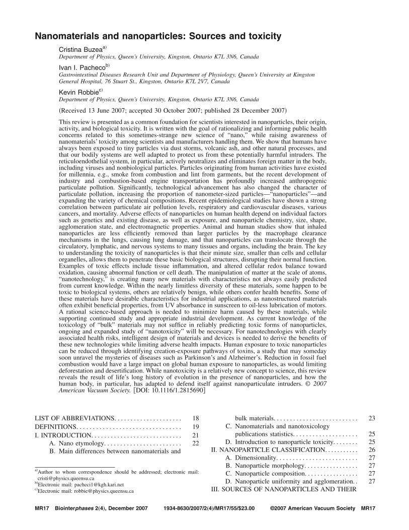
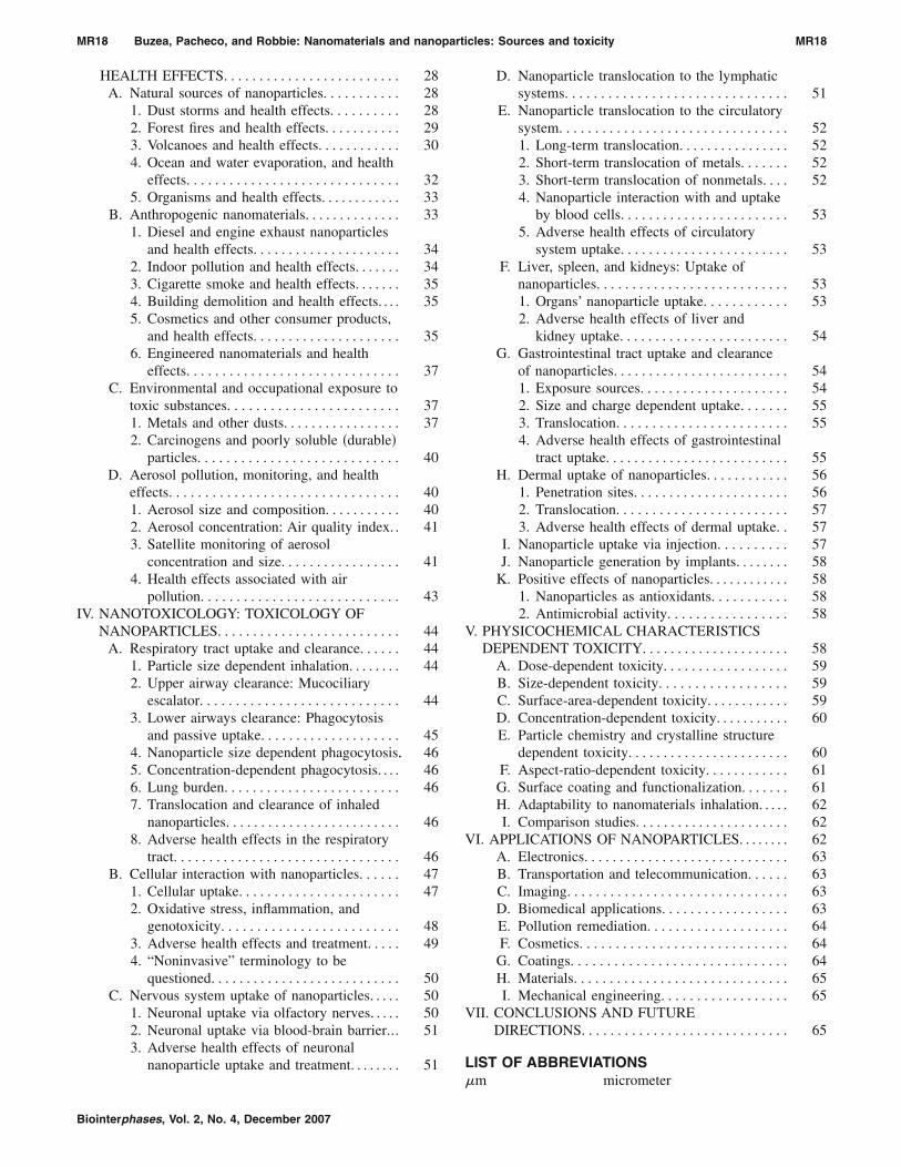
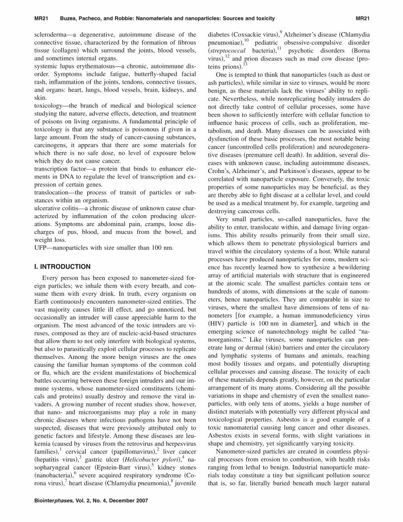
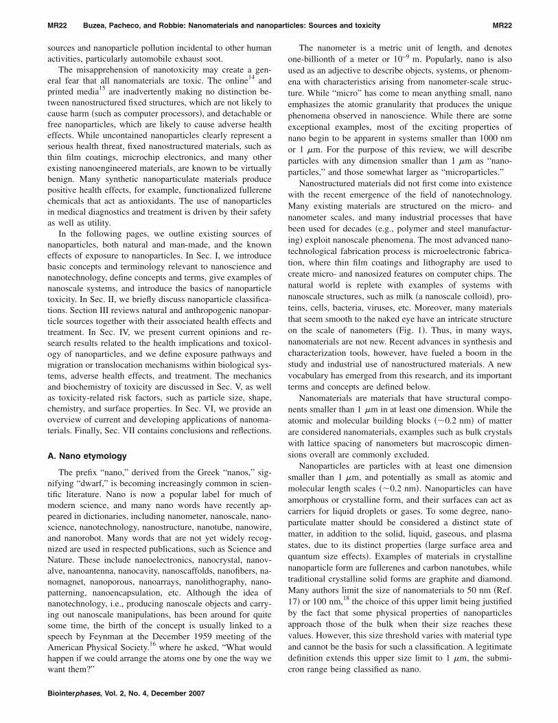
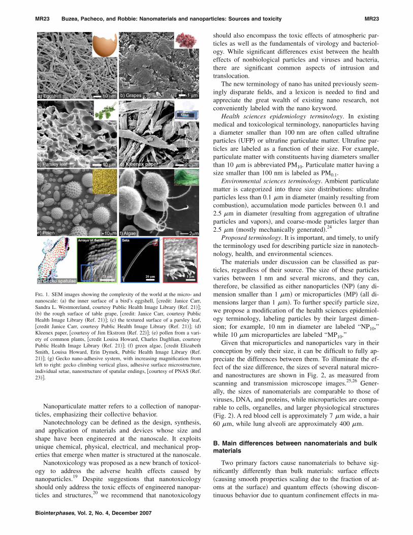
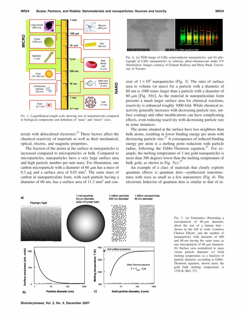
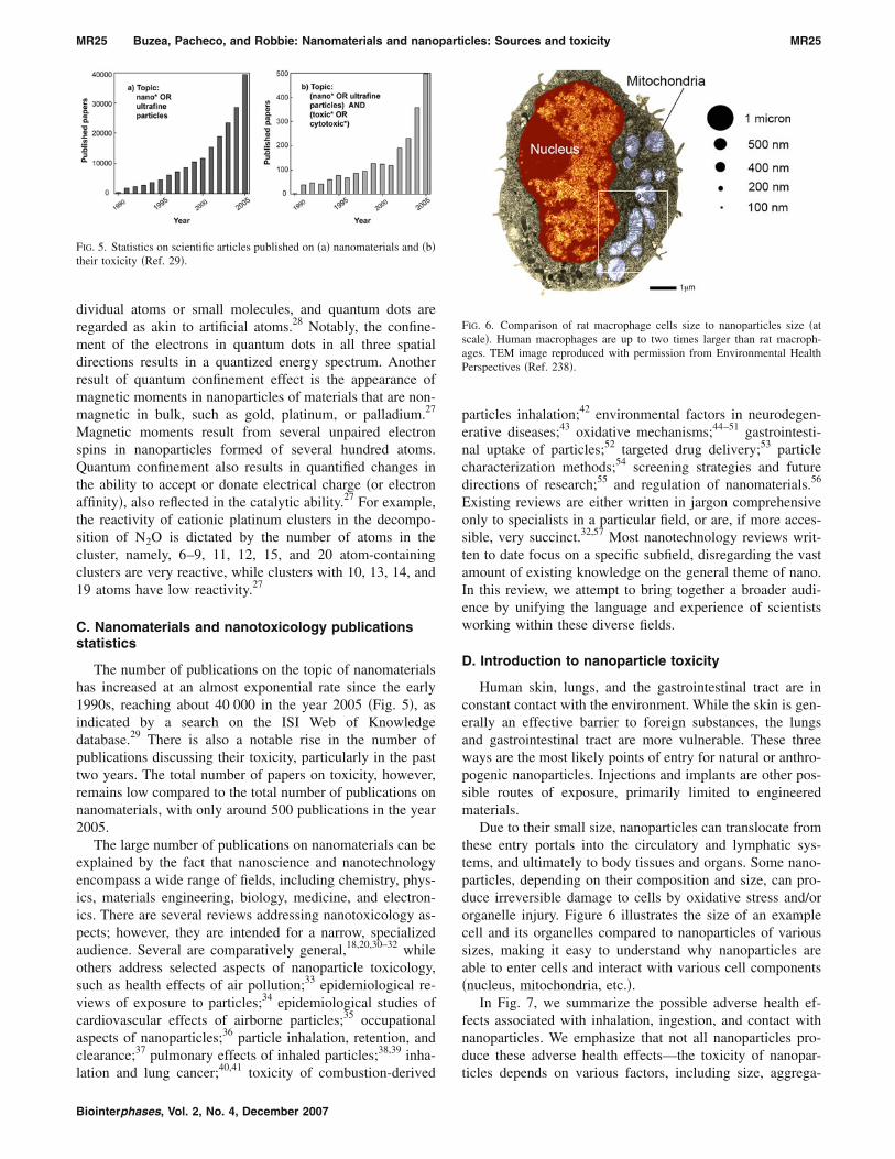
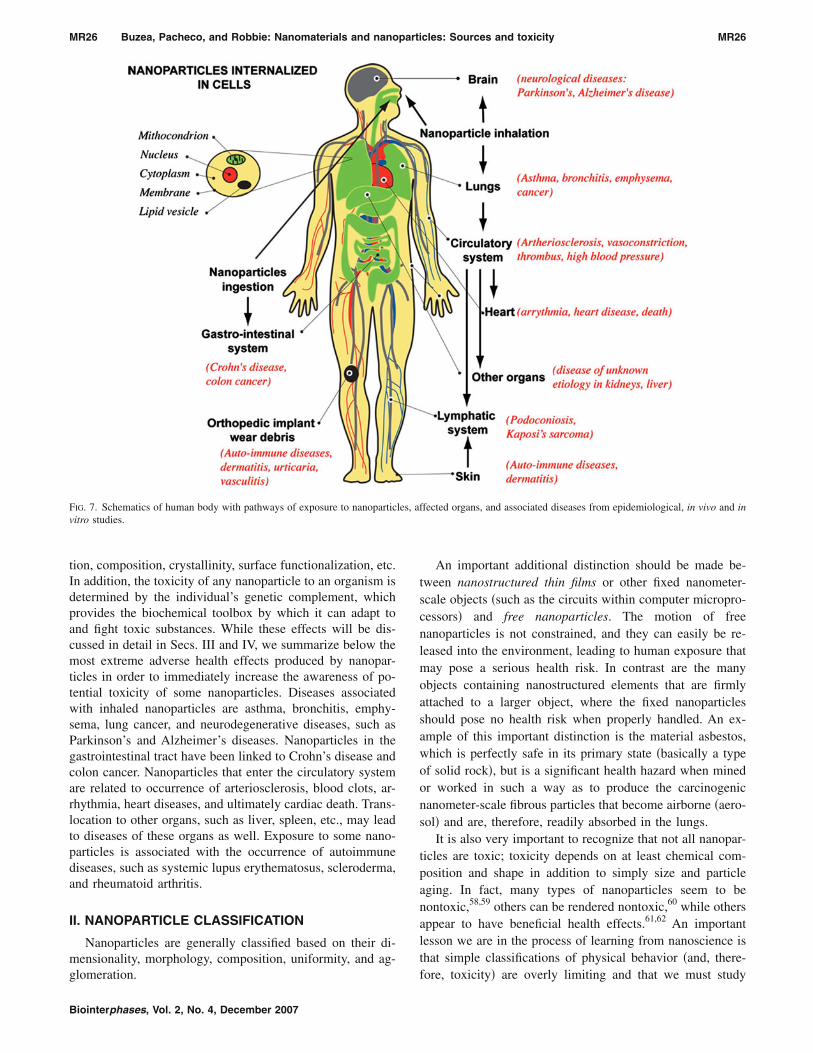
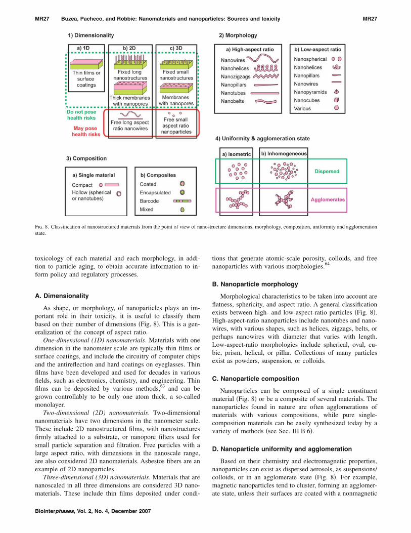
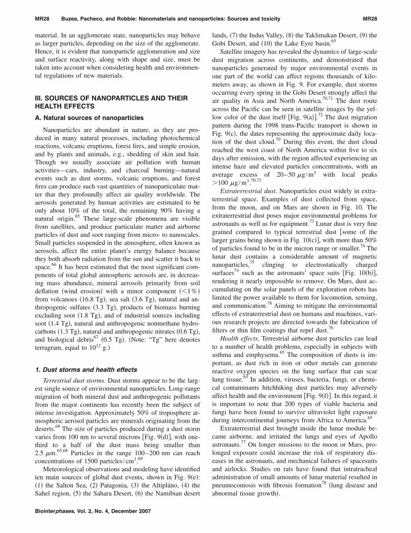
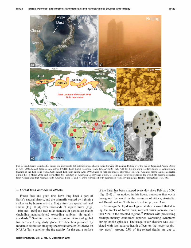
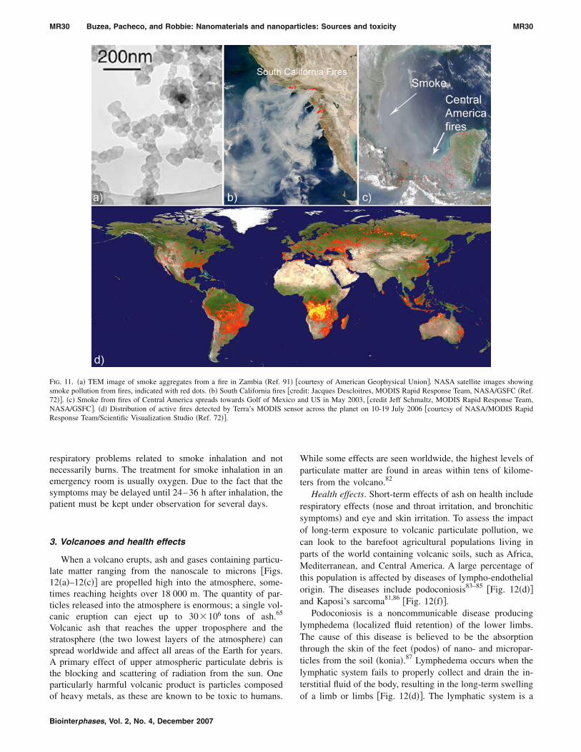
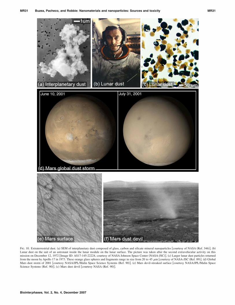
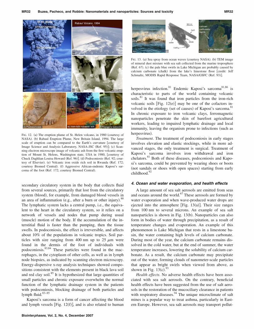
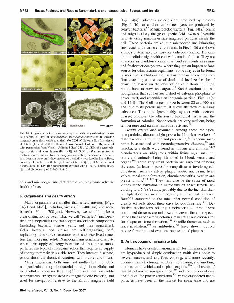
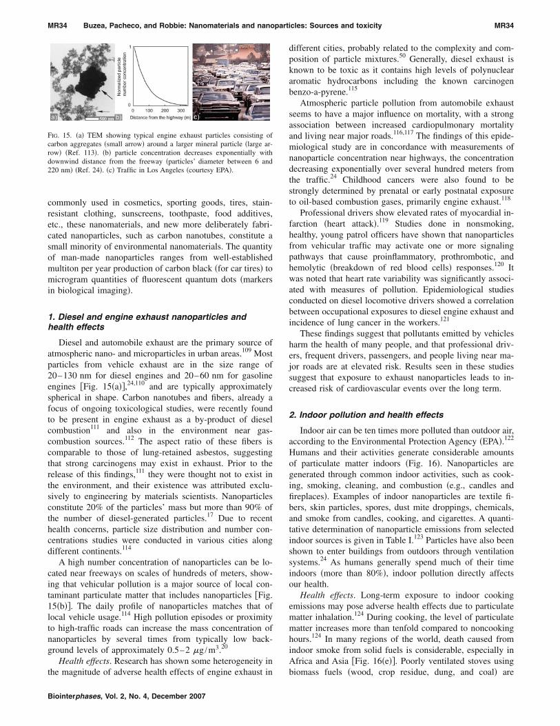
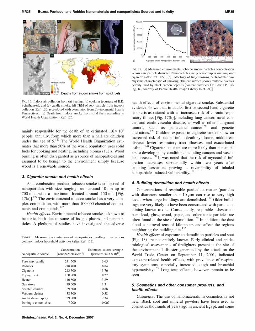
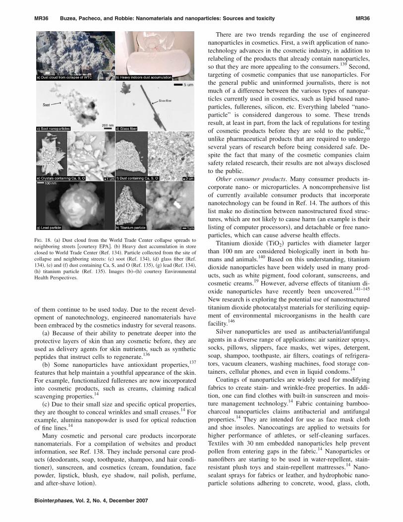
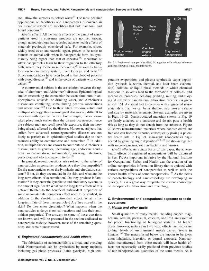
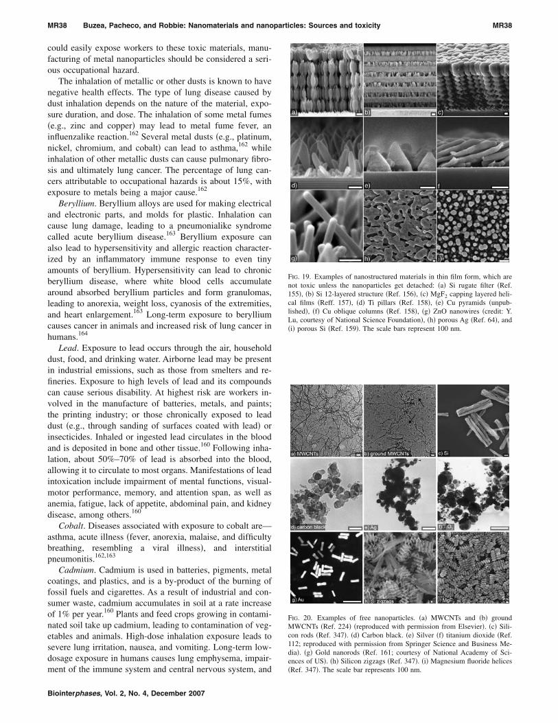
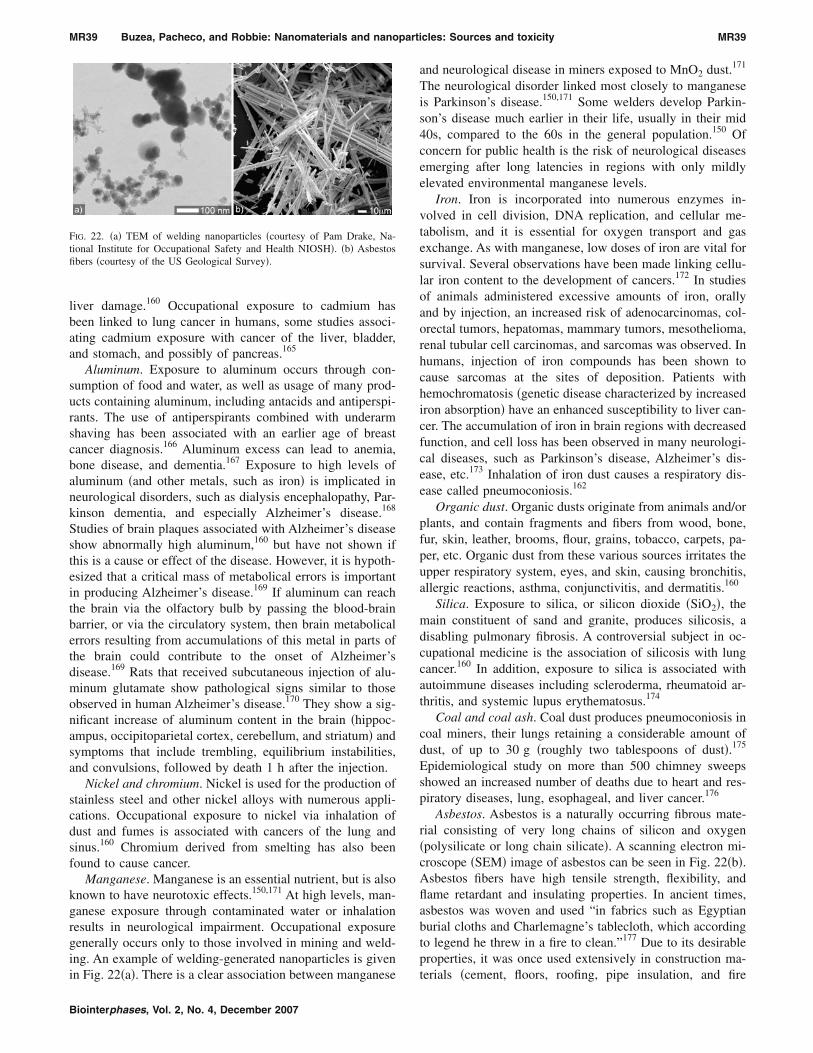
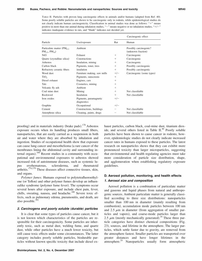
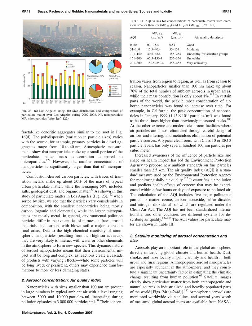
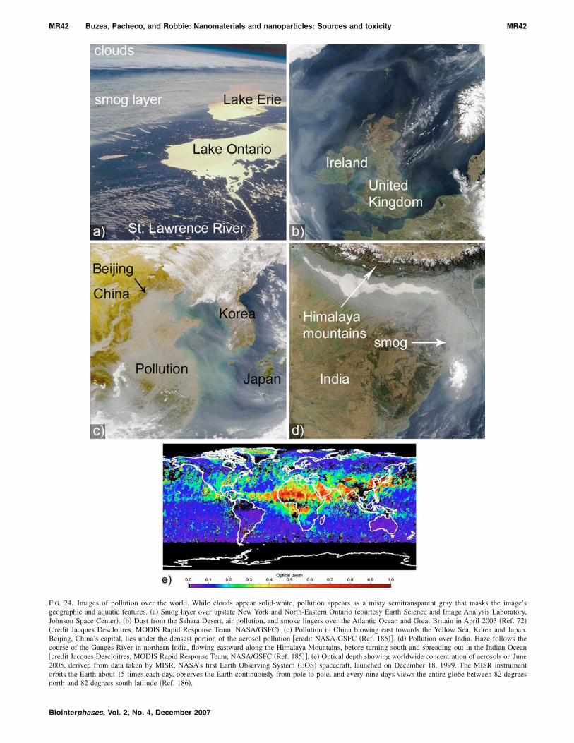
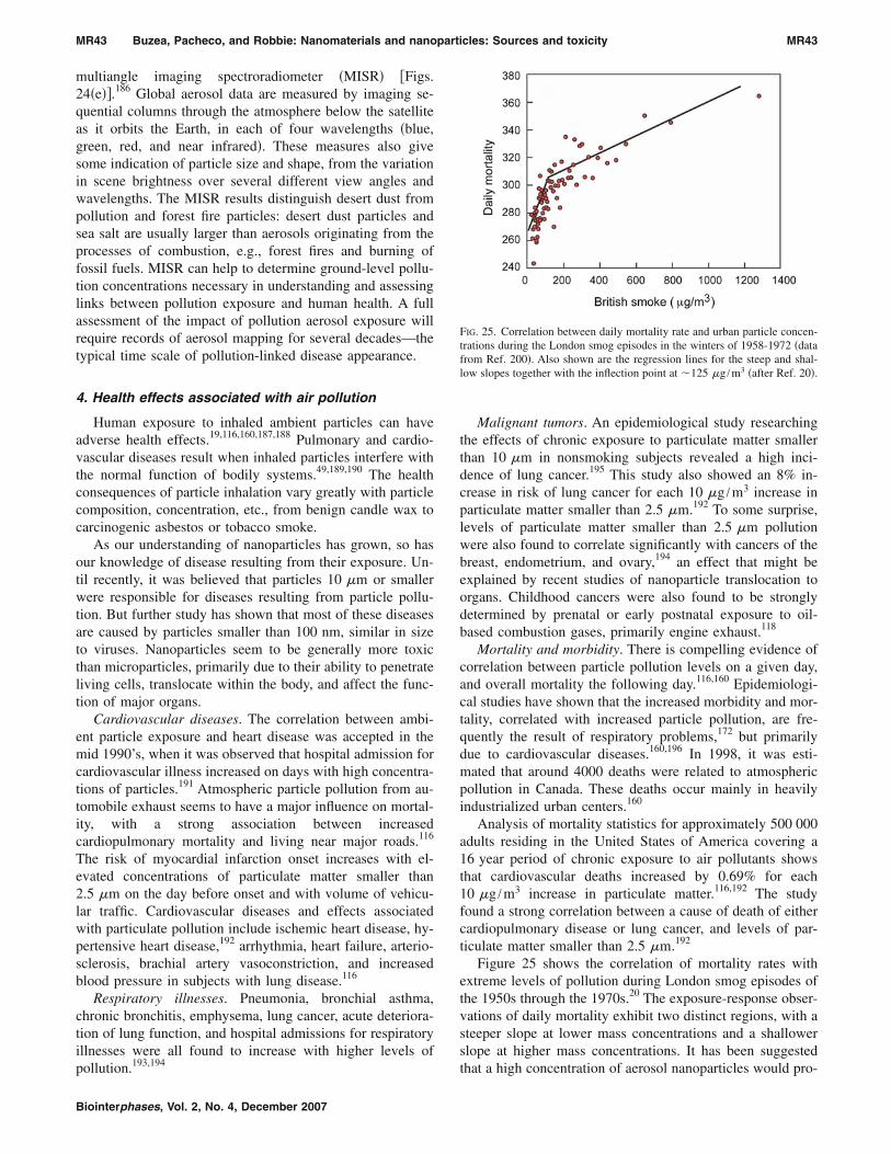
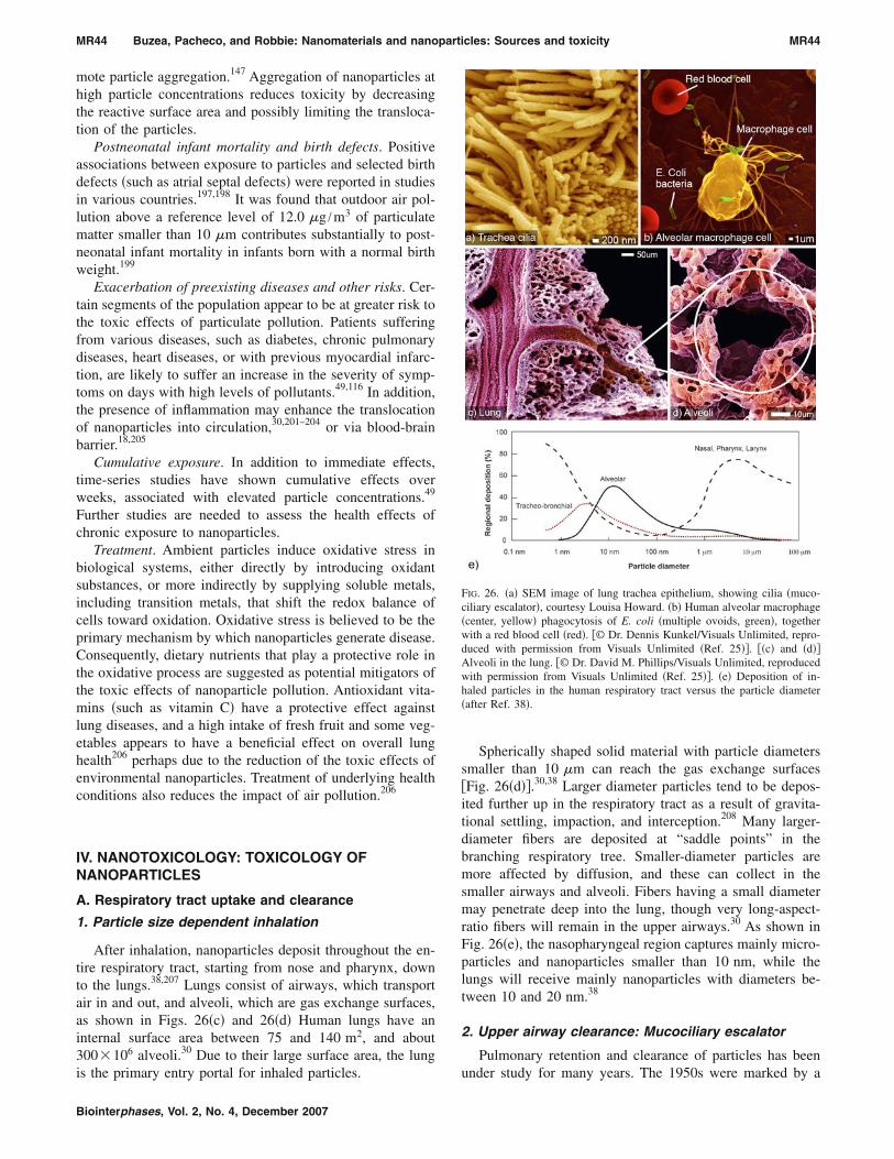
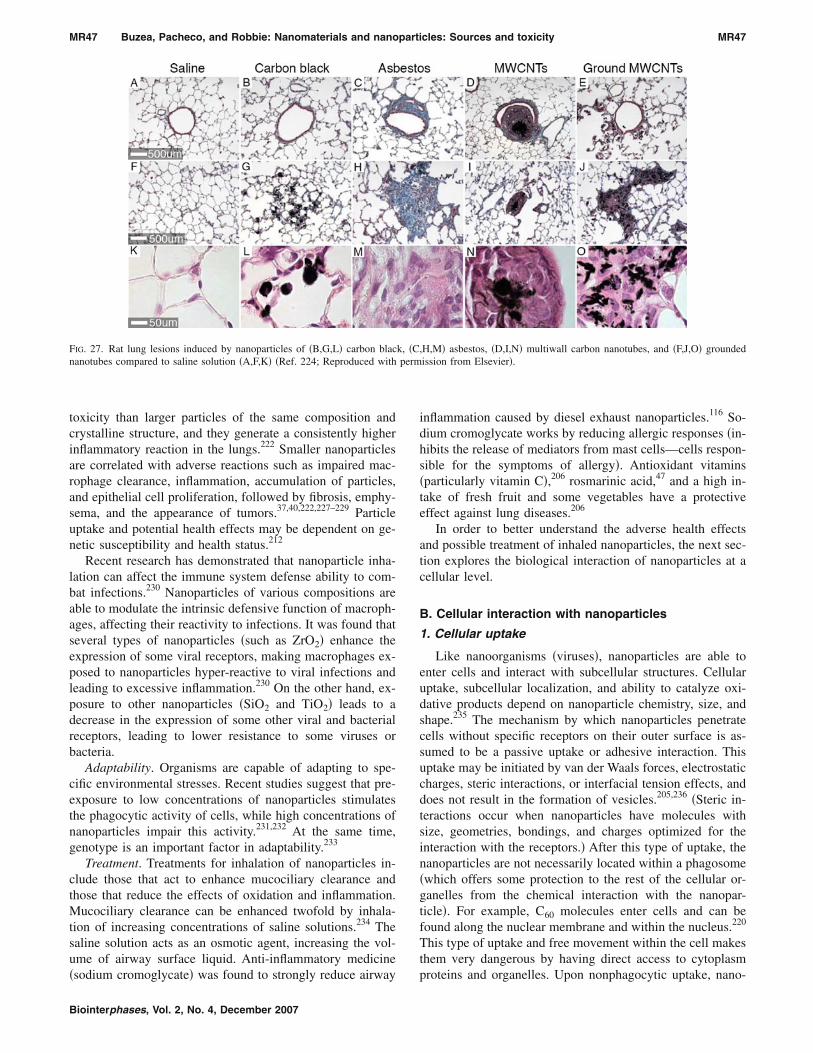
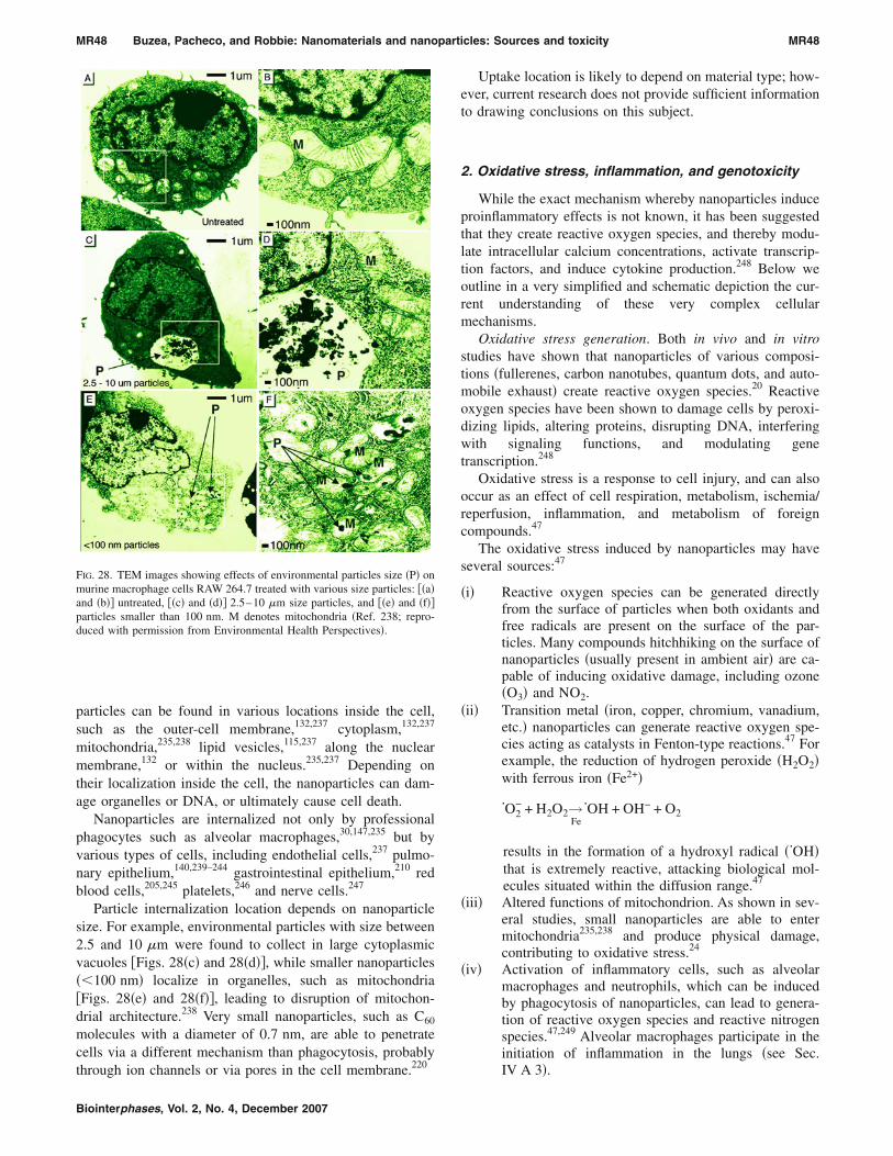
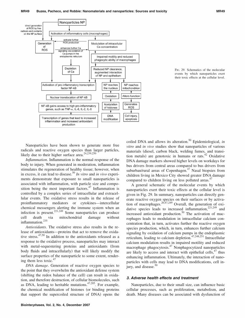
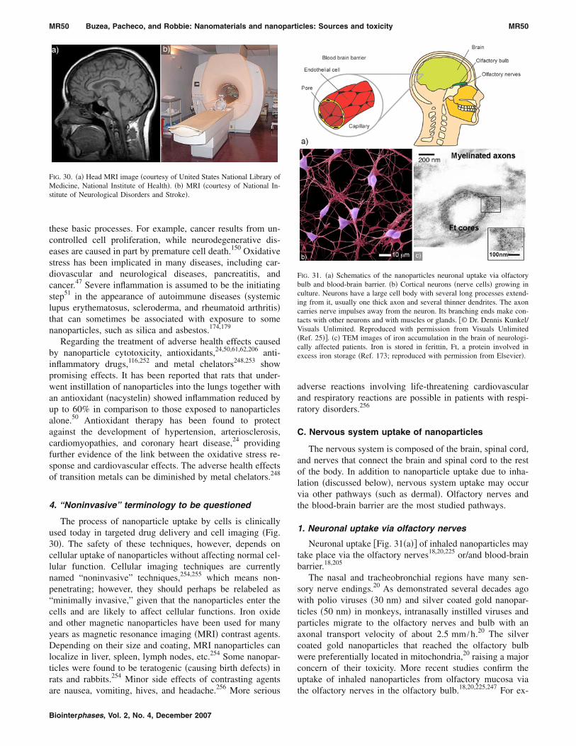
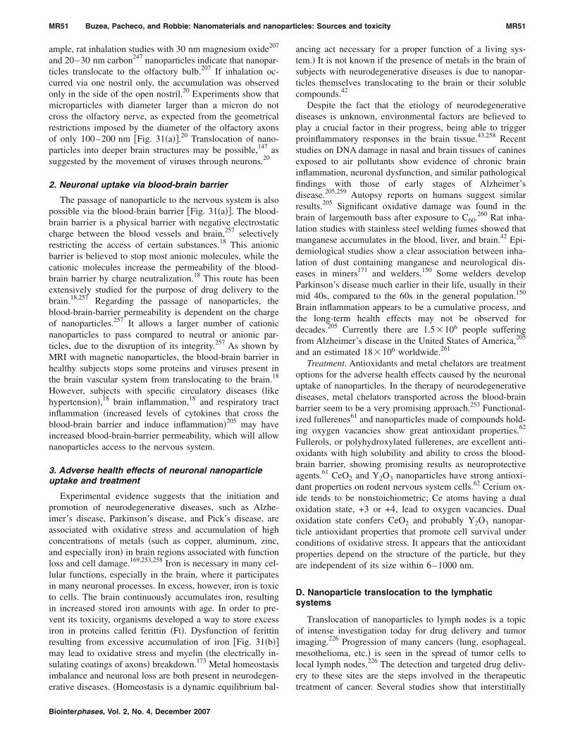
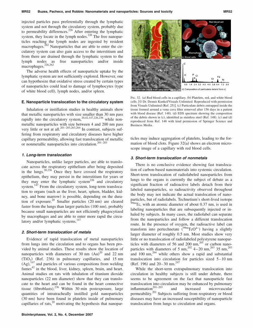
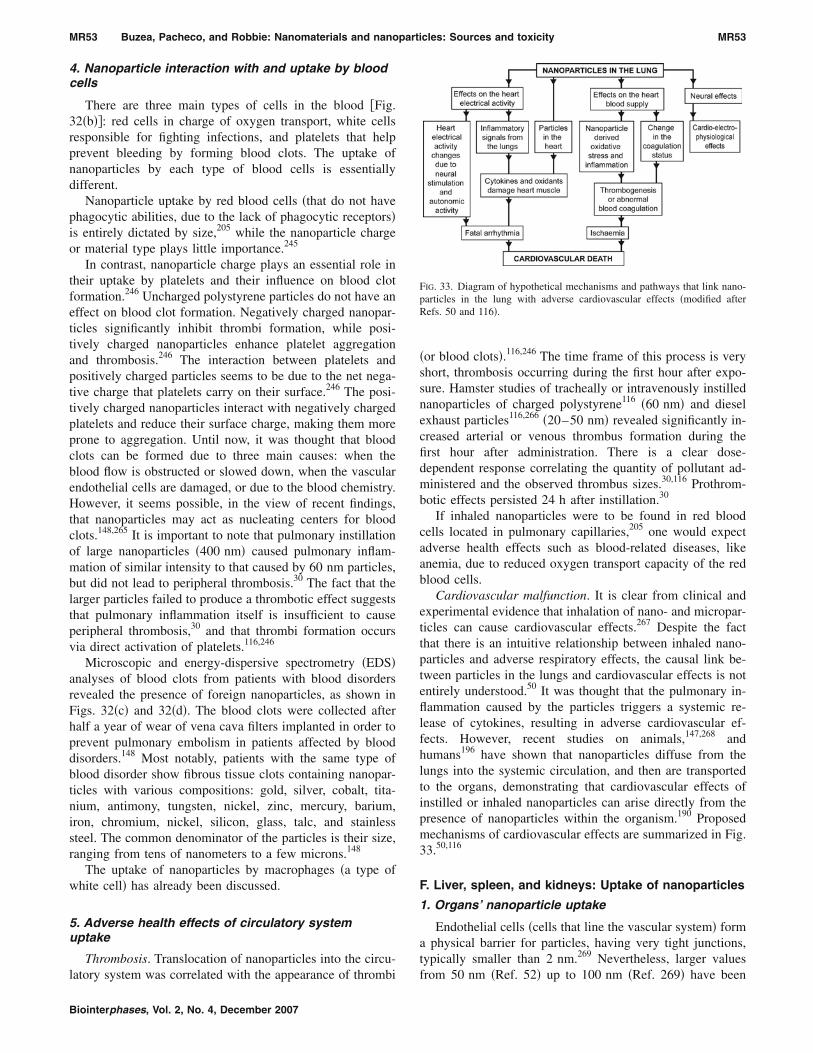
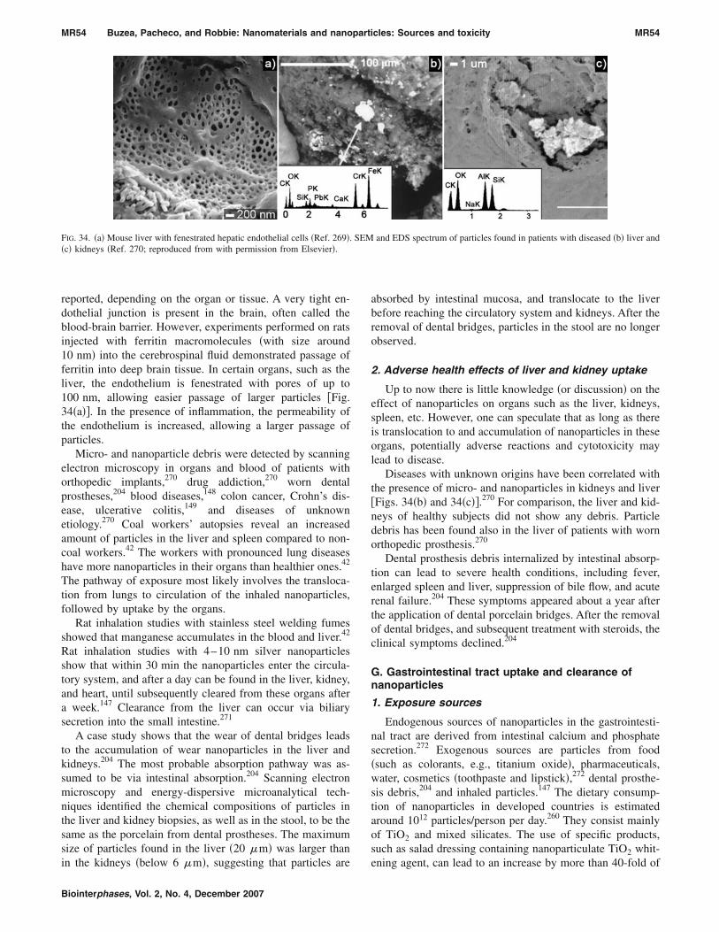
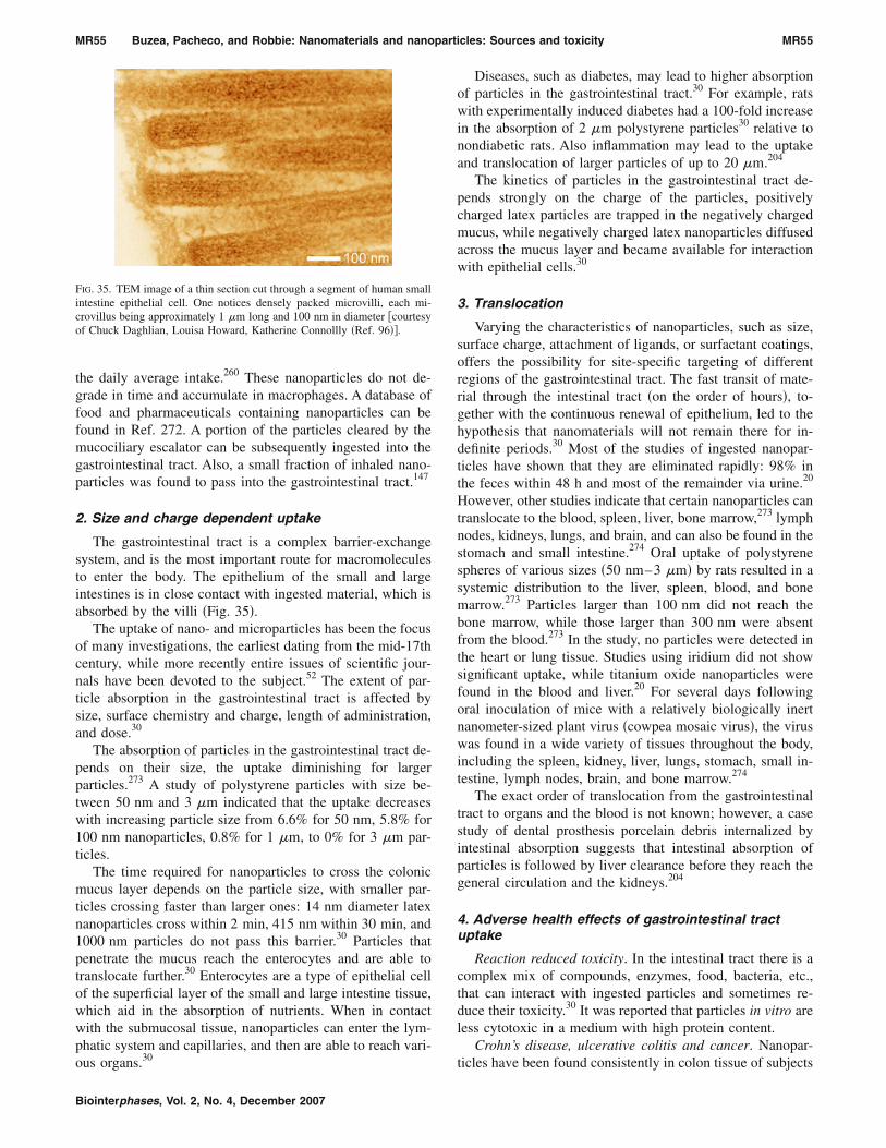
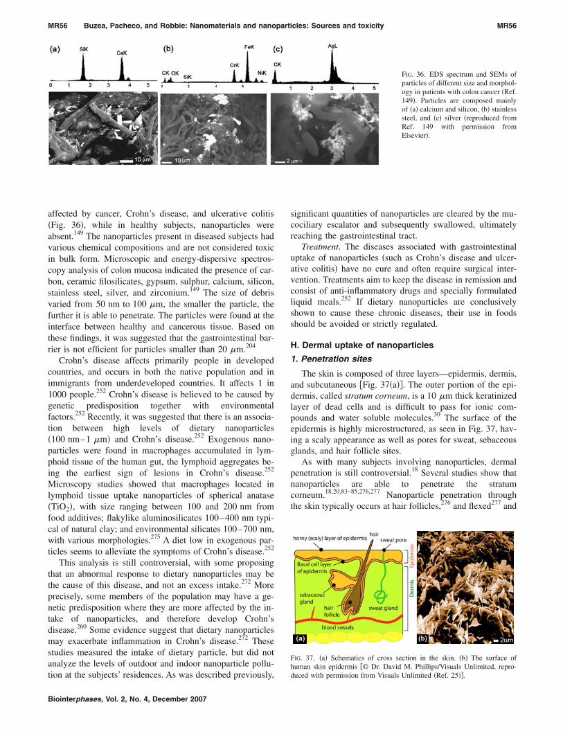
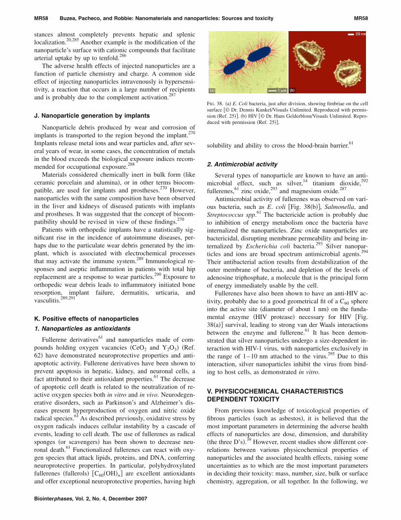
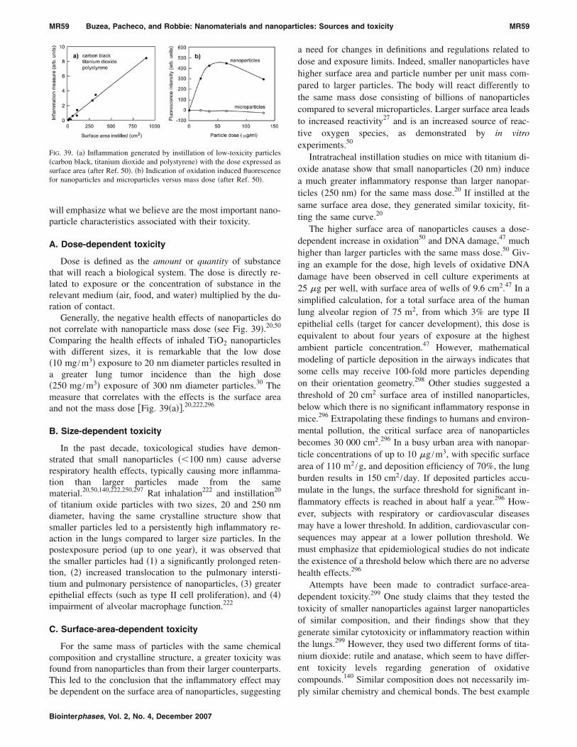
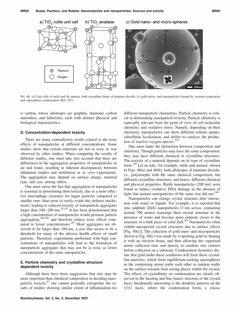
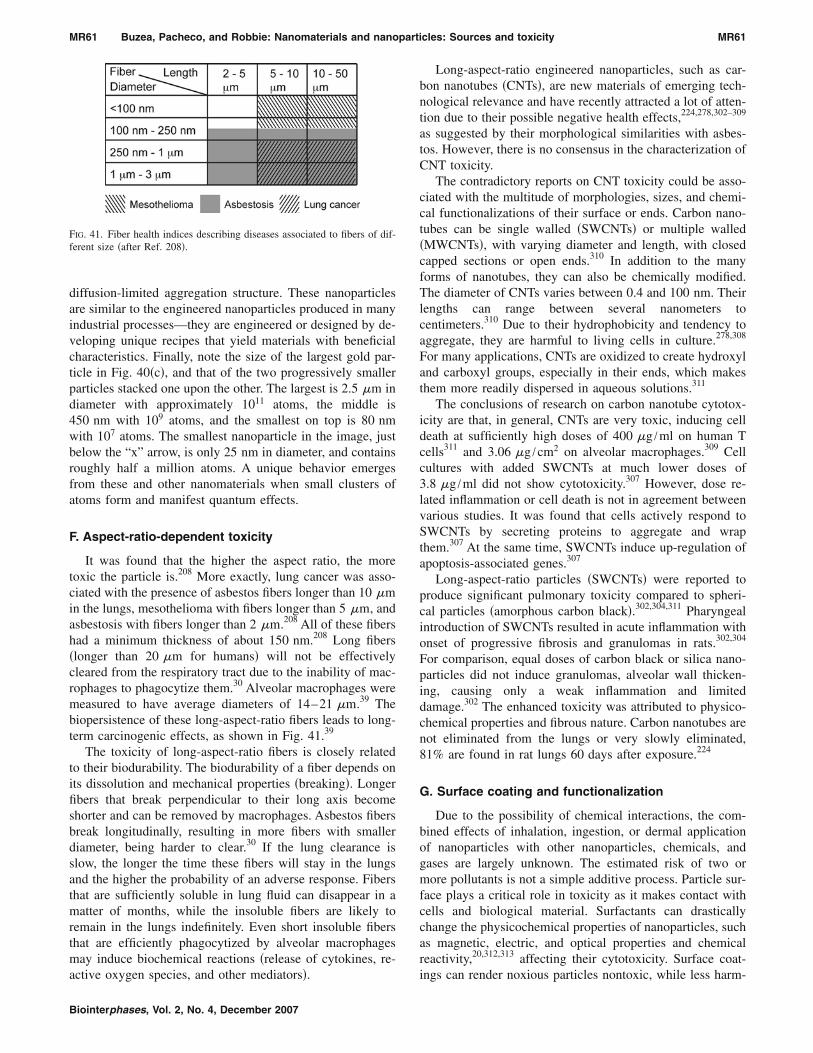
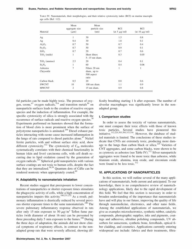


![Targeted Nanomaterials for Phototherapy · upconversion nanoparticles (UCNPs) [47, 48], gold nanoparticles [49, 50], mesoporous silica nanoparticles [51, 52], and carbon nanomaterials](https://static.documents.pub/doc/80x56/5f71f70b5cd47d2b1b7523e3/targeted-nanomaterials-for-phototherapy-upconversion-nanoparticles-ucnps-47.jpg)


