Nanometal-Glass Hybrid Nanocomposites: Synthesis, Properties and Applications Basudeb Karmakar * , Tirtha Som, Shiv Prakash Singh and Mithun Nath Glass Science and Technology Section, Central Glass and Ceramic Research Institute (Council of Scientific & Industrial Research, CSIR, India), Kolkata 700032, India In recent past research on nanometal-glass hybrid composites has been the centre of attraction across the globe particularly in the area of nanoscience and for the future nanotechnology. In this review, with a short historical background, its preparation by various multi-step techniques, properties and applications are briefly described. In addition, recently developed single-step in-situ thermochemical reduction methodology by these authors for synthesis of various nanometal-glass hybrid nanocomposites are described in details with their significant characteristic properties, relevant theories and applications. Here Au, Ag, and Bi metals are considered and the synthesized glasses are mostly based on antimony, bismuth and phosphorus oxides. Some of them are dichroic in nature, that is, they exhibit blue to green colourations in transmitted light and brown to reddish brown colourations in reflected light. The appropriate reasons for their dichroic character are still remained unsolved. Nanometal-antimony oxide glass nanocomposites have been found to enhance the photoluminescence upconversion intensities up to 11 fold when co-doped with rare-earth (RE) ions due to the plasmonic induced local field as enhanced by the effects of doped metal nanoparticles. The nanometal-glass hybrid nanocomposites, therefore, seem to be very promising for various nanophotonic applications (such as nanometal enhanced rare-earth luminescence, solar cell, light emitting diode, plasmonic integrated circuit, plasmon slot waveguide, etc.) that have presently emerged into a major field called ‘plasmonics’. [Keywords: Nanometal, Glass, Nanocomposites, Single-step synthesis, SPR band, Plasmonics, Enhanced photoluminescence, Nanophotonics] ______________________________________________________________________ * Corresponding author, e-mail: [email protected]
Transcript
Nanometal-Glass Hybrid Nanocomposites: Synthesis, Properties and Applications
Basudeb Karmakar*, Tirtha Som, Shiv Prakash Singh and Mithun Nath
Glass Science and Technology Section, Central Glass and Ceramic Research Institute (Council of Scientific & Industrial Research, CSIR, India), Kolkata 700032, India
In recent past research on nanometal-glass hybrid composites has been the
centre of attraction across the globe particularly in the area of nanoscience and
for the future nanotechnology. In this review, with a short historical background,
its preparation by various multi-step techniques, properties and applications are
briefly described. In addition, recently developed single-step in-situ
thermochemical reduction methodology by these authors for synthesis of various
nanometal-glass hybrid nanocomposites are described in details with their
significant characteristic properties, relevant theories and applications. Here Au,
Ag, and Bi metals are considered and the synthesized glasses are mostly based
on antimony, bismuth and phosphorus oxides. Some of them are dichroic in
nature, that is, they exhibit blue to green colourations in transmitted light and
brown to reddish brown colourations in reflected light. The appropriate reasons
for their dichroic character are still remained unsolved. Nanometal-antimony
oxide glass nanocomposites have been found to enhance the photoluminescence
upconversion intensities up to 11 fold when co-doped with rare-earth (RE) ions
due to the plasmonic induced local field as enhanced by the effects of doped
metal nanoparticles. The nanometal-glass hybrid nanocomposites, therefore,
seem to be very promising for various nanophotonic applications (such as
nanometal enhanced rare-earth luminescence, solar cell, light emitting diode,
plasmonic integrated circuit, plasmon slot waveguide, etc.) that have presently
However, for real application in nanophotonic devices, a major goal is to produce stable
metal nanoclusters within suitable encapsulating hosts. It is well known that glasses possess
several incredible properties like high transparency, ease of fabrication in desirable shapes and
sizes, high durability and inertness, prevention of air oxidation of nanometals, low cost, ability of
tailoring of properties, absence of high energy bond vibrations, etc. Besides these, they are most
important from basic viewpoints. These have made glasses not only promising encapsulating
hosts for lasing RE3+
ions but also for metal NPs. Consequently, nanometal- RE3+
-glass hybrid
nanocomposites have created a center of enormous attention and many investigations are
currently in progress.18, 19
Historical Background
Curiosity in the optical properties of colloidal metals in glass dates back to the Roman
Era (753 BC to 476 AD). Although at that time there was no understanding of the underlying
mechanisms which give rise to the spectacular colors of small metal particles but nanosized
coinage metal (e.g., Au, Ag and Cu) particles were used as colorants in the stained glass
windows of Cathedrals (red colored ruby glass), to color ceramics, enamel pottery, aesthetic
4
items, ornaments, etc. Sometimes they were also used to create complex optical effects such as
dichroism. Perhaps the most famous example is the ‘Lycurgus Cup’ that was manufactured
around 400 AD.20
The cup can still be seen in the British Museum and possesses the unique
feature of changing color depending upon the light in which it is viewed. It is ruby red in
transmitted light and green in reflected light. Analysis of the glass reveals that it contains a very
small amount of tiny Au and Ag nanocrystals (~70 nm).19, 20
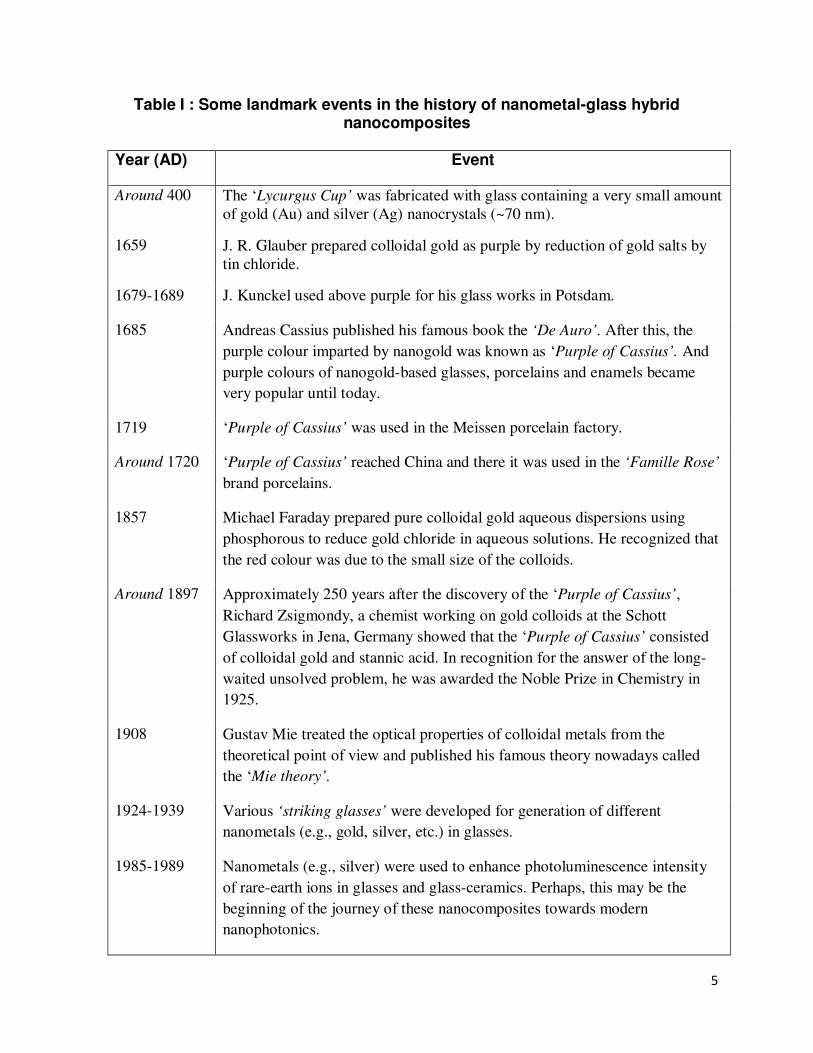
Some landmark events in the history
of nanometal-glass hybrid nanocomposites are listed in Table I.
In 1659, Johann Rudolf Glauber prepared colloidal gold as purple by reduction of gold
salts by tin chloride in Germany. During 1679-1689, Johann Kunckel21
used this purple gold for
his glass works in Potsdam. In 1685, Andreas Cassius published the book ‘De Auro’
incorporating the gold-based glass and enamel colours. After then it is known as ‘Purple of
Cassius’. It was used in Meissen porcelain factory from and onward 1719. Around 1720, the
‘Purple of Cassius’ reached China and was used in ‘Famille Rose’ porcelain. The ‘Purple of
Cassius’ became, until today, the most popular enamel colour for pottery and porcelain.
However, its chemical nature was a challenge for the scientists of the 19th
century. In 1857,
Michael Faraday prepared pure colloidal gold using phosphorous to reduce gold chloride. He
recognized that the colour was due to the small size of the colloids. Around 1897, nearly 250
years after the discovery of the ‘Purple of Cassius’, Richard Zsigmondy, a chemist working on
gold at the Schott Glassworks in Jena, Germany showed that the ‘Purple of Cassius’ consisted of
colloidal gold and stannic acid. In recognition, he was awarded the Noble Prize in Chemistry in
1925. Various ‘striking glasses’ were developed during 1924-1939 for generation of nanometals
in glasses.22
However, the advent of modern characterization facilities like transmission electron
microscopy (TEM was discovered in 1931) and scanning tunneling microscopy (STEM was
discovered in 1938 but successfully re-developed for analysis of materials only in 1993)
techniques, and introduction of quantum electrodynamics theories (Mie theory, Drude model,
Maxwell Garnet Theory, Gans theory, etc) have directed research of metal-glass hybrid
nanocomposites along with other branches of nanoscience and nanotechnology towards a new
dimension23,24
. Currently nanometal-glass hybrid nanocomposite is an interesting emerging area
of research.25-28
5
Table I : Some landmark events in the history of nanometal-glass hybrid nanocomposites
Year (AD) Event
Around 400 The ‘Lycurgus Cup’ was fabricated with glass containing a very small amount
of gold (Au) and silver (Ag) nanocrystals (~70 nm).
1659 J. R. Glauber prepared colloidal gold as purple by reduction of gold salts by
tin chloride.
1679-1689 J. Kunckel used above purple for his glass works in Potsdam.
1685 Andreas Cassius published his famous book the ‘De Auro’. After this, the
purple colour imparted by nanogold was known as ‘Purple of Cassius’. And
purple colours of nanogold-based glasses, porcelains and enamels became
very popular until today.
1719 ‘Purple of Cassius’ was used in the Meissen porcelain factory.
Around 1720 ‘Purple of Cassius’ reached China and there it was used in the ‘Famille Rose’
brand porcelains.
1857 Michael Faraday prepared pure colloidal gold aqueous dispersions using
phosphorous to reduce gold chloride in aqueous solutions. He recognized that
the red colour was due to the small size of the colloids.
Around 1897 Approximately 250 years after the discovery of the ‘Purple of Cassius’,
Richard Zsigmondy, a chemist working on gold colloids at the Schott
Glassworks in Jena, Germany showed that the ‘Purple of Cassius’ consisted
of colloidal gold and stannic acid. In recognition for the answer of the long-
waited unsolved problem, he was awarded the Noble Prize in Chemistry in
1925.
1908 Gustav Mie treated the optical properties of colloidal metals from the
theoretical point of view and published his famous theory nowadays called
the ‘Mie theory’.
1924-1939 Various ‘striking glasses’ were developed for generation of different
nanometals (e.g., gold, silver, etc.) in glasses.
1985-1989 Nanometals (e.g., silver) were used to enhance photoluminescence intensity
of rare-earth ions in glasses and glass-ceramics. Perhaps, this may be the
beginning of the journey of these nanocomposites towards modern
nanophotonics.
6
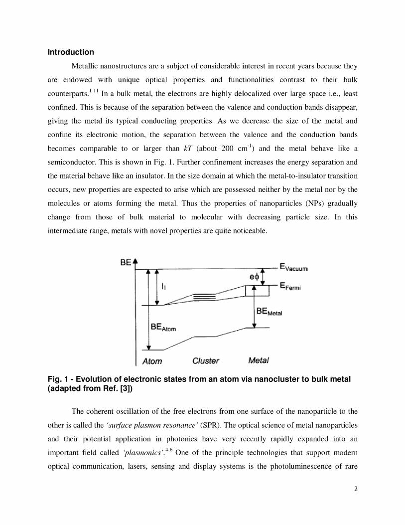
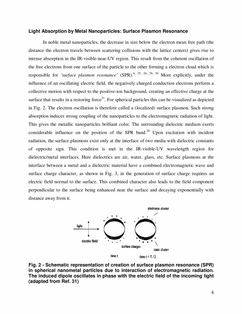
Light Absorption by Metal Nanoparticles: Surface Plasmon Resonance
In noble metal nanoparticles, the decrease in size below the electron mean free path (the
distance the electron travels between scattering collisions with the lattice centers) gives rise to
intense absorption in the IR-visible-near-UV region. This result from the coherent oscillation of
the free electrons from one surface of the particle to the other forming a electron cloud which is
responsible for ‘surface plasmon resonance’ (SPR).6, 25, 26, 29, 30
More explicitly, under the
influence of an oscillating electric field, the negatively charged conduction electrons perform a
collective motion with respect to the positive-ion background, creating an effective charge at the
surface that results in a restoring force31
. For spherical particles this can be visualized as depicted
in Fig. 2. The electron oscillation is therefore called a (localized) surface plasmon. Such strong
absorption induces strong coupling of the nanoparticles to the electromagnetic radiation of light.
This gives the metallic nanoparticles brilliant color. The surrounding dielectric medium exerts
considerable influence on the position of the SPR band.28
Upon excitation with incident
radiation, the surface plasmons exist only at the interface of two media with dielectric constants
of opposite sign. This condition is met in the IR-visible-UV wavelength region for
dielectric/metal interfaces. Here dielectrics are air, water, glass, etc. Surface plasmons at the
interface between a metal and a dielectric material have a combined electromagnetic wave and
surface charge character, as shown in Fig. 3, in the generation of surface charge requires an
electric field normal to the surface. This combined character also leads to the field component
perpendicular to the surface being enhanced near the surface and decaying exponentially with
distance away from it.
Fig. 2 - Schematic representation of creation of surface plasmon resonance (SPR) in spherical nanometal particles due to interaction of electromagnetic radiation. The induced dipole oscillates in phase with the electric field of the incoming light (adapted from Ref. 31)
7
Fig. 3 - Surface plasmons at the interface between a metal and a dielectric material (adapted from Ref. 32)
Factors affecting Surface Plasmon Resonance
The frequency and intensity of the surface plasmon resonance (SPR) absorption bands are
distinctive of the type of material (metal).32
Metals only with free electrons (essentially the
coinage metals Au, Ag and Cu, and the alkali metals) possess plasmon resonances in the visible
spectrum, which give rise to such intense colors. For many metals like Pb, In, Hg, Sn and Cd the
plasma frequency is in the UV; so no color effects are observed.32
Since coinage metal (Au, Ag
and Cu) nanoparticles and others like Bi can be easily detected by UV-Vis absorption and this is
one of the reasons why they are more studied. These resonances are determined four factors: the
density of electrons, the effective electron mass, and the shape and size of the charge
distribution. In addition, the surrounding dielectric medium also exerts considerable influence on
the position of the SPR band. Thus the optical effects in metallic nanostructures results from
electrodynamics effects and from modifications of the dielectric environment.
Thus, the physical basis of light absorption by metallic nanoparticles or the intriguing
optical properties of metal nanoparticles is due to localized SPR near the boundary between the
metal nanostructures and the surrounding (dielectric) matrix. The surface plasmons exist only at
the interface of two media with dielectric constants of opposite sign. This condition is met in the
IR-visible wavelength region for air/metal, water/metal and glass/metal interfaces (where the
frequency-dependent dielectric constant of a nano metal, ε(ω), is negative and that of air or water
(medium), εm, is positive).33
The SPR bands are highly susceptible to the changes in sizes, spatial distribution and
geometry of the nanostructures, particle density, inter-particle distance as well as the surrounding
8
environment.34-36
Accordingly, the optical properties of metal nanoparticles can be controlled by
adjusting these parameters. Generally, a red-shift of the absorption maxima is induced by
increasing particle size, number density and increasing refractive index of the host matrix.34-36
But in some cases, the blue-shifting (decrease in wavelength) of the SPR bands is also observed
particularly for nanoparticles having diameter less than 20 nm.35-38
This anomaly from the
general behavior is due to electromagnetic field enhancement, called the dielectric confinement
of the medium and quantum confinement (decrease in electron density) which is the direct
consequence of the wave nature of the electrons. The shape of the plasmon band also changes
with particle shape. In case of the small spherical particles a single plasmon band due to dipolar
resonance is observed due to dipole plasmon resonance (sometimes denoted “dipole particle
plasmon resonance” to distinguish from plasmon excitation that can occur in bulk metal or metal
surfaces). But elongated nanoparticles may display two distinct plasmon bands related to
transverse and longitudinal electron oscillations. Elongated particles may show two maxima if
the aspect ratio is ≥ 4. The longitudinal resonance signal in case of gold, with an aspect ratio of
4, shifts from 520 to 770 nm.39, 40
The longitudinal oscillation is very sensitive to the aspect ratio
of the particles, so that slight deviations from spherical geometry can lead to impressive color
changes.
The SPR peak of non-spherical metal NPs is generally red-shifted compared to spherical
ones.2, 5
As the size increases, the field across the particle become non-uniform, and this phase
retardation broadens the dipole resonance and excites higher multipole resonances, such as the
quadrupole, octupole, etc. leading to several peaks in the spectra.40, 41
Higher modes of plasmon
excitation can occur, such as the quadrupole mode where half of the electron cloud moves
parallel to the applied field and half moves antiparallel. It is apparent that the dipole maximum
rapidly shifts to longer wavelengths as the particle size increases beyond 70 nm (450 nm spectral
maximum) revealing the quadrupole peak at about 420 nm. The observed spectral shift results
from the ‘spreading’ of the particle’s surface charge over a larger surface area so that the
surrounding medium better compensates the restoring force thus slowing the electron
oscillations.40, 41
Several processes can damp the plasmon oscillations, such as electron scattering by
lattice phonon modes, inelastic electron–electron interactions, scattering of the electrons at the
particle surface, and excitation of bound electrons into the conduction band (interband
9
transitions).40
Interband transitions can cause a substantially decreased efficiency of plasmon
excitation, as is the case for Au and Cu, where there is significant overlap between the interband
absorption edge and the plasmon resonance. For Ag, however, the absorption edge is in the UV
(ca. 320 nm) and has little impact on the SPRs, which appear at wavelengths larger than 370 nm,
accounting for the fact that excitation of the SPR in Ag particles is more efficient than for Au
and Cu.41
The bi-SPR peaks can also be generated due to the bi-modal distribution of metal
nanoparticles.42
Broadening of the SPR band is also observed for arbitrary shaped particles.43
Advantages of Glasses as Encapsulating Hosts
Although during the last two decades there has been an astounding progress in the wet-
chemical synthesis of diverse metal particles of different shapes and sizes to understand their
fundamental general properties, functionalities and modeling their optical responses4, 7, 10, 44-47
but
realization of the above emerging applications of plasmonic nanoparticles in active and real
functional devices requires their synthesis/insertion within solid-state preferably transparent
environment. Besides, metal nanoparticles must be embedded in solid dielectrics in order to
avoid aggregation and the formation of the thermodynamically favored bulk material.
Exploitation of glasses as encapsulating hosts for plasmonic metal nanoparticles (fabrication of
metallo-dielectric nanocomposites) provides an opportunity to create a breed of nanoscale
devices with attractive properties often due to the amalgamation of the properties of the glass
host and the nano metal.
Glasses present some superior inherent advantages over other dielectrics. High
transparency, mechanical strength, ease of fabrication in desirable shapes and sizes, ability to
withstand high intensity radiation, preventing air oxidation of metal nanoparticles, etc. make
glasses excellent encapsulating hosts for metal NPs for practical applications.48-51
Glasses
containing nanosized metal NPs are increasingly being appreciated for their potential
application48-53
as optical data recording disks and memory devices, optical waveguides, optical
switches based on their nonlinear optical properties, photochromatic and color glass recycling
industry, three-dimensional multicolored industrial art objects, etc. In all these applications the
size, shape, number density, and distribution of the nanoparticles critically determine
performance and properties of the nanocomposites.48, 54-61
Such metal-glass nanocomposites also
find other applications like solid-state lasers, sensors, dichroic polarizers, colored glasses,
10
ophthalmic lenses, display devices and optoelectronic materials due to nonlinear optical
properties.48-61
Since glasses are also promising hosts for encapsulating rare-earth (RE3+
) ions,
consequently, an emerging application of metal-glass nanocomposites in the field of plasmonics
is their development as substrates (hosts) capable of providing large electromagnetic
enhancements or ‘hot spots’ formation for nanometal-enhanced luminescence of RE3+
ions.
Multi-Step Synthesis of Nanometal-Glass Nanocomposites
Pioneering works on preparation of metal-glass nanocomposites and evaluation of their
optical properties were performed by Doremus.54, 55
Thereafter the metal-dielectric (glass)
nanocomposites kept receiving significant exposures and are currently occupy a significant area
of material science and nanotechnology.48-53, 56-64
Preparation of nano metal doped conventional
glass systems are not simple and demand multi-step techniques like sol-gel process, metal-
dielectric co-sputtering deposition, direct metal-ion implantation, radio-frequency magneton
sputtering, pulsed laser deposition, ion-exchange of thin plates followed by long time heat
treatment at high temperatures in reducing (hydrogen) atmosphere or UV-light/X-ray/60
Co γ-
radiation or high energy laser/synchrotron irradiation.48-64
However, the above customary techniques face some serious disadvantages. Firstly, only
thin glass plates doped with metal nanoparticles can be obtained. Particularly the metal-dielectric
co-sputtering deposition, direct metal-ion implantation and radio-frequency magneton sputtering
methods are very costly. So, large-scale industrial production calls for simplified cost-effective
techniques. Secondly, the sample may often get damaged on exposure to high intensity radiation.
Thirdly, these methodologies are generally used to incorporate thin layers of spherical or quasi-
spherical metal nanoclusters within high phonon (resonance vibration energy of the matrix)
matrices like silicate, borate and phosphate glasses and the nanoparticle formation are only
limited to the surface layers.61
Fourthly, even on heat-treatment of the samples in hydrogen
atmosphere for several hours at high temperature or laser irradiation for long duration there is
hardly any shift of the SPR maxima.62, 63
This is a severe disadvantage for many applications,
like optical filters and surface enhanced fluorescence, where tuning of SPR band is essential.7
Further it cannot be applied to low melting or low softening glasses. This is one of the reasons
why mainly silicate and borate glasses have so far been used to fabricate metal-glass
nanocomposites.
11
Moreover, it is known that the maximum local field enhancement occurs at mid-point
between two interacting spherical metallic particles.23-27
However, the field enhancement for a
non-spherical nanoparticle is considerably greater than that of a spherical particle of comparable
size.23-27
Consequently fabrication of anisotropic or non-spherical nanoparticles (ellipsoids, rod-
shaped, hexagons etc) with tunable optical properties is currently underway. So, development of
simplified preparation techniques leading to the fabrication of new bulk dielectric (glass)
matrices incorporating anisotropic metal nanoparticles in high yield with significant applications
in the area of plasmonics is of paramount importance. Although anisotropic nanoparticles have
been prepared in solutions32-34
but they have hardly been prepared with solid matrices like
glasses.
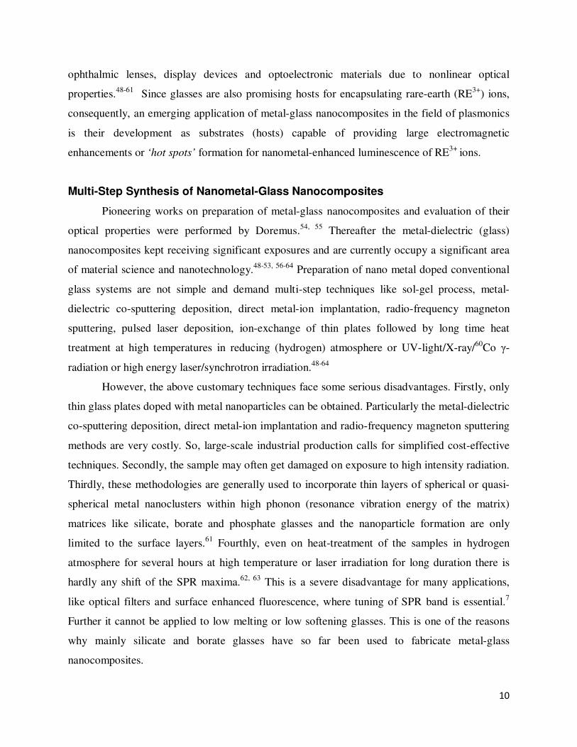
The evolution of SPR band of silver nanoclusters at 410 nm in optical absorption spectra
of silver ion exchanged silicate glasses with heat-treatment temperature62
is shown in Fig. 4.
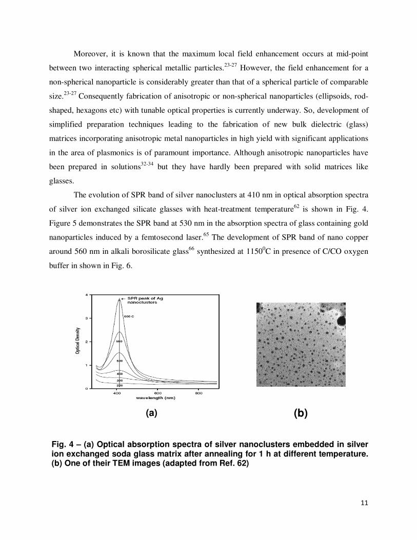
Figure 5 demonstrates the SPR band at 530 nm in the absorption spectra of glass containing gold
nanoparticles induced by a femtosecond laser.65
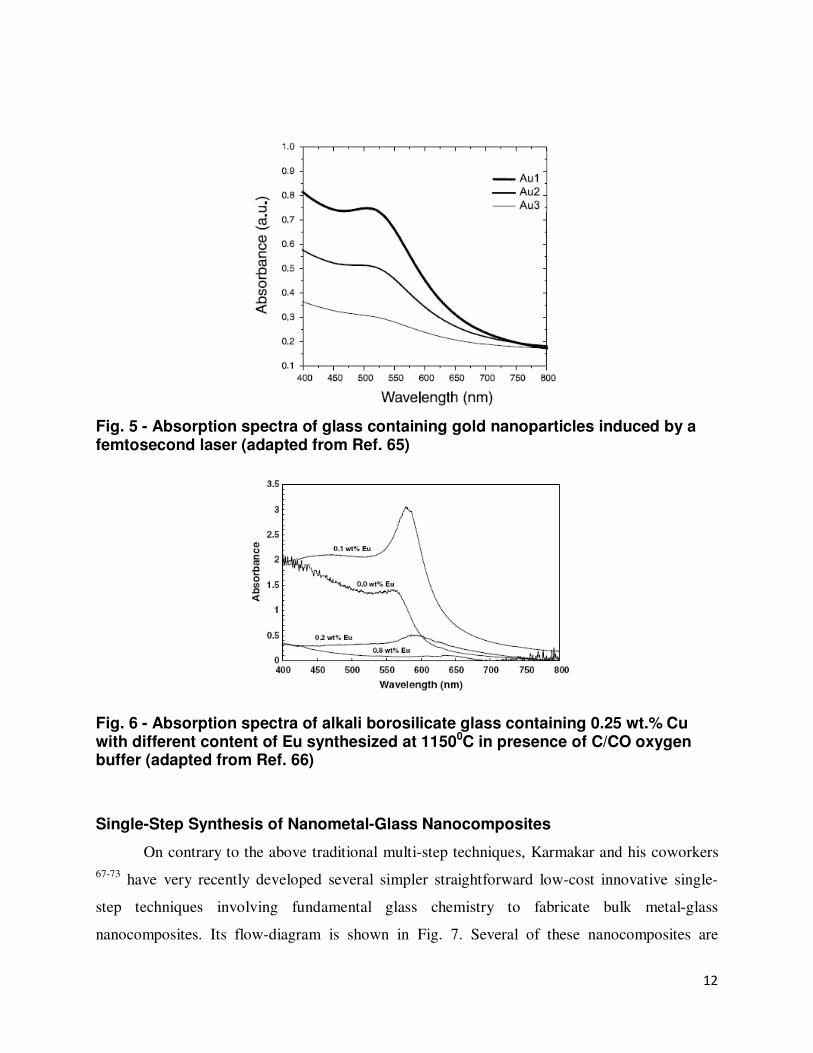
The development of SPR band of nano copper
around 560 nm in alkali borosilicate glass66
synthesized at 11500C in presence of C/CO oxygen
buffer in shown in Fig. 6.
Fig. 4 – (a) Optical absorption spectra of silver nanoclusters embedded in silver ion exchanged soda glass matrix after annealing for 1 h at different temperature. (b) One of their TEM images (adapted from Ref. 62)
(a)
(b)
12
Fig. 5 - Absorption spectra of glass containing gold nanoparticles induced by a femtosecond laser (adapted from Ref. 65)
Fig. 6 - Absorption spectra of alkali borosilicate glass containing 0.25 wt.% Cu with different content of Eu synthesized at 11500C in presence of C/CO oxygen buffer (adapted from Ref. 66)
Single-Step Synthesis of Nanometal-Glass Nanocomposites
On contrary to the above traditional multi-step techniques, Karmakar and his coworkers
67-73 have very recently developed several simpler straightforward low-cost innovative single-
step techniques involving fundamental glass chemistry to fabricate bulk metal-glass
nanocomposites. Its flow-diagram is shown in Fig. 7. Several of these nanocomposites are
13
dichroic as well. This would be discussed in details in the next section. The techniques employed
as well as the materials (glass based nanocomposites) can further be employed as suitable hosts
for nanophotonics applications, particularly nano-metal enhanced fluorescence of rare-earth ions.
In this review, we shall discuss each of these techniques along with their superiority and
thermochemical mechanism. Besides, the effects of particle size, shape and concentration,
refractive index of the dielectric (here glass) matrix, quantum and dielectric confinements on the
surface plasmon resonance (SPR) bands of the mental nanoparticles have been considered and
explained with relevant electrodynamics theories.
Mixing of Raw Materials
↓↓↓↓
Melting in a Suitable Crucible
↓↓↓↓
Casting
↓↓↓↓
Annealing
↓↓↓↓
Nanometal-Glass Hybrid Nanocomposites
Fig. 7 - Flow-chart of single-step methodology for synthesis of nanometal-glass hybrid nanocomposites
Dichroic Nanometal-Glass Nanocomposites
The best example of dichroic nanometal-glass nanocomposites is the ‘Lycurgus Cup’
which is green when illuminated from outside and red when illuminated from inside.20
. It is
made up of glass nanocomposites with about 70 nm sized gold and silver metal nanoparticles.
This fact was mentioned earlier. The deep red color of gold-ruby red ‘striking glasses’ on further
heat-treatment starts to fade and changes into dichroic ‘saphirin glasses’. It produces weak blue
14
transmission colors and strong brown reflection colors. The blue and brown colors were
attributed to small and large spherical gold colloidal particles respectively.22
Regarding the origin of dichroism, earlier researchers observed that the color of this well-
renowned gold-ruby glass varies with heat treatment. It was assumed that small sized gold
particles (3-5 nm) act as nuclei and they grow under proper conditions (heat treatment) through
the deposition of gold atoms on their surfaces. At 700 °C, some of the gold crystals grow at the
expense of smaller ones. At optimum sizes of 5–60 nm a strong ruby color is yielded. Particles
which become considerably larger, 200-500 nm, contribute very little to the absorption. Their
main function is scattering and reflection of light. Scattering occurs whenever a beam of light is
passed through a transparent medium containing in suspension of small particles, the refractive
index of which differs from that of the medium. Glasses containing particles of this range
(Saphirin glass) give blue color in the transmitted and brown color in the reflected light and
absorption wavelength shifts from 530 to 570 nm with successive crystal growth.22
At that time
visual examination by means of ultra-microscope did not reveal the actual shape of the
particles.22
Later on the advent of commercial transmission electron microscopy (TEM), which
was discovered in 1931, made possible the determination of actual shapes of gold particles.74
In
1940, by means of TEM, Borries and Kausche22
confirmed the presence of anisodimentional Au
crystals. Particles which are sufficiently large and which deviate in shape from sphere orient
themselves parallel and produce birefringence. Recently, although Schreiber et al.66
have
proclaimed that (Cu)n0
nanoparticles of certain sizes (no information about shapes was given)
formed by a combination of redox and nucleation processes give rise to dichroism but now it is
also a well acknowledged fact that prolate ellipsoid Ag NPs having an aspect ratio around 1.2
exhibits the phenomenon of dichroism.39, 62
Production of dichroic glasses generally involves deformation of embedded spherical
nanoparticles (in silicate based glasses) into ellipsoidal NPs by intense irradiation with ultrashort
laser pulses59
or stretching metal-doped glasses in their softening range.58
In recent times,
Hofmeister and his coworkers58, 75
have produced silicate based dichroic glasses by deformation
of embedded spherical nanoparticles into ellipsoidal nanoparticles by these techniques. Their
elliptical morphologies were confirmed by the TEM analysis. Fort, et al76
have also established
that the dichroic character is caused by the elliptical shaped nanoparticles and arises due to
difference in polarizations, that is, electron oscillations along the major (longitudinal) and minor
15
(transverse) axes of a polarizable ellipsoidal nanoparticles during interaction with
electromagnetic waves.
The present authors67-70, 72, 73
have carried out several studies in this view. It is understood
that the dichroic character of nanogold-antimony oxide glass nanocomposites67-69
caused by the
elliptical nanogold particles (see Figs. 9 a-c). On other hand, the dichroic nanogold-tin phosphate
glasses73
contain spherical nanogold particles of wide size variation (see Figs. 9 d-f). In the case
of nanosilver-bismuth oxide glass nanocomposites,72
dichroism arises due to both hexagonal and
spherical silver nanoparticles (see Figs. 9 g-i).
From these facts, it is clear that both spherical and non-spherical gold or silver
nanoparticles could exhibit dichroism. It is very difficult to assign the dichroism exclusively
either to the spherical or to the nonspherical shaped nanometallic particles. The appropriate
reasons for their dichroic character are, therefore, remained unsettled. Therefore it needs further
studies.
16
(a)
(b)
(c)
(d)
(e)
(f)
(g)
(h)
(i)
Fig. 8 - Nanogold-antimony oxide glass hybrid nanocomposite: (a) transmitted blue color, (b) reflected brown color, and (c) TEM image shows the elliptical shape of its nanogold particles (adapted from Ref. 67-69). Nanogold-tin phosphate glass hybrid nanocomposite: (d) transmitted deep blue color, (e) reflected reddish brown color, and (f) TEM image shows the spherical shape of its nanogold particles (adapted from Ref. 73). Nanosilver-bismuth oxide glass hybrid nanocomposite: (g) transmitted blue color, (h) reflected brown color, and (i) TEM image shows the combination of hexagonal and spherical shapes of its nanosilver particles (adapted from Ref. 72)
17
Nanometal-Antimony Glass Hybrid Nanocomposites: Synthesis, Effects of Particle Size, Composition and Refractive Index of Glass Host
Preparation of high antimony(III) oxide (Sb2O3) containing monolithic glasses encounter
serious limitations. Available reports on synthesis of antimony glasses show that they are usually
yielded as tiny pieces or pulverized form incompatible for practical applications in photonics.
This is firstly because of the low field strength (0.73) of Sb3+
cation makes Sb2O3 a poor glass
former. Secondly, the intense vaporization of Sb2O3 during melting and high devitrification
during casting has made their synthesis extremely difficult Hence, the area of nano metal-
embedded Sb2O3 based glasses and nanocomposites had remained totally unexploited because of
their difficulties in preparation particularly in the bulk monolithic form.
Very recently, Som and Karmakar67-70
exemplified for the first time a new single-step
melt-quench thermochemical reduction technique for the fabrication of monometallic and
bimetallic (Au-Ag) antimony oxide glass hybrid nanocomposites. The idea involved the
employment of a suitable antimony oxide based glass matrix, K2O-B2O3-Sb2O3 (KBS) and K2O-
B2O3-Sb2O3-ZnO (KBSZ), which itself has a reducing property mild enough to generate nano
sized metallic particles. It must be mentioned here that oxides having very high reduction
capabilities assists in the formation of larger particles. It is noteworthy that we are the first to
report the synthesis and plasmonic properties of nanometal-antimony oxide glass
nanocomposites.
It was observed that all Au-doped antimony oxide glasses were dichroic, i.e., they
transmitted the blue color (see Fig. 8a) and reflected the brown to reddish-brown (see Fig. 8b)
light. The intensity of the reflected brown color increases with increase in Au0 concentration.
The UV-Visible absorption spectrum is the most important tool to detect the formation of noble
metal NPs and SPR bands. Typical UV-Vis-NIR absorption spectra of elliptical Au nanoparticles
with increasing gold concentration and particle sizes in K2O-B2O3-Sb2O3 (KBS) glasses are
shown in Fig. 9. The absorption spectrum of the undoped glass (curve-a) shows absence of any
features indicating the base glass matrix is transparent in the spectral region of interest to this
study. The absorption spectra of the Au doped glasses (curves b-g) display well-defined broad
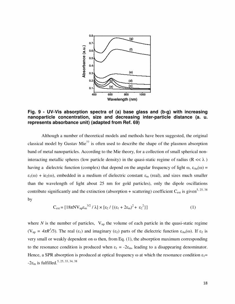
Fig. 9 - UV-Vis absorption spectra of (a) base glass and (b-g) with increasing nanoparticle concentration, size and decreasing inter-particle distance (a. u. represents absorbance unit) (adapted from Ref. 69)
Although a number of theoretical models and methods have been suggested, the original
classical model by Gustav Mie77
is often used to describe the shape of the plasmon absorption
band of metal nanoparticles. According to the Mie theory, for a collection of small spherical non-
interacting metallic spheres (low particle density) in the quasi-static regime of radius (R << λ )
having a dielectric function (complex) that depend on the angular frequency of light ω, εAu(ω) =
ε1(ω) + iε2(ω), embedded in a medium of dielectric constant εm (real), and sizes much smaller
than the wavelength of light about 25 nm for gold particles), only the dipole oscillations
contribute significantly and the extinction (absorption + scattering) coefficient Cext is given5, 25, 38
by
Cext = [18πNVnpεm3/2
/ λ] × [ε2 / {(ε1 + 2εm)2 +
ε2
2}] (1)
where N is the number of particles, Vnp the volume of each particle in the quasi-static regime
(Vnp = 4πR3/3). The real (ε1) and imaginary (ε2) parts of the dielectric function εAu(ω). If ε2 is
very small or weakly dependent on ω then, from Eq. (1), the absorption maximum corresponding
to the resonance condition is produced when ε1 = -2εm, leading to a disappearing denominator.
Hence, a SPR absorption is produced at optical frequency ω at which the resonance condition ε1=
-2εm is fulfilled.5, 25, 33, 34, 38
19
According to Eq. (1), the SPR band is independent of size within the dipole
approximation. But practically the plasmon band width increases with decrease in particle size
for particles smaller than 20 nm. The size dependence arises from the size dependence of the
dielectric constant εAu(ω, d) of the metal. This is referred as “intrinsic size effect”.5, 38
For
nanoparticles of larger dimension (>25 nm for gold nanoparticles), significant contributions are
made by higher-order (quadrupolar) charge cloud distortion of conduction electrons. These
contributions induce drastic red-shift (from 610 to 681 nm) of the SPR peak with the increase in
particle diameter D (30-40 nm). This is practically observed for nanocomposites having a higher
concentration of Au (see Fig. 10, curves e-g). This effect for larger size particles is called
“extrinsic size effect”.5, 25
The shape and position of the SPR band is also influenced by the dielectric constant of
the surrounding environment (here glass) as the resonance condition is ε1 = -2εm. This constitutes
the basis of “immersion spectroscopy”.14
The SPR absorption maxima λmax, is susceptible to the
changes of refractive index, nm of the surrounding medium as:34, 38, 68
λmax = 2πc meff εo [(ε∝ + 2nm2)]
1/2 / neff e
2 (2)
where nm = (εm)1/2
, c is the velocity of light and meff is the effective mass of the electrons. Thus,
the SPR peak exhibits a red-shift with increase in medium refractive index.5, 25
Our antimony
glass having a refractive indices in the range 1.9-2.1, radically red-shifts the plasmon peak to
around 610 nm. Beside increase in εm it is also expected to result an increase in plasmon band
intensity and band width.5, 25
This enables tuning of the SPR peak as shown in Fig. 10. As
mentioned earlier, elongated particles may show two maxima if the aspect ratio is ≥ 4.25, 33, 34, 38
From the TEM images (see Fig. 8c), it is seen that the average aspect ratio of the elliptical Au
nanoparticles synthesized within antimony oxide glasses is 1.2. So here only one SPR maxima is
displayed.
20
400 500 600 700 8000.10
0.15
0.20
0.25
1.85 1.90 1.95 2.00585
590
595
600
605
610
615
(d)
(c)
(b)
(a)
SPR
Pea
k
Refractive Index
(d)
(c)
(b)
(a)
Ab
so
rba
nc
e (
a.u
.)
Wavelength (nm)
Fig. 10 - SPR band tuning by refractive index control of host glasses. Inset plot shows the red shift of Au0-SPR band with increase in refractive index of the embedding glass matrix (adapted from Ref. 68).
Nanometal-Bismuth Glass Hybrid Nanocomposites: Synthesis and Effect of Particle Morphology In recent times, glass researchers have also focused their extensive interest in the bismuth
glasses as it is one of the most important among the heavy metal oxide (HMO) glasses due to
their several properties78-81
such as high refractive index, wide transmission window, broad band
near infrared (NIR) luminescence, etc. Bismuth glasses have also been found to be very effective
for generation of photo induced second harmonic (SHG) as reported by Kityk et al.82
Bismuth
oxide glasses are usually obtained in darkbrown or black color, which deepens with increasing
Bi2O3 content and when melted at higher temperature.78, 79, 83
This browning or blackening of
bismuth glasses when melted at higher temperature is a serious problem for controlling of Bi0
NPs and its SPR band which limits their use in optical applications. The intensity of browning or
blackening increases with increase in melting temperature as well as Bi2O3 concentration due to
auto-thermo reduction of Bi3+
ions to bismuth metal (Bi0) during the melting process. The
reduction of Bi2O3 occurs through the following thermal dissociation reaction71, 78, 79
2Bi2O3↔ 4Bi0 + 3O2↑ (3)
21
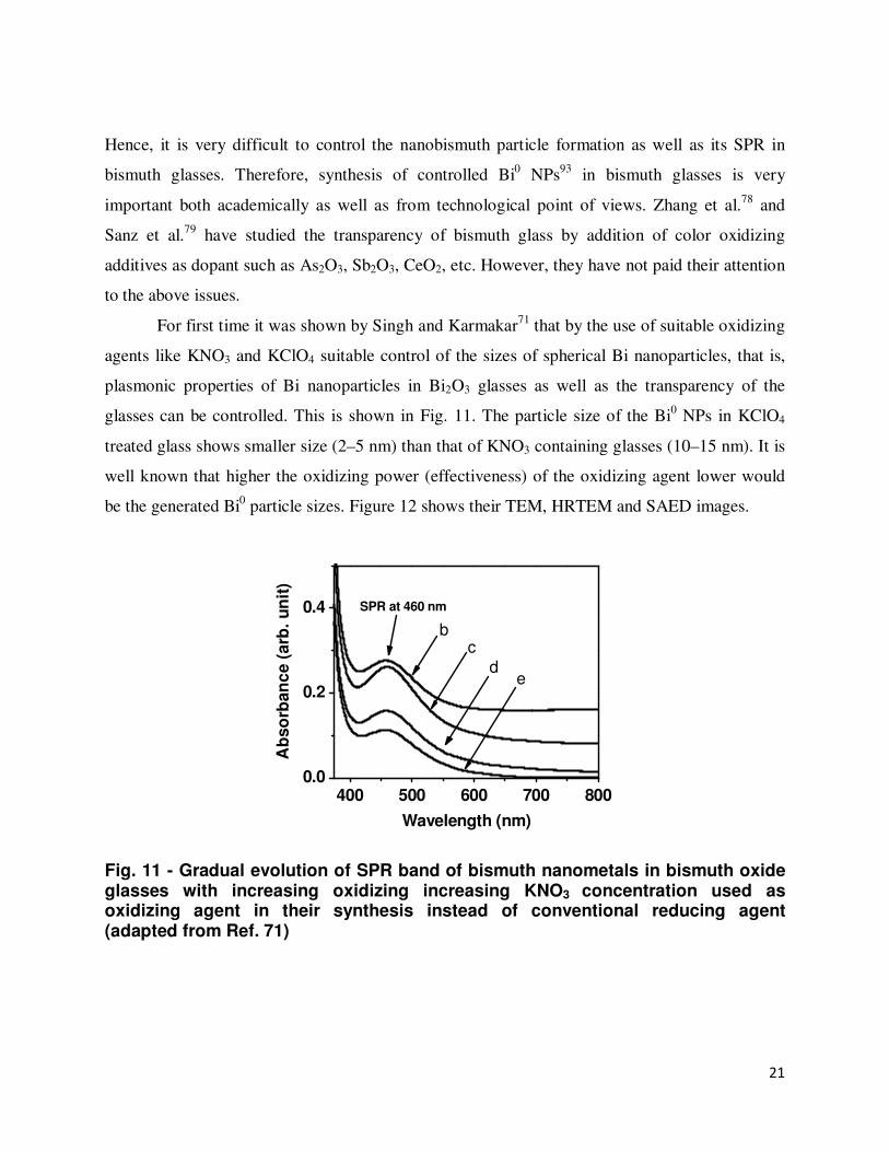
Hence, it is very difficult to control the nanobismuth particle formation as well as its SPR in
bismuth glasses. Therefore, synthesis of controlled Bi0 NPs
93 in bismuth glasses is very
important both academically as well as from technological point of views. Zhang et al.78
and
Sanz et al.79
have studied the transparency of bismuth glass by addition of color oxidizing
additives as dopant such as As2O3, Sb2O3, CeO2, etc. However, they have not paid their attention
to the above issues.
For first time it was shown by Singh and Karmakar71
that by the use of suitable oxidizing
agents like KNO3 and KClO4 suitable control of the sizes of spherical Bi nanoparticles, that is,
plasmonic properties of Bi nanoparticles in Bi2O3 glasses as well as the transparency of the
glasses can be controlled. This is shown in Fig. 11. The particle size of the Bi0 NPs in KClO4
treated glass shows smaller size (2–5 nm) than that of KNO3 containing glasses (10–15 nm). It is
well known that higher the oxidizing power (effectiveness) of the oxidizing agent lower would
be the generated Bi0 particle sizes. Figure 12 shows their TEM, HRTEM and SAED images.
400 500 600 700 8000.0
0.2
0.4
Ab
so
rban
ce (
arb
. u
nit
)
Wavelength (nm)
bc
de
SPR at 460 nm
Fig. 11 - Gradual evolution of SPR band of bismuth nanometals in bismuth oxide glasses with increasing oxidizing increasing KNO3 concentration used as oxidizing agent in their synthesis instead of conventional reducing agent (adapted from Ref. 71)
22
Fig. 12 - TEM image (left), HRTEM (middle) and SAED of bismuth nanometals in bismuth oxide glass nanocomposites (adapted from Ref. 71).
We also synthesized for the first time of hexagonal shaped bismuth coated silver
nanoparticles in bismuth glass dichroic (brown in reflection and sky blue in transmission)
nanocomposites by a novel and simple one-step melt quench technique.72
Bismuth glasses on
doping with Ag, Ag nanoparticles are first precipitated due to their higher reduction potential
followed by precipitation of Bi of it due to their similar lattice constant. At lower concentration
of Ag a single SPR maxima is observed (Fig. 13, curve a) due to spherical nanoparticles, but at
higher concentration of Ag mixture of spherical and hexagonal nanoparticles are generated (see
TEM image Fig. 8i). Formation of hexagonal particles of large aspect ratios is reflected in the
UV-vis absorption spectra, which show splitting of the SPR maxima into two peaks (Fig. 13,
curve b). The higher energy transverse mode oscillation of the electrons along the minor axis of
which absorption maxima is at 575 nm while the lower energy longitudinal mode absorption
maxima is at 785 nm corresponding to electron oscillations perpendicular to the major axis. The
spectral behavior of the hexagonal nanoparticles is similar to that of a nanorod.5, 25
400 600 800 10000
1
2
3
4
5
(b)
Ab
orb
an
ce (
arb
. u
nit
)
Wavelength (nm)
(a)
Fig. 13 – Evolution of SPR bands of bismuth coated silver nanoparticles (adapted
from Ref. 72).
23
Nanogold-Tin Phosphate Glass Hybrid Nanocomposites: Effect of Quantum and Dielectric Confinements
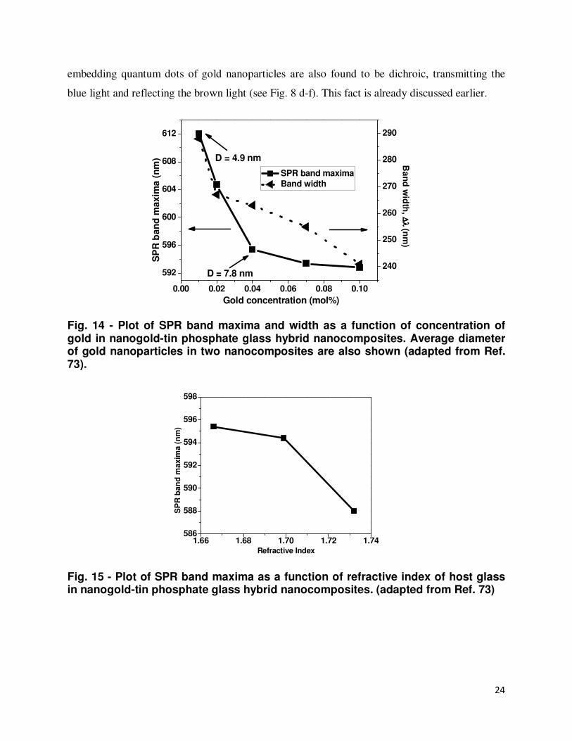
According to Eqs. (1) and (2), the SPR maxima are expected to exhibit a red shift with
increase in nanoparticle diameter and refractive index respectively. However, a blue-shift is
observed in these cases when the particle size remains less than 10 nm. This occurs due to
quantum confinement and dielectric confinement effects respectively arising due to the
difference in dielectric constant of the confined nanometal and the confining potential barrier
around it. We have demonstrated such a blue shift of quantum confined Au nanoparticles in the
prototypical tin phosphate glass system.
We fabricated sub-10 nm gold nanoparticles inside tin phosphate glasses by using a new
in-situ melt-quench technique without applying any external reducing reagent.73
UV-visible
spectra of these glass nanocomposites show blue shifts (towards lower wavelength) in the SPR
band with increase in both nanogold particle size (Fig. 14) and refractive index of the medium
(Fig. 15), which are contrary to the common trends according to Eqs. (1) and (2). It is happened
due to quantum and dielectric confinements and can be explained by electrodynamics theory.
SPR is due to the contribution of the inter-band electronic term and the Drude term. The
SPR involve both conduction electrons and bound electrons. In the visible and ultra-violet region
the optical response is dominated by the inter-band term whereas in the red and infrared region
the intra-band term (εDrude) predominates. Polarizations induced by the quasi-unbound and bound
electrons bring about the shifting of the SRP band. The plasmon shift is related with both the
polarizations induced by the quasi-free and the bound electrons. When the nanoparticle sizes are
significantly reduced (quantum confinement), the electronic distribution and polarizability near
the surface of the nanoparticle changes due to modification of the electronic wave functions. The
plasmonic oscillations (electronic wave functions) extend beyond the nanosphere into the
dielectric surroundings over a distance of the order of 0.1 nm since the spherical potential related
with the medium is finite.35
Therefore, the quasi-unbound electrons travel the region which is
greater than the diameter D of the nanoparticle and the of electron diameter De > D.35
This is
called electronic spill-out effect which results in a decrease in the average electron density in the
nanoparticle. Hence their contribution to inter-band term is reduced which in turn induces a blue
shifting in the SPR bands.35
They are shown in Figs. 14 and 15. These nanocomposites
24
embedding quantum dots of gold nanoparticles are also found to be dichroic, transmitting the
blue light and reflecting the brown light (see Fig. 8 d-f). This fact is already discussed earlier.
Fig. 14 - Plot of SPR band maxima and width as a function of concentration of gold in nanogold-tin phosphate glass hybrid nanocomposites. Average diameter of gold nanoparticles in two nanocomposites are also shown (adapted from Ref. 73).
Fig. 15 - Plot of SPR band maxima as a function of refractive index of host glass in nanogold-tin phosphate glass hybrid nanocomposites. (adapted from Ref. 73)
1.66 1.68 1.70 1.72 1.74586
588
590
592
594
596
598
SP
R b
an
d m
axim
a (
nm
)
Refractive Index
0.00 0.02 0.04 0.06 0.08 0.10
592
596
600
604
608
612
Gold concentration (mol%)
SP
R b
an
d m
axim
a (
nm
)
240
250
260
270
280
290
SPR band maxima
Band width
D = 7.8 nm
D = 4.9 nm Ba
nd
wid
th, ∆
λ
∆λ
∆
λ
∆λ
(nm
)
25
Nanophotonic Application: Nanometal Enhanced Photoluminescence of Rare-Earth Ions Malta, et al.
84-88 first reported the enhanced photoluminescence (PL) for Eu
3+ ions in
glasses containing silver nanoparticles (NPs). It was also pointed out by them that such
enhancements occurred due to the surface plasmon resonance (SPR) of the particles and long
range electromagnetic interactions associated with SPR excitation were considered significant in
obtaining an enhanced rare-earth (RE) photoluminescence. Hayakawa et al.89-91
also investigated
on Ag–Eu3+
co-doped glasses and attributed the observed photoluminescence enhancements to
local-field effects owing to the SPR of the metal particles. The plot of electric field amplitude as
a function of wavelength of nanogold particles of various sizes92
is shown in Fig. 16 (a). The
SPR absorption band of nanogold as a function of wavelength is also shown in Fig. 16 (b). It is
noteworthy that the electric field maxima are at the SPR band peak wavelength. Here it is around
530 nm for gold nanoparticles. It is seen that the electric field amplitude is low at the lower and
higher wavelengths of the SPR peak position. These different behaviours are a consequence of a
shift in the phase of the nanoparticle polarizability near the SPR band peak wavelength leading
to interference effects that vary strongly with wavelength.92
(a)
(b)
Fig. 17 – (a) Plot of electric field amplitude as a function of wavelength of gold nanoparticles of sizes: 50, 80 and 100 nm. (b) SPR absorption band of nanogold as a function of wavelength (adapted from Ref. 92])
26
This initiated investigation of metal enhanced fluorescence of RE3+
ions in glasses by
several groups in the world. Of late, the presence of metallic NPs has also been suggested as
accountable for the improved photoluminescence of RE ions in glasses.93-98
On the other hand,
Kassab et al.99-109
in their investigations of luminescence enhancement of several RE ions in a
variety of heavy metal oxide glass matrices (tellurite, lead and bismuth) attributed local field
enhancement induced by SPR of metal NPs to be the main cause.
Rare-earth (RE) doped glasses represent an important class of photonic materials owing
to their laser amplification and upconversion properties which are critically important to develop
short wavelength (visible) lasers, color displays, remote sensing, optical communication, bar-
code reading, laser printing, etc.110-113
However the small absorption cross-section of these RE
ions has spawned numerous attempts to enhance the efficiency of these ions. Most concepts rely
on energy transfer to the RE3+
ions from another species with a large absorption cross-section
(Yb3+
),114, 115
energy transfer from another species like SnO2 nanocrystals,116
and/or employing
low phonon energy host.110-113
Coupling RE3+
ions with metal nanoclusters have recently
developed as an interesting alternative strategy to enhance the luminescence intensity of RE3+
ions and are likely to bring a renaissance in the field of solid state lasers.117-120
The conducting plasmonic metal nanostructures in the vicinity of RE ions are found to
alter their free space spectral properties and greatly enhance the yield of their weak optical
transitions by precise generation of intense electric fields, i.e., by local field enhancement (LFE)
induced by SPR.10,121
This phenomenon is termed as ‘nano metal enhanced fluorescence’
(NMEF). This had initially triggered the numerous basic studies on the effect of metal NPs on
the emission properties of RE ions in solution phases and polymers.122, 123
For example, Wang et
al.122
and Nabika and Deki123
have reported the enhancement of emissions of Eu3+
-complex in
solution in presence of Ag. They have mentioned the local field enhancement around the Eu3+
ions by the Ag NPs to be the cause of luminescence enhancement.
However, for real application in nanophotonic devices, a major goal has been to produce
stable metal nanoclusters within suitable encapsulating hosts. As mentioned earlier, we have
witnessed that glasses possess some fantastic properties like high transparency, ease of
fabrication in desirable shapes and sizes, inertness, absence of high energy bond vibrations, etc.
These have made glasses not only promising encapsulating hosts for lasing RE3+
ions but also for
27
metal NPs. Consequently, nano-metal: rare-earth ions hybrid-glass nanocomposites are currently
underway.
Malta et al.84
and Hayakawa et al.90
pioneered the nanometal enhanced rare-earth
luminescence strudies in silica glasses. Of late, several groups worldwide like Kassab et al.103
Polman and his co-workers,124
have initiated this research with heavy metal oxides as glass host.
Among the heavy metal oxides, the works so far have mainly been restricted to tellurite, lead and
bismuth oxide glasses mainly due to the difficulty of fabrication of antimony glasses. Besides
they have mainly employed the traditional techniques to grow spherical nanoparticles within the
glasses and the SPR bands. However anisotropic nanoparticles are more potential candidates for
luminescence studies because local surface electric fields are drastically increased and confined
near the sharp edges of anisotropic nanostructures which act as light-harvesting nanoantennas
converting visible light into large localized electromagnetic radiation. This effect is termed as
‘lightning-rod effect’.121
But the production of anisotropic nanostructures embedded in solid
matrices like glasses being a greater challenge, which has been met with a very limited success.
Continuing the process of development, we recently studied the spectroscopic properties
of Sm3+
, Nd3+
, Eu3+
and Er3+
in a melt-quenched antimony oxide glass system containing nano
gold and silver, with the objective of developing a new class of advanced functional
nanomaterials and furthering the understanding of optical interactions in these systems.117-120
We
established that the mild reduction capability of Sb2O3 enables selective reduction of Au3+
(HAuCl4.xH2O) to Au0 or Ag
+ (AgNO3 to Ag
0), as the case may be, than RE
3+ (Sm
3+, Er
3+, Eu
3+
and Nd3+
) ions (Sm2O3, Er2O3, Eu2O3 and Nd2O3) in a single-step during the melting process
thereby providing for a straightforward, low-cost strategy for the fabrication of bulk nano metal:
RE3+
hybrid nanocomposites for application in plasmonics technologies. Besides antimony
glasses also have lower phonon energies (602 cm-1
) than other heavy metal oxide glasses. This
enables efficient upconversion of rare-earth ions reducing the multiphonon relaxation rate. The
example of nano-metal enhanced photoluminescence upconversion spectra of Nd3+
ions in
nanogold-antimony oxide glass nanocomposites is shown Fig. 17. The extent of
photoluminescence upconversion intensity enhancement as a function of concentration of gold is
also shown in Fig. 18. Maximum amplifications of the 536 nm green and 645 nm red emissions
are found to be 8 and 11 fold respectively for nanocomposite containing 0.3 wt.% Nd2O3 + 0.03
wt.% Au (Fig. 17, spectrum-c).
28
Fig. 17 - Upconversion spectra of glass and nanocomposite containing (a) 0.3 wt.% Nd2O3, (b) 0.3 wt.% Nd2O3 + 0.003 wt.% Au, (c) 0.3 wt.% Nd2O3 + 0.03 wt.% Au, (d) 0.3 wt.% Nd2O3 + 0.3 wt.% Au, (e) 0.3 wt.% Au and (f) base glass under excitation wavelength at λex = 805 nm The bases of the emission curves a, b, c and e have been uplifted for better visibility (adapted from Ref. 119).
Fig. 18 - Plot of log intensity as a function of concentration of Au (wt %) for 536 and 645 nm emission bands. (adapted from Ref. 119).
Currently there has been a growing controversy regarding the mechanism of
enhancement of rare-earth luminescence- whether plasmonic enhancement or energy transfer,125
500 550 600 650 7000.0
5.0x10⁴
1.0x10⁵
1.5x10⁵
2.0x10⁵
2.5x10⁵
3.0x10⁵
Inte
nsit
y (
cp
s)
Wavelength (nm)
(a)
(b)
(c)
(d)
(e)
(f)
(c)
(b)
(e)
(a)
(d)(f)
4G7/2→
4I15/2
4G7/2→4I9/2 λex= 805 nm
4G
7/2
→4I 1
1/2
29
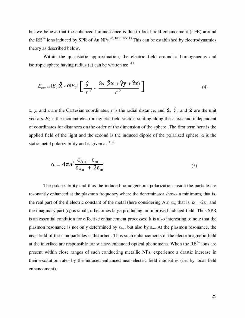
but we believe that the enhanced luminescence is due to local field enhancement (LFE) around
the RE3+
ions induced by SPR of Au NPs.90, 103, 110-113
This can be established by electrodynamics
theory as described below.
Within the quasistatic approximation, the electric field around a homogeneous and
isotropic sphere having radius (a) can be written as:1-11
Eout = |E0|x - α|E0|[ x
r 3-
3x (xx + yy + zz)]r 5
^ ^ ^ ^ ^
(4)
x, y, and z are the Cartesian coordinates, r is the radial distance, and , , and are the unit
vectors. E0 is the incident electromagnetic field vector pointing along the x-axis and independent
of coordinates for distances on the order of the dimension of the sphere. The first term here is the
applied field of the light and the second is the induced dipole of the polarized sphere. α is the
static metal polarizability and is given as:1-11
α = 4πa3εAu - εm
εAu + 2εm
(5)
The polarizability and thus the induced homogeneous polarization inside the particle are
resonantly enhanced at the plasmon frequency where the denominator shows a minimum, that is,
the real part of the dielectric constant of the metal (here considering Au) εAu that is, ε1= -2εm and
the imaginary part (εi) is small, α becomes large producing an improved induced field. Thus SPR
is an essential condition for effective enhancement processes. It is also interesting to note that the
plasmon resonance is not only determined by εAu, but also by εm. At the plasmon resonance, the
near field of the nanoparticles is disturbed. Thus such enhancements of the electromagnetic field
at the interface are responsible for surface-enhanced optical phenomena. When the RE3+
ions are
present within close ranges of such conducting metallic NPs, experience a drastic increase in
their excitation rates by the induced enhanced near-electric field intensities (i.e. by local field
enhancement).
30
All of the above categories of metal-glass nanocomposites make them potential for
applications in the optical memory devices with high storage density and ultrafast recording
speed, various other nanophotonic and optoelectronic devices.
Plasmon-Based Nanophotonic Applications
“Nanophotonics” is another emerging multidisciplinary field that enables exploitation of
study light–matter interactions on a scale much smaller than the wavelength of light.5 The
applications of nanophotonics are ample. Several nanophotonic applications thrive on the
exploitation of plasmonics which currently is seriously being considered as a promising approach
to tackle important global issues such as energy generation and even healthcare.1-12, 124
When surface plasmons are localized in or around metal nanostructures, a wealth of
interesting optical effects arises, such as local electric field enhancement near the interface of the
nanometal and dielectric.5,25
This local field enhancement has fostered great expectations in new
applications of plasmonic structures.5,6
Some of the most prevalent application of nanostructured
materials which utilizes the evanescent field at the surface are surface enhanced fluorescence,
surface enhanced Raman scattering (SERS), surface plasmon resonance imaging and
spectroscopy, optical/ molecular sensors, surface enhanced second harmonic generation,
photothermal imaging and therapy, photonic circuitry, optical communications and non-linear
optical elements.1-12, 25
The major advantage of plasmon-based nanostructures is that light can be
confined to and manipulated on a scale smaller than the wavelength of light (a few hundred
nanometers), i.e., smaller than would be possible by conventional optics.32, 92
Metal nanoparticle
arrays supported by suitable matrices (optical glasses, fibers or polymers) can serve to transport
/guide electromagnetic energy in the visible and infrared wavelengths over distances of tens to
hundreds of micrometers.32
Very tight lateral confinement of light can be achieved by use of
linear arrays of metal nanoparticles.8-10
Surface plasmon based photonic devices not only offer
the opportunity to develop high quality optical communication systems by integrating photonic
and electronic circuits but can also result in intense light generation from luminescent
materials.11-17
31
Conclusions
In this review, with a short historical background, preparation of a variety of nanometal-
glass hybrid nanocomposites by different multi-step techniques, properties and applications are
briefly described. In addition, recently developed single-step in-situ thermochemical reduction
methodology by these authors for their synthesis is described in details with their characteristic
properties, relevant theories and applications. Here Au, Ag, and Bi metals are considered and the
glasses addressed are based on antimony, bismuth and phosphorus oxides. Some of them are
dichroic in nature, that is, they exhibit blue to green colorations in transmitted light and brown to
reddish brown colorations in reflected light. Both spherical and non-spherical gold or silver
nanoparticles could exhibit dichroism. The appropriate reasons for their dichroic character are
still remained unsettled. The solution leftovers open for the researchers. Nanometal-antimony
oxide glass nanocomposites have been found to enhance the photoluminescence upconversion
intensities up to 11 fold when co-doped with rare-earth oxides (for example, Nd2O3, Er2O3, and
Sm2O3) due to the plasmonic induced local field enhancement effects of metal nanoparticles.
These studies have revealed that nanometal-glass hybrid nanocomposites are very promising for
various nanophotonic applications, such as nanometal enhanced rare-earth luminescence, solar
cell, light emitting diode, plasmonic integrated circuit, plasmon slot waveguide, etc. The reasons
for the present excitement in the nanometal-glass hybrid nanocomposites research are because of
their several inherent advantages over other dielectric nanocomposites, exceptional properties,
unique functions in nanoscience and future nanotechnology.
Acknowledgements: The authors are thankful to Prof. Indranil Manna, Director, CGCRI for
his kind permission to publish this paper. TS gratefully thanks CSIR, New Delhi for the financial
support in the form of NET-SRF Research Fellowship. The financial support of the NMITLI
Scheme of CSIR is thankfully acknowledged. SPS would also like to thank CSIR for the award
of Senior Research Fellowship (SRF).
32
References
1. A. M. Schwartzberg and J. Z. Zhang, J. Phys. Chem., C112, 10323-10337 (2008).
2. X. Lu, M. Rycenga, S. E. Skrabalak, B. Wiley and Y. Xia, Ann. Rev. Phys. Chem,. 60, 167192
(2009).
3. M. Bäumer and H. –J. Freund, Prog. Surf. Sci., 61, 127-139 (1999).
4. E. Ozbay, Science, 311, 189-193 (2006).
5. P. N. Prasad, Nanophotonics, pp. 129-151, Wiley, New Jersey, 2004.
6. W. A. Murray and W. L. Barnes, Adv. Mater., 19, 3771–3782 (2007).
7. S. A. Maier and H. A. Atwater, J. Appl. Phys. 98, 011101 (10 pp.) (2005).
8. W. L. Barnes, A. Dereux and T. W. Ebbesen, Nature, 424, 824-830 (2003).
9. F. Le, D. W. Brandl, Y. A. Urzhumov, H. Wang, J. Kundu, N. J. Halas, J. Aizpurua and P.
Nordlander, ACS Nano, 2, 707-718 (2008).
10. S. Lal, S. Link and N. J. Halas, Nature Photonics, 1, 641-648 (2007).
11. C. D. Geddes and J. R. Lakowicz, J. Fluoresc. 12, 121-129 (2002).
12. K. Aslan, I. Gryczynski, J. Malicka, E. Matveeva, J. R Lakowicz and C. D Geddes, Current
Opinion Biotechnol. 16, 55–62 (2005).
13. J. R. Lakowicz, K. Ray, M. Chowdhury, H. Szmacinski, Y. Fu, J. Zhang and K. Nowaczyk,
Analyst, 133, 1308–1346 (2008).
14. C. D. Geddes, I. Gryczynski, J. Malicka, Z. Gryczynski and J. R. Lakowicz, Combinat.
Chem. High Throughput Screening, 6, 109-117 (2003).
15. J. R. Lakowicz, B. P. Maliwal, J. Malicka, Z. Gryczynski and I. Gryczynski, J. Fluoresc. 12,
431-437 (2002).
16. E. G. Matveeva, I. Gryczynski, A. Barnett, Z. Leonenko, J. R. Lakowicz and Z. Gryczynski,
Anal. Biochem. 363, 239-245 (2007).
17. A. V. Zayats, I. I. Smolyaniov and A. A. Maradudin, Phys. Reports, 408, 131-314 (2005).
18. F. E. Wagner, S. Haslbeck, L. Stievano, S. Calogero, Q. A. Pankhurst and K.-P. Martinek,
Nature, 407, 691-692 (2000).
33
19. L. M. Liz-Marzán, Mater. Today, 7, 26-31 (2004).
20. Freestone, N. Meeks, M. Sax and C. Higgitt, Gold Bull., 40, 270-277 (2007).
21. J. Kunckel, Ars Vitraria Experimentalis oder vollkommene Glasmacher-Kunst, Berlegung
Christoph Riegels, Frankfurt,1689.
22. W. A. Weyl, Coloured Glasses, Society of Glass Technology, Sheffield, 1951.
23. M. Valden, X. Lai and D. W. Goodman, Science, 281, 1647–1650 (1998).
24. P. Buffat and J. P. Borel, Phys. Rev. A, 13, 2287–2298 (1976).
25. U. Kreibig, and M. Vollmer, Optical Properties of Metal Cluster, Springer: Berlin, 1995.
26. E. Hutter and J. H. Fendler, Adv. Mater., 16, 1685-1706 (2004).
27. F. Gonella and P. Mazzoldi,“Metal Nanocluster Composite Glasses” In: Handbook of
Nanostructured Materials and Nanotechnology, H. S. Nalwa (editor), vol 4, Academic Press,
San Diego, 2000.
28. K. L. Kelly, E. Coronado, L. L. Zhao and G. C. Schatz, J. Phys. Chem. B, 107, 668-677
(2003).
29. G. C. Papavassiliou, Prog. Solid State Chem., 12, 185-271 (1979).
30. C. F. Bohren and D. R. Huffman, Absorption and Scattering of Light by Small Particles,
Wiley: New York, 1983.
31. S. K. Ghosh and T. Pal, Chem. Rev., 107, 4797-4862 (2007).
32. W. L. Barnes, A. Dereux, and T. W. Ebbesen, Nature, 424, 824-830 (2003).
33. P. Mulvaney, Langmuir, 12, 788-800 (1996).
34. C. Noguez, J. Phys. Chem. C, 111, 3806-3819 (2007).
35. F. Vallée, in; Nanomaterials and Nanochemistry, C. Bréchignac, P. Houdy and M. Lahmani
(Eds.), Springer-Verlag, Berlin Heidelberg, 2007.
36. C. Bréchignac, Ph. Cahuzac, J. Leygnier and A. Sarfati, Phys. Rev. Lett., 70, 2036-2039
(1993).
37. C. Voisin, D. Christofilos, N. Del Fatti and F. Vallée, Phys. Rev. Lett., 85, 2200-2204 (2000).
38. S. Link and M. A. El-Sayed, Internat. Rev. Phys. Chem., 19, 409-453 (2000).
34
39. J. Pérez-Juste, I. Pastoriza-Santos, L. M. Liz-Marzán and P. Mulvaney, Coordinat. Chem.
Rev. 249 1870–1901 (2005).
40. G. Schmid, “General Features of Metal Nanoparticles Physics and Chemistry” In: Metal
Nanoclusters in Catalysis and Materials Sscience: The Issue of Size Control B. Corain, G.
Schmid, and N. Toshima (eds), Elsevier, Amsterdam, 2008, pp. 3-20.
41. D. D. Evanoff, Jr. and G. Chumanov, Chem. Phys. Chem., 6, 1221–1231 (2005).
.
42. S. Inasawa, M. Sugiyama and Y. Yamaguchi, J. Phys. Chem. B, 19, 9404-9410 (2005).
43. E. A. Coronado and G. C. Schatz, J. Phys. Chem., 119, 3926-3934 (2003).
44. Y. Hamanaka, K. Fukata, A. Nakamura, L. M. Liz-Marzán and P. Mulvaney, Appl. Phys.
Lett., 84, 4938 (3 pp.) (2004).
45. L. M. Liz-Marzán, Langmuir, 22, 32-39 (2006).
46. M. C. Daniel and D. Astruc, Chem. Rev., 104, 293-346 (2004).
47. V. Myroshnychenko, J. Rodríguez-Fernández, I. Pastoriza-Santos, A. M. Funston, C. Novo,
P. Mulvaney, L. M. Liz-Marzán and F. J. G. de Abajo, Chem. Soc. Rev., 37, 1792–1805
(2008).
48. S. Chen, T. Akai, K. Kadono and T. Yazawa, Appl. Phys. Lett., 79, 3687 (3 pp.) (2001).
49. H. Zeng, J. Qiu, S. Yuan, Y. Yang, G. Chen, Ceram. Internat., 34, 605–608 (2008).
50. H. Zeng, J. Qiu, X. Jiang, C. Zhu and F. Gan, J. Cryst. Growth, 262, 255–258 (2004).
51. H. Zeng, G. Chen, J. Qiu, X. Jiang, C. Zhu and F. Gan, J. Non-Cryst. Solids, 354, 1155–1158
(2008).
52. M. V. Roldán, A. Frattini, O. de Sanctis, H. Troiani and N. Pellegri, Appl. Surf. Sci., 254,
281–285 (2007).
53. S. Chen, T. Akai, K. Kadono and T. Yazawa, Chem. Commun. 20, 2090–2091 (2001).
54. R. H. Doremus, J. Chem. Phys., 40, 2389-2396 (1964).
55. R. H. Doremus, J. Appl. Phys., 35, 3456-3463 (1964).
56. J. Shin, K. Jang, K. –S. Lim, I. –B. Sohn, Y. –C. Noh and J. Lee, Appl. Phys, A, 93, 923–927
(2008).
35
57. G. Speranza, L. Minati, A. Chiasera, M. Ferrari, G. C. Righini and G. Ischia, J. Phys. Chem.
C, 113, 4445-4450 (2009).
58. H. Hofmeister, W. –G. Drost and A. Berger, Nanostruct. Mater., 12, 207-210 (1999).
59. M. Kaempfe, T. Rainer, K. -J. Berg, G. Seifert and H. Graener, Appl. Phys. Lett., 74, 1200-
1202 (1999).
60. D. P. Peters, C. Strohhöfer, M.L. Brongersma, J. van der Elsken, and A. Polman, Nucl. Inst.
Methods Phys. Res. B, 168, 237-244 (2000).
61. M. Dubiel, H. Hofmeister and E. Wendler, J. Non-Cryst. Solids, 354, 607–611 (2008).
62. P. Gangopadhyay, P. Magudapathy, R. Kesavamoorthy, B.K. Panigrahi, K.G.M. Nair and