NANOTOXICOLOGY: An Emerging Discipline Evolving from Studies of Ultrafine Particles Günter Oberdörster, Eva Oberdörster, Jan Oberdörster doi:10.1289/ehp.7339 (available at http://dx.doi.org/) Online 22 March 2005 The National Institute of Environmental Health Sciences National Institutes of Health U.S. Department of Health and Human Services ehponline.org
Transcript
NANOTOXICOLOGY:An Emerging Discipline Evolving from Studies of
Ultrafine Particles
Günter Oberdörster, Eva Oberdörster, Jan Oberdörsterdoi:10.1289/ehp.7339 (available at http://dx.doi.org/)
Online 22 March 2005
The National Institute of Environmental Health SciencesNational Institutes of Health
U.S. Department of Health and Human Services
ehponline.org
NANOTOXICOLOGY:An Emerging Discipline Evolving from Studies of Ultrafine Particles
Günter Oberdörster1, Eva Oberdörster2, Jan Oberdörster3
1University of Rochester
Department of Environmental Medicine
Rochester, NY2Southern Methodist University
Department of BiologyDallas, TX3Bayer CropScience
Toxicology DepartmentResearch Triangle Park, NC
Corresponding Author:Dr. Günter Oberdörster
University of Rochester
Department of Environmental Medicine575 Elmwood Avenue, MRBx Bldg., Box 850
IARC = International Agency for Research on Cancer
ICON = International Council on Nanotechnology
ICRP = International Commission on Radiological Protection
ILSI = International Life Sciences Institute
ISC = Inter System Crossing
kg = kilogram
LPO = lipid peroxidation
m = meter
mg = microgram
mg = milligram
mm = micrometers
mm = millimeter
MMAD = Mass Median Aerodynamic Diameter
Mn = Manganese
MPPD = Multiple Path Particle Deposition
MSDS = Material Safety Data Sheet
MWNT = multi-walled Carbon Nanotubes
ng = nanogram
nm = nanometer
NNI = National Nanotechnology Initiative
NP = engineered nanoparticles
4
NRC = National Research Council
NSP = nano-sized particles
P450s = cytochrome P450s
PNS = Peripheral Nervous System
ppb = parts per billion
PTFE = polytetrafluoroethylene
ROS = Reactive Oxygen Species
ssDNA = single-stranded DNA
SWNT = Single-walled Carbon Nanotube
TiO2 = titanium dioxide
UFP = ultrafine particle
UV = ultraviolet radiation
WHO = World Health Organization
5
Outline of Nanotoxicology manuscript
Abstract1. Introduction
1.1 Naturally-Occurring and Anthropogenic Nano-Sized Particles1.2 Physico-Chemical Characteristics as Determinants of Biological Activity
1.3 Human Exposure to Nano-Sized Materials
1.4 Manufactured Nanomaterials in the Environment2. Toxicology of Airborne Ultrafine Particles, Overview
3. Concepts of Nanotoxicology3.1 Laboratory Rodent Studies
3.2 Ecotoxicological Studies
3.3 ROS Mechanisms of Nano-Sized Particle Toxicity3.4 Exposure-Dose-Response Considerations
4. Portals of Entry and Target Tissues
4.1 Respiratory Tract4.1.1 Efficient Deposition of Inhaled Nano-Sized Particles
4.1.2 Disposition of NSP in the Respiratory TractClassical Clearance Pathways
Epithelial Translocation
Translocation to the Circulatory SystemNeuronal Uptake and Translocation
4.2 GI Tract and Skin5. Risk Assessment
6. Summary and Outlook
7. References8. Tables
9. Figure Legends
6
ABSTRACTAlthough humans have been exposed to airborne nano-sized particles (NSP; <100 nm)
throughout their evolutionary stages, such exposure has increased dramatically over the last
century due to anthropogenic sources. The rapidly developing field of nanotechnology is likely tobecome yet another source through inhalation, ingestion, skin uptake, and injection of engineered
nanomaterials. Information about safety and potential hazards is urgently needed. Results of
older biokinetic studies with NSP and newer epidemiologic and toxicologic studies with airborneultrafine particles can be viewed as the basis for the expanding field of nanotoxicology, which can
be defined as safety evaluation of engineered nanostructures and nanodevices. Collectively, someemerging concepts of nanotoxicology can be identified from the results of these studies: When
inhaled, specific sizes of NSP are efficiently deposited by diffusional mechanisms in all regions
of the respiratory tract. The small size facilitates uptake into cells, transcytosis across epithelialand endothelial cells into the blood and lymph circulation to reach potentially sensitive target sites
such as bone marrow, lymph nodes, spleen, heart. Access to CNS and ganglia via translocation
along axons and dendrites of neurons has also been observed. NSP penetrating the skin distributevia uptake into lymphatic channels. Endocytosis, and biokinetics are largely dependent on NSP
surface chemistry (coating) and in vivo surface modifications. The greater surface area per masscompared to larger-sized particles of the same chemistry renders NSP more active biologically.
This activity includes a potential for inflammatory and pro-oxidant, but also anti-oxidant, activity,
which can explain early findings showing mixed results in terms of toxicity of NSP toenvironmentally-relevant species. Evidence of mitochondrial distribution and oxidative stress
response following NSP endocytosis points to a need for basic research about their interactionswith subcellular structures. Additional considerations for assessing safety of engineered NSP
include careful selections of appropriate and relevant doses/concentrations, the likelihood of
increased effects in a compromised organism, but also the benefits of possible desirable effects.An interdisciplinary team approach (e.g., toxicology, materials science, medicine, molecular
biology, and bioinformatics, to name a few) is mandatory for nanotoxicology research to arrive atan appropriate risk assessment.
7
1. Introduction1.1 Naturally-Occurring and Anthropogenic Nano-Sized Particles
Exposures to airborne nano-sized particles (NSP, <100 nm) have been experienced byhumans throughout their evolutionary stages, but it is only with the advent of the industrial
revolution that such exposures have increased dramatically due to anthropogenic sources such as
internal combustion engines, power plants, and many other sources of thermodegradation. And,most recently, the rapidly developing field of nanotechnology is likely to become yet another
source for human exposures to NSP – engineered nanoparticles (NP) – by different routes, i.e.,inhalation, ingestion, dermal or even injection. Table 1 summarizes some of the natural and
anthropogenic sources of NSP, the latter divided into unintentional and intentional sources.
Biologically-based or naturally-occurring molecules that are found inside organisms sincethe beginning of life can serve as model nano-sized materials. For example, biogenic magnetite is
a naturally occurring nano-sized particle that occurs in many species ranging from Bacteria to
Protozoa and to Animals (Blakemore 1975; Kirschvink et al. 2001). Biogenic magnetite has evenbeen found in brains of humans (Dunn et al. 1995; Kirschvink et al. 1992; Schultheiss-Grassi et
al. 1999), and has been associated with neurodegenerative diseases (Dobson 2001; Hautot et al.2003). A biological-model of coated nanomaterials can be found in ferritin, which is an ~12 nm
large iron storage protein that contains 5-7 nm sized hydrous ferric oxide-phosphate inside a
protective protein shell (Donlin et al. 1998). Nano-sized materials, including fullerenes, occurnaturally from combustion processes such as forest fires and volcanoes.
Obvious differences between unintentional and intentional anthropogenic NSP are thepolydispersed and chemically complex nature (elemental, soluble, and volatile carbon
compounds; soluble and poorly soluble inorganics (Cyrys et al. 2003; Hughes et al. 1998)) of the
former, in contrast to the monodisperse and precise chemically engineered characteristics andsolid form of the latter, generated in gas or liquid phase (NNI, 2004). However, despite these
differences the same toxicological principles are likely to apply for NP, since not only size but anumber of other particle parameters determine their biological activity. The multitude of
interactions of these factors has yet to be assessed, and this article is an attempt to summarize our
present knowledge.
8
NSP are variably called ultrafine particles (UFPs) by toxicologists (EPA 2004), Aitken
mode and nucleation mode particles by atmospheric scientists (Kulmala 2004; NRC 1983), andengineered nanostructured materials by materials scientists (NNI 2004). Figure 1 depicts the
range of sizes of airborne ambient particulate matter, including the nucleation mode, Aitkenmode, accumulation mode and coarse mode particles. Ambient particles below 0.1 µm, defined
as ultrafine particles in the toxicological literature, consist of transient nuclei or Aitken nuclei
(NRC 1983). More recently, even smaller particles in the nucleation mode with peak diametersaround 4 nm have been observed (McMurry and Woo 2002). The distinction between NSP
generated by internal combustion engines and engineered nanoparticles becomes further cloudedby the finding of nanotubes in diesel exhaust (Evelyn et al. 2003). The use of the term “nano” in
this review only reflects on particle size and not on chemical composition. For the purposes of
this review, we will use the following terms: “nano-sized particle” includes all engineered andambient nano-sized spherical particles below 100 nm. “Engineered nanoparticles” (NP) includes
only spherical NSP specifically engineered in the laboratory; other engineered nano-sized
structures will be labeled according to their shape, e.g., nanotubes, -fibers, -wires, -rings, etc.“Ultrafine Particles (UFP)” includes ambient and laboratory-generated NSP that are not produced
in a controlled, engineered way.Table 2 shows the tremendous differences in particle number concentrations and particle
surface areas for particles of the four ambient modes, assuming an airborne concentration of 10
µg/m3 of unit density particles of each size. The extraordinarily high number concentrations ofNSP per given mass will likely be of toxicological significance when these particles interact with
cells and subcellular components. Likewise, their increased surface area per unit mass can betoxicologically important if other characteristics like surface chemistry and bulk chemistry, are
the same. Although the mass of UFP in ambient air is very low, approaching only 0.5 - 2 µg/m3
at background levels (Hughes et al. 1998), it can increase several-fold during high pollutionepisodes or on highways (Brand et al. 1991; Shi et al. 2001; Zhu et al. 2002).
1.2 Physico-Chemical Characteristics as Determinants of Biological Activity
The small size and corresponding large specific surface area of solid NSP (Table 2)
confers specific properties to them, for example, making them desirable as catalysts for chemicalreactions. The importance of surface area becomes evident when considering that surface atoms
9
or molecules play a dominant role in determining bulk properties (Amato 1989); the ratio of
surface to total atoms or molecules increases exponentially with decreasing particle size (Figure2). Increased surface reactivity predicts that NSP exhibit greater biological activity per given
mass compared to larger particles, should they be taken up into living organisms and providedthey are solid rather than solute particles. This increased biological activity can either be positive
and desirable (e.g., antioxidant activity, carrier of capacity for therapeutics, penetration of cellular
barriers for drug delivery), or negative and undesirable (e.g., toxicity, induction of oxidative stressor of cellular dysfunction) or a mix of both. Not only may adverse effects be induced, but
interactions of NSP with cells and subcellular structures and their biokinetics are likely to be verydifferent from those of larger-sized particles. For example, virologists have described more than
60 years ago the translocation of 30-50 nm sized virus particles along axons and dendrites of
neurons and across epithelia (Bodian and Howe 1941), whereas first reports about increasedinflammatory activity and epithelial translocation of man-made 20 and 30 nm solid particles
appeared only more recently (Ferin et al. 1990; Oberdörster et al. 1990).
The characteristic biokinetic behaviors of NP are attractive qualities for promisingapplications in medicine as diagnostic and therapeutic devices, and as tools to investigate and
understand molecular processes and structures in living cells (Akerman et al. 2002; deLorenzo1970; Foley et al. 2002; Kreuter 2001; Li et al. 2003). For example, targeted drug delivery to
tissues which are difficult to reach (e.g., CNS), NP for the fight against cancer, intravascular
nanosensor and nanorobotic devices, diagnostic and imaging procedures are presently underdevelopment. The discipline of nanomedicine –defined as medical application of nanotechnology
and related research – has arisen to design, test, and optimize these applications so that they caneventually be used routinely by physicians (Freitas 1999).
However, in apparent stark contrast to the many efforts aimed at exploiting desirable
properties of NP for improving human health are the limited attempts to evaluate potentialundesirable effects of NP when administered intentionally for medicinal purposes, or following
unintentional exposure during manufacture or processing for industrial applications: The sameproperties that makes NP so attractive for development in nanomedicine and for specific
industrial processes could also prove deleterious when NP interact with cells. Thus, evaluating
the safety of NP should be of highest priority given their expected world-wide distribution forindustrial applications and the likelihood of human exposure, directly or through release into the
10
environment (air, water, soil). An emerging discipline – nanotoxicology, which can be defined as
“Science of engineered nanodevices and nanostructures that deals with their effects and problemsinvolved” — is gaining increased attention. Nanotoxicology research will not only provide data
for safety evaluation of engineered nanostructures and devices but will also help to advancefurther the field of nanomedicine by providing information about their undesirable properties and
means to avoid them.
1.3 Human Exposure to Nano-Sized Materials
In addition to natural and anthropogenic sources of UFP in the ambient air, certainworkplace conditions also generate NSP which can reach much higher exposure concentrations,
up to several hundred µg/m3, than ambient levels. In contrast to airborne UFP exposures
occurring via inhalation at the workplace and from ambient air, not much is known about levels ofexposure via different routes for NP, whether it is by direct human exposure or indirectly through
contamination of the environment. For example, are there or will there be significant exposures
to airborne singlet engineered carbon nanotubes or C60 fullerenes? First measurements at a modelworkplace gave only very low concentrations, less than 50 µg/m3, and these were most likely in
the form of aggregates (Maynard et al. 2004). However, even very low concentrations of nano-sized materials in the air represent very high particle number concentrations, as is well known
from measurements of ambient ultrafine particles (Hughes et al. 1998), For example, a low
concentration of 10 µg/m3 of unit density 20 nm particles translates into more than 1x10⁶
particles/cm3 (Table 2). Inhalation may be the major route of exposure for NP, yet ingestion and
dermal exposures also need to be considered during manufacture, use, and disposal of engineerednanomaterials, and specific biomedical applications for diagnostic and therapeutic purposes will
require i.v., s.c. or i.m. administration (Table l). It can be assumed, though, that the toxicology of
NP can build on the experience and data already present in the toxicology literature of ambientUFP.
(See web-materials for additional details.)
1.4 Manufactured Nanomaterials in the Environment
Manufactured nanomaterials are likely to enter the environment for several reasons: i)some are and others will be produced by the ton, and some of any material produced in such mass
11
quantities is likely to reach the environment from manufacturing effluent or from spillage during
shipping and handling; ii) they are being used in personal-care products such as cosmetics andsunscreens and can, therefore, enter the environment on a continual basis from washing off of
consumer products (Daughton and Ternes 1999); iii) they are being used in electronics, tires, fuelcells, and many other products and it is unknown whether some of these materials may leak out or
be worn off over the period of use; iv) they are being used in disposable materials such as filters
and electronics and may therefore reach the environment through landfills and other methods ofdisposal.
Scientists have also found ways of using nanomaterials in remediation. Although many ofthese are still in testing stages (Chitose et al. 2003; Jaques and Kim 2000; Joo et al. 2004;
Nagaveni et al. 2004; Nghiem et al. 2004; Tungittiplakorn et al. 2004), dozens of sites have
already been injected with various nanomaterials, including nano-iron (Mach 2004). Testing todetermine the safety of these NP used for remediation to environmentally-relevant species has not
yet been done. Although most people are concerned with effects on large wildlife, the basis of
many food chains depends on the benthic and soil flora and fauna, which could be dramaticallyimpacted by such NP injections. In addition, as noted by Lecoanet and Wiesner (2004a), nano-
sized materials may not migrate through soils at rapid enough rates to be valuable in remediation.Future laboratory and field trials will help clear up the line between remediation and
contamination.
(See web-materials for additional details.)
2. Toxicology of Airborne Ultrafine Particles, OverviewIn recent years, interest in potential effects of exposure to airborne UFP has increased
considerably, and studies have shown that they can contribute to adverse health effects in the
respiratory tract as well as in extrapulmonary organs. Results on direct effects of ambient andmodel UFP have been reported from epidemiological studies and controlled clinical studies in
humans, inhalation/instillation studies in rodents, or in vitro cell culture systems. For example,several epidemiological studies have found associations of ambient UFP with adverse respiratory
and cardiovascular effects resulting in morbidity and mortality in susceptible parts of the
population (Pekkanen et al. 1997; Penttinen et al. 2001; Peters et al. 1997a, b; von Klot et al.2002; Wichmann et al. 2002), whereas other epidemiological studies have not seen such
12
associations (Pekkanen et al. 1997; Tiittanen et al. 1999). Controlled clinical studies evaluated
deposition and effects of laboratory-generated UFP. High deposition efficiencies in the totalrespiratory tract of healthy subjects were found, and deposition was even greater in asthmatic and
COPD subjects. In addition, effects on the cardiovascular system including blood markers ofcoagulation and systemic inflammation and pulmonary diffusion capacity were observed
following controlled exposures to ultrafine carbonaceous particles (Anderson et al. 1990;
Wichmann et al., 2000; Brown et al. 2002; Chalupa et al. 2004; Jaques and Kim 2000; Pietropaoliet al. 2004; Pekkanen et al., 2002; Henneberger et al., 2005).
Studies in animals using laboratory-generated model UFP or ambient UFP showed thatUFP consistently induced mild yet significant pulmonary inflammatory responses as well as
effects in extrapulmonary organs. Animal inhalation studies included the use of different
susceptibility models in rodents, with analysis of lung lavage parameters and lung histopathology,effects on the blood coagulation cascade and translocation studies to extrapulmonary tissues
(Elder et al. 2000; Elder et al. 2002, 2004; Ferin et al. 1991; Ferin and Oberdörster 1992; Kreyling
et al. 2002; Li et al. 1999; Nemmar et al. 1999; Nemmar et al. 2002a; Nemmar et al. 2002b;Nemmar et al. 2003; Oberdörster et al. 1992a; Oberdörster et al. 1995; Oberdörster et al. 2000;
Oberdörster et al. 2002; Oberdörster et al. 2004; Semmler et al. 2004; Zhou et al. 2003).In vitro studies using different cell systems showed to varying degrees pro-inflammatory
and oxidative stress–related cellular responses after dosing with laboratory-generated or filter
collected ambient UFP (Brown et al. 2000; Brown et al. 2001; Li et al. 2003). Collectively, the invitro results have identified oxidative stress related changes of gene expression and cell signaling
pathways as underlying mechanisms of UFP effects, as well as a role of transition metals andcertain organic compounds on combustion generated UFP (Figure 3). These can alter cell
signaling pathways, including Ca++ signaling and cytokine signaling (e.g., IL-8) (Donaldson et
al., 2002; Donaldson and Stone, 2003). Effects were on a mass basis greater for ultrafine modelparticles than for those of fine particles, whereas for ambient UFP cellular responses sometimes
were greater and sometimes less than those of fine and coarse particles. The interpretation of thein vitro studies is oftentimes difficult because particles of different chemical compositions were
used, target cells were different, duration, endpoints, and generally high dose levels also differed.
Results from high doses in particular should be viewed with caution if they are orders ofmagnitude higher than predicted from relevant ambient exposures (see section 3.4).
13
(See web-materials for additional details.)
3. Concepts of Nanotoxicology3.1 Laboratory Rodent Studies
With respect to potential health effects of NSP, two examples should serve to illustrate i)
that these particles have a higher inflammatory potential per given mass than larger particles,
provided they are chemically the same, and ii) that UFP generated under certain occupationalconditions can elicit severe acute lung injury.
The first example involves studies with ultrafine and fine TiO2 particles which showedthat ultrafine anatase TiO2 (20 nm), when instilled intratracheally into rats and mice, induced a
much greater pulmonary-inflammatory neutrophil response (determined in the lung lavage 24 hrs.
after dosing) than fine anatase TiO2 (250 nm) at the same instilled mass dose of both types ofparticles (Figure 4a). However, when the instilled dose was expressed as particle surface area it
became obvious that the neutrophil response in the lung for both ultrafine and fine TiO2 fitted the
same dose-response curve (Figure 4b), suggesting that particle surface area for particles ofdifferent sizes but of the same chemistry, such as TiO2, is a better dosemetric than particle mass or
particle number (Oberdörster 2000). Moreover, normalizing the particle surface dose to lungweight shows excellent agreement of the inflammatory response in both species (Figure S-2 on
web). The better fit of dose-response relationships by expressing the dose as surface area rather
than mass when describing toxicological effects of inhaled solid particles of different sizes hasbeen pointed out repeatedly, especially when particles of different size ranges – nano to fine –
were studied (Brown et al. 2001; Donaldson et al. 1998; Donaldson et al. 2002; Oberdörster andYu 1990; Oberdörster et al., 1992; Driscoll, 1996; Tran et al. 1998; Tran et al. 2000) (see web for
additional material).
Particle chemistry, and specifically surface chemistry, play a decisive role in addition toparticle size as is shown in the second example, i.e., exposure of rats to polytetrafluoroethylene
fume (PTFE). PTFE fume (generated by heating PTFE) has long been known to be of high acutetoxicity to birds and mammals, including humans (Cavagna et al. 1961; Coleman et al. 1968;
Griffith et al. 1973; Nuttall et al. 1964; Waritz and Kwon 1968). Analysis of these fumes
revealed the nano-sized nature of the particles generated by heating PTFE to about 480°C, with acount median diameter (CMD) of 18 nm. They were highly toxic to rats, causing severe acute
14
lung injury with high mortality within 4 hours after a 15-min. inhalation exposure to 50 µg/m3
(Oberdörster et al. 1995) This short exposure resulted in an estimated deposited dose in thealveolar regions of only ~60 ng. In humans, acute lung injury, known as polymer fume fever, can
result from exposures to PTFE fumes (Auclair et al. 1983; Goldstein et al. 1987; Lee et al. 1997;Williams et al. 1974; Woo et al. 2001). Additional rat studies showed that the gas phase alone
was not acutely toxic and that aging of the PTFE fume particles for 3 minutes increased their
particle size to >100 nm due to accumulation which resulted in a loss of toxicity (Johnston et al.2000). However, it is most likely that changes in particle surface chemistry during the aging
period contributed to this loss of toxicity, and that this is not just an effect of the accumulatedlarger particle size. (Additional information on web).
These examples seem to represent the extremes of NSP in terms of pulmonary toxicity,
one (TiO2) being rather benign yet still inducing significantly greater inflammatory effects on amass basis than fine particles of the same chemical make-up; the other (PTFE fumes) inducing
very high acute toxicity, possibly related to reactive groups on the large surface per unit mass.
Engineered nanomaterials can have very different shapes, e.g., spheres, fibers,tubes, rings, planes. Toxicological studies of spherical and fibrous particles have well established
that natural (e.g., asbestos) and man-made (e.g., biopersistent vitreous) fibers are associated withincreased risks of pulmonary fibrosis and cancer following prolonged exposures (Greim et al.
2001; IARC 2002). Critical parameters are the three D’s: Dose, Dimension and Durability of the
fibers. Fibers are defined as elongated structures with a diameter to length ratio (aspect ratio) of1:3 or greater and with a length of >5 µm and diameter < 3 µm (WHO 1985). Carbon nanotubes
have aspect ratios of up to 100 and greater, and length can exceed 5 µm with diameters rangingfrom 0.7 to 1.5 nm for single-walled nanotubes, and 2 nm to 50 nm for multi-walled nanotubes.
Results from three studies using intratracheal dosing of carbon nanotubes in rodents indicate
significant acute inflammatory pulmonary effects which either subsided in rats (Warheit et al.2004) or were more persistent in mice (Lam et al. 2004; Shvedova et al. 2004b). Administered
doses were very high, ranging from 1 to 5 mg/kg in rats, and in mice from 3.3 to 16.6 mg/kg(Lam et al. 2004) or somewhat lower from 0.3 to 1.3 mg/kg (Shvedova et al. 2004b). Granuloma
formation as a normal foreign body response of the lung to high doses of a persistent particulate
material was a consistent finding in these studies. Metal impurities (e.g., Fe) from the nanotubegeneration process may also have contributed to the observed effects. Although these in vivo first
15
studies revealed high acute effects, including mortality, this was explained by the large doses of
the instilled highly aggregated nanotubes which caused death by obstructing the airways andshould not be considered a nanotube effect per se (Warheit et al. 2004). In vitro studies with
carbon nanotubes also reported significant effects. Dosing keratinocytes and bronchial epithelialcells in vitro with single walled carbon nanotubes resulted in oxidative stress, as evidenced by the
formation of free radicals, accumulation of peroxidative products, and depletion of cell
antioxidants (Shvedova et al. 2004a). Multiwalled carbon nanotubes showed proinflammatoryeffects and were internalized in keratinocytes (Monteiro-Riviere et al. 2005). Again, relatively
high doses applied in these studies need to be considered when discussing the relevancy of thesefindings for in vivo exposures. A most recent study in macrophages comparing single walled and
multi-walled carbon nanotubes with C60 fullerenes found a cytotoxicity ranking on a mass basis in
the order SWNT>MWNT>C60. Profound cytotoxicity (mitochondrial function, cell morphology,phagocytic function) was seen for SWNT even at a low concentration of 0.38 µg/cm2. The
possible contribution of metal impurities of the nanotubes still needs to be assessed. Therefore,
whether the generally recognized principles of fiber toxicology apply to these nanofiber structuresneeds still to be determined (Huczko et al. 2001).
Future studies should be designed to investigate both effects and also the fate of nanotubesfollowing deposition in the respiratory tract, preferentially by inhalation using well dispersed
(singlet) airborne nanotubes. In order to design the studies using appropriate dosing, it is
necessary to assess the likelihood and degree of human exposure. It is of utmost importance tocharacterize human exposures in terms of the physico-chemical nature, the aggregation state and
concentration (number; mass; surface area) of engineered nanomaterials and perform animal andin vitro studies accordingly. If using direct instillation into the lower respiratory tract a large
range of doses, which include expected realistic exposures of appropriately prepared samples,
needs to be considered.(See web-materials for additional details.)
3.2 Ecotoxicological Studies:
Studies have been carried out to date only on a few species that have been accepted by
regulatory agencies as models for defining ecotoxicological effects. Tests with un-coated, water
soluble, colloidal fullerenes (nC60) show that the 48 hour LC50 in Daphnia magna is 800 ppb
16
(Oberdörster E, 2004), using standard EPA protocols (EPA 1994). In largemouth bass
(Micropterus salmoides), although no mortality was seen, lipid peroxidation in the brain and
glutathione depletion in the gill was observed after exposure to 0.5 ppm nC60 for 48 hours
(Oberdörster 2004). There are several hypotheses as to how lipid damage may have occurred in
the brain, including direct redox activity by fullerenes reaching the brain via circulation or axonal
translocation (see also section 4.1.2) and dissolving into the lipid-rich brain tissue; oxyradical
production by microglia; or reactive fullerene metabolites may be produced by cytochrome P450
metabolism. Initial follow-up studies using suppressive subtractive hybridization of pooled
control fish vs. pooled 0.5 ppm-exposed fish liver mRNA were also performed. Proteins related
to immune responses and tissue repair were up-regulated, and several proteins related to
homeostatic control and immune control were down-regulated. A cytochrome P450 (CYP2K1)
involved in lipid metabolism was up-regulated. (See web-materials for additional details).
In addition to these biochemical and molecular-level changes in fish, bactericidalproperties of fullerenes have also been reported, and are being explored as potential new anti-
microbial agents (Yamakoshi et al. 2003). Engineered nanomaterials used as anti-microbials may
shift microbial communities if they are released into the environment via effluents. As we knowfrom anthropogenic Endocrine Disrupting Compounds, interference of signaling between
nitrogen-fixing bacteria and their plant hosts could be extremely harmful both ecologically andeconomically in terms of crop production (Fox et al. 2001).
Aqueous fullerenes and coated SWCNT are stable in salt solutions (Cheng et al. 2004;
Warheit et al. 2004), cell culture media (Lu et al. 2004; Sayes et al. 2004), Reconstituted HardWater and MilliQ water (Dieckmann et al. 2003; Oberdörster 2004). NSP will tend to sorb onto
sediment and soil particles and be immobilized due to their high surface area:mass ratio (Lecoanetand Wiesner 2004b). Biological transport would occur from ingested sediments, and one would
expect movement of nanomaterials through the food chain (Figure 5).
To make engineered nanomaterials more biocompatible, both surface coatings andcovalent surface modifications have been incorporated. Some studies have shown that both the
surface coating and the covalent modifications can be weathered by either exposure to the oxygenin air or by UV irradiation for one to four hours (Derfus et al. 2004; Rancan et al. 2002).
Therefore, although coatings and surface modifications may be critically important in drug-
delivery devices, the likelihood of weathering under environmental conditions makes it important
17
to study toxicity under UV and air exposure conditions. Even coatings used in drug delivery of
NP may not be biopersistent or could be metabolized to expose the core NP material.(See web-materials for additional details.)
3.3 ROS Mechanisms of Nano-sized Particle Toxicity
Both in vivo and in vitro, NSP of various chemistries have been shown to create reactive
oxygen species (ROS). ROS production has been found in NP as diverse as C60 fullerenes,SWNT, quantum dots, and UFP, especially under concomitant exposure to light, UV or transition
metals. (Brown et al. 2000; Brown et al. 2001; Derfus et al. 2004; Joo et al. 2004; Li et al. 2003;Nagaveni et al. 2004; Oberdörster 2004; Rancan et al. 2002; Sayes et al. 2004; Shvedova et al.
2004b; Wilson et al. 2002; Yamakoshi et al. 2003). It has been demonstrated that NSP of various
sizes and various chemical compositions preferentially mobilize to mitochondria (DeLorenzo1970; Gopinath et al. 1978; Foley et al. 2002; Li et al. 2003; Rodoslav et al. 2003). Since
mitochondria are redox active organelles, there is a likelihood of altering ROS production and
thereby over-loading or interfering with anti-oxidant defenses (Fig. 3).Figure 6 diagrams some of the anti-oxidant defense systems that occur in animals, and
possible areas where NSP may create oxyradicals. The C60 fullerene is shown as a model NPproducing superoxide, as has been shown by Yamakoshi (2003) (Yamakoshi et al. 2003). The
exact mechanism by which each of these diverse NP cause ROS is not yet fully understood, but
suggested mechanisms include i) photo excitation of fullerenes and SWNT causing Inter-SystemCrossing (ISC) to create free electrons; ii) metabolism of NP to create redox active intermediates,
especially if metabolism is via cytochrome P450s; iii) inflammation responses in vivo may causeoxyradical release by macrophages. Likely other mechanisms will emerge as studies on NP
toxicity continue.
The small size and respective large specific surface area of NP, like those of ambientairborne UFP, gives them unique properties with respect to a potential to cause adverse effects.
Certainly, as we learned from studies with UFP, chemical composition and other particleparameters are additional important effect modifiers. Results from these studies will, therefore
serve as a basis for future studies in the field of nanotoxicology, for example, the propensity of
NSP to translocate across cell layers and along neuronal pathways (see section 4.1.2).3.4 Exposure Dose–Response Considerations:
18
A careful evaluation of exposure-dose-response relationships is critical to the
toxicological assessment of NSP. This includes not only questions about the dosemetric – massor number or surface of the particles as discussed before - but most importantly also the
relevance of dose levels. For example, it is tempting, and continuously being done, to doseprimary cells or cell-lines in vitro with very high doses without any consideration or discussion of
realistic in vivo exposures; for instance, 100 µg NSP per ml of culture medium – labeled as a low
dose – is extremely high and is unlikely to be encountered in vivo. Likewise, intratracheallyinstilling several hundred µg into a rat does not resemble a relevant in vivo inhalation exposure,
both dose and dose rate cause high bolus dose artifacts. While such studies may be used in a firstproof of principle approach, it is mandatory to follow up and validate results using orders of
magnitude lower concentrations resembling realistic in vivo exposures, including worst case
scenarios. The 500 year-old phrase “the dose makes the poison” can also be paraphrased as “the
dose makes the mechanism”: Mechanistic pathways operating at low realistic doses are likely to
be different at very high doses when the cell’s or organism’s defenses are overwhelmed.
Therefore, in vivo and in vitro studies will only provide useful data on the toxicity andmode of action of NSP provided that justifiable concentration/doses are considered when
designing such studies. This approach is particularly important for the proper identification of thedose-response curve. When data are generated only at high concentrations/doses, it is difficult to
determine whether the dose response curve in question is best described by a linear (no
threshold), supralinear, threshold, or hormetic model (Figure 7). Study designs should includedoses that most closely reflect the expected exposure levels. A critical gap that needs to be filled
urgently in this context is the complete lack of data for human or environmental exposure levelsof NSP. Furthermore, some knowledge about the biokinetics of NSP is required in order to
estimate appropriate doses. Do specific engineered NP reach certain target sites? If so, what are
the doses, dose rates, their persistence? Further, while it may be tempting to extrapolate from invitro results to an in vivo risk assessment, it is important to keep in mind that in vitro tests are
most useful in providing information on mechanistic processes and to elucidatemechanisms/mode of actions suggested by studies in whole animals. . A combination of in vitro
and in vivo studies with relevant dose levels will be most useful in identifying the potential
hazards of engineered NP, and a thorough discussion and justification of selected dose levelsshould be mandatory.
19
4. Portals of Entry and Target TissuesMost of the toxicity research on NSP in vivo has been carried out in mammalian systems,
with a focus on respiratory system exposures for testing the hypothesis that airborne UFP causesignificant health effects. With respect to NP, other exposure routes, via skin and GI tract, also
need to be considered as potential portals of entry. Portal of entry specific defense mechanisms
protect the mammalian organism from harmful materials. However, these defenses may notalways be as effective for NSP as will be discussed in this section.
4.1 The Respiratory TractIn order to appreciate what dose the organism receives when airborne particles are inhaled,
information about their deposition as well as on their subsequent fate is needed. This section will
focus on the fate of inhaled nano-sized materials both within the respiratory tract itself, andtranslocation out of the respiratory tract. As will be pointed out, there are significant differences
between NSP and larger particles regarding their behavior during deposition and clearance in the
respiratory tract.(See web-materials for additional details.)
4.1.1 Efficient Deposition of Inhaled NSP
The main mechanism for deposition of inhaled NSP in the respiratory tract is by diffusion
due to displacement when they collide with air molecules. Other deposition mechanisms ofimportance for larger particles, such as inertial impaction, gravitational settling, and interception,
do not contribute to NSP deposition, and electrostatic precipitation occurs only in cases whereNSP carry significant electric charges. Figure 8 shows the fractional deposition of inhaled
particles in the nasopharyngeal, tracheobronchial and alveolar regions of the human respiratory
tract under conditions of nose-breathing during rest, based on a predictive mathematical model(ICRP 1994). These predictions apply to particles which are inhaled as singlet particles of a given
size and not as aggregates; the latter obviously will increase particle size and change depositionsite. In each of the three regions of the respiratory tract significant amounts of a certain size of
NSP (1-100 nm), are deposited. For example, 90% of inhaled 1 nm particles are deposited in the
nasopharyngeal compartment, only ~10% in the tracheobronchial region, and essentially none inthe alveolar region. On the other hand, 5 nm particles show about equal deposition of ~30% of the
20
inhaled particles in all three regions; 20 nm particles have the highest deposition efficiency in the
alveolar region (~50%), whereas in tracheobronchial and nasopharyngeal regions this particle sizedeposits with ~15% efficiency. These different deposition efficiencies should have consequences
for potential effects induced by inhaled NSP of different sizes as well as for their disposition toextrapulmonary organs, as will be discussed later.
4.1.2 Disposition of NSP in the Respiratory TractThe previous section summarized data demonstrating that inhaled NSP of different sizes
can target all three regions of the respiratory tract. Several defense mechanisms exist throughoutthe respiratory tract aimed at keeping the mucosal surfaces free from cell debris and particles
deposited by inhalation. Several reviews describe the well-known classic clearance mechanisms
and pathways for deposited particles, (EPA 1996; Kreyling and Scheuch 2000; Schlesinger et al.1997), so the following paragraphs will only briefly mention those mechanisms and point out
specific differences that exist with respect to inhaled NSP.
(See web-materials for additional details.)Once deposited, NSP – in contrast to larger-sized particles – appear to translocate readily
to extrapulmonary sites and reach other target organs by different transfer routes and mechanisms.One involves transcytosis across epithelia of the respiratory tract into the interstitium and access
to the blood circulation directly or via lymphatics resulting in distribution throughout the body.
The other is a not generally recognized mechanism which appears to be distinct for NSP andwhich involves their uptake by sensory nerve endings embedded in airway epithelia, followed by
axonal translocation to ganglionic and CNS structures.
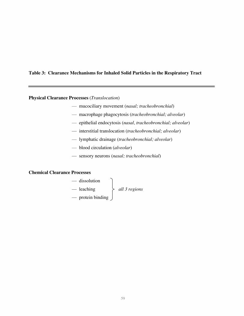
Classical Clearance Pathways
The clearance of deposited particles in the respiratory tract is basically due to twoprocesses (Table 3): i) physical translocation of particles by different mechanisms and (ii)
chemical dissolution or leaching. Chemical dissolution is directed at biosoluble particles orcomponents of particles that are either lipid soluble or soluble in intracellular and extracellular
fluids. Solutes and soluble components can then undergo absorption and diffusion or binding to
proteins and other subcellular structures, and may be eventually cleared into blood and lymphaticcirculation. This mechanism of clearance for biosoluble materials can happen at any location
21
within the three regions of the respiratory tract, although to different degrees, depending on local
extracellular and intracellular conditions (pH). In contrast, a number of diverse processesinvolving physical translocation of inhaled particles exist which are different in the three regions
of the respiratory tract. Figure 9 summarizes these clearance processes for solid particles. Aswill be pointed out, some of them show significant particle size–dependent differences, making
them uniquely effective for a certain particle size but very inefficient for other sizes.
The most prevalent mechanism for solid particle clearance in the alveolar region ismediated by alveolar macrophages, through phagocytosis of deposited particles. The success of
macrophage-particle encounter appears to be facilitated by chemotactic attraction of alveolarmacrophages to the site of particle deposition (Warheit et al. 1988). The chemotactic signal is
most likely C5a, derived from activation of the complement cascade from serum proteins present
on the alveolar surface (Warheit et al. 1986; Warheit and Hartsky 1993). This is followed bygradual movement of the macrophages with internalized particles towards the mucociliary
escalator. The retention halftime of solid particles in the alveolar region based on this clearance
mechanism is about 70 days in rats and up to 700 days in humans. The efficacy of this clearancemechanism depends highly on the efficiency of alveolar macrophages to “sense” deposited
particles, move to the site of their deposition, and then phagocytize them. This process ofphagocytosis of deposited particles takes place within a few hours, so that by 6-12 hours after
deposition essentially all of the particles will be phagocytized by alveolar macrophages, to be
cleared subsequently by the slow alveolar clearance mentioned above. However, it appears thatthere are significant particle size–dependent differences in the cascade of events leading to
effective alveolar macrophage mediated clearance.Figure 10 displays results of several studies in which rats had been exposed to different
sized particles (for the 3 and 10 µm particles intratracheal instillation of 40 µg and 10 µg
polystyrene beads were used) (Kreyling et al. 2002; Oberdörster et al. 1992b; Oberdörster et al.2000; Semmler et al. 2004). Twenty-four hours later the lungs of the animals were lavaged
repeatedly retrieving about 80% of the total macrophages as determined in earlier lavageexperiments (Ferin et al. 1991). As shown in Figure 10, ~80% of 0.5, 3 and 10 µm particles could
be retrieved with the macrophages, whereas only ~20% of nano-sized 15-20 nm and 80 nm
particles could be lavaged with the macrophages. In effect, ~80% of the ultrafine particles wereretained in the lavaged lung after exhaustive lavage, while ~20% of the larger particles > 0.5 µm
22
remained in the lavaged lung. This indicates that NSP were either in epithelial cells or had further
translocated to the interstitium.(See web-materials for additional details.)
Epithelial Translocation
Because of the apparent inefficiency of alveolar macrophage phagocytosis of NSP, one
might expect that these particles interact instead with epithelial cells. Indeed, results from severalstudies show that NSP deposited in the respiratory tract readily gain access to epithelial and
interstitial sites. This was also shown in studies with ultrafine PTFE fumes when shortly after a15-min. exposure the fluorine-containing particles could be found in interstitial and submucosal
sites of the conducting airways as well as in the interstitium of the lung periphery close to the
pleura (Oberdörster 2000). Such interstitial translocation represents a shift in target site awayfrom the alveolar space to the interstitium, potentially causing direct particle-induced effects
there.
Indeed, a surprising finding in a study evaluating the pulmonary inflammatory response ofTiO2 particles, ranging from NP TiO2 to pigment grade TiO2 (12-250 nm) was that 24 hours after
intratracheal instillation of different doses, higher doses induced a lower effect (Oberdörster et al.1992a). This was explained by the additional finding that at the higher doses (expressed as
particle surface area) of the nano-sized TiO2, 50% or more had reached the pulmonary
interstitium, causing a shift of the inflammatory cell response from the alveolar space to theinterstitium (more information on web). The smaller particle size of 12 and 20 nm vs. 220 and
250 nm also means that the administered particle number was more than 3 orders of magnitudehigher for the NSP, a factor that seems to be an important determinant for particle translocation
across the alveolar epithelium, as are the delivered total dose and the dose rate (Ferin et al. 1992).
Since interstitial translocation of fine particles across the alveolar epithelium is more prominent inlarger species (dogs, non-human primates) than in rodents (Kreyling and Scheuch 2000; Nikula et
al. 1997), it is reasonable to assume that the high translocation of NSP observed in rats occurs inhumans as well.
(See web-materials for additional details.)
Translocation to the Circulatory System
23
Once the particles have reached pulmonary interstitial sites, uptake into the blood
circulation in addition to lymphatic pathways can occur, a pathway that again is dependent onparticle size, favoring NSP. Berry et al.(1977) were the first to describe translocation of NSP
across the alveolar epithelium using intratracheal instillations of 30 nm gold particles in rats.They found within 30 minutes post-exposure large amounts of these particles in platelets of
pulmonary capillaries; they suggested that this is an elimination pathway for inhaled particles
which is of significance for transporting the finest air pollutant particles, in particular particles oftobacco smoke, to distant organs. They also hypothesized that this “might predispose to platelet
aggregation with formation of microthrombi atheromatous plaques”.Since then, a number of studies with different particle types have confirmed the existence
of this translocation pathway, as summarized in Table 4. Collectively, these studies indicate that
particle size and surface chemistry (coating), and possibly charge, govern translocation acrossepithelial and endothelial cell layers. In particular, the studies summarized by Mehta (2004) and
those performed by Heckel (2004)using intravenous administration of albumin-coated gold
nanoparticles in rodents demonstrated receptor-mediated transcytosis (albumin binding proteins)via caveolae (Figure11). These 50-100 nm vesicles, first described by Simionescu et al (1975)
form from indentations of the plasmalemma, and are coated with the caveolin-1 protein.Albumin, as the most abundant protein in plasma and interstitium, appears to facilitate NP
endocytosis, as does lecithin, a phospholipid: Even 240 nm polystyrene particles translocated
across the alveolo-capillary barrier when coated with lecithin, whereas uncoated particles did not(Kato et al. 2003). The presence of both albumin and phospholipids in alveolar epithelial lining
fluid may, therefore, be important constituents for facilitated epithelial cell uptake of NSP afterdeposition in the alveolar space.
Rejman et al. (2004) reviewed a number of different endocytic pathways for
internalization of a variety of substances, including phagocytosis, macropinocytosis, clathrin-mediated endocytosis, and caveolae-mediated endocytosis. They found in non-phagocytic cells in
vitro that internalization via clathrin-coated pits prevailed for latex microspheres <200 nm,whereas with increasing size up to 500 nm caveolae became the predominant pathway. However,
as shown in Table 4, surface coating of NSP with albumin clearly causes even the smallest
particles to be internalized via caveolae. The presence of caveolae on cells differs, they areabundant in lung capillaries and alveolar type l cells, but not in brain capillaries (Gumbleton
24
2001). In the lung, during inspiratory expansion and expiratory contraction of the alveolar walls,
caveolae with openings around 40 nm disappear and reappear, forming vesicles which are thoughtto function as transport pathways across the cells for macromolecules (Patton 1996). Knowledge
from virology about cell entry of biological NSP (viruses) via clathrin-coated pits and caveolaemechanisms should also be considered (Smith and Helenius, 2004), and can shed light on the
mechanism by which engineered NP may enter cells and interact with subcellular structures.
Evidence in humans for the translocation of inhaled NSP into the blood circulation isambiguous, with one study showing rapid appearance in the blood and significant accumulation
of label in the liver of humans inhaling 99Tc-labelled 20 nm carbon particles (Nemmar et al.2002a), while another study using the same labeled particles reported no such accumulation
(Brown et al. 2002). Taken together all of the evidence from animal and human studies for
alveolar translocation of NSP, it is likely that this pathway exists in humans as well; however, theextent of extrapulmonary translocation is highly dependent on particle surface
characteristics/chemistry, in addition to particle size. Translocation to the blood circulation could
provide a mechanism for a direct particle effect on the cardiovascular system as an explanationfor epidemiological findings of cardiovascular effects associated with inhaled ambient UFP
(Pekkanen et al., 2002; Wichmann et al., 2000) and for results of clinical studies showing vascularresponses to inhaled ultrafine elemental carbon particles ( Pietropaoli et al. 2004). In addition to
direct alveolar translocation of NSP, cardiovascular effects may also be the corollary of a
sequence of events starting with particle-induced alveolar inflammation initiating a systemic acutephase response with changes in blood coagulability and resulting in cardiovascular effects (Seaton
et al. 1995).Once NSP have translocated to the blood circulation, they can be distributed throughout
the body. The liver is the major distribution site via uptake by Kupffer cells followed by the
spleen as another organ of the reticuloendothelial system, although coating with PEG preventsalmost completely hepatic and splenic localization so that other organs can be targeted (Akerman
et al., 2002). Distribution to heart, kidney and immune-modulating organs (spleen, bone marrow)have been reported. For example, several types of NP, ranging from 10-240 nm, localized to a
significant degree in bone marrow following i.v. injection into mice (Table 5). Such target
specificity may be extremely valuable for drug delivery; for example, drug delivery to the CNSvia blood-borne NP requires NP surface modifications in order to facilitate translocation across
25
the tight blood-brain barrier via specific receptors (e.g., apolipoprotein coating for LDL receptor
mediated endocytosis in brain capillaries) (Kreuter. 2001, 2004; Kreuter et al. 2002). Such highlydesirable properties of NP must be carefully weighed against potential adverse cellular responses
of targeted NP drug delivery, and a rigorous toxicological assessment is mandatory.(See web-materials for additional details.)
Neuronal Uptake and TranslocationA translocation pathway for solid particles in the respiratory tract involving neuronal
axons is apparently specific for NSP. Respective studies are summarized in Table 6. Thispathway was already described more than 60 years ago, yet it has received only little or no
attention by toxicologists. It is depicted in Figure 9 for the nasal and tracheobronchial regions,
comprising sensory nerve endings of the olfactory and the trigeminus nerves and of an intricatenetwork of sensory nerve endings in the tracheobronchial region. These early studies concerned a
large series of studies with 30 nm polio virus intranasally instilled into chimpanzees and Rhesus
monkeys (Bodian and Howe 1941; Bodian and Howe 1941; Howe and Bodian 1940). Theirstudies revealed that the olfactory nerve and olfactory bulbs are, indeed, portals of entry to the
CNS for intranasally-instilled nano-sized polio virus particles, which could subsequently berecovered from the olfactory bulbs. The close proximity of nasal olfactory mucosa and olfactory
bulb requires only a short distance to be covered by neuronal transport (Fig. 12). Bodian and
Howe (Bodian and Howe 1941) determined the transport velocity of the virus in the axoplasm ofaxons to be 2.4 mm/hr., which is very well in agreement with neuronal transport velocities
measured later by Adams and Bray (1983) for solid particles (up to 500 nm) directlymicroinjected into giant axons of crabs, and by deLorenzo (1970) for silver-coated colloidal gold
(50 nm) in squirrel monkeys.
The de Lorenzo (1970) study demonstrated in squirrel monkeys that intranasally-instilledsilver-coated colloidal gold particles (50 nm) translocated anterogradely in the axons of the
olfactory nerves to the olfactory bulbs. The 50 nm gold particles even crossed synapses in theolfactory glomerulus to reach mitral cell dendrites within one hour after intranasal instillation. An
interesting finding in this study — and important for potential adverse effects — was that the NSP
in the olfactory bulb were no longer freely distributed in the cytoplasm but were preferentiallylocated in mitochondria (see also section 3.3).
26
Newer studies indicated that this translocation pathway is also operational for inhaled
NSP. Inhalation of ultrafine elemental 13C particles (CMD=35 nm) resulted in a significantincrease of 13C in the olfactory bulb on day 1, which increased further throughout day 7 post-
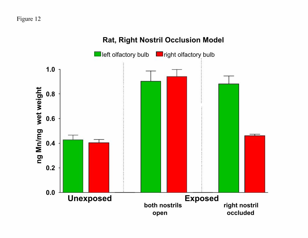
exposure (Oberdörster et al. 2004). Results of another inhalation study with solid nano-sized(CMD=30 nm) manganese oxide particles in rats showed after a 12-day exposure a more than 3.5-
fold significant increase of Mn in the olfactory bulb, compared to only a doubling of Mn in the
lung. When one nostril was occluded during a 6-hr. exposure, Mn accumulation in the olfactorybulb was restricted to the side of the open nostril only (Figure 13) (Feikert et al. 2004). This
result contrasts with 15-day inhalation of larger-sized MnO2 particles in rats (1.3 and 18 µmMMAD) where no significant increases in olfactory Mn was found (Fechter et al. 2002). This
was to be expected given that the individual axons of the fila olfactoria (forming the olfactory
nerve) are only 100-200 nm in diameter (De Lorenzo, 1957; Plattig, 1989Collectively, these studies point out that the olfactory nerve pathway should also be
considered a portal of entry to the CNS for humans under conditions of environmental and
occupational exposures to airborne NSP. However, there are important differences betweenrodents and humans. The olfactory mucosa of the human nose comprises only 5% of the total
nasal mucosal surface as opposed to 50% in rats – which in addition are obligatory nose breathers(Table 7). One can argue that the olfactory route may, therefore, be an important transfer route to
the CNS for inhaled NSP in animals with a well-developed olfaction system, yet at the same time
its importance for humans with a more rudimentary olfactory system can be questioned.However, estimates using a predictive particle deposition model and data from Table 7 show that
concentrations of 20 nm translocated particles in the human olfactory bulb can, indeed, be 1.6 –10 times greater than in rats. (Additional information on web).
Translocation into deeper brain structures may possibly occur as well, as shown in rats for
soluble manganese (Gianutsos et al. 1997), but requires further confirmatory studies with respectto solid NSP. Further evidence for movement of NSP along axons and dendrites in humans is
provided by knowledge accumulated by virologists who have long understood the movement ofhuman meningitis virus through olfactory and trigeminal neurons, and, similarly, herpes virus
movement up and down the trigeminal neuron to trigger outbreaks of herpes cold sores in humans
(Kennedy and Chaudhuri 2002; Terasaki et al. 1997).
27
There are additional neuronal translocation pathways for solid NSP via the trigeminus
nerve and tracheobronchial sensory nerves (Table 6). A study by Hunter and Dey (1998) in ratsdemonstrated the translocation of intranasally-instilled rhodamine-labelled microspheres (20-200
nm) to the trigeminal ganglion inside the cranium via uptake into the ophthalmic and maxillarybranches of the trigeminus nerve which supplies sensory nerve endings throughout the nasal
mucosa. Hunter and Undem (1999) in another study instilled the same microparticles
intratracheally into guinea pigs; they found neuronal translocation of these solid microparticles tothe ganglion nodosum in the neck area which is networked into the vagal system. This finding
may be relevant for ambient UFP since it can be hypothesized that cardiovascular effectsassociated with ambient particles in epidemiological studies (Utell et al. 2002) are in part due to
direct effects of translocated UFP on the autonomic nervous system via sensory nerves in the
respiratory tract.In the context of potential CNS effects of air pollution, including ambient UFP, two recent
studies with exposures of mice to concentrated ambient fine and ultrafine particles should be
mentioned. The authors found significant increases of TNFa or decreases in dopaminergic
neurons, supporting the hypothesis of ambient PM causing neurodegenerative disease (Campbellet al., 2005; Veronesi et al. 2005). A study by Calderon-Garcidueñas et al. (2002) may also point
to an interesting link between air pollution and CNS effects: These authors describe significantinflammatory or neurodegenerative changes in the olfactory mucosa, olfactory bulb and cortical
and subcortical brain structures in dogs from a heavily polluted area in Mexico City, whereas
these changes were not seen in dogs from a little polluted rural control city. However, whetherdirect effects of airborne UFP are the cause of these effects remains to be determined.
Although the existence of neuronal translocation of NSP has been well established, itneeds to be emphasized that size alone is only one particle parameter governing this process.
Surface characteristics of NSP (chemistry, charge, shape, aggregation) are essential determinants
as well, and it needs to be cautioned to assume that all NSP, when inhaled, will be distributing bythe mechanism described here. It should be kept in mind though, that the unique biokinetic
behavior of NSP — cellular endocytosis, transcytosis, neuronal and circulatory translocation anddistribution — which makes them desirable for medical therapeutic or diagnostic applications —
may be are associated with potential toxicity. For example, NP facilitated drug delivery to the
CNS raises the question of the fate of NP following their translocation to specific cell types or to
28
subcellular structures in the brain, e.g., does mitochondrial localization induce oxidative stress?
How persistent is the coating or the core of the NP? A respective safety evaluation is key. (Seeweb-materials for additional details.)
4.2 Exposure via GI Tract and Skin
NSP cleared from the respiratory tract via the mucociliary escalator can subsequently be
ingested into the gastrointestinal (GI) tract. Alternatively, nanomaterials can be ingested directly,for example if contained in food or water or if used in cosmetics or as drugs or drug delivery
devices. Only a few studies have investigated the uptake and disposition of nanomaterials by theGI tract, and most have shown that NSP pass through the GI tract and are eliminated rapidly. In
rats dosed orally with radiolabeled functionalized C-60 fullerenes, water solubilized using PEG
and albumin (18 kBq in 100 mL), 98% were cleared in the feces within 48 hours, while the rest
was eliminated via urine, indicating some uptake into the blood circulation (Yamago et al. 1995).In contrast, in this same study, 90% of the same radiolabeled fullerenes administered i.v. (9.6
kBq, ~50mL or 14-18 kBq in 215 mL) were retained after one week, with the majority (73-80%,
depending on time course) found in the liver. Studies by Kreyling (Kreyling et al., 2002,
Semmler et al., 2002) using ultrafine 192Ir did not show significant uptake in the GI tract, whileearlier studies with larger TiO2 particles (150-500 nm) found uptake into the blood and movement
to the liver (Jani et al., 1994 and Böckmann et al. 2000). Likely there are both particle surfacechemistry and particle size dependent differences in GI tract uptake.
A potentially important uptake route is through dermal exposure. The epidermis,
consisting of the outer horny layer (stratum corneum), the prickle cell layer (stratum spinosum)and basal cell layer (stratum basale) forms a very tight protective layer for the underlying dermis
(Fig. 14). The dermis has a rich supply of blood and tissue macrophages, lymph vessels, dendriticcells (Langerhans, also in stratum spinosum of epidermis), and five different types of sensory
nerve endings. Broken skin represents a readily available portal-of-entry even for larger (0.5 – 7
µm) sized particles, as evidenced by reports about accumulation of large amounts of soil particlesin inguinal lymph nodes of barefoot walking/running people; this can be associated with
hypothesized that unbroken skin when flexed — as in wrist movements — would make theepidermis permeable for NSP. They demonstrated in a proof of concept experiment that, indeed,
29
flexing the skin, but not flat skin, resulted in penetration of even 1 µm fluorescent beads to the
dermis. The followup question about access of particles in the dermis to the circulation isanswered by the aforementioned reports of podoconiosis, i.e., uptake into the lymphatic system
and regional lymph nodes. Subsequent translocation of NSP beyond lymph nodes to the bloodcirculation is likely to occur as well, as shown in studies with small asbestos fibers (Oberdörster
et al. 1988).
Newer studies by Kim et al. (2004) in mice and pigs with intradermally-injected nearinfrared quantum dots confirmed that NP, once in the dermis, will localize to regional lymph
nodes, which makes these particles very useful for in vivo imaging. Likely transport mechanismsto the lymph nodes are skin macrophages and dendritic (Langerhans) cells (Sato et al. 1998; Ohl
et al. 2004); this raises a question about potential modulation of immune responses, following
interaction of these NP containing macrophages and dendritic cells with T-lymphocytes. Forexample, Chen et al. (1998) were able to raise antibodies in mice specific for C60 after i.p.
injections of C60 conjugated to thyroglobulin and serum albumin. Clearly, research is needed to
determine whether and under what conditions NP can be recognized by the immune system,following any route of uptake into the organism.
Another question relates to the potential of sensory skin nerves to take up and translocateNP: Given that this mechanism has been demonstrated for the nasal and tracheobronchial regions
of the respiratory tract, how likely is this to occur in the dermis layer of the skin with its dense
supply of different types of sensory nerves? It may be conceivable, considering data on neuronaluptake and translocation of NSP after intramuscular injection. For example, nano-sized ferritin
and iron-dextran, after injection into the tongue of mice, labeled the neurons of the hypoglossalnuclei; and injection of both of these NSP into facial muscles of mice also resulted in synaptic
uptake; cationized ferritin was also detected in cell bodies of facial neurons indicating that
electrical charge is of importance for incorporation into axons and axonal transport (Arvidson1994; Malmgren et al. 1978; Olsson and Kristensson 1981). Other studies using intra-muscular
injection of ferritin (~112 nm), iron-dextran (11 or 21 nm) and gold protein (20-25 nm)nanoparticles also showed rapid penetration through the basal lamina into the synaptic clef of the
neuromuscular junction, but this was restricted only to the smaller nanoparticles, implying that
there may be a size-dependent penetration of the basal lamina with a threshold somewherebetween 10 and 20 nm (Oldfors and Fardeau 1983).
30
Neuronal transport of NSP along sensory skin nerves is well established for herpes virus.
After passing through the skin — especially broken skin — they are transported retrogradelyalong dendrites of sensory neurons to the dorsal root ganglion, remain dormant there until a stress
situation triggers anterograde translocation along the dendrites back to the skin (Kennedy andChaudhuri 2002; Terasaki et al. 1997). Future studies need to determine as to whether and to
what degree such translocation along sensory skin neurons also occurs with NP penetrating the
epidermis.
5. Risk AssessmentThe lack of toxicology data on engineered NP does not allow for adequate risk
assessment. Because of this, some may even believe engineered NP so risky that they call for a
precautionary halt in NP-related research. However, the precautionary principle should not beused to stop research related to nanotechnology and NP. Instead, we should strive for a sound
balance between further development of nanotechnology and the necessary research to identify
potential hazards in order to develop a scientifically defensible database for the purpose of riskassessment. To be able to do this, a basic knowledge about mammalian and eco-toxicological
profiles of NP is necessary, rather than attempting to assess NP risks based on some popularscience fiction literature. Most importantly, sufficient resources should be allocated by
governmental agencies and industries to be able to perform a scientifically based risk assessment
and then establish justifiable procedures for risk management. The data needed for this riskassessment should be determined a priori so that limited resources can be used efficiently to
develop useful and well-planned studies.At this point, governmental regulation is not possible given the lack of needed information
on which to base such regulations. However, academia and industry and regulatory governmental
agencies should seriously consider the view that engineered nanoparticles have new and uniquebiological properties and that the potential risks of engineered NP are not the same as those of the
bulk material of the same chemistry. Assigning a unique identifier to nano-sized materials wouldindicate that the toxicology profile of the material in question may not be the same as the bulk
material. Toxicological tests and the resulting data base would provide information for MSDS
sheets for NP as well as a basis for potential NP risk assessments and risk management.Obviously, this approach may not be appropriate for all NP, for example, when embedded in a
31
matrix, and the feasibility of this proposed strategy needs to be thoroughly discussed and
considered. For discussing this, and for developing and deciding upon a reasonable battery oftests for toxicological profiling, it would be very useful to convene international multidisciplinary
workshops of experts from industry, academia, and regulatory agencies (including materialscientists, chemists, chemical engineers, toxicologists, physicians, regulators, statisticians, and
others) to establish a NP classification scheme and testing guidelines. A multidisciplinary and
multi-national collaborative team approach is critical. Respective efforts have been initiatednationally by the American Standards Institute (ANSI 2004) and internationally by the
International Council on Nanotechnology (ICON 2004) as well as the International Organizationfor Standardization.
As many regulatory agencies do not consider a nanotechnologically manufactured
substance different from the conventional substance, the manufacture and use of nanotechnologyproducts are currently not specifically regulated. Typically, nano-sized substances are treated as
variations of the technical material or existing formulation and thus do not require a separate
registration. A main reason for producing nano-sized form of a registered substance, however, isthat conversion of a substance to a nanoparticle imparts new properties to the substance (e.g.,
enhanced mechanical, electrical, optical, catalytic, biological activity). Thus, as stated earlier,while the toxicology of the base material may be well defined, the toxicity of the nanotechnology
form of the substance may be dramatically different from its parent form. As a result, new
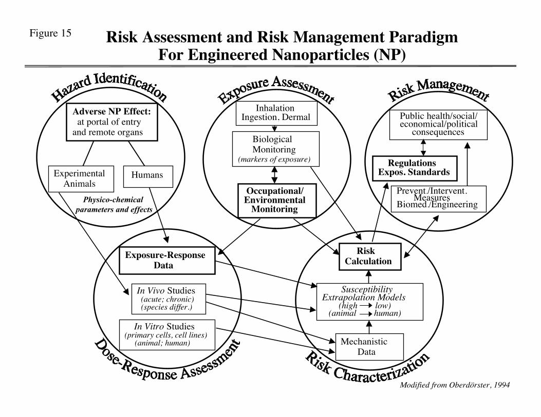
toxicology data on the nano-size form of a substance is likely to result in a different hazardassessment for the NP. Figure 15 depicts a Risk Assessment/Risk Management paradigm
pointing out different steps and data required for this process. As described in the previous sections, the difference in toxicological profile of NP
compared to its parent form is not only due to its intrinsic chemical properties, but to a large
degree to differing kinetics in vivo. While larger particles may not enter the CNS, the potentialexists for inhaled nano-sized particles to be translocated to the CNS via the axons of sensory
neurons in the upper respiratory tract. Furthermore, while the toxicity per unit mass of aparticular substance may vary depending on the nano vs. large form, it will be important to take
into account not only new biological activities, but also potential new target organs and routes of
exposure. To what degree does the nano-form of a substance have enhanced dermal penetration,or increased systemic uptake via the lung or GI-tract? What determines how many nanoparticles
32
that enter the systemic circulation distribute throughout the body, reach the bone marrow, cross
the blood brain barrier, cross the placenta, and affect the developing offspring, or sequestereffectively in the liver? Do nanoparticles released into the environment affect species that are
important in food chain dynamics? What are the long-term consequences of exposure tonanoparticles? Changes in toxicity profile and new target organs can be expected, and it will then
be necessary to establish new risk assessments for nanoparticles in addition to the bulk material.
Currently there exists a paucity of data to effectively address these questions but it will beimportant to determine whether there exist common modes of action/behavior of NP to establish
baseline assumptions for use in risk assessments.The use of nanotechnology products will likely increase dramatically over the next
decade. In fact, nanomaterials are already being used in applications ranging from burn and
wound dressings to dental-bonding agents to sunscreens and cosmetics to fuel cells, tires, optics,clothing, and electronics. While currently there exists little public awareness of nanotechnology
in everyday life (e.g., stain-free clothing), it would be prudent to examine and address
environmental and human health concerns before the widespread adoption of nanotechnology.Both the societal benefits and potential risks of nanotechnology should be evaluated and clearly
communicated to the general public and regulators. This type of open communication andrisk/benefit evaluation will avoid the pitfalls encountered with genetically modified organisms
recently experienced in the field of biotechnology. In that instance, the benefits of the emerging
field of biotechnology were not communicated effectively before the introduction of thetechnology. As the public's awareness of this new technology grew, regulators and producers of
biotechnology failed to effectively acknowledge public concerns that genetically modifiedorganisms could adversely affect ecosystem balance. As a result, the public support of
genetically modified organisms, particularly in the EU, is low. For nanomaterial producers it will
be important to demonstrate that what they may perceive as a new and potentially harmless formof a familiar material has, indeed, an acceptable risk profile. If such proactive steps are not taken,
nanomaterials may be regarded as dangerous by the public and regulators, which could lead toinappropriate categorization and unnecessarily burdensome regulations. Such action (or inaction
on the side of producers), in turn, could result in significant barriers to commercialization and the
widespread acceptance of otherwise useful nanotechnology materials.
33
6. Summary & OutlookResearch on ambient UFP has laid the foundation for the emerging field of nano-
toxicology, with the goal to study the biokinetics and the potential of engineered nanomaterials
(particles, tubes, shells, quantum dots, etc.) to cause adverse effects. Major differences betweenambient UFP and NP are the polydisperse nature of the former vs. the monodisperse size of the
latter; and particle morphology, oftentimes a branched structure from combustion vs. spherical
form of NP, although other shapes (tubes, wires, rings, planes) are also manufactured. Inaddition, combustion derived volatile organic compounds and inorganic constituents (e.g., metals,
nitrates, sulfates) of different solubilities on UFP predict differences in the toxicological profilebetween UFP and NP. However, as far as the insoluble particle is concerned, concepts of NSP
kinetics, including cell interactions, will most likely be the same for UFP and NP (Figure 16).
The introduction of nanostructured materials for biomedical and electronics applicationsopens tremendous opportunities for biomedical applications as therapeutic and diagnostic tools as
well as in the fields of engineering, electronics, optics, consumer products, alternative energy,
soil/water remediation and others. However, very little is known yet about their potential to causeadverse effects or humoral immune responses once they are introduced into the organism —
unintentionally or intentionally. Nano-medicine products will be well-tested prior to introductioninto the marketplace. However, for the manufacturers of the majority of current nano-tech
products, regulations requiring nanomaterial-specific data on toxicity prior to introduction into the
marketplace are an evolving area and presently under discussion (Bergeson and Auerbach, 2004;Foresight and Governance Project, 2003). During a product’s Life Cycle (manufacture, use,
disposal), it is probable that nanomaterials will enter the environment, and currently there is nounified plan to examine ecotoxicological effects of NP. In addition, the stability of coatings and
covalent surface modifications need to be determined both in ecological settings and in vivo.
(Additional information on web).Results of older biokinetic studies and some new toxicology studies with NSP (mostly
ambient UFP) can be viewed as the basis for the expanding field of nanotoxicology. Thesestudies showed that the greater surface area per mass renders NSP more active biologically than
larger sized particles of the same chemistry, and that particle surface area and number appear to
be better predictors for NSP-induced inflammatory and oxidative stress responses. The followingemerging concepts of nanotoxicology can be identified from these studies:
34
• The biokinetics of nano-sized particles are different from larger particles:
– when inhaled:they are efficiently deposited in all regions of the respiratory tract;
they evade specific defense mechanisms;they can translocate out of the respiratory tract via different pathways
(endocytosis and transcytosis)
– when in contact with skin:there is evidence of penetration to the dermis;
they translocate via lymph to regional lymph nodes;a possible uptake into sensory nerves needs to be investigated
– when ingested:
there appears to be little uptake into the organism, mostly excreted via feces– when in blood circulation:
they can distribute throughout the organism,
they are taken up into liver, spleen, bone marrow, heart, and other organs– in general, translocation rates are largely unknown, they are probably very low but
are likely to change in a compromised/diseased state• The biological activity and biokinetics are dependent on many parameters:
– agglomeration state; biopersistence; dose• These parameters are likely to modify responses and cell interactions such as:
– a greater inflammatory potential than larger particles per given mass
– translocation across epithelia from portal-of-entry to other organs– translocation along axons and dendrites of neurons
– induction of oxidative stress– pro-oxidant and antioxidant activity of NSP in environmentally-relevant species
– binding to proteins, receptors
– localization in mitochondria
35
The principles of cellular and organismal interactions that have been discussed in this
manuscript, should be applicable for both ambient UFP and NP, even if the latter are coated witha biocompatible material. Knowledge about the biopersistence of this coating is as essential as is
knowledge about the bioavailability of the core material that could have intrinsic toxic properties,e.g., semiconductor metal compounds in sub 10 nm quantum dots consisting of cadmium and lead
compounds. The very small size of these materials makes them available to the same
translocation processes described here for polydisperse NSP, possibly even in a more efficientway because of their uniform size. When studying biological/toxicological effects, new processes
of interactions with subcellular structures, e.g., microtubuli, mitochondria, will likely bediscovered. The diversity of engineered nanomaterials and of the potential effects represents
major challenges and research needs for nanotoxicology, including also the need for assessing
human exposure during manufacture and usage. The goal to exploit positive aspects ofengineered nanomaterials and avoid potential toxic effects can best be achieved through a
multidisciplinary team effort involving researchers in toxicology, materials science, medicine,
molecular biology, bioinformatics and their subspecialties.
36
7. References:Adams RJ, Bray D. 1983. Rapid transport of foreign particles microinjected into crab axons.
Nature 303:718-720.
Akerman MA, Chan WCW, Laakkonen P, Bhatia SN, Ruoslahti E. 2002. Nanocrystal targeting in
vivo. PNAS 99(No. 20):12617-12621.
Amato I. 1989. Making the Right Stuff. Science News 136:108-110.
Anderson PJ, Wilson JD, Hiller FC. 1990. Respiratory tract deposition of ultrafine particles in
subjects with obstructive or restrictive lung disease. Chest 97:pp. 1115-1120.
ANSI. 2004. American National Standards Institute. Available: http://www.ansi.org. [accessed
March 16, 2005].
Arvidson B. 1994. A review of axonal transport of metals. Toxicol 88:1-14.
Auclair F, Baudot P, Beiler D, Limasset J. 1983. Accidents benines et mortetels dus aux
"traitement" du polytetrafluoroethylene en millieu industriel: Observations cliniques et
measures physio-chemiques des atmosphere pollues. Toxicol European Res 1:43-48.
Table 4: Particle Size and Surface Chemistry-related Alveolar-Capillary Translocation
Particle size Type Translocation Localization/Effect Reference (nm)
5-20 nm Gold, albumin coated yes via caveolae Mehta et al., 2004
8 nm Gold, albumin coated yes via “vesicles” Konig et al., 1993
8 nm Gold, albumin coated yes via caveolae Heckle et al., 2004
18 nm Iridium yesa extrapulm. org. Kreyling et al., 2002
30 nm Gold yes platelet? Berry et al., 1977
35 nm Carbon yes liver Oberdörster et al., 2002
60 nm Polystyrene, b yes thrombus, early Nemmar et al., 2002bSilva et al., 2005
60 nm Polystyrene ? no thrombus Nemmar et al., 2002b
80 nm Iridium yesa extrapulm. org. Kreyling et al., 2002
240 nm Polystyrene, lecithin yes monocyte Kato et al., 2003
240 nm Polystyrene, uncoated no Kato et al., 2003
400 nm Polystyrene no thrombus, late Nemmar et al., 2004
a = minimal; b = indirect evidence
Surface coating (chemistry) charge, size govern translocation
61
Table 5: Translocation of Nano-sized Particles in the Blood Circulation to Bone Marrowin Mice.
Particle Size Type Finding Reference
~10 nm PEG-quantum dots Fast appearance of QDs in Ballou et al., 2004liver, spleen, lymph nodes
and bone marrow (mouse)
<220 nm Metallo-fullerene Highest accumulation in bone Cagle et al., 1999
marrow after liver; continued
increase in bone marrow butdecrease in liver (mouse)
90 – 250 nm HAS coated polylactic Significant accumulation in Bazile et al., 1992
acid nanoparticles bone marrow, second to liver
(rat)
240 nm Polystyrene (non Rapid passage through Gibaud et al.,biodegradable) endothelium in bone marrow, 1996
polylisohexylcyon- uptake by phagocytizing cells 1998
acrylate (biodegradable) in tissue (mouse) 1994
62
Table 6: Studies of Neuronal Translocation of UFP from Respiratory Tract
1941: Bodian and Howe: Olfactory axonal transport of Polio-virus (30 nm) afterintranasal instillation in chimpanzee.
Transport velocity: 2.4 mm/h
1970: de Lorenzo: Olfactory axonal transport of 50 nm silver-coated gold after
intranasal instillation in squirrel monkey.Transport velocity: 2.5 mm/h
1998: Hunter and Dey: Retrograde tracing of trigeminal neurons from nasal epitheliumwith microspheres
1999: Hunter and Undem: Rhodamine-labelled microspheres (20-200 nm) translocation
via sensory nerves of TB region to ganglion nodosum in hamster after
intratracheal instillation
2004a: Oberdörster et al.: 13C particles (CMD ~36 nm) in olfactory bulb after whole-body inhalation exposure in rats
63
Table 7: Rat vs. Human Nasal and Olfactory Parameters.
Rat Human
Breathing mode obligatory nose nasal/oro-nasal
Area of nasal mucosa ~16 cm3 ~105 cm2
Area of olfactory mucosa ~8 cm3 ~5.25 cm2
(% of total mucosa) (50) (5)
% of nasal airflow going to
olfactory mucosa ~15 ~10
Weight of olfactory bulb ~85 ng ~168 ng
Based on Keyhani et al., 1997; Kimbell et al., 1997; Turetsky et al., 2003
64
9. FIGURE LEGENDS
Figure 1: Idealized size distribution of traffic-related particulate matter (EPA, 2004). The fourpolydisperse modes of traffic-related ambient particulate matter span approximately 4
orders of magnitude from below 1 nm to above 10 µm. Nucleation and Aitken modeparticles are defined as ultrafine particles (<~100 nm). Source-dependent chemical
composition is not well controlled and varies considerably. In contrast engineered
nanoparticles (1-100 nm) have well controlled chemistry and are generallymonodispersed.
Figure 2: Surface molecules as function of particle size. Surface molecules increase
exponentially when particle size decreases below 100 nm, reflecting the importance
of surface area for increased chemical and biological activity of NSP. The increasedbiological activity can either be positive and desirable (e.g., antioxidant activity,
carrier capacity for therapeutics, penetration of cellular barriers) or negative andundesirable (e.g., toxicity, induction of oxidative stress or of cellular dysfunction) or
also a mix of both. (Figure courtesy of Dr. Fissan, personal communication)
Figure 3: Hypothetical cellular interaction of NSP (adapted from Donaldson and Tran (2002).Inflammation and oxidative stress can be mediated by several primary pathways: 1)
Particle surface causes oxidative stress resulting in increased intracellular calcium and
gene activation, 2) Transition metals released from particles result in oxidative stress,increased intracellular calcium, and gene activation, 3) Cell surface receptors are
activated by transition metals released from particles, resulting in subsequent geneactivation, 4) Intracellular distribution of NSP to mitochondria generates oxidative
stress.
Figure 4: Percent of neutrophils in lung lavage of rats and mice as indicators of inflammation
24 hrs. after intratracheal instillation of different mass doses of 20 nm and 250 nm
65
TiO2 particles in rats and mice. (a) The steeper dose–response of nano-sized TiO2 is
obvious when dose is expressed as mass. (b) The same dose–response relationshipbut with dose expressed as particle surface area, indicating that particle surface area
seems to be a more appropriate dosemetric for comparing effects of different sizedparticles provided they are of the same chemical structure (anatase TiO2 in this case).
( 20 nm TiO2; 250 nm TiO2, saline control)
Figure 5: Routes of exposure, uptake, distribution and degradation of NSP in the Environment.
Solid lines indicate routes which have been demonstrated in the laboratory or field; orwhich are currently in use (remediation). Italics indicate possible degradation routes,
while blue lettering indicates possible sinks and sources of NSP.
Figure 6: Engineered NP have been shown to release oxyradicals (pictured here is the
mechanism of C60 as determined by Yamakoshi et al. (2003), which can interact with
the antioxidant defense system. In addition to fullerenes, metals such as Cd, Fe, or Niquantum dots, or Fe from SWNT manufacturing, could also act in Fenton-type
Figure 7: Some basic shapes of exposure-response or dose-response relationships.
Prerequisites for establishing these relationships for NSP from in vitro or in vivo
studies include a sufficient number of data points, i.e., over a wide range of exposure
concentrations or doses; knowledge about exposure levels; and information about
correlation of exposure with doses at the organismal or cellular level (an exposure isnot a dose!). Dose-response curves of different shapes can be extrapolated when only
response data at high dose levels (indicated by dashed oval) are available. Lack ofdata in the low – oftentimes the most relevant – dose range can result in severe
misinterpretation if a threshold or even a hormetic response is present. Consideration
also needs to be given to the likelihood that the shape or slope of exposure-dose-response relationships change for susceptible parts of the population.
66
Figure 8: Predicted fractional deposition of inhaled particles in the nasopharyngeal,tracheobronchial and alveolar region of the human respiratory tract during nose-
breathing (based on data from ICRP (1994). (Drawing courtesy of J. Harkema)
Figure 9: Pathways of particle clearance (disposition) within and out of the respiratory tract.
There are significant differences between nano-sized and larger particles for some ofthese pathways (see text). (Drawing courtesy of J. Harkema).
Figure 10: In vivo retention of inhaled nano-sized and larger particles in alveolar macrophages
(left side) and in exhaustively lavaged lungs (epithelial and interstitial retention, right
side) 24 hrs. post-exposure. The alveolar macrophage is a most important defensemechanism in the alveolar region for fine and coarse particles, yet inhaled singlet
NSP are not efficiently phagocytized by alveolar macrophages.
Figure 11: Different forms of caveolae and cellular tight junctions function as translocation
mechanisms across cell layers. Depending on particle surface chemistry, NSP havebeen shown to transcytose across alveolar type I epithelial cells and capillary
endothelial cells (Table 4), but not via cellular tight junctions. However, in a
compromised or disease state (e.g., endotoxin exposure) translocation across widenedtight junction occurs as well (Heckel et al., 2004). This indicates that assessing
potential effects of NSP in the compromised state is an important component ofNanotoxicology.
Figure 12: Close proximity of olfactory mucosa to olfactory bulb of the CNS. Inhaled NSP,especially below 10 nm, deposit efficiently on the olfactory mucosa by diffusion,
similar to airborne “smell” molecules which deposit in this area of olfactory dendriticcilia. Subsequent uptake and translocation of solid NSP along axons of the olfactory
nerve has been demonstrated in non-human primates and rodents. Surface chemistry
of the particles may influence their neuronal translocation. (Figure used with
permission from McGraw-Hill)
67
Figure 13: Occlusion of the right nostril of rats during 6 hr. inhalation of nano-sized Mn-oxideparticles (~30 nm CMD, ~450 µg/m3) resulted in accumulation of Mn only in the left
olfactory bulb only at 24 hrs. after dosing (Feikert et al., 2004).
Figure 14: The epidermis represents a tight barrier against NSP penetration. Quantitatively,
dermal translocation will, therefore, be minimal or non-existent under normalconditions but increases in areas of skin flexing (Tinkle et al., 2003) and broken skin.
Once in the dermis, lymphatic uptake is a major translocation route, likely facilitatedby uptake in dendritic cells (epidermis) and macrophages; other potential pathways
may include the dense networks of blood circulation and sensory nerves in the dermis.(Figure adapted from: http/www.essentialdayspa.com/Skin_Anatomy_and_Physiology.htm)
Figure 15: Risk assessment (NRC 1983) and risk management paradigm for engineered
nanoparticles (NP). The four steps of risk assessment require answers to the
questions: Do NP have adverse effects? What are the dose–response relationships?What are occupational/environmental levels in different media? What is the
calculated risk? Once a risk is determined, risk management decision can be
established, including exposure standards and regulations and efforts for effective riskcommunication.
Figure 16: Biokinetics of NSP. While many uptake and translocation routes have been
demonstrated, others still are hypothetical and need to be investigated. Largely
unknown are translocation rates as well as accumulation and retention in criticaltarget sites and their underlying mechanisms. These as well as potential adverse
effects will be largely dependent on physicochemical characteristics of the surfaceand core of NSP. Both qualitative and quantitative changes in NSP biokinetics in a
diseased or compromised organism need also to be considered.
Figure 1
0
20
40
60
1 10 100 1000 10000
% S
urfa
ce M
olec
ules
Diameter (nm)
Figure 2
particle releasestransition metals
activation ofreceptor (e.g. EGFR)
increased cytosoliccalcium and
oxidative stress
phagocytosis
particle surface causesoxidative stress
signaling pathways
NF-_B
inflammatory mediators
inflammation
Figure 3
0 50 100 150 200 2500
10
20
30
40
50
ultrafine TiO2 (~20nm) fine TiO 2 (~250nm) saline
Particle Surface Area, cm2
(b)Rats
Mice
0 500 1000 1500 20000
10
20
30
40
50Pe
rcen
t Neu
trop
hils
(a)
Figure 4
µg
µg
cm2
cm2
TiO2 Mass TiO2 Surface Area0 100 200 300 400
0
10
20
30
40
50
0 10 20 30 40 500
10
20
30
40
50(a) (b)
20 nm
250 nm
20 nm250 nm
Air
water
sediment
soil
aquifer
deposition/spillage injection for
remediation
sorption toorganic matter
volatilization/dust
(soil) floraand fauna
benthic organismsbiodegradation?
chemical degradation?
food chain
wildlife &humans
UV degradation
leaching
wildlife &humans
food chain
Legend: Routes of exposure, uptake, distribution and degradation of NSP in the Environment. Solidlines indicate routes which have been demonstrated in the laboratory or field; or which are currently inuse (remediation). Italics indicate possible degradation routes, while blue lettering indicates possiblesinks and sources of NSP.
filter feeders
foodchain
biodegradation?
Figure 5
Protein oxidation
Redox Cycle
Nanoparticles or metals
superoxide
O2.
SODO2
H2O2
•OH+OH-
H2O + O2
GSH
GSSG
GSH Reductase GS- GSSG
.+O2
DNA strand breaks, covalent modification of DNA
catalaseH2O + O2
GPx
Lipid Peroxidation
Metallothionein Induction
Intermediates from oxyradical attacks
RH2 + GS• RH•
hv ISC C60 3C60
. 1C60. 1C60
O2
not shown: phase II biotransformation, ascorbic acid, vitamin E, beta carotene etc.