NIH Comparative Biomedical Scientist Training Program Symposium April 18–20, 2012 Natcher Conference Center, National Institutes of Health Bethesda, Maryland Translational Perspectives on Melanomas in Humans and Dogs TP B C NIH Comparative Biomedical Scientist Training Program An NCI Symposium with support from Animal Cancer Foundation, Inc., Canine Comparative Oncology and Genomics Consortium, Inc., and NHLBI, NIAID, and NINDS U.S. DEPARTMENT OF HEALTH AND HUMAN SERVICES National Institutes of Health National Cancer Institute
Transcript
NIH Comparative Biomedical Scientist Training Program SymposiumApril 18–20, 2012Natcher Conference Center, National Institutes of Health Bethesda, Maryland
Translational Perspectives on Melanomas in Humans and Dogs
TPBCNIH
Comparative Biomedical Scientist Training Program
An NCI Symposium with support from Animal Cancer Foundation, Inc., Canine Comparative Oncology and Genomics Consortium, Inc., and NHLBI, NIAID, and NINDS
U.S. DEPARTMENT OF HEALTH AND HUMAN SERVICES
National Institutes of Health
Natio
nal C
ance
r Ins
titut
e
T GG T G A A CTC C T G G A A A A A A A C C T G G T C C C A T A A T T A
The cover photograph (Wright-Leishman stain of malignant melanoma) byHazelyn Patterson, DVM and Kenneth S. Latimer, DVM, PhD University of Georgia, Athens
September 8, 2010
Welcome invitees, to this scientific symposium and training retreat, being held in the NIH Natcher Conference Center September 8-10, 2010, honoring the partnership established for interdisciplinary training of veterinarians in comparative pathology and biomedical research. Welcome to Michigan State University, North Carolina State University, University of Maryland, University of Illinois, and Purdue University from your hosts, The Intramural Research Divisions of The National Cancer Institute, The National Institute of Diabetes and Digestive and Kidney Diseases, The National Institute of Allergy and Infectious Diseases, The National Heart, Lung and Blood Institute, and The National Institute of Neurological Disorders and Stroke. As we commence this symposium, the training consortium is robust with scientifically and academically diverse institutional partners. Each member of the partnership contributes significantly to our mission.
The format of the symposium will be expanded from the 1st symposium held October 2-3, 2008. The scientific sessions, including presentations by outstanding investigators from our partnership universities and by NIH principal investigators, will be supplemented by additional presenters. This year we anticipate graduating 6 veterinarians who have successfully achieved training as veterinary pathologists and who plan to defend their Doctor of Philosophy dissertations this Fall. Presentations of their original scholarship in research will be provided by imminent graduates including: Drs. Schantel Hayes and Yava Jones, of Michigan State University, Tanasa Osborne, of the University of Illinois, Kevin Woolard, of North Carolina State University, and Heather Shive and Philip Martin, of the University of Maryland. Our objectives in this second symposium include 1) highlighting research and research training being accomplished by veterinarians training in the NIH GPP interdisciplinary DVM/PhD training program, 2) informing our veterinary college-GPP university partners about NIH research, and for 3) developing interactive collaborations among partnership university faculty and NIH investigators. Toward this end, our six anticipated program graduates will present their dissertation projects during scientific sessions on Thursday and Friday. We will have 4 poster sessions for our veterinary pathologists-in-training and invited veterinary students.
Molecular Pathology Unit Laboratory of Cancer Biology and Genetics Bldg. 37, Room 2000 37 Convent Drive Bethesda, MD 20892 Phone: 301-435-7176 Fax: 301-480-1138
April 18, 2012
Welcome to the 2012 Comparative Biomedical Scientist Training Program Symposium. This CBSTP scientific symposium, being held in the NIH Natcher Conference Center April 18-20, 2012, is a component of our partnership graduate program for interdisciplinary education of veterinarians in comparative pathology and biomedical research. Welcome to the university partners: Michigan State University, North Carolina State University, University of Maryland, University of Illinois, and Purdue University, from your hosts: The Intramural Research Divisions of The National Cancer Institute, The National Institute of Allergy and Infectious Diseases, The National Heart, Lung and Blood Institute, and The National Institute of Neurological Disorders and Stroke. Each member of the partnership contributes significantly to our mission. We also welcome our invited speakers and NIH Comparative Melanoma Tumor Board collaborating pathologists.
The symposium highlights Ph.D. research work-in-progress presentations by CBSTP Fellows Ian Moore, D.V.M., Leah Zadrozny, D.V.M., and Sally Davis, D.V.M. This will be followed by a poster session featuring research work-in-progress, as well as presentations of clinical case studies undertaken during diagnostic training at partnership university teaching hospitals, by the complete class of CBSTP trainees.
The symposium’s Animal Models of Human Disease Series will feature scientific presentations on mouse biology, provided by renowned faculty and investigators, offering the state of the art technology and foresight into creating and utilizing predictive disease models for genomic medicine.
The comparative melanoma biology scientific session will serve as a preface to help inform the Comparative Pathology Tumor Board, serve to create a forum for open discussion among a wide range of interested researchers and clinicians, and to serve as a prototype for future, similar comparative biomedical conferences. This day-long scientific session will incorporate platform presentations on mouse and fish models of melanoma, as well as the clinical behavior and pathobiology of spontaneous melanomas in humans and dogs in order to stimulate examination of the disease from a comparative perspective. The aim is to improve the comparative characterization of melanoma across different species and to appraise
We appreciate the participation by our NIH and university invited speakers in this year’s symposium. Once again partnership university research deans have nominated a faculty member from their institution to contribute a scientific talk on their research. Naturally a broad range of topics results, particularly as we include presentations from NIH investigators, chosen for their scientific interests and enthusiastic support for integrating knowledge on the biology of diseases affecting human beings and animals. Our featured presentation will be delivered by Michael Lenardo, M.D. of the National Institute of Allergy and Infectious Diseases, Laboratory of Immunology. Dr. Lenardo directs another NIH Graduate Partnership training program known as the NIH Oxford Cambridge Scholars Program. He will highlight benefits being derived as the NIH becomes a community for partnership graduate training, in his talk entitled “International Collaboration Elucidates Severe Acute Respiratory Syndrome (SARS) Pathogenesis”. This year, we will also include a September the 8th pre-meeting workshop on the Biology of Animal Models of Human Diseases that will focus on nonhuman primate biology and pathology. The afternoon of Friday September 10 will feature a career planning/development session. A program directors meeting will be held on Thursday morning, September 9th. Also new this year, through a competitive essay contest, we have provided 9 veterinary students with travel awards to allow them to participate in the symposium and present their current work as a poster presentation. These invitees from across the U.S., are listed in our meeting proceedings booklet. Thank you for participating in our training program symposium. R. Mark Simpson
the utility of each model organism for preclinical studies useful to pilot therapeutic development by examining morphology, pathogenesis, and biology of both induced and spontaneous melanomas in multiple species.
Friday is a closed session, by invitation only, for the Collaborative Comparative Melanoma Tumor Board. A panel of invited collaborating physician and veterinary pathologists will convene as a Comparative Melanoma Tumor Board to study dog and human mucosal melanomas, a specialized form of the human disease for which there may be considerable shared characteristics. By addressing mucosal melanoma in the context of multiple species, the board will seek consensus analysis aimed to further inform progress in melanoma research and to assess the utility of the dog as a model for mucosal melanomas in people, in order to pilot experimental new therapies with potential to benefit both animal and human patients.
Support for the meeting includes provision by The National Cancer Institute, The Animal Cancer Foundation, Inc., The Canine Comparative Oncology and Genomics Consortium, Inc., The National Institute of Allergy and Infectious Diseases, The National Institute of Neurological Disorders and Stroke, and The National Heart, Lung, and Blood Institute.
Comparative Biomedical Scientist Training Program Symposium
Acknowledgements
I would like to acknowledge and thank all the participants and sponsors of this symposium and the training program. Most noteworthy are those in-training, the NIH and university faculty investigators serving as mentors and graduate committee members, and the NIH staff members whose daily contributions make the program the valuable success it is. Many of these individuals are listed in the symposium participants list.
The program’s success and continuing advancement flows from the enthusiastic support of our partners and NIH scientific leadership. Faculty from each university not only help to recruit, train, and mentor the trainees with proven educational content developed over long personal tenures, but they also provide vision to the pathologists-in-training, helping to elucidate the unique benefits of this kind of novel interdisciplinary training. We are thankful for the support of faculty and administration at Michigan State University, North Carolina State University, University of Maryland, University of Illinois and Purdue University for helping us to accomplish the partnership educational agenda.
In addition to our partnership university faculty, we benefit from invaluable educational contri-butions of our scientific staff members in the Molecular Pathology Unit, Laboratory of Cancer Biology and Genetics, Center for Cancer Research. My colleagues including Shelley B. Hoover, Jennifer E. Dwyer, Bih-Rong Wei, Joshua D. Webster and John Hickerson are instrumental in operating the program and in providing training content to trainees. Holding this symposium is in large part due to the collective efforts of this outstanding group.
This years comparative melanoma tumor board study arose out of collaborative interactions initiated on two fronts. Jaime Rodriguez-Canales, M.D. brought his foresight and energy to create integration among medical and veterinary pathologists at the NIH, recruiting Stephen Hewitt, M.D., Ph.D. and others in this interaction. The tumor board is an outgrowth of this collaboration with Jaime and Stephen, as well as the collaboration begun with the Canine Comparative Oncology and Genomics Consortium, Inc., to develop a biobank for comparative oncology.
As we continue to strive to add value to the public health research agenda in the United States, I am indebted to the sustained backing from two critical NIH sources of support. I thankfully acknowledge those Scientific Directors of our partnership institutes with whom I have the pleasure of working. These include Robert Wiltrout, Ph.D., NCI, Kathy Zoon, Ph.D., NIAID, Robert Balaban, Ph.D., NHLBI, and Alan Koretsky, Ph.D., NINDS. Their leadership motivates me to continue the excellence brought about by the many others contributing and participat-ing in the program. I am extremely grateful for the ongoing supportive, collegial direction and advisement from our Director of the Center for Cancer Training, Jonathan S. Wiest, Ph.D. Finally, I appreciatively acknowledge the support and encouragement of my mentors and lab chiefs Glenn Merlino, Ph.D., Stuart Yuspa, M.D., and Beverly Mock, Ph.D. We acknowledge the generous support of The National Cancer Institute, The Animal Cancer Foundation, Inc. and The Canine Comparative Oncology and Genomics Consortium, Inc. for their sponsorship of these activities.
R. Mark Simpson
The Molecular Pathology Graduate Partnership
Program Consortium Members
NIH CBSTP Symposium 9 April 18–20, 2012
Comparative Biomedical Scientist Training Program Symposium
The Molecular Pathology Graduate Partnership Program Consortium Members
NIH CBSTP Symposium Agenda
NIH CBSTP Symposium 13 April 18–20, 2012
The National Cancer Institute Molecular Pathology Graduate Partnership Program in collaboration with NIAID, NHLBI, and NINDS announces the
NIH Comparative Biomedical Scientist Training Program (CBSTP) Scientific Symposium
and the NIH Comparative Melanoma Tumor Board
April 18–20, 2012 The NIH Natcher Conference Center, Building 45, Bethesda, MD
Four Parts of the Symposium
Part 1:
Presentation of Ph.D. Dissertation Research: CBSTP Fellows
Wednesday, April 18 (morning)
Part 2:
Biology of Animal Models of Human Disease: Mouse Biology
Wednesday, April 18 (afternoon)
Part 3:
Comparative Melanoma Biology: Scientific Session and Tumor Board
Thursday, April 19
Part 4:
Comparative Melanoma Tumor Board Session Friday, April 20
NIH CBSTP Symposium 15 April 18–20, 2012
2012 NIH Comparative Biomedical Scientist Training Program (CBSTP)Scientific Symposium
April 18–20, 2012 The NIH Natcher Conference Center, Bethesda, MD
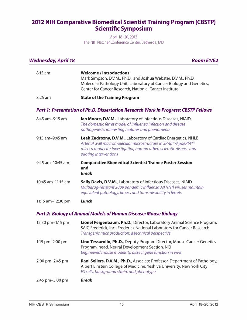
Wednesday, April 18 Room E1/E2
8:15 am Welcome / Introductions Mark Simpson, D.V.M., Ph.D., and Joshua Webster, D.V.M., Ph.D., Molecular Pathology Unit, Laboratory of Cancer Biology and Genetics, Center for Cancer Research, Nation al Cancer Institute
8:25 am State of the Training Program
Part 1: Presentation of Ph.D. Dissertation Research Work in Progress: CBSTP Fellows
8:45 am–9:15 am Ian Moore, D.V.M., Laboratory of Infectious Diseases, NIAID The domestic ferret model of influenza infection and disease pathogenesis: interesting features and phenomena
9:15 am–9:45 am Leah Zadrozny, D.V.M., Laboratory of Cardiac Energetics, NHLBI Arterial wall macromolecular microstructure in SR-BI-/-/ApoeR61h/h mice: a model for investigating human atherosclerotic disease and piloting interventions
9:45 am–10:45 am Comparative Biomedical Scientist Trainee Poster Session and Break
10:45 am–11:15 am Sally Davis, D.V.M., Laboratory of Infectious Diseases, NIAID Multidrug-resistant 2009 pandemic influenza A(H1N1) viruses maintain equivalent pathology, fitness and transmissibility in ferrets
11:15 am–12:30 pm Lunch
Part 2: Biology of Animal Models of Human Disease: Mouse Biology
12:30 pm–1:15 pm Lionel Feigenbaum, Ph.D., Director, Laboratory Animal Science Program, SAIC-Frederick, Inc., Frederick National Laboratory for Cancer Research Transgenic mice production: a technical perspective
1:15 pm–2:00 pm Lino Tessarollo, Ph.D., Deputy Program Director, Mouse Cancer Genetics Program, head, Neural Development Section, NCI Engineered mouse models to dissect gene function in vivo
2:00 pm–2:45 pm Rani Sellers, D.V.M., Ph.D., Associate Professor, Department of Pathology, Albert Einstein College of Medicine, Yeshiva University, New York City ES cells, background strain, and phenotype
2:45 pm–3:00 pm Break
NIH CBSTP Symposium 16 April 18–20, 2012
3:00 pm–3:45 pm Harm HogenEsch, D.V.M., Ph.D., Professor, Purdue University, West Lafayette Immunodeficient mouse models in biomedical research
3:45 pm–4:30 pm Wanda Hasheck-Hock, D.V.M., Ph.D., Professor Emerita, University of Illinois, Urbana-Champaign The pathologist and rodent toxicology studies in drug development
4:30 pm–5:00 pm Closing Remarks and Additional Opportunity to Visit Posters
Thursday, April 19 Room E1/E2
Part 3: Comparative Melanoma Biology: Scientific Session and Tumor Board
8:00 am Director’s Welcome Richard G. Wyatt, M.D., Deputy Director, Office of Intramural Research, Office of the Director, NIH, Bethesda
Introductions Mark Simpson, D.V.M., Ph.D., and Stephen Hewitt, M.D., Ph.D., Center for Cancer Research, NCI
Session 1: Animal Models of Melanoma
8:15 am–8:55 am Craig J. Ceol, Ph.D., Assistant Professor, University of Massachusetts Medical School, Worcester Understanding melanoma initiation using the zebrafish Danio rerio
8:55 am–9:35 am Marcus W. Bosenberg, M.D., Ph.D., Associate Professor, Yale University Medical School, New Haven Comparative pathology of genetically engineered mouse models of melanoma
9:35 am–9:50 am Break — Visit Fellow Posters
9:50 am–10:30 am Glenn Merlino, Ph.D., Chief, Laboratory of Cancer Biology and Genetics, NCI, Bethesda Modeling UV-initiated melanoma in the mouse
10:30 am–11:10 am Sheri Holmen, Ph.D., Associate Professor, Huntsman Cancer Institute, University of Utah, Salt Lake City A somatic cell gene delivery mouse model allows rapid evaluation of genes implicated in human melanoma
11:10 am–12:00 pm Keynote Speaker Boris Bastian, M.D., Ph.D., Gerson and Barbara Bakar Distinguished Professor of Cancer Biology, University of California, San Francisco Integrating molecular and clinical features to improve the taxonomy of melanocytic neoplasms
12:00 pm–1:15 pm Lunch
NIH CBSTP Symposium 17 April 18–20, 2012
Session 2: Human Melanomas
1:15 pm–1:50 pm Xiaohong Rose Yang, Ph.D., M.P.H., Investigator, Genetic Epidemiology Branch, Division of Cancer Epidemiology and Genetics, NCI, Rockville Genetic epidemiology of melanoma
1:50 pm–2:25 pm Adriano Piris, M.D., Assistant in Pathology, Massachusetts General Hospital, Instructor in Pathology, Harvard Medical School, Boston Metastatic malignant melanoma and selective BRAF inhibitor therapy
2:25 pm–3:00 pm Victor Prieto, M.D., Ph.D., Professor of Pathology and Dermatology Section Chief, Dermatopathology, Department of Pathology, MD Anderson Cancer Center, Univ of Texas, Houston Histopathology and prognostic factors
3:00 pm–3:15 pm Break — Visit Fellow Posters
Session 3: Canine Melanomas
3:15 pm–3:55 pm Philip J. Bergman, D.V.M., Ph.D., Chief Medical Officer, BrightHeart Veterinary Centers, Armonk Of mice & men (and dogs!): xenogeneic DNA vaccines for canine melanoma & other spontaneous malignancies
3:55 pm–4:35 pm Rebecca Smedley, D.V.M., M.S., Academic Specialist, Diagnostic Center for Population and Animal Health, Michigan State University, Lansing Review of the prognostic evaluation of canine melanomas
4:35 pm Closing Remarks
Thursday, April 19 (6:00 pm – 9:00 pm ) Bethesda North Marriott
Tumor Board Commences Working Session, including Evening Speaker (by invitation only)
7:00 pm Evening Speaker Chand Khanna, D.V.M., Ph.D., Head, Tumor and Metastasis Biology Section, Pediatric Oncology Branch, NCI, Bethesda A comparative approach to cancer biology and therapy
Friday, April 20 (8:00 am – 2:30 pm) NIH Natcher Conference Center, Room D
Part 4: Comparative Melanoma Tumor Board Session (limited space, by invitation only)
8:00 am–2:30 pm Tumor Board Working Session Moderated by Mark Simpson, D.V.M., Ph.D., Laboratory of Cancer Biology and Genetics, NCI, and Stephen Hewitt, M.D., Ph.D., Laboratory of Pathology, NCI
2:30 pm Meeting Adjourn
Biographies and Research Abstracts(Trainees with Dissertation Projects)
NIH CBSTP Symposium 21 April 18–20, 2012
A. Sally Davis, D.V.M.
Dr. Davis is currently a graduate scholar in the NCI Comparative Biomedical Scientist Training Program in partnership with North Carolina State University and the National Institute of Allergy and Infectious Diseases, 2007-present.
She conducts her Ph.D. research in the laboratory of Jeffery K. Taubenberger, M.D., Respiratory Virus Pathogenesis and Evolution Section, Laboratory of Infectious Diseases, NIAID. Dr. Davis received her D.V.M. and Residency Certificate in veterinary anatomic pathology in 2007 and 2009 respectively, from North Carolina State University College of Veterinary Medicine. She also has a BA in Computer Science modified with education and a graduate certificate in second-ary school science education from Dartmouth College. Her current research focuses on comparative pathogenesis and response of a diversity of mammalian species to a variety of influenza A viruses, including reconstructed 1918 and 2009 H1N1 pandemic strains. She is interested in influenza receptor identification, viral binding and entry. Additionally, she studies the interspecies variability in host response to influenza A virus via in vivo experimentation, digital and light microscopy, immunohistochemistry and immunofluorescence. Her academic advisors are Jeffery Taubenberger, M.D., Ph.D., J. Mac Law, D.V.M., Ph.D., Diplomate, The American College of Veterinary Pathologists (chair), Mark Simpson, D.V.M., Ph.D., Diplomate, The American College of Veterinary Pathologists and Fred Fuller, Ph.D.
NIH CBSTP Symposium 22 April 18–20, 2012
Multidrug-resistant 2009 Pandemic Influenza A(H1N1) Viruses Maintain Equivalent Pathology, Fitness and Transmissibility in Ferrets
A. Sally Davis, Matthew J. Memoli, Kathleen Proudfoot, Daniel S. Chertow, Rachel J. Hrabel, Tyler Bristol, Jeffery K. Taubenberger
Laboratory of Infectious Diseases, National Institute of Allergy and Infectious Diseases, National Institutes of Health, Bethesda, MD 20892
Background: Influenza A viruses are significant human pathogens, both in their sporadic pandemic and endemic (seasonal) forms. For the last few decades, two classes of antiviral therapies, adamantanes and neuraminidase inhibitors (NAIs), have been used to treat both severe illness in high-risk patients and self-limited influenza infection. Adamantanes target the matrix 2 ion channel involved with release of influenza A virus from the endocytic pathway into the infected cell’s cytoplasm and NAIs block the release of newly matured virions from the cell’s surface. By the 2005-2006 influenza season, over 90% of seasonal H3N2 viruses in circulation had gained adamantane resistance, rendering this class of drug ineffective as a viable treatment for seasonal H3N2 influenza infections. Treatment of choice shifted to NAIs. By the end of the 2008–2009 influenza season, ~98% of seasonal H1N1 isolates worldwide exhibited resistance to oseltamivir. Over the past 5 years, a smaller number of NAI-resistant H3N2 viruses have been identified. These H3N2 NAI-resistant viruses demonstrate equivalent fitness and transmissibility to wild-type viruses, suggesting that widespread seasonal H3N2 NAI resistance is also a threat. The 2009 influenza A (H1N1) pandemic called attention to the limited influenza treatment options available, especially in individuals at high risk of severe disease. 2009 H1N1 pandemic viruses are adamantine resistant, but have sporadically appeared containing resistance to neuraminidase inhibitors. The ability of these antiviral resistant viruses to replicate, transmit, and cause disease in mammalian hosts has not been fully characterized. We sought to investigate if dual drug class resistant 2009 H1N1 pandemic viruses would have equivalent replication, transmission and pathogenicity characteristics to their NAIs susceptible peers in the ferret model.
Methods: Two pre-treatment wild-type viruses and 2 post-treatment multidrug-resistant viruses containing the neuraminidase H275Y mutation conferring NAI resistance collected from immunocompromised patients infected with pandemic influenza H1N1 were tested for viral fitness, pathogenicity, and transmissibility in ferrets. Thirty-two influenza A virus–seronegative, 4-month-old, male ferrets divided into 8 groups of 4 ferrets each were inoculated intranasally with 105 plaque-forming units of virus: 1 group with each of the pretreatment viruses and each of the posttreatment viruses. The remaining 4 groups of ferrets were placed into contact with the aforementioned inoculated ferrets at 48 hours post-inoculation to assess contact transmission. Daily temperatures, weights, clinical scores and nasal washes were collected. A pair of ferrets from each group of inoculated ferrets was euthanized on day 4 post-inoculation, a pair from each contact group on day 4 post-exposure, and the remaining pairs were euthanized on day 14 post-inoculation (day 12 post-exposure). Whole lungs, trachea, tracheobronchial lymph nodes, rostral nasal turbinates, heart, and spleen were harvested.
Quantification of viral shedding was determined using 1-step real-time reverse-transcription polymerase chain reaction for the influenza A virus matrix 1 gene. Amplification and sequencing of the surface glycoprotein genes hemagglutinin (HA) and neuraminidase (NA) was performed on each viral isolate recovered to determine if the HA and NA gene sequence remained stable after infection and transmission. The HA and NA genes were amplified and sequenced from virus isolated from the ferret nasal washes and sequences were compared with those generated from the viral isolates used for inoculation. Viral titers in lung tissue were determined after tissue homogenization by standard plaque assays. Hematoxylin and eosin stains were preformed for all tissues for histopathologic analysis.
NIH CBSTP Symposium 23 April 18–20, 2012
Brown and Hopps tissue Gram stain was applied to select sections to confirm absence of bacterial co-infection. Immunohistochemistry for influenza A antigen distribution was performed on all sections from ferrets euthanized at the earlier time point.
Results: The pre-treatment wild-type viruses and post-treatment resistant viruses containing the H275Y mutation all demonstrated significant pathogenicity and equivalent viral fitness and transmissibility. HA and NA gene sequences of viruses isolated from the ferret nasal washes contained sequences identical to those of the inoculating viruses. All ferrets inoculated with virus showed signs of clinical disease and weight loss 2 days after inoculation. Clinical signs in ferrets inoculated with wild-type and antiviral resistant viruses from the same human patient were equivalent. Viral shedding and lung titers were equivalent across all four viruses in both inoculated and exposed ferrets, 102–103 pg of viral RNA and mean peak titers of 102–103 pfu/g of tissue respectively at the early harvest time point. There were no discernible differences in gross pathology attributable to specific viruses or route of viral inoculation. Histopathological analysis of ferrets euthanized on day 4 and 14 demonstrated no significant differences between ferret groups attributable to a specific virus or route of inoculation. Influenza viral antigen distribution was similar across ferrets at the same timepoint regardless of infecting virus.
Conclusions: The admantane-resistant 2009 pandemic influenza A(H1N1) virus can develop the H275Y mutation in the neuraminidase gene conferring resistance to both oseltamivir and peramivir without any loss in fitness, transmissibility, or pathogenicity. This suggests that the dissemination of widespread multidrug resistance similar to neuraminidase inhibitor resistance in seasonal H1N1 is a significant threat.
NIH CBSTP Symposium 24 April 18–20, 2012
Joy Gary, D.V.M.
Dr. Gary is currently a graduate scholar in the NCI Comparative Biomedical Scientist Training Program in partnership with Michigan State University and the National Cancer Institute, 2009-present.
Joy Gary grew up in Colorado and attended Davidson College in North Carolina, where she received her B.S. in Biology. She received her D.V.M. from Colorado State University in 2009. She completed her didactic graduate studies and core diagnostic experience in veterinary anatomic pathology at Michigan State University. She is continu-ing her training in pathology and research, having just joined the Laboratory of Cancer Biology and Genetics for her dissertation work under the mentorship of Dr. Beverly Mock. She is currently studying the role of an allelic variant of mTOR (R628C; C1977T) in BALB/c mice in the development of mesenteric plasma cell tumors. She is also inter-ested in differential miRNA expression in mice with this allele. Her academic advisors are Matti Kiupel, Dr. Med. Vet., Ph.D., Diplomate, The American College of Veterinary Pathologist (chair), Michigan State University; Beverly Mock, Ph.D., Laboratory of Cancer Biology and Genetics, NCI.
NIH CBSTP Symposium 25 April 18–20, 2012
Allelic Variant of Mechanistic Target of Rapamycin (mTOR) Induced Differential Expression of Multiple miRNAs Implicated in Interactions with Upstream and Downstream Targets of mTOR
Joy Gary1,2, Shuling Zhang1, Dena Tran1, Aleksandra Michalowski1, and Beverly Mock1
1Laboratory of Cancer Biology and Genetics, NCI, CCR, NIH; 2Department of Pathobiology and Diagnostic Investigation, College of Veterinary Medicine, Michigan State University
miRNAs are small, non-coding RNAs that regulate multiple genes involved in cell growth, development, and disease states by binding to the 3’ untranslated region of the target mRNA and inhibiting translation or inducing degradation. The PI3K/AKT/mTOR pathway is frequently dysregulated in cancer and mechanistically often involves activation of growth factor receptor pathways, mutations, PTEN loss, and AKT amplification. Allelic variation and mutations in mTOR are relatively rare. Our lab discovered an allelic variant of mTOR (R628C; C1977T) in BALB/c mice, which predisposes the mice to development of pristane-induced plasma cell tumors (PNAS 100:14982). We recently developed a knock-in (KI) B6;129 mouse by homologous recombination that carries the BALB/c allele (C at nucleotide 1977) of mTOR (Blood 117:1228). Since miRs tend to control several genes simultaneously, we compared miR expression between wild-type (WT; mTORT1977) and mTORC1977 KI mice. B220+ splenic B-lymphocytes were isolated from WT and KI mice with and without pristane induction. miRNA was extracted from the splenocytes and a Nanostring miRNA expression assay was performed. BRB-ArrayTools was used to analyze differences in expression, and pathway analyses were performed with Ingenuity. In the Nanostring assay, several miRs were found to be differentially expressed. Those that were found to be significantly higher (p < 0.05) in the KI were miR30a (2-fold), miR101 (3-fold), and miR423-5p (1.5-fold). Similar trends were found with qRT-PCR. Pathway analysis in Ingenuity Pathway Analysis and evaluation of predicted targets in TargetScan revealed that miR30a likely interacts indirectly with AKT1 which is upstream of mTOR, and EIF4E, which is downstream. miR101 likely interacts with mTORC1. Finally, miR423-5p likely interacts indirectly with AKT1 and directly with 4EBP-1 (downstream of mTOR and hypophosphorylated in KI mice). Further exploration of the relationship of these miRs with the allelic variant of mTOR is needed to establish their role in the mTOR pathway.
NIH CBSTP Symposium 26 April 18–20, 2012
Ian Moore, D.V.M.
Dr. Moore is a graduate scholar in the NCI Comparative Biomedical Scientist Training Program in partnership with Michigan State University and the National Institute of Allergy and Infectious Diseases, 2007-present.
Dr. Moore received his B.S. from Tuskegee University and his D.V.M. from Tuskegee University School of Veterinary Medicine in 2006, and following graduation, entered a residency in anatomic pathology the Diagnostic Center for Population and Animal Health, Michigan State University. Following completion of residency training, Dr. Moore is currently a fellow in the NIAID’s Laboratory of Infectious Diseases (LID) where, under the guidance of Dr. Kanta Subbarao, he studies the pathologic and immunologic responses of the ferret to wild-type Influenza virus infection. Dr. Moore’s research interests include mechanisms of infectious diseases in animal models. Dr. Moore’s Ph.D. committee is chaired by Kurt Williams DVM, Ph.D., Diplomate, The American College of Veterinary Pathologists.
NIH CBSTP Symposium 27 April 18–20, 2012
The Domestic Ferret Model of Influenza Virus Infection and Disease Pathogenesis
Ian N. Moore1,2, Kurt J. Williams2, Kanta Subbarao1
1Laboratory of Infectious Disease National Institute of Allergy and Infectious Diseases, NIAID, NIH, Bethesda, MD, and 2Diagnostic Center for Population and Animal Health, Michigan State University East Lansing, MI
In 2009, a newly emerged swine-origin influenza A virus caused a pandemic associated with severe disease and mortality in humans. The ferret model was used to investigate the kinetics of virus replication, disease pathogenesis and anti-viral and vaccine efficacy for this strain of influenza. However, disease outcomes following experimental infection with the pandemic virus varied widely. We hypothesize that dose of virus, volume of inoculum, age of ferrets, and viral subtype contribute to the clinical and pathologic severity of experimental influenza A virus infection in ferrets. We evaluated the effect of these variables on disease outcomes by administering virus to young (8-12 week old) ferrets at doses of 106 TCID50/ml or 107 TCID50/ ml. Each dose was administered in one of three different inoculum volumes (0.2, 0.5 and 1.0ml). At days 1, 3 and 6, animals were humanely euthanized and tissues from the upper and lower respiratory tract were collected for virus titration and histopathology. The virus replicated to high titer in the upper and lower respiratory tract but was associated with minimal clinical illness and no mortality. We observed consistently higher virus titers in the right caudal lung lobe. We did not observe a significant effect of virus dose on viral replication. Similar experiments were performed in older (6 month old) ferrets and the clinical outcomes contrasted those observed in the younger cohort. Based on our findings, we will propose parameters to reduce inter and intra-laboratory reproducibility for ferret studies.
NIH CBSTP Symposium 28 April 18–20, 2012
Tiffany Reed, D.V.M.
Dr. Reed is currently a graduate scholar in the NCI Comparative Biomedical Scientist Training Program in partnership with Purdue University and the National Cancer Institute, 2009-present.
Dr. Reed received her B.S. from the University of Georgia and her D.V.M. from the University of Georgia (2008). Following graduation, she initiated an anatomic pathology residency at Purdue University’s Animal Disease Diagnostic Laboratory. Following one year of training, Dr. Reed enrolled in the CBSTP as an NCI Cancer Research Training Fellow. She completed her diagnostic pathology training and didactic coursework in 2011. Dr. Reed is continuing her research training at NCI. Her academic program advisors are Margaret A. Miller, D.V.M., Ph.D., Diplomate, The American College of Veterinary Pathologists; Jose Ramos-Vara, D.V.M., Ph.D., Diplomate, The European College of Veterinary Pathologists; and Stephen Lenz, D.V.M., Ph.D., Diplomate, The American College of Veterinary Pathologists; and Patricia Steeg, Ph.D., Head, Womens Cancers Section, Laboratory of Molecular Pharmacology. Dr. Reed’s research interests include breast cancer metastasis to the brain and brain microenvironment.
NIH CBSTP Symposium 29 April 18–20, 2012
The Role of Molecular Interactions of Rad51 in Brain Metastasis from Breast Cancer
L. Tiffany Reed1,2, Stephan Woditchka1, Patricia S. Steeg1
1Women’s Cancers Section, Laboratory of Molecular Pharmacology, NCI, NIH, Bethesda, MD; 2Department of Comparative Pathobiology, Purdue University, West Lafayette, IN
Brain metastases occur historically in 10-16% of breast cancer patients with metastatic disease, however the incidence is rising. Patients with the highest risk factors for brain metastasis include those with metastatic Her2+ or triple negative breast cancer, with over one third of patients developing lesions. Brain metastatic lesions can occur in the leptomeninges, subarachnoid space, or neuroparenchyma.
I am investigating the role of the Rad51 and Bard1 proteins in brain metastasis. Rad51 overexpression was identified as a component of a 13-gene signature, predictive of rapid brain metastasis development (<3 years vs. ≥ 3 years) in primary tumors from Her2+ metastatic breast cancer patients. The 13 genes were separated into three groups: Her2-related, DNA double-strand break repair, and uncategorized genes. The three DNA double-strand break repair genes included Rad51 (RecA homolog, E. coli), BARD1 (BRCA1 associated ring domain protein 1), and FANCG (Faconia anemia group G).
Rad51 has been identified as a critical component of the homologous recombinational DNA repair pathway (HRR). For HRR to occur at a site of double-strand breakage, the homologous sequence of the sister chromatid is used for regeneration of the site and restoration of the DNA sequence. There are no identifiable mutations in RAD51 in neoplasia, but defects in other HRR genes are factors in carcinogenesis. The most prominent genes are BRCA1 and BRCA2, which are key players in homologous recombination and breast cancer predisposition syndrome. Rad51 overexpression has also been reported to decrease the functional effects of mediators of HRR. To functionally test the role of Rad51 expression in brain metastasis of breast cancer, it was overexpressed in brain-tropic derivatives of the MDA-MB-231 human breast carcinoma cell line and the 4T1 murine mammary carcinoma cell line. The Rad51 overexpressing polyclonal populations or similarly created empty vector controls were injected into the left cardiac ventricle of mice, and experimental brain metastases were quantified 2-4 weeks post-injection. Overexpression of Rad51 increased brain metastasis by 3-4 fold (p=0.01) in 231-BR7 cells and 2-3 fold (p<0.05) in 4T1-BR5 cells.
Despite this strong in vivo phenotype, the overexpression of RAD51 does not appear to modulate the expression levels of any of its co-mediators, necessary for homologous recombination. I therefore hypothesized, that RAD51 overexpression confers its phenotype by interacting with co-mediators more frequently, thus, forming more functional HRR complexes. To determine the function of Rad51 overexpression, I am examining its protein-protein expression patterns. Co-immunoprecipiation, formation of an immune complex against a target and interacting macromolecules, and two imaging modalities, immunofluorescence and confocal microscopy, were used to identify macromolecules interacting with the Rad51 DNA repair pathway in two cell lines: 231-Br7-Vector and 231-Br7-Rad51 overexpressor. Current experiments are focusing on BRCA2 as the most important co-mediator of Rad51 in HRR. This macromolecule binds Rad51 and drives it to the DNA double-strand break. The DNA double-strand break is transformed into two single-stranded DNA sequences by BRCA1, ATM, and the MRE11/Rad50/NBS1 (MRN) complex. PALB2, a macromolecule bound to BRCA2 and BRCA1, attracts BRCA2/Rad51 to the single-stranded DNA filament. Rad51 then forms a Rad51—single stranded DNA filament which finds an identical DNA template to achieve homologous recombinational repair. Other mediators of HRR include RPA, which is a single-strand DNA binding protein; this protein binds to the 3’ overhang at the site of the double-strand break. Displacement of RPA by Rad51 is the critical signaling indicating DNA repair. Recombination mediators, that assist Rad51 in displacement of RPA, include
NIH CBSTP Symposium 30 April 18–20, 2012
BRCA2, Rad51B, Rad51C, Rad51D, XRCC2, XRCC3, and Rad52. Co-mediators of HRR, or those that assist in localizing mediators to the site of the DNA double-strand break include BRCA1, Chk2, PALB2, Ku70 and SWS1.
I will determine whether Rad51 overexpression alters the relative number and composition of protein-protein complexes. Rad51 has been immunoprecipitated from both untreated and x-treated 231-BR cells. Identification and characterization of the interactions of Rad51 is ongoing and will assist in explanation of the upregulation in carcinogenesis, hopefully providing an option in therapy and control of brain metastasis.
x = doxorubicin, carboplatin, which both induce DNA ds breaks
NIH CBSTP Symposium 31 April 18–20, 2012
Heather Sheppard-Tillman, D.V.M.
Dr. Tillman is currently a graduate scholar in the NCI Comparative Biomedical Scientist Training Program in partnership with the Michigan State University and the National Cancer Institute, 2008-present.
Dr. Tillman received her B.S.A. in Animal Science from the University of Georgia (2005), her D.V.M. also from the University of Georgia (2008), and her residency certificate in veterinary anatomic pathol-ogy from Michigan State University (2011). She is pursuing her dissertation research and training in the molecular pathology of advanced prostate cancer. Her research focuses on understanding the signal transduction relating to epithelial-mesenchymal plasticity and metastatic progression using the PbCre4;PTENfl/flTP53fl/fl mouse model. Members of her graduate committee include: Matti Kiupel, Dr. Med. Vet., Ph.D., Diplomate, The American College of Veterinary Pathologist (chair), Michigan State University; Kathleen Kelly, Ph.D., Cell and Cancer Biology Branch, NCI; Vilma Yuzbasiyan-Gurkan, Ph.D., Michigan State University; Ingeborg Langohr, D.V.M. Ph.D., Diplomate, The American College of Veterinary Pathologists, Michigan State University; Joshua Webster, Ph.D., Diplomate, The American College of Veterinary Pathologists, Laboratory of Cancer Biology and Genetics, NCI.
NIH CBSTP Symposium 32 April 18–20, 2012
Investigation of the signaling factors involved in Ras/MAPK promoted metastasis in the PTEN-/-TP53-/- mouse model
Heather S. Tillman1, Juan Juan Yin1, Yen-Nien Liu1, Musaddiq Awan1, Philip Martin1, and Kathleen Kelly1
1Cell and Cancer Biology Branch, National Cancer Institute, National Institutes of Health, Bethesda, Maryland
The most significant challenge for advanced prostate cancer patients is resistance to androgen-deprivation therapy and the dissemination of cancer cells leading to metastatic disease. This study addresses how enhanced Ras signaling cooperates with the two most common prostate cancer genetic deletions, TP53 and PTEN, in metastasis. To study metastatic progression we will use the well established PbCre4;PTENfl/fl/TP53fl/fl mouse prostate cancer model system. PbCre4;PTENfl/fl/TP53fl/fl mice develop prostatic intraepithelial neoplasia (mPIN) as early as 8 weeks with progression to invasive adenocarcinoma by 12 weeks. From 15 to 19 weeks adenocarcinomas undergo an irreversible epithelial to mesenchymal transition (EMT) to form sarcomatoid carcinomas. In this model of transforming growth factor β (TGFβ)-induced prostatic EMT, the transcription factor SLUG (Snai2) is the dominant regulator of EMT initiation in vitro and in vivo, as demonstrated by the inhibition of irreversible EMT following Slug depletion. While vascular invasion is observed in this model metastasis does not occur. Because both PTEN and TP53 deletion alone are not sufficient for metastasis, we asked what other signaling pathways are required.
Ras pathway activation is implicated in prostate cancer metastatic progression in comprehensive genetic analyses of human prostate tumors and from in vivo xenograft models. To develop a metastatic prostate model, we introduced a lentiviral vector expressing K-rasG12V into a PTEN-/-/ TP53-/- adenocarcinoma clonal cell line (clone 2). Constitutive Ras signaling was shown by enhanced Erk phosphorylation as detected by western blot analysis. Orthotopic injection of clone2/Ras expressing cells resulted in 100% pulmonary metastasis. The metastatic phenotype was paralleled by an inhibition of sarcomatoid carcinoma transformation. Thus we hypothesized that Ras signaling either inhibited or modified TGFβ-mediated EMT signaling.
There are multiple pathways downstream of TGFβ initiated signaling, including proteosome dependent degradation of KLF4 leading to SLUG induction, SMAD4 dependent transcription, and other non-SMAD mediated pathways such as TAK1 induced NF-κB activation. To determine whether TGFβ signaling occurs in clone2/Ras cells, parental and Ras lines were stimulated with TGF-β in vitro. In both cell lines, the Smad signaling arm of the TGFβ pathway was activated as demonstrated by enhanced SMAD2 phosphorylation and nuclear SMAD4 translocation. In addition, TGF-β stimulation led to enhanced SLUG expression in both lines. In primary tumors, low numbers of clone 2 and clone2/Ras tumor cells demonstrated nuclear Slug labeling, although SMAD2/3/4 dependent signaling was not observed. Relevant to this finding is a recent report that loss of the TGFβ signaling component, Smad4, results in vertebral metastasis in the PbCre4;PTENfl/fl/TP53fl/fl model. Thus, we are using genetically modified clone 2 and clone2/Ras cells in metastasis assays to determine 1) whether TGFβ signaling is required by expressing a dominant negative TGFβ type 2 receptor, and 2) whether SLUG expression is required using shRNA-mediated Slug depletion.
In both lung and breast cancer models, NF-κB activation has been demonstrated as a necessary pathway downstream of Ras transformation. NF-κB activation occurs through multiple mechanisms, including, but not limited to, TGFβ initiated signaling. Interestingly, p65 nuclear labeling, an indication of canonical NF-κB activation, was detected by immunohistochemistry (IHC) in the clone2/Ras primary tumors and in approximately 50% of lung metastases. No nuclear p65 labeling was observed in clone 2 tumors. These in vivo findings suggest that canonical NF-κB signaling may be a downstream effector
NIH CBSTP Symposium 33 April 18–20, 2012
of the Ras pathway. To test the importance of canonical NF-κB signaling in our metastatic model we are using an inducible dominant negative IKB lentiviral vector in the clone 2 and clone 2/Ras lines to analyze in vivo metastatic ability. If successful, the above studies will establish the requirement for TGFβ-mediated signaling, for the SLUG invasion pathway, and for the canonical NF-κB pathway in Ras-mediated metastasis. The above approaches also begin to address the intersection of TGFβ and NF-κB pathways in Ras-mediated metastasis.
NIH CBSTP Symposium 34 April 18–20, 2012
Leah Zadrozny, D.V.M.
Dr. Zadrozny is currently a graduate scholar in the NCI Comparative Biomedical Scientist Training Program in partnership with North Carolina State University and the National Heart, Lung, and Blood Institute, 2008 - present.
Dr. Zadrozny received her B.S. from the University of Vermont and her D.V.M. from North Carolina State University in May, 2008. She has recently completed her third-year of training as a graduate scholar in comparative pathology at the NHLBI Laboratory of Cardiac Energetics, where she focused on studying the aortic macromolecular microstructure in healthy and diseased SR-BI KO/ApoER61h/h mice, a recently defined model of diet-induced occlusive coronary athero-sclerosis and coronary heart disease. Her research interests are in modeling the functional pathophysiology of cardiovascular disease, with an emphasis on atherosclerosis, with her Ph.D. dissertation studies headed by principal investigator mentor Robert Balaban, Ph.D. Members of her graduate guidance committee include Robert Balaban, Ph.D., NHLBI; John Cullen, V.M.D., Ph.D., Diplomate, The American College of Veterinary Pathologists; Mark Simpson, D.V.M., Ph.D., Diplomate, The American College of Veterinary Pathologists; and Edward Neufeld, Ph.D., Laboratory of Cardiac Energetics, NHLBI.
NIH CBSTP Symposium 35 April 18–20, 2012
Arterial Wall Macromolecular Microstructure in SR-BI-/-/Apoe61h/h Mice: A Model for Investigating Human Atherosclerotic Disease and Piloting Interventions
Leah M. Zadrozny1, Edward B. Neufeld1, Zu-Xi Yu2, Robert S. Balaban1
1Laboratory of Cardiac Energetics and 2Pathology Core, NHLBI, NIH, Bethesda, MD
Atherosclerosis is a disease in which deposits of yellowish-tan plaques, containing fatty substances and necrotic cellular debris, expand the intima and superficial media of the large and medium sized elastic and muscular arteries. This arteriopathy has been implicated in 75% of cardiovascular disease-related deaths in the United States. Vascular lesions can be initiated as early as in utero as regions of intimal thickenings and intracellular lipid accumulation often referred to as fatty streaks. The most pronounced thickenings develop in regions near branch vessel ostia, at the aortic and carotid bifurcations, and within the descending thoracic aorta (TA). Spontaneous regression of early lesions often occurs; however, advanced disease is characterized by the conversion of fatty streaks into atheromas or atherosclerotic plaques. Untoward effects of advanced lesions include vascular remodeling, acute and chronic luminal obstruction, plaque rupture, thrombosis and diminished oxygen supply to major organs. Although mechanical factors such as pressure-induced vascular wall stress and blood flow disturbances are important in lesion development, the vascular extracellular matrix (ECM), including collagen, elastin and proteoglycans (PGs) largely contribute to vascular remodeling and disease progression. Furthermore, PG-mediated subendothelial retention of low-density lipoprotein (LDL) is currently thought to serve as an initiating step in atherosclerosis.
The scavenger receptor class B, type I (SR-BI) deficient, hypomorphic apolipoprotein ER61 transgenic mouse (SR-BI KO/ApoER61h/h) is a recently described model of diet-induced occlusive coronary atherosclerosis and coronary heart disease (CHD). To investigate the role of the ECM in atherosclerotic disease initiation and progression, longitudinal analysis of the macromolecular microstructure of the developing vascular bed within the TA of SR-BI KO/ApoER61h/h neonatal and adult mice fed normal chow (NC) will be completed. Analysis will be performed utilizing multiphoton imaging, including two-photon excitation fluorescence (TPEF) and second-harmonic generation (SHG), coupled with histology and PG immunohistochemistry (IHC).
TPEF is a powerful method of label-free imaging based on intrinsic fluorescence of living tissues and cells as well as fresh and formalin-fixed postmortem samples. Elastin is readily visualized with TPEF due to autofluorescence provided by the presence of endogenous chromophores found for example within certain amino acid side chains (e.g. tryptophan and tyrosine). SHG is a coherent process allowing for the visualization of unstained, endogenous, noncentrosymmetric proteins such as fibrillar collagens. Together, these imaging modalities afford increased three-dimensional spatial resolution, decreased photobleaching and phototoxicity, and increased tissue penetration depth.
Preliminary multiphoton images obtained from SR-BI KO/ApoER61h/h mice fed NC show a variably confluent, wavy meshwork of elastin throughout the tunica media around which variably dense, fibrillar strands of collagen are circumferentially arranged forming layers of elastic lamellar units. Both the matrix density and the overall abundance of elastin and collagen qualitatively appear to vary with increasing age. Intravascular injection of Nile Red (NR), a lipophilic probe that fluoresces in a hydrophobic environment, reveals both punctuate and small aggregates of lipid extravasating the vessel wall. Statistical analysis will be completed.
In mice fed a HFHC diet, significant lesion development within the TA occurs within less than 7 days in mice aged 1month to 8 months old. Although early lesion initiation was not captured and NR was not administered during this pilot study, advanced plaque-like lesions were readily appreciated both by multiphoton microscopy and histopathology as well as grossly. Even without NR staining, atherosclerotic lesions were identified in the TA with multiphoton microscopy as disruption and
NIH CBSTP Symposium 36 April 18–20, 2012
regionally extensive loss of the elastin meshwork coupled with the circumferential knot-like accumulation of thick sheets of collagen surrounding intercostal ostia. Collagen within the intercostal space became more disorganized and linearly arranged as compared to mice fed NC. In mice fed HFHC and NC diets, decorin (PG) immunostaining was abundant in the adventitial space; however, mice fed a HFHC diet exhibited copious deposition within the luminally obstructive atherosclerotic plaques admixed with intra- and extracellular lipid that expanded the subintimal space.
Evaluation of the micromolecular microstructure in the developing and diseased TA of SR-BI KO/ApoER61h/h will provide a solid base for future experiments involved in the development and administration of therapeutic compounds designed to block PG-mediated subendothelial LDL retention.
NIH CBSTP Symposium 37 April 18–20, 2012
Philip Martin, D.V.M.
Dr. Martin is a graduate scholar in the NCI Comparative Biomedical Scientist Training Program in partnership with University of Maryland and the National Cancer Institute, 2005–present.
Dr. Martin received his B.S. from Northwestern University, his M.S. from Ohio University and his D.V.M. from Kansas State University in 2003. After completing the D.V.M. Dr. Martin went to the University of California, Davis for residency training in anatomic pathology in the Department of Veterinary Pathology. As an anatomic pathology resident Dr. Martin pursued specialty track training in the pathol-ogy of laboratory animals and undertook training in the UC Davis Comparative Pathology Laboratory and the Pathology Department of the California Regional Primate Center. In 2005 Dr. Martin began training in comparative pathology through the Graduate Partnership Program at the National Cancer Institute, Bethesda, MD. The first year of the GPP program was spent completing graduate course work at the University of Maryland and in additional anatomic pathology training while working in the Comparative Molecular Pathology Unit with Dr. Mark Simpson. Dr. Martin accomplished board certifica-tion in Anatomic Pathology by The American College of Veterinary Pathologists in 2006. Dr. Martin is currently pursuing dissertation research in the NCI Cancer and Cell Biology Branch headed by Kathy Kelly, PhD. His Ph.D. dissertation research involves developing an in vivo bioluminescent transgenic mouse model of prostate cancer metastasis for the purpose of investigating the molecular signaling mechanisms responsible for driving prostate cancer metastasis. Members of his graduate guidance committee include Siba Samal, B.V.Sc., Ph.D., Diplomate, The American College of Veterinary Microbiologists (chair); Xiaoping Zhu, D.V.M., Ph.D.; Robert Dooling, Ph.D.; Kathy Kelly, Ph.D.; and Mark Simpson, D.V.M., Ph.D, Diplomate, The American College of Veterinary Pathologists. Dr. Martin is currently the veterinary pathologist for the NCI Frederick’s Center for Advanced Preclinical Research.
Dr. Martin is unable to attend the symposium, as he is an invited participant/panelist in a prostate cancer meeting.
Biographies and Research Abstracts(Trainees on University Campuses)
NIH CBSTP Symposium 41 April 18–20, 2012
Laura Baseler, D.V.M.
Dr. Baseler is currently a graduate scholar in the NCI Comparative Biomedical Scientist Training Program in partnership with Purdue University and the National Institute of Allergy and Infectious Diseases, July 2010-present.
Dr. Baseler received her B.S. and M.S. from Iowa State University. She also completed her D.V.M. degree at Iowa State University (2010). She began her graduate fellowship and pathology training in July of 2010 at Purdue University. After completion of the init ial diagnostic pathol-ogy and pre-dissertation research training at the university, she will be relocating to Hamilton, MT to begin research on highly pathogenic viruses, with Heinz Feldmann, M.D., Ph.D. at the National Institute of Allergy and Infectious Diseases, Rocky Mountain Laboratories in July of 2012.
1Indiana Animal Disease Diagnostic Laboratory and Departments of 1Comparative Pathobiology and 2Veterinary Clinical Sciences, Purdue University, West Lafayette, IN; 3National Institute of Allergy and Infectious Diseases, Rocky Mountain Laboratories, Hamilton, MT
Twelve beef cows had access to a pasture where construction lumber had been burnt and were observed ingesting the ashes. Within 24 hours of exposure, one animal (cow 1) became recumbent and died without overt clinical signs. A second cow (cow 2) died but was not necropsied. Cows 3-6 subsequently developed ataxia, hind limb weakness, dehydration, hematuria and diarrhea, and were admitted to the Purdue University Veterinary Teaching Hospital for treatment.
Lesions in the first cow included abomasal erosions and acute renal proximal tubular necrosis. Cow 6 was euthanized on day 5 post-exposure. Gross lesions included abomasal erosions and marked submucosal edema, segmental jejunal mucosal congestion, and urinary bladder mucosal petechiae. Histologically, there was mild proximal renal tubular necrosis with prominent tubular regeneration. Cow 3 developed acute hemolysis and icterus, and subsequently died 27 days post-exposure. At necropsy, the subcutis and perivisceral adipose tissue and mucocutaneous junctions were diffusely icteric.
Arsenic was detected in the lumber ashes (> 2,400 ppm), and toxic concentrations were detected in whole blood from sick cows (747 ppb), kidney from cow 1 (15 ppm), and kidney (7.4 ppm), liver (8.5 ppm) and urine (3.7 ppm) from cow 6. Elevated concentrations of arsenic were also detected in kidney and urine from cow 3.
In general, the lesions caused by arsenic toxicosis are caused by two main mechanisms of action. The predominate mechanism of arsenic toxicosis is inactivation of sulfhydryl-containing enzymes involved in cellular respiration in mitochondria leading to decreased metabolic activity; this most severely affects tissues with a high metabolic rate such as the gastrointestinal tract and kidneys. Arsenic toxicosis also causes dilation and degeneration of capillaries, producing gastrointestinal tract edema and congestion, and hypotension resulting in hypoxia and renal tubular necrosis. The renal proximal straight tubules (S3 segment) and medullary ascending limb of the nephron are most sensitive to hypoxia due to their minimal capacity for anaerobic glycolysis. The exact mechanism of the vascular changes is unknown.
Arsenic toxicosis in these cattle was attributed to ingestion of ashes from burnt copper-chromium-arsenic treated lumber. Other common causes of arsenic toxicosis in ruminants are ingestion of old pesticides, herbicides and fungicides containing arsenic. In this herd, four deaths occurred 1 (2 animals), 5, and 27 days post-exposure; three animals were necropsied. Cows 1 and 6 had a spectrum of lesions that represented different post-exposure stages of tissue damage and regeneration. The lesions in cow 3 could not be directly attributed to arsenic ingestion.
NIH CBSTP Symposium 43 April 18–20, 2012
Kara Corps, D.V.M.
Dr. Corps is a graduate scholar in the NCI Comparative Biomedical Scientist Training Program in partnership with North Carolina State University and the National Institute of Neurological Disorders and Stroke, July 2010-present.
Dr. Corps received her B.S. from Michigan State University. After completing the didactic portion of veterinary school, Dr. Corps spent 2008 working towards a M.S. in Comparative Medicine and Integrative Biology under Jack Harkema, D.V.M., Ph.D.,Diplomate, ACVP at Michigan State University. She defended her M.S. thesis in December 2008. Dr. Corps received her D.V.M. from Michigan State University in May 2010. She is currently a graduate student and training in anatomic pathology at North Carolina State University and is interested in neuroimmunology. She will continue pursuing her Ph.D. dissertation research training with Dorian McGavern, Ph.D. in the Laboratory of Viral Immunology and Intravital Imaging, NINDS, beginning Summer 2012.
NIH CBSTP Symposium 44 April 18–20, 2012
Immunohistochemical Labeling of Two Canine Dysgerminomas with Protein Gene Product (Pgp) 9.5 and C-Kit
K. N. Corps1,2, J. M. Cullen1
1Population Health and Pathobiology, College of Veterinary Medicine, North Carolina State University, Raleigh, NC; 2NIH Comparative Biomedical Scientist Training Program, National Institute of Neurological Disorders and Stroke, Bethesda, MD
Dysgerminomas are rare ovarian tumors arising from primordial germ cells and are considered the ovarian correlate of the more common testicular seminoma. Dysgerminomas have been reported in most domestic species and humans, with canines overrepresented among veterinary patients. Mitotic rate and pleomorphism do not correlate with clinical behavior and all dysgerminomas are therefore considered malignant. Dysgerminomas rarely metastasize in most veterinary species; however metastases have been identified in 20% of affected canine patients and are relatively common in human patients. Immunohistochemical features of dysgerminomas are not well characterized and no studies have established potential immunohistochemical markers.
A recent report described immunohistochemical findings in one bitch, with a dysgerminoma characterized by positive labeling for vimentin and alkaline phosphate (ALP) and a lack of labeling for cell surface markers CD3, CD79a, or other cytoplasic proteins including creatine kinase (CK, an enzyme that catalyzes production of phosphocreatine, an energy reservoir in several tissues including spermatozoa), inhibin-a (a protein complex that inhibits production and secretion of follicle stimulating hormone), and S-100 (a marker associated with neural differentiation).
We elected to evaluate two novel markers for this tumor type. The first, protein gene product (PGP) 9.5, is a member of the ubiquitin carboxyl-terminal hydrolase family. PGP 9.5 has been detected immunohistochemically in healthy neuronal tissue as well as human oocytes and spermatogonia. PGP 9.5 labeling has previously been demonstrated in neuroendocrine tissues and in neoplastic testicular germ cells in a study of mixed germ cell sex-cord stromal tumors in dogs.
The second marker was c-Kit. Upregulation of c-Kit, a receptor tyrosine kinase, has been demonstrated in a number of human cancers. Increased expression of the c-KIT gene via activating mutations has been demonstrated in human seminomas. C-Kit was also recently suggested as a diagnostic marker for dysgerminoma as it has shown positive immunohistochemical labeling in seminomas in several species.
Antibodies directed against PGP 9.5 and c-Kit were applied to sections from two canine dysgerminomas. We demonstrate diffuse, intense cytoplasmic labeling of neoplastic cells with PGP 9.5 and multifocal, granular, cytoplasmic labeling with c-Kit. S-100, applied to these sections as a negative marker for comparison, had only rare cytoplasmic labeling of neoplastic cells and strong, cytoplasmic labeling of stromal spindle cells. For additional comparison, PGP 9.5 antibodies were applied to sections of a canine granulosa cell tumor and a canine ovarian carcinoma. Neoplastic cells in these two tumors do no exhibit PGP 9.5 labeling.
Given strong labeling of dysgerminoma neoplastic cells and a lack of labeling in other common canine ovarian neoplasms, PGP 9.5 clearly has potential as a diagnostic marker for canine dysgerminomas. C-Kit may provide additional support for a diagnosis of dysgerminoma, though due to the variable labeling we observed, it likely has limited use as a solo marker for canine dysgerminomas.
NIH CBSTP Symposium 45 April 18–20, 2012
Sarah Cramer, D.V.M., Diplomate, ACVP
Sarah Cramer is a graduate scholar in the NCI Comparative Biomedical Scientist Training Program in partnership with the University of Maryland and the National Cancer Institute, July 2011–present.
Currently, Dr. Cramer is completing graduate course work and pre-dissertation research training at the University of Maryland and the National Cancer Institute, Comparative Molecular Pathology Unit. Prior to joining the program, Dr. Cramer received her B.A. in biology from St. Mary’s College of Maryland (2002) and her D.V.M. from Cornell University College of Veterinary Medicine (2008). She completed a residency in veterinary anatomic pathology at Oklahoma State University (2008–2011) and became board-certified by The American College of Veterinary Pathologists in 2011. She will pursue her Ph.D. dissertation research with Giorgio Trinchieri, M.D., in the Laboratory of Experimental Immunology, within the Cancer and Inflammation Program CCR, NCI.
NIH CBSTP Symposium 46 April 18–20, 2012
The role of S100A6 in growth and metastasis of ovarian carcinoma and breast carcinoma cells
Sarah D. Cramer, Jennifer E. Dwyer, Joshua D. Webster, Shelley B. Hoover, Bih-Rong Wei, R. Mark Simpson
Molecular Pathology Unit, Laboratory of Cancer Biology and Genetics, Center for Cancer Research, National Cancer Institute, Bethesda, MD
Recently, we identified S100A6 as a potential serum biomarker for detection and monitoring of ovarian cancer (OVCa). Serum levels of S100A6 were correlated with tumor burden in mice with human OVCa xenografts, and with disease stage in human patients with ovarian carcinoma. S100A6 is a member of the S100 family of calcium binding proteins. Deregulation of S100A6 during malignant transformation has been reported in human cancers including breast cancer, ovarian cancer, pancreatic cancer, lung cancer, colorectal carcinoma, and malignant thyroid neoplasms. In addition, S100A6 overexpression has been associated with poor prognosis of melanoma and gastric cancer patients. The role of S100A6 during neoplastic transformation and progression is unclear, however. To investigate the potential role of S100A6 in development of cancer, shRNA interference was used to knock down S100A6 in SKOV-3 (OVCa) and MDA-MB-231 (breast cancer) cell lines. Both cells express high levels of S100A6.
In vitro growth rate analyses using MTS assay demonstrated no effect of S100A6 knockdown on either cell line. Wound healing and Boyden chamber migration assays showed that the mobility of SKOV-3 S100A6 knockdown cells was unaltered, while the migration rate of MDA-MB-231 knockdown cells was reduced. S100A6 knockdown appears to affect the SKOV-3 cells differently from the MDA-MB-231 cells in vitro. In vivo studies are being performed next to assess whether these differences observed in in vitro assays can be translated to tumor growth and metastasis in vivo.
SKOV-3 and MDA-MB-231 cell xenografts are established in nude mice. SKOV-3 cells are injected subcutaneously or intraperitoneally. The latter is to mimic the disseminated growth of OVCa in human patients. MDA-MB-231 cells are injected into the mammary fat pad and the tail vein to imitate tumor growth at primary and metastatic sites, respectively. Cells are transduced with luciferase expressing lentivirus for the purpose of in vivo imaging. Whole body bioluminescent imaging of mice is performed at weekly intervals to monitor tumor growth and incidence of metastases. Tumors grown in the subcutis of the flank (SKOV-3) or mammary fat pad (MDA-MB-231) are surgically removed 6-7 weeks after cell inoculation to permit development of metastases at distal sites.
SKOV-3 cells exhibited no difference in tumor development and growth between S100A6 knockdown cells and control cells in either localized or disseminated xenograft models. A differential growth was observed, however, in MDA-MB-231 xenograft models. When injected intravenously, S100A6 knockdown cells exhibited earlier onset of tumor establishment and growth than control cells, although there is no difference in tumor progression once tumors are established. When injected into mammary fat pad, S100A6 knockdown MDA-MB-231 cells showed rapid growth and/or metastases in all mice while control cells exhibited low tumor take rate (~50%) and slower tumor growth.
The knockdown of S100A6 in MDA-MB-231 cells impacted cell growth and metastatic potential in vitro and in vivo while it showed minimal influence in SKOV-3 cells. These results suggest that the functions of S100A6 are dependent on cell type. How S100A6 affects cancer cell growth and metastases is currently under investigation.
NIH CBSTP Symposium 47 April 18–20, 2012
Amy McCalla, D.V.M.
Dr. McCalla is currently a graduate scholar in the NCI Comparative Biomedical Scientist Training Program in partnership with North Carolina State University and the National Cancer Institute, July 2010-present
Dr. McCalla received her B.S. in Microbiology and Molecular Cell Sciences from The University of Memphis in 2001. Following this degree she was a member of Dr. Stephen Skapek’s lab at St. Jude Children’s Research Hospital until 2004. During this time she devel-oped two Arf transgenic mouse models as well as an Arf null knock-out mouse model. She coauthored four publications with the Skapek lab and was the primary author on an additional publication. Research undertaken in this lab examined the role of p19Arf in vascular remod-eling and development of the disease persistent hyperplastic primary vitreous.
In 2005, Dr. McCalla joined Dr. Jorge Piedrahita’s lab at North Carolina State University. She coauthored two publications with this lab on research involving porcine microarray systems and the correlation between intrauterine growth restriction and somatic cell nuclear transfer techniques in swine. In January of 2010, she was primary author on a publication through this lab which described the Gli2 transgenic pig.
Dr. McCalla began the DVM program at NCSU in the fall of 2006. During her D.V.M. program she continued to work with the Skapek and Piedrahita labs and presented research from both labs at the annual ACVP conferences in 2007 and 2008. She is currently a second year anatomic pathology trainee, graduate fellow at NCSU-CVM. In July of 2012 she will transition to Bethesda, MD for completion of graduate training in comparative molecular pathology through the Graduate Partnership Program at the National Cancer Institute.
NIH CBSTP Symposium 48 April 18–20, 2012
Diplomyelia and Myelodysplasia in a German Shepherd
1North Carolina State University College of Veterinary Medicine, Raleigh, NC, USA; 2National Cancer Institute, Bethesda, MD, USA; 3WIL-Biotechnics, Hillsborough, NC, USA
A 2-year-old, male castrated, German Shepherd dog presented at 8 months of age with bilateral coxofemoral joint luxations, symmetric hind limb muscle atrophy and inability to flex the tarsal or stifle joints. On post mortem examination, the spinal cord was found to abruptly terminate in the cranial lumbar region. Three centimeters prior to termination, a second cord arose within the same sheath continuing caudally. Histologic changes observed in the caudal cord included: paramidline location of the ventral median fissure, paracentral location of the central canal, absence of dorsolateral sulci, and absence of identifiable grey matter horns. There was swirling and abnormal arrangement of the grey and white matter. Within the caudal lumbar segments, two cords were seen which were fused by a region of shared white matter. One cord had irregularities of the white and grey matter and the other cord had a normal presentation with identifiable dorsal and ventral horns. Both cords in the caudal lumbar region contained central canals. Post mortem and histologic evaluation of the animal revealed significant developmental spinal cord disease. Hind limb motor function was likely impaired as a result of the spinal abnormalities resulting in significant skeletal muscle atrophy and secondary joint and bone degenerative changes. Spinal myelodysplasia and dysraphism have been reported in the Weimaraner breed, due to a co-dominant mutation with variable penetration that is lethal in the homozygous animal. These animals are not reported to have joint flexion abnormalities (as in this case) but alternatively, present with arthrogryposis-like lesions. To our knowledge, the spinal lesion and associated degenerative changes, observed in this German Shepherd dog, have not been reported in the canine species.
Training Program Mentors
NIH CBSTP Symposium 51 April 18–20, 2012
Training Program Directors, Faculty Major Professors, and NIH Mentors
NIH Partnership DirectorsR. Mark Simpson, D.V.M., Ph.D. Diplomate, The American College of Veterinary Pathologists Head, Comparative Molecular Pathology Unit, Laboratory of Cancer Biology and Genetics, NCI Center for Cancer Research 37 Convent Drive, 2000 Bethesda, MD 20892 Phone: 301-435-7176 E-mail: [email protected]
Joshua D. Webster, D.V.M., Ph.D. Diplomate, The American College of Veterinary Pathologists Comparative Molecular Pathology Unit, Laboratory of Cancer Biology and Genetics, NCI Center for Cancer Research 37 Convent Drive, 2000 Bethesda, MD 20892 Phone: 301-402-0475 E-mail: [email protected]
Jonathan S. Wiest, Ph.D. Investigator, Laboratory of Cancer Biology and Genetics Director, Center for Cancer Training, Office of the Director, NCI and Associate Director, Office of Training and Education, Center for Cancer Research, Office of the Director, NCI 31 Center Drive, 4A48 Bethesda, MD 20892-2440 Phone: 301-451-9638 E-mail: [email protected]
University Program DirectorsMatti Kiupel, Dr. Med. Vet., Ph.D. Diplomate, The American College of Veterinary Pathologists Associate Professor, Michigan State University
Scott Fitzgerald, D.V.M., Ph.D. Diplomate, The American College of Veterinary Pathologists Professor, Michigan State University
John Cullen, V.M.D., Ph.D. Diplomate, The American College of Veterinary Pathologists Professor, North Carolina State University
NIH CBSTP Symposium 52 April 18–20, 2012
Margaret Miller, D.V.M., Ph.D. Diplomate, The American College of Veterinary Pathologists Professor, Purdue University
Harm HogenEsch, D.V.M., Ph.D. Diplomate, The American College of Veterinary Pathologists Professor and Associate Dean for Research, Purdue University
Matt Wallig, D.V.M., Ph.D. Diplomate, The American College of Veterinary Pathologists Professor, University of Illinois at Urbana-Champaign
Wanda Haschek-Hock, D.V.M., Ph.D. Diplomate, The American College of Veterinary Pathologists Professor Emeritus, University of Illinois at Urbana-Champaign
Siba K. Samal, B.V.Sc., Ph.D. Diplomate, The American College of Veterinary Microbiologists Professor and Associate Dean, University of Maryland
List of Participating NIH Mentors and University Major Professors with Veterinary Pathologists Who Are Conducting Dissertation Research
Jeff Taubenberger, M.D., Ph.D. Senior Investigator, Respiratory Viruses Section, Laboratory of Infectious Diseases, NIAID
Mac Law, D.V.M., Ph.D. Diplomate, The American College of Veterinary Pathologists Professor, North Carolina State University
Graduate Scholar Sally Davis, D.V.M.
Bevery Mock, Ph.D. Senior Investigator and Deputy Laboratory Chief, Laboratory of Cancer Biology and Genetics, CCR, NCI
Matti Kiupel, Dr. Med. Vet., Ph.D. Diplomate, The American College of Veterinary Pathologists Associate Professor, Michigan State University
Graduate Scholar Joy Gary, D.V.M.
NIH CBSTP Symposium 53 April 18–20, 2012
Kathy Kelly, Ph.D. Senior Investigator and Chief, Cell and Cancer Biology Branch, CCR, NCI
Siba K. Samal, B.V.Sc., Ph.D. Diplomate, The American College of Veterinary Microbiologists Professor and Associate Dean, University of Maryland
Graduate Scholar Philip Martin, M.S., D.V.M. Diplomate, The American College of Veterinary Pathologists
Kanta Subbarao, M.D., M.P.H. Senior Investigator, Emerging Respiratory Viruses Section, Laboratory of Infectious Diseases, NIAID
Kurt Williams, D.V.M., Ph.D. Diplomate, The American College of Veterinary Pathologists Associate Professor, Michigan State University
Margaret Miller, D.V.M., Ph.D. Diplomate, The American College of Veterinary Pathologists Professor, Purdue University
Graduate Scholar Tiffany Reed, D.V.M.
Kathy Kelly, Ph.D. Senior Investigator and Chief, Cell and Cancer Biology Branch, CCR, NCI
Matti Kiupel, Dr. Med. Vet., Ph.D. Diplomate, The American College of Veterinary Pathologists Associate Professor, Michigan State University
Graduate Scholar Heather Sheppard-Tillman, D.V.M.
NIH CBSTP Symposium 54 April 18–20, 2012
Robert Balaban, Ph.D. Scientific Director of Laboratory Research, Division of Intramural Research, Senior Investigator and Chief, Laboratory of Cardiac Energetics, NHLBI
John Cullen, V.M.D., Ph.D. Diplomate, The American College of Veterinary Pathologists Professor, North Carolina State University
Graduate Scholar Leah Zadrozny, D.V.M.
List of Participating NIH Mentors And University Major Professors with Veterinary Pathologists Who Are Currently On University Campuses
Heinz Feldmann, M.D., Ph.D. Chief, Laboratory of Virology, NIAID
Margaret Miller, D.V.M., Ph.D. Diplomate, The American College of Veterinary Pathologists Professor, Purdue University
Graduate Scholar Laura Baseler, D.V.M.
Dorian McGavern, Ph.D. Chief, Viral Immunology and Intravital Imaging Unit, NINDS
John Cullen, V.M.D., Ph.D. Diplomate, The American College of Veterinary Pathologists Professor, North Carolina State University
Graduate Scholar Kara Corps, D.V.M.
Giorgio Trinchieri, M.D. Director, Cancer and Inflammation Program, CCR, NCI Chief, Laboratory of Experimental Immunology, CCR, NCI
Siba K. Samal, B.V.Sc., Ph.D. Diplomate, The American College of Veterinary Microbiologists Professor and Associate Dean, University of Maryland
Graduate Scholar Sarah Cramer, D.V.M., Diplomate, ACVP
NIH CBSTP Symposium 55 April 18–20, 2012
- NCI Ph.D. dissertation laboratory to be determined -
John Cullen, V.M.D., Ph.D. Diplomate, The American College of Veterinary Pathologists Professor, North Carolina State University
Graduate Scholar Amy McCalla, D.V.M.
NIH Comparative Melanoma Tumor Board Members
NIH CBSTP Symposium 59 April 18–20, 2012
Tumor Board OrganizersR. Mark Simpson, D.V.M., Ph.D. Head, Molecular Pathology Unit Director, NIH Comparative Biomedical Scientist Training Program Laboratory of Cancer Biology and Genetics, Center for Cancer Research, National Cancer Institute, Bethesda, MD E-mail: [email protected]
Stephen M. Hewitt, M.D., Ph.D. Head, Tissue Array Research Program Head, Advanced Molecular Pathology Section Laboratory of Pathology, Center for Cancer Research, National Cancer Institute, Bethesda, MD E-mail: [email protected]
Jaime Rodriguez-Canales, M.D. Pathologist Laser Microdissection Laboratory, Laboratory of Pathology, Center for Cancer Research, National Cancer Institute, Bethesda, MD E-mail: [email protected]
Joshua D. Webster, D.V.M., Ph.D. Staff Scientist Laboratory of Cancer Biology and Genetics, Center for Cancer Research, National Cancer Institute, Bethesda, MD E-mail: [email protected]
Joy Gary, D.V.M. Fellow, NIH Comparative Biomedical Scientist Training Program Laboratory of Cancer Biology and Genetics, Center for Cancer Research, National Cancer Institute, Bethesda, MD E-mail: [email protected]
Collaborating Physician and Veterinary Pathologist Diagnostic SpecialistsBoris C. Bastian, M.D., Ph.D. Clinical Professor, Baker Distinguished Professor Department of Dermatology, University of California, San Francisco E-mail: [email protected]
Marcus W. Bosenberg, M.D., Ph.D. Associate Professor of Dermatology and Pathology Yale University School of Medicine, Yale Cancer Center, New Haven, CT E-mail: [email protected]
NIH CBSTP Symposium 60 April 18–20, 2012
D. Glen Esplin, D.V.M., Ph.D. Vice President and Medical Director Animal Reference Pathology, ARUP Laboratories, University of Utah Salt Lake City, UT E-mail: [email protected]
Michael H. Goldschmidt, B.V.M.S, M.Sc. Professor of Pathology Head, Surgical Pathology Department of Pathobiology, University of Pennsylvania School of Veterinary Medicine, Philadelphia, PA E-mail: [email protected]
Matti Kiupel, Dr. Med. Vet, Ph.D. Associate Professor Department of Pathobiology and Diagnostic Investigation, Michigan State University College of Veterinary Medicine, East Lansing, MI E-mail: [email protected]
Chyi-Chia Richard Lee, M.D., Ph.D. Staff Clinician Laboratory of Pathology, National Cancer Institute, Bethesda, MD Capitol Dermatopathology Laboratories, Rockville, MD E-mail: [email protected]
Donald J. Meuten, D.V.M., Ph.D. Professor Department of Population Health and Pathobiology, North Carolina State University College of Veterinary Medicine, Raleigh, NC E-mail: [email protected]
Adriano Piris, M.D. Assistant in Pathology Massachusetts General Hospital Instructor in Pathology Harvard University Medical School, Boston, MA E-mail: [email protected]
Manju Prasad, M.D., MBBS Associate Professor of Pathology Director, Endocrine, Head & Neck Pathology Director, Immunohistochemistry Laboratory Department of Pathology, Yale University, New Haven, CT E-mail: [email protected]
NIH CBSTP Symposium 61 April 18–20, 2012
Victor G. Prieto, M.D., Ph.D. Professor of Pathology and Dermatology Section Chief, Dermatopathology Department of Pathology, MD Anderson Cancer Center, University of Texas, Houston, TX E-mail: [email protected]
Rebecca C. Smedley, D.V.M., M.S. Academic Specialist Diagnostic Center for Population and Animal Health, Michigan State University, Lansing, MI E-mail: [email protected]
Tumor Board Adjunct Pathologists Miriam Anver, D.V.M., Ph.D. Senior Pathologist SAIC-Frederick, Inc., Frederick National Laboratory for Cancer Research, Frederick, MD E-mail: [email protected]
Alfredo Alberto Molinolo, M.D., Ph.D. Staff Scientist, Pathologist Oral & Pharyngeal Cancer Branch, National Institute of Dental and Craniofacial Research, Bethesda, MD E-mail: [email protected]
Jerrold Ward, D.V.M., Ph.D. Global VetPthology Montgomery Village, MD E-mail: [email protected]
Symposium and Tumor Board Participants
NIH CBSTP Symposium 65 April 18–20, 2012
NIH CBSTP Symposium and the NIH Comparative Melanoma Tumor Board
April 18–20, 2012 Natcher Conference Center, Bethesda, Maryland
Participant List
University of Maryland
Robert Dooling University of MarylandPhone: 301-405-5925E-mail: dooling@psyc .umd .edu
Siba Samal University of MarylandPhone: 301-314-6813E-mail: ssamal@umd .edu
Xiaoping ZhuUniversity of MarylandPhone: 301-314-6814 E-mail: xzhu1@umd .edu
Purdue University
Laura BaselerPurdue UniversityPhone: 765-743-0245E-mail: lbaseler@purdue .edu