Page 1
National Neonatal Protocol
A Manual of Neonatal Care in Palestine
A Manual for Palestinian Nurseries, NICUs and Obstetric Wards
For Residents, Pediatricians, Neonatologists and Neonatal Nurses:
Towards Better Survival and Better Neurodevelopmental Outcomes
Copy @ 2019
Page 3
2
Acknowledgments
Contributors
Dr. Hatem Khammash, Chief Neonatologist, Makassed Hospital and MOH
Dr. Amir Atawna, Neonatologist, National Team Leader
Dr. Shireen Abed, Head of NICU, Nassr Hospital, Gaza Neonatal Network, Team Leader and
Coordinator, Gaza
Dr. Motee Abu Awwad, Neonatologist, Technical Team Member
Dr. Sudqi Hamada, Neonatologist, Technical Team Member
Ms. Jameleh Tlaib, Chief Neonatal Nurse, MOH
Mr. Iyad Alsayyed, Senior Neonatal Nurse, Makassed Hospital
Mr. Hamdi Alkhodari, Senior Neonatal Nurse, Nassr hospital, Gaza
Dr. Nabil Al Barqouni, Consultant Pediatrician, Head of Gaza Neonatal Network
Dr. Tareq Hawash, Palestinian Ministry of Health
Dr. Ahmad Shatat, General Directorate of Hospitals, Gaza, MOH
Dr. Hania Al Jouzy, Head of St Joseph Hospital NICU
Dr. Allam Abu Hamda, Neonatologist, Head of Al Shifa Hospital NICU
Dr. Micheline Al Qassiss, Neonatologist, Head of Holy Family Hospital NICU, Bethlehem
Dr. Hatem Dhair, Pediatrician, Head of Al Tahreer Hospital NICU
Mr. Omar Al Mahmoud, Neonatal Nursing Instructor, Birzeit University
Ms. Ishraf Farraj, Head of Neonatal Nursing, Holy Family Hospital
Dr. Rashed Idkedek, Head of NICU, Hebron Governmental Hospital
Dr. Hasan Eideh, Palestine Medical Complex, Ramallah
Dr. Samer Dahdoul, Palestine Medical Complex, Ramallah
Dr. Imad Dweikat, Metabolic Disease Consultant, Makassed Hospital.
Dr. Majed Abu Jaish, Al Itihad Hospital Nablus
Dr. Jawad Hassoun, Head of Pediatric Department, Rafidia Hospital, Nablus
Dr. Abdul Kareem Abu Shuaib, Arabi Hospital, Nablus
Page 4
3
Special thanks for the great input and contribution are due to:
Dr. Tomomi Kitamura (Ms), Child Survival & Development Specialist, UNICEF Regional Office
for the Middle East & North Africa
Dr. Selena Bajraktarevic, Chief, Health and Nutrition, UNICEF
Dr. Nirvana Pistoljevic, Columbia University and Director of Education for All ―EDUS‖
Dr. Silvia Pivetta, WHO Pediatric Expert, EENC Project
Dr. Ayadil Saparbekov, WHO Health Emergencies (WHE) Team Leader at WHO
Dr. Izzidin Goutta, Consultant Neonatologist, Director and Founder, Gaza Neonatal Network
Dr. Khaled Elian, Pediatrician, Pediatric Society – Palestine
Special thanks for endless support are due to:
Mrs. Maha Awwad, Women‘s Health Department, Ministry of Health
Dr. Umaiyeh Khammash, Head of Juzoor for Health and Social Development
Dr. Amin Thalji, Consultant Neonatologist, Secretary General of Palestinian Medical Council
Ms. Kanar Qadi, Health and Nutrition Specialist, UNICEF, Palestine
Dr. Elias Habash, OBGYN Specialist, Public Health Expert, Protocol Implementation Team
Leader
Dr. Dina Nasser, Juzoor for Health and Social Development
Dr. Yahia Abed, Juzoor for Health and Social Development, Gaza
Dr. Younis Awadallah, Health Specialist, UNICEF, Gaza
Dr. Malek Qutteina, Consultant Pediatrician, Protocol Editor
Thanks for technical support are due to:
Ms. Ladana Dirini, Juzoor
Ms. Faten Tannous, Juzoor
Mr. Shaaban Mortaja, Juzoor, Gaza
Page 5
4
Table of Contents
Forward by Palestinian Minister of Health .................................................................................................... 1
Acknowledgments ......................................................................................................................................... 2
Acronyms and Abbreviations ........................................................................................................................ 8
Introduction ................................................................................................................................................. 13
SECTION A: Normal Nursery and Well Newborn Protocols ..................................................................... 14
Early Essential Newborn Care................................................................................................................. 15
Cord around Neck at Time of Delivery (Nuchal Cord) ........................................................................... 27
Physical Examination of the Newborn .................................................................................................... 28
Well Baby Nursery Comprehensive Care ............................................................................................... 32
Breastfeeding Procedure and Maternal Support ...................................................................................... 35
No Urine Output in 24-48 Hours ............................................................................................................. 38
No Stool in 48 Hours ............................................................................................................................... 39
Newborn Blood Spot Testing (Screening)............................................................................................... 40
Newborn Hearing Screening ................................................................................................................... 41
Sacral Dimples ........................................................................................................................................ 43
Developmental Dysplasia of the Hip (DDH) .......................................................................................... 44
Early Onset Sepsis ................................................................................................................................... 46
Umbilical Cord Care ............................................................................................................................... 53
Neonatal Jaundice .................................................................................................................................... 54
Antenatal Diagnosis of Hydronephrosis .................................................................................................. 62
Hypoglycemia ......................................................................................................................................... 64
Developmental Care in Normal Newborn ............................................................................................... 71
Critical Congenital Heart Disease Screening .......................................................................................... 73
Neonatal Conjunctivitis ........................................................................................................................... 75
Identifying Newborn with Danger Signs ................................................................................................. 77
SECTION B: Labor Ward and Resuscitation Guidelines ........................................................................... 79
Labor Ward Calls .................................................................................................................................... 80
Antenatal Neonatal Counselling .............................................................................................................. 81
Neonatal Resuscitation ............................................................................................................................ 82
SECTION C: Neonatal Intensive Care Protocols ........................................................................................ 86
Page 6
5
PART I: Admission, Discharge and Referral Protocols .......................................................................... 86
Referral Protocol among Palestinian Hospitals in the West Bank and Gaza .......................................... 87
Transport Protocol ................................................................................................................................... 90
Admission Criteria to NICU .................................................................................................................... 94
Discharge Criteria from NICU: ............................................................................................................... 95
PART II: Prematurity and Care of Premature Baby Protocols ................................................................ 96
Babies Born at Margins of Viability ....................................................................................................... 97
Golden Hour for Premature Babies Born <30 Weeks ............................................................................. 99
Pain Management in NICU ................................................................................................................... 103
Developmental Care for Premature Babies ........................................................................................... 106
Retinopathy of Prematurity ................................................................................................................... 110
PART III – Respiratory Problems and Ventilation ............................................................................... 113
Neonatal Respiratory Distress ............................................................................................................... 114
Mechanical Ventilation and Oxygen Therapy ....................................................................................... 116
Continuous Positive Airway Pressure (CPAP) ...................................................................................... 118
High Frequency Oscillatory Ventilation (HFOV) ................................................................................. 121
Surfactant Replacement Therapy .......................................................................................................... 125
Apnea of Prematurity ............................................................................................................................ 127
Chronic Lung Disease ........................................................................................................................... 130
Air Leak Syndromes .............................................................................................................................. 132
Persistent Pulmonary Hypertension of the Newborn (PPHN) ............................................................... 135
Congenital Diaphragmatic Hernia (CDH) ............................................................................................. 138
Congenital Chylothorax ........................................................................................................................ 141
Hydrops Fetalis ...................................................................................................................................... 143
Pulmonary Hemorrhage in Premature Infants ....................................................................................... 146
Babies Affected by Intrauterine Growth Restriction (IUGR) ............................................................... 147
Late Preterm Infant – Care and Management ........................................................................................ 149
PART IV: Cardiovascular Problems ..................................................................................................... 152
Hypotension in Neonatal Population ..................................................................................................... 153
Duct-Dependent Congenital Heart Disease Lesions ............................................................................. 156
Patent Ductus Arteriosus ....................................................................................................................... 160
Supraventricular Tachycardia ................................................................................................................ 165
Page 7
6
PART V: Gastrointestinal Problems ..................................................................................................... 169
Total Parenteral Nutrition: ..................................................................................................................... 170
Feeding Protocol .................................................................................................................................... 172
Breast Milk Fortification ....................................................................................................................... 175
Necrotizing Enterocolitis (NEC) ........................................................................................................... 178
Liver Dysfunction in Neonates .............................................................................................................. 183
PART VI: Hematology .......................................................................................................................... 185
Blood Group Incompatibility ................................................................................................................ 186
Coagulopathy in Newborns ................................................................................................................... 188
Polycythemia in Newborn ..................................................................................................................... 193
Neonatal Thrombocytopenia ................................................................................................................. 194
Blood Transfusion in NICU .................................................................................................................. 196
PART VII: Infectious Diseases ............................................................................................................. 199
Late Onset Neonatal Infections (after 72 Hours of Age) ....................................................................... 200
Maternal Varicella ................................................................................................................................. 204
RSV Prophylaxis for Prevention of Bronchiolitis ................................................................................. 208
Infection Control and Prevention .......................................................................................................... 210
PART VIII: Neurology .......................................................................................................................... 213
Cranial Ultrasonography ....................................................................................................................... 214
Hypoxic Ischemic Encephalopathy (HIE) ............................................................................................. 217
Intraventricular Hemorrhage (IVH): ..................................................................................................... 223
Neonatal Seizures .................................................................................................................................. 225
Congenital Hypotonia ............................................................................................................................ 228
PART IX: Fluids and Electrolytes and Metabolic Disorders ................................................................ 231
IV Fluids Principles ............................................................................................................................... 232
Hyperglycemia in the Newborn ............................................................................................................. 235
Sodium Imbalances ............................................................................................................................... 237
Potassium Imbalances ........................................................................................................................... 241
Approach to Newborn with Suspected Metabolic Disorder .................................................................. 245
Acute Kidney Injury .............................................................................................................................. 249
PART X: Common Surgical Problems .................................................................................................. 253
Esophageal Atresia ................................................................................................................................ 254
Page 8
7
Omphalocele .......................................................................................................................................... 256
Gastroschisis .......................................................................................................................................... 259
Myelomeningocele (MMC) ................................................................................................................... 262
Inguinal Hernia ...................................................................................................................................... 264
SECTION D: Nursing Protocols ............................................................................................................... 266
Introducing IV Access ........................................................................................................................... 267
Arterial Puncture/Sampling ................................................................................................................... 271
Arterial Line Sampling .......................................................................................................................... 274
Heel Stick Capillary Blood Sampling ................................................................................................... 276
Extravasation Injury .............................................................................................................................. 279
Central Line Procedures (Umbilical Venous Line, Umbilical Arterial Line and PICC Line) ............... 282
Preparation for Endotracheal Intubation ............................................................................................... 287
Care of Mechanically Ventilated Baby ................................................................................................. 290
Suction ................................................................................................................................................... 293
Preparation for Chest Tube and Care of Chest Drains .......................................................................... 295
Blood Gas Monitoring ........................................................................................................................... 297
Respiratory Care: Respiratory Assessment and Monitoring of Sick Newborn ..................................... 298
Cooling Therapy for HIE – Nursing Perspectives ................................................................................. 301
Neonatal Skin Care ................................................................................................................................ 304
Thermoregulation and Neutral Thermal Environment .......................................................................... 311
Incubator Humidity ............................................................................................................................... 318
Early Preparation for Expected Admission of Premature or Sick Newborn ......................................... 319
Prevention of IVH – Nursing Perspectives ........................................................................................... 321
Common Medication Infusions ............................................................................................................. 323
Tips to Avoid Medications Errors ......................................................................................................... 332
Page 9
8
Acronyms and Abbreviations
AAP American Academy of Pediatrics
ABO Blood groups
ABR Auditory Brain Stem Reponses
ACOG American College of Obstetricians and Gynecologists
ACTH Adrenocorticotropic Hormone
ADH Antidiuretic Hormone
AED Anti-Epileptic Drug
AFASS Acceptable, Feasible, Affordable, Sustainable, and Safe
AGA Appropriate for Gestational Age
ALT Alanine Transaminase
ANC Absolute Neutrophil Count
Apgar Appearance, Pulse, Grimace, Activity, Respiratory Effort
APTT Activated Partial Thromboplastin Time
ASD Atrial Septal Defect
AST Aspartate Transaminase
ATN Acute Tubular Necrosis
AV Atrioventricular
A/C Assist/Control
BFHI Baby-Friendly Hospital Initiative
BP Blood Pressure
BPD Bronchopulmonary Dysplasia
BSA Body Surface Area
BUN Blood Urea Nitrogen
C Celsius
CAH Congenital Adrenal Hyperplasia
CB Conjugated Bilirubin
CBC Complete Blood Cell Count
CDC Centers for Disease Control and Prevention
CH Congenital Hypothyroidism
CHARGE Coloboma of the Iris, Choroid, and/or Microphthalmia, Heart Defect, Atresia of
Choanae, Retarded Growth and Development, Genitourinary Abnormalities, Ear
Defects
CHD Congenital Heart Disease
CHF Congestive Heart Failure
CMV Cytomegalovirus
CNS Central Nervous System
CONS Coagulase-Negative Staphylococci
CPAP Continuous Positive Airway Pressure
CRIES Crying, Requires oxygen saturation, Increased vital signs, Expression, and
Sleeplessness
CRP C-Reactive Protein
CSF Cerebrospinal Fluid
CT Computed Tomography
Page 10
9
CVP Central Venous Pressure
DDH Developmental Dysplasia of the Hip
DIC Disseminated Intravascular Coagulation
DINAMAP Automated Blood Pressure Machine
DNA Deoxyribonucleic Acid
EA Esophageal Atresia
EBM Expressed Breast Milk
ECF Extracellular Fluid
ECG Electrocardiography
ECMO Extracorporeal Membrane Oxygenation
EEG Electroencephalography
EH Epidural Hemorrhage
ELBW Extremely Low Birth Weight
EMG Electromyography
EMLA Eutectic Mixture of Local Anesthetics
EOS Early-Onset Sepsis
ESR Erythrocyte Sedimentation Rate
ETT Endotracheal Tube
FDP Fibrin Degradation Product
FiO2 Fraction of Inspired Oxygen
FFP Fresh Frozen Plasma
FRC Functional Residual Capacity
GA Gestational Age
GALT Galactose-1-Phosphate Uridyl Transferase
GBS Group B β-Hemolytic Streptococcus
GER Gastroesophageal Reflux
GERD Gastroesophageal Reflux Disease
GFR Glomerular Filtration Rate
GGT Gamma-Glutamyl Transpeptidase
GI Gastrointestinal
GIR Glucose Infusion Rate
GIT Gastrointestinal Tract
GM Germinal Matrix
GMH Germinal Matrix Hemorrhage
HAI Healthcare-Associated Infections
HBIG Hepatitis B Immunoglobulin
HBV Hepatitis B Virus
Hct Hematocrit
HDN Hemorrhagic Disease of the Newborn
HELLP Hemolytic Anemia, Elevated Liver Enzymes, and Low Platelet Count
HIE Hypoxic Ischemic Encephalopathy
HIV Human Immunodeficiency Virus
HMD Hyaline Membrane Disease
HMF Human Milk Fortifier
HR Heart Rate
HSV Herpes Simplex Virus
Page 11
10
HSV-1 Herpes Simplex Virus Type 1
IAP Intrapartum Antimicrobial Prophylaxis
ICF Intracellular Fluid
IDM Infants of Diabetic Mothers
IEM Inborn Errors of Metabolism
IM Intramuscular
IMV Intermittent Mandatory Ventilation
INR International Normalized Ratio
IPH Intraparenchymal Hemorrhage
IPPV Intermittent Positive Pressure Ventilation
ITP Idiopathic Thrombocytopenic Purpura
IUGR Intrauterine Growth Restriction
IV Intravenous
IVH Intraventricular Hemorrhage
IVIG Intravenous Immunoglobulin
IWL Insensible Water Loss
I&O Input and Output
I:E Ratio Inspiratory Time/Expiratory Time Ratio
I:T Ratio Immature/Total Neutrophil Ratio
KMC Kangaroo Mother Care
LBW Low Birth Weight
LES Lower Esophageal Sphincter
LGA Large for Gestational Age
LOS Late-Onset Sepsis
LP Lumbar Puncture
L/S Lecithin/Sphingomyelin
MAP Mean Airway Pressure
MAS Meconium Aspiration Syndrome
MCT Medium Chain Triglycerides
MOH Ministry of Health
MRI Magnetic Resonance Imaging
MRSA Methicillin-Resistant Staphylococcus Aureus
NCPAP Nasal Continuous Positive Airway Pressure
NEC Necrotizing Enterocolitis
NFCS Neonatal Facing Coding System
NG Nasogastric
NGO Non-Governmental Organization
NICU Neonatal Intensive Care Unit
NIH National Institutes of Health
NIPS Neonatal Infant Pain Scale
NKH Non-Ketotic Hyperglycinemia
NMDA N-Methyl D-Aspactate
NNS Non-Nutritive Sucking
NO Nitric Oxide
N-PASS Neonatal Pain Agitation and Sedation Scale
NPO Nothing Per Orem (Nothing by Mouth)
Page 12
11
NRP Neonatal Resuscitation Program
NTE Neutral Thermal Environment
OTC Ornithine Transcarbamolase
PAF Platelet Activating Factor
PCR Polymerase Chain Reaction
PDA Patent Ductus Arteriosus
PEEP Positive End Expiratory Pressure
PFO Patent Foramen Ovale
PHHI Persistent Hyperinsulinemic Hypoglycemia of Infancy
PIE Pulmonary Interstitial Emphysema
PIH Pregnancy Induced Hypertension
PIP Peak Inspiratory Pressure
PIPP Premature Infant Pain Profile
PN Parenteral Nutrition
PO By Mouth
PPHN Persistent Pulmonary Hypertension of the Newborn
PPV Positive Pressure Ventilation
PSV Pressure Support Ventilation
PT Prothrombin Time
PTT Partial Thromboplastin Time
PTU Propylthiouracil
PTV Patient-Triggered Ventilation
PUV Posterior Urethral Valve
PVD Post-Hemorrhagic Ventricular Dilation
PVHI Periventricular Hemorrhagic Infarction
RBC Red Blood Cell
RDS Respiratory Distress Syndrome
Rh Blood Type
Rh-EPO Recombinant Human Erythropoietin
RNA Ribonucleic Acid
ROM Rupture of Membranes
ROP Retinopathy of Prematurity
RR Respiratory Rate
SAH Subarachnoid Hemorrhage
SC Subcutaneous
SCM Sternocleidomastoid Muscle
SDH Subdural Hemorrhage
SGA Small for Gestational Age
SGH Subgaleal Hemorrhage
SIADH Syndrome of Inappropriate Release of Antidiuretic Hormone
SIDS Sudden Infant Death Syndrome
SIMV Synchronized Intermittent Mandatory Ventilation
SIPPV Synchronized Intermittent Positive Pressure Ventilation
SLE Systemic Lupus Erythematosus
SSC Skin-To-Skin Contact
SVT Supraventricular Tachycardia
Page 13
12
TAR Thrombocytopenia with Absent Radii
TBW Total Body Water
TEF Tracheoesophageal Fistula
TG Triglycerides
TGA Transposition of the Great Arteries
Te Expiratory Time
Ti Inspiratory Time
TMS Tandem Mass Spectrometry
TORCH Toxoplasmosis, Other, Rubella Virus, Cytomegalovirus, Herpes Simplex Virus
TPN Total Parenteral Nutrition
TR Tricuspid Regurgitation
TRH Thyrotropin-Releasing Hormone
TSB Total Serum Bilirubin
TSH Thyroid Stimulating Hormone
TTN Transient Tachypnea of the Newborn
UAC Umbilical Artery Catheter
UCB Unconjugated Bilirubin
UDPG-T Uridine Diphosphate Glucuronyl Transferase
UNICEF United Nations Children‘s Fund
USAID U.S. Agency for International Development
VACTERL Vertebral Anomalies, Anal Atresia, Cardiac Defect, Tracheoesophageal Fistula
with Esophageal Atresia, Renal Dysplasia, Limb Anomalies
VEGF Vascular Endothelial Growth Factor
VG Volume Guarantee
VLBW Very Low Birth Weight
VLCFA Very-Long-Chain Fatty Acids
VSD Ventricular Septal Defect
Vt Tidal Volume
VWD Von Willebrand Disease
VWF Von Willebrand Factor
VZIG Varicella-Zoster Immune Globulin
VZV Varicella-Zoster Virus
V/Q Ventilation/Perfusion
WBC White Blood Cell
WHO World Health Organization
Page 14
13
Introduction
Despite numerous advances in decreasing childhood mortality, neonatal mortality remains one of
the largest contributors to under-five mortality in Palestine. Neonatal health and survival remain a
major challenge in the West Bank and Gaza.
In Palestine, there is a unique opportunity to develop and implement best practices in care for
those in the earliest stage of life. The Ministry of Health (MOH), UNICEF and WHO have
strongly prioritized decreasing maternal and neonatal mortality, and this impetus has led to the
creation of the present protocols.
The following guidelines offer national standardization of neonatal care in Palestine. The
knowledge and guidance found within these protocols offer those caring for newborns important
resources and methods for reducing mortality and morbidity in the first month of life. In addition,
these guidelines affirm the vitality of the Baby-Friendly Hospital Initiative (BFHI), early
essential newborn care and neuroprotection, which have significant impact on the long-term
neurodevelopmental outcomes of Palestinian children.
The protocol consists of four sections: Section A addresses normal nursery and well newborn
protocols; Section B provides labor ward and resuscitation guidelines; Section C provides
neonatal intensive care protocols, including care for premature babies, management of
respiratory, cardiovascular and gastrointestinal problems, among others; and Section D provides
relevant nursing protocols.
There remains much to be done in improving the quality of care provided to newborns and their
mothers in order to achieve the needed reduction in infant mortality and morbidity and
improvement in overall neonatal health. May this publication contribute to improving awareness
and knowledge around neonatal care for all those involved in the health sector, and to improving
the lives of Palestinian population as a whole.
Page 15
14
SECTION A: Normal Nursery and Well
Newborn Protocols
Page 16
15
Early Essential Newborn Care "EENC is the recommended care for mothers and newborns during labor, delivery
and immediately after birth. Evidence shows that these practices dramatically improve neonatal
outcomes and shall be delivered to every newborn. The EENC guide (WHO/WPRO 2014) has
been adapted to the Palestinian context:
EENC - First Embrace - Clinical Practice Pocket Guide (MoH, 2018)
Early Essential Newborn Care: Summary Table
I. Preparing for birth
Intervention Action
TIME BAND: Upon arrival of the woman to the facility
Introduce yourself to the woman.
Obtain pregnancy history and birth plan.
Identify the companion(s) of choice.
Perform proper handwashing.
Examine the woman, check for pallor and take her BP, HR, RR and temperature.
Assess fetal heart rate.
Assess presence of labor and stage.
TIME BAND: Upon confirmation labor has begun
Check results of woman‘s lab tests including hemoglobin, blood group, RH,
hepatitis, syphilis, RPR or VDRL and HIV.
Fill out WHO partograph.
If the diastolic blood pressure (DBP) is ≥ 90 mmHg on two readings AND ≥2+
proteinuria, STABILIZE the woman.
If diastolic BP ≥ 110 mmHg AND +3 proteinuria OR diastolic BP ≥ 90 mmHg
AND +2 proteinuria AND ANY of the following:
- severe headache
- visual disturbance
- epigastric pain
START magnesium sulfate.
If the gestational age is < 34 weeks:
- START antenatal steroids:
- Betamethasone 12 mg IM q 24 hours x 2 doses OR
- Dexamethasone 6 mg IM q 12 hours x 4 doses.
- START tocolytics to slow down labor, if no contraindications.
- START antibiotics for pPROM.
- CALL for help; inform neonatal unit staff.
- Prepare for resuscitation and management of a preterm baby.
If there are any of the following:
- Maternal temperature > 38o C
- Foul smelling vaginal discharge
- Ruptured membranes > 18 hours
Page 17
16
START IM OR IV ANTIBIOTICS.
If late labor, deliver then REFER.
- Start prophylactic antibiotics on the newborn.
If any of the following are present:
- The fetus in transverse lie,
- Vaginal bleeding (If yes, DO NOT perform internal exam),
- Continuous contractions,
- Constant pain between contractions,
- Sudden and severe abdominal pains,
- A horizontal ridge across the lower abdomen,
STABILIZE and REFER accordingly for caesarean section.
If labor has lasted for more than 24 hours or the cervical dilatation is at the WHO
partograph action line:
STABILIZE and do vacuum / forceps extraction.
TIME BAND: During labor
Encourage birth companion(s) to be present.
Encourage the woman to:
- move around and assume a position she is comfortable in,
- take in light snacks and oral fluids,
- empty her bladder.
Every 30 mins – plot HR, contractions and fetal HR.
Every 4 hours – plot temperature, BP and cervical dilatation.
TIME BAND: Prepare for the birth
Ensure privacy.
Ensure that delivery area is between 25o C–28
o C using a non-mercury room
thermometer.
Test whether the delivery area is draft-free. Eliminate draft if present, e.g. turn
off fans and/or air-conditioning units.
Introduce yourself to the mother and her companion of choice or support person.
Review with her what care to expect for herself and her newborn in the
immediate postpartum period.
Wash hands with clean water and soap.
Place a dry cloth, on her abdomen or within easy reach.
Prepare the following:
- Clean linen or towel(s),
- Bonnet,
- Syringe,
Page 18
17
- Ampule of 10 IU of oxytocin,
- Basin with 0.5% chlorine solution for decontamination,
- Sterile umbilical clamp or tie,
- Sterile instrument clamp,
- Sterile scissors.
Prepare newborn resuscitation area:
- Clear a flat, firm surface.
- Check that resuscitation equipment including bag and masks and a
suction device (preferably single-use) are within reach, clean and
functional.
TIME BAND: Prior to delivery at perineal bulging, with presenting part visible (second stage
of labor, perineal phase)
Prepare for the
delivery
Perform proper handwashing.
Put on sterile double gloves if lone birth attendant.
Allow the mother to push as she wishes with contractions.
Do not perform routine episiotomy.
Episiotomy should be considered only in the case of:
- complicated vaginal delivery (breech, shoulder dystocia, vacuum or
forceps extraction),
- scarring in the female genitalia or poorly healed third or fourth degree
tears,
- fetal distress.
Provide good perineal support with controlled delivery of the head.
II. Immediate newborn care (the first 90 minutes)
Intervention Action
TIME BAND: Within the first 30 sec
Dry, assess and
provide warmth
Call out time of birth.
Deliver the baby onto the dry cloth.
Immediately dry the baby within the first 5 secs after birth, as follows:
- Use a clean, dry cloth and thoroughly dry the baby. Wipe the eyes, face,
head, front, back, arms and legs.
- Do a quick check of newborn‘s breathing while drying.
- Remove wet cloth and place in skin to skin contact.
Cover the baby and mother with a clean warm cloth.
Cover the baby‘s head with a bonnet.
NOTE:
Do not do routine suctioning.
During the first 30 seconds:
Do not suction unless the mouth/nose is/are blocked.
Do not suction meconium unless the baby is not vigorous.
Page 19
18
TIME BAND: 30 sec to three minutes
If after
thorough drying
and stimulation
(as close to 30
seconds as
possible),
newborn is
gasping or is
not breathing:
Start positive
pressure
ventilation
Call for help.
Clamp and cut the cord.
Transfer to warm, firm surface.
Inform the mother in a kind and gentle tone that the newborn has difficulty
breathing and that you will help the baby to breathe.
Start ventilation.
If breathing or
crying, continue
skin-to-skin
contact
If baby is breathing normally or crying, avoid manipulation such as routine
suctioning that may cause trauma or introduce infection. Postpone routine
procedures like weighing, measurements.
Continue skin-to-skin contact on mother‘s abdomen. Keep the newborn prone
on the mother‘s abdomen or chest skin-to-skin. Turn the baby‘s head to one
side.
Keep the newborn‘s back covered with a blanket and head with a bonnet.
Notes:
- Do not separate newborn from the mother as long as the newborn is well –
does not exhibit severe chest in-drawing, gasping or apnea or severe
malformation and the mother does not need urgent medical stabilization, e.g.
emergent hysterectomy.
- Do not wipe off the vernix if present.
- Do not bathe the newborn earlier than 24 hours of life.
- If identification band is used, place on ankle.
- If the newborn must be separated from his/her mother, clamp and cut the
cord and put the baby on a warm surface in a safe place close to the mother.
Inject the
mother with
oxytocin
Explain to the mother that you will be injecting her with oxytocin to make her
uterus contract and protect her from excessive bleeding.
After excluding a second baby, inject oxytocin 10 IU IM. If a trained second
health worker is available, s/he should inject the oxytocin.
Put soiled instruments into a decontaminating solution.
Assist with
multiple births
If there is another baby/-ies, get help. Deliver the next baby. Manage as in multi-
fetal pregnancy.
Do
appropriately
timed cord
Ensure gloves are sterile when touching or handling the cord;
If single health worker with double sterile gloves, remove the first set of gloves
prior to touching or handling the cord. If other health worker, wash hands and
Page 20
19
clamping and
cutting
use sterile gloves.
Clamp and cut the cord after cord pulsations have stopped, between 1-3 minutes,
as follows:
- Apply a sterile plastic clamp or tie around the cord at 2 cm from the
umbilical base.
- Drain the cord of blood by stripping away from the baby.
- Apply the second clamp at 5 cm from the umbilical base (or 3 cm from
the first clamp).
- Cut close to the first clamp or tie using sterile scissors.
- Apply a second tie if there is oozing blood.
Put soiled instruments into a decontaminating solution.
TIME BAND: Within 90 mins from birth
Leave the newborn on mother‘s chest in skin-to-skin contact with the baby‘s
head turned to one side and mother in a semi-upright position.
Observe the newborn. Only when the newborn shows feeding cues (e.g. opening
of the mouth, tonguing, licking, rooting), suggest to the mother to encourage her
baby and nudge him/her towards the breast.
Provide breastfeeding support to ensure good positioning and attachment.
If attachment or suckling is not good, try again and reassess.
Do not leave the mother and newborn alone. Monitor breathing and warmth.
If the baby has signs of illness or does not show readiness to feed, i.e., feeding
cues within 90 minutes, EXAMINE the newborn and MANAGE urgent
conditions.
If the breast is engorged, express a small amount of breast milk before starting
breastfeeding to soften the nipple area so that it is easier for the baby to attach.
Notes:
- Do not touch the newborn unless there is a medical indication.
- Do not give sugar water, formula or other prelacteals.
- Do not give bottles or pacifiers.
- Do not throw away colostrum.
- If the mother is HIV positive, take measures to prevent mother-to-child
transmission (PMTCT), do counselling and testing.
Provide
additional care
for a small baby
or twin
For a visibly small newborn or a baby born > one month early:
- Encourage the mother to keep the small newborn in skin-to-skin contact with
her as much as possible.
- Provide extra blankets to keep the baby warm.
- If mother cannot keep the baby skin-to-skin because of complications, wrap
Page 21
20
the baby in a clean, dry, warm cloth and place in a cot. Cover with a blanket.
Use a radiant warmer if room below 28o C.
- Do not bathe the small baby. Ensure hygiene by wiping with a damp cloth
but only after 24 hours.
Prepare a very small baby (< 1.5 kg) or a baby born > two months early for
referral. Keep the baby in skin-to-skin contact or in an incubator while waiting
for referral.
Note:
- LBW neonates weighing >1200 g who do not have complications should be
maintained in skin-to-skin contact with the mother immediately after birth
and after drying them thoroughly to prevent neonatal hypothermia.
III. Essential newborn care from 90 min to six hours
Intervention Action
TIME BAND: From 90 mins to six hours
Examine the
baby
After the baby has detached from the breast:
- Wash hands.
- Thoroughly examine the baby.
- Put an identification tag around the ankle.
- Weigh the baby and record.
Explain to the mother that you will be examining her baby and checking for
birth injuries and/or malformations, especially those that need additional care or
early referral.
Check for breathing difficulty:
- Normal breathing rate is 30–60 per minute.
- Check for grunting.
- Check for chest in-drawing.
Check the baby‘s temperature:
- Normal axillary temperature of 36.5o C to 37.5
o C.
Check the baby‘s eyes:
- Is there redness, swelling or pus draining?
Check the baby‘s umbilicus:
- Is there oozing blood?
Check for abdominal distention.
Look at the head, trunk and all limbs of the baby. Check for possible birth
injury:
Page 22
21
- Bumps on one or both sides of the head.
- Bruises, swelling on the buttocks.
- Abnormal position of legs (after breech extraction) or asymmetrical arm
movement or arm that does not move.
If present:
- Explain to parents that this does not hurt the newborn, is likely to
disappear in a week or two and does not need special treatment.
- Gently handle the limb that is not moving.
- Do not force the legs into a different position.
Look for fracture:
- Swelling.
- Baby crying when part touched or manipulated.
- If suspected fracture, refer.
Look for malformations:
- Club foot (talipes).
- Odd looking, unusual appearance.
- Open tissue on head, abdomen or back.
- No anal opening.
- Other abnormal appearance.
If present:
- Cover any open tissue with sterile gauze before referral and keep warm.
- Place a nasogastric tube and keep it open during referral if abdominal
malformation or no anal opening.
Look at the baby‘s skin:
- Cuts or abrasions.
Look into the baby‘s mouth:
- Cleft palate or lip.
Inform the mother of your examination findings. Reassure her as necessary.
If the baby:
- Weighs <1800 g,
- Is not feeding well,
- Has a danger sign,
MANAGE urgent conditions as follows:
- Start resuscitation if necessary.
- Re-warm and keep warm during referral for additional care.
- Give first dose of ampicillin and gentamycin.
- Stop any bleeding.
- Give oxygen if available.
Page 23
22
Refer for special treatment and/or evaluation.
Help the mother to breastfeed. If not successful, teach her alternative feeding
methods, always with breastmilk.
Give vitamin K
prophylaxis
Inject hepatitis
B and BCG
vaccinations at
birth
Wash hands.
Explain to the mother that you will be injecting vitamin K to prevent bleeding,
hepatitis B vaccine to prevent her baby from catching an infection of the liver
that can cause cancer later in life, and BCG vaccine to prevent serious
infections due to tuberculosis.
Explain to her that there may be soreness at the injection site or other minor
side effects but that these are uncommon and that the benefits of getting the
injections far outweigh the risks.
Inject a single dose of vitamin K (phytomenadione) 1 mg IM.
Inject hepatitis B vaccine IM and BCG intradermally per national guidelines.
Ensure that there is no excessive bleeding before you leave the newborn and
mother.
Wash hands.
Record the injections.
If the baby has other problems: MANAGE other problems accordingly.
Note:
Neonates requiring surgical procedures, those with birth trauma, preterm
newborns and those exposed in utero to maternal medication known to interfere
with vitamin K are at especially high risk of bleeding and must be given vitamin
K 1 mg IM.
Dry cord care Wash hands.
Instruct the mother to:
- Fold diaper below the stump. Keep cord stump loosely covered with clean
clothes.
- Put nothing on the stump.
- If stump is soiled, wash it with clean water and soap. Dry it thoroughly with
a clean cloth.
- Explain to the mother that she should seek care if the umbilicus is red or
draining pus.
- If probability of infection or poor hygiene is highly suspected, advise
mother to use 4% chlorhexidine.
Page 24
23
- Teach the mother to treat local umbilical infection three times a day.
o Wash hands with clean water and soap.
o Gently wash off pus and crusts with boiled and cooled water and
then soap.
o Dry the area with clean cloth.
o Wash hands.
o If pus or redness worsens or does not improve in two days, refer
urgently to the hospital.
Notes:
- Do not bandage the stump or abdomen.
- Avoid touching the stump unnecessarily.
Provide
additional care
for a small baby
or twin
If the newborn is delivered before 32 weeks gestation or weights <1500 g, refer
to specialized hospital.
If the newborn is delivered 1–2 months earlier or weighs 1500–2500 g (or
visibly small where scale not available), see Additional care for small
newborns.
Notes:
- Encourage the mother to keep her small baby in skin-to-skin contact.
- If mother cannot keep the baby in skin-to-skin contact because of
complications, another family member (grandmother or father) should be
instructed on how to do so.
- Do not bathe the small baby. Keep the baby clean by wiping with a damp
cloth but only after 24 hours.
- Measure the newborn‘s temperature every 2 hours until stable, then every
six hours.
IV. Care prior to discharge (but after the first 90 minutes)
Intervention Action
TIME BAND: After 90 minutes of age but prior to discharge
Advice on stay
in the facility
Advise the mother that after her uncomplicated vaginal birth, she and her
healthy newborn should receive care in the birthing facility for at least 24 hours.
Support
unrestricted, on
demand
breastfeeding,
day and night
Keep the newborn in the room with his/her mother, in her bed or within easy
reach. Do not separate them.
Support exclusive breastfeeding on demand, day and night.
Assess breastfeeding in every baby before planning for discharge. Ask the
mother to alert you if with difficulty breastfeeding.
Praise any mother who is breastfeeding and encourage her to continue
exclusively.
Explain that exclusive breastfeeding is the only feeding that protects her baby
Page 25
24
against serious illness. Define that exclusive breastfeeding means no other food
or water except for breast milk.
Notes:
- Do not discharge if baby is not feeding well.
- Do not give sugar water, formula or other liquids.
- Do not give bottles or pacifiers.
Ensure warmth
of the baby
Ensure the room is warm (25o C–28
o C) and draft-free.
Explain to the mother that keeping baby warm is important for the baby to
remain healthy.
Keep the baby in skin-to-skin contact with the mother as much as possible.
Dress the baby or wrap in a soft dry clean cloth. Cover the head with a bonnet
for the first few days, especially if baby is small.
If a thermometer is not available, assess warmth every four hours by touching
the baby‘s feet; if feet are cold, use skin-to-skin contact, add extra blanket and
reassess.
Washing and
bathing
(hygiene)
Wash your hands.
Wipe the baby‘s face, neck and underarms with a damp cloth daily.
Wash the buttocks when soiled. Dry thoroughly.
Bathe after 24 hours (after checking the baby‘s temperature), ensuring that the
room is warm and draft-free, using warm water for bathing and thoroughly
drying the baby, then dressing and covering after the bath.
If the baby is small, ensure that the room is warmer when changing, wiping or
bathing him/her.
Sleeping Let the baby sleep on his/her back or side.
Keep the baby away from smoke and from people smoking.
Look for danger
signs
Re-examine the baby before discharge
Look for signs of serious illness:
- stopped feeding well,
- convulsions,
- fast breathing (>60 breaths per min) apnea or difficult breathing,
- severe chest in-drawing,
- no spontaneous movements,
- raised temperature > 37,5 o C,
- low temperature < 35,5 o
C,
- Oozing or bleeding from umbilical stump.
If any of the above is present, consider possible serious illness.
Page 26
25
MANAGE urgent conditions.
- Start resuscitation, if necessary.
- Re-warm and keep warm during referral for additional care.
- Give first dose of IM/IV ampicillin and gentamycin antibiotics.
- Stop bleeding.
- Give oxygen, if available.
Look for signs
of jaundice and
local infection
Look at the skin. Is it yellow?
- Observe in good daylight. Jaundice will look more severe if observed in
artificial light and may be missed in poor light.
- Refer urgently, if jaundice present:
- On face of <24 hour old newborn,
- On palms and soles at any age,
- In preterm babies.
- Encourage breastfeeding.
- If feeding difficulty, give expressed breast milk by cup.
Look at the eyes: Are they swollen and draining pus?
- If present, give antibiotic for eye infection.
- Teach mother to treat the eyes.
- Follow up in two days. If pus or swelling worsens or does not improve, refer
urgently.
- Assess and treat mother and her partner for possible gonorrhea.
Look at the umbilicus:
- What has been applied to the umbilicus?
- Advise the mother on proper cord care.
- Is there redness, pus draining or hardness of the skin around the umbilicus?
Refer the baby urgently to neonatal unit.
- If the umbilicus is draining pus, then consider possible serious illness. Give
the first dose of two IM antibiotics. Refer baby urgently to neonatal unit.
Look at the skin, especially around the neck, armpits, inguinal area:
- Are there pustules? Refer the baby urgently to the neonatal unit.
- Is there fluctuant swelling? Consider abscess or cellulitis and urgently refer
for evaluation.
Look into the baby‘s mouth. If whitish lesions are present:
- Consider oral thrush due to a yeast infection. Remember to observe a breast
feed and examine the mother‘s breasts for signs of yeast infection.
- Prescribe Nystatin 1 ml every 6 hours for 5 days and teach the mother how
to treat at home.
Page 27
26
Discharge
instructions
Provide counseling. Do a thorough examination prior to discharge.
Discharge no earlier than 24 hours after birth.
Promote birth registration and timely vaccinations according to national
guidelines.
Counsel the mother on prompt recognition of the following danger signs, and to
return or go to hospital immediately if baby has any of the following:
- stopped feeding well,
- convulsions,
- fast breathing (breathing rate 60 per minute),
- severe chest in-drawing,
- no spontaneous movement,
- fever (temperature >37.5 °C),
- low body temperature (temperature <35.5 °C),
- any jaundice in first 24 hours of life, or yellow palms and soles at any
age.
The family should be encouraged to seek health care early if they identify any
of the above danger signs in between postnatal care visits.
Schedule home
visits
All babies should be examined within 24 hours after birth. At least three
additional postnatal contacts are recommended for all mothers and newborns
Schedule routine visits as follows:
- Between 72 hours – 7 days of life,
- 1 month,
- In each vaccination visit.
Advise newborn screening tests per national guidelines.
Schedule additional follow up visits depending on baby‘s problems:
- After two days – if with breastfeeding difficulty, Low Birth Weight in 1st
week of life, red umbilicus, skin infection, eye infection, thrush or other
problems, or
- After seven days – if Low Birth Weight discharged more than a week of age
and gaining weight adequately.
Home visits after birth are recommended for care of the mother and newborn.
These home visits can be done by midwives
Reference: World Health Organization, Early Essential Newborn Care, Clinical Practice Pocket
Guide, 2014.
Page 28
27
Cord around Neck at Time of Delivery (Nuchal Cord)
When an umbilical cord becomes wrapped around the neck, the loop is referred to as the nuchal
cord. The term "nuchal" relates to the nape or back of the neck
A nuchal cord might interrupt blood flow, oxygen, and nutrients to the fetus and cause
complications.
Fortunately, most nuchal cords will resolve before delivery.
Even in cases where they do not resolve, the potential for problems is low.
The main causes are excessive fetal movements, an abnormally long umbilical cord, a
weak cord structure, excessive amniotic fluid and having twins or multiples.
The most common risk from a nuchal cord is decreased heart rate of the baby during
delivery. This is usually the result of reduced oxygen and blood flow through the
entangled cord during contractions. Even if there is a decreased heart rate, most babies
will still be born healthy.
In most cases there is loose nuchal cord but in rare instances it can be tight.
The most important thing is to do thorough physical exam of the baby after birth – if the
baby has normal physical exam, there is no need to intervene.
Sometimes there may be facial congestion due to capillary and venous stasis. In these
cases, you may need to take CBC to check hemoglobin and platelets.
Reference:
Peesay M. Cord around the Neck Syndrome. Review Article, BMC Pregnancy and Childbirth.
2012;12 (Suppl. 1): A6.
Page 29
28
Physical Examination of the Newborn Use a systematic approach to examine the newborn where possible. A recommended systematic
approach is ‗head to toe‘ and ‗front to back.‘ Undress the newborn down to the nappy as it is not
possible to fully examine a dressed baby for all abnormalities.
Table 1 includes aspects of the clinical assessment and possible indications for further
investigation or follow up. Indications for urgent follow-up are identified but the list is not
exhaustive. Use clinical judgment when determining the need and the urgency of follow-up for
all abnormal or suspicious findings.
Table 1: Newborn examination
Aspect Clinical assessment Indications for further investigation
Urgent follow-up
General appearance
While the newborn is quiet, alert, not hungry or crying observe:
o Skin cooler/warmth/perfusion
o State of alertness/responsiveness
o Activity o Range of spontaneous movement
o Posture
o Muscle tone
Dysmorphic features
Growth status and
feeding
Document on the appropriate centile charts:
o Weight
o Length
o Head Circumference
Document weight loss
Skin
Color
Trauma
Congenital or subcutaneous skin lesions
Edema
Any jaundice at less than 24
hours of age
Central cyanosis
Petechiae not fitting with mode of birth or newly appearing or associated with purpura
Pallor
More than 3 café-au-lait spots in a Caucasian, more than 5 in a black African newborn
Multiple hemangioma
Hemangioma on nose or forehead (in distribution of ophthalmic division of trigeminal nerve)
Hemangioma or other midline skin defect over spine
Edema of feet (consider Turner syndrome)
Head
Shape and symmetry
Scalp
Anterior and posterior fontanelle
Sutures
Scalp lacerations/lesions
Enlarged, bulging or sunken
fontanelle
Microcephaly/macrocephaly
Subgaleal hemorrhage
Caput/cephalohematoma (consider potential for jaundice)
Fused sutures
Face
Symmetry of structure, features and
movement Asymmetry on crying
Eyes
o Size and structure
o Position in relation to the nasal
Hazy, dull cornea
Absent red reflex
Pupils unequal, dilated or
Page 30
29
bridge
o Red eye reflex
constricted
Purulent conjunctivitis
Yellow sclera
Nose
o Position and symmetry of the nares
and septum
Nasal flaring
Nasal obstruction especially if
bilateral
Dacryocyst
Mouth o Size, symmetry and movement
o Shape and structure
Teeth and gums
Lips
Palate (hard/soft)
Tongue/frenulum
Cleft lip/palate
Mouth drooping
Ears
o Position
o Structure including patency of the
external auditory meatus
o Well-formed cartilage
Jaw size
Unresponsive to noise
Absent external auditory canal or microtia
Drainage from ear
Neck
Structure and symmetry
Range of movement
Thyroid or other masses
Masses/swelling
Neck webbing
Shoulders, arms and
hands
Length
Proportions
Symmetry
Structure and number of digits
Swelling over clavicle/fractured clavicle
Hypotonia
Palsy (e.g. Erb‘s palsy, Klumpke‘s paralysis)
Contractures
Palmar crease pattern
Chest,
Cardiorespiratory
Chest
o Chest size, shape and symmetry
o Breast tissue
o Number and position of nipples
Respiratory
o Chest movement and effort with
respiration
o Respiratory rate
o Breath sounds
Cardiac
o Pulses – brachial and femoral
o Skin color/perfusion
o Heart rate
o Heart rhythm
o Heart sounds
o Pulse oximetry (optional)
Signs of respiratory distress Apneic episodes
Variations in rate, rhythm or regularity
Murmurs
Poor color/mottling
Weak or absent pulses
Positive pulse oximetry
Abdomen
Shape and symmetry
Palpate for enlargement of liver, spleen, kidneys and bladder
Bowel sounds
Umbilicus including number of arteries
Tenderness
Organomegaly Gastroschisis/exomphalos
Bilious vomiting
Inguinal hernia
Less than 3 umbilical vessels
Erythema or swelling at base of umbilicus onto anterior abdominal wall
Page 31
30
Genitourinary
Has the newborn passed urine?
Male genitalia
o Penis including foreskin
o Testes (confirm present bilaterally
and position of testes) including
any discoloration
o Scrotal size and color
o Other masses such as hydrocele
Female genitalia (discuss pseudomenses)
o Clitoris
o Labia
o Hymen
No urine passed within 24 hours
Ambiguous genitalia
Bilateral undescended testes
Testicular torsion
Hypospadias, penile chordee
Penile torsion greater than 60%
Micropenis (stretched length less than 2.5 cm)
Unequal scrotal size or scrotal discoloration
Testes palpable in inguinal canal
Anus Has the newborn passed meconium?
Anal position
Anal patency
No meconium passed within 24
hours
Hips, legs and feet
Use Ortolani and Barlow‘s maneuvers
A firm surface to examine hips is necessary
Assess legs and feet for:
o Length
o Proportions
o Symmetry
o Structure and number of digits
Risk factors for hip dysplasia: breech presentation, fixed talipes, fixed flexion deformity, severe oligohydramnios, 1st degree relative with developmental hip dysplasia
Positive/abnormal Barlow‘s and/or Ortolani maneuvers
Hypotonia/contractures
Fixed talipes
Back Spinal column
Scapulae and buttocks for symmetry
Skin
Curvature of spine
Non-intact spine
Tufts of hair or dimple along intact spine
Neurologic
Observe throughout:
o Behavior
o Posture
o Muscle tone
o Movements
o Cry
Examine reflexes
o Moro
o Suck
o Grasp reflex
Weak, irritable, high pitched cry
No cry
Does not respond to consoling
Inappropriate carer response to crying
Absent/exaggerated reflexes
Seizures Altered state of consciousness
Page 32
31
Isolated abnormalities
The following abnormalities are usually of no concern when isolated (3 or more such
abnormalities are of concern):
Folded-over ears,
Hyperextensibility of thumbs,
Syndactyly of second and third toes,
Single palmar crease,
Polydactyly, especially if familial,
Single umbilical artery,
Hydrocele,
Fifth finger clinodactyly,
Simple sacral dimple just above the natal cleft (less than 2.5 cm from anus and less than 5 mm wide),
Single café-au-lait spot,
Single ash leaf macule,
Third fontanelle,
Capillary hemangioma apart from those described in table above.
Page 33
32
Well Baby Nursery Comprehensive Care
This protocol is based upon the strongest up-to-date evidence in neonatology – see references at
bottom of the protocol.
Well Baby Nursery (WBN) is intended for term and near-term infants who are sufficiently stable
for rooming-in with their mothers. All infants admitted to the WBN must satisfy all of the
following criteria:
Gestation ≥ 35 weeks (babies of 34-35 weeks can be kept in nursery if stable and after
consultant decision)
Birth weight ≥ 2000 grams
No major congenital anomalies
Temperature is stable without the need of an incubator or warmer
No need for continuous electronic cardio-respiratory monitoring
No need for intravenous lines, nasal/oral gastric tubes, or similar interventions
Breastfeeding
Breastfeeding (BF) is to be encouraged. Our goal is to assure that all families who decide to
breastfeed their infants will have a successful and satisfying experience.
Please note the following:
Infants are to be put to breast as soon after birth as feasible for both mother and infant, ideally
within the first hour after birth.
Breastfeeding mother-infant pairs are encouraged to room-in together on a 24-hour basis.
Encourage the infant to nurse whenever the infant is hungry or as often as the infant wants, to feed
every 2-3 hours for a minimum of 8 feedings per 24 hours is also a reasonable approach.
No supplementary dextrose solutions or milk is to be given unless ordered by a physician for
medical indications.
Bathing
Tub-Baths are preferred more than sponge baths for the first 2 weeks or until the umbilical
cord completely falls off and the umbilicus has healed.
The bath water at approximately 37.8°C (100°F) is safe to use.
An unperfumed, mild soap should be used and kept on a soap dish or paper towel, not added to
the water. Before the bath, the newborn should be wrapped in a blanket, with a T-shirt and
diaper on, to keep her or him warm and secure.
Soap is not to be used on the face.
Time of bathing: first bathing should be delayed for first 24 hours as recommended globally.
Page 34
33
Vitamin K1 Administration
Every neonate should receive a single parenteral 0.5-1.0 mg dose of vitamin K IM after 1 hour
of birth for prevention of vitamin K deficiency bleeding (Hemorrhagic Disease of the
Newborn).- delayed to after first hour to allow time for skin to skin care.
Orally administered vitamin K has not been shown to be as effective and puts infants at risk
for late onset vitamin K deficiency bleeding, which is associated with a higher incidence of
intracranial hemorrhage.
Eye Prophylaxis
Eye prophylaxis against gonococcal opthalmia neonatorum is necessary.
A sterile ophthalmic ointment containing 0.5% erythromycin should be administered after the
first hour of life in order not to interrupt skin to skin contact or until first breast feeding.
Hyperbilirubinemia
Refer to hyperbilirubinemia and jaundice protocol.
Serum bilirubin or transcutaneous bilirubin has to be checked for every newborn at least once
before discharge and even more than that for babies at risk.
Hepatitis B Vaccine and Immunoglobulin
- Immunoprophylaxis:
Infants of mothers who are known to be hepatitis B surface antigen (HBsAg) negative should
be immunized per the usual schedule of infant immunizations
For infants of mothers who are HBsAg positive, give HepB Vaccine and hepatitis B immune
globulin (HBIG) before age 12 hours. Complete the HepB Vaccine series in the first 6
months.
Infants of mothers with unknown HBsAg status:
A. Term infants:
HepB Vaccine before age 12 hours..
If mother is found to be HBsAg positive, give HBIG before age 7 days.
Complete HepB Vaccine series in the first 6 months.
B. Preterm infants (BW < 2 kg):
HepB Vaccine and HBIG before age 12 hours.
If mother is found to be HBsAg positive, complete 3 additional doses of HepB Vaccine
series per usual preterm schedule.
For preterm babies (BW< 2 kg), administer 3 additional vaccine doses per the usual
preterm immunization schedule.
HBIG only effective if given within 7 days of birth.
HepB Vaccine series is highly effective alone.
Note: Breastfeeding by HBsAg positive mother is not known to increase risk of transmission
and, therefore, is not contraindicated.
Page 35
34
References:
1. Benitz et al, AAP Committee of Fetus and Newborn, Hospital Stay for Healthy Term
Newborn Infants, Pediatrics, 2015, Vol 135/issue 5.
2. USCF Children‘s Hospital, Intensive Care Nursery House Staff Manual: Cross-Covering the
Well Baby Nursery Guideline, 2004.
3. Royal Children‘s Hospital of Melbourne, Clinical Guidelines/Neonate, Ehandbook, March
2018.
4. WHO and UNICEF, The Revised Baby Friendly Hospital Initiative Implementation Guidance:
Protecting, Promoting and Supporting Breastfeeding in Facilities Providing Maternity and
Newborn Services, 2018.
5. Canadian Pediatric Society, Guidelines for Vitamin K Prophylaxis in Newborns, Updated
2018.
6. Ministry of Health. Immunization Schedule for Palestine, Update of 2012.
7. Schillie S, et al. Prevention of Hepatitis B Virus Infection in the United States:
Recommendations of the Advisory Committee on Immunization Practices. MMWR Recomm
Rep 2018; 67 (No. RR-1):1–31.
8. Journal of Obstetric, Gynecologic & Neonatal Nursing 2004 ―Tub bathing versus Traditional
Sponge Bathing for the Newborn
Page 36
35
Breastfeeding Procedure and Maternal Support
The purpose of this protocol is to promote and support breastfeeding practices in Palestinian units
as part of the Baby Friendly Hospital Initiative lead by UNICEF.
What is good about breastfeeding?
Breast milk provides all the nutrients that a baby needs for the first six months of life to
grow and develop.
Breast milk continues to provide high-quality nutrients and helps protect against infection
up to two years of age or more.
Breast milk protects babies from infections and illnesses.
Babies find breast milk easy to digest.
The baby's body uses breast milk efficiently.
Breastfeeding can contribute to birth spacing.
Breastfeeding helps the mother's uterus to contract, reducing the risk of bleeding after
birth.
Breastfeeding lowers the rate of breast and ovarian cancer in the mother.
Breastfeeding promotes a faster return to mother's pre-pregnancy weight.
Breastfeeding promotes the emotional relationship, or bonding, between mother and
infant.
Risks for not to breastfeed
Babies may get sick more often with diarrhea, malnutrition and pneumonia and are at
increased risk of dying.
Babies do not get natural protection from illnesses.
Equipment
Extra pillow, comfortable chairs, foot stools, and hat for the baby
Preparation
1- Explain benefits of BF (direct or even expressed breast milk).
2- Rooming-in (baby with his mother all time).
3- No supplementary water or milk will be given unless specifically ordered by the physician, If
supplements are ordered, they should be administered via slow syringe, syringe and finger, or
a cup to avoid nipple confusion.
Procedure (see breastfeeding leaflets with illustrations)
1- Position the mother comfortably, well supported with pillows. Remind her to bring the baby to
her breast rather than leaning forward to the baby.
2- For the best way to ensure adequate body position for baby and mother, observe these items:
Mother relaxed and comfortable
Baby's body close, facing the breast
Baby's head and body straight
Baby's chin touching the breast
Page 37
36
Baby's bottom supported
3- Have the mother hold her breast with four fingers below the nipple/areola and the thumb
above.
4- Baby reaches for the breast if hungry. If not hungry, baby roots for the breast.
5- Be sure that the baby explores the breast with tongue and is calm and alert at the breast, With a
sleepy baby, unwrap the baby, encourage lots of skin-to-skin contact between mom and baby,
and have mom rest with her baby near her breast so that the baby can feel and smell the breast.
6- Encourage mom to watch for feeding cues. The earliest sign is drooling, followed by mouth
opening, tonguing, licking, rooting and biting of fingers or hand).
7- When suckling starts, be sure that good latching-on the breast occurs by checking these items:
Mouth wide open
Lower lip turned outwards
Tongue cupped around breast
Cheeks round
More areola above baby's mouth
Slow deep sucks, bursts with pauses
Can see or hear swallowing
8- Be sure that adequate emotional bonding is found by checking these items:
Secure, confident hold
Face-to-face attention from mother
Much touching by mother
9- Keep the baby for 10 to 20 minutes on the first breast. The infant may feed only a few minutes
on the second breast or not at all.
10- Burp the infant (sitting up on lap or over the shoulder).
11- After feeding, check mother‘s breast for the following items:
Breasts soft after the feed
Nipples stand out, protractile
Skin appears healthy
12- Babies are probably getting enough milk if:
They are nursing at least eight times in 24 hours.
The number of wet diapers increases daily by a minimum of one additional diaper until
the fifth day after birth; after day 5, the infant should have six to eight wet diapers.
Their stools are beginning to lighten in color by the third day after birth, or have changed
to yellow no later than a day.
They are relaxed at the end of the feeding and sleep until the next feeding is due (at least
an hour).
Page 38
37
References:
1- WHO and UNICEF, The Revised Baby Friendly Hospital Initiative Implementation Guidance:
Protecting, Promoting and Supporting Breastfeeding in Facilities Providing Maternity and
Newborn Services, 2018.
2- WHO, Counseling for Maternal and Newborn Health Care: A Handbook for Building Skills,
Breastfeeding Session, 2013.
3- Canadian Pediatric Society, Nutrition for the Healthy Term Infants, Birth to Six Months,
Canadian Pediatric Society Guidelines, Posted 2013, Reaffirmed 2018.
4- American College of Obstetricians and Gynecologists, Breastfeeding in Underserved Women:
Increasing Initiation and Continuation of Breastfeeding, ACOG Guidelines, Committee
Opinion, 2013.
Page 39
38
No Urine Output in 24-48 Hours
Definition
One hundred percent of healthy premature, full-term, and post-term infants void by 24 hours of
age.
Immediate questions
1. Is the bladder palpable? If a distended bladder is present, it is usually palpable.
2. Has bladder catheterization been performed? Catheterization determines whether urine is
present in the bladder. It is commonly done in more mature infants.
3. What is the blood pressure? Hypotension can cause decreased renal perfusion and urine
output.
4. Has the infant ever voided? Did the infant void and was it not recorded on the bedside
chart? If the infant has never voided, consider bilateral renal agenesis, renovascular accident,
or obstruction. Approximately 13–21% of infants void in the delivery room.
5. Did the mother have oligohydramnios? One of the etiologies of oligohydramnios (decrease
in amniotic fluid) can be caused by a decrease in fetal urine production.
6. Is there gross hematuria? Gross hematuria suggests intrinsic renal disease.
7. What medications was the mother on during her pregnancy? Certain medications (ACEI,
NSAIDS), if given to the mother during her pregnancy, may interfere with fetal
nephrogenesis.
8. Does the infant have a congenital renal disease? Did the prenatal ultrasound suggest
kidney disease? Acute renal failure in the newborn may have a prenatal onset.
Management
Always consider hypovolemia, sepsis, congenital renal anomalies, and neurogenic bladder.
1- History and physical exam according to questions above.
2- Lab workup: CBC, BUN, creatinine, electrolytes, blood gases.
3- Abdominal ultrasound should be performed.
4- Treat according to underlying cause.
5- Must admit to NICU for strict observation.
Reference:
1- Gomella TL et al., Neonatology: Management, Procedures, On-Call Problems, Diseases and
Drugs, 7th
edition, 2013.
2- Warren el al, Care of the Well Newborn, Review Article, Pediatrics in Review, 2012.
Page 40
39
No Stool in 48 Hours
Definitions
- Ninety-nine percent of term infants, 100% of post-term infants, and 76% of premature infants
(majority are >32 weeks) pass a stool in the first 24 hours of life.
- The majority of preterm infants have delayed passage (37% in 24 hours, 32% beyond 48
hours, and 99% by 9 days .(
- The time when the first meconium stool passes has been used as a marker for normal
gastrointestinal functioning.
- Delay can occur because of gestational immaturity, a severe illness, a bowel obstruction, or
other causes.
- When meconium is not passed by 48 hours of life, the possibility of an anatomic or
neuromuscular abnormality must be considered, such as Hirschsprung disease.
History
1- Gestational age?
2- Never passed or passed then stopped?
3- Maternal drugs, such as Mg sulfate.
4- Physical exam: abdominal distension, associated down syndrome features, hypothyroid
features,
Investigations
If meconium doesn‘t pass in 48 hours in a baby who is feeding properly, consider the following:
1- CBC, CRP and blood culture, as sepsis induced ileus can cause this.
2- Electrolytes, Mg and calcium.
3- Thyroid function tests.
4- Abdominal X-rays; if any suspicion about intestinal pathology, may need contrast enema and
surgical consultation.
5- Spinal ultrasound and if suspecting any abnormality, MRI may be needed.
Reference:
Gomella TL et al., Neonatology: Management, Procedures, On-Call Problems, Diseases and
Drugs, 7th
edition, 2013.
Page 41
40
Newborn Blood Spot Testing (Screening)
Newborn blood spot (NBS) screening identifies babies who may have rare but serious
conditions.
For the small number of babies affected, early detection, referral and treatment can help to
improve their health and prevent severe disability or even death.
Without early treatment, the conditions screened for can result in permanent brain damage and
serious learning disabilities.
Good quality blood spots are those where the circle is filled and evenly saturated by a single
drop of blood that soaks through to the back of the blood spot card.
Obtain pre-transfusion blood spot samples as previous blood transfusions can affect results.
What do we screen for? Many diseases can be screened for by blood spot cards:
o Sickle cell disease (SCD), cystic fibrosis (CF), congenital hypothyroidism (CHT)
o Six inherited metabolic diseases (IMDs) such as phenylketonuria (PKU)
In Palestine we only screen for:
o Phenylketonuria (PKU)
o Congenital hypothyroidism (CHT)
When to do it (timing)?
o Between 72 hours and day 5
o Not before 36 hours
o In some centers not less than 3 days
References:
1. UK National Screening Committee, Standards for Newborn Blood Spot Screening, Updated
2013.
2. Perinatal Services BC, Neonatal Guideline: Newborn Metabolic Screening, Provincial Health
Services Authority, BC, Canada. 2018.
Page 42
41
Newborn Hearing Screening
Hearing loss is one of the most common congenital anomalies, occurring in approximately 2-4
infants per 1000.
50% of infants born with hearing loss have no known risk factors.
The occurrence of hearing loss has been estimated to be more than twice that of other
screenable newborn disorders combined.
Risk factors for newborns are as follows:
Family history of permanent childhood sensorineural hearing loss
Prematurity
TORCH infections
Craniofacial anomalies
Neonatal jaundice, ventilation, aminoglycoside infusion
Postnatal infections, such as meningitis
Findings associated with a syndrome known to be associated with hearing loss
Methods of Screening
Auditory brainstem response (ABR), otoacoustic emissions (OAEs), and automated ABR
(AABR) testing have all been used in newborn hearing-screening programs.
The 2 methods used in most universal hearing-screening programs are automated OAEs and
AABR.
Otoacoustic emissions
OAEs are used to assess cochlear integrity and are physiologic measurements of the response
of the outer hair cells to acoustic stimuli.
They serve as a fast objective screening test for normal preneural cochlear function.
OAEs are fast, efficient, and frequency-specific measurements of peripheral auditory
sensitivity.
The effectiveness of the test is reduced by
Contamination with low-frequency ambient noise in a busy nursery,
Vernix in the ear canal,
Any middle ear pathology.
OAEs are not a sufficient screening tool in infants who are at risk for neural hearing loss
(e.g., auditory neuropathy/dyssynchrony).
Any infant in the NICU or in the hospital for more than 5 days should undergo an ABR
screening so that the presence of auditory neuropathy is not missed.
OAE measurements, are usually normal in infants and children with this type of hearing
loss.
Diagnostic auditory brainstem response
Diagnostic ABR tests can be used to determine:
o The degree of hearing loss.
o The nature of the loss.
Page 43
42
Not efficient for a screening program. ABR techniques should be used for follow-up
diagnostic procedures.
Follow-up testing
Infants who do not pass an initial hearing screening at birth should return for follow-up
testing within 1 month.
This follow-up allows for multiple testing sessions, medical intervention, parent
counseling, and appropriate amplification measures to be initiated before the age of 6
months.
Any infant with one risk factor or more should undergo diagnostic ABR even if OAE
screening is normal.
References:
1. Vohr BR, Screening the Newborn for Hearing Loss, UpToDate, 2018.
https://www.uptodate.com/contents/screening-the-newborn-for-hearing-loss.
2. CDC, Hearing Screening: Recommendations and Guidelines, 2017.
Page 44
43
Sacral Dimples
Background
Shallow sacral dimples are a normal variant in 4.3% infants and Occult Spinal Dysraphisms are
unlikely in blind ending dimples and pits within the natal cleft. Routine ultrasound of the spine is
not indicated.
Which sacral dimples or pits can be safely ignored and parents reassured?
Simple sacral dimples/pits (solitary dimple, < 5 mm in diameter, situated in the midline
and < 25 mm from anus).
Which sacral dimples or pits should we worry about?
Complex sacral dimples or pits (multiple cutaneous markers in combination, > 5 mm in
diameter, situated above the natal cleft and > 25 mm from anus, base of the pit shows a
discharging sinus).
Abnormal antenatal ultrasound scans of spinal column.
Associated suspicious signs or symptoms:
o Neurological (weakness, spasticity or loss of sensation – difficult to demonstrate
in neonates).
o Urological.
o Orthopedic (scoliosis, pes cavus, talipes, congenital dislocation of the hip).
If abnormal ultrasound or any neurologic or urologic sigs – need to do spine MRI.
References:
1. Gomella TL et al., Neonatology: Management, Procedures, On-Call Problems, Diseases
and Drugs, 7th edition, 2013.
2. Sunnybrook Hospital, NICU Protocol, 2015 Updates.
Page 45
44
Developmental Dysplasia of the Hip (DDH)
Introduction
Screening for DDH is part of the Newborn and Infant Physical Examination (NIPE).
Screening for DDH
All babies are offered a newborn exam to be completed before discharge from nursery:
- Questions to the parents to identify risk factors for DDH and a thorough examination for
hip abnormalities.
- Ortolani and Barlow tests, to detect an unstable hip, or a hip that is dislocated or subluxed
but reducible. IT will not detect irreducible hip, which is best detected by identifying
limited abduction of the flexed hip.
Criteria for urgent ultrasound screening (≤2 weeks)
Abnormal examination
Difference in leg length
Knees at different levels when hips and knees bilaterally flexed
Difficulty abducting hip to 90°
Asymmetry of skin folds in the buttocks and posterior thighs when baby is in ventral
suspension
Palpable ‗clunk‘ when undertaking Ortolani or Barlow maneuvers
Criteria for non-urgent screening (≤6-8 weeks)
Normal examination but risk factors for DDH:
Family history of first degree relative with DDH
Breech presentation during pregnancy at ≥ 34 weeks
Structural foot abnormality
Significant intrauterine molding
Birth weight >5 kg
Oligohydramnios
Multiple pregnancy
Prematurity
Neuromuscular disorders
Female gender
Page 46
45
Process
Preterm babies to be scanned at term +4 weeks.
Babies with normal hip scan require no further action and will be re-examined at their 6-8
week check.
Babies with abnormal hip scan require an expert consultation at age ≤8 weeks.
References:
1. Hansen AR, Cloherty and Stark's Manual of Neonatal Care, 8th edition, 2017,
2. Gomella TL et al., Neonatology: Management, Procedures, On-Call Problems, Diseases
and Drugs, 7th edition, 2013.
3. Auckland District Health Board, Newborn Services Clinical Guideline, NZ, 2018 Update.
Page 47
46
Early Onset Sepsis
Definition:
Systemic inflammatory response syndrome (SIRS): Systemic inflammatory response to a
variety of clinical insults, manifested by 2 out of 4 criteria, 1 of which must be abnormal
temperature or abnormal leukocyte count:
1. Temperature instability (core temperature <36 or >38.5°C)
2. Tachypnea >2 SD above the mean for age
3. HR >2 SD above the mean for age
4. Leukocyte count elevated or depressed for age or >10% immature neutrophils.
Sepsis: SIRS plus suspected or proven evidence of infection.
Severe sepsis: Sepsis combined with organ dysfunction.
Septic shock: Severe sepsis plus the persistence of hypoperfusion or hypotension for >1
hour despite adequate fluid resuscitation or a requirement for inotropic agents or
vasopressors.
Multiple organ dysfunction syndrome (MODS): Presence of altered organ function so
that homeostasis cannot be maintained without medical intervention.
Guidelines:
If clinical concerns about possible early-onset neonatal infection arise during pregnancy
or in the first 72 hours after birth (for example, in relation to risk factors or clinical
indicators):
Tell the baby's parents and carers.
Explain the reason for concern (including the nature of early-onset neonatal
infection).
Discuss the preferred options for management (for example, observation,
investigations or antibiotic treatment).
Risk factors:
Maternal GBS positive
Previous baby with invasive GBS and unknown current maternal GBS status
PPROM >18 hours
Preterm labor <37 weeks and unknown GBS status.
Inadequate intrapartum antibiotic prophylaxis (adequate if given more than 4 hours of
ampicillin, penicillin or cefazolin- any other duration or antibiotic is considered
inadequate).
Page 48
47
In babies given antibiotics because of risk factors for infection or clinical indicators of
possible infection, measure the C-reactive protein concentration 18–24 hours after
administration of antibiotics.
A web-based Neonatal Early-Onset Sepsis Calculator (Fig 1B) (https://
neonatalsepsiscalculator.kaiserpermanente.org) that includes recommended clinical
actions to be taken at specific levels of predicted risk, is currently widely used as an
acceptable method to assess newborn at risk of GBS sepsis. Go to the website and
calculate the risk and identify actions after entering the specified variables.
Preterm infants (<35 weeks) at highest risk for EOS: Infants born preterm because of
cervical insufficiency, preterm labor, PROM, intraamniotic infection, and/or acute and
otherwise unexplained onset of nonreassuring fetal status are at the highest risk of EOS
and GBS EOD. The administration of GBS IAP may decrease the risk of infection among
these infants, but the most reasonable approach for those is to take blood culture and start
empirical antibiotic treatment.
Preterm infants (<35weeks) who are born due to non-infectious maternal disease or
placental insufficiency: It is acceptable not to take labs and not to start antibiotics if they
are clinically stable. Once they develop any symptoms, take blood culture and start
antibiotic treatment.
Reference for following figures 1 and 2:
Puopolo KM, Lynfield R, Cummings JJ, AAP Committee on Fetus and Newborn, AAP
Committee on Infectious Diseases. Management of Infants at Risk for Group B Streptococcal
Disease. Pediatrics. 2019; 144(2):e20191881.
Page 51
50
When to do LP?
Consider performing a lumbar puncture (see table below)
When to stop antibiotics?
In babies given antibiotics because of risk factors for infection or clinical indicators of
possible infection, consider stopping the antibiotics at 36 hours if:
The blood culture is negative,
The initial clinical suspicion of infection was not strong,
The baby's clinical condition is reassuring with no clinical indicators of possible
infection,
The levels and trends of C-reactive protein concentration are reassuring.
Duration of treatment?
Positive blood culture: 10-14 days;
Group B Strep: 10 days
Gram negative: 10-14 days
Listeria: 21 days
Meningitis: 14-21 days
Negative blood culture, consider stopping antibiotics after 36-48 hours.
if baby was unwell and when the suspicion of sepsis is high, clinicians should
consider continuing antibiotic therapy for a course of 5-10 days despite negative
blood cultures.
Early onset sepsis <72 hours of age: Which babies need treatment?
One ―red flag‖ of the following, in any infant (irrespective of intrapartum antibiotic
prophylaxis):
1- Suspected or confirmed maternal infection requiring intravenous antibiotics
from 24 hours before to 24 hours after delivery.
2- Suspected or confirmed infection in a co-twin/triplet.
3- Respiratory distress starting more than 6 hours after birth.
4- Need for mechanical ventilation in a term baby.
5- Signs of shock
6- Seizures.
Or
Two or more risk factors/clinical indicators form the tables above.
Page 52
51
Perform investigations and start antibiotic treatment. Do not delay starting antibiotics
pending the test results.
Investigations Antibiotics
Blood culture before administering the first dose.
C-reactive protein concentration at presentation.
Lumbar puncture for CSF before starting antibiotics if
it is thought safe to do so and:
1- There is a strong clinical suspicion of infection,
2- There are clinical symptoms or signs suggesting
meningitis e.g. seizures,
3- A positive blood culture (except coagulase
negative Staph sepsis in which the incidence of
meningitis is very low),
4- Laboratory data strongly suggestive of sepsis e.g.
significantly high CRP(>40 mg/dl), or
5- Worsening clinical status while on antibiotic
therapy.
If a baby needs antibiotic treatment, it
should be given as soon as possible and
always within 1 hour of the decision to
treat.
Use benzyl penicillin and
aminoglycoside (usually gentamycin)
for empirical treatment of suspected
early infection, unless microbiological
surveillance data reveal local bacteria
and resistance patterns indicating the
need for different antibiotics.
Meningitis is diagnosed if a lumbar puncture has
any of the following:
Positive Gram stain or positive culture, or
More than 22 white blood cells/L.
If performing the lumbar puncture would unduly
delay starting antibiotics, perform it as soon as
possible after starting antibiotics. Postpone LP
while initiating antibiotics if the baby‘s condition
is unstable (cardiopulmonary compromise) or if
bleeding tendency.
If there is microbiological evidence
of Gram-negative bacterial sepsis,
add another antibiotic to the
penicillin and gentamicin regimen
that is active against Gram-negative
bacteria (e.g. cefotaxime). If Gram-
negative infection is confirmed,
stop ampicillin.
Indications for repeat LP (2012 AAP):
1. if the patient has not responded clinically after
2-3 days and is experiencing seizures or
continued fever,
2. Before stopping antibiotics in patients with
more complicated courses,
3. Gram-negative bacterial meningitis
Do not routinely perform urine microscopy or culture
as part of the investigation for early-onset infection.
Other cultures (as indicated):
Skin and eye swabs only if clinical signs of
infection (take skin swabs only if clinical signs
of localized infection).
Gastric aspirate cultures (at birth only),
Tracheal aspirate culture (in intubated patients
with pneumonia and in overwhelming early-
onset sepsis).
If meningitis is suspected from
lumbar puncture but the causative
pathogen is unknown (for example,
because the CSF Gram stain is
uninformative), treat with
intravenous ampicillin/amoxicillin
and cefotaxime.
Continued therapy is based on
cultures/sensitivity, clinical
condition and progress.
Page 53
52
References:
1. Puopolo KM, Lynfield R, Cummings JJ, AAP Committee on Fetus and Newborn, AAP
Committee on Infectious Diseases. Management of Infants at Risk for Group B
Streptococcal Disease. Pediatrics. 2019; 144(2):e20191881.
2. Committee on Infectious Disease American Academy of Pediatrics. Group B
Streptococcal Infections. In: Pickering LK, Baker CJ (Eds.), Red Book: 2012 Report of
the Committee on Infectious Diseases, 29th ed., American Academy of Pediatrics, Elk
Grove Village. 2012.
3. Gaza Neonatal Network. Early-Onset Sepsis, Gaza NN Protocols, May 2014.
4. Polin RA, Committee on Fetus and Newborn. Management of Neonates with Suspected or
Proven Early-Onset Bacterial Sepsis. Pediatrics 2012; 129:1006.
5. Nizet V, Klein JO. Bacterial Sepsis and Meningitis. In: Remington JS et al (Ed),
Infectious Diseases of the Fetus and Newborn Infant, 7th ed., Elsevier Saunders,
Philadelphia. 2010.
6. Verani JR, McGee L, Schrag SJ, Division of Bacterial Diseases, National Center for
Immunization and Respiratory Diseases, Centers for Disease Control and Prevention
(CDC). Prevention of Perinatal Group B Streptococcal Disease--Revised Guidelines from
CDC, 2010. MMWR Recomm Rep 2010; 59:1.
7. Hofer N. Zacharias E. Müller W. Resch B. Early-Onset Neonatal Sepsis: Current Insights
and New Tasks. Neonatology 2012;102:25–36.
Page 54
53
Umbilical Cord Care
1. Delayed cord clamping after birth have been affirmed to have very beneficial effects on
morbidity and mortality rates – clamping should be delayed till the cord becomes
pulseless or for maximum of 3 minutes in a vigorous well newborn.
2. Use proper hand hygiene and sterile blade to cut the cord.
3. Clamp at 2 cm from the baby abdominal wall.
4. Regarding routine care, dry cord care is the option but in case you suspect poor hygiene
or in home delivery setting, use diluted chlorhexidine.
5. Instruct the mother to:
Keep cord stump loosely covered with clean clothes;
Fold diaper below the stump;
Put nothing on the stump;
Wash the stump with clean water and soap only if it is soiled and dry it thoroughly
with a clean cloth;
Seek care if the umbilicus is red or draining pus;
Treat local umbilical infection 3 times a day;
Wash hands with clean water and soap;
Gently wash off pus and crusts with boiled and cooled water, and then soap; dry the
area with a clean cloth.
6. Always upon checking or examining a baby, look for signs of omphalitis including
swelling and redness of the skin around, foul smell, pus and other non-specific signs of
neonatal infection.
7. If omphalitis is suspected, the baby needs admission and parenteral antibiotics.
References:
1. Wong, DL, Whaley & Wong's Nursing Care of Infants and Children, (10th edition), 2015,
Mosby, St. Louis.
2. Association of Women‘s Health, Obstetric and Neonatal Nurses: Evidence-based Clinical
Practice Guideline: Neonatal Skin Care, ed. 3, Washington, DC, 2013.
Page 55
54
Neonatal Jaundice
Definitions:
Neonatal hyperbilirubinemia in infants ≥ 35 weeks gestation is defined as total serum bilirubin >
95th
percentile in hour-specific normogram.
Severe neonatal hyperbilirubinemia is defined as total serum bilirubin > 25 mg/dL and is
associated with risk of bilirubin-induced neurologic dysfunction (BIND).
Acute Bilirubin encephalopathy (ABE): describes the acute manifestations of BIND.
Kernicterus: describes the chronic and permanent sequelae of BIND.
Prolonged hyperbilirubinemia: Jaundice lasting more than 14 days in term babies and more than
21 days in preterm babies.
Features suggestive of pathologic jaundice:
1. Jaundice recognized in the first 24 hours.
2. Total serum bilirubin > 95th percentile in hour-specific normogram.
3. Rate of rise of TB greater than 0.2 mg/dL/hour.
4. Prolonged hyperbilirubinemia.
5. Direct hyperbilirubinemia (> 2mg/dL or > 20% of total if TSB > 5mg/dL),
Evaluation of jaundice (unconjugated)
1. Initial evaluation:
o Total and direct bilirubin
o Blood type and Rh (infant & mother)
o Hematocrit - Direct Antiglobulin (Coombs) Test on infant
2. Later evaluation (as indicated):
o RBC smear, reticulocyte count (if evidence or suspicion of hemolytic disease)
o Urinalysis, urine culture (unexplained > 5 days old)
o Thyroid function tests, G6PD assay, Hgb electrophoresis
Page 56
55
Clinical algorithm for pathologic indirect hyperbilirubinemia in the first 2 weeks of life
(Exceeding the level of phototherapy)
History & P/E
CBC, Blood group, Coombs test
Coombs +Ve
RH / ABO
Subgroup
Coombs –Ve
Retic
Blood film
G6 PD level*
*Unexplained indirect hyperbilirubinemia
not explained by any of the above
causes: ASK
Urine culture
T4, TSH
Management of unconjugated hyperbilirubinemia
1. Healthy term newborn:
Recent data suggest that even healthy term infants may suffer mild neurologic damage with
bilirubin concentrations >20-22 mg/dL.
2. Sick term newborns: Start therapies at lower total serum bilirubin levels.
3. Preterm infants: Because of increased risk of bilirubin encephalopathy, therapy should be
started at lower bilirubin concentrations.
NB: In general, for premature babies < 1.8 kg, bilirubin should not be allowed to exceed the
infant‘s weight in kg x 10 (e.g., for 1.0 kg infant, keep bilirubin <10 mg/dL).
Phototherapy as per guideline
Exchange transfusion as per guideline
IVIG when there is immune hemolytic disease and bilirubin is 1-3 below exchange level
Page 57
56
Nomogram of hour-specific serum total bilirubin (STB) concentration in healthy term and
near-term newborns
The red, blue, and green lines denote the 95th, 75th, and 40th percentiles, respectively.
Risk zones are designated according to percentile: high (STB ≥ 95th), high intermediate
(95th > STB ≥ 75th), low intermediate (75th > STB ≥ 40th), and low (STB < 40th).
Infants with values in the high risk zone are at increased risk for the development of
clinically significant hyperbilirubinemia requiring intervention.
Guidelines for phototherapy in hospitalized infants of 35 or more weeks gestation
Page 58
57
Use total bilirubin. Do not subtract direct reacting or conjugated bilirubin.
Risk factors include isoimmune hemolytic diseases, G6PD deficiency, asphyxia,
significant lethargy, temperature instability, sepsis, acidosis, or albumin <3.0 g/dL (if
measured).
For well infants 35-37 6/7 week can adjust TSB levels for intervention around the medium
risk line. It is an option to intervene at lower TSB levels for infants closer to 35 weeks and
at higher TSB levels for those closer to 37 6/7 week.
It is an option to provide conventional phototherapy in hospital or at home at TSB levels
2-3 mg/dL (35-50 mmol/L) below those shown but home phototherapy should not be used
in any infant with risk factors.
Note: These guidelines are based on limited evidence and the levels shown are
approximations. The guidelines refer to the use of intensive phototherapy which should be
used when the TSB exceeds the line indicated for each category.
Infants are designated as "higher risk" because of the potential negative effects of the
conditions listed on albumin binding of bilirubin, the blood-brain barrier, and the
susceptibility of the brain cells to damage by bilirubin.
Guidelines for exchange transfusion in infants 35 or more weeks gestation
The dashed lines for the first 24 hours indicate uncertainty due to a wide range of clinical
circumstances and a range of responses to phototherapy.
Immediate exchange transfusion is recommended if infant shows signs of acute bilirubin
encephalopathy (hypertonia, arching, retrocollis, opisthotonos, fever, high pitched cry) or
if TSB is ≥5 mg/dL (85 µmol/L) above these lines.
There is no recommended guidelines from the AAP for infants less than
35 weeks gestation – use table for recommendations of phototherapy and
exchange transfusion according to infant weight.
Page 59
58
Risk factors include: isoimmune hemolytic disease, G6PD deficiency, asphyxia,
significant lethargy, temperature instability, sepsis, and acidosis.
Measure serum albumin and calculate B/A ratio. Use total bilirubin. Do not subtract direct
reacting or conjugated bilirubin. If infant is well and 35-37 6/7 week (median risk), can
individualize TSB levels for exchange based on actual gestational age.
Note that these suggested levels represent a consensus of most of the committee but are
based on limited evidence, and the levels shown are approximations.
During birth hospitalization, exchange transfusion is recommended if the TSB rises to
these levels despite intensive phototherapy. For readmitted infants, if the TSB level is
above the exchange level, repeat TSB measurement every 2 to 3 hours and consider
exchange if the TSB remains above the levels indicated after intensive phototherapy for 6
hours.
There is no recommended guidelines from the AAP for infants less than 35
weeks gestation – use table for recommendations of phototherapy and
exchange transfusion according to infant weight.
Page 60
59
Phototherapy guidelines for premature baby: each 1mg/dl = 18mmol/L
Page 61
60
Approach to neonatal cholestasis
I- Detailed history and physical examination
Age of the infant? – Evaluate for cholestasis if jaundice after 2 weeks, except in normal
breast-fed neonates (at 3 weeks).
What is the color of urine and stools?
Have any risk factor been identified? (TPN, LBW, sepsis...)
II- Laboratory studies
1. Assess the extent of hepatobiliary dysfunction: Total and conjugated bilirubin, SGPT,
SGOT, serum alkaline phosphatase, and gamma-glutamyl transpeptidase (GGTP), PT,
PTT, serum albumin and glucose, blood ammonia.
2. Other studies to look for the cause, include: CBC, urinalysis with testing for reducing
substances (to evaluate galactosemia), thyroid function tests, bacterial cultures of urine
and blood, TORCH, Acid-base status as initial step in the evaluation for metabolic
disease.
3. Further tests: alpha-1 antitrypsin, and screening for cystic fibrosis (sweat chloride or
mutation analysis), metabolic screen.
III- Imaging studies
Ultrasonography – as a general rule, abdominal ultrasonography is commonly used as the
initial test.
CXR – check for cardiovascular or situs anomalies that may suggest biliary atresia.
Scintigraphy – hepatobiliary scintigraphy with technetium-labeled iminodiacetic acid
analogs (HIDA) can be helpful for distinguishing biliary atresia from neonatal hepatitis
and other causes of cholestasis.
Pretreatment for five days with phenobarbital (5 mg/kg per day) increases the accuracy of
this test by enhancing isotope excretion
IV- Additional tests
ERCP, MRCP, duodenal aspirate – not routinely recommended.
Liver biopsy – If the results are equivocal and biopsy was performed when the infant was
<6 weeks of age, repeat biopsy may be necessary.
Exploratory laparotomy and operative cholangiography-should be considered if all
previous tests are inconclusive and biliary atresia needs to be diagnosed.
Referral to a pediatric gastroenterologist is recommended for all infants with cholestasis of
unknown etiology.
Management
1. Treatment of the cause
Page 62
61
2. Dietary:
Special formula as MCT-based oil (ex: Progestimil) and supplementation with fat-
soluble vitamins (ADEK)
3. Medications:
Ursodeoxycholic acid: 20 mg/kg/d in divided doses PO.
Cholestyramine: 240 mg/kg/d in 3 dived doses PO.
4. Surgery:
Hepatic portoenterostomy (Kasai procedure) is the treatment of choice for biliary
atresia and should be performed in the first 45-60 days of life.
Liver transplantation for advanced liver failure.
References:
1. Hansen AR, Cloherty and Stark's Manual of Neonatal Care, 8th edition, 2017.
2. Gleason CA, Devaskar SU (Ed.), Avery’s Diseases of the Newborn, 9th edition, 2012.
3. Gomella TL et al., Neonatology: Management, Procedures, On-Call Problems, Diseases
and Drugs, 7th edition, 2013.
4. Auckland District Health Board, Newborn Services Clinical Guideline, NZ, 2018 Update.
5. Bedside Clinical Guidelines Partnership, Neonatal Guidelines 2017-19, Staffordshire,
Shropshire and Black Country Neonatal Operational Delivery Network and Southern
West Midlands Neonatal Operational Delivery Network.
Page 63
62
Antenatal Diagnosis of Hydronephrosis
ANH is present if the Anteroposterior diameter (APD) is ≥4 mm in second trimester and
≥7 mm in the third trimester.
Classification Renal pelvic anteroposterior diameter (APD)
Second trimester Third trimester
Mild <7 mm <10 mm
Moderate 7-10 mm 10-15 mm
Severe > 10 mm > 15 mm
In fetuses with unilateral hydronephrosis, at least one follow-up ultrasound should be
performed in the third trimester.
All newborns with history of ANH should have postnatal ultrasound examination within
the first week of life.
In neonates with suspected posterior urethral valves, oligohydramnios or severe bilateral
hydronephrosis, ultrasonography should be performed within 24-48 hours of birth.
In all other cases, the ultrasound should be performed preferably within 3-7 days, or
before hospital discharge.
Neonates with normal ultrasound examination in the first week of life should
undergo a repeat study at 4-6 weeks.
Infants with isolated mild unilateral or bilateral hydronephrosis (APD < 10 mm or
SFU grade 1-2) should be followed-up by sequential ultrasound alone, for
resolution or progression of findings.
Micturating cystourethrogram (MCUG)
MCUG should be performed in patients with unilateral or bilateral hydronephrosis with
renal pelvic APD > 10 mm, SFU grade 3-4 or ureteric dilatation.
o MCUG should be performed early, within 24-72 hours of life, in patients with
suspected lower urinary tract obstruction. In other cases, the procedure should be
done at 4-6 weeks of age.
o We recommend MCUG for infants with antenatally detected hydronephrosis who
develop a urinary tract infection.
Antibiotic prophylaxis
a. We recommend that parents of all infants with antenatal or postnatal hydronephrosis be
counseled regarding the risk of urinary tract infections and need for prompt management.
b. We recommend that infants with postnatally confirmed moderate or severe
hydronephrosis (SFU 3-4; renal APD > 10 mm) or dilated ureter receive antibiotic
prophylaxis while awaiting evaluation.
c. We recommend that all patients detected to have VUR receive antibiotic prophylaxis
through the first year of life.
Page 64
63
References:
1- Sinha A et al, Revised Guidelines on Management of Antenatal Hydronephrosis. Indian J.
Nephrol, 2013 Mar-Apr; 23(2): 83–97.
2- Capolicchio JP, Braga LH, Szymanski KM. Canadian Urological Association/Pediatric
Urologists of Canada Guideline on the Investigation and Management of Antenatally
Detected Hydronephrosis, Can Urol Assoc J. 2018 Apr;12(4):85-92.
3- Baskin LS, Postnatal Management of Fetal Hydronephrosis, UpToDate, 2017,
http://enjoypregnancyclub.com/wp-
content/uploads/2017/06/Postnatal%20management%20of%20fetal%20hydronephrosis.pdf.
4- Schlomer BJ, Copp HL, Antenatal Hydronephrosis, NeoReviews, November 2013, Vol 14 /
Issue 11.
Page 65
64
Hypoglycemia
Definition:
The definition of hypoglycemia (controversial): is a blood glucose level (BGL) < 45 mg/dl.
Persistent hypoglycemia:
Persistently low glucose beyond the first 48 hours of life or the requirement of parental glucose
infusion to treat hypoglycemia beyond 48 hours of life.
Asymptomatic hypoglycemia is a common transient problem in most neonates.
Symptomatic hypoglycemia is an emergency and requires intravenous treatment.
Symptoms include
o CNS excitation: irritability, jitteriness, seizures.
o CNS depression: hypotonia, lethargy, poor feeding, apneas.
o Non-specific: temperature instability, sweating, tachycardia.
The fetus under normal conditions derives all its glucose from the mother.
At birth most infants are able to mobilize glycogen, initiate gluconeogenesis and produce
glucose at a rate of 4-6 mg/kg/min. This is usually adequate to maintain euglycemia
(normal blood glucose).
Persistent or recurrent hypoglycemia (>2 episodes of hypoglycemia) warrants further
investigation.
The cause/risk factors for hypoglycemia can be divided into:
Inadequate supply or reduced
glycogen stores
Increased utilization Hormone/Metabolism imbalance
Prematurity Infection Persistent hyperinsulinemia
hypoglycemia of infancy
Small for gestational age RDS Inborn errors of metabolism
Poor feeding Hypothermia Pancreatic tumor
Perinatal asphyxia Congenital adrenal hyperplasia
Hypothermia Hypopituitarism
Erythroblastosis fetalis Syndromes: Beckwith-
Wiedemann
Babies <37 weeks’ gestation:
Management of these babies should follow the guidance below
Use blood glucose threshold of 45 mg/dl.
Continue to monitor blood sugar pre-feed until 4 consecutive values > 45 mg/dl.
Screen ALL infants <37 weeks for hypoglycemia.
Use nasogastric (NG) feeds in preference to IV fluids for a well-baby who is unable to
take sufficient milk volumes orally.
If baby 34-36+6 weeks is unable to tolerate NG feeds, admit to NICU for IV fluids.
Page 66
65
Babies ≥37 weeks’ gestation:
Follow the guidance below which is based on Identification and Management of Neonatal
Hypoglycemia in the Full Term Infant.
Risk factors for hypoglycemia:
Infants at risk of hypoglycemia that require early energy provision and BGL monitoring:
Infants of mother‘s with diabetes (insulin-dependent, type 2 DM or GDM).
Infants that are small for gestational age (below the 10th percentile).
Preterm infants (less than 37 weeks gestation).
Infants large for gestational age (>90th centile).
Infants of mother‘s who have received antenatal steroids, beta-blockers
Hemolytic disease and polycythemia
Clinical signs suggestive of hypoglycemia:
Presence of ≥1 of the following clinical signs/diagnoses is an indication to measure blood
glucose:
Perinatal acidosis (cord arterial or baby pH <7.1 and base deficit ≥-12)
Hypothermia (≤36.5°C) not attributable to environmental factors
Suspected/confirmed early neonatal sepsis
Cyanosis, apnea
Altered level of consciousness, seizures, hypotonia, lethargy, high-pitched cry
Abnormal feeding behavior (not waking for feeds, not sucking effectively, appearing
unsettled, demanding very frequent feeds), especially after a period of feeding well, may
be indicative of hypoglycemia.
Jitteriness (excessive repetitive movements of ≥1 limb which are unprovoked and not in
response to stimulus) is common and is not by itself an indication to measure blood
glucose.
Measurement of blood glucose:
Whole blood glucose (blood gas analyzer) or plasma glucose (biochemistry lab) should be
performed.
For at risk infants:
o First sample done pre-second feed (2-3 hours of age).
o If infant feeding well and plasma glucose level (PGL) ≥ 45mg/dl, then repeat
PGLs 6 hourly (pre-feed) – if 2 consecutive PGLs are ≥ 45 mg/dL, then stop
regular monitoring and test only if infant becomes symptomatic.
Initial management of baby at risk of hypoglycemia:
Ensure baby is kept warm and commence skin-to-skin contact.
Ensure baby offered feed within first hour.
Encourage early first breast feed followed by 3 hourly feeds/more frequent if demanding.
If poor breast feeding, consider supplemented enteral feeding 3 hourly.
o Start at 60-80 mL/kg/day or 12.5 mL/kg/feed if not contra-indicated.
If enteral feeding is not possible, admit to NICU and give IV 10% Glucose.
Page 67
66
o Start at 60-80 mL/kg/day (providing 4.9 mg/kg/min of glucose).
o Goal: PGL ≥ 50mg/dl in the first 48 hours, PGL ≥ 60 mg/dl after 48 hours.
Management of hypoglycemia:
Asymptomatic infants with PGL 30-45 mg/dl
Enteral feeding:
Start enteral feeding at 60-80 mL/kg/day if no contra-indications.
If persistent or recurrent, then increase feed volume to 15 mL/kg/feed.
o Provides GIR of 7 mg/kg/min; total fluids 120 mL/kg/day.
o Consider more regular feeds (2 hourly).
o If no contraindications, then feeds can be fortified.
Admit to NICU if:
o PGL remains between 30-45 mg/dl despite the increased feeds to 15 mL/kg/feed
(THIRD READING IS LOW).
o Infant is symptomatic (lethargic with inadequate feeds, seizure).
Parenteral Supplementation
If unable to obtain IV access, consider glucagon (IM 100 micrograms/kg) and UVC.
Commence IV supplementation with 10% dextrose at 80-100 mL/kg/day (5.6-7
mg/kg/hour).
Consider bolus of 2 mL/kg of 10% dextrose.
Monitoring:
o Repeat BGL after 30 minutes of treatment; if normal then check at 3 hours.
o If 30 minute and 2 consecutive 3 hourly BGL are normal, then can monitor 6
hourly.
Asymptomatic Infants with PGL < 30mg/dl
Admit to NICU immediately for IV supplementation.
Take hypoglycemia screen if it does not delay treatment significantly.
Commence IV supplementation with 10% dextrose at 100-120 mL/kg/day (7-8.3
mg/kg/hour).
o If unable to obtain IV access then consider glucagon (IM 100 micrograms/kg) and
UVC. or buccal dextrose 40% gel 200 mg/kg (0.5 mL/kg of 40% gel).
o Consider bolus of 2 mL/kg of 10% dextrose.
o If hypoglycemia continues then aim to increase GIR by 2-3 mg/kg/min (increase
total fluids by 20-30 mL/kg/day or increase dextrose concentration by 2.5-5%).
o If needing > 12.5% dextrose, then central access is required.
Monitoring:
o Recheck BGL at 30 minutely intervals until PGL is ≥ 45 mg/dL.
o Once BGL is ≥ 45 mg/dl, check 3 hourly.
o If 2 consecutive 3 hourly BGL are normal, then can extend to 6 hourly BGLs.
Symptomatic infants – seizures, reduced consciousness
Admit to NICU for urgent IV supplementation.
Take hypoglycemia screen if it does not delay treatment significantly.
Page 68
67
Commence IV supplementation with 10% dextrose at 100-120 mL/kg/day (7-8.3
mg/kg/hour).
o If unable to obtain IV access then consider glucagon (IM 100 micrograms/kg). or
buccal dextrose 40% gel 200 mg/kg (0.5 mL/kg of 40% gel).
o Give bolus of 2 mL/kg of 10% dextrose; repeat until seizure has stopped.
Monitoring:
o Recheck PGL at 15-30 minutely intervals until PGL is ≥ 45mg/dl.
o Once BGL is ≥ 45 mg/dl, check 3 hourly.
o If 2 consecutive 3 hourly BGL are normal, then can extend to 6 hourly BGLs.
Investigations for hypoglycemia:
Indications:
Persistent hypoglycemia
Severe hypoglycemia (<20 mg/dl) at any time
Signs of acute neurological dysfunction and blood glucose < 45 mg/dl at any time
Investigations:
Perform the following investigations during the period of hypoglycemia:
Blood:
Glucose, insulin, cortisol, growth hormone, fatty acids, ketone bodies, carnitine,
acylcarnitine profile, amino acids, ammonia, lactate
Urine:
Ketones, organic acids
Review need to screen for/treat sepsis (see Infection in the first 72 hours of life guideline).
Further investigations based on results of initial screen and following specialist advice.
Persistently low blood glucose measurement:
Defined as >2 measurements < 40 mg/dl after the first 48 hours of life
May be the first sign of hyperinsulinism or another metabolic disorder characterized by
hypoglycemia
If blood glucose concentration remains low on ≥3 occasions in the first 48 hours, despite
adequate energy provision and a feeding plan, or a glucose dose >8 mg/kg/min (glucose
10% 115 mL/kg/day infusion) is required, suspect hyperinsulinism.
If GIR > 10 mg/kg/min is needed, consider glucagon infusion at 10-20 mic/kg/hr.
Glucocorticoids: hydrocortisone is considered in infants requiring dextrose ≥12
mg/kg/min. dose 2-6 mg/kg/day divided in 2-3 doses PO/IV for 1-2 days only unless
having adrenal insufficiency.
If hyperinsulinism suspected or confirmed, aim to maintain blood glucose >3.0 mmol/L
o Hyperinsulinism confirmed if paired insulin and glucose measurements taken
whilst hypoglycemic give a glucose: insulin ratio <0.3, or if insulin >10
picomole/L when glucose <40 mg/dl.
o If baby is suspected of having hyperinsulinism, give glucose >12.5% infusion via
a central line to obtain GIR of up to 10-12 mg/kg/min.
o Consult endocrinology service and start:
Page 69
68
Oral diazoxide: 10–25 mg/kg/24 hours given in divided doses every 6
hours.
A long-acting somatostatin analog: Octreotide is administered
subcutaneously every 6–12 hours in doses of 20–50 μg in neonates and
young infants.
o Partial or near-total pancreatectomy if unresponsive to medical therapy (not
optimal therapy).
Calculation of glucose infusion rate (GIR):
Glucose infusion rate in mg/kg/min = % glucose x fluid volume in mL/kg/day / 144 or % glucose
x fluid rate (in mL/hour) / weight x 6
If >12.5% glucose required, give via a central line (see Umbilical venous catheter insertion and
removal and Long line insertion guidelines).
(GIR = Concentration% X Rate (ml/hour)/6Xweight)
Page 71
70
References:
1- Rozance PJ, Hay Jr. WW, New Approaches to Management of Neonatal Hypoglycemia,
Matern Health Neonatol Perinatol. 2016; 2: 3.
2- Kelly JC, Murata P, AAP Sets Guidelines for Neonatal Hypoglycemia, Medscape,
3/17/2011.
3- Geneva Foundation for Medical Education and Research, Obstetrics and Gynecology
Guidelines: Neonatal Hypoglycemia,
https://www.gfmer.ch/Guidelines/Neonatology/Neonatal_hypoglycemia.htm
4- Thompson-Branch A, Havranek T, Neonatal Hypoglycemia, Pediatrics in Review, April
2017, Vol 38 / Issue 4. https://pedsinreview.aappublications.org/content/38/4/147
Page 72
71
Developmental Care in Normal Newborn
Tips to enhance neurodevelopment of newborn in nurseries
1- Enhance exclusive breast feeding (or feeding of expressed breast milk) and avoid formula
feeds unless ordered by physician for certain recommendations.
2- Room in with the mother and try to maximize time of skin to skin contact.
3- Avoid all unnecessary heel sticks and sampling.
4- Use pre-procedural pain management maneuvers through using buccal dextrose gel or
colostrum.
Comforting techniques include:
Non-nutritive sucking (cotton bud with breast milk).
Sucrose can only be used for procedural pain as heelstick or venipuncture
Containment of infant's arms and or legs (Positioning or swaddling or gently holding
hands together on chest and/or holding legs tucked up).
Grasping a finger.
5- Limit environment noise and light as much as possible.
6- Use of Positioning or nesting to provide boundaries whilst ensuring a safe sleeping
environment.
7- Clustering of cares encourages a minimum handling approach and protects periods of
deep sleep.
Discharge from normal nursery
Before discharge of a baby from normal nursery to home you have to do a discharge checklist:
1- Age must be >24 hours, typically 48-72 hours for well babies.
2- Baby has no danger signs for disease.
3- Physical exam by physician within 6 hours before discharge at maximum; if done before
more than 6 hours, the baby must be examined again.
4- Feeding well and mother should be satisfied about feeding of the baby.
5- Must have passed urine and stool.
6- Weight loss should be less than 8-10% from birth weight at time of discharge. Excessive
weight. loss can be a sign for poor intake or other condition.
7- Baby should have passed critical congenital heart disease screening.
Page 73
72
8- If any labs have been taken, they should be reviewed by physician, like serum bilirubin,
platelets and hemoglobin.
9- Safe social and mental background of the caring persons/parents (not drug abusers, no
mental or psychological problems).
References:
1- Gardner S et al. Merenstein and Gardner’s Handbook of Neonatal Intensive Care. 8th
ed.,
2015, St. Louis, London, Syelrey: Mosby.
2- Kenner C and McGrath JM, Developmental Care of Newborns and Infants, 2nd Edition,
2010.
3- Symington et al. ―Developmental care for promoting development and preventing
morbidity in preterm infants‖
Page 74
73
Critical Congenital Heart Disease Screening
Some infants with CCHD are discharged from the nursery to home, where they quickly
decompensate. To improve early detection of CCHD, we recommend that CCHD screening be
added to the uniform newborn screening panel.
All newborns at risk for undetected CCHD should be screened.
Screening should begin after 24 hours of age or shortly before discharge if the baby is less
than 24 hours of age. Waiting until 24 hours of life will decrease the false-positive results.
The screening should occur in the right hand and either foot. If using only one pulse
oximeter, test one right after the other.
The baby passes screening if the oxygen saturation is 95% or greater in the right hand and
foot and the difference is three percentage points or less between the right hand and foot.
The screen is immediately failed if the oxygen saturation is less than 90% in the right
hand. If the oxygen saturation is greater than 90% and less than 95% in the right hand and
foot, or there is more than a three percent difference between the right hand and foot, then
repeat the screen in one hour and follow the same process as above. Rarely, some babies
will require three screens.
A baby whose oxygen saturation is from 90% to less than 95% in either the right hand and
foot, or who has more than a three percent difference between the right hand and foot after
the third screen, will be considered to have failed screening.
What should we do if failed screening?
The first step is to examine the infant to make sure the baby is hemodynamically
stable.
Then begin the process of evaluation for hypoxemia. Depending on the status of
the baby, this could involve evaluating for sepsis or pneumonia.
Any signs or symptoms of congenital heart defect should prompt rapid evaluation,
including potential urgent echo and transfer to a center with advanced care
capabilities.
If the baby is asymptomatic and otherwise well, with no obvious cause for
hypoxemia, a cardiologist or neonatologist should be consulted and an
echocardiogram should be performed.
Newborns should not be discharged home until the underlying reason for
hypoxemia has been identified or the hypoxemia has resolved.
Remember, these babies will often appear normal and have no clinical findings
other than the low oxygen saturation. Still, a careful and thorough evaluation is
necessary.
Page 75
74
Does passing the screening means there is no CCHD?
As with any screening test, both false positives and false negatives can occur.
Health care providers should not consider a passed normal CCHD screen to mean
that the baby does not have CCHD.
It is critical to remember that CCHD screening does not detect all cases of serious
congenital heart defect. For example, coarctation of the aorta can be life
threatening in early infancy, but may not be associated with hypoxemia.
The seven primary targets for CCHD screening are:
Hypoplastic left heart syndrome
Pulmonary atresia with intact ventricular septum,
TGA, Truncus arteriosus, Tricuspid atresia
Tetralogy of Fallot, Total anomalous pulmonary venous return
References:
1- Kemper et al. Strategies for Implementing Screening for Critical Congenital Heart
Disease. Pediatrics. 2011; 128:e1-8.
2- American Academy of Pediatrics, Critical Congenital Heart Defect (CCHD) Screening
Resource for Primary Care Providers, 2012.
Page 76
75
Neonatal Conjunctivitis
Conjunctivitis is a potentially blinding condition with associated systemic manifestations.
Recognition and assessment
Conjunctival redness
Swelling of conjunctiva and eyelids
Purulent or mucopurulent discharge
Differential diagnosis
Sticky eye with blocked tear duct in which there is no inflammation of the conjunctiva
Congenital glaucoma in which there is corneal opacity
Swelling of the conjunctiva and eyelids as part of preseptal or orbital cellulitis
Etiology
Bacterial
o Staphylococcus aureus
o Hemophilus influenzae
o Streptococcus pneumoniae
o Serratia spp, E. Coli, Pseudomonas spp
o Neisseria gonorrhoeae – typical onset aged 0–5 days; mild inflammation with
sero-sanguineous discharge to thick, purulent discharge with tense edema of
eyelids
o Chlamydia trachomatis – typical onset aged 5–14 days; mild to severe swelling
with purulent discharge (may be blood-stained)
Chemical: silver nitrate
Viral: herpes simplex virus (HSV)
Management
Sticky eye/blocked tear duct:
o 4-6 hourly eye toilet using sterile saline
o Cooled, boiled tap water acceptable for home use
Suspected conjunctivitis (see signs above):
o Swab for:
Gram stain and bacterial culture and sensitivities
If other suspicions of HSV (e.g. vesicles etc.), viral swab
Chlamydia swab (specific for Chlamydia PCR)
o Treat both Eyes with:
Frequent eye toilet as necessary
Chloramphenicol 0.5% eye drops
Fusidic acid 1% eye drops for staphylococcus
o Presentation within first 24 hours suggests gonococcal infection
Subsequent management
In severe non-resolving cases:
Page 77
76
Take throat and eye swabs for viral culture (viral transport medium).
If herpes suspected, look for other signs of herpetic infection:
o Treat suspected herpes with IV acyclovir for 14 days.
o Refer to ophthalmology.
Neisseria gonorrhea suspected:
o Request Gram stain and culture.
o Swab in viral transport media for PCR.
o Assess baby for septicemia.
Neisseria gonorrhea confirmed:
o Give single dose cefotaxime 100 mg/kg IV stat.
o For severe cases, frequent sodium chloride 0.9% irrigation of the eyes and
continue treatment with cefotaxime IV for up to 5 days (consultant decision).
o Refer to ophthalmology.
o If due to Neisseria gonorrhea or chlamydia, discuss referral to the genitourinary
medicine services.
Chlamydia result positive:
o Treat with
Azithromycin 20 mg/kg single dose, or
Erythromycin 12.5 mg/kg/dose orally 6 hourly for 2 weeks.
This will treat the conjunctivitis and prevent most cases of chlamydia
pneumonitis.
o Refer mother and partner to genitourinary medicine for immediate treatment.
Gonococcal versus chlamydial conjunctivitis
Gonococcal Chlamydial
2-5 days incubation 5-14 days incubation
Transmission vaginal or from contaminated
fingers after birth
Transmission vaginal or from contaminated
fingers after birth
Mild inflammation with sero-sanguineous
discharge
Progression to thick, purulent discharge with
tense edema of eyelids
Varies from mild inflammation to severe
swelling of eyelids with copious purulent
discharge
Complications include corneal ulceration and
perforation
Meningitis and sepsis
Corneas rarely affected
1 in 5 untreated will develop chlamydial
pneumonitis
References:
1- Birmingham and Midland Eye Centre, Treatment of Ophthalmic Infection
(BMEC/Ophth/09), 2013.
2- Auckland District Health Board, Newborn Services Clinical Guideline, NZ, 2018 Update.
3- Goldman MA. Treatment and Prevention of Ophthalmia Neonatorum. Can Fam
Physician. 2013;59(11):1187–1190.
Page 78
77
Identifying Newborn with Danger Signs
Illness in the newborn can become serious quickly and put the baby‘s life in danger. The mother
and family members must know how to recognize danger signs in the newborn that require
immediate care by a qualified health care provider. As soon as someone sees even one of these
danger signs, the baby should be taken immediately to a health service provider, day or night.
Ensure that the baby is kept warm and fed during the journey.
Danger signs in the newborn
1. Trouble feeding and has less
energy
Sucks less or poorly, or refuses to breastfeed. Doesn‘t act
hungry; sleeps through feedings.
Looks sluggish and drowsy; moves less or only when
stimulated; sleeps longer than normal and does not wake
easily when aroused.
2. Fits Abnormal movements in a baby.
These movements may not be obvious (abnormal stare,
blinking of eyes, unusual movement of lips) or may be
more obvious (abnormal tightening of feet or jerking of
arms).
3. Baby too cold or too hot Compare baby‘s temperature to your own.
If baby‘s belly and feet are cold, baby may be sick. Warm
baby quickly by placing on mother‘s bare chest and
covering with blankets. The baby will warm up, but if he
or she still looks unwell, this is a warning sign.
Baby will feel hot to the touch compared to your own
temperature. Face and body may look flushed. Cool by
taking off baby‘s clothes briefly. If baby is still too hot, or
looks unwell, this is a warning sign.
4. Trouble breathing Breathes too fast; chest and belly have a rocking motion;
grunting.
5. Umbilical redness A moist cord with redness or swelling around the base of
the cord is often the first sign.
This can become serious quickly. The redness can spread
and there can be swelling, pus discharge, and a bad odor.
The belly can swell.
6. Jaundice, skin pustules,
and/or red swollen eyes with
drainage
Yellow coloring of the skin and eyes is a sign of jaundice.
Take the baby outside and press firmly on the soles of
his/her feet. If you see a yellow color, this is a warning
sign.
Light yellow pus-filled bumps (pustules) are a sign of skin
infection.
In a serious eye infection, eyelids will appear swollen and
red with yellow pus drainage.
Page 79
78
Any baby with one or more of these signs should be evaluated for one or more of the following,
as guided by history and physical exam:
Infection
Congenital heart disease
Possible metabolic disease
Intestinal obstruction
Liver disease
Bleeding diathesis and anemia
Congenital adrenal hyperplasia
Possible ongoing shock
Management:
1- Check vital signs.
2- Stabilize the baby before transfer (ABCs).
3- Transfer to appropriate facility (NICU or pediatric ward).
4- Counsel the parents.
5- Call for help in emergency situations like pending shock or arrhythmia.
References:
1- Ekwochi U, et al. Knowledge of Danger Signs in Newborns and Health Seeking Practices
of Mothers and Care Givers in Enugu State, South-East Nigeria. Ital J Pediatr.
2015;41:18. Published 2015 Mar 21. doi:10.1186/s13052-015-0127-5.
2- Palestinian Ministry of Health. Identifying Newborn with Danger Signs, MOH Booklet,
2013.
Page 80
79
SECTION B: Labor Ward and Resuscitation
Guidelines
Page 81
80
Labor Ward Calls
1- Encourage obstetric team to warn neonatal team of expected problems well in advance.
2- Decide:
- Who should attend (e.g. first on-call, middle grade or consultant).
- Degree of urgency.
3- Delivery rooms should be prepared for resuscitation station. If not, be sure that there is
emergency bag for main resuscitation equipment present and all residents and nurses have
easy access to it.
Neonatal team should attend the following deliveries:
1. Non-reassuring electronic fetal monitoring (EFM) trace.
2. Significant fresh meconium in liquor.
3. Caesarean section.
4. Major congenital abnormalities (minor abnormalities will wait until working hours).
5. Vacuum extraction or instrumental deliveries.
6. Preterm delivery <36 weeks.
7. Abnormal antenatal scans.
8. Low-birth-weight baby.
The following factors may require neonatal team to attend birth or assess baby soon after birth:
A. Maternal illness likely to affect baby: diabetes mellitus / systemic lupus erythematous /
myasthenia gravis / myotonic dystrophy / hepatitis B carriage.
B. Suspected sepsis treated with IV antibiotics.
C. Maternal medications that may affect baby, e.g. antidepressants.
Reference:
Bedside Clinical Guidelines Partnership, Neonatal Guidelines 2017-19, Staffordshire, Shropshire
and Black Country Neonatal Operational Delivery Network and Southern West Midlands
Neonatal Operational Delivery Network.
Page 82
81
Antenatal Neonatal Counselling
- Decisions regarding care of sick or extreme premature infants should ideally be well
informed, ethically sound, and consistent within medical teams, and consonant with
the parents‘ wishes.
- The approach should be shared decision-making with the family, guided by
considering both the likelihood of death or morbidity and the parents‘ desires for their
unborn child.
- If a decision is made not to resuscitate, providing comfort care, encouraging family
bonding, and palliative care support are appropriate.
- This counselling should include not only expected outcomes for the infant but also a
discussion of available options.
- Because of the uncertain outcomes for infants born at 23 to 24+6 weeks‘ gestation, it
is reasonable that decision-making regarding the delivery room management be
individualized and family centered, taking into account known fetal and maternal
conditions and risk factors as well as parental beliefs regarding the best interest of the
child.
- In Palestine (West Bank), according to our experience, age of viability would not be
less than 24 weeks in best centers, and gestational age of 25 weeks is the most
appropriate limit at our setting.
- In Palestine (Gaza Strip), according to our experience, age of viability would not be
less than 24 weeks in best centers, and gestational age of 26 weeks is the most
appropriate limit at our setting.
- For any premature baby or any baby with antenatal diagnosis of congenital anomaly,
antenatal counselling regarding outcomes and management options should be essential
part of neonatal care.
- For counselling, a form (antenatal neonatal counselling) has been developed and
attached with the supplementary material of this protocol and it has to be available as
an official form in each unit.
- This counselling can play an important role in the training and learning process of
residents and physicians.
Page 83
82
Neonatal Resuscitation
Effective resuscitation requires anticipation, adequate preparation of equipment and personnel,
and teamwork.
At every delivery, there should be at least one person capable of initiating resuscitation, whose
only responsibility is the baby.
Perinatal risk factors increasing the likelihood of neonatal resuscitation
Antepartum risk factors
Gestational age < 36 weeks.
Gestational age > 41 weeks
Preeclampsia or eclampsia – maternal
hypertension
Multiple gestation
Fetal anemia
Polyhydramnios
Oligohydramnios
Fetal hydrops
Fetal macrosomia
Intrauterine growth restriction
Significant fetal malformations or
anomalies
No prenatal care
Intrapartum risk factors
Emergency caesarean delivery
Forceps or vacuum-assisted delivery
Breech or other abnormal presentation
Category II or III fetal heart rate
pattern
Maternal general anesthesia
Maternal magnesium therapy
Placental abruption
Intrapartum bleeding
Chorioamnionitis
Narcotics administered to mother
within 4 hours of delivery
Shoulder dystocia
Meconium-stained amniotic fluid
Prolapsed umbilical cord
Any normal pregnancy may become high risk at the onset of previously unexpected or
undetected intrapartum complications.
Resuscitation equipment, supplies, and drugs (see equipment checklist below) should
always be readily available, functional, and assembled for immediate use in a designated
location prior to every delivery.
Page 84
83
Equipment checklist
Warmth Preheated warmer
Warm towels or blankets
Temperature sensor and sensor cover for prolonged resuscitation
Hat
Plastic bag or plastic wrap (less than 32 weeks‘ gestation)
Thermal mattress (less than 32 weeks‘ gestation)
Clear airway Bulb syringe
10F or 12F suction catheter attached to wall suction, set at 80 to
100 mm Hg
Meconium aspirator
Auscultation Stethoscope
Ventilation Flowmeter set to 10 L/min
Oxygen blender set to 21% (21%-30% if 35 weeks‘ gestation)
Positive-pressure ventilation (PPV) device
Term- and preterm-sized masks
8F feeding tube and large syringe
Oxygenation Equipment to give free-flow oxygen
Pulse oximeter with sensor and cover
Target oxygen saturation table
Intubation Laryngoscope with size-0 and size-1 straight blades (size 00,
optional)
Stylet (optional)
Endotracheal tubes (sizes 2.5, 3.0, 3.5)
Carbon dioxide (CO2) detector
Measuring tape and/or endotracheal tube insertion depth table
Waterproof tape or tube-securing device
Scissors
Laryngeal mask (size 1) and 5-mL syringe
Medications 1:10,000 (0.1 mg/mL) epinephrine
Normal saline
Supplies for placing emergency umbilical venous catheter and
administering medications
Electronic cardiac (ECG) monitor leads and ECG monitor
Prepare to facilitate normal transition or provide neonatal resuscitation by performing the
following:
• Conduct a pre-briefing to review pregnancy history and designate roles among the
resuscitation team.
• Prepare the mother for skin-to-skin care and preheat the radiant warmer..
• Assemble basic supplies: warm linens, head covering for infant, suction device, cord
clamp, and appropriate personal protection.
• Check suction equipment for function; set the vacuum regulator control not to exceed 100
mm Hg.
Turn on the air/oxygen flow to the ventilation.
Page 85
84
See next page – Algorithm of NRP 2015
Page 86
85
Notes:
- For babies born with meconium in the amniotic fluid routine suction below cords is no
more recommended unless the baby is flaccid and not responding well to PPV
- Naloxone is no more recommended for babies of mothers received opiates, as it may
cause seizures, pulmonary edema and myocardial depression, instead continue PPV till
the baby starts breathing spontaneously.
References:
1- American Heart Association and American Academy of pediatrics. Textbook of Neonatal
Resuscitation, 7th ed.. 2016, USA: Library of Congress Catalog.
2- Gardner S et al. Merenstein and Gardner’s Handbook of Neonatal Intensive Care. 8th
ed.,
2015, St. Louis, London, Syelrey: Mosby.
Page 87
86
SECTION C: Neonatal Intensive Care
Protocols
PART I: Admission, Discharge and Referral Protocols
Page 88
87
Referral Protocol among Palestinian Hospitals in the West
Bank and Gaza
Purpose:
Clinical protocol for referrals in neonatal services is deemed necessary to outline eligibility of any
referral decision and to provide a road map for clinicians taking care of sick newborns to identify
easily which cases have to be referred and where to be referred.
This protocol was intended to create a levelling system for NICUs in Palestine, depending on
equipment, trained nurses, doctors, neonatologists and presence of other pediatric and surgical
subspecialties in certain centers. This levelling system can make it easier for the decision maker
to identify resources in the country that can guarantee standard care for certain newborns among
governmental and nongovernmental hospitals.
Leveling system:
Hospitals that have neonatal units in the West Bank and Gaza Strip are categorized to levels
according to human resources, equipment and their ability to provide a standard management.
These levels can be categorized from level 1 to level 4 according to the following criteria:
Indicator Criteria Level 1 Level 2 Level 2 M Level 3 Level 4
Criteria satisfied
Neonatal
admission
Hospital accepts VLBW
neonates
-- -- √ √ √
Hospital accepts ELBW
neonates
-- -- -- √ √
Equipment
Hospital has a C-PAP
and/or mechanical
ventilator
--
√ √ √ √
Hospital can keep neonate
on mechanical ventilator
for 48 hours or more
--
-- √ √ √
Staff Hospital has
neonatologists
-- -- -- √ √
Hospital has pediatricians √ √ √ √ √
Hospital has pediatric
medical sub-specialties
-- -- -- √ √
Hospital has pediatric
surgical sub-specialties
-- -- -- -- √
Page 89
88
Guideline for referral of neonates and pregnant women
The best ambulance to transfer a baby is the mother
The best incubator for the baby is the uterus
Page 90
89
Referrals of newborns
Page 91
90
Transport Protocol
Introduction
The aim of a safe transfer policy is to ensure the highest standard, streamlined care. In the
majority of cases, transfer will be performed by a dedicated transfer team but, in certain cases, the
referring team may perform the transfer.
Aim:
Safe passage of newborn from one location to another:
Transport of critically ill baby from NICU to higher level NICU
Transport of stabilized growing newborn from higher level NICU to Intermediate care
unit
Process:
ALL cases need to be transferred in a safe environment (skilled team, transport equipment,
ambulance).
In All cases, you can follow ACCEPT model:
Assessment: Assess breathing, airway, circulation, and try to stabilize before getting into
ambulance, Examples:
o If on CPAP or FiO2 >40% or RDS score of >5 intubate, if on ETT fix it properly.
o Thermal regulation especially LBWt infants (use warm pads, pre-warmed
incubator, plastic wraps for ELBWt infants).
o Secure IV lines (central and peripheral lines very well).
o If there are signs of hemodynamic instability, give fluids bolus and start inotropes
accordingly.
o Keep medication and fluid infusions during transport (i.e. fluids and inotropes of
prostaglandin).
o Vitamin K should be given (if not already given after birth) before transport.
o Monitor temperature throughout transport process.
o Discuss the process with parents.
Control: Identify a qualified person for emergency situation who can deal with any
situation that may develop during transport.
Communication: The referring center should provide all related information to the center
of referral, including demographics, history, exam, current status and medications.
Evaluation of the urgency of transfer (within hours, the same day or can wait longer?)
Page 92
91
Preparation and packing: Transport equipment, secure tubes and lines, oxygen source.
Baby must be secured in the transport incubator.
Transport: Before leaving, recheck equipment, vital signs and record them. Ensure
temperature of ambulance to be warm enough. If any deterioration happens during
transport, better to stop at any safe place and work with baby for safety of the team.
References:
1- Auckland District Health Board, Newborn Services Clinical Guideline, NZ, 2018 Update.
2- Bedside Clinical Guidelines Partnership, Neonatal Guidelines 2017-19, Staffordshire,
Shropshire and Black Country Neonatal Operational Delivery Network and Southern
West Midlands Neonatal Operational Delivery Network.
Page 93
92
1- Neonatal Transport Checklist
Patient Name___________________________________ Referral Date________________
Referring Hospital_______________________________ Referring Doctor______________
Referral Diagnosis_____________________________________________________________
DOB_________________ *Birth Time_____________ *Birth Weight_______________
*GA_________________ Present Weight__________ Allergies___________________
*Apgars_______________ Parents Name_____________________________________
Parents Phone________________________
Cultures (include date obtained):
Blood______________ Urine_____________ CSF_______________
ETT______________ Other________________________________
Laboratory Data (include date & time):
CBC_________________________________
Diff/Plts______________________________
Electrolytes___________________________
Oxygenation/Ventilation:______________
FiO2_______ Hood______ NC_____ LPM____
CPAP________ Face Mask_______
Mechanical Ventilation_____________
Vent Settings_____________________
ETT Size______ Lip-Tip______
X-ray Placement_______________
Latest ABG: (date/time)___________________
Present Status:
VS: T______ HR______ RR _______ BP ___________
Level of Consciousness_________________________
Glucose_______________ HCT___________________
Last Void (time) _________ Last Stool (time)________
Last Fed (time/type/amount)______________________
Page 94
93
Immunization ___________ Vitamin K _______________
Discharge Summary __________________
Medications:
Name Dose Route Time Last given dose
IV Access/ Arterial
Type Site Fluid type Rate X ray position
for central lines
Page 95
94
Admission Criteria to NICU
Avoid unnecessary separation of mother and baby.
Decide level of care according to local protocol.
Cases to be admitted should fulfil one or more of the following criteria:
1. Any newborn who needs resuscitation at birth for >10 minutes &/or cord pH <7.0.
2. Any preterm less than 35 weeks (babies 34-35 weeks can be kept in nursery if stable,
consultant decision needed).
3. Any newborn with B.W ≤ 2 kg.
4. Any newborn with RD &/or cyanosis, regardless of GA.
5. Apnea/cyanotic episodes.
6. Any critically ill neonate who needs NICU management (ex: respiratory support,
inotropes. etc.)
7. Any newborn with major congenital anomalies that is most likely to threaten immediate
survival.
8. Any newborn with jaundice appearing within the 24 hours of life.
9. Babies for mothers with high-risk factors for early onset sepsis (ex: maternal fever,
PROM >18 hours, UTI).
10. Any Baby with family history of neonatal deaths (suspected IEM).
11. Signs of encephalopathy.
12. Any neonate with seizures.
13. Any unwell baby.
14. Vomiting & abdominal distension.
15. Hypoglycemia not responding to feeding.
16. Neonatal abstinence syndrome requiring treatment.
Page 96
95
Discharge Criteria from NICU:
1. Age: postconceptional age of at least 35 weeks.
2. Weight: weight on discharge at least 2000 gm (earlier discharge of 1800- 2000, only by
consultant decision).
3. Physiologic stability of the infant indicating that the neonate is ready for discharge:
Vital signs should be within normal range for the 24 hours preceding discharge:
Respiratory rate less than 60 breaths/min
Heart rate of 100 to 160 beats/min
Axillary temperature of 36.5° to 37.4° C measured in an open crib with
appropriate clothing and normal ambient temperature (24 to 25ºC).
Demonstrates maturity of respiratory control without episodes of apnea and
bradycardia with at least 5 days after the discontinuation of methylxanthine therapy.
Demonstrates mature oral feeding skills (breast or bottle): Twenty-four hours of
successful feeding with ability to coordinate sucking, swallowing, and breathing while
feeding. Each feeding shouldn‘t take more than 30 minutes for caretaker and infant‘s
comfort.
Demonstrates a consistent pattern of appropriate weight gain: goal of at least 15 g/day.
Adequate urine output and stool passage.
4. Parental readiness and acquisition of the skills required for home care of their infant:
This includes demonstration of feeding techniques, positioning, medication
administration, and respiratory treatments (if needed).
Some may need additional training in gastrostomy and/or tracheostomy care and in the
use of cardiorespiratory monitoring equipment.
5. Review of the hospital course:
The results of diagnostic studies, such as cranial ultrasound examinations and
echocardiograms, including those that require outpatient follow-up, should be
reviewed.
If possible, subspecialty consultants who will provide follow-up care should see the
infant prior to hospital discharge
Page 97
96
PART II: Prematurity and Care of Premature Baby
Protocols
Page 98
97
Babies Born at Margins of Viability
Introduction
Outcomes for premature babies at borderline viability improve with each additional week of
gestational age.
Ultrasound-estimated fetal weight within a week before delivery of <500 g at any gestation
between 22+0 and 24+6 weeks is associated with a very poor outcome.
Ultrasound carried out in first trimester of pregnancy is the most reliable method of
estimating gestational age.
Once baby is delivered, further resuscitation and management decisions should be made in
baby‘s best interests, taking into account clinical condition at birth, e.g. heart rate, breathing,
weight, severity of bruising to skin etc.; obtain urgent senior advice.
Discussion with parents before birth, if possible, should precede any action, preferably by
obstetric and pediatric teams jointly.
Document all discussions in case records.
Management
An experienced neonatologist/pediatrician ideally to be present at delivery of extremely
premature babies (<27 completed weeks‘ gestation) and assess whether the physical
appearance and size of the infant are consistent with the presumed gestational age.
For babies born at 24-25+6 weeks in a resource-limited setting, where maternal or postnatal
referral for such babies is difficult, decision about initiation of resuscitation of such babies
should be a consultant opinion.
≥25 weeks’ gestation
Unless baby has a severe abnormality incompatible with any significant period of
survival, initiate intensive care and admit to neonatal intensive care unit (NICU).
Be prepared to provide full, invasive, intensive care and support from birth and admit to
NICU, unless parents and clinicians agree that, in view of baby‘s condition (or likely
condition) or response to initial resuscitation, intensive care is not in his/her best interests.
<25 weeks’ gestation
Discuss with parents national and local statistical evidence for survival in babies with
range of disabilities found in this age group.
Explain that statistics indicate most babies born <25 weeks‘ gestation are likely to die and
a significant proportion of survivors are likely to have some form of neurological
impairment.
24+0 to 24+6 weeks’ gestation
Be prepared to provide full, invasive, intensive care and support from birth and admit to
NICU, unless parents and clinicians agree that, in view of baby‘s condition (or likely
condition) or response to initial resuscitation, intensive care is not in his/her best interests.
Page 99
98
23+0 to 23+6 weeks’ gestation
Give consideration to parents‘ wishes regarding resuscitation and invasive intensive care
treatment.
However, when condition at birth indicates that baby will not survive for long, clinicians
are not legally obliged to proceed with treatment that is wholly contrary to their clinical
judgment, if they consider treatment would be futile.
<23 weeks’ gestation
Resuscitation should not occur in routine clinical practice.
Any attempt to resuscitate babies born at this gestational age should take place only within
the context of an approved research study with caretakers consent as a must.
When intensive care is not given, clinical team must provide palliative care until baby dies.
References:
1- Sunnybrook Hospital, NICU Protocol, Toronto, 2015.
2- Auckland District Health Board, Newborn Services Clinical Guideline, NZ, 2018 Update.
3- American College of Obstetricians and Gynecologists, Society for Maternal-Fetal
Medicine. Obstetric Care consensus No. 6: Periviable Birth. Obstet Gynecol 2017;
130:e187.
4- Mercurio MR, Periviable birth (Limits of Viability),.UpToDate, 2019.
Page 100
99
Golden Hour for Premature Babies Born <30 Weeks
The care preterm babies receive within the first few hours and days of life has a significant
impact on their long-term outcomes..
Aim: To stabilize baby and perform all procedures required within the first hour of life.
Before delivery
Nurses Doctors
Identify nurse responsible for admission
and redistribute existing babies.
Ensure incubator set up and pre-warmed
with humidity set at maximum.
Check monitor and appropriate
connections.
Set oxygen saturation limits at 90-94%.
Ensure that appropriate size face masks are
available.
Prepare suction and catheters.
Ensure that transport incubator tube (ETT)
sizes 2.5 and 3.0 are available.
Set up trolley for umbilical arterial catheter
(UAC) and umbilical venous catheter
(UVC) beside incubator.
Prepare infusion fluids for UAC and UVC.
Take resuscitation bag and saturation
monitor to delivery.
Prepare Trans-warmer mattress.
Physician is responsible for early care of
babies <28 weeks gestation.
Counsel parents appropriate to gestation.
<26 weeks: discuss delivery with
consultant, who will attend whenever
possible.
Prescribe infusions for UAC and UVC.
Check resuscitator in delivery suite.
Ensure overhead heater switched on (if
manual mode used observe closely for
overheating, servo is always preferred).
Set peak inspiratory pressure (PIP) at 20 cm
H2O and FiO2 at 0.21-0.3.
Check that saturation monitor and probe are
available.
Check ECG monitor and leads (if
available).
Prepare plastic bag.
After delivery
Nurses Doctors
Keep baby warm with plastic bag and hat.
Assist with resuscitation.
Accurate time-keeping, including
resuscitation and procedures.
Attach oxygen saturation probe to right
hand.
Attach ECG leads (if available).
Assist with ETT fixation.
Set up transport incubator and transfer baby
to it.
Ensure baby labels are in place before
Competent doctor to attend.
If baby uncompromised, delay clamping of
cord till it becomes pulseless or for max of
3 minutes, keeping baby warm.
If baby compromised, cut cord
immediately and take baby to
resuscitation.
Place baby in plastic bag.
Use warmed humidified gases and thermal
mattress as required.
Cover baby‘s head with appropriate size
Page 101
100
transport.
Ensure midwives have taken cord gases.
Transfer baby to NNU.
warmed hat.
Assess color, tone, heart rate and
breathing.
If baby is breathing regularly, commence
CPAP at 5-6 cm H2O.
If baby is not breathing regularly, give 5
inflation breaths at 20-25 cm H2O using T-
piece and face mask.
Monitor response: check heart rate, color
and respiratory effort.
If no improvement and no chest rise check
for corrective steps (MRSOPA)
If baby does not start to breathe (but chest
moving with inflation breaths), give
ventilation breaths with pressure of 20/5
and rate of 40-60/min.
If heart rate is not above 100 bpm or falls,
observe chest movement and if poor,
increase pressures to 25/5. Observe chest
movement throughout and consider
reducing inspiratory pressure if necessary
(e.g. to 16-18).
When heart rate > 100 bpm or chest
movement seen, check saturation monitor
and adjust FiO2 aiming to bring saturation
close to NLS guidance.
If continued IPPV is necessary, intubate.
If unit policy is to give surfactant on labor
ward, ensure appropriate ETT position and
fix securely before administering
surfactant.
Review baby once placed in transport
incubator:
o Air entry
o Color
o Heart rate
o Saturation.
Complete joint resuscitation record and
obtain signature from maternity team.
Show baby to parents.
Senior member of staff to talk briefly to
parents.
Page 102
101
First hour
Nurses Doctors/ANNPs
Aim for at least 1:1 nursing care for first
hour.
Transfer to incubator in plastic bag.
Weight baby in plastic bag.
Leave baby in plastic bag until incubator
reaches adequate humidity.
Attach baby to ventilator or CPAP driver
and reassess ABC.
If ventilated, pre-warm surfactant and
prepare surfactant administration equipment
(e.g. Trach Care Mach TM
).
Monitor heart rate and saturation.
Record blood pressure + baseline
observations.
Do not use ECG leads on babies <26 weeks
gestation.
Measure axillary temperature on arrival.
Insert nasogastric tube (NGT).
Assist doctor/ANNP with lines.
Give vitamin K.
Give first dose of antibiotics.
Commence prescribed infusions – do not
wait for X-ray confirmation of umbilical
lines.
Reassess ABC.
Split tasks between doctors/nurses.
Prescribe weight-dependent drugs and
infusions, and vitamin K.
Write blood test form.
Prepare blood bottles.
Start admission notes.
Check ETT position clinically and
administer surfactant if not previously
given on labor ward.
Check ventilation – review tidal volume
and chest movement.
Commence with tidal volume of 5 ml/kg.
PIP not important, providing there is a flow
sensor.
If not oxygenating/ventilating, consider
increasing tidal volumes.
If tidal volume >5 mL/kg or vigorous chest
movement, reduce PIP without waiting for
first gas.
Check saturation and adjust FiO2 to keep
saturation 90-94%.
Insert UAC and UVC through hole in
plastic bag- if it took longer time to insert
may introduce fluids through a PIV till you
insert lines to avoid hypoglycemia).
Commence infusions as soon as line
secured.
Take blood for:
o FBC, blood group, culture, ABGs,
glucose
Defer peripheral IV cannula insertion
unless unable to gain umbilical access.
Once lines inserted, request X-rays.
Document:
o ETT position
o NGT length
o UAC and UVC positions at time X-
ray taken
Write X-ray report in notes.
Update parents and document in notes.
Page 103
102
References:
1- Sunnybrook Hospital, NICU Protocol, Toronto, 2015.
2- Auckland District Health Board, Newborn Services Clinical Guideline, NZ, 2018 Update.
3- Sharma D. Golden Hour of Neonatal Life: Need of the Hour. Matern Health Neonatol
Perinatol. 2017;3:16.
Page 104
103
Pain Management in NICU
Introduction
Discomfort, pain or stress can be associated with routine care and invasive procedures.
Babies are unable to report pain. Use observational skills and clinical judgment.
Key recommendations
- Routine assessments to detect pain using a validated assessment tool.
- Reduce number of painful procedures.
- Prevent/reduce acute pain from invasive procedures using non-pharmacological and
pharmacological methods.
- Anticipate and treat post-operative pain.
Symptoms and signs
- Lack of behavioral responses does not exclude pain.
- Sudden pain and distress may indicate acute deterioration, e.g. bowel perforation.
- Physiological changes cannot be sustained long-term.
Pain assessment
- Assess within 1 hour of admission.
- Frequency of further assessments will depend on baby‘s clinical condition, underlying
diagnosis and pain score.
Pain assessment tools
Premature Infant Pain Profile (PIPP)
Process Indicator 0 1 2 3 Total
Score
Chart Gestational
age
36 weeks
or more
32-35
weeks, 6
days
28-31
weeks, 6
days
Less than
28 weeks
Observe infant
for 15 seconds
Behavioral
state
Active,
awake,
eyes open,
facial
movement
Quiet,
awake, eyes
open, no
facial
movement
Active,
sleep, eyes
closed,
facial
movements
Quiet,
sleep, eyes
closed, no
facial
movements
Observe
baseline heart
rate & oxygen
saturation for
30 seconds
Heart rate
maximum
0 beats per
minute
increase
5-15 beats
per minute
increase
15-24 beats
per minute
increase
25 beats
per minute
increase
Oxygen
saturation
minimum
92-100% 89-91% 88-85% < 85%
Observe
infant‘s facial
actions for 30
seconds
Brow bulge None Minimum Moderate Maximum
Eye squeeze None Minimum Moderate Maximum
Nasolabial
furrow
None Minimum Moderate Maximum
Page 105
104
Score 0-6 generally indicates the infant has minimal or no pain: No action.
Score 7-12 generally indicates slight to moderate pain: Institute comfort measures.
Score >12 may indicate severe pain: Pharmacological intervention.
Frequency of assessment
All babies to have pain assessment within 1 hour of admission; score generated will
dictate frequency of assessment.
Intensive care: Hourly with observations.
High dependency: 4-hourly or if signs of distress/discomfort.
Special care: As condition dictates or subsequently if signs of distress/discomfort.
Post-operatively: Hourly for first 8 hours, then 4-hourly until 48 hours post-op (more
frequently if signs of distress/discomfort).
Pain management
Indications
Birth trauma; Iatrogenic injury; before, during and after any painful procedure
Severe illness, e.g. NEC, meningitis
To aid ventilation
Babies undergoing therapeutic hypothermia
Post-operatively
End-of-life care
Begin with non-pharmacological techniques. If moderate-severe pain evident (exceptions
include post-surgery, severe illness, major injury, congenital malformations and palliative
care), progress to pharmacological agents
Non-pharmacological pain relief
Gently repositioning baby, light swaddling (blanket/nest)
Comfort/containment holding (nesting)
Reducing light, noise, and activity around baby
Soothing voice during and after procedures, nappy change
Non-nutritive sucking (dummy or gloved finger)
Kangaroo care, breastfeed, sucrose or mother‘s expressed breast milk (MEBM)
Reassess after 30 min
If pain score in upper range, institute comfort measures and administer prescribed
analgesia/seek medical review.
If score continues to rise, consider increasing dose of analgesia and reassess after 30 min.
If clinical concerns, medical review.
Sucrose
Sucrose 24% solution and breast milk provide a quick, short-term analgesic effect.
Non-nutritive sucking increases effectiveness.
Use in conjunction with measures mentioned above.
May be given to ventilated babies with care.
Page 106
105
Ineffective if not given orally.
Contraindications to sucrose
Do not use May not be effective
< 28 weeks gestation – use MEBM
High risk of NEC – use MEBM
Nil-by-mouth (if due to surgical problem, sucrose
may be appropriate. Discuss with surgeon)
Sedated or on other pain medications
Diabetic mother (until blood glucose stabilized)
Known carbohydrate malabsorption or enzyme
deficiency
Baby with neonatal abstinence
syndrome
Baby just been fed
Exposed to chronic in-utero stress
> 6 months
Administration
Maximum 8 doses in 24 hours.
Avoid risk of choking/aspiration – ensure baby is awake.
Drop dose onto tongue, buccal membrane, or dummy and wait 2 min before starting
procedure.
For procedures lasting >5 min, repeat dose (maximum 2 further doses).
Gestation Dose of sucrose 24%
28 -30+6
weeks 0.1 mL (max 0.3 mL per procedure)
≥ 31+0
weeks and 1000 – 2000 g 0.2 mL (max 0.6 mL per procedure)
≥ 2000 g 0.5 mL (max 1.5 mL per procedure)
Suggested medication for procedures
Fentanyl, Morphine (don‘t use in hypotensive baby), muscle relaxant when indicated.
Don‘t use Midazolam in premature babies as it may intervene with the brain development.
References:
1. Auckland District Health Board, Newborn Services Clinical Guideline, NZ, 2018 Update.
2. Hansen AR, Cloherty and Stark's Manual of Neonatal Care, 8th edition, 2018..
3. SickKids Hospital, NICU Protocol, Toronto, 2016.
4. Hall RW, Anand KJ. Pain Management in Newborns. Clin Perinatol. 2014;41(4):895–
924. doi:10.1016/j.clp.2014.08.010.
Page 107
106
Developmental Care for Premature Babies
Developmental care is an approach to individualized care of infants to maximize
neurological development and reduce long-term cognitive and behavioral problems.
While advances in perinatal care have resulted in decreased mortality rates in preterm
neonates, morbidity rates remain significantly high.
Goals:
A. Infant: early screening and detection of Neurodevelopmental disabilities in prematurely born
infants
The goals of developmental care for the infant are to:
Reduce stress,
Conserve energy and enhance recovery,
Promote growth and wellbeing,
Protect sleep,
Support emerging behaviors at each stage of neurodevelopmental maturation.
B. Family
The goals of developmental care for the family are to:
Encourage and support parents in the primary caregiver role,
Enhance family emotional and social wellbeing.
Concepts:
Developmental care refers to interventions that:
Support the behavioral organization of the individual infant,
Enhance physiological stability,
Protect sleep rhythms,
Promote growth and maturation.
Developmental care interventions include:
Optimal handling and positioning measures,
Reduction of noxious environmental stimuli,
Cue-based care.
Interventions:
1- Limiting environmental noise
Monitoring and reducing noise levels should be encouraged. The threshold for cochlear
damage for adults is 80-85 decibels, and the newborn will have a lower threshold than this
Page 108
107
as the immature cochlear is more sensitive. In the nursery, noises of this magnitude
include closing portholes with a snap or placing bottles on the top of the incubator.
Background noise should not exceed an hourly Leq 40-45 DB.
Noise reduction tips
Avoid tapping on or writing on incubators, and close incubator doors and portholes
carefully.
Encourage staff and visitors to talk quietly.
Set monitor alarm limits and tone at appropriate levels and try to silence alarms as soon as
possible.
Purchase equipment with a low noise criterion.
2- Limiting environmental light
Constant bright light in the nursery can interfere with natural diurnal rhythms and
overstimulate the infant.
Light reduction tips
Maintain appropriate individualized lighting.
Use adjustable light levels within each baby cot plus procedure light for observation and
procedures.
Monitor ambient light levels.
Shield infants from bright light with cot covers, eye covers and dimmed lights.
Reduce light levels generally in the nursery, maintaining a safe level for accurate clinical
observation as necessary.
Make use of available natural lighting.
3- Positioning
Infants should be provided with developmentally supportive positioning to optimize
musculoskeletal development and behavioral organization. The primary goals of positioning
should include:
A variety of symmetrical postures (supine/prone/side lying).
Trunk flexion, shoulder and hip flexion and adduction.
Shoulder protraction, hands near face.
Neutral alignment of ankles and hips.
The use of swaddling or nesting to provide boundaries whilst ensuring a safe sleeping
environment- encourage Back to sleep policy to reduce SIDS especially after discharge.
Use head water/gel pillows for infants less than 34 weeks (with respiratory monitoring).
4- Parental involvement
Parents should be involved in decisions about interventions where possible. This promotes their
understanding of their infant's behavior and allows them also to practice cue-based care. This
Page 109
108
allows them to experience positive interactions with their baby and empowers them to recognize
behavioral cues and become more confident caring for their baby.
5- Cue-based care and clustering of cares
This involves caring for the infant while recognizing the behavioral cues or stress responses and
providing an appropriate strategy such as timeout or modification of care as appropriate
Clustering of cares encourages a minimum handling approach and protects periods of deep sleep
by minimizing the number of times an infant needs to be woken up or disturbed.
6- Sleep protection
As sleep is the main organizational state of the preterm baby, sleep protection is very important
for optimal brain development. Sleep deprivation can have long term effects on growth and
development. Understanding the infant's unique sleep/wake cycle and providing cue-based and
clustered cares enables longer periods of uninterrupted sleep.
7- Minimize Stressful or painful procedures (pain management protocol)
8- Feeding support
Provide support for breastfeeding or alternatives as required.
9- Non-nutritive sucking
Provides opportunity for self-calming and is helpful in the transition to suck feeds. Offer the
infant opportunities to suck on a dummy/pacifier or other suitable object, such as a (gloved)
finger or their own hands and fingers.
10- Staffing practices
Provide continuity of caregivers whenever possible. Develop caregiver groups for longer stay
infants.
11- Handling:
Contain the infant using hands or a light swaddle to keep them in a flexed and contained
position.
Move infant slowly and keep them in contact with the supporting surface whenever
possible.
Ensure opportunity for positive touch is given to the infant by parents and caregivers.
12- Noxious stimuli
Minimize infant‘s exposure to noxious stimuli such as strong fragrances, open alcohol swabs
outside the incubator, clinical procedures and adhere to lighting and noise guidelines.
Page 110
109
13- Kangaroo care
Provide opportunities for kangaroo care when possible. Kangaroo care is early, prolonged and
continuous skin-to-skin contact between a parent and a low-birthweight infant. Kangaroo care has
been shown to:
Improve state organization,
Reduce oxygen needs, improve respiratory patterns,
Reduce apneas and bradycardias,
Improve thermal regulation,
Enhance parent infant bonding and a parental sense of competence,
Enhance cognitive and motor development.
Kangaroo care can be used to facilitate early breast contact/feeding or be used for the simple
pleasure of closeness, attachment and bonding.
References:
1- Kenner C and McGrath JM, Developmental Care of Newborns and Infants, 2nd Edition,
2010.
2- Als H, McAnulty GB. The Newborn Individualized Developmental Care and Assessment
Program (NIDCAP) with Kangaroo Mother Care (KMC): Comprehensive Care for
Preterm Infants. Curr Women’s Health Rev. 2011;7(3):288–301.
Page 111
110
Retinopathy of Prematurity
Retinopathy of prematurity (ROP) screening
Retinopathy of prematurity (ROP), a proliferative disorder of the developing retinal blood
vessels in preterm infants, may lead to poor visual acuity or blindness.
Stages
Stage 1 Demarcation line separating avascular from vascularized retina
Stage 2 Ridge arising in region of demarcation line
Stage 3 Extraretinal fibrovascular proliferation/neovascularization extending into the
vitreous
Stage 4 Partial retinal detachment
Stage 5 Total retinal detachment
Plus disease Increased vascular dilatation and tortuosity of posterior retinal vessels in at least
two quadrants of the retina
Pre-plus
disease
More vascular dilatation and tortuosity than normal but insufficient to make the
diagnosis of plus disease
Type 1 ROP Zone I – any stage ROP with plus disease as well as stage 3 ROP without plus
disease
Zone II – stage 2 or 3 ROP with plus disease
Type 2 ROP Zone I – stage 1 or 2 ROP without plus disease
Zone II – stage 3 ROP without plus disease
Screening
Neonates with birth weight of <1501 g or less and/or gestational age of <32 weeks or less
(as defined by the attending neonatologist).
In our setting:
o Preterm neonates (≤36 weeks) receiving ventilation or significant supplemental
O2.
o Special attention should be paid to larger preterm neonates at risk of ROP who
receive frequent RBC transfusions or exchange transfusions to treat anemia of
prematurity or Rh hemolytic disease of the newborn.
Page 112
111
Examination
Ophthalmological notes should be made after each ROP examination.
Pupil dilatation is done by using
o A combination of Phenylephrine 2.5% and Cyclopentolate 0.5% eye drops. 1 drop
of each only in each eye at 60 mins before the examination. Repeat the dose 5
minutes after the initial dose. Some babies with dark colored eyes may require a
third dose 5 minutes after the second dose.
All babies <32 weeks‘ gestation age or birth weight <1501 g should have their first ROP
screening examination prior to discharge.
Timing of first examination
Any preterm neonate of gestational age of 27 weeks or less should have the first fundus
examination at postmenstrual age of 31 weeks.
Any preterm neonate of gestational age of 28 weeks or more should have the first fundus
examination at 4–6 weeks of chronological (postnatal) age.
Any eligible stable preterm neonate planned for discharge prior to the scheduled fundus
examination should have the first fundus examination before the time of discharge.
Follow-up examinations as scheduled by experienced ophthalmologist.
ROP treatment
Treatment may also be initiated for the following retinal findings:
Zone I ROP: Any stage with plus disease
Zone I ROP: Stage 3 – no plus disease
Zone II ROP: Stage 2 or 3 with plus disease
Anti-vascular endothelial growth factor (anti-VEGF) therapy is a recent development in
the treatment of ROP
Page 113
112
Intravitreal injection of bevacizumab (Avastin), a recombinant humanized monoclonal
antibody, was shown to be more effective than conventional laser therapy in decreasing
the recurrence of zone I but not posterior zone II ROP.
The first post-operative examination should take place 5–7 days after treatment and should
be continued weekly for signs of decreasing activity and regression.
Re-treatment or seeking another opinion should be done 10–14 days after initial treatment
when there has been a failure of the ROP to regress.
.
References:
1- Al-Amro SA et. al Practical Guidelines for Screening and Treatment of Retinopathy of
Prematurity in Saudi Arabia, Saudi J Ophthalmol. 2018 Jul-Sep; 32(3): 222–226.
2- AAP, Updated Recommendations on Retinopathy of Prematurity, AAP News, November
26, 2018.
3- AAP, Retinopathy of Prematurity: AAP Updates Screening Policy, Medscape. Monday,
July 1, 2019.
Page 114
113
PART III – Respiratory Problems and Ventilation
Page 115
114
Neonatal Respiratory Distress
Respiratory problems are the most common problem seen in NICU, especially in preterm infants.
Birth initiates a dramatic change from the intrauterine state, in which the placenta is the primary
organ of respiration, to life outside the uterus, in which the lung is the organ of gas exchange.
Causes of respiratory distress in the newborn
Pulmonary causes
TTN.
RDS.
Pneumonia.
MAS.
Air leak syndromes.
Pulmonary hemorrhage.
Extra-pulmonary causes
Cardiac causes:
o CHD (i.e., cyanotic or acyanotic).
o CHF.
PPHN.
Neurological causes (e.g., prenatal asphyxia, meningitis).
Diaphragmatic disorders (e.g., congenital diaphragmatic hernia, diaphragmatic paralysis).
Chest wall deformities.
Metabolic causes (e.g., hypoglycemia, hypothermia, or hyperthermia).
Disturbances of acid-base equilibrium (e.g., metabolic acidosis).
Hematological causes (e.g., anemia, polycythemia).
Management of neonatal respiratory distress
The initial approach to a neonate with respiratory distress, regardless of etiology, consists of:
Relieving cyanosis with supplemental oxygen and providing assisted ventilation, if
needed.
A neonate with obvious respiratory distress needs continuous monitoring with pulse
oximetry to decide when intubation and mechanical ventilation are required.
Obtaining a chest radiograph to assist in diagnosis and to identify complications such as
pneumothorax that may require urgent treatment.
Providing an appropriate fluid management.
Correction of any metabolic abnormalities (e.g., acidosis, hypoglycemia), if present.
Providing adequate nutrition. In general, infants with sustained RRs over 60 breaths per
minute should not be fed orally; these infants should be maintained on gavage feedings.
Obtaining a blood culture and beginning antibiotic coverage with ampicillin and
gentamicin while awaiting the results of the culture, in case of a preterm infant with
respiratory distress or a term infant with respiratory distress that persists longer than four
Page 116
115
to six (4-6) hours, or if sepsis or pneumonia is suspected by history or physical
examination.
Providing an appropriate specific therapy that is directed at the underlying disorder.
Respiratory distress syndrome
Introduction:
RDS is the commonest respiratory disorder in preterm infants. The clinical diagnosis is made in
preterm infants with respiratory difficulty that includes tachypnea, retractions, grunting
respirations, nasal flaring and need for ↑ FiO2.
In the last three decades, introduction of antenatal steroids and exogenous surfactant has greatly
improved outcomes in RDS; however, it remains a principal clinical problem.
Epidemiology:
RDS affects 40,000 infants each year in the US and accounts for approximately 20% of neonatal
deaths. RDS typically affects premature infants.
Clinical:
Respiratory difficulty at birth that gets progressively worse
Cyanosis (blue coloring)
Flaring of the nostrils
Tachypnea (rapid breathing)
Grunting sounds with breathing
Chest retractions (pulling in at the ribs and sternum during breathing)
Diagnosis:
Clinical and radiological findings.
Prophylactic measures:
Antenatal steroids preferably betamethasone given 24 hours to one week prior to delivery.
Try to delay unnecessary preterm delivery of scheduled cases as much as possible like
multiples and maternal disease.
Treatment:
Respiratory support (see respiratory support protocol)
Surfactant (see surfactant section)
Fluids
Nutrition
References:
1- Polin R, Yoder M, Workbook in Practical Neonatology, 5th edition, 2014. Saunders.
2- Donn SM, Sinha SK. (Eds.), Manual of Neonatal Respiratory Care, 2017, Springer.
3- Rimensberger P, Pediatric and Neonatal Mechanical Ventilation: From Basic to Clinical
Practice, 2015, Springer.
Page 117
116
Mechanical Ventilation and Oxygen Therapy
Always try not to intubate unless really indicated and if so try to extubate as early as
possible
Conventional PPV:
Always volume targeted ventilation is preferred to be used in neonates and premature
babies over pressure controlled, a VT of 4-6ml/kg and a set maximum PIP of 25-30 cm H2O
In infants, PPV is usually pressure-limited and time-cycled (if new machines are available
volume targeted ventilation is much better option).
This means that no more than the pre-set pressure can be generated and that respiratory
rate is set by adjusting inspiratory (Ti) and expiratory (Te) times.
With each breath, there is a PIP (VT in volume targeted), a PEEP, and a PAW.
Raising PEEP increases FRC (in most cases) and reduces/prevents atelectasis. If PIP is
not changed, raising PEEP reduces tidal volume and lowering PEEP increases tidal
volume, when ventilation is pressure-limited.
Prolonging Ti allows more time for expansion of the lungs and is useful to treat
widespread atelectasis. With localized atelectasis, a long Ti may worsen the infant‘s
condition by over-expanding non-atelectatic areas of lung and reducing venous return
and pulmonary blood flow.
It is seldom necessary to use gas flows >10 L/min. Flows of 6 to 8 L/min are usual.
Tidal volume is generated by the difference between PIP and PEEP. Changing PIP or
PEEP independently of the other may increase or decrease tidal volume.
Oxygenation is primarily a function of FiO2 and PAW (mean airway pressure).
Changes in PAW during a single breath by changing one or each of the following: (1)
inspiratory flow, (2) PIP, (3) Ti, and (4) PEEP.
Ti is usually set at 0.3 to 0.4 sec (almost always).
As lung disease improves (especially after surfactant administration), reduce ventilator
pressures to prevent lung injury.
With each change in ventilator settings, document the infant‘s responses in SpO2 and
arterial blood gas tensions.
Measurements of arterial pH and blood gas tensions should be made 15-30 min after each
ventilator change during the acute phase of the disease.
Major Modes – SIMV, AC, PSV, SIMV+PSV.
Page 118
117
Table 1: Adjustment of ventilation
Table 2: Relation between FiO2 requirement and PAW to be set
References:
1- Rimensberger P, Pediatric and Neonatal Mechanical Ventilation: From Basic to Clinical
Practice, 2015, Springer.
2- USCF Benioff Children‘s Hospital, Intensive Care Nursery House Staff Manual,
Mechanical Ventilation, 2004.
3- Polin R, Yoder M, Workbook in Practical Neonatology, 5th edition, 2014. Saunders.
Page 119
118
Continuous Positive Airway Pressure (CPAP)
Definition
Non-invasive respiratory support utilizing continuous distending pressure during inspiration and
expiration in spontaneously breathing babies
Benefits
- Improves oxygenation
- Reduces work of breathing
- Maintains lung volume
- Lowers upper airway resistance
- Conserves surfactant
Indications
- Early onset respiratory distress in preterm babies
- Respiratory support following extubation
- Respiratory support in preterm babies with evolving chronic lung disease
- Recurrent apnea (in preterm babies)
- Atelectasis
- Tracheomalacia
CPAP following extubation
- Consider in babies of <32 weeks‘ gestation.
Contraindications
- Any baby fulfilling the criteria for ventilation
- Irregular respirations
- Pneumothorax without chest drain
- Nasal trauma/deformity that might be exacerbated by use of nasal prongs
- Larger, more mature babies often do not tolerate application of CPAP devices well
- Congenital anomalies:
o Diaphragmatic hernia
o Choanal atresia
o Tracheoesophageal fistula
o Gastroschisis
Types of CPAP
1. Standard CPAP
2. Two-level CPAP
3. Bubble CPAP
Pressure range
Start at 5-6 cm H2O initially and increase by 1 cm H2O increments.
Page 120
119
Optimum pressure depends on illness type and severity – watch baby and use lowest
pressure required to improve work of breathing.
High pressures (≥10 cm H2O) may restrict pulmonary blood flow, increase air leak risk and
cause over-distension.
CPAP 'failure'
'Failure of CPAP' implies a need for ventilation. Consider intubation for preterm babies on CPAP
if any of the following apply:
- FiO2 >0.4
- Marked respiratory distress
- Persistent respiratory acidosis
- Recurrent significant apnea
- Irregular breathing
Checks: Before accepting apparent CPAP 'failure' exclude:
- Pneumothorax
- Insufficient pressure
- Insufficient circuit flow
- Inappropriate prong size or placement
- Airway obstruction from secretions
- Open mouth
Complications
- Erosion of nasal septum: reduce risk by careful prong placement and regular
reassessment.
- Gastric distension: benign, reduce by maintaining open nasogastric tube.
Weaning CPAP
When
- Start when baby consistently requiring FiO2 <0.30, pressure 5 cm H2O and stable
clinical condition.
- If nasal tissue damage is significant, consider earlier weaning
How: 'Pressure reduction' or 'Time off'
- Pressure reduction
- More physiological approach although can increase the work of breathing if pressure
is too low.
- Has been shown to be quicker than ‗time off‘ mode.
- Wean pressures in steps of 1 cm H2O every 12–24 hours till pressure reaches 3 cm
H2O
- If no deterioration, discontinue CPAP after 24 hours of 4 cm H2O and minimal oxygen
requirement.
Page 121
120
- Time off CPAP
- Plan using 2 x 12 or 3 x 8 hour time periods
- The following regimen of cycling CPAP can be adapted to individual situations:
-
1 hour off twice a day (1 off, 11 on) Day 1
2 hour off twice a day (2 off, 10 on) Day 2
3 hour off twice a day (3 off, 9 on) Day 3
4 hour off twice a day (4 off, 8 on) Day 4
6 hour off twice a day (6 off, 6 on) Day 5
Off CPAP Day 6
Note: High-flow humidified oxygen therapy
Increasingly used as non-invasive respiratory support.
Offers theoretical advantages over CPAP in ventilating upper airway spaces and
producing less nasal tissue damage.
When weaning CPAP, consider using 5–6 L/min of high-flow humidified oxygen rather
than low-flow nasal cannulae oxygen or lower pressure CPAP.
Failure of weaning
- Increased oxygen requirement,
- Increasing frequency of apneas,
- Increasing respiratory distress and/or worsening respiratory acidosis.
References:
1- Rimensberger P, Pediatric and Neonatal Mechanical Ventilation: From Basic to Clinical
Practice, 2015, Springer.
2- Polin R, Yoder M, Workbook in Practical Neonatology, 5th edition, 2014. Saunders.
Page 122
121
High Frequency Oscillatory Ventilation (HFOV) Definition:
High frequency oscillatory ventilation (HFOV) is a method of mechanical ventilation that
employs supra-physiological breathing rates and tidal volumes that are frequently less than dead
space.
Ventilation strategies:
The preferred method used in the application of high frequency ventilation is the high lung
volume strategy.
- This means that with higher mean airway pressure, alveolar recruitment and
elimination of atelectasis there is improved oxygenation (high lung volume strategy).
- Low lung volume strategies should NOT be used.
The table below documents differences between HFOV and Conventional ventilation (CV):
Differences Conventional HFOV
Respiratory Rates 0-150/min 180-900/min
Tidal Volume 4-20 ml/kg 0.1 -3 ml/kg
Alveolar pressure 0-50 cm H2O 0.1-5 cm H2O
End expiratory volume Normalized Low
Gas flow Low High
Indications
- Rescue following failure of conventional ventilation (e.g. PPHN, MAS).
- To reduce barotrauma when conventional ventilator settings are high.
- Air leak (pneumothorax, PIE).
Terminology
High frequency ventilation rate (Hz, cycles/sec) Frequency
Mean airway pressure (cm H2O) MAP
Delta P or Power is the variation around the MAP Amplitude
Mechanism
Oxygenation and CO2 elimination are independent.
MAP provides constant distending pressure equivalent to
CPAP, inflating the lung to constant and optimal lung volume,
maximizing area for gas exchange and preventing alveolar
collapse in the expiratory phase
Oxygenation depends on
MAP and FiO2
The wobble superimposed around the MAP achieves alveolar
ventilation and CO2 removal
Ventilation (CO2 removal)
depends on amplitude
Page 123
122
Management
Initial settings on HFOV MAP
Optimal (high) lung volume
strategy (aim to maximize
recruitment of alveoli)
If changing from conventional ventilation, set MAP 2-4
cm H2O above MAP on conventional ventilation
If starting immediately on HFOV, start with MAP of 8 cm
H2O and increase in 1-2 cm H2O increments until optimal
SpO2 achieved
Set frequency to 10 HZ
Low volume strategy (aim to
minimize lung trauma)
Set MAP equal to MAP on conventional ventilation
Set frequency to 10 Hz
Optimal (high) volume strategy is preferred but consider low volume strategy when air leaks are
present.
Amplitude (delta P on SLE ventilator)
- Gradually increase amplitude until chest is seen to wobble well.
- Obtain early blood gas (within 20 min) and adjust settings as appropriate.
- Frequency 8-12 in term, and 10-14 in premature.
Making adjustments once HFOV established
Poor oxygenation Over-oxygenation Under-ventilation Over-ventilation
Either Adjust MAP (+/-
1-2 cm H2O)*
Decrease MAP (1-
2 cm H2O) when
FiO2 <0.4
Increase amplitude Decrease
amplitude
Or Increase FiO2 Decrease FiO2 Decrease
frequency (2nd
line)
Increase frequency
(2nd line)
Chest X-ray
- Within 1 hour to determine baseline lung volume on HFOV (aim for 8 ribs at
midclavicular line).
- If condition changes acutely and/or daily to assess expansion/ETT position, repeat
chest X-ray.
Troubleshooting on HFOV
Chest wall movement
- Suction indicated for diminished chest wall movement indicating airway or ETT
obstruction.
- Always use an in-line suction device to maintain PEEP.
- Increase FiO2 following suctioning procedure.
Page 124
123
- MAP can be temporarily increased by 2-3 cm H2O until oxygenation improves.
Low PaO2
- Suboptimal lung recruitment
o Increase MAP
o Consider chest X-ray
- Over-inflated lung
o Reduce MAP: does oxygenation improve? Check blood pressure
o Consider chest X-ray
- ETT patency
o Check head position and exclude kinks in tube
o Check for chest movement and breath sounds
o Check there is no water in ETT/T-piece
- Air leak/pneumothorax, transillumination: urgent chest X-ray
High PaCO2
- ETT patency and air leaks (as above)
o Increase amplitude: does chest wall movement increase?
- Increased airway resistance (MAS or BPD) or non-homogenous lung disease: is
HFOV appropriate?
Persisting acidosis/hypotension
- Over-distension
o Exclude air leaks; consider chest X-ray
o Reduce MAP: does oxygenation improve?
Spontaneous breathing
- Usually not a problem but can indicate suboptimal ventilation (e.g. kinking of ETT,
build-up of secretions) or metabolic acidosis.
Weaning
- Reduce FiO2 to <0.4 before weaning MAP (except when over-inflation is evident).
- When chest X-ray shows evidence of over-inflation (>9 ribs), reduce MAP.
- Reduce MAP in 1-2 cm decrements to 8-9 cm 1–2 hourly or as tolerated.
- If oxygenation is lost during weaning, increase MAP by 3-4 cm and begin weaning
again more gradually. When MAP is very low, amplitude may need increasing.
- In air leak syndromes (using low volume strategy), reducing MAP takes priority over
weaning the FiO2.
- Wean the amplitude in small increments (5-15%) depending upon PCO2
Do not wean the frequency
- When MAP <8 cm H2O, amplitude 20–25 and blood gases satisfactory, consider
switching to conventional ventilation or extubation to CPAP.
Page 125
124
References:
1- Rimensberger P, Pediatric and Neonatal Mechanical Ventilation: From Basic to Clinical
Practice, 2015, Springer.
2- USCF Benioff Children‘s Hospital, Intensive Care Nursery House Staff Manual,
Mechanical Ventilation, 2004.
3- Polin R, Yoder M, Workbook in Practical Neonatology, 5th edition, 2014. Saunders.
Page 126
125
Surfactant Replacement Therapy
Early CPAP and selective administration of surfactant is preferable to routine intubation
and prophylactic surfactant.
Early administration of natural surfactant decreases the risk of acute pulmonary injury and
neonatal mortality.
Use of INSURE (INtubate–SURfactant–Extubate to CPAP) technique for early surfactant
administration reduces the need for ventilation and improves survival.
Natural surfactant preparations are superior to protein-free synthetic preparations
containing only phospholipids for reducing mortality and air leaks.
Multiple rescue doses result in greater improvements in oxygenation and ventilatory
requirements, a decreased risk of pneumothorax and a trend toward improved survival.
Indications
Intubation should be reserved for babies who have not responded to positive pressure
ventilation via face mask. Babies who require intubation for stabilization should be given
surfactant.
Babies with RDS should be given rescue surfactant early in the course of the disease. A
suggested protocol would be to treat babies ≤26 weeks‘ gestation when FiO2 requirements
>0.30 and babies >26 weeks‘ when FiO2 requirements >0.40.
INSURE (INtubation-SURfactant-Extubation) should be considered for infants who are
failing on CPAP.
Less Invasive Surfactant Administration (LISA) or Minimally Invasive Surfactant
Therapy (MIST) may be used as alternatives to INSURE for spontaneously breathing
infants.
A second and sometimes a third dose of surfactant should be administered if there is
evidence of ongoing RDS such as persistent oxygen requirement and need for MV.
Other babies that can be considered for surfactant therapy (after discussion with
consultant):
- Ventilated babies with meconium aspiration syndrome (may need repeat dose after 6–
8 hours).
- Term babies with pneumonia and less compliant lungs.
Procedure
Preparation
- Calculate dose of surfactant required and warm to room temperature.
- Ensure correct endotracheal tube (ETT) position.
o Check ETT length at lips.
o Listen for bilateral air entry and look for chest movement.
o If in doubt, ensure ETT in trachea using laryngoscope and adjust to ensure
bilateral equal air entry.
Page 127
126
- Chest X-ray before first dose.
- Invert surfactant vial gently several times, without shaking, to re-suspend the material.
- Draw up required dose.
Instillation
- With baby supine, instill prescribed dose down ETT.
- Wait for recovery of air entry/chest movement and oxygenation between boluses.
Post-instillation care
- Do not suction ETT for first 6 hours after instillation (routine frequent suction is not
indicated in surfactant-deficiency disease for 48 hours, can be done if there is signs of
blockade of ETT).
- Be ready to adjust ventilator/oxygen settings in response to changes in chest
movement, tidal volume and oxygen saturation.
Subsequent management
- If baby remains ventilated at FiO2 >0.3 with a mean airway pressure of >7 cm H2O,
give further dose of surfactant 6-12 hours after first dose.
- Third dose should be given only at request of attending consultant.
References:
1- Rimensberger P, Pediatric and Neonatal Mechanical Ventilation: From Basic to Clinical
Practice, 2015, Springer.
2- Polin R, Yoder M, Workbook in Practical Neonatology, 5th edition, 2014. Saunders.
Page 128
127
Apnea of Prematurity
Definition
Apnea is defined as no effective respiratory effort for 20 seconds or shorter if associated with
bradycardia <100 bpm, cyanosis or pallor.
Classification:
1. Central apnea: a pause of alveolar ventilation due to immaturity of neurological controls.
There is a complete cessation of both chest movement and airflow.
2. Obstructive apnea: a pause in alveolar ventilation due to obstruction of the upper airway
(usually at the level of the pharynx). There may or may not be respiratory effort but there
is no airflow – not detected by motion sensing monitors (10-25% of all apnea).
3. Mixed apnea: a combination of central and obstructive apnea (50-75% of all apnea).
Apnea of prematurity:
Is a diagnosis of exclusion <37 weeks.
Apnea occurs in:
o Most infants < 30 weeks
o About 50% of infants at 30-32 weeks
o About 10% of infants at 34 weeks
Apnea usually resolves by the time the infant is 36 weeks of postmenstrual age.
Infants at risk of apnea should have cardiorespiratory monitoring +/- oxygen saturation
monitoring. Alarms should be set appropriately with heart rate 100 beats per minute and
apnea delay at 20 seconds. When alarms are triggered, the infant should be assessed for
color, perfusion, position, respiratory rate and effort, heart rate, oxygen saturation and
state.
Consider causes other than apnea of prematurity if apnea occurs in:
o Term, near-term babies >34 weeks,
o On day 1 of life in preterm <34 weeks,
o After 7 days of age in preterm <34 weeks.
Investigations
A thorough physical examination is mandatory with emphasis on cardiorespiratory and
neurological status.
Usually a septic screen and blood glucose estimation will be required.
Further tests are determined by the need to look for specific conditions causing or
aggravating apnea.
Management
Managing the acute apneic episode
1. Stimulate, e.g. tickle or flick the feet or stroke the abdomen.
Page 129
128
2. Position the neck in a neutral position or slightly extended to minimize airway
obstruction.
4. Bag and mask ventilation if still no response, using the amount of oxygen the infant
was receiving prior to the apnea (not 100%). Only increase the concentration of oxygen
(by steps of 5-10%) if the infant‘s condition fails to improve despite effective bag and
mask ventilation.
5. Use ongoing positive pressure ventilation if there is still no response.
Managing specific causes
Treatments will depend on the specific cause of the apnea.
Symptomatic management:
Attention should be given to positioning the infant to avoid obstruction of the
upper airway.
Feeds may be given more frequently as smaller boluses to avoid excessive
distension of the stomach.
Some infants benefit from maintaining their thermal environment in the lower part
of the neutral thermal range.
Positive pressure ventilation: CPAP; if failed try biphasic CPAP.
When uncontrolled by other means, intubation and positive pressure ventilation
will be required.
Pharmacological management
Dosages Theophylline (orally) / aminophylline
(IV infusion)
Caffeine citrate (dose expressed as
caffeine base)
Loading dose 6 mg/kg A loading dose of 20 mg/kg of caffeine
citrate (equivalent to 10 mg/kg caffeine
base) is given intravenously, or enterally.
Dose 2.5 mg/kg/dose every 12 hours
(increased if necessary to 3.5 mg
/kg/dose 12 hourly) staring 24 hours
after initial dose
5 to 10 mg/kg per dose (equivalent to 2.5
to 5 mg/kg caffeine base) is started 24
hours after the loading dose, which can
also be administered either intravenously
or orally.
Side effects Jitteriness, irritability, seizures,
hyperglycemia, vomiting, abdominal
distension/feeding intolerance,
electrolyte imbalances
Irritability, Seizures, tachycardia,
hypertension, hypo-/hyperglycemia, fluid
and electrolyte imbalance, GI irritation
When to stop the medication?
Medication is usually stopped when the infant is ≥34 weeks gestation and apnea free for 1
to 2 weeks. Monitoring is continued for a further week after medication is stopped. Data
are lacking on when to discontinue caffeine therapy. You may consider discontinuing
caffeine earlier between 32 and 34 weeks and there have been no apneic episodes
requiring intervention for approximately five days. It takes up to seven days for caffeine
Page 130
129
to be totally eliminated from the neonate, so it should be stopped at least 5 days before
discharge.
References:
1- Gaza Neonatal Network, Apnea of Prematurity, Gaza Neonatal Network Protocols 2015.
2- Auckland District Health Board, Newborn Services Clinical Guideline, NZ, 2018 Update.
3- Schmidit et al, Caffeine for Apnea of Prematurity, RCT, 2006.
Page 131
130
Chronic Lung Disease
Definition
Target saturations ≥94% at 36 weeks CGA.
Investigations at time of assessment.
- Blood gas
- Chest X-ray: homogenous opacification of lung fields developing after first week of
life or coarse streaky opacities with cystic translucencies in lung fields.
- Echocardiography to rule out pulmonary hypertension or structural pathology
- Electrocardiography to rule out pulmonary hypertension
- Overnight oximetry study
Treatment
Preventive measures as Optimizing ventilation strategies
- Volume-targeted/volume-guarantee ventilation is preferred mode for surfactant-
deficient lung disease.
- If using pressure limited ventilation, use lowest possible ventilator pressures to deliver
appropriate tidal volumes to minimize volutrauma/barotraumas.
Optimize nutrition
- Ensure adequate calorie intake (≥120 kcal/kg/day) because of increased work of
breathing.
- If growth unsatisfactory, involve dietitian.
- Avoid fluid overload.
Corticosteroids
- If ventilator-dependent and requiring increasing or persistently high oxygen intake,
consider using corticosteroids.
Page 132
131
- Treatment with corticosteroids (dexamethasone/hydrocortisone) is a consultant-led
decision.
- Inform parents of potential short-term and long-term adverse effects.
- Obtain oral consent and record in notes.
- The DART protocol for dexamethasone administration of
intravenous dexamethasone (cumulative dose 0.89 mg/kg) is as follows:
o 0.075 mg/kg per dose 12 hourly for three days, then,
o 0.05 mg/kg per dose 12 hourly for three days, then,
o 0.025 mg/kg per dose 12 hourly for two days, then,
o 0.01 mg/kg per dose 12 hourly for two days, then cease.
- If respiratory status worsens after initial improvement, consider repeating course of
corticosteroids.
Monitoring while on corticosteroids
- Daily BP and urinary glucose.
Diuretics
- Use of diuretics to improve lung function. Diuretics of choice are chlorothiazide and
spironolactone (use of spironolactone can be guided by serum potassium). Stop if no
response in 1 week.
Monitoring treatment
Continuous
- Aim for SpO2 of 90–94% until 36 weeks CGA.
- After 36 weeks CGA, maintain SpO2 ≥94% to prevent pulmonary hypertension.
- Warm and humidify supplemental oxygen unless on low-flow oxygen.
- Monitor weight and head growth.
- Assess for gastro-esophageal reflux.
- Aim to stop diuretic therapy before discharge.
Discharge and follow-up
- If still oxygen-dependent at time of discharge.
- Long-term neuro-developmental and respiratory follow-up.
References:
1. Eichenwald EC, Bronchopulmonary Dysplasia, UpToDate, April 2019.
2. Sunnybrook Hospital, NICU Protocol, Toronto, 2015.
Page 133
132
Air Leak Syndromes
The air leak syndromes, such as pneumomediastinum, pneumothorax, pulmonary interstitial
emphysema (PIE) and pneumopericardium, comprise a spectrum of diseases with the same
underlying pathophysiology.
Over-distension of alveolar sacs or terminal airways leads to disruption of airway integrity,
resulting in dissection of air into the surrounding spaces.
These syndromes are most commonly seen in infants with lung diseases who are on ventilator
supports; however, they can also occur spontaneously.
The more severe the lung disease, the higher is the incidence of pulmonary air leak.
Risk factors
Ventilatory support.
Lung over distention from high tidal volume (volutrauma) is more injurious than high
peak inspiratory pressure (PIP) (barotrauma).
MAS.
Surfactant therapy without decreasing pressure support in ventilated infants.
Vigorous resuscitation.
Prematurity with stiff lungs.
Pneumonia.
Types
Pneumothorax
Pneumothorax refers to a collection of gas or air in the pleural sac resulting in collapse of
the lung on the affected side. It may be seen as an isolated finding or may be associated
with other forms of lung disease (particularly RDS and MAS).
Spontaneous pneumothorax:
o May be asymptomatic or only mildly symptomatic (i.e., tachypnea and increasing
O2 needs, progressing to classic signs of respiratory distress).
o If the infant is on ventilatory support, he/she will have sudden onset of clinical
deterioration characterized by cyanosis, hypoxemia, hypercarbia, and respiratory
acidosis associated with decreased breath sounds and shifted heart sounds.
Tension pneumothorax:
o This is a life-threatening condition caused by air in the pleural space that is under
pressure.
o With tension pneumothorax, compression of major veins and decreased cardiac
output occurs, and signs of obstructive shock will be evident.
o In this circumstance, urgent drainage prior to a radiograph is mandatory.
A chest X-ray may show just minimal differences in lucence of lung fields (in the case of
spontaneous pneumothorax) or may show jet black lung and shift of mediastinum to the
opposite side (in the case of tension pneumothorax).
Page 134
133
Pulmonary interstitial emphysema (PIE)
PIE is dissection of air into the perivascular tissues of the lung. This interstitial air can be
localized or can spread to involve a significant portion of the lung.
PIE is most commonly seen in small preterm infants with significant RDS, usually in the
first 48 hours of life.
With over distention of the alveoli, rupture may occur and there may be dissection of the
air into the interstitial tissues. Air can dissect towards the hilum and the pleural surface
through connective tissue surrounding the lymphatics and pulmonary vessels or through
the lung interstitium. Pneumothorax and pneumomediastinum may occur.
PIE affects pulmonary mechanics by decreasing compliance and enhancing
ventilation/perfusion (V/Q) mismatch. BPD is a common sequel.
Chest X-ray may reveal radiolucencies that are either linear or cyst-like in nature. Linear
lucencies radiate from the lung hilum and the cyst-like lucencies vary from 1 to 4 mm in
diameter.
Pneumomediastinum
Pneumomediastinum is defined as free air or gas contained within the mediastinum,
almost invariably originating from the alveolar space or the conducting airways.
Pneumomediastinum can occur with aggressive ETT insertion or Nasogastric feeding tube
insertion, neonatal lung disease, mechanical ventilation, or chest surgery or other invasive
procedures such as in TEF.
Pneumopericardium
Pneumopericardium is the least common form.
Air accumulates in the pericardium around the heart with gradual heart compression and
tamponade. Pneumopericardium should be suspected in any ventilated newborn with
acute hemodynamic deterioration.
Others
Pneumoperitoneum.
Subcutaneous emphysema.
Systemic air embolism.
Clinical manifestations
Clinical diagnosis is based on the presence of respiratory distress or sudden deterioration
in the infant's clinical status with an alteration in vital signs such as cyanosis or
bradycardia.
In cases with unilateral pneumothorax, asymmetry of the thorax will be noted with hyper-
resonant chest on percussion and shift of mediastinum to the opposite side.
Investigations
Blood gas analysis may show respiratory and/or metabolic acidosis with hypoxemia.
Page 135
134
Chest X-ray (anterior-posterior and lateral views) is the gold standard test for diagnosis of
air leak and differentiation of the type.
With the transillumination test, using a fiber-optic light source to the side of the chest, in
pneumothorax cases the light will spread all over the affected side. This test is more
sensitive in preterm babies as they have thin chest walls.
N.B.: Needle aspiration should be done for suspected cases of pneumothorax with deteriorating
general condition until intercostal tube is inserted.
Management
Prevention:
o Judicious use of ventilatory support; close attention to distending pressures, both
PIP and positive end expiratory pressure (PEEP); inspiratory time; and appropriate
weaning of ventilatory support as the clinical condition improves.
General management of respiratory distress (as mentioned before).
Specific therapy:
o Conservative therapy: Close observation of the degree of respiratory distress as
well as oxygen saturation, without any other intervention aiming at spontaneous
resolution and absorption of air. This management plan can be used more in
spontaneous pneumothorax and non-ventilated cases.
o Decompression of air leak according to the type (intercostal tube insertion in case
of pneumothorax).
References:
1- Hansen AR, Cloherty and Stark's Manual of Neonatal Care, 8th edition, 2017.
2- Gleason CA, Devaskar SU (Ed.), Avery’s Diseases of the Newborn, 9th edition, 2012.
3- Gomella TL et al., Neonatology: Management, Procedures, On-Call Problems, Diseases
and Drugs, 7th edition, 2013.
4- Auckland District Health Board, Newborn Services Clinical Guideline, NZ, 2018 Update.
Page 136
135
Persistent Pulmonary Hypertension of the Newborn (PPHN)
Definition
Failure of normal postnatal fall in pulmonary vascular resistance.
Leads to right-to-left shunting and subsequent hypoxia.
Can be primary (idiopathic) or secondary.
Severe hypoxemia.
Complex condition with varied causes and degrees of severity.
Echocardiogram: structurally normal heart (may show right ventricular hypertrophy),
right-to-left or bidirectional shunt at PFO and/or patent ductus arteriosus (PDA).
Idiopathic
Degree of hypoxia disproportionate to degree of hypercarbia.
Mild lung disease (in primary/idiopathic PPHN).
Secondary
May be associated with:
Severe lung disease [e.g. meconium aspiration (MAS), surfactant deficiency].
Perinatal asphyxia.
Infection [e.g. group B streptococcal (GBS) pneumonia].
Structural abnormalities: pulmonary hypoplasia, congenital diaphragmatic hernia, A-V
malformations, congenital cystic adenomatoid malformation (CCAM).
Maternal drugs: aspirin, non-steroidal anti-inflammatory drugs, SSRIs.
Clinical features
Usually present in first 12 hours of life.
SpO2 <95% or hypoxia (PaO2 <6 kPa) in FiO2 1.0.
Mimics cyanotic heart disease.
CVS: tricuspid regurgitant murmur, right ventricular heave, loud second heart sound and
systemic hypotension.
Idiopathic PPHN: respiratory signs mild or absent.
Secondary PPHN: features of underlying disease.
Investigations
Blood gas shows hypoxemia (PaO2 <6 kPa) (mmHg value = kPa value x 7.50062) with
oxygenation index <20 (underlying disease will produce a mixed picture).
SpO2 >5% difference in pre and postductal saturations (pre > post).
Hyperoxia test (100% oxygen for 5 min).
SpO2 may improve to ≥95% in early stage or may not respond, i.e. staying <95% in
established PPHN (as in cyanotic heart disease).
Page 137
136
Chest X-ray: variable findings depending on underlying diagnosis (normal or minimal
changes in idiopathic PPHN).
Electrocardiograph: often normal. Can sometimes show tall P waves in lead 2/V1/V2 or
features of RVH (i.e. tall R waves V1/V2, right axis deviation or upright T waves in
V1/V2).
Echocardiogram (although not mandatory for initial diagnosis and management) is useful:
o To exclude cyanotic heart disease,
o To assess pulmonary pressure,
o To evaluate ventricular function,
One or more of the following confirm PPHN in presence of normal cardiac structures:
a) Significant tricuspid regurgitation,
b) Dilated right side of heart,
c) Right-to-left shunting across PFO and/or PDA,
d) Pulmonary regurgitation.
Management
Aims of management are to:
Decrease pulmonary vascular resistance,
Increase systemic blood pressure,
Treat any underlying condition.
General measures
Minimal handling, nurse in quiet environment.
Sedation, pain management and possibly muscle relaxants
Secure arterial and central venous access.
Maintain normal temperature, biochemistry and fluid balance.
Keep Hb ≥120 g/L.
Give antibiotics (sepsis, particularly GBS, is difficult to exclude).
Surfactant may be beneficial in MAS or GBS sepsis.
If perfusion poor, fluid bolus (sodium chloride 0.9% 10 mL/kg).
Ventilation
Use conventional ventilation to start with (targeted tidal volume 5-6 mL/kg).
Use sedation and muscle relaxation in babies with high ventilatory and oxygen
requirements and/or ventilator asynchrony.
PaCO2 4.5-5.5 kPa (accept up to 6 kPa in parenchymal lung disease). Avoid hypocarbia.
Start in 100% oxygen and reduce as tolerated. Maintain SpO2 at 96-100% and PaO2 at
10-12 kPa.
High frequency oscillatory ventilation (HFOV) may further improve oxygenation.
Monitor oxygenation index (OI).
Inotropes
Use inotropes early (consider milrinon).
Page 138
137
In significant PPHN, adrenaline or noradrenaline can be useful in increasing systemic
blood pressure without increasing pulmonary vascular resistance.
Maintain systemic mean BP 45-55 mmHg in term baby and systemic systolic BP 60-70
mmHg or above estimated pulmonary pressures.
Pulmonary vasodilatation
If OI >20 or needs 100% oxygen, or significant PPHN on echo, use inhaled nitric oxide
(NO) as a selective pulmonary vasodilator.
There is no good evidence supporting use of Magnesium sulfate for PPHN in neonates
Severe and resistant PPHN not responding to conventional management may benefit from
ECMO.
Criteria for considering ECMO:
Baby born ≥34 weeks or ≥2 kg with PPHN,
Not responding or OI >30 despite NO, inotropes and/or HFOV, or
Unable to maintain BP with inotropes or persistent need for adrenaline/noradrenaline
infusion, or
No significant progression after 3 days.
Criteria for ECMO
Baby born ≥34 weeks or ≥2 kg with PPHN,
Oxygenation index >40,
Reversible lung disease (<10 days high pressure ventilation),
No lethal congenital malformation.
Exclusion criteria (if in doubt, discuss with ECMO team)
Major intracranial hemorrhage,
Irreversible lung injury or mechanical ventilation >10 days,
Lethal congenital or chromosomal anomalies,
Severe encephalopathy,
Major cardiac malformation.
References:
1- Stark et al, Persistent Pulmonary Hypertension of the Newborn, UpToDate, Dec 2018.
2- Hansen AR, Cloherty and Stark's Manual of Neonatal Care, 8th edition, 2017.
3- Gleason CA, Devaskar SU (Ed.), Avery’s Diseases of the Newborn, 9th edition, 2012.
4- Gomella TL et al., Neonatology: Management, Procedures, On-Call Problems, Diseases
and Drugs, 7th edition, 2013.
5- Auckland District Health Board, Newborn Services Clinical Guideline, NZ, 2018 Update.
Page 139
138
Congenital Diaphragmatic Hernia (CDH)
Introduction
CDH is a congenital defect in the diaphragm resulting in herniation of abdominal contents into
the thoracic cavity; associated with a high risk of mortality and morbidity. A combination of
hypoplasia and abnormal morphology of the pulmonary vasculature leads to severe respiratory
insufficiency and increased risk of developing persistent pulmonary hypertension.
Recognition and assessment
Antenatal diagnosis
Delivery to be planned at level III neonatal intensive care unit (NICU).
Pediatric surgeon to provide antenatal counselling.
Neonatal team to meet parents before delivery.
Postnatal diagnosis
In some babies, the lesion develops later in gestation; these babies tend to have a better
prognosis.
Postnatal presentation can be with clinical features ranging from inability to resuscitate
baby at birth to incidental finding on chest X-ray.
In cases diagnosed postnatally, there may be early respiratory distress in association with a
scaphoid abdomen and heart sounds shifted usually to the right. Mask inflation will often cause
deterioration as air is delivered into herniated gut resulting in cardiorespiratory embarrassment.
Investigations
Pre and post ductal SpO2
Chest X-ray
Arterial blood gas
Echocardiogram
Immediate management at delivery
Key principles
Intubate all antenatally diagnosed babies promptly (intubation to be carried out by most
experienced and reliable operator present).
Optimize endotracheal tube position and size, aiming for little or no leak, with largest size
tube feasible.
Do not give mask ventilation.
Maintain low peak pressure <25 cm H2O to avoid lung damage.
Avoid high airway pressures.
Establish adequate perfusion and oxygenation.
Aim for pre ductal SpO2 85-95%.
Page 140
139
Insert large gauge 8-10 Fr nasogastric tube.
Aspirate at least every 5 min to decompress stomach until baby is established on
ventilation, then place on free drainage.
Examine baby for other associated abnormalities:
o Cardiac
o Trisomy 18/21
o Urogenital
o Musculoskeletal
Management on NICU
Babies with CDH fare better with minimal handling.
Weigh baby.
Ventilate on HFOV, or SIMV TTV if well.
Sedation: morphine 20 micrograms/kg/hour and muscle relaxant.
Umbilical venous and arterial catheters.
o If not possible to site umbilical arterial catheter (UAC), insert peripheral arterial
line.
Monitor pre and post ductal SpO2.
On admission, maintain arterial blood pressure at normal level for gestational age.
Cardiac echocardiogram (ideally within 6 hours of birth) to:
o Exclude associated congenital cardiac disease,
o Assess right ventricular function,
o Look for evidence of persistent pulmonary hypertension,
o Identify patent ductus arteriosus and assess shunting.
Avoid peak pressures >25 cm H2O.
o If greater peak pressures required to maintain preductal SpO2 >85%, discuss
HFOV with consultant.
HFOV [see Ventilation: high frequency oscillatory (HFOV) guideline]
Initial setting:
o MAP: 12 cm H2O (do not increase >16 cm H2O),
o Rate/frequency: 10 Hz, delta P 25.
Chest X-ray 1 hour after commencing HFOV.
o If >8 rib spaces visible, lungs are hyper-inflated: reduce MAP.
Systemic blood pressure support
Invasive blood pressure monitoring required.
In the presence of PPHN, maintain mean arterial pressure >55 mmHg.
Treat hypotension or poor tissue perfusion (rising lactate, urine output <1 mL/kg/hour)
with sodium chloride 0.9% 10-20 mL/kg fluid bolus.
In persistent hypotension give inotropes:
o Start dobutamine 10 microgram/kg/min and increase to 20 microgram/kg/min.
o Start dopamine at 10 microgram/kg/min.
Page 141
140
o If hypotension persists, increase to 20 microgram/kg/min.
If right ventricular failure on echocardiogram, start adrenaline.
Metabolic acidosis
Review vasoconstrictor effects versus benefits of inotropes.
Correct metabolic acidosis with sodium bicarbonate; give full correction over 12-24
hours.
References;
1- Hansen AR, Cloherty and Stark's Manual of Neonatal Care, 8th edition, 2017.
2- Gleason CA, Devaskar SU (Ed.), Avery’s Diseases of the Newborn, 9th edition, 2012.
3- Gomella TL et al., Neonatology: Management, Procedures, On-Call Problems, Diseases
and Drugs, 7th edition, 2013.
4- Auckland District Health Board, Newborn Services Clinical Guideline, NZ, 2018 Update.
Page 142
141
Congenital Chylothorax
Definition:
Chylothorax is caused by disruption or obstruction of the thoracic duct that results in leakage of
chyle (lymphatic fluid of intestinal origin) into the pleural space. Incidence is 1 in 10,000 births.
Mortality is 10-30%.
Drainage of congenital chylothorax results in the loss of lymphocytes and bears a high risk of
sepsis, malnutrition and electrolyte disturbance.
Causes:
It is usually idiopathic but may be associated with Turners, Downs and most commonly
encountered after cardiac surgery.
Management:
Antenatal management:
Refer to a tertiary maternity unit: Consider thoracentesis or pleural-amniotic shunts to
prevent pulmonary hypoplasia.
After Birth
Provide respiratory support as necessary.
Investigations: CXR shows pleural effusion. Consider ultrasound to estimate the size of
the effusion.
If there is significant respiratory compromise (RR>60, O2 Saturations <92%, FiO2 >40%,
requiring CPAP or ventilation), insert a chest drain to drain pleural fluid.
Biochemical analysis of fluid indicates chylothorax: Triglycerides >100 mg/dl and total
cell count of >1000 cells/ml with >80% lymphocytes.
If infant is not feeding, triglycerides and chylomicrons may be normal. Total cell count &
lymphocyte count may be used for diagnosis.
Check FBC with differential for evidence of lymphopenia.
Ongoing management:
Refer to a tertiary neonatal unit/ children hospital.
Investigations: Echocardiogram, chromosomal analysis, TORCH.
Medium chain triglyceride formula (Monogen) or TPN for 4 weeks. If clinical
deterioration or failure to resolve after 24 hours, consider insertion of further chest drain.
After 5 days conservative management if fails to resolve, start Octreotide (1-10
ug/kg/hour IV gradually).
When stopping to drain chyle from chest tube, wean over 48 hours.
Refractory cases (without resolution after 2 weeks conservative management) require
cardiothoracic surgical assessment.
Page 143
142
Surgical interventions include pleurodesis, thoracic duct ligation and pleuroperitoneal
shunt.
Administer immunizations as normal.
References:
1- Bedside Clinical Guidelines Partnership, Neonatal Guidelines 2017-19, Staffordshire,
Shropshire and Black Country Neonatal Operational Delivery Network and Southern
West Midlands Neonatal Operational Delivery Network.
2- Auckland District Health Board, Newborn Services Clinical Guideline, NZ, 2018 Update.
Page 144
143
Hydrops Fetalis
Definitions:
- Diagnostic criteria are: fluid in at least two body cavities (pleural space, pericardial space,
peritoneal space, skin or placental enlargement).
- Immune Vs, non-immune.
- Immune hydrops is mainly caused by Rh isoimmunization (refer to Rh isoimmunization
treatment).
- High mortality rates despite optimal treatment (up to 60%).
Delivery and resuscitation:
All babies with hydrops fetalis diagnosed antenatally should be delivered in a tertiary center with
level 4 NICU.
It is important to prepare equipment before delivery (including chest tubes, high frequency
ventilation).
Causes:
A very long list of causes: 25% of cases are due to cardiovascular problems, including fetal
arrhythmia or congenital heart disease, chromosomal anomalies, congenital infections,
hematological genetic and metabolic causes. 20% of cases remain idiopathic.
Investigations:
1- CBC, hemoglobin electrophoresis if evidence of anemia.
2- Biochemistry including liver and kidney function.
3- ECG and echocardiography.
4- TORCH profile and viral PCR if CMV IgM is positive.
5- Ultrasound of the chest and abdomen.
6- X-rays of the chest, abdomen and skeletal survey in some cases.
7- Karyotype and DNA for separation.
8- Specific metabolic testing according to metabolic disease department consultation.
Treatment:
Immediate neonatal management
An expert team, including a neonatal consultant must attend delivery of a baby diagnosed
with having hydrops fetalis as resuscitation and stabilization can be difficult.
Manage according to Neonatal resuscitation program(NRP)).
Ventilation
Ensure adequate oxygenation and ventilation, consider chest tubes.
Page 145
144
May require high frequency oscillatory ventilation [see High frequency oscillatory
ventilation (HFOV) guideline] and muscle relaxation.
If pulmonary hypertension present, may require nitric oxide (see Nitric oxide guideline).
Cardiovascular system
Use inotropes to support heart and blood pressure
If intravascular fluid depletion, give colloid.
Strict fluid balance.
If severe compromise may require further pleural and ascitic taps.
References:
1- Bedside Clinical Guidelines Partnership, Neonatal Guidelines 2017-19, Staffordshire,
Shropshire and Black Country Neonatal Operational Delivery Network and Southern
West Midlands Neonatal Operational Delivery Network.
2- Auckland District Health Board, Newborn Services Clinical Guideline, NZ, 2018 Update.
Page 146
145
Oxygen Saturation Targets in NICU
Supplemental oxygen must always be monitored.
There are risks of too little or too much oxygen.
1. Preterm infants are at risk of ROP with high levels.
2. Term infants are at risk of pulmonary hypertension if hypoxemic.
3. Babies with chronic lung disease are at risk of pulmonary vascular disease if hypoxic.
Infants Saturation Range Alarm Limits
Preterm <36 weeks 90-94% 89-95%
Term (≥ 36 weeks) or post-
term
90-99%
In the first 24 hours,
95-99%
90-99%
In the first 24 hours, 95-
99%
CLD AND 36 weeks PMA 90-95% 88-96%
- Always wean FiO2 whenever max target limit has been reached.
- Target Fio2 is always 21%.
Reference:
Auckland District Health Board, Newborn Services Clinical Guideline, NZ, 2018 Update.
Page 147
146
Pulmonary Hemorrhage in Premature Infants
Definition:
Pulmonary hemorrhage is an acute, catastrophic event characterized by discharge of bloody fluid
from the upper respiratory tract or the endotracheal tube. The incidence is 1 in 1,000 live births.
When evident clinically, P-Hem is usually massive, is associated with bleeding in other sites,
involves more than one third of the lungs, and has a high mortality rate.
Etiology & pathogenesis:
Prematurity is the most common risk factor. Hypothermia, infection, and Respiratory Distress
Syndrome are strongly associated factors.
Clinical features:
The onset of P-Hem is characterized by oozing of bloody fluid from the nose and mouth or
endotracheal tube with associated rapid worsening of the respiratory status, cyanosis and, in
severe cases, shock. Bleeding may be noted from other sites. Radiographic findings range from
patchy infiltrates to complete opacification of lung fields. Hematocrit of the P-Hem fluid is
usually 15 to 20% less than blood.
Treatment & outcome:
1- Immediate treatment of P-Hem should include tracheal suction, oxygen and positive
pressure ventilation.
2- To assist in decreasing P-Hem, mean airway pressure should be increased, either by a
relatively high PEEP (i.e., 6 to 10 cm H2O) or by high frequency ventilation.
3- Correct underlying abnormalities, especially disorders of coagulation.
4- When blood loss is large, prompt blood transfusion may be needed to maintain an
adequate circulating blood volume.
The outcome is dependent on the cause of P-Hem.
Mortality is 30 to 40% in best centers and reaches 60% in very preterm infants.
Reference:
Gomella TL et al., Neonatology: Management, Procedures, On-Call Problems, Diseases and
Drugs, 7th edition, 2013.
Page 148
147
Babies Affected by Intrauterine Growth Restriction (IUGR)
Definitions and classification:
- AGA, appropriate for gestational age: Birth weight is between 10th and 90th percentile for
infant‘s gestational age (GA).
- LGA, large for gestational age: Birth weight >90th percentile for GA.
- SGA, small for gestational age: Birth weight <10th percentile for GA. Other definitions
are sometimes used for SGA, including <3rd percentile for GA or more than 2 S.D below
the mean.
- IUGR vs. SGA: IUGR refers to deviation and reduction in expected fetal growth Pattern.
Multiple adverse conditions inhibit normal fetal growth potential. Not all IUGR infants
are SGA.
Asymmetric vs. symmetric growth retardation:
Most growth retarded infants have asymmetric growth restriction with a relative ―head sparing‖
effect.
This asymmetric growth is more commonly due to extrinsic influences that affect the fetus later
in gestation, such as preeclampsia, chronic hypertension, and uterine anomalies.
Postnatal growth after IUGR depends on cause of growth retardation, postnatal nutritional intake,
and social environment.
Symmetric growth retardation affects all growth parameters. In the human brain, most neurons
develop prior to the 18th week of gestation. Early gestational growth retardation would be
expected to affect the fetus in a symmetric manner, and thus have permanent neurologic
consequences for the infant.
Examples of etiologies for symmetric growth retardation include genetic or chromosomal causes,
early gestational intrauterine infections (TORCH) and maternal alcohol use.
Assessment and management:
- Treat asphyxia if present.
- Measure weight, head circumference and length to categorize the type of IUGR.
- Careful physical examination for anomalies and dysmorphic features.
- Blood glucose and hematocrit to detect hypoglycemia and polycythemia.
- Serum Ca++, WBC count with differential and platelet count.
- Infants with IUGR due to placental factors have ↑ O2 consumption. This ↑ insensible
water loss to a variable degree (as much as 20-30%). Compensate for this by increasing
IV fluid intake.
Page 149
148
- These infants may also need greater intake (>150 mL/kg/d and >100 kcal/kg/d) to achieve
adequate growth.
- Further workup and treatment depends on abnormalities identified on history and physical
examination: TORCH profile, placental pathology, genetic consult.
Reference:
Hansen AR, Cloherty and Stark's Manual of Neonatal Care, 8th
edition, 2017.
Page 150
149
Late Preterm Infant – Care and Management
Late preterm infants are infants born at a gestational age between 34 0⁄7 weeks and 36 6⁄7 weeks.
They have higher morbidity and mortality rates than term infants (gestational age ≥37 weeks) due
to their relative physiologic and metabolic immaturity, even though they are often the size and
weight of some term infants.
"Late preterm" has replaced "near term" to describe this group of infants, since near term
incorrectly implies that these infants are "almost term" and only require routine neonatal care.
Risks associated with the late preterm infant
The late preterm infant is at increased risk of the following:
1. Hypoglycemia.
2. Hypothermia.
3. Respiratory distress and/or apnea.
4. Sepsis.
5. Hyperbilirubinemia.
6. Feeding difficulties.
7. Poor weight gain.
8. Psychosocial issues.
Birth suite management
A pediatrician or clinician with experience in neonatal resuscitation should be in
attendance if the gestation is 34.0 - 36.6 weeks or the baby is expected to have a birth
weight < 2.5 kg.
Provide immediate skin-to-skin contact with mother, dry infant on mother's chest.
If no respiratory distress, attempt feeding within the first hour of life, and subsequently
feed 3 hourly.
Perform weight and blood glucose level (BGL) prior to leaving birthing suite (within 2-4
hours of birth) or BGL earlier if clinical signs of hypoglycemia.
Determine if small for gestational age, appropriate for gestational age, or large for
gestational age.
Hourly axillary temperatures and vital signs should be performed for 4 hours from birth.
Identify maternal or fetal risk factors that may impact the infant's ability to transition to
extra-uterine life, and /or impact the ability for these babies to be managed on the post-
natal ward.
Observations
Vital signs
The baby should have temperature, heart rate and respiratory rate/effort performed
following admission to the ward and then prior to feeds for at least 24 hours.
Assessment of level of activity and color should be documented with vital signs.
Page 151
150
Any abnormal vital signs should be reported to the pediatric team for medical review.
Temperature checks should continue before each feed.
Weighing of baby
The baby should be weighed after the first hour to allow time for skin to skin contact.
A weight loss of greater than 10% requires a medical review and feeding plan instigated.
After day 3, it is expected that the baby will gain at least 10g/kg/day.
Babies of 34 - 35.6 week's gestation at birth should not be discharged until they have
regained their birth weight.
Babies 36 - 36.6 week's gestation at birth should not be discharged unless they are gaining
weight and heading back towards their birth weight.
More mature babies (weighing < 2.5 kg) should not be discharged until they are
approaching birth weight.
Consider the need for fortification or supplementation of breast milk or formula if infant is
not meeting targeted weight gains.
Temperature control
The newborn baby may require an extra blanket or cardigan to stay warm (on top of a
singlet, nappy, jump suit, blanket).
The baby should only have their first bath once their observations and blood glucose
levels are normal.
If the baby's temperature remains ≤ 36.0°C despite warming (e.g., radiant heater), they
should have a medical review and be admitted to the NICU.
Staff need to be aware that temperature instability may be a sign of sepsis and should be
reported to the medical team for further investigation.
Feeding the late preterm infant
Breastfeeding is recommended and should be supported and encouraged.
Breastfed babies should not be offered complementary feeds unless frequent (at least 3
hourly) breastfeeding is associated with:
o Hypoglycemia,
o There is significant weight loss (>10% birth weight), or
o Poor weight gain despite frequent breastfeeding.
The baby should be fed as soon as stable within the first hour after birth and 3 hourly until
they have regained their birth weight.
Parent education
Provide ongoing support and education for parents (feeding, clothing, bathing, sleep position,
weight gain).
Discharge of the late preterm infant
The baby may be discharged home when the following criteria are met:
Page 152
151
Baby is medically stable, assessed by pediatrician.
Most often ≥ 36 weeks corrected age.
Has had adequate weight gains and approaching birth weight.
Feeding well at the breast and / or bottle or cup.
Sucking all feeds for 48 hours.
Temperature maintained for 48 hours.
Age appropriate urine and stool output.
Parent(s) are agreeable to taking the baby home and able to manage daily care.
Parents are supported and encouraged to contact the hospital if they have any concerns
following discharge.
Arrangements are made for pediatric follow up.
References:
1- Hansen AR, Cloherty and Stark's Manual of Neonatal Care, 8th edition, 2017.
2- Gleason CA, Devaskar SU (Ed.), Avery’s Diseases of the Newborn, 9th edition, 2012.
3- Gomella TL et al., Neonatology: Management, Procedures, On-Call Problems, Diseases
and Drugs, 7th edition, 2013.
Page 153
152
PART IV: Cardiovascular Problems
Page 154
153
Hypotension in Neonatal Population
Background
Hypotension is a common problem in the neonatal period and is associated with multiple adverse
outcomes, including increased mortality, intraventricular hemorrhage, adverse
neurodevelopmental outcomes and increased incidence of hearing loss. Nevertheless, there is no
clear evidence that treating isolated hypotension in the first 24 hours of life improves outcome.
Definition:
There is no universal definition of ‗normal‘ blood pressure for neonates; a frequently used
definition of hypotension is that of a mean blood pressure below the gestational age in weeks, or
alternatively a value below the 5th or 10th centile in a birth weight and gestational age reference
range. Blood pressure itself is a poor marker for the more important consideration of systemic
blood flow, but it is the only measure available to us easily in the neonatal unit.
Clinical significance
1- In a well-baby, i.e. one that is passing urine, has good perfusion, is easy to
ventilate, is not acidotic, does not have a high lactate, or is not septic, taking the
mean blood pressure as around the gestational age is appropriate. Alternatively use
the birth weight and the table below.
Birth weight 10th
percentile for mean blood pressure
500-750 grams 26 mmHg
750-1000 grams 28 mmHg
1000-1250 grams 29 mmHg
1250-1500 grams 30 mmHg
2000-2999 grams 32 mmHg
3000-3999 grams 36 mmHg
4000 grams 42 mmHg
If the blood pressure is lower than these criteria, treat it.
Birth weight Average mean arterial blood pressure
500-750 grams 35 mmHg
750-1000 grams 38 mmHg
1000-1250 grams 39 mmHg
1250-1500 grams 40 mmHg
2000-2999 grams 41 mmHg
3000-3999 grams 47 mmHg
4000 grams 52 mmHg
Page 155
154
Technical Issues and considering possible causes
Invasive monitoring is the most accurate. Cuff blood pressures are usually higher than arterial
line readings, anywhere up to 15 mmHg, and may be falsely reassuring.
Consider:
Patent ductus arteriosus, hypovolemia, blood loss, pneumothorax, sepsis (particularly in persistent
or late hypotension), adrenocortical insufficiency in extreme prematurity, high mean airway
pressure on mechanical ventilation, intraventricular hemorrhage, necrotizing enterocolitis,
metabolic disorders, electrolyte problems and congenital heart disease.
Treatment guideline:
Normal Saline Give one to two boluses of 10-15 ml/kg over 20 minutes.
Equal response to 5% albumin infusion but fewer repeat doses of volume
were needed and a lower weight gain was seen over 48 hours, with no
difference in serum [Na+] in the one trial.
Saline similar to albumin/FFP in effect.
Volume (albumin in the published paper) is less consistent at increasing BP
than dopamine and starting dopamine should not be delayed if hypotension
persists.
Dopamine Start at 5 mcg/kg/min and increase incrementally to a maximum of 20
mcg/kg/min.
Increases BP. More consistent response at a lower dose with a bigger
increase in BP than dobutamine.
Dobutamine Start at 5 mcg/kg/min and increase incrementally to a maximum of 20
mcg/kg/min.
Dobutamine is added to dopamine in persistently hypotensive infants, when
the dopamine dose has been increased to 10-20 mcg/kg/min.
Some evidence that improves systemic blood flow better than dopamine.
4% Albumin This is an alternative to Normal Saline.
There is limited data on the relative merits of each in the treatment of
neonatal hypotension. A small study showed a trend towards increased
mortality in those treated with albumin consistent with the findings in larger
adult studies.
Page 156
155
Blood Use ‗whole‘ blood as volume support if the baby is also relatively anemic.
If volume expansion is desired, do not give furosemide with the blood.
Hydrocortisone 2.5 mg/kg IV 4 hourly x 2 then 6 hourly x 48 hours, then 1.25 mg/kg 6
hourly x 48 hours, then 0.625 mg/kg 6 hourly x 48 hours.
Not for routine use. Consider in preterm infants with refractory hypotension
who may have relative adrenocortical insufficiency. Increases BP a little less
consistently but to a greater extent than with dopamine.
Dexamethasone 0.1-0.25 mg/kg/dose. Studies of dexamethasone for ventilator dependence
shows a fairly rapid increase in BP and ability to wean off inotropes.
Adrenaline 100-300 nanograms/kg/minute, incrementally, higher doses occasionally
required.
1. May be considered as additional therapy in refractory hypotension such
as septic shock or asphyxial cardiac compromise.
2. As effective as dopamine as single therapy at low doses but with
increased side effects. Use with caution and only after a decision by a
specialist.
Noradrenaline 0.05-0.5 mcg/kg/min, incrementally.
Primarily raises systemic vascular resistance.
It may produce a reflex reduction in cardiac output and can produce
coronary artery changes.
Very little (if anything) published on its use in the newborn. Use with
caution and only after a decision by a specialist.
References:
1- Batton B et al, Use of Antihypotensive Therapies in Extremely Preterm Infants. Pediatrics
2013;131.
2- Hansen AR, Cloherty and Stark's Manual of Neonatal Care, 8th edition, 2017.
3- Gaza Neonatal Network, Gaza Neonatal Network Protocols, Revised 2014.
Page 157
156
Duct-Dependent Congenital Heart Disease Lesions
Duct-dependent congenital heart disease can be broadly divided into 3 categories:
- Mixing lesions, e.g., transposition of great arteries. Usually presents as cyanosis (‗blue
baby‘).
- Obstruction to pulmonary circulation, e.g., pulmonary or tricuspid atresia, Fallot‘s
tetralogy, critical pulmonary stenosis. Usually presents as cyanosis (‗blue baby‘).
- Obstruction to systemic circulation, e.g., HLHS, critical aortic stenosis, coarctation of
aorta, interrupted aortic arch. Usually presents as poor perfusion (shock).
Differential diagnosis of central cyanosis ('blue baby') or persistently low SpO2 (<95%)
Cyanosis is the abnormal blue discoloration of skin and mucous membranes.
Without echocardiography, clinical distinction between significant persistent pulmonary
hypertension (PPHN) and a duct-dependent pulmonary circulation can be extremely challenging,
If duct-dependent lesion, discuss commencing prostaglandin with a consultant even if in doubt
about cause.
Cardiac causes of central cyanosis
o Duct-dependent lesions (see above)
o Other cardiac conditions e.g., anomalous pulmonary venous drainage, Fallot‘s
tetralogy, truncus arteriosus, etc.
Respiratory causes of central cyanosis
o Persistent pulmonary hypertension
o Other respiratory conditions, e.g., congenital pneumonia, pneumothorax,
meconium aspiration, congenital diaphragmatic hernia, respiratory tract
obstruction
Other rare causes of central cyanosis
o Methemoglobinemia
Differential diagnosis of babies presenting with poor perfusion (shock)
Cardiac causes of shock
o Duct-dependent lesion (see above)
o Other cardiac causes, e.g., arrhythmias (supraventricular/ventricular) tachycardia),
cardiomyopathy etc.
Other causes of shock
o Sepsis, bleeding, dehydration, metabolic
Recognition and assessment of duct-dependent lesions
In-utero (antenatal) diagnosis:
o Deliver at neonatal intensive care unit (NICU) equipped for serious congenital
heart disease.
Page 158
157
o Stabilize before non-urgent transfer to regional pediatric cardiac center for full
cardiology assessment.
o If urgent septostomy anticipated for closed or small (restrictive) atrial septum,
cardiologists may recommend delivery at tertiary center.
o Neonatal team meet parent(s) pre-delivery.
o In some cases of HLHS or complex congenital heart disease, comfort care plan
may be in place antenatally – clarify with cardiac team and parents before
delivery.
o When delivery expected, notify on-call neonatal consultant and pediatric
cardiology team.
Postnatal:
o Some babies, particularly if left heart lesion developed later in gestation, will
present when duct closes.
o Can happen at any time during neonatal period and early infancy.
o Baby often asymptomatic before duct closes.
o A baby presenting with cyanosis or shock is a neonatal emergency requiring
consultant input. These babies can deteriorate very quickly.
Symptoms and signs of duct-dependent cardiac disease:
o Central cyanosis and/or SpO2 <95%,
o Poor perfusion and shock,
o Weak or absent femoral pulses,
o Usually limited signs of respiratory distress,
o Murmur (in some),
o Hepatomegaly or other signs of cardiac failure.
Investigations
Chest X-ray
o Oligemia/plethora/congenital anomaly
o ‗Classic‘ appearance (e.g. ‛boot-shaped‘ heart) is unusual
Blood gas including lactate
Echocardiogram if available
Blood pressure in right upper limb and a lower limb (>20 mmHg difference between
upper and lower limb is abnormal)
Preductal (right upper limb) and postductal (lower limb) saturations (SpO2 of <95% in
both limbs or >3% difference is significant)
Hyperoxia test to differentiate between respiratory (parenchymal) and cardiac cause of
cyanosis including baseline saturation (and blood gas if arterial line in situ)
o Place baby in 100% ambient oxygen for 10 min.
o If there is respiratory pathology, PaO2 >150 make cyanotic CHD unlikely.
Immediate management
Discuss commencement of prostaglandin infusion urgently with consultant.
Discuss urgently with cardiac center.
Immediate post-delivery and resuscitation.
Page 159
158
If antenatally diagnosed duct-dependent lesion, neonatal team should be present at
delivery.
If baby requires resuscitation do not delay.
Check SpO2 using pulse oximetry.
Once stable, transfer baby to NICU immediately in transport incubator (if on saturation
monitor, SpO2 75–85% should be acceptable for babies with antenatal diagnosis of duct-
dependent cyanotic heart lesion).
If cyanotic heart lesions suspected and not confirmed postnatally, manage initially by
trying to achieve maximum SpO2 possible.
Stable babies with normal breathing and SpO2 ≥75% may not require intubation.
Management in NICU
Aim to maintain patency of (or open a closed) ductus arteriosus, and optimize systemic
perfusion
Commence prostaglandin infusion (as per antenatal plan if known) through peripheral IV
line, or long line:
o 2 venous lines access recommended to ensure reliable infusion.
o Unless access extremely difficult, avoid umbilical venous line [cardiac unit may
need umbilical venous catheterization (UVC) for septostomy].
Use dinoprostone (prostaglandin E2, prostin E2).
o Start IV infusion at 5-15 nanogram/kg/min as indicated; dose may be increased up
to 50 nanogram/kg/min if no response within 1 hour.
o If dinoprostone not available, use prostaglandin E1 (Alprostadil).
o Make fresh solution every 24 hours.
o If apnea occurs secondary to a prostaglandin infusion, intubate baby but do not
reduce infusion dose.
Discuss management with cardiac team.
Echocardiogram if available.
Monitor
SpO2, Heart rate and ECG, blood gases (including lactate) and avoid acidosis, blood
pressure (preferably using a peripheral arterial cannula – avoid umbilical lines).
Avoid hypothermia.
Ventilation:
o Severe hypoxemia, acidosis and cardiorespiratory failure
o Apnea after starting prostaglandin infusion dose >20 nanogram/kg/min
o Features of high pulmonary flow in case of HLHS
o Elective ventilation, if preferred by pediatric cardiologist or retrieval team lead
o Technique:
Use sedation/muscle relaxants as needed.
Avoid hyperventilation, can increase pulmonary blood flow.
Use supplemental oxygen judiciously if SpO2 <75%.
Aim for: SpO2 75–85% (although many will run higher in room air).
Page 160
159
Inotropes:
o If signs of peripheral under-perfusion, discuss using fluid boluses and inotropes
(e.g. dobutamine, milrinone etc.) with cardiac center.
o Arrange local echocardiography (if available) to assess contractility.
Restrictive atrial septum
Signs:
Severe cyanosis.
Cool peripheries.
Pallor.
Respiratory distress.
X-ray signs of oligemic lung with relatively normal heart size.
In contrast, if atrial septum is nonrestrictive, pulmonary congestion with cardiomegaly
and prominent right heart border is likely.
May require balloon atrial septostomy as an urgent procedure.
High pulmonary blood flow (especially in left-sided lesions such as HLHS)
Presentation
If there is too much pulmonary blood flow due to pulmonary ‗steal‘ phenomenon, baby may
have:
High or near normal saturations,
Metabolic acidosis with a rising lactate,
Low blood pressure (especially low diastolic),
Cool peripheries.
Tachycardia.
Management
Aim is to improve perfusion and acidosis by balancing systemic versus pulmonary
circulation.
Discuss urgently with cardiac center.
Intubate and ventilate (technique as above).
Fluid boluses and inotropes as needed.
References:
1- Altman CA, Identifying Newborns with Critical Congenital Heart Disease, UpToDate,
2015, https://www.uptodate.com/contents/identifying-newborns-with-critical-congenital-
heart-disease.
2- Cucerea M. et. al, Congenital Heart Disease Requiring Maintenance of Ductus Arteriosus
in Critically Ill Newborns Admitted at a Tertiary Neonatal Intensive Care Unit, J Crit
Care Med (Targu Mures). 2016 Oct; 2(4): 185–191.
3- Tomar M. Neonatal Cardiac Emergencies: Evaluation and Management. J Intensive &
Crit Care 2016, 2:1.
Page 161
160
Patent Ductus Arteriosus
Persistent patency of the ductus arteriosus (PDA) is a failure of functional ductal closure by 48
hours or anatomical closure by age of 3 weeks.
Factors associated with delayed closure:
Prematurity (significant PDA affects 30% of VLBW babies)
Lack of antenatal corticosteroid prophylaxis
Surfactant-deficient lung disease
Hypoxemia
Volume overload
Adverse effects of PDA
Hemodynamic consequences of left-to-right shunt in preterm babies can prolong
ventilatory support and are associated with mortality and morbidity (CLD, pulmonary
hemorrhage, IVH, NEC, and ROP).
Increased pulmonary blood flow (leading to increased work of breathing and respiratory
deterioration).
Reduced systemic blood flow (leading to acidosis and hypotension).
Symptoms and signs
Can be absent even in the presence of a significant duct in first 7 days of life.
A significant left-to-right shunt is suggested by:
o Bounding pulses and wide pulse pressure (i.e. >25 mmHg).
o Hyperdynamic precordium (excessive movement of precordium).
o Low-pitched systolic or continuous murmur over left upper sternal edge (absence
of a murmur does not exclude significant PDA).
Signs of cardiac failure (tachypnea, tachycardia, hepatomegaly, pulmonary edema,
generalized edema etc.)
Poor perfusion (hypotension, poor capillary refill, mottled skin and persistent acidosis).
Increased or persistent ventilatory requirements.
Investigations
SpO2 monitoring
Chest X-ray (cardiomegaly? pulmonary plethora?).
Echocardiography: to detect duct-dependent cardiac lesions and other cardiac pathologies
that is difficult to exclude clinically.
o If considering treatment with prostaglandin inhibitor
o Echocardiographic assessment of significant PDA includes:
Size of PDA (>1.5 mm),
Volume loading of left atrium (LA/aorta ratio >1.5),
Volume loading of left ventricle,
Page 162
161
Velocity and flow pattern of ductal flow.
Immediate treatment
General measures
Optimize oxygenation by appropriate ventilatory management.
o Use of a higher PEEP (i.e. ≥5 cm H2O) can help minimize effects of pulmonary
edema and risk of pulmonary hemorrhage.
Treat anemia – maintain Hb ≥10.0 g/L with blood transfusion (consider concurrent dose
of furosemide IV).
Before starting medication, restrict fluid intake to 60–80% (e.g. from 150 mL/kg/day to
90-120 mL/kg/day).
If fluid overload or pulmonary edema, give 1 dose of furosemide IV.
Specific measures
Aim to convert hemodynamically significant PDA into insignificant PDA as complete
duct closure may take weeks or months.
Pharmacological treatment with prostaglandin inhibitor to initiate closure.
Ibuprofen
The drug of choice in UK (Indomethacin is not currently available in the UK).
o Best used aged ≤2 weeks but can be effective ≤6 weeks.
o Given to babies born <34 weeks‘ gestation with significant PDA – on clinical
and/or echocardiographic assessment.
o Includes ventilatory/CPAP dependent babies or PDA with hemodynamic effects
(i.e. cardiac failure or poor perfusion).
o Monitor babies with non-significant PDA carefully and treat if becomes
significant.
Contraindications to ibuprofen:
o Duct-dependent cardiac lesion.
o Significant renal impairment: urine output <1 mL/kg/hour or creatinine >120
micromol/L.
o Significant thrombocytopenia, i.e., platelet count <50 x109 /L (course started or
next dose given only after platelet transfusion).
o Suspected or definite necrotizing enterocolitis (NEC).
o Active phase of significant bleeding (gastrointestinal or severe intracranial – treat
coagulopathy before starting).
Dose:
o Calculate carefully and prescribe individually on single dose part of prescription
chart so that contraindications are checked before each dose.
o Administer in accordance with Neonatal Formulary.
o Ibuprofen has similar efficacy to indomethacin but fewer renal side effects (can be
used in babies with mild or previous renal dysfunction).
o The standard dosing of ibuprofen for PDA closure for both oral and intravenous
administration is an initial dose of 10 mg/kg followed by two additional doses of 5
Page 163
162
mg/kg given at 24-hour intervals. Ibuprofen is typically given as an intravenous
(IV) preparation in developed countries.
o However, the IV preparation is expensive and many nurseries in developing
countries use oral ibuprofen for PDA closure. In a systematic review, it appears
that the oral administration of ibuprofen is equally as effective as IV
administration.
Indomethacin
Initial dose of Indomethacin 200 micrograms, followed by 100 or 200 micrograms per
dose every 24 hours.
A repeat echocardiogram should be performed within 24 hours of the third dose and a
further 2 doses of indomethacin given if the DA remains patent.
Contraindicated in renal failure, thrombocytopenia (platelets < 60) or in infants with
hypocoagulation (for these infants, we administer a course of acetaminophen
(paracetamol) when it is decided that directed pharmacologic therapy should be given).
Indomethacin is not a contraindication to feeding.
If the DA remains patent, a second course of indomethacin may be used, preferably at the
higher dose.
Infants are very unlikely to respond to a third course.
Paracetamol
There is growing evidence for the efficacy of paracetamol in DA closure but no evidence
to suggest superiority to indomethacin and not been shown to protect against IVH.
The safety profile of paracetamol is better than indomethacin.
Paracetamol may also be considered:
o When Indomethacin is contraindicated.
o In infants who have previously not responded to indomethacin.
A five day course of 15 mg/kg 6 hourly is recommended.
Subsequent management
Monitoring pharmacological treatment
Check before each dose:
Creatinine (maintained <120 micromol/L)
Urine output (maintained >1 mL/kg/hour)
Platelet count (kept ≥50 x 109/L with platelet infusions if needed)
Concomitant nephrotoxic drug, e.g., gentamicin/vancomycin (monitor levels carefully or
use alternative non-nephrotoxic drug)
Feed tolerance (feeds cautiously initiated or continued during treatment – briefly stopped
during actual infusion)
Clinical signs of PDA and baby‘s progress
Echocardiography (if clinically indicated), repeated after 2–3 days of completion
Fluid gradually liberalized after treatment based on:
o Daily weight (weight gain suggests fluid retention)
Page 164
163
o Serum sodium (dilutional hyponatremia common)
Persistence or recurrence of asymptomatic PDA
Persistence of murmur does not necessarily indicate return of PDA.
Echocardiogram sometimes demonstrates physiological branch pulmonary stenosis.
If baby with asymptomatic murmur is making progress, plan echocardiography before
discharge to decide follow-up.
Persistent significant PDA and surgical referral
If PDA significant after 48 hours of completion of first course of prostaglandin inhibitor,
use second course of ibuprofen.
If PDA still significant but baby making progress (i.e. can be extubated or come off
CPAP):
o Commence regular diuretics (furosemide + amiloride/spironolactone) to help
control hemodynamic,
o Monitor closely.
If PDA still significant and baby ventilatory or CPAP dependent, discuss with cardiac center for
surgical ligation when:
Prostaglandin inhibitor contraindicated.
Prostaglandin inhibitor not indicated (≥34 weeks with cardiac failure not controlled by
diuretics).
Prostaglandin inhibitor ineffective (usually after giving second course).
Discharge policy for persistent PDA
If PDA persistent clinically or echocardiographically at discharge or at 6 weeks follow-up,
arrange further follow-ups in cardiac clinic (locally or at cardiac center depending on local
practice).
If PDA reviewed locally still persistent at aged 1 year or if clinically significant during
follow-up (cardiac failure or failure to thrive), refer to pediatric cardiologist at regional
cardiac center to consider closure.
First option is usually catheter closure.
See Algorithm below
References:
1- Benitz WE and Committee on Fetus and Newborn. Patent Ductus Arteriosus in Preterm
Infants. Pediatrics. 2016;137(1):e20153730
2- Geneva Foundation for Medical Education and Research, Obstetrics and Gynecology
Guidelines: Patent ductus arteriosus,
https://www.gfmer.ch/Guidelines/Neonatology/Patent_ductus_arteriosus.htm.
3- Doyle T. et al., Management of Patent Ductus Arteriosus in Term Infants, Children, and
Adults, UpToDate, Jan 12, 2018, https://www.uptodate.com/contents/management-of-
patent-ductus-arteriosus-in-term-infants-children-and-adults.
Page 165
164
From: Neonatal Clinical Guidelines, 2017-2019, NHS.
Page 166
165
Supraventricular Tachycardia
Introduction
Supraventricular tachycardia is the most common pathological tachycardia in newborns; can be
new presentation or commenced in fetal life.
Recognition and assessment
Sustained, accelerated non-sinus rhythm, regular and narrow-complex, originating above
the level of the atrioventricular junction.
Heart rate >200 bpm.
May be 1 of 3 tachycardias:
o Atrial
o Atrioventricular nodal re-entry (AVNRT)
o Atrioventricular re-entrant (AVRT), most common form
Can be presenting feature of a congenital heart defect; do not wait to exclude this before
commencing treatment.
Symptoms and signs
Acute onset in a baby in heart failure/shock with no previous signs and symptoms.
Fetal tachycardia during pregnancy.
Baby with irritability, poor feeding, sweating and breathlessness for hours/days before
presentation.
SVT can cause reduced cardiac output due to reduced diastolic filling time.
Many babies tolerate SVT well, however if tachycardia is sustained for >6 hours, signs of
congestive heart failure may develop, with irritability, tachypnea and pallor.
Causes
No known cause in majority of babies.
Idiopathic SVT is more common in neonates than older children.
Wolf-Parkinson-White pre-excitation, only becomes visible after conversion to sinus
rhythm.
Congenital heart defect, including Ebstein‘s and TGA.
Triggers
Co-existing infections e.g. LRTI
Examination
Heart rate >200 bpm.
Capillary refill.
Blood pressure.
Respiratory rate, may be normal/abnormal depending on:
Page 167
166
o Signs of heart failure.
o Co-existing respiratory conditions.
o Infections.
SpO2 may be normal, low, or of poor signal in hemodynamic compromise.
Cardiovascular and respiratory examination; may be normal aside from fast heart rate.
Examine baby for other reasons of tachycardia, including pain and environmental factors
e.g. pyrexia, (particularly in premature baby in incubator).
Investigations
12-lead ECG to confirm SVT diagnosis in hemodynamically stable cases.
If baby hemodynamically unstable, or if ECG not available, defibrillator can record and
print rhythm strips from 3 different leads.
o Once SVT terminated, perform repeat ECG to assist with identification of pre-
excitation and any other underlying rhythm abnormality.
Blood gas for acid-base balance.
Electrolytes, ionized calcium.
Echocardiogram to assess structural anatomy and cardiac function.
Management
If hemodynamic compromise:
If no hemodynamic compromise:
Page 168
167
Additional information
Adenosine
Give via cannula into large vein in upper limb, followed by rapid sodium chloride 0.9%
flush; very short half-life of 10-30 sec, must get to the heart as quickly as possible.
Acts by slowing conduction time through the atrioventricular (AV) node.
Intraosseous access ineffective due to time taken for venous return.
Use 3-way lines; 1 syringe for adenosine and 1 for sodium chloride 0.9% flush.
Never test cannula by aspirating blood into syringe with adenosine before injection; will
lead to breakdown of adenosine.
Keep defibrillator nearby. Capture and print rhythm while adenosine given via
defibrillator rhythm strip or ECG recording.
Starting dose 100 micrograms/kg; repeat after 2 min; if no effect increase to maximum
dose of 300 micrograms/kg.
If experienced clinician present, maximum dose 500 micrograms/kg.
Vagal maneuvers
Cold stimulation of the trigeminal nerve (afferent branches) instigates stimulation of the
vagal nerve (efferent branches); slows AV node conduction.
o Wrap bag of ice in towel and apply to baby‘s face. or
Page 169
168
o Wrap baby in towel and immerse entire head in ice-cold water for 5 sec.
Unilateral carotid massage not recommended; difficult to perform in neonates and has
limited effect.
DC cardioversion
Applies direct current of electricity to the heart, synchronized to R wave of QRS complex
on ECG.
Reduces risk of inducing ventricular fibrillation.
Synchronized shock starting at 1 J/kg; if no response increase to 2 J/kg.
Chemical cardioversion:
Discuss with pediatric cardiology if:
Hemodynamically unstable and unresponsive to adenosine IV or DC cardioversion.
Hemodynamically stable and unresponsive to adenosine IV.
Prophylactic medication
When SVT has terminated, it is vital to commence medication to prevent further episodes.
Choice of prophylactic medication based on:
o Previous history of SVT (including in fetal life),
o Assessment of ECG, both in SVT and once terminated,
o Cardiac function. Discuss with pediatric cardiology center and send
ECG/echocardiogram for review
Follow-Up
Any episode of SVT, follow-up with pediatrician with expertise in cardiology/pediatric
cardiologist.
Baseline echocardiogram in outpatient clinic (if not already done).
Holter monitor.
References:
1- Salerno JC, Seslar SP. Supraventricular Tachycardia, Arch Pediatr Adolesc Med.
2009;163(3):268-274.
2- St. George-Hyslop S, Morton C, Daley E, Neonatal and Pediatric Guidelines for
Arrhythmia Management: What the Neonatal and Pediatric Critical Care Nurse Needs to
Know, Canada: Pediatric Cardiac Intensive Care Society, 2014,
https://www.pcics.org/wp-
content/uploads/2014/12/Neo_Pedia_Guidelines_Arrhythmia.pdf.
3- Malekian A et. al, Evaluation and Management of Neonatal Supraventricular Tachycardia,
Journal of Comprehensive Pediatrics. 2016 August;7(3):e35982.
Page 170
169
PART V: Gastrointestinal Problems
Page 171
170
Total Parenteral Nutrition: - Total Parenteral Nutrition (TPN) is an effective way of maintaining nutrition in infants
who cannot be fed or who will be slow establishing enteral feeds in the first days of
life. Preterm infants have low calorific reserves (<4 days in infants <1000g) and
maintaining nutrition is an essential part of intensive care.
- TPN improved survival by 40% in neonates of 28-30 weeks gestation with respiratory
distress syndrome (RDS). TPN is also associated with shorter hospital stay. Full nutrition
requirements can be safely started on the first day of life.
- The risks of TPN include line infections, metabolic disturbances and cholestatic jaundice
and these risks need to be balanced against the benefits. As the benefits require TPN to
be used for at least 7 days, TPN should not be used unless the infant is unlikely to be on
full enteral feeds by day 7.
- TPN is also important in maintaining nutrition in infants with necrotizing enterocolitis
and infants with short bowel syndrome, where enteral feeding is harmful or not tolerated.
- Components of TPN:
Calories Goal: 80-90 kcal/kg/day to term and 90-100 kcal/kg/day to VLBW and
100-110 to ELBW.
A minimal of 70 kcal/kg/day (goal 90-120 kcal/kg/d)/
Carbohydrates
(40-50% of total
calories)
Dextrose: 3.4
kcal/gm
Peripheral PN: D 5-12.5%
Central PN: up to D 25%
Start 6-8 mg/kg/min in preterm (3-4 mg/kg/min in full term), and
increase over 2-7 days by 0.5-1 mg/kg/min each day up to 9-12
mg/kg/min (13-17 gm/day with a maximum of 18 gm/day).
ELBW infants usually need 8-10 mg/kg/min (if total fluid requirement
>120 cc/kg/day, use D 5% to avoid hyperglycemia).
Provide glucose to maintain blood sugar >40 mg/dl and < 150-200
mg/dl
PROTEIN
(7-16%of total
calories)
PROTEIN: 4
kcal/gm
Should be started as soon as possible on the 1st day of life in sick
infants, preterm and VLBW infants.
Type: aminosol 10% is the currently available solution.
Start by 3 gm/kg/d on day 1 and increase to 3.5 g/kg on day 2, then
advance to a max of 4gm/kg/day
Fat
(40-50% of total
calories)
Fat: 9 kcal/gm
Type: intralipid 20% is the preferred preparation;
Start within 24 hours of birth.
Dose: start by 1 gm/kg/day and increase by 0.5-1.0 gm/kg/day as
tolerated up to a max of 4 gm/kg/day.
Infuse over 24 hours (don‘t exceed 0.12 gm/kg/hour).
Monitor TG: level (should be <200mg/dl and <150gm/dl in
jaundice). Stop infusion when exceeding these levels.
No need to stop lipids in septic neonates, monitor TG level.
Electrolytes Sodium and chloride are usually not necessary till day 3 unless
needed (according to frequent measurement of Serum Na, Cl): give
2-4 meq/kg/day of NaCl after initial dieresis.
Potassium: should not be started until renal function is clearly
Page 172
171
established (good UOP) especially in ELBW: give 2-3
meq/kg/day.
Calcium: give 60-80 mg/kg/day of elemental calcium (6.5-8.6
cc/kg/day of calcium gluconate 10%).
Vitamins and trace
elements
IV multivitamin solution and trace elements as below
Solivito N
(FK) (Water
soluble)
1/10th vial per kg provides per kg/day:
Thiamine (B1) 0.32 mg, Riboflavin
(B2) 0.36 mg, Nicotinic acid 4.0
mg, Vitamin B6 0.4 mg, Pantothenic
acid 1.5 mg
Biotin 6.0 micrograms, Folic acid 0.04
mg, Vitamin B12 0.5 micrograms,
Vitamin C 10.0 mg
Vitlipid N
Infant (FK)
(Fat soluble)
4 ml/kg (to a maximum daily dose of 10 ml) is added to the lipid solution.
Infants <2.5 kg will receive 4 ml/kg,
providing per kg per day:
Vitamin A 276 micrograms (920 iu),
Vitamin D 4.0 micrograms (160 iu),
Vitamin E 2.56 mg (2.8 iu), Vitamin
K 80 micrograms
Infants >2.5 kg will receive 10 ml
daily, providing a total daily intake of:
Vitamin A 690 micrograms (2300 iu),
Vitamin D 10 micrograms (400 iu),
Vitamin E 6.4 mg (7 iu), Vitamin K
200 micrograms
Trace minerals Peditrace 1 ml/kg/day provides a full range of trace minerals and electrolytes
(Zinc, Copper, Manganese, Selenium, Fluoride, Iodine, chloride). Magnesium
sulphate is added routinely at 0.3 mmol/kg/d. If serum calcium level is low,
need to ensure magnesium levels are adequate.
Suggested monitoring schedule for neonates receiving TPN
Measurement Frequency of measurement
Weight Daily (if condition allows)
Length, H.C Weekly
Intake and Output Daily
Glucose 2-4 hours after IV running, then 2-3 times/day in the
first week, then daily
Week 1 Daily U+E/LFT/Bone Profile
Subsequent weeks: U+E 2-3/week,
Weekly LFT/Bone/Triglycerides/Magnesium
Monthly: Selenium/Copper/Zinc/Manganese/Carnitine
Ammonia (If on high protein)
TG 1-2 weekly
Reference:
Gaza Neonatal Network, Gaza Neonatal Network Protocols, Updated 2014.
Page 173
172
Feeding Protocol
Aim
The aim of this guideline is to provide a standardized approach to feeding in preterm infants.
Background
The goal of nutrition is to achieve optimal growth.
Following a standardized approach by having guidelines for enteral feeding in preterm
neonates and thereby reducing variation may reduce risk of NEC.
Risk Groups
This guideline uses a risk factor approach. Increasing prematurity, growth restriction and
abnormal dopplers are risk factors for NEC.
Phases of nutritional support
There are four phases of nutritional support of premature infants, these are
1. Phase 1: Parenteral nutrition + Gut priming
2. Phase 2: Transition feeding: Enteral phased in, parenteral out
3. Phase 3: Full Enteral (late)
4. Phase 4: Post-discharge
Commencing feeds
There is a limitation of evidence regarding the timing of initiation of any feed.
Units with prolonged periods of enteral fasting have been shown to have the lowest rates
of necrotizing enterocolitis. However, prolonged periods of parenteral nutrition has
disadvantages with increased rates of catheter acquired infection and cholestasis.
Infants receiving inotropes for hypotension may be at increased risk of reduced gut
perfusion; therefore feeds should be avoided in this situation.
Use of umbilical catheters is not a contraindication to feeding.
Use the patient‘s actual weight when calculating nutrient requirements, If actual weight is
lower than the birth weight, then birth weight should be used.
GIT priming (Trophic feeding):
Start priming feeds on day 1 or 2 and start with low volume, e.g., 10 cc/kg/day.
Withholding and delaying of priming feeds increase risk of NEC and is associated with
longer days of TPN infusion
Feeding route:
Most infants <35 weeks gestation will require orogastric or nasogastric tube feeding.
No evidence to suggest an advantage of continuous feeding over bolus feeding.
Continuous feeding (rather than bolus feeding) may be useful in infants with gut resection,
severe respiratory problems & high output stomas.
Page 174
173
Naso-jejunal feeding not recommended but can be considered for preterm infants
with very severe GOR. When used, continuous feeding must be used.
Advancing feeds
It is suggested that increments to be limited to 20 mL/kg/day in infants considered at
highest risk, but allowing up to 30 mL/kg day in those infants of lesser risk.
Feeding advancement in VLBW:
o Start priming feeds on day 1 or 2.
o Start with low volume, e.g., 10 cc/kg/day.
o Monitor gastric residuals.
o If feeds tolerated then increase feeds slowly in frequency and/or size as residuals
subside.
o Do not hold feedings because of occasional large residuals in a well-baby (see
below).
o Pay attention to passage of meconium.
Feeds can be increased if:
o Gastric aspirates <25% of volume given since last residual check.
o No bilious aspirate.
o No significant vomiting (>50% of feed).
o No significant abdominal distension.
Stop feeds if:
o GA >50% of volume given since last replacement of residual.
o Dark bile stained aspirate.
o Suspected or confirmed NEC
o If inotropes are being used to maintain a normal blood pressure.
o Exchange transfusion in last 24 hours.
Action with the gastric residuals
Aspirate NG tube every 4-6 hours dependent on cares.
Bile:
o Dark bilious? If so then NPO, check NG position, clinical exam and assessment
the position of NGT in case of inadvertent advancement past the pylorus. AXR
should be ordered if any doubt.
o Light bile stained? Acceptable if volume aspirated OK.
Volume:
o Less than 1.5 mL or less than 25% of volume given since last replacement of
residual?
Replace entire amount and continue with feeds. Increment feeds if due.
o Volume 25-50% of volume given since last replacement of residual?
If well and no clinical evidence of NEC, replace the hourly volume due
only without increments of feeds till GRV has been below 25% in two
consecutive occasions.
Consider glycerin tip if no stools passed in last 24 hours and no
abdominal tenderness.
o Volume >50% of volume given since last replacement of residual?
Page 175
174
Empty the stomach, put NBM for at least 4 hours and clinically assess for
NEC.
Consider withholding feeds for 12-24 hours even if clinical assessment is
normal if there has been recent feed intolerance.
Recommence feeds at 0.5-1 mL/hour for 6 hours, then 50% of previously
attained feeds for 6 hours then back to previously attained feed rate.
No further increments should be attempted for a further 24 hours.
If either abdominal distension or large aspirates present in absence of no
opened bowels for 24 hours then glycerin tip rectally can be given.
Type of milk:
AAP recommends that All preterm infants should receive human milk (breast milk).
Human milk should be fortified, with protein, minerals, and vitamins to ensure optimal
nutrient intake for infants weighing <1500 g at birth. Accordingly the following is the
list of available milk for preterms:
o Breast milk (BM) is the best – expressed.
o Fortification of breast milk needed in preterm, i.e., multi-nutrient fortifiers as
preterm breast milk is different composition and may not be enough to sustain
growth.
o Preterm formula – adapted to mimic breast milk.
o ‗Special‘ feeds – e.g., pre-digested formula for surgical neonates, additives
to add calories, etc.
References:
1. Gaza Neonatal Network, Feeding the Preterm Babies, January 2015.
2. Patole SK, de Klerk N. Impact of Standardized Feeding Regimens on Incidence of
Neonatal Necrotizing Enterocolitis: A Systematic Review and Meta-Analysis of
Observational Studies. Arch Dis Child Fetal Neonatal Ed. 2005 Mar;90(2):F147-51.
Review.
3. Pietz J, Achanti B, Lilien L et al. Prevention of Necrotizing Enterocolitis in Preterm
Infants: A 20-Year Experience. Pediatrics. 2007 Jan;119(1):e164-70. Epub 2006 Dec 4.
4. Patole S, McGlone L, Muller R. Virtual Elimination of Necrotizing Enterocolitis for 5
Years – Reasons? Med Hypotheses. 2003 Nov-Dec;61(5-6):617-22.
5. Hospital for Sick Children, Neonatal Guidelines, 2016.
Page 176
175
Breast Milk Fortification
Breast milk fortification is considered for:
o <1500 g (birth weight) or below 34 weeks gestation who are receiving breast
milk.
o >1500 g (birth weight) when weight gain is less than 15 g/kg/day and if weight is
falling off centile chart after day 14.
o With consistent serum urea levels <2 mmol/l and >2 weeks old.
o Only when baby has had EBM for 2 weeks. and is receiving full enteral feeds, i.e.
the maximum tolerated – aim for minimum of 180 ml/kg/day EBM.
Caution about use of fortifiers:
o Family history of cow‘s milk protein allergy – use hypoallergenic formula powder
instead.
o When using potassium acid phosphate supplements.
o Babies with NEC – if NEC suspected do not use fortifier due to fortifier
increasing osmolarity of BM. If baby is recovering from NEC, use fortifier with
great caution for same reason.
o Dexamethasone therapy – unless plasma urea and amino acid levels are within
acceptable range.
How to use fortifiers:
o Use freshly EBM when possible and fortify minimum amount of EBM as close as
possible to baby‘s feed time as the fortifier may alter the composition of the
EBM.
o Do not make up more fortified milk than is required as storage reduces the
effectiveness of some anti-infective components of EBM and may lead to
increased osmolality.
o Check the expiry date of the fortifier before use.
o Fortify at half-strength for first 24 hours (i.e., half a sachet of fortifier to 50 ml
EBM); increase to full-strength as tolerated. One sachet provides 2.1 g of fortifier.
Smaller volumes of fortifier must be weighed on medical weighing scales.
o Shake breastmilk gently to distribute fortifier.
o Discard any unused open sachets of fortifier.
When to stop breast milk fortifiers:
o Infant must be sustaining adequate growth, has an adequate breast milk intake
and must have blood values within normal limits (Schanler 2005).
o If baby receiving >50% of feed requirement as formula milk, fortifier is not
necessary (Jones & King 2005).
Preterm nutritional requirements include:
Energy requirement, protein requirement, protein to energy ratio, long chain
polyunsaturated fat, prebiotics and probiotics, minerals and vitamins.
Page 177
176
Energy requirements:
o Preterm infants: 110-135 kcal/kg/day.
o Term infants 96-120 kcal/kg/day (range for term infants depends on method of
feeding).
o IUGR babies do not necessarily have higher requirements than their appropriately
grown counterparts.
Recommended protein supply for preterm infants:
o Infant body weight 1-1.8kg:3.5-4g/kg/day
o Infant body weight <1kg:4-4.5g/kg/day
o There is no benefit to feeding >4.5g/kg/day
Protein to energy ratio must be considered for all preterms:
o Infant body weight 1-1.8 kg: 3.2-3.6 g/100 kcal (12.8-14.4%).
o Infant body weight <1 kg: 3.6-4.1 g/100 kcal (14.4-16.4%).
Vitamin A 400-1000 µg RE/kg/day
Vitamin D 20-25 µg/day, difficult to achieve (Aim to provide 10 µg/day vitamin D)
Iron 1-3mg/kg/day at two to six weeks of age (two to four weeks in ELBW infants)
Folic acid 35-100 µg/kg/day
Sodium 69-115 mg/kg/day (monitor serum/urine sodium)
Calcium 120-140 mg/kg/day, calcium to phosphorus ratio between 1.5 and 2
Phosphorus 60-90 mg/kg/day
Monitoring: How to ensure adequate nutrition?
Monitor protein intakes.
Measure weight three times a week: Tue/Thu/Sun.
Plot birth weight on admission and weekly weights thereafter on centile charts.
Measure & plot length weekly (Tue) until 40 weeks post-conceptional age and then every
two weeks until discharge.
Measure and plot head circumference weekly: Tuesday.
Lab tests: Weekly monitoring of serum Na, K, phosphorus, calcium, urea and creatinine,
CRP, Hb as well as urinary sodium, required for nutritional assessment.
References:
1- Schanler et al, Breast Milk Feeding and Fortification for Premature Infants, UpToDate,
2018.
2- University of Iowa Children‘s Hospital, Guidelines for the Use of Human Milk Fortifier
in the Neonatal Intensive Care Unit, 2011.
Page 178
177
Guideline for initiation and advancement of enteral feeding:
Page 179
178
Necrotizing Enterocolitis (NEC)
Definitions
- Acute inflammatory disease in newborn intestine characterized by hemorrhagic necrosis, which
may lead to perforation and destruction of the gut.
- Clinical presentation usually comprises triad of abdominal distension, gastrointestinal bleeding
and pneumatosis intestinalis (air in bowel wall on abdominal X-ray).
- Necrotizing Enterocolitis (NEC) is when sections of the bowel tissue die and is among the most
common and devastating gastrointestinal (GI) emergency in neonates and can present late in tiny
babies.
- As ―Early‖ or ‗suspected‘ NEC is difficult to diagnose, if in doubt treat early and conservatively
(nil by mouth and broad spectrum antibiotics).
- NEC is predominantly a disease of the very low birth weight infant and is most common in
babies < 1000 g or those that are both preterm and growth restricted.
- In general, the age of onset is inversely proportional to gestation; therefore smaller babies
present later.
- The mortality rate of NEC is 20-40% with the highest rate amongst those that require surgery.
- Note: Spontaneous perforation in preterm infants probably represents a different disease entity
with a different pathogenesis. The more premature the infant, the later the disease occurs after
birth.
- Diagnostic criteria using Bell‘s staging or Vermont-Oxford criteria both have similar
shortcomings since severe NEC can develop without meeting the advance criteria.
Modified Bell’s criteria
Stage 1: Suspected NEC: clinical signs suggestive but X-ray non-diagnostic.
Stage 2: Definite NEC: mild to moderately ill – abdominal X-ray demonstrates pneumatosis
intestinalis.
Stage 3: Advanced NEC: severely ill, bowel intact or perforated.
Diagnosis and treatment of NEC
A. Suspected NEC in a clinically stable infant should be investigated and treated as
NEC.
B. Suspected NEC in an unwell/unstable infant OR confirmed NEC in a clinically
stable infant should be resuscitated, stabilized, investigated, monitored and
treated for 5-10 days. Consider discussion with surgical colleagues +/- early
transfer in cases not responding to medical management.
C. Confirmed NEC in a sick/clinically deteriorating infant requires urgent
resuscitation and stabilization, close monitoring of clinical, hematological and
biochemical status; urgent discussion with surgical and neonatal colleagues and
transport team.
Page 180
179
Clinical tips:
- Early symptoms may be non-specific: Increased episodes of bradycardia, apnea and
desaturations; temperature instability, tachycardia, lethargy. mild abdominal distension, feed
intolerance, e.g., large bilious or large bloody aspirates, vomiting.
- Classic NEC in a preterm (usually after 10 days of age):
1- Feed intolerance.
2- Abdominal distension (may be shiny +/- periumbilical erythema), tenderness.
3- Bloody stools.
Symptoms and signs may progress rapidly, often within hours: Increased crying; abdominal
discoloration; visible loops, absent bowel sounds, intestinal perforation; systemic
hypotension, acidosis, DIC plus signs of generalized peritonitis, marked tenderness and
distension of abdomen, right lower quadrant mass.
Investigations:
1. Blood: Monitoring of white cells, differentials (neutropenia), platelet counts (sudden
decreases suggest disease progression) + inflammatory markers; blood cultures; coagulation
screen; blood gases; sugar, electrolytes and lactate.
2. Abdominal radiograph (AP +/- left lateral decubitus) +/- radiological opinion. Findings:
• Early imaging signs: Dilated loops of bowel; paucity of gas; gas-filled loops of
bowel unaltered on repeated examinations (fixed loops).
• More advance imaging signs: Pneumatosis intestinalis (submucosal bubbly or cystic
appearance and/or subserosal linear or curvilinear appearance); portal venous gas;
pneumoperitoneum.
Diagnosis, Signs and Symptoms, Severity Treatment Strategy
Suspected/unconfirmed NEC
Abdominal distension (no radiographic
evidence)
Unexpected onset of feeding
intolerance
Unwell; increased bradycardia episodes; raised
inflammatory markers; decreasing neutrophil &
platelets; blood in stools
Close observation for worsening signs and
abdominal distension and feeding
intolerance;
Consideration of bowel decompression and
brief stopping of feeds;
AXR; monitor white cells, differentials,
platelet counts; blood cultures & antibiotics.
Definitive medical NEC
Unwell; abdominal distension with pneumatosis
intestinalis, portal venous gas, or both; fixed
dilated loops of intestines and ileus patterns;
decreasing platelet & neutrophil counts; lactate
acidosis
Bowel decompression and stop enteral
feeds for 5-10 days, parenteral nutrition.
Close monitoring of white cells‘ differentials
and platelet counts; blood cultures and IV
antibiotics for 5-10 days; monitor AXR.
Failure of medical management (after 3-4 days) e.g. persistent ileus pattern, abdominal
distension,
AXR showing absence of bowel gas-consider elective discussion with surgical
team
Surgical NEC
Free intraperitoneal air on top of above clinical
picture; shock/ deteriorating clinical condition
and biochemical status, e.g., abnormal electrolytes
Urgent surgical referral: exploratory
laparotomy +/- resection.
Placement of drain.
Page 181
180
Treatment of NEC
1. Management depends on clinical presentation and severity as detailed in Table 1.
2. Medical intervention includes: abdominal decompression, bowel rest, parenteral
nutrition, supportive care and antibiotics.
3. Nil by mouth – bowel rest, depending on degree of suspicion, severity of
symptoms/signs may be a brief stopping of enteral feeds, e.g., 24-48 hours (for
unconfirmed NEC) or 5-10 days. Refer to refeeding after NEC below.
4. IV fluid/total parenteral nutrition (TPN) at ≤ 150 cc/kg (3rd space loss).
5. Fluid resuscitation in shocked babies (10 mL/kg normal saline)
6. Strict fluid balance, monitor urine output, consider catheterization.
7. Nasogastric tube on free drainage.
8. Start broad spectrum intravenous antibiotics – including anaerobic and gram negative
cover. The antibiotics used may change from one unit to another and there is inadequate
evidence to suggest specific regimen.
9. Analgesia and pain management.
10. Intensive care monitoring.
11. Appropriate resuscitation.
12. Ventilatory support. Also, consider ventilation for infants on CPAP to avoid further
abdominal distension.
13. Surgery (25% to 50% of cases) – Early discussion with surgical team regarding on-going
management, urgency of surgical assessment / intervention and the need for transfer –
e.g., when not responding to/failure of medical management, where perforation/stricture
is suspected.
Monitoring treatment
Observe general condition closely and review at least 12-hourly.
Daily:
o Acid-base.
o Fluid balance (twice daily if condition unstable).
o electrolytes (twice daily if condition unstable).
o FBC and coagulation (twice daily if condition unstable).
o repeat X-ray daily or twice daily until condition stable. Discuss with
consultant/surgeons.
Long-term management
Advise parents about signs of bowel obstruction.
Medical +/- surgical follow-up after discharge.
Contrast studies if clinically indicated for strictures.
Appropriate developmental follow-u
Page 182
181
Re-feeding after NEC
- In a clinically stable infant with unconfirmed NEC, consider restarting feeding after 5
days of nil by mouth.
- In definitive NEC, consider restarting feeding after 10 days of nil by mouth.
- Rate of feed increment no more than 30 ml/kg/day (refer to feeding guideline).
- Use EBM or donor breast milk; if these unavailable, consider elemental formula, e.g.,
Pepti Junior.
- After resolution of clinical and radiological features in medically managed NEC, if feeds
cannot be established, i.e., recurrent/large aspirates with abdominal distension, etc. plus
dilated loops on plain AXR, surgical opinion should be sought to exclude a post-NEC
stricture.
Prevention
Factors conferring a predisposition to NEC include genetic factors and several immature
characteristics of the fetal intestine, including altered microbiota, inadequate intestinal barrier
function, and an excessive inflammatory response. Because of the fulminating nature of NEC,
preventive approaches are extremely important:
1. Rapid increases in feeding increases the likelihood of NEC.
2. Complete withholding of feeds lead to prolonged use of parenteral nutrition, intestinal
atrophy, increased permeability and inflammation, and late onset sepsis..
3. Exclusive use of human milk enterally may lower the incidence of NEC..
4. Recommendation: early initiation of minimal enteral feeds of expressed human milk
avoiding rapid increments: refer to Feeding guideline.
5. Prolonged empirical use of intravenous antibiotics increases the incidence of NEC.
6. Enteral supplementation of probiotics reduces incidence of severe NEC and mortality.
Prebiotics enhance the proliferation of endogenous flora such as bifidobacteria and
appear to alter the consistency and frequency of stools, but their efficacy in prevention of
NEC is unclear.
7. Antenatal corticosteroids.
8. Early intervention (nil orally) for suspected NEC.
9. Infection control practices may limit the size of disease clusters
References:
1- Gaza Neonatal Network, Necrotizing Enterocolitis (NEC) in Neonates, February 2015.
2- Neu J, Walker WA. Necrotizing Enterocolitis. N Engl J Med 2011;364:255-64.
3- Tarnow-Mordi WO, Wilkinson D et al., Probiotic Reduce All-Cause Mortality in
Necrotizing Enterocolitis: It is Time to Change Practice. Pediatrics 2010;125:1068-70.
Page 183
182
4- Taeusch HW, Ballard RA. Avery’s Diseases of the Newborn, 7th Ed. W.B. Saunders
Company: Philadelphia, 1998.
5- Morgan J, Young L, McGuire W, Slowly Advancing Milk Feeds does not Reduce the Risk
of Necrotizing Enterocolitis in Very Low Birth Weight Infants, Cocharane Summaries,
2012.
Page 184
183
Liver Dysfunction in Neonates
Definitions:
Cholestasis: conjugated hyperbilirubinemia ≥ 2 mg /dL and/or ≥20% of total bilirubin.
Acute liver failure with raised transaminase and coagulopathy unresponsive to vitamin K.
Causes:
Symptoms and signs
Pale or acholic stools.
Prolonged jaundice (defined as visible jaundice at day 14 in term and day 21 or older in
preterm babies).
Bleeding, including intraventricular hemorrhage from vitamin K deficiency.
Green jaundice on any day of life.
Acute collapse with liver failure.
Failure to thrive.
Family history is important.
Investigations:
Coagulation screen.
Transaminases, bilirubin (total and conjugated), albumin, gamma GT, and alkaline
phosphates.
Galactosemia and tyrosinemia screen (reducing substances and urine organic acids).
T4 and TSH and 8 am cortisol.
Abdominal ultrasound scan, after 4 hour fast if possible, to include liver and gallbladder
examination.
If clinical suspicion is high, toxoplasma serology, CMV IgM or PCR or urine PCR for
CMV, syphilis serology, viral PCR from swabs of any vesicles for herpes simplex,
hepatitis E serology.
If metabolic disorder is suspected, plasma lactate, plasma and urine amino acids, and
urine organic acids.
Consult pediatric GI if possible.
Page 185
184
Treatment:
According to cause, ADEK vitamins, Ursolit, MCT based formulas, referral to GI
consultant.
Flow diagram: from NHS Neonatal Protocols, 2017-2019
Reference:
Bedside Clinical Guidelines Partnership, Neonatal Guidelines 2017-19, Staffordshire, Shropshire
and Black Country Neonatal Operational Delivery Network and Southern West Midlands
Neonatal Operational Delivery Network.
Page 186
185
PART VI: Hematology
Page 187
186
Blood Group Incompatibility
Postnatal monitoring
Babies at risk
Those with mothers with known blood group antibodies including:
D (Rhesus), c, C, s, E, e, Duffy
Kell: causes bone marrow suppression in addition to hemolysis
Management of babies at risk of hemolysis
Antenatally: prepare a plan based on antibody titers, middle cerebral artery Dopplers and
evidence of hydrops
In severely affected cases, order blood in advance for exchange transfusion.
Send cord blood urgently for: Hb, blood group, direct Coombs‘ test (DCT) and bilirubin.
In all babies who have had an in-utero blood transfusion (IUT), send cord blood also for a
Kleihauer test.
If pale with abnormal cardiorespiratory signs (e.g. tachycardia), admit to neonatal unit
(NICU).
o If cord bloods not available, check baby‘s blood immediately for bilirubin, Hb and
DCT.
o Monitor serum bilirubin, usually at 6-hourly intervals until level is both
stable/falling and 2 consecutive values are >50 micromol/L below the treatment
threshold.
o Plot bilirubin values on gestational age-specific charts.
o Decide whether baby needs phototherapy or exchange transfusion as determined
by the gestational age-specific charts.
If baby has negative DCT and had no IUT, no further action required; baby is not
affected.
Phototherapy
Refer to jaundice guideline table and treatment charts.
Prophylactic phototherapy (e.g. from birth) is not beneficial.
DO NOT subtract the direct/conjugated bilirubin value from the total.
Administer phototherapy (see Jaundice guideline).
Check bilirubin 6 hours after onset of phototherapy and at least 6-hourly until level is both
stable/falling and 2 consecutive values of >50 micromol/L below treatment threshold.
Intravenous immunoglobulin (IVIG)
Always discuss indications with consultant.
See guideline of hyperbilirubinemia.
Exchange transfusion
Always discuss indications with consultant.
See Exchange transfusion guideline.
Page 188
187
Before discharge
Check discharge Hb, bilirubin and review need for folic acid.
Follow-up and treatment of late anemia
Babies with weakly positive DCT:
If baby did not require treatment for jaundice, do not give folic acid and no follow-up is
needed.
If baby required treatment for jaundice, follow guidance below.
All babies with hemolytic anemia:
Arrange Hb check and review at age 2 weeks.
Dependent on rate of fall of Hb from discharge Hb, frequency of Hb checks planned (may
need to be as frequent as weekly).
For babies who had intrauterine Tx, IVIG or exchange transfusion, follow up with Hb
check every 2 weeks initially, and until age 3 months; thereafter arrange developmental
follow-up (see below).
For all other babies who had Coombs‘ tests strongly positive, review with Hb check at 2
and 6 weeks; once Hb stable, discharge from follow-up and discontinue folic acid if this
has been prescribed.
Indication for top-up transfusion for late anemia:
Symptomatic anemia
Hb <7.5 g/dL
Ongoing neuro-developmental follow-up and hearing test
Arrange for any baby:
With definite red cell anomalies.
Who has undergone an exchange transfusion.
Who has had an IUT.
Who required IVIG.
With serum bilirubin at or above exchange transfusion threshold.
References:
1- Bennardello F et al, Recommendations for the Prevention and Treatment of Hemolytic
Disease of the Fetus and Newborn, Blood Transfus. 2015 Jan; 13(1): 109–134.
2- White J et al, Guideline for Blood Grouping and Red Cell Antibody Testing in Pregnancy.
British Committee for Standards in Hematology, 2016.
3- UCSF Children‘s Hospital, Hemolytic Disease of the Newborn: Intensive Care Nursery
House Staff Manual, https://www.ucsfbenioffchildrens.org/pdf/manuals/42_Hemol.pdf.
Page 189
188
Coagulopathy in Newborns
Clotting abnormalities are commonly seen on the neonatal intensive care unit.
The aim of this guideline is to advise on the initial management and investigation of the
neonate with coagulation abnormalities.
Clotting screen
PT- prothrombin time
o This assesses the function of factors II, V, VII and X- which includes the vitamin
K dependent factors,
o Prolonged in vitamin K deficiency, warfarin usage, and fibrinogen deficiency.
APTT- activated partial thromboplastin time.
o This measures the activity of factors II, V, VIII, IX, X, XI and XII.
o It is also abnormal in fibrinogen deficiency.
TT- Thrombin time
o This tests for fibrinogen deficiency/dysfunction, such as in disseminated
intravascular coagulopathy (DIC).
o Prolonged with heparin.
Reference ranges
Coagulation problems are common in preterm infants.
There remains a scarcity of data on the acceptable normal ranges.
It is known that the components of the coagulation system vary widely from adult ranges
across different gestations; for example fetal fibrinogen is known to be more active.
Corrected
Gestational Age
Age PT (s) APTT (s) Fibrinogen (g/l)
23-27 14.4 – 36.7 40.5 – 158.5 0.7 – 4.8
28 – 34 13.9 – 20.6 30 – 57 0.87 – 4.7
34 - 36
1 day 10.6 -16.2 27.5-79.4 1.5-3.7
5 days 10-15.3 26.9-74.1 1.6-4.2
30 days 10-13.6 26.9-62.5 1.5-4.1
Temp
1 day 11.6-14.4 26.8-48.7 2.3-3.4
5 days 10.9-13.9 34.0-51.2 2.4-3.9
30 days 10.6-13.1 33-47.8 2.2-3.2
Clotting abnormalities in term infants
A specific bleeding disorder should be suspected in infants with bleeding in healthy term
or late preterm infants,
Page 190
189
o Oozing from umbilical stump,
o Bleeding following capillary heel prick sampling, or attempts at cannulation,
o Excessive cephalohematoma
o Unexplained intracranial hemorrhage.
o A family history may be helpful; however 1/3 of infants with severe hemophilia
have no previous family history.
Expert hematology advice may be required for further investigation and treatment.
Although rare, a congenital bleeding disorder must be considered (and appropriate tests
requested before blood product support) in a bleeding infant or with an unexplained
severe coagulopathy. The infant should not be discharged until the results are available.
For mild conditions, testing may be required at a later date as definitive diagnosis may not
be possible until the hemostatic system has matured.
Clotting
Abnormality
PT alone APTT
alone
TT alone PT and
APTT
PT.
ASPTT. TT
All
Normal
Factor
abnormality
Factor VII
def
Factor VIII
IX, XI,
XLL def
Low
fibrinogen
Factor II,
V, X def
Fibrinogen
deficiency
XIII def
Diagnosis Vitamin K
def
Hemophilia
A or B Von
Willebrand
disease
Heparin
contamination
Liver
disease
DIC Platelet
defects
Warfarin Heparin Severe liver
disease
Heparin
Clotting abnormalities in preterm infants
There is no evidence to support the routine testing of coagulation in preterm infants, or
literature to suggest which infants should be tested.
It would be reasonable to perform a coagulation profile in:
o Infants at high risk of abnormalities e.g. those who may have a consumptive
coagulopathy (DIC, sepsis especially gram negative infections, NEC),
o Who have suffered a significant hemorrhage (e.g. severe bruising, pulmonary
hemorrhage, grade 4 IVH).
o Those undergoing surgical procedures such as laparotomy.
It should be noted that the wide ranges for ―normal‖ clotting times quoted for preterm
infants overlap with values that could occur in neonates with congenital bleeding
disorders e.g. hemophilia.
The possibility of a bleeding disorder should therefore still be considered if there is a
family history or if there is bleeding symptoms even if the clotting screen is normal.
Specific factor assays may be required which should be discussed with a hematologist.
There is no evidence for routine ―screening‖ coagulation profiles or prophylactic
administration of FFP.
Page 191
190
Treatment
Empirical blood product support may be indicated if there is bleeding or the clotting times
are significantly prolonged (more than 1.5 times the mean normal range) and the infant is
considered to be at higher risk of bleeding, e.g., bruised, septic, has suffered a recent
hemorrhage (e.g., significant IVH or pulmonary hemorrhage) or is to undergo an invasive
procedure.
When interpreting coagulation results, take into account heparin contamination as this will
falsely prolong APTT and in some cases PT.
Suggested treatment threshold / action
Fibrinogen g/L PT (s) APTT (s)
>28/40 gestation < 0.7 21 113
28-34/40 gestation < 0.9 20 80
34-37/40 birth <1.0 20 80
34-37/40 day 5 <1.0 19 76
34-37 day 30 <1/0 18 67
Term (birth) <1.0 20 64
Term day 5 <1.0 19 64
Term day 30 <1.0 18 61
Action-bleeding/high
risk of hemorrhage
Cryoprecipitate Vitamin K+- FFP FFP
Evidence
strength/category
4D 3C 3C
FFP- Fresh Frozen Plasma
This is a blood product containing fibrinogen, factors II, VII, VIII IX, X, XII, XIII.
There is evidence that routine use of FFP for fluid boluses/blood pressure support for
prophylaxis of IVH does not offer benefit in the short or long term and therefore cannot
be recommended.
The dose of FFP is 10-20 ml/kg delivered over 30 minutes.
Cryoprecipitate:
This is precipitate that forms following the thawing of FFP.
It contains a high concentration of factor VIII, von Willebrand factor, fibrinogen factor
XIII and fibronectin.
Cryoprecipitate is often used for infants with low fibrinogen due to DIC,
However there is no neonatal evidence to support this use.
The dose used is 5-10ml/kg infused over 30 minutes.
Specific factor concentrates
Factor VIII, IX and VWF containing
o For severe hemophilia or type 2 VWD
Page 192
191
o Should be referred to tertiary centers.
o Must be discussed with a consultant pediatric hematologist.
Specific coagulopathies
Vitamin K deficiency
This is a common acquired cause of coagulopathy which can result in vitamin K
deficiency bleeding (VKDB).
It occurs due to low placental transfer of vitamin K, low bacterial colonization of the gut
leading to low endogenous supply, and low intake of vitamin K due to the low quantities
in breast milk.
There are 3 subtypes:
o Early VKDB
This presents within the first 24 hours with serious bleeding as intracranial
bleeding, in infants of mothers on vitamin K inhibitors such as
anticoagulants, anticonvulsants and anti-tuberculosis drugs.
o Classical
Occurs between days 1 and 7 with GI bleeding and intracranial
hemorrhage.
Often these infants have not received vitamin K at birth. The incidence of
classical VKDB is 0.01-1.5% without vitamin K prophylaxis.
o Late
This occurs between 2 and 12 weeks of age.
The majority of these infants present with intracranial hemorrhage. The
incidence of late VKDB is 4-10 per 100,000 births.
Investigation
o Initially PT will be prolonged, however in more severe forms; the APTT will also
be increased.
Treatment
o Urgent vitamin K is the treatment of choice given by slow intravenous injection.
o Unless venous access cannot be established in which case this can be given
subcutaneously..
o The intramuscular route should not be used when there is a coagulopathy.
o In infants who are bleeding, FFP should be given in addition to Vitamin K.
o In the presence of life-threatening hemorrhage or intracranial hemorrhage, the use
of Prothrombin Complex Concentrate should be discussed with a hematologist.
It is recommended that all infants receive 1 mg vitamin K in the first 6 hours after birth to
prevent VKDB.
Infants receiving this orally require a further dose to prevent the late form.
Disseminated Intravascular Coagulation (DIC)
This is the most common form of coagulopathy seen on the neonatal unit.
It results from activation of the coagulation cascade leading to consumption of
coagulation factors and subsequent hemorrhage.
Page 193
192
Diagnosis is made in infants with a prolonged PT, APTT, reduced fibrinogen, and
thrombocytopenia and raised D-dimers.
o Note, not all parameters may be abnormal especially in early DIC.
Treatment
o Supportive
The mainstay of treatment is reversing the underlying condition- this is
commonly sepsis or NEC, hypoxia, etc.
o Blood products
Beyond supportive care there are no clear guidelines on the optimal
management of neonatal DIC.
Blood product support may be indicated if there is bleeding or the infant is
at risk of bleeding from an invasive procedure and the clotting times are
significantly prolonged.
Those more than 1.5 times the mean normal range would usually trigger
empirical therapy.
Mild DIC usually does not warrant blood product support.
o Platelets
Thrombocytopenia should be treated as per guideline, aiming for a platelet
count of 30-50 x109/l.
o FFP- 10-20 ml/kg of FFP can be given to improve coagulation tests by about 30%.
o Cryoprecipitate 5-10 ml/kg – this can be used for infants with low fibrinogen
levels; however this practice is extrapolated from adult literature as there are no
trials of this in newborns.
References:
1- UCSF Children‘s Hospital, Neonatal Coagulation Disorders: Intensive Care Nursery
House Staff Manual, https://www.ucsfbenioffchildrens.org/pdf/manuals/40_Bleeding.pdf
2- Auckland District Health Board, Newborn Services Clinical Guideline, NZ, 2018 Update.
3- Wong W, Glader B, Disseminated Intravascular Coagulation in Infants and Children,
UpToDate, 2018, https://www.uptodate.com/contents/disseminated-intravascular-
coagulation-in-infants-and-children/print.
Page 194
193
Polycythemia in Newborn
Definition: Central hematocrit > 65% (venous sample to confirm).
Causes:
A. Falsely elevated Hct: heel stick sample.
B. Dehydration. Weight loss and decreased urine output are sensitive indicators of dehydration.
C. True polycythemia
1- Placental transfusion occurs with delayed cord clamping, twin-twin transfusion, feto-
maternal transfusion, or perinatal asphyxia.
2- Iatrogenic polycythemia: too much blood was transfused.
3- Intrauterine hypoxia may be caused by placental insufficiency. It may be seen in
postmature or small for gestational age infants, preeclampsia/eclampsia, and infants of
diabetic mothers as well.
4- Maternal use of the drug propranolol, maternal smoking and severe maternal heart
disease.
5- Chromosomal abnormalities as trisomy 21.
6- Idiopathic.
Clinical presentation:
Many infants with polycythemia are asymptomatic. Symptoms and signs of polycythemia include
respiratory distress, tachypnea, hypoglycemia, lethargy, irritability, apnea, seizures, jitteriness,
vomiting, weak sucking reflex, poor feeding, and cyanosis.
Treatment:
1- Obtain CBC, glucose monitoring, sodium, BUN, calcium, bilirubin.
2- Symptomatic infant: Consider partial exchange transfusion.
3- Asymptomatic infant:
HCT 65 to 75%, start IV hydration, monitor for symptoms and keep checking
HCT every 12 hours.
HCT >75 to 80%, then partial exchange transfusions is needed.
4- To calculate the volume of Plasmanate that must be exchanged, use the following
formula:
Volume exchanged = Weight (kg) X 80 X (HCT of patient – desired HCT) / HCT of
patient
(Blood volume = 80 mL/kg), desired HCT is 60% always, take out blood and transfuse
normal saline:
References:
1- Auckland District Health Board, Newborn Services Clinical Guideline, NZ, 2018 Update.
2- Hansen AR, Cloherty and Stark's Manual of Neonatal Care, 8th edition, 2017.
Page 195
194
Neonatal Thrombocytopenia
Introduction
Normal platelet count for neonates is 150–400 x 10 9/L. Population–based studies on cord
blood suggest 2% of term infants have a platelet count < 150, and 0.2% have platelets < 50.
Thrombocytopenia (e.g. platelets <100) should be investigated and may be a symptom of
underlying disease.
The commonest cause of a falsely low platelet count is a clot in the sample. Repeat if in
doubt, especially if capillary sample or difficult peripheral venepuncture.
The natural history of thrombocytopenia in sick infants is very consistent. Platelets fall by
day 2 of life in 75% of affected babies, and usually reach their nadir around day 4. By day 10,
the platelet count has recovered to normal in 90% of cases.
In an otherwise well term infant, the commonest cause of thrombocytopenia is
alloimmune. In a preterm or systemically unwell baby, the commonest cause is sepsis.
Causes:
Timing Most common etiology Less common etiology
Early-Onset
Neonatal
(<72 hours)
Placental insufficiency
Perinatal asphyxia
DIC
Alloimmune (NAIT)
Autoimmune
Congenital infection (e.g. CMV, toxoplasma,
rubella)
Thrombosis (e.g. aortic, renal vein)
Bone marrow replacement (e.g. congenital leukemia)
Metabolic disease (e.g. propionic and methylmalonic
acidemia)
Congenital/inherited
Late-Onset
Neonatal
Late-onset sepsis
NEC
Congenital infection (e.g. CMV, toxoplasma,
rubella)
Autoimmune
Kasabach-Merritt Phenomenon
Treatment:
Platelet count (x109) Indications for transfusion
<20 1. All neonates
<30 1. Neonates <1000 g and <7 days
2. Clinically unstable (e.g. fluctuating BP)
3. Previous major bleeding (e.g. Grade 3-4 IVH, pulmonary
hemorrhage)
4. Current minor bleeding
5. Concurrent coagulopathy
6. Requiring surgery or exchange transfusion
7. NAIT
<50 1. Major Hemorrhage
Page 196
195
Practice points
a. Consider clinical risk factors in addition to absolute platelet count:
1. Timing of onset (early versus late),
2. Primary origin (maternal, placental or neonatal/fetal),
3. Individual risk for bleeding (gestational age, postnatal age, NEC/sepsis, surgery,
signs of bleeding).
b. In a clinically stable, term neonate without NAIT, signs of infection or pre-existing
IVH the threshold for transfusion of <20 x 109/l seems safe.
c. Persistent unexplained thrombocytopenia should be investigated for uncommon
causes.
d. For platelet transfusion dose, see the Blood Product Guideline.
Reference:
Auckland District Health Board, Newborn Services Clinical Guideline, NZ, 2018 Update.
Page 197
196
Blood Transfusion in NICU
Common causes of neonatal anemia
1. Preterm delivery before establishment of normal red cell and iron stores in last trimester.
2. Blood loss for Laboratory testing.
3. Expansion of blood volume with growth.
4. Physiological cessation of red cell synthesis in the first 6 weeks after birth.
Although these factors combine to result in the ‗normal‘ fall in hemoglobin concentration in the
first 6-9 weeks after birth, this is accompanied by improved oxygen unloading capacity as 2,3-
DPG levels rise, so that tissue oxygen delivery may improve despite reduced oxygen carrying
capacity. Thus the possible benefits of transfusion need to be balanced against the known (and
unknown) risks for each individual baby.
Indications for transfusion
1. Shock due to blood loss. If severe see the Massive transfusion protocol. (Term baby
estimated total blood volume is 80ml/kg and extremely preterm baby is 100ml/kg).
2. Exchange transfusion for severe hemolytic anemia – See neonatal jaundice protocol.
3. Anemia of prematurity - transfusion thresholds.
Table 1: Hemoglobin concentration, g/dL (hematocrit, %)
Respiratory Support No Respiratory Support
<7 days 11 (35) 10 (30)
8-14 days 10 (30) 9 (25)
>15 days 9 (25) 7 (23)
These thresholds are for guidance only and should be individualized considering the following
factors:
Severity of respiratory disease (and other condition)
1. Any symptoms that might relate to anemia e.g. increasing apneas.
2. The reticulocyte count (marker of RBC production).
3. Any evidence of on-going blood loss or hemolysis (e.g. a rhesus baby who‘d been
managed with intra-uterine transfusions).
4. Possible or proven sepsis.
5. Baby to have surgery – It remains uncertain what the optimal preoperative hemoglobin
should be for neonates undergoing major surgery.
6. Stable growing babies > 6 weeks old: Transfuse only if hematocrit <23 and symptomatic.
Page 198
197
Irradiated components are required for:
1. Infants with known or suspected immunodeficiency disorders.
2. Infants who have received intrauterine transfusions.
3. Infants transfused with directed donations (from family relatives).
4. Exchange transfusion where the requirement to irradiate will not unduly delay transfusion.
Volume to Transfuse:
1. In general a dose of 15 ml/kg can be expected to raise the hemoglobin concentration by
about 20g/L.
2. Small babies (<1,500 g) in the first week of life: 10- 15 ml/kg over 2 hours.
3. Larger babies and older babies: 15-20 ml/kg over 2 hours to maximize hemoglobin rise
while minimizing numbers of transfusions required.
4. There is no indication for routine furosemide. If there is risk for fluid overload consider
giving 1 mg/kg at the start of the transfusion
Transfusion reactions
Type of reaction Symptoms and signs
Febrile 1. Pyrexia, rigors.
Circulatory
overload
1. Increase in blood pressure, heart rate and respirations.
2. Pulmonary edema, dyspnea, increase in urinary output
Allergic 1. Urticaria, facial edema, dyspnea, hypotension
2. TRALI (transfusion associated lung injury)
Hemolytic 1. Collapse with hypotension.
2. Shock, pyrexia, rigors, hemoglobinuria, hemoglobinemia, oliguria,
later uremia.
Infected blood 1. Pyrexia, profound collapse and shock, pallor, dyspnea, low blood
pressure, rapid pulse.
Pre-Administration Checklist
1. The unit of blood/plasma must be checked by a Registered Nurse with a current IV
certificate and checked by a RN or Enrolled Nurse, Registered Obstetric Nurse with
current IV certificate
2. Informed consent must be obtained unless the situation is life threatening. Consent is
documented on the Agreement for Treatment (CR0111) form and signed by parent.
However verbal consent by a parent is acceptable provided it is documented in baby's
chart, until written consent can be obtained.
3. Check maternal antibody status and baby's blood group if known.
4. Baby's identity – the baby's identification band is checked against front sheet of clinical
records.
Page 199
198
5. Swinging label is checked with baby identity and component issue form.
6. Swinging label is checked with label on bag for donation number, donation group, expiry
date and/or collection date to determine age of blood. MUST BE LESS THAN 35 DAYS
OLD. To limit donor exposure, dedicated units may be available for small babies who
will require up to eight transfusions in the first four weeks of life.
7. Check blood is leukodepleted and CMV negative.
8. The unit of blood/plasma/platelets is checked for abnormalities (visual inspection).
9. Check prescription order and amount to be given (blood transfusion/IV fluid balance
chart).
10. If administering blood via a luered cannula, check patency of luer prior to ordering blood.
11. Administration of each pediatric pack should take no longer than 2 hours – no exceptions.
If the amount of blood required must be given over 3 hours, a further pack of blood will
need to be obtained from blood bank.
12. If a Guthrie card (NBST) has not yet been taken, ensure this is done prior to the first RBC
transfusion.
References:
1- Auckland District Health Board, Newborn Services Clinical Guideline, NZ, 2018 Update,
2- Sunnybrook Hospital, NICU Guidelines, Toronto, 2015.
Page 200
199
PART VII: Infectious Diseases
Page 201
200
Late Onset Neonatal Infections (after 72 Hours of Age)
- The majority of late onset infections are hospital acquired.
- Prevention of these infections is the first line of management.
- The incidence in Palestine needs to be estimated and varies from unit to another.
- This high infection rate is due to a combination of factors – a susceptible host (preterm
infant), and exposure to high risk interventions (prolonged hospital admission, indwelling
central lines, handing by multiple personnel, exposure to prolonged courses of
antibiotics).
Risk factors
- Risk of infection is inversely related to gestational age and birth weight, and directly
related to severity of illness at birth, reflecting need for invasive interventions e.g.
prolonged ventilation, central venous access and parenteral nutrition.
- Delayed introduction of enteral feeds is associated with higher infection rates.
- Increased risk of sepsis after gut surgery especially if enteral feeds slow to establish e.g.
post-gastroschisis or necrotizing enterocolitis (NEC) with stoma
Signs and symptoms of sepsis in infants:
- The signs and symptoms of sepsis in infants are varied and subtle.
- Not all of those listed below will definitely be due to infection, and do not necessarily
mean that investigations must be performed, equally some infants will have an infection
without any of these signs however they should be used as triggers for a further clinical
assessment of the infant.
General CNS
Pyrexia; Temperature instability (non-
environmental), e.g. need for increase in
incubator temperature.
―Quiet‖ (usually identified by parents),
change in cry/weak cry; irritability; seizures,
Respiratory Cardiovascular
Respiratory distress/grunting; apneas;
desaturations (a change in the previous pattern);
increased ventilatory requirements/need for
CPAP or intubation.
Tachycardia; bradycardias (a change from
the previous pattern); hypotension; poor
peripheral perfusion/mottling ; increased
metabolic acidosis/lactic acidosis.
Metabolic Renal
Hyperglycemia; hypoglycemia.
Jaundice.
Poor urine output (usually secondary to
hypotension/poor perfusion).
GI Hematological
Poor feeding/weak suck; Vomiting; Poor feed
tolerance e.g., increased aspirates; bilious
aspirates/vomiting; loose stools.
White cells – increase or decrease, especially
neutrophils; thrombocytopenia; coagulopathy
Page 202
201
Causes (organisms):
Late onset sepsis in babies who were not admitted to NICU, still GBS, E.coli, listeria should be
considered.
In NICU patients those who develop sepsis after 72 hours, The most common are Coagulase
negative Staphylococcus (most common cause of line sepsis), Staph Aureus and Gram negative
bacilli (Klebsiella, E Coli & Enterobacter Cloacae.
Suspected late onset (>72hrs) sepsis
A. Full Septic work up.
Blood culture peripheral & lines if present.
LP (Required for all babies).
Urine (SPA, or catheter).
Wound swabs, ET secretions (if ventilated); consider stool for virology (consider case by
case).
B. Babies admitted from community – Ampicillin plus Gentamicin. If meningitis is suspected or
LP postponed give ampicillin plus Cefotaxime.
C. All other babies: according to the microbiome in the hospital and the common organism in
the unit. Vancomycin and amikacin should be good empiric treatment in general. But where
there is higher incidence of gram negative organism or baby is very sick,
piperacillin/tazobact or Meropenem should be started with amikacin.
D. In units where CRE is present, colistin to be considered.
E. In those with positive cultures take new control cultures and reassess inflammatory markers
after 48 hours.
F. Stop antibiotics according to the following:
Baby is well, cultures are negative and normal labs in 36-48 hours.
Baby is well, cultures are negative but high inflammatory markers 5-7 days.
Gram negative sepsis 14 days after negative control culture.
Gram negative meningitis 21 days after negative control CSF culture but if there was
abscess or ventriculitis at brain imaging, duration should be 4-6 weeks.
Gram positive sepsis 7-10 days are usually enough.
Gram positive meningitis 10-14 days after negative control CSF culture.
If baby is not improving or deteriorating, consider upgrading antibiotics after taking new cultures
and consider fungal sepsis (adding fluconazole or amphotericin).
What about the baby not getting better?
For infants failing to improve after 24-48 hours, consider the following:
Page 203
202
1. Persistent line infection? – Re-culture or change lines with adequate period free of
lines.
2. Fungal infection? – Especially in presence of thrombocytopenia.
3. Another focus? E.g., bone, liver abscess, endocarditis.
4. Consider ultrasound screening of above sites.
5. Consider viral screen.
6. Consider immune defect.
7. Is the baby on the correct treatment (with therapeutic drug levels?)
8. Discuss with microbiology.
9. May be appropriate to change antibiotic therapy to meropenem, vancomycin and
fluconazole.
Meningitis
Meningitis is diagnosed if a lumbar puncture has any of the following: positive gram stain or
positive culture or more than 22 white blood cells/L.
1. Infants with suspected meningitis should have their antibiotics changed to Cefotaxime and
ampicillin/amoxicillin pending CSF culture results, unless microbiological surveillance
data reveal local bacteria and/or resistance patterns indicating the need for different
antibiotics. Consider also the risk of herpes encephalitis and the need for aciclovir.
2. Meningitis is diagnosed if a lumbar puncture has any of the following:
- Positive Gram stain.
- Positive culture.
- More than 22 white blood cells/L.
If < 22 cells/microliter, still consider bacterial meningitis if other symptoms and signs, e.g.,
seizures until final results from cultures, etc.
3. Meningitis due to a Gram-positive infection shown by CSF Gram stain, continue treatment
with intravenous ampicillin/ amoxicillin and Cefotaxime while awaiting the CSF culture
result and seek expert microbiological advice.
4. Meningitis due to a Gram-negative infection either by CSF Gram stain or culture, stop
ampicillin/amoxicillin and give at least 14 days of treatment with Cefotaxime alone or
alternative antibiotics based on microbiology advice.
5. Meningitis due group B streptococcus in CSF culture treated with 5 days of gentamicin and 14
days of benzyl penicillin. Penicillin is the treatment of choice. In cases of GBS meningitis,
use high doses of penicillin (450,000-500,000 U/kg/day) or ampicillin (150-200 mg/kg/day
for sepsis, 300–400 mg/kg/day for meningitis).
Page 204
203
6. Positive for listeria from CSF culture or blood culture, consider stopping Cefotaxime and
treating with ampicillin/amoxicillin and gentamicin for 21 days. Gentamicin continued until
the patient improves (usually 7 to 14 days) or, in poor responders, for up to three weeks if
there are no signs of nephrotoxicity or ototoxicity.
7. If positive for a Gram-positive bacterium other than group B streptococcus or listeria form the
cerebrospinal fluid culture, seek expert microbiological advice on management.
8. Other organism: Treatment of other organisms should be guided by microbiology advice.
9. Suspected meningitis (organism not known, i.e. negative CSF culture): If clinical or CSF
findings are suggestive of meningitis, babies should complete 14 days of antibiotics, e.g.,
Meropenem, amoxicillin and topical antifungals. Treat meningitis for 14 days.
Central line infection
With Coagulase negative staphylococcus – 5 days vancomycin and topical antifungals
after line removal.
Other organisms – follow microbiology advice.
Confirmed Staph aureus infections:
Use Cloxacillin (culture positive Staph Aureus sepsis should be treated for three weeks.
Actively exclude the possibility of osteomyelitis and septic arthritis which will require antibiotic
treatment for six weeks).
UTI
Treatment of confirmed UTI should be based on susceptibility results.
UTI prophylaxis: trimethoprim is first line. Discuss with consultant microbiologist and
neonatologist if known trimethoprim resistance.
References;
1- Gaza Neonatal Network, Late-Onset Sepsis, May 2014.
2- Polin RA, Committee on Fetus and Newborn. Management of Neonates with Suspected or
Proven Early-Onset Bacterial Sepsis. Pediatrics 2012; 129:1006.
3- Nizet V, Klein JO. Bacterial Sepsis and Meningitis. In: Remington JS et al (Ed),
Infectious Diseases of the Fetus and Newborn Infant, 7th ed., Elsevier Saunders,
Philadelphia 2010.
4- NICU Women‘s Hospital Hamad Medical Corporation, Clinical Guidelines: Use of
Antibiotics in NICU. Approved by: HMC Corporate Antibiotic Committee, April 30th,
2012.
5- Pilling E., North Trent Neonatal Network Clinical Guideline: Management of Late Onset
Infection on the Neonatal Unit, February 2012.
Page 205
204
Maternal Varicella
The aim of this protocol is to provide guidance on the diagnosis and management of Varicella
Zoster virus in the newborn.
Varicella-zoster virus (VZV) is the human herpesvirus that causes primary varicella
infection, known as chickenpox.
Latent VZV infection can reactivate as herpes zoster also called shingles.
Varicella pneumonia is the most common cause of mortality,
The incubation period for varicella typically is between 10 and 21 days, although the
administration of varicella-zoster immune globulin (VZIG) or intravenous immune
globulin (IVIG) can extend the incubation period up to 28 days,
Patients with varicella are considered contagious from 1 to 2 days preceding the
development of the rash until all lesions have crusted over.
The preferred method for the diagnosis of VZV is polymerase chain reaction or direct
fluorescent antibody testing of vesicular swabs or scrapings.
There are 2 separate presentations depending on timing of infection:
Fetal varicella syndrome (FVS): maternal chickenpox infection before 20 weeks‘
gestation.
Neonatal varicella: maternal infection in perinatal period or close contact with chickenpox
or shingles in first 7 days after birth.
Fetal varicella syndrome FVS
Approximately 25% to 36% of women infected during pregnancy will transmit VZV to their
fetuses, but fewer than 2% of maternal infections result in FVS.
Symptoms and signs
o Neurological abnormalities (e.g., mental retardation, microcephaly, hydrocephalus,
seizures, Horner‘s syndrome).
o Limb hypoplasia.
o Scarring of skin in a dermatomal distribution.
o Cortical atrophy, microcephaly, bowel and bladder sphincter dysfunction, vocal
cord paralysis.
o Chorioretinitis, cataracts and microphthalmia.
o Intra-uterine growth restriction.
Investigations
o Maternal
If no history of chickenpox, check maternal VZ IgG at time of contact.
If mother develops chickenpox rash, send swab from base of vesicle in viral
transport media for varicella zoster PCR.
o Neonatal
≤7 days VZ IgM (can be done on cord blood), or
>7 days VZ IgG (even if VZ IgM negative at birth).
Page 206
205
Persistence of VZV IgG beyond seven months of age.
If vesicles present send swab from base of vesicle in viral transport media for
varicella zoster PCR.
Management
o Management is supportive and requires long-term multidisciplinary follow-up.
o Varicella zoster immunoglobulin (VZIG) or aciclovir have no role in the
management.
Neonatal varicella (NV)
NV is a serious illness with high mortality (approximately 30%).
Most commonly occurs in babies born to mothers with chickenpox or close contact with
chickenpox or zoster within 7 days of birth.
Management of exposure to chickenpox/zoster.
o Requires VZIG.
Management of baby born to mother who develops chickenpox rash (but not zoster)
within 7 days before birth, or 7 days after birth:
o Give VZIG 250 mg (1 vial approximately 1.7 mL) IM (not IV).
o Up to date, 125 units (1 vial) intramuscularly for neonates weighing >2.1 kg to 10
kg and 62.5 units (0.5 vial) for children weighing ≤2 kg.
o Antenatal chickenpox: give as soon as possible after delivery (must be within 72
hour).
o Postnatal chickenpox: give as soon as possible and within 10 days after initial
exposure.
o Consider giving in different sites in small babies.
o Can be given without antibody testing of baby.
o Of no benefit once neonatal chickenpox has developed.
o Not needed for babies born after 7 days of appearance of maternal chickenpox, or
where mother has zoster; these babies should have transplacental antibodies.
o May not prevent NV, but can make the illness milder
o If VZIG not available or IM injection contraindicated, give 0.2 g/kg IVIG (less
effective).
Management of baby exposed to chickenpox after birth from non-maternal source (see
Decision pathway for VZV contact):
o Significant exposure: household, face-to-face for 5 min, in same room for >15
min.
o A case of chickenpox or disseminated zoster is infectious from 48 hours before
onset of rash until crusting of lesions.
o Give VZIG in the following cases of postnatal exposure to varicella:
Varicella antibody-negative babies (determined by testing mother for varicella
antibodies) exposed to chickenpox or herpes zoster from any other contact
other than mother, in first 7 days of life.
VZ antibody-negative babies of any age, exposed to chickenpox or herpes
zoster while still requiring intensive or prolonged special care nursing.
For babies exposed postnatally, regardless of maternal chickenpox history,
who:
Page 207
206
weighed <1 kg at birth, or
Were ≤28 weeks‘ gestation at birth, or
Are >60 days old, or
Have had repeated blood sampling with replacement by packed cell
infusions, perform VZ IgG assay and, if negative, give VZIG (because they
are at risk of not having received or retained sufficient maternal VZ IgG).
Symptoms and signs of NV
Mild: vesicular rash.
Severe: pneumonitis, pulmonary necrosis, fulminant hepatitis.
o Mortality 30% without VZIG.
Treatment
Aciclovir.
Indications:
o Babies with signs and symptoms of NV.
o If high risk (e.g. premature) and mother develops chickenpox 4 days before to 2
days after delivery.
o Chickenpox in baby:
Currently treated with corticosteroids.
Born prematurely.
Immunocompromised.
Dosage:
o 20 mg/kg IV (over 1 hour) 8-hourly, diluted to 5 mg/mL.
o For renal impairment, refer to Neonatal Formulary.
o Treat for ≥7 days; up to 21 days if severe.
Page 208
207
Subsequent management
Where:
On postnatal ward, unless baby requires neonatal intensive care support:
o Isolate mother and baby together in separate room until 5 days after onset of rash
and all lesions crusted over.
o If baby already exposed, breastfeeding can continue, but explain to mother
possible risk of transmission.
Staff:
Exposed staff with no history of chickenpox, VZ vaccination or of unknown VZ IgG
status to have VZ IgG measured by occupational health.
o If VZ IgG negative,
Immunize with varicella vaccine.
Remove from clinical duties during days 7–21 following exposure.
If in high-risk group for complications (immunocompromised), offer
VZIG.
Monitoring treatment
Aciclovir:
o Ensure good hydration.
o Stop once clinical improvement occurs, or when all lesions crusted.
Discharge and follow-up
Maternal infection:
After baby has had VZIG, discharge.
Advise mother to seek medical help if baby develops chickenpox, preferably via an open-
access policy where available.
Fetal infection:
Diagnosed with positive VZ IgM or positive VZV PCR.
Ophthalmic examination.
Cranial ultrasound.
Developmental follow up.
References:
1- Institute of Obstetricians and Gynecologists, Royal College of Physicians of Ireland and
the Clinical Strategy and Programmes Division, Health Service Executive, Clinical
Practice Guideline, Chickenpox in Pregnancy, Nov 2015.
2- Lamont R, Sobel J, Carrington D, Mazaki-Tovi S, Kusanovic J, Vaisbuch E, Romero R.
Varicella-Zoster Virus (Chickenpox) Infection in Pregnancy. BJOG 2011;118:1155–1162.
3- Cobelli Kett J., Perinatal Varicella, Pediatrics in Review, January 2013, Vol 34 / Issue 1.
Page 209
208
RSV Prophylaxis for Prevention of Bronchiolitis
Palivizumab was licensed in June 1998 by the Food and Drug Administration for the
reduction of serious lower respiratory tract infection caused by respiratory syncytial virus
(RSV) in children at increased risk of severe disease.
For qualifying infants: 5 doses given, a dose beginning in November and continuation for
a total of 5 monthly doses will provide protection for most infants through April.
If prophylaxis is initiated in October, the fifth and final dose should be administered in
February, which will provide protection for most infants through March.
If prophylaxis is initiated in December, the fifth and final dose should be administered in
April, which will provide protection for most infants through May.
Summary of guidance
In the first year of life, palivizumab prophylaxis is recommended for infants born before
29 weeks, 0 days‘ gestation.
o Palivizumab prophylaxis is not recommended for otherwise healthy infants born at
or after 29 weeks, 0 days‘ gestation.
In the first year of life, palivizumab prophylaxis is recommended for preterm infants with
CLD of prematurity, defined as birth at <32 weeks, 0 days‘ gestation and a requirement
for >21% oxygen for at least 28 days after birth.
Clinicians may administer palivizumab prophylaxis in the first year of life to certain
infants with hemodynamically significant heart disease.
Clinicians may administer up to a maximum of 5 monthly doses of palivizumab (15
mg/kg per dose) during the RSV season to infants who qualify for prophylaxis in the first
year of life.
o Qualifying infants born during the RSV season may require fewer doses. For
example, infants born in January would receive their last dose in March.
Palivizumab prophylaxis is not recommended in the second year of life except for
children who required at least 28 days of supplemental oxygen after birth and who
continue to require medical intervention (supplemental oxygen, chronic corticosteroid, or
diuretic therapy).
Monthly prophylaxis should be discontinued in any child who experiences a breakthrough
RSV hospitalization.
Children with pulmonary abnormality or neuromuscular disease that impairs the ability to
clear secretions from the upper airways may be considered for prophylaxis in the first year
of life.
Page 210
209
Children younger than 24 months who will be profoundly immunocompromised during
the RSV season may be considered for prophylaxis.
Insufficient data are available to recommend palivizumab prophylaxis for children with
cystic fibrosis or Down syndrome.
Palivizumab prophylaxis is not recommended for prevention of health care-associated
RSV disease.
References:
1- AAP, RSV Recommendations Unchanged after Review of New Data, AAP News, October
19, 2017.
2- Committee on Infectious Diseases and Bronchiolitis Guidelines Committee, Updated
Guidance for Palivizumab Prophylaxis among Infants and Young Children at Increased
Risk of Hospitalization for Respiratory Syncytial Virus Infection, Pediatrics, August
2014, Vol 134 / Issue 2.
Page 211
210
Infection Control and Prevention
Purpose and intent:
To provide a process to reduce the risk of cross-contamination between patients in neonatal areas.
Practice outcome:
Decrease the incidence of nosocomial infections in vulnerable newborns.
Guidelines:
Key factors:
Hand hygiene
Personal protective equipment
Cleaning and sterilization
Visitors
Environment
Screenings
Isolation precautions
Adhere to the “4 Moments for Hand Hygiene”:
Before initial patient / patient environment contact
Before aseptic / clean procedures
After body fluid exposure risk
After patient / patient environment contact
Staff and Visitors:
Upon entering the Neonatal Intensive Care Unit, follow the instructions.
Instruct all visitors in appropriate hand hygiene and infection prevention measures.
Remove all rings, watches and other jewelry on hands and wrists before performing hand
hygiene and handling of neonatal patients.
Keep natural nails clean with the nail not showing past the end of the finger, with any nail
polish fresh and in good condition and no artificial nails or nail enhancements.
Keep arms bare to the elbows during direct patient care.
Central lines and indwelling catheters:
Wear clean gloves for all handling of invasive devices such as peripheral IVs, chest tubes,
etc., that are indwelling.
Management of central lines is done according to the Neonatal Guideline ―Central Venous
Access Device Management in Neonates‖.
Page 212
211
Discontinue invasive devices including chest tubes, drains and urinary catheters as early
as possible to diminish the infection risk.
Provide skin antisepsis prior to invasive procedures using the appropriate antiseptic
options.
For umbilical catheter insertions prevent potential chemical burns by using care to cleanse
the umbilical cord only and not the abdominal skin.
Isolation of infected baby:
Cover gowns or another appropriate barrier must be worn if:
o An infant is on isolation technique.
o A staff member will be holding an infant in close contact and then will move on to
hold a second infant with close contact.
o A patient care activity is likely to generate splashed or sprays of anybody fluid.
Environment:
Gather all clean supplies with clean hands and assemble them on a clean surface.
Use medication bedside bins for medications only.
Clean bins daily and when visibly soiled.
Any papers or lines or bedding of the baby are part of his own environment and hand
hygiene measures should be followed when touching these.
Cleaning and disinfection:
Use hospital approved disinfectant to clean contaminated surfaces and objects.
Clean surface with hospital approved disinfectant before and after infant feeding
preparation.
Wipe patient bedside area at the start of every shift and as often as necessary.
o This includes bedside table or cart, counter tops, monitor, ventilators, IV pump
control pads and any computer keyboard that are in the area. Clean incubators /
infant warmers according to a schedule and additionally as required.
When cleaning an incubator or warmer, remove and scrub all detachable parts. If the
incubator has a fan, clean and disinfect it according to the manufacturer‘s instructions.
Maintain the air filter as recommended by the manufacturer. Replace mattresses when the
surface covering is broken. Clean and disinfect portholes, cuffs and sleeves frequently.
Infection spread from staff and visitors:
Staff members who have cold sores (herpetic lesions) do not work with newborns until
the lesion is crusted and dry.
Visitors who have been identified as having an infection can visit only after consultation
with the Infection Prevention and Control department and the attending neonatologist.
Families who wish to bring toys in to the hospital are encouraged to bring in toys that are
easily cleaned. Plush toys are sent home with the family to be laundered weekly and when
visibly soiled or contaminated.
Page 213
212
Keep plush toys away from direct contact with patients who have invasive lines.
References:
1- Hospital for Sick Children, Neonatal Guidelines, Toronto, 2015.
2- Winnipeg Health Authority, Acute Care Manual, Infection Control and Prevention,
Canada, 2018.
Page 214
213
PART VIII: Neurology
Page 215
214
Cranial Ultrasonography
Purpose
To detect brain injury in at-risk babies in order to provide appropriate medical
management.
To detect lesions associated with long-term adverse neuro-developmental outcome.
Additional scans
If routine scans show a significant abnormality, discuss serial scanning with consultant.
If intraventricular (grade 2+) or intraparenchymal hemorrhage: follow-up at least
weekly until stable (more frequently if unstable posthemorrhagic hydrocephalus and
clinically indicated.
Perform additional scans as clinically indicated or following a significant clinical event:
o Necrotizing enterocolitis
o Major collapse
o Repeated severe episodes of apnea and bradycardia
o Unexplained sharp fall in hemoglobin
o Change in neurological status
o Abnormal head growth
o Pre and post-operatively
Follow-up
If scan is abnormal at 6 weeks, discuss the need for further imaging with consultant.
Indications for scanning term/near term babies
Neonatal encephalopathy/ischemic brain injury
Neonatal seizures
Multiple congenital abnormalities (except trisomy 21)
Unexplained poor feeding at term
Page 216
215
Unexplained hypoglycemia, looking for pituitary and midline structures
Meningitis, congenital viral infection
Metabolic disorders
Suspected brain malformations
Consider further imaging e.g. MRI scan or, if ultrasound abnormal, CT scan of brain
Significant maternal alcohol intake during pregnancy.
Scan reporting
Appropriately trained staff must interpret cranial ultrasound scans.
Intraventricular hemorrhage
None
Localized IVH without dilatation (germinal matrix hemorrhage, subependymal
hemorrhage)
IVH with ventricular dilatation
Large IVH with parenchymal infarction
Ventricular size
Normal
Enlarged (measure and plot ventricular index)
Parenchymal lesions
None
Periventricular flare
Cystic lesions
o Single large porencephalic cyst
o Multiple cysts (cystic periventricular leukomalacia)
Documentation
Documentation is extremely important. Archive digital copies of scans for future review –
each image must contain patient identifiers.
Record following information on investigation chart:
o Date scan requested
o Date scan carried out
o Record ultrasound result (or file a written report) in baby‘s notes.
o Record a plan for performing future scans.
Record in notes any discussion with parents, especially of abnormal scans.
Include results of all scans in discharge summary, even if normal.
If eligible baby transferred to another hospital before scanning, communicate need for
scan in transfer summary.
Page 217
216
Cranial U/S has poor sensitivity in detection of the following (ask CT/MRI): cerebral edema,
subdural and subgaleal hemorrhage, after HIE, white matter injury in preterm babies.
References;
1. Hansen AR, Cloherty and Stark's Manual of Neonatal Care, 8th edition, 2017.
2. Gleason CA, Devaskar SU (Ed.), Avery’s Diseases of the Newborn, 9th edition, 2012.
3. Gomella TL et al., Neonatology: Management, Procedures, On-Call Problems, Diseases
and Drugs, 7th edition, 2013,
4. Auckland District Health Board, Newborn Services Clinical Guideline, NZ, 2018 Update.
5. Bedside Clinical Guidelines Partnership, Neonatal Guidelines 2017-19, Staffordshire,
Shropshire and Black Country Neonatal Operational Delivery Network and Southern
West Midlands Neonatal Operational Delivery Network.
Page 218
217
Hypoxic Ischemic Encephalopathy (HIE)
Definition:
Hypoxic-ischemic encephalopathy (HIE) is a type of neonatal encephalopathy caused by systemic
hypoxemia and/or reduced cerebral blood flow resulting from an acute peripartum or intrapartum
event. It is a condition which can cause significant mortality and long-term morbidity. HIE can be
a clinical consequence of perinatal, birth and/or neonatal asphyxia.
Risk factors:
History of non-reassuring cardiotocography (CTG)
Fetal heart rate abnormalities during labor
Low Apgar score
Acidotic umbilical arterial or venous gas
Need for prolonged resuscitation
Diagnostic Criteria:
The American College of Obstetricians and Gynecologists (ACOG) developed a consensus
statement regarding the criteria needed to define an intrapartum hypoxic-ischemic insult that is
severe enough to cause a neonatal encephalopathy that subsequently leads to cerebral palsy.
Markers of acute hypoxia-ischemia – Neonatal encephalopathy (SEIZURE, STUPOR, COMA,
HYPOTONIA) is most likely due to acute hypoxia-ischemia when one or more of the
following conditions are present (Essential criteria)
1. Metabolic acidosis (pH<7, or base deficit ≥12 mmol/L in fetal, cord or within 60
minutes of life.
2. Apgar score 0-3 for longer than 5 minutes or Apgar score of <5 at 5 minutes and 10
minute.
3. Acute brain injury seen on brain MRI or magnetic resonance spectroscopy (MRS)
consistent with hypoxia-ischemia (limited use in our settings, as it is usually done late
after 72 hours for prognostic purposes).
4. Presence of multisystem organ failure consistent with hypoxic-ischemic
encephalopathy
5. Exclusion of other identifiable etiologies, such as trauma, coagulation disorders,
infectious conditions, or genetic disorders.
The cornerstone of all statements is the presence of severe metabolic acidosis (pH < 7.0 and
base deficit ≥ 12 mmol/L) at birth in a newborn exhibiting early signs of moderate or severe
encephalopathy.
Page 219
218
Severity staging of HIE: (modified from Sarant-1976)
Signs Stage I (Mild) Stage II (Moderate) Stage III (Severe)
LOC Hyper alert Lethargic Stuporous, coma
Muscle tone Normal Hypotonic Flaccid
Posture Normal Flexion Decerebrate
DTR/clonus Hyperactive Hyperactive Absent
Myoclonus Present Present Absent
Moro reflex Strong Weak Absent
Pupils Mydriasis Miosis Unequal, poor light reflex
Seizures None Common Decerebration
EEG Normal Low voltage changing to
seizure activity
Burst suppression to
isoelectric
Duration <24-48 hours Resolve within 5 days Days to weeks
From: Hansen AR, Cloherty and Stark's Manual of Neonatal Care, 8th
edition, 2017, pp. 796-797.
Investigations (Some tests may not be available locally):
Day one Subsequent days
1st line on admission 2nd line same day
Blood glucose
Blood cultures
Coagulation
(PT / APPT / TT /
Fibrinogen)
Arterial blood gas
Lactate FBC / film /
platelets Hb / PCV
U&Es and LFTs
CRP
Magnesium
Troponin I (peaks 6-12 hours post insult)
Congenital infection screen
Maternal Kleihauer if anemic or history of APH
To exclude other causes of neonatal
encephalopathy consider: Lumbar
puncture/Metabolic screen including
ammonia, amino acids/Urine for amino and
organic acids, ketones, reducing
substances/Genetic investigations.
Daily FBC
Coagulation
Renal function
LFTs
Imaging:
Head ultrasound soon after admission to rule out intracranial hemorrhage as it may contraindicate
cooling therapy, at 24-48 hours you may see signs of brain edema.
In general head ultrasound has little prognostic utility but relative increase of end diastolic blood
flow velocity compared to peak systolic blood flow velocity (Resistive Index<0.55) in anterior
cerebral artery predicts poor outcome.
DW MRI at day 5 of life or just after rewarming is a good indicators for future outcome.
Page 220
219
Management of HIE:
Optimal management is prevention through recognition of risk factors and appropriate
timely intervention. However, many cases of perinatal asphyxia are unanticipated and
unpreventable.
The clinical management of infants with HIE is a combination of therapeutic hypothermia
(where appropriate) and supportive management, dependent on the extent of organ
compromise. Each baby‘s management should be individualized, with close monitoring of
cardiorespiratory status and early identification and treatment of multi- organ system
complications where appropriate.
If the baby meets the criteria for cooling discuss as soon as possible with the consultant on
call
This discussion includes: selection of patients who need cooling and initiation of passive
cooling pending transfer to the treatment center and the steps that should be undertaken to
ensure that cooling is safely initiated and maintained.
Eligibility criteria for therapeutic hypothermia (Refer to Cooling guidelines):
The following criteria will be used as a guide when choosing to provide neonatal hypothermia.
Inclusion Criteria:
A. Infants ≥ 35 completed weeks gestation admitted to the NICU with at least one of the
following:
(1) Apgar score of ≤ 5 at 10 minutes after birth
(2) Continued need for resuscitation, including endotracheal or mask
ventilation, at 10 minutes after birth
(3) Acidosis within 60 minutes of birth (defined as any occurrence of
umbilical cord, arterial or capillary pH < 7.00)
(4) Base Deficit ≥ 16 mmol/L in umbilical cord or any blood sample (arterial,
venous or capillary) within 60 minutes of birth.
(5) Infants that meet criteria A will be assessed for whether they meet the
neurological abnormality entry criteria
B. Moderate to severe encephalopathy (Seizure, hypotonia, coma)
C. Timing: Initiation of this therapy should begin within 3-6 hours of birth. New data suggest
that cooling before 12 hours of age may have benefits.
Passive cooling:
For all infants born outside of the Cooling Centers, passive cooling should only be
commenced after discussion with treatment center and continued until the infant is
transferred.
Care is taken to not overheat the infant prior to cooling
Turn off radiant heat source and allow to cool passively- turn off incubator and open doors
Page 221
220
Umbilical venous and arterial lines should be inserted as venous access may not be easy to
establish once the infant is cooled and frequent blood samples +/- invasive BP monitoring
may be required
Insert approved rectal probe to 3 cm
Turn off radiant heat source and allow to cool passively
Cooling (core temperature between 33.0 and 34.0°C) normally continues for a period of
72 hours before careful rewarming. Infants with severe PPHN may require warming
earlier.
Infants are nil by mouth during their initial stabilization and assessment. Consideration of
starting trophic feeds (20ml/kg/day) during active cooling is at the discretion of the
consultant and may depend on the availability of EBM.
Infants do not need to be routinely intubated and ventilated during therapeutic cooling.
This should be assessed on an individual case basis.
Cold is a noxious stimulus. During cooling, patients may be kept comfortable on a
morphine infusion of lowest possible dose (5mic per kg per hour, titrate as needed.
Monitor rectal temperature continuously aiming for a rectal temperature of 33.5°C (range
33-34°C – do not allow it to drop below this). Record rectal temperature every 15
minutes. Please ensure you use a low reading thermometer.
Continuous monitoring of HR, Respiration, BP, is advisable during cooling. Obtain
central access (UVC/UAC).
Perform cranial ultrasound (CUSS) to investigate for any other major structural causes of
encephalopathy. Discuss any unexpected findings with consultant.
Active cooling (Criticool device):
Assemble the criticool system according to device manual available in your unit.
Set temperature at 33.5°C;
Cooling for 72 hours.
Intensive care monitoring during cooling;
Whole panel of blood work (CBC, electrolytes, calcium, kidney function, liver function,
coagulation, blood gases, lactate) every 12 hours;
Gradual rewarming of 0.2 to 0.5°C per hour till you reach 36.5°C;
Brain MRI (DWMRI) to be performed after rewarming (3-5 days of age);
Continuous EEG monitoring or aEEG (CFM) during and after cooling;
Manage seizures accordingly;
General management of the infant is provided according to the routine clinical practice
guidelines and protocols such as:
respiratory support,
cardiovascular support,
management of seizures,
infection,
fluid and electrolyte balance.
Page 222
221
Identification and selection of patients for cooling flowchart * ∞
*CONSENT – Verbal parental assent should be sought for cooling treatment which requires
transfer to the cooling center and parents may be given a copy. Details of all discussion with
parents about their infant‘s treatment with cooling should be documented in the infant‘s notes.
Page 223
222
References:
1. AAP and ACOG Task Force. Guidelines for Hypoxic Ischemic Encephalopathy, Updated
2014.
2. Gaza Neonatal Network, Hypoxic Ischemic Encephalopathy, July 2014.
3. Yvonne Wu, MD, MPH: Clinical features, diagnosis, and treatment of neonatal
encephalopathy, UpToDate 2013
4. National Institute for Health and Clinical Excellence. IPG347. Therapeutic hypothermia
with intracorporeal temperature monitoring for hypoxic perinatal brain injury.
file://publications.nice.org.uk/therapeutic-hypothermia-with-intracorporeal-temperature-
monitoring- for-hypoxic-perinatal-brain-ipg347 (Accessed on November 29, 2011).
5. Perlman JM, Wyllie J, Kattwinkel J, et al. Neonatal resuscitation: 2010 International
Consensus on Cardiopulmonary Resuscitation and Emergency Cardiovascular Care
Science with Treatment Recommendations. Pediatrics 2010; 126:e1319.
6. Yorkshire Neonatal Network HIE guideline
7. North Trent HIE Neonatal Network Guideline
Page 224
223
Intraventricular Hemorrhage (IVH):
Introduction:
IVH, the most common type of neonatal intracranial hemorrhage, occurs mainly in preterm
infants ≤32 weeks of gestation.
The incidence ranges from 13-65% in different centers, decreases with advancing gestational age
and is influenced by certain perinatal risk factors (see below).
Pathogenesis is related to:
(1) Intra-vascular factors:
Impaired cerebral autoregulation.
Fluctuating cerebral blood flow (related to fluctuating arterial blood pressure).
↑ Cerebral blood flow (e.g., due to hypercarbia, excess volume expansion).
↑ Cerebral venous pressure (e.g., with pneumothorax, asphyxial heart failure).
Hypotension and reperfusion.
Coagulation abnormalities.
(2) Vascular factors:
Germinal matrix, a highly vascular structure with poor capillary support, is present 50%
of ventricular area or distends ventricle).
Intra-parenchymal echodensity (IPE) represents periventricular hemorrhagic infarction
and is often referred to as Grade IV IVH.
Severity and grading of IVH by head ultrasound:
The following grading system is used:
Grade I – Bleeding is confined to the GM.
Grade II – GMH and IVH occupies between 10 to 50 percent of the lateral ventricle
volume.
Grade III – GMH and IVH occupies more than 50 percent of the lateral ventricle volume
and is associated with acute ventricular distension.
Prevention of IVH
Prevention of IVH is primary goal of management and important factors are:
Perinatal:
Prevention of prematurity.
Improved perinatal management, including:
Page 225
224
o Maternal transport of women in preterm labor to regional center.
o Antenatal glucocorticoids: accelerate lung maturation and decrease IVH.
o Optimal obstetrical management
Postnatal:
Skilled resuscitation to avoid hypoxia and hypercarbia.
Circulatory support to avoid hypotension and fluctuating arterial blood pressure.
Correction of coagulation abnormalities.
Management:
Other than early diagnosis and careful supportive care (including correction of coagulopathies,
circulatory and respiratory support), there is no therapy for IVH. Consider consultation with
Neurology for all IVH cases except Grade I and mild Grade II. For progressive ventricular
dilatation (post-hemorrhagic hydrocephalus), the essential point is early recognition. Head
circumference does not increase until after there has been considerable ventricular dilatation.
Therefore, do serial head U/S examinations in infants with IVH ≥grade II. Some cases of
ventricular dilatation will respond to serial lumbar punctures and/or acetazolamide (carbonic
anhydrase inhibitor) or other diuretics (to decrease CSF production). Persistent, progressive
ventricular dilatation requires a ventricular reservoir or ventriculo-peritoneal shunt by a
neurosurgeon.
General measures include the following:
Maintenance of arterial perfusion to avoid hypotension or hypertension and preserve
cerebral blood flow without significant perturbations.
Adequate oxygenation and ventilation with specific avoidance of hypocarbia, hypercarbia,
and acidosis.
Provision of appropriate fluid, metabolic, and nutritional support.
Seizures should be treated to avoid any associated impairment of cerebral oxygenation
and cerebral perfusion, or elevations of systemic blood pressure.
Detection of posthemorrhagic ventricular dilatation (PHVD), which is the major
complication of severe IVH.
References:
1. Hansen AR, Cloherty and Stark's Manual of Neonatal Care, 8th edition, 2017.
2. Gleason CA, Devaskar SU (Ed.), Avery’s Diseases of the Newborn, 9th edition, 2012.
3. Gomella TL et al., Neonatology: Management, Procedures, On-Call Problems, Diseases
and Drugs, 7th edition, 2013.
4. Auckland District Health Board, Newborn Services Clinical Guideline, NZ, 2018 Update.
5. Bedside Clinical Guidelines Partnership, Neonatal Guidelines 2017-19, Staffordshire,
Shropshire and Black Country Neonatal Operational Delivery Network and Southern
West Midlands Neonatal Operational Delivery Network.
Page 226
225
Neonatal Seizures
The incidence of neonatal seizures in term infants is 0.7-2.8 per 1000 live births and is higher in
the preterm population. In term infants hypoxic-ischemic encephalopathy is the most common
cause, but other causes include intracranial hemorrhage, infection, metabolic abnormalities, CNS
malformations and drug withdrawal.
Types of seizures
The clinical manifestations of neonatal seizures differ from those in older children. Five major
varieties are described:
1- Subtle,
2- Generalized tonic,
3- Multifocal clonic,
4- Focal clonic, and
5- Myoclonic.
Jitteriness is not a seizure but is frequently confused with one and may be a sign of cerebral
irritation.
History, examination, and investigation
It is important to obtain a careful perinatal history and perform a physical examination.
Hypoglycemia or meningitis should be recognized and treated promptly.
Investigation should include:
1- Blood glucose.
2- FBC and PCV,
3- Blood gas.
4- Sodium, potassium, magnesium, calcium and phosphate.
5- Further investigation including lumbar puncture, cerebral ultrasound, metabolic
screen and CT or MRI will depend on the individual case.
6- Neuroimaging:
Cranial ultrasound scans recommended for all babies with seizures to
exclude intracranial hemorrhage
Magnetic Resonance Imaging (MRI) to evaluate the cause of seizures in
preterm babies and to provide the definitive diagnosis in term babies
7- Neurophysiology
EEG is the only way to identify electrographic seizures and to monitor
response to therapy
Indication for treatment
Untreated seizures may continue for extended periods of time, interfere with ventilation or
precipitate cardiovascular collapse.
Page 227
226
Cardio-respiratory compromise may impair cerebral vascular autoregulation and
predispose to secondary brain injury.
In general if seizure duration >3 min or frequency or >3 per hour treatment is required.
Ensure that ventilation and perfusion are adequate and any hypoglycemia is corrected.
Drugs should be given intravenously to achieve a rapid onset of action and predictable
blood levels.
Usual sequence of therapy
1 Phenobarbitone 20 mg/kg loading dose (slow IV infusion over 30 mins)
- If the initial 20 mg/kg dose is ineffective, additional doses of 5-10 mg/Kg can be
administered until either seizures have ceased or a total dose of 40 mg/Kg has
been given.
2 Phenytoin 20 mg/kg (slow IV infusion over 30 mins, i.e. < 1 mg/kg/min)
- Cardiac rate and rhythm should be monitored during the infusion.
3 Livetricetam (keppra) 20-60 mg/kg/day
- Infuse over 1 hour in a 5% solution made up in 5% dextrose.
OR Clonazepam 100-200 micrograms/kg (intravenously over 30 seconds)
- And if control not achieved then Clonazepam intravenous infusion 10-30
micrograms/kg/hour.
OR Midazolam 0.05 to 0.15 mg/kg as a slow push over 5 minutes
- Can be repeated 2-4 hourly as required or given as a continuous infusion (10-60
micrograms/kg/hour).
Convert to maintenance therapy when seizures controlled (usually not >48 hours infusion).
N.B. If there are recurrent seizures with no obvious cause consider pyridoxine dependency.
A therapeutic trial of pyridoxine IV 50 -100 mg may be helpful (this may be considered during
EEG).
EEG
It is not necessary to defer therapy until an EEG can be obtained. However, EEG may assist in
confirming that subtle phenomena are seizures or to determine if a paralyzed infant is having
seizures.
The interictal EEG may be useful in estimating prognosis particularly in HIE.
Page 228
227
Maintenance therapy
Phenobarbitone 3-6 mg/kg/day. IV, IM, or oral.
Phenytoin 3-8 mg/kg/day IV only (oral absorption is erratic).
Maintenance therapy should begin 12 hours after the loading dose and is given divided q
12 hours.
Drug levels are important when these drugs are used for maintenance.
Slow elimination rates in asphyxiated infants, secondary to hepatic and/or renal
involvement, may lead to drug accumulation.
Also maintenance administration of phenytoin is difficult because of its nonlinear kinetics
and rapid decrease in elimination rates in the first weeks of life.
Duration of therapy
Optimal duration of therapy depends principally on the likelihood of recurrence of
seizures.
Following HIE there is a low risk of seizure recurrence after early withdrawal of
anticonvulsant in the neonatal period.
Hence, it is usual to discontinue the anticonvulsant prior to discharge.
However, infants with prolonged or difficult seizures and those who continue to show
abnormality on EEG may benefit from continuing treatment (usually monotherapy with
phenobarbitone).
The neurodevelopmental outcome depends on the cause of seizures. Major cerebral
malformations have a poor prognosis whilst the outcome from HIE, infection, and
metabolic abnormalities will be variable. It is important that all infants with neonatal
seizures receive adequate follow up.
References:
1. Queensland Maternity and Neonatal Clinical Guidelines Program. Neonatal Seizure
Guideline No.MN10.10-V2-R15. Queensland Health. 2010. Available from:
http://www.health.qld.gov.au/qcg.
2. Hospital for Sick children, Neonatal Guidelines, Toronto, 2105.
Page 229
228
Congenital Hypotonia
Congenital hypotonia is a relatively common diagnosis in the newborn period. It is defined as a
subjective decrease of resistance to passive range of motion in a newborn and can be due to a
defect at any level of the nervous system.
Causes include (but are not limited to):
Central
(most common)
1. Hypoxic ischemic encephalopathy
2. Intracranial hemorrhage
3. Cerebral malformations
4. Chromosomal abnormalities (e.g. Trisomy 21, Prader-
Willi syndrome)
5. Congenital infections (TORCH)
6. Acquired infections
7. Peroxisomal disorders
8. Drug effects (e.g. benzodiazepines)
Spinal cord 1. Birth trauma (especially Breech delivery)
2. Syringomyelia
Anterior Horn Cell 1. Spinal Muscular Atrophy
2. Pompe‘s disease (acid maltase deficiency)
Neuromuscular
junction
1. Myasthenia gravis (transient/ congenital)
2. Infantile botulism
Muscle 1. Muscular dystrophies (including congenital myotonic
dystrophy)
2. Congenital myopathies (e.g. central core disease)
Peripheral nerves 1. Hereditary motor and sensory neuropathies
Metabolic myopathies 1. Acid maltase deficiency
2. Carnitine deficiency
3. Cytochrome-c-oxidase deficiency
The first goal in diagnosing the source of neonatal hypotonia is to ascertain if it is central (upper
motor neuron) or peripheral (lower motor neuron). Central causes are the most common. This
delineation will determine the investigations most likely to yield a diagnosis.
History
1. Any significant family history – affected parents or siblings, consanguinity, stillbirths,
childhood deaths.
2. Maternal disease – diabetes, epilepsy, myotonic dystrophy (may not be recognized).
3. Pregnancy and delivery history – drug or teratogen exposure.
4. Decreased fetal movements.
5. Abnormal presentation.
6. Polyhydramnios/ oligohydramnios.
Page 230
229
7. Apgar scores.
8. Resuscitation requirements.
9. Cord gases.
10. History since delivery:
1. Respiratory effort.
2. Ability to feed.
3. Level of alertness.
4. Level of spontaneous activity.
5. Character of cry.
Physical examination
A detailed physical examination should be performed, assessing muscle tone, any asymmetry, the
infant‘s strength, deep tendon reflexes (DTR), and any dysmorphic or unusual features.
Central Anterior Horn
Cell Nerve
Neuromuscular
Junction Muscle
Normal strength Generalized
weakness
Weakness,
distal>proximal
Weakness, face/
eyes/ bulbar
Weakness, proximal
> distal, face, EOM
Normal /
increased DTRs +
Decreased/
absent DTRs
Decreased/ absent
DTRs Normal DTRs Decreased DTRs
+/- seizures Fasciculations +/- Fasciculations No fasciculations
+/- dysmorphic
features, reduced
alertness
Often
described as
alert
+/- arthrogryposis +/- contractures
+ At times babies with profound central hypotonia may have absent DTR, therefore absent DTR
at least in the first few days of life would not rule out a central cause for the hypotonia.
* Note that the presence of profound weakness as well as hypotonia suggests a disorder of the
lower motor neuron. A sign of this may be a weak cry. Weakness is uncommon in central
hypotonia except in the acute stages.
Arthrogryposis (the fixation of joints at birth) may be associated with neonatal hypotonia, more
commonly with lower motor neuron unit or multisystem abnormalities.
Additional clues which may direct to a specific diagnosis:
1. Hepatosplenomegaly – storage disorders, congenital infections
2. Renal cysts, high forehead, wide fontanelles – Zellweger‘s syndrome
3. Hepatomegaly, retinitis pigmentosa – neonatal adrenoleukodystrophy
4. Congenital cataracts, glaucoma – oculocerebrorenal (Lowe) syndrome
5. Abnormal odor – metabolic disorders
6. Hypopigmentation, undescended testes – Prader Willi
Page 231
230
Examination of the mother is also important in suspected cases of congenital myotonic dystrophy
or myasthenia gravis.
Most studies have found that central causes account for 60-80% of cases and that the diagnosis
can usually be made by a careful history and examination. However, there may be a mixed
picture. Infants with a peripheral cause for their hypotonia may be at increased risk for problems
during labor, delivery and resuscitation and develop hypoxic ischemic encephalopathy.
Investigations
Further investigation needs to be guided by history and examination.
1. If the infant is hypotonic but has a degree of strength, a central cause is most likely and
investigations should be directed toward this.
2. If the infant is hypotonic and weak a peripheral cause is possible and an early review the
neurology service is warranted.
Reference;
Auckland District Health Board, Newborn Services Clinical Guideline, NZ, 2018 Update.
Page 232
231
PART IX: Fluids and Electrolytes and Metabolic
Disorders
Page 233
232
IV Fluids Principles
Principles
Postnatal physiological weight loss is approximately 5–10% in first week of life
Preterm babies have more total body water and may lose 10–15% of their weight in first
week of life.
Postnatal diuresis is delayed in respiratory distress syndrome (RDS) and in babies who
had significant intrapartum stress.
Preterm babies have limited capacity to excrete sodium in first 48 hours.
Sodium chloride 0.9% contributes a significant chloride (Cl-) load which can exacerbate
metabolic acidosis.
Liberal sodium and water intake before onset of natural diuresis is associated with
increased incidence of patent ductus arteriosus (PDA), necrotizing enterocolitis (NEC)
and chronic lung disease (CLD).
After diuresis, a positive sodium balance is necessary for tissue growth.
Preterm babies, especially if born <29 weeks‘ gestation, lose excessive sodium through
immature kidneys.
Babies <28 weeks have significant transepidermal water loss (TEW).
Monitoring
Weight
On admission.
Daily for intensive care babies: twice daily if fluid balance is a problem.
Use in-line scales if available.
Serum sodium
Daily for intensive care babies.
If electrolyte problems or ≤26 weeks, measure twice daily.
Admission electrolytes reflect maternal status: need not be acted upon but help to interpret
trends.
Serum urea not useful in monitoring fluid balance: reflects nutritional status and nitrogen
load.
Serum creatinine
Reflects renal function over longer term.
Trend is most useful.
Tends to rise over first 2–3 days.
Gradually falls over subsequent weeks.
Absence of postnatal drop is significant.
Urine output
Review 8-hourly for intensive care babies.
2–4 mL/kg/hour normal hydration.
<1 mL/kg/hour requires investigation except in first 24 hours of life.
Page 234
233
>6–7 mL/kg/hour suggests impaired concentrating ability or excess fluids.
Normal requirements
Humidification
If <29 weeks, humidify incubator to 60-80%.
If ventilated or on CPAP ventilator, set humidifier at 38°C
Normal fluid volume requirements:
Day Pre-term Full-term
1 80 ml/kg/day 60 ml/kg/day
2 100 ml/kg/day 80 ml/kg/day
NO ADDED ELECTROLYTES IN THE 1ST 48 HOURS*
3# 120 ml/kg/day 100 ml/kg/day
After 48 hours, add potassium (1-2 mEq/kg**) and sodium (2-3
mEq/kg/d)
4 150 ml/kg/day 120 ml/kg/day
5 150 ml/kg/day 150 ml/kg/day
* Add calcium in the first 24 hours ONLY for ELBW infants.
** Start K supplements only after UOP has been established, usually by 3rd day of life to 1-2
mEq/kg/d and increase over 1-2 days to 2-3 mEq/kg/d
# manage fluids by increments or decrements of 20-40 cc/kg/day after 3rd day of life
depending on weight changes and Na values as following:
Fluids should be increased if:
Weight loss is >1-2% in term and >2-3 % in preterm infants.
UOP is low.
Serum Na is rising.
Fluids should be decreased if:
Weight is not falling appropriately (physiologic decrease).
Serum Na is decreasing.
Type of fluid
Glucose 10% is the first choice in full-term & pre-term, use D 5% if birth weight <1000 g
& has glucose intolerance.
Modify fluid rate in:
Radiant warmer (open system): add 50%.
Phototherapy: controversial in full-term, may be needed in LBW to add 10-20ml/kg/day.
Hyperthermia: increase by 30%.
Page 235
234
GIT losses (ex: pyloric stenosis): replace losses by NaCl 0.9% and add K if good urine
output.
Other ongoing losses: replace 1 mL by 1 mL; choice of fluid depends on electrolyte
content of the body fluid.
Birth asphyxia (see protocol): Restrict total fluids to 40 mL/kg/day for first 48 hours.
Liberalization will then depend upon clinical progress.
Oliguric AKI (see AKI protocol) – Restrict input to insensible losses plus previous 6
hours urine output, to be replaced over following 6 hours. Fluid balance must be re-
calculated every 6 hours.
Fluid overload: The diagnosis is based on weight gain, edema, and often hyponatremia
management is usually effected by 10% to 20% decrements of total daily fluid intake,
while carefully monitoring clinically and laboratory.
References:
1. Hansen AR, Cloherty and Stark's Manual of Neonatal Care, 8th edition, 2017.
2. Gleason CA, Devaskar SU (Ed.), Avery’s Diseases of the Newborn, 9th edition, 2012.
3. Gomella TL et al., Neonatology: Management, Procedures, On-Call Problems, Diseases
and Drugs, 7th edition, 2013,
4. Auckland District Health Board, Newborn Services Clinical Guideline, NZ, 2018 Update.
5. Bedside Clinical Guidelines Partnership, Neonatal Guidelines 2017-19, Staffordshire,
Shropshire and Black Country Neonatal Operational Delivery Network and Southern
West Midlands Neonatal Operational Delivery Network.
Page 236
235
Hyperglycemia in the Newborn
Definition
Defined as blood glucose level of more than 145 mg/dl (Hansen AR, Cloherty and Stark's
Manual of Neonatal Care, 8th edition, 2017, page 321).
Causes of hyperglycemia
- Iatrogenic,
- Extremely low birth weight <1000 gm,
- Stress e.g. infection,
- Side-effect of medication e.g. glucocorticoid therapy,
- Hypoxia,
- Neonatal diabetes.
Complications of hyperglycemia
- Associated with periventricular hemorrhage, retinopathy of prematurity and death.
- Undernutrition leading to faltering growth.
- Osmotic diuresis leading to dehydration.
- Exacerbation of hypoxic ischemic brain injury.
Management of hyperglycemia
The first step is to investigate and treat underlying pathology as infection.
1. Reduce dextrose intake: For babies in the first 72 hours of life, or who are receiving a
large volume of intravenous fluids, consider changing 10% dextrose to 5% dextrose (at
GIR of 4 mg per kg per minute).
NB. Hypotonic fluids with glucose concentration less than DW 5% should be avoided.
Begin TPN as soon as possible as some aminoacids may promote insulin secretion.
2. Insulin:
Criteria for insulin use: Persistent blood glucose concentration ≥ 250 mg/dl
despite minimal glucose infusion rate.
Boluses of 0.05 -0.1 unit per kg of short acting insulin Q 4-6 hours may be used initially
as needed.
Management of insulin infusion
1. Administer in same line as intravenous fluids, so if there are any interruptions, both are
interrupted together.
Page 237
236
2. Starting dose usually 0.05 units/kg/hour, then adjusted according to requirements by
increase of 0.01 unit/kg/hour if glucose is >180mg/dl.
3. Do not include insulin in the total daily fluid intake - it should be titrated on top of the
prescribed fluid intake.
4. Monitor the blood glucose concentration Q 1 hour till you have stable blood glucose
readings then you can space, if glucose decreases to <145 mg/dl, wean insulin by 0.01
unit rate and continue hourly monitoring.
5. If develops hypoglycemia, stop insulin infusion immediately and administer DW 10%.
6. Aim for a blood glucose concentration between 145-180 mg/dl while on insulin infusion.
7. Once the blood glucose concentration is stable within the target range, wean insulin dose
at least daily, more often if tolerated.
References
1. Alsweiler JM, Harding JE, Bloomfield FH. Tight Glycemic Control with Insulin in
Hyperglycemic Preterm Babies: A Randomized Controlled Trial. Pediatrics.
2012;129(4):639-47.
2. Hansen AR, Cloherty and Stark's Manual of Neonatal Care, 8th edition, 2017.
Page 238
237
Sodium Imbalances
Management of hyponatremia
Normal Range: 135-145 mEq/L
Definition of hyponatremia: serum sodium < 130mEq/L
Causes
Excessive free water
o Reflection of maternal electrolyte status in first 24 hours.
o Failure to excrete fetal extracellular fluid will lead to edema without weight gain.
o Water overload: diagnose clinically by edema and weight gain.
o Excessive IV fluids.
o Inappropriate secretion of ADH in babies following major cerebral insults, or with
severe lung disease.
o Treatment with indomethacin or ibuprofen.
Excessive losses
o Prematurity (most common cause after age 48 hours).
o Adrenal insufficiency.
o GI losses.
o Diuretic therapy (older babies).
o Inherited renal tubular disorders.
Inadequate intake
o Preterm breast fed babies aged >7 days.
Management:
Management depends on the cause
1- Emergency measures (hyponatremic seizures):
o Give NaCl 3%* (1 cc = 0.5 mEq) 4-6 mL/kg IV push over 15 min.
* To prepare NaCl 3%, add 1 ml NaCl 23.4% to 9 ml NaCl 0.9%.
2- If serum Na < 120 mEq/L REGARDLESS the cause, it is recommended to use NaCl 3% to
correct Na deficit using the below formula over 4- 6 hours.
Total Na deficit = (125 – measured Na) X B.W (kg) X 0.6
Note: In asymptomatic hyponatremia with serum Na > 120 mEq/L, hypertonic infusions are NOT
indicated.
3- Correct sodium deficit using the above formula
Give only 2/3 the amount over 24 hours with added maintenance of sodium (3 mEq/kg) and the
remaining 1/3 over the next 24 hours.
Follow-up serum Na every 4-6 hours till correction
Page 239
238
4- Excessive IV fluids and failure to excrete fetal ECF. Management of the cause:
o Reduce fluid intake to 75% of expected
Inappropriate ADH
Clinical features
Weight gain, edema, poor urine output.
Serum osmolality low (<275 mOsm/kg) with urine not maximally dilute (osmolality >100
mOsm/kg).
Management
Reduce fluid intake to 75% of expected.
Consider sodium infusion only if serum sodium <120 mmol/L.
Acute renal failure
Management
Reduce intake to match insensible losses + urine output.
Seek advice from middle grade doctor/consultant.
Excessive renal sodium losses
Management
If possible, stop medication (diuretics, caffeine) that causes excess losses.
Check urinary electrolytes.
Calculate fractional excretion of sodium (FE Na+ %):
o FE Na+ = [(urine Na x plasma creatinine)/(urine creatinine x plasma Na)] × 100
o Normally <1% but in sick preterm babies can be up to 10%.
o Affected by sodium intake: increased intake leads to increased fractional
clearance.
o If >1%, give sodium supplements.
Calculate sodium deficit
o = (135 – plasma sodium) x 0.6 x weight in kg
Replace over 24 hours unless sodium <120 mmol/L or symptomatic (apnea, fits,
irritability).
Initial treatment should bring serum sodium up to about 125 mmol/L.
Use sodium chloride 30% (5 mmol/mL) diluted in maintenance fluids. Ensure bag is
mixed well before administration.
Management of hypernatremia
Definition:
Serum sodium > 145 mEq/L
Mild: 146–149 mmol/L
Moderate: 150–160 mmol/L
Severe: >160 mmol/L
Page 240
239
Most common cause is failure to establish adequate oral intake while attempting breastfeeding.
Hypernatremia does not reflect total body sodium (may be normal, high or low depending on the
cause).
Neonates with chronic hypernatremia often do not demonstrate overt signs of intravascular
volume depletion and dehydration until late in the course of the condition.
Management:
Assess fluid status, based on clinical and laboratory criteria
Mild hypernatremia (Manage in postnatal ward/at home):
Put baby to the breast and encourage mother to express breast milk (EBM).
Top up baby with EBM/formula milk 100 mL/kg/day by cup or syringe.
Check U&E and calcium after 12 hours.
Monitor blood glucose as per Hypoglycemia guideline.
Aim to establish breastfeeding and reduce top-up once sufficient EBM.
Admit to the neonatal unit and give IV fluid if:
o Oral feeds not tolerated.
o Baby unwell.
o Repeat U&E shows worsening hypernatremia, moderate or severe hypernatremia.
o Associated hypocalcemia.
Moderate to severe hypernatremia:
Emergency phase (if signs of shock): 1st priority is to restore intravascular volume with
isotonic fluid (normal saline is preferred) – give 20 cc/kg over 20 min and repeat as
needed.
Rehydration phase: replace fluid deficit and the maintenance needs plus ongoing losses:
o Administer fluid at a constant rate over time for correction.
o Typical rate: 1.25-1.5 times maintenance (1.25-1.5 x maintenance fluid).
o Example: weight 2 kg, day 5 of life: fluid required = 1.5 x 2 x 150= 450 mL
o Refer to fluid management guideline according to weight and postnatal age.
o Feeding to be considered in calculation of maintenance fluid.
o For proper adjustment of sodium level, keep NPO for 24 hours if serum Na >175
mEq/L.
Follow serum Na after shock therapy then every 2-4 hours till desired rate of decline of
serum Na then every 4-6 hours till serum Na < 150 mEq/L.
Goal of drop of serum sodium is 12 mEq/L/day (<0.5 mEq/L/hour).
Increase the rate if slow drop of serum Na.
Decrease the rate if rapid drop of serum Na.
The only indication for rapid correction of hypernatremia (reducing serum Na by 1
mEq/L/hour or 24 mEq/L/day) without risk of brain edema is acute hypernatremia due to
accidental sodium loading.
Page 241
240
Choose appropriate type of fluids (suggested by Avery according to serum sodium):
o Na < 165 mEq/L: pediatric dextrose/saline (D4.3%-0.18 NS) (31 mEq/L) or
o Na > 165 mE/L: use D 5%-NS* (154 mEq/L).
o If this solution is not available, prepare by adding 15.5 mL NaCl 23.4% to 500 mL
pediatric dextrose/saline (this equals (62+ 15.5) x 2 = 155 mEq).
o If sodium drop is higher than desired, add hypertonic saline to IV fluid to keep Na
concentration of the solution 10-15 mEq/L less than serum Na.
o Example: serum sodium 180, sodium concentration of fluid should be 165-170
mEq (Add 21-22 mL NaCl 23.4% to each 500 mL NS).
o Once serum Na, UOP and renal function are normalized, give standard IV or oral
fluids according to the patient condition with monitoring of serum Na for
additional 24 hours.
Emergency management of seizures (usually due to rapid correction):
Give NaCl 3% as 4-6 cc/kg IV push over 15 minutes.
Symptomatic management
Hypernatremia is usually associated with hyperglycemia and hypocalcemia.
o Hypocalcemia should be corrected.
o It is NOT recommended to give insulin for hyperglycemia.
Treat any underlying cause;
References:
1. Hansen AR, Cloherty and Stark's Manual of Neonatal Care, 8th edition, 2017.
2. Gleason CA, Devaskar SU (Ed.), Avery’s Diseases of the Newborn, 9th edition, 2012.
3. Gomella TL et al., Neonatology: Management, Procedures, On-Call Problems, Diseases
and Drugs, 7th edition, 2013,
4. Auckland District Health Board, Newborn Services Clinical Guideline, NZ, 2018 Update.
5. Bedside Clinical Guidelines Partnership, Neonatal Guidelines 2017-19, Staffordshire,
Shropshire and Black Country Neonatal Operational Delivery Network and Southern
West Midlands Neonatal Operational Delivery Network.
Page 242
241
Potassium Imbalances
Management of hyperkalemia
Definition:
Serum K > 6.0 mEq/L in a non-hemolyzed specimen (normal 3.5-5.5 mEq/L).
Babies often tolerate concentrations up to 7.5–8.0 mmol/L without ECG changes.
Symptoms and signs
Cardiac arrest
ECG abnormalities (see below):
o Tall peaked T waves
o Widened QRS complex
o Sine waves (widened QRS complex merging with T wave)
o Prolonged PR interval, bradycardia, absent P wave
Causes
Renal failure: secondary to hypoxic ischemic encephalopathy, sepsis and hypotension, or
structural abnormalities
Cellular injury with potassium release, e.g. large intraventricular hemorrhage, hemolysis.
Very-low-birth-weight babies without renal failure (non-oliguric hyperkalemia) in first
12–48 hours
Excess +K in IV solutions
Endocrine (congenital adrenal hyperplasia)
Management:
Serum potassium >6.0 mmol/L (stable with normal ECG)
o 1st confirm potassium level through a STAT serum sample.
o Stop all K+ IV solutions, oral supplements and potassium-sparing diuretics.
o Institute continuous ECG monitoring.
Serum potassium >7.0 mmol/L without ECG changes
o As above.
o If available, give salbutamol 4 microgram/kg IV in glucose 10% over 5–10 min:
effect evident within 30 min but sustained benefit may require repeat infusion after
at least 2 hours.
o If IV access difficult/IV salbutamol is unavailable, give nebulized salbutamol 2.5-
5 mg/dose as a single dose and repeat if necessary until serum K < 5.0 mEq/L
(max. 12 doses).
o Give furosemide (controversial): 1 mg/kg IV in infants with adequate renal
function.
Page 243
242
o If serum potassium still >7.0 mmol/L, give soluble insulin; start regular insulin
0.05 U/kg with 2 ml/kg D10% then IVI of insulin 0.1-0.2 U/kg/hour, diluted in
D10% 2 ml/kg/hour. Repeat insulin infusion as necessary until K+ <7.0 mmol/L.
o Monitor blood glucose every 15 min for first 2 hours during and after infusion.
Serum potassium>7.5 mmol/L with ECG changes
o As above, but first institute emergency measures below:
Give 10% calcium gluconate 100-200 mg/kg over 5-10 min (1-2 ml/kg)
over 5–10 min.
Flush line with sodium chloride 0.9% or preferably use a different line.
Give IV sodium bicarbonate (1-2 mEq/kg over 10-30 min). This is
effective even in babies who are not acidotic (2 mL of sodium bicarbonate
4.2% = 1 mmol).
Further treatments: discuss with consultant
o A cation-exchange resin, such as kayexalate (1g/kg/6h orally or 500 mg/kg
rectally, with removal by colonic irrigation 8-12 hourly, repeat every 12 hours.
Dose can be doubled at least once to 1 g/kg in severe hyperkalemia). Useful for
sustained reduction in serum potassium but takes many hours to act and is best
avoided in sick preterms who are at risk of necrotizing enterocolitis.
o If severe hyperkalemia persists despite above measures in term babies with
otherwise good prognosis, contact renal team for consideration of dialysis.
o Exchange transfusion using fresh blood or washed red blood cells is another
strategy for sustained and reliable reduction in serum K+ concentration.
Subsequent management
o Recheck serum K+ 4–6 hourly; when arrhythmias present with renal failure,
monitor hourly.
o Monitor urine output and maintain good fluid balance.
o If urine output <1 mL/kg/hour, unless baby volume depleted, give furosemide 1
mg/kg IV until volume corrected.
o Treat any underlying cause (e.g. renal failure).
Management of hypokalemia
Normal range: 3.5 – 5.9 mEq/L.
Definition: serum K < 3.5 mEq/L.
Hypokalemia is rarely symptomatic until serum K < 2.5 mEq/L.
Slowly replace K either IV or orally (AVOID rapid K administration).
Symptoms and signs
Muscle weakness and paralysis
ECG changes
o Increased amplitude and width of P wave
o Prolongation of PR interval
Page 244
243
o T wave flattening and inversion
o ST depression
o Prominent U waves (best seen in precordial leads)
o Apparent long QT interval due to fusion of T and U waves
Arrhythmias (premature atrial and ventricular beats, sinus bradycardia, paroxysmal atrial
or junctional tachycardia, atrioventricular block, and ventricular tachycardia or
fibrillation)
Causes
Low intake/K+ concentration in IV fluids.
Alkalosis (approximately 0.4 mmol/L fall in K+ for every 0.1 unit rise in pH).
Insulin administration.
Salbutamol administration (high dose, nebulizer/IV).
Diarrhea (Note: K+ content of lower GI losses is >upper GI losses).
Renal losses – diuretics, bicarbonate administration or renal tubular acidosis, Bartter
syndrome.
Increased mineralocorticoid activity – as in hypovolemia, 11- beta-hydroxylase
deficiency, (rarer form of congenital adrenal hyperplasia; presents with virilization,
hypertension, and hypokalemia), primary hyperaldosteronism.
Immediate management
Rule out metabolic alkalosis (causes potassium shift) and discontinue medications that
lowers potassium (if possible).
Give additional supplements: 3-4 mEq/kg/day.
Treat underlying pathology.
Symptomatic babies
Give the available IV K+ supplementation..
Potassium chloride is the usual choice for supplementation.
Must be diluted at least 50-fold with sodium chloride 0.9% or a mixture of sodium
chloride 0.9% in glucose prior to administration.
Maximal peripheral concentration 40 mmol/L (1 mmol in 25 mL).
Maximal central concentration 150-200 mEq/L.
Rate 0.2 mmol/kg/hour (Maximal IV infusion rate: 1 mEq/kg/hour (24 mEq/kg/day) with
EEG monitoring when infusion rate > 0.5 mEq/kg/hour).
Recheck potassium at 2–4 hours and assess continuing need for infusion.
Asymptomatic babies
Potassium replacement given according to how baby is being fed.
Orally fed babies:
Page 245
244
o Oral supplementation should be given e.g. potassium chloride 1 mmol/kg – 12
hourly dose – increased/titrated according to response.
Babies on intravenous fluids:
o Potassium chloride 3–5 mmol/kg/day, depending on electrolyte levels, may be
added to intravenous fluid.
Babies receiving parenteral nutrition:
o Increase K+ concentration in the PN to 3–5 mmol/kg/day.
o If modified PN not available, run K+ infusion 3–5 mmol/kg/day to run alongside
current PN.
Subsequent management
Monitor potassium levels according to clinical need.
Well babies receiving oral K+, check level 1–2 weekly.
Babies on IV fluids or PN with mild hypokalemia (potassium 3-3.5 mmol/L), check daily.
Check more frequently in significant hypokalemia (serum level <3 mmol/L), symptomatic
hypokalemia or if concentrations of potassium >5 mmol/kg/day are being given.
Once plasma/serum potassium level is normal, continue potassium supplementation for a
further week if baby is orally fed to allow replenishment of total body potassium
(intracellular) stores, or reduce potassium down to 2 mmol/kg/day if baby is on IV
fluids/TPN.
Re-check the potassium level following this to ensure hypokalemia does not recur.
References:
1. Hansen AR, Cloherty and Stark's Manual of Neonatal Care, 8th edition, 2017.
2. Gleason CA, Devaskar SU (Ed.), Avery’s Diseases of the Newborn, 9th edition, 2012.
3. Gomella TL et al., Neonatology: Management, Procedures, On-Call Problems, Diseases
and Drugs, 7th edition, 2013,
4. Auckland District Health Board, Newborn Services Clinical Guideline, NZ, 2018 Update.
5. Bedside Clinical Guidelines Partnership, Neonatal Guidelines 2017-19, Staffordshire,
Shropshire and Black Country Neonatal Operational Delivery Network and Southern
West Midlands Neonatal Operational Delivery Network.
Page 246
245
Approach to Newborn with Suspected Metabolic Disorder
Inborn errors of metabolism (IEM) comprise a group of disorders, in which a single gene defect
causes a clinically significant block in a metabolic pathway resulting either in accumulation of
substrate behind the block or deficiency of the product. All IEMs are genetically transmitted,
typically in an autosomal recessive or X-linked recessive fashion.
Always when metabolic disease is suspected, try to consult a metabolic disease specialist and
consider referral to tertiary unit where metabolic and genetic service is available.
Clinical findings suggestive of an IEM include:
History of consanguinity, mental retardation, or SIDS.
Symptom onset with institution of feedings or formula change (symptom free interval
after birth, then symptoms start hours to days after initiation of feeds).
History of growth disturbances, lethargy, recurrent emesis, poor feeding, rashes, seizures,
hiccoughs, apnea, tachypnea.
Physical findings: tachypnea, apnea, lethargy, hypertonicity, hypotonicity,
hepatosplenomegaly, ambiguous genitalia, jaundice, dysmorphic or coarse facial features,
rashes or patchy hypopigmentation, ocular findings (cataracts, lens dislocation or
pigmentary retinopathy), intracranial hemorrhage, unusual odors.
Laboratory findings:
- Metabolic acidosis with increased anion gap.
- Hyperammonemia.
- Hypoglycemia, ketosis or ketonuria, direct hyperbilirubinemia.
- Lactic acidosis, high lactate/pyruvate ratio.
- Non-glucose-reducing substances in urine, elevated liver function tests including PT and
PTT, neutropenia and thrombocytopenia.
Initial approach:
- Rule out non-metabolic causes of symptoms, such as infection or asphyxia, at the same
time obtain samples for metabolic screen as mentioned below: lab assessment).
- Obtain consult from metabolic service.
Laboratory assessment prior to therapy
Blood glucose, newborn screen card, CBC with differential, platelets, blood gas,
electrolytes for anion gap.
Liver function tests, total and direct bilirubin, PT, PTT, uric acid..
Blood ammonia, lactate should be collected without a tourniquet, kept on ice and
analyzed immediately.
Urine: color, odor, pH, glucose, ketones, reducing substances (positive for
galactosemia, fructose intolerance, tyrosinemia and others).
Page 247
246
Save samples for plasma amino acids and urine organic acids (best time during
decompensation or according to metabolic specialist recommendation).
CSF: glycine (for nonketotic hyperglycinemia), lactate, pyruvate if appropriate. These
flow charts are guides to the differential diagnosis of hyperammonemia.
NB: Not all inborn errors of metabolism will present with acidosis, hyperammonemia, or
hypoglycemia. Neurological signs (e.g., seizures, obtundation) may be the predominant feature in
several IEMs (e.g., nonketotic hyperglycinemia, molybdenum cofactor deficiency, peroxisomal
disorders).
Page 248
247
Treatment:
Every baby with suspected metabolic disorder should be managed with the following steps, along
with consultation with a metabolic specialist:
- Hydration /nutrition/acid-base management:
Rehydrate infant and stop all oral intake to eliminate protein, galactose and
fructose.
Provide calories with IV glucose at 8-10 mg/kg/min (even if insulin is required to
keep the blood glucose level normal).
Give IV lipids only after ruling out a primary or secondary fatty acid oxidation
defect.
Withhold all protein for 48 to 72 hours, while the patient is acutely ill, and until an
aminoacidopathy, organic aciduria or urea cycle defect has been excluded.
Page 249
248
Special enteral formulas and parenteral amino acid solutions are available for
many disorders.
Treat significant acidosis (pH<7.2 or base deficit > 12) with NaHCO3 drip.
Treatment of hyperammonemia is urgent. The severity of neurological
impairment in infants with urea cycle defects depends upon the duration of the
hyperammonemic coma.
Treatment of coexisting/precipitating factors (e.g., infection, thrombocytopenia).
Cofactor replacement: Certain enzyme deficiencies are vitamin-responsive, so they
can be started early after consultation and recommendation of the metabolic
disease specialist, examples:
The vitamin-responsive form of propionic acidemia, holocarboxylase
synthetase deficiency, and biotinidase deficiency: biotin (5 mg daily, oral
or parenteral).
Vitamin-responsive methylmalonic acidemia: Vitamin B12 (1 mg daily,
IM).
Vitamin-responsive maple syrup urine disease: thiamine (50 mg daily,
oral).
It is important to make a specific diagnosis, even in a dying child, to help parents
understand what happened and to provide information that might affect future
reproductive planning.
If an autopsy is not permitted, request consent for pre-mortem or immediately
postmortem specimens.
Blood should be centrifuged and the plasma should be frozen.
Urine and spinal fluid should be refrigerated.
References:
1. Hansen AR, Cloherty and Stark's Manual of Neonatal Care, 8th
edition, 2017.
2. USCF Children‘s Hospital, Neonatal Guidelines, Updated 2004
3. Kliegman R, Stanton, B, St. Geme J, Schor N (eds.), Nelson Textbook of Pediatrics, 20th
edition, 2016.
Page 250
249
Acute Kidney Injury
(Previously referred to as acute renal failure)
Definition
Sudden impairment in kidney function, that results in the retention of nitrogenous waste
products (e.g., urea) and alters the regulation of extracellular fluid volume, electrolytes,
and acid-base homeostasis.
In neonates, serum Creatinine cut-point used to define AKI is 1.5 mg/dL or greater,
independent of day of life and regardless of urine output.
AKI may be anuric (self-explanatory), oliguric (urine volume less than 0.5-1 mL/kg/h) or
non-oliguric (adequate but poor quality urine output), depending upon the severity of the
reduction in GFR and the degree of tubular reabsorption.
AKI may be pre-renal (85%), renal (11%), or post-renal (3%).
N.B.: Neonatal chronic kidney disease is diagnosed when sustained derangements of glomerular
filtration or tubular function occur with minimal to no resolution over time (evidence of kidney
damage for more than 3 months).
Evaluation
Take history of:
o Oligo-/polyhydramnios.
o Risk factors: prematurity, HIE, RDS, sepsis, umbilical artery catheterization,
drugs.
o Volume depletion, urine output, urine stream.
o Family history of renal disease.
Measure blood pressure.
Examine for: signs of dehydration, dysmorphic features, edema, renal masses, and
palpable bladder.
Laboratory:
Page 251
250
1. Urea, creatinine* & serum electrolytes: each 8 hours.
* Creatinine before 48 hours postpartum is misleading.
2. Blood gases each 4-8 hours.
3. Glucose each 4 hours.
4. Ca, Ph, Mg, albumin, uric acid.
5. CBC CRP, blood culture.
6. Urinalysis, urine culture, and a spot urine sample for sodium, creatinine, and
osmolality can help to differentiate the cause (see table), urine dipstick for protein,
casts, blood.
Imaging studies:
A renal and bladder ultrasound.
Abdominal X-ray if UAC in place.
Doppler ultrasound.
Voiding cystourethrogram (VCUG).
Scintigraphy can be used to demonstrate renal structure and function (DTPA,
DMSA. MAG3).
Management of AKI
Prevention:
Monitor blood urea, serum creatinine and electrolytes in neonates at risk for renal
impairment (ex.: sepsis, HIE).
Ensure adequate fluid balance especially in preterm babies.
Early treatment of hypovolemia.
Relief of obstruction.
Avoid nephrotoxic drugs if possible, discontinue if AKI.
Treatment:
1. Insert urine Catheter to R/O lower urinary tract obstruction and to monitor UOP.
2. Monitor: weight/12 hours, BP/12 hours or more frequent if abnormal, cardiac monitor
(for arrhythmia).
3. Fluid challenge with 10 to 20 mL/kg of isotonic fluids (usually normal saline) over 30
minutes should be given to all infants with oligo-anuria* with close observation to
vital signs and urine output.
Page 252
251
* Avoid fluid challenges if suspected urinary outlet obstruction or signs of fluid
overload**.
** Tachypnea, edema, excessive weight gain, raised blood pressure, Gallop rhythm,
hepatomegaly.
A positive response, which indicates a prerenal cause, is an increase in urine
output to ≥1 mL/kg per hour.
In infants who respond, the urine output should be maintained by adequate fluid
maintenance and replacement that takes into account ongoing urinary and
insensible losses, and allows for other losses.
The fluid challenge should be repeated in infants who do not respond to the initial
infusion and do not have clinical signs of fluid overload.
Absence of a response to a repeated challenge generally indicates intrinsic renal
failure which necessitates fluid restriction to estimated insensible water losses plus
the urine output.
4. Drug therapy:
Few data are available to support the use of low-dose dopamine, or diuretics for
the treatment or prevention of AKI.
Loop diuretics: use high dose furosemide: (2-5 mg/kg/dose over 5-10 minutes up
to 4 times daily – do not exceed 0. 5 mg/kg/minute) if oliguria present, then 0.5–1
mg/kg every 6 hours in term babies, every 12 hours in preterm ≥32 weeks and
every 24 hours if postmenstrual age under 31 weeks.
o Contraindicated in anuria.
o An initial diuretic response can be seen in 2–5 min after intravenous
administration of the drug, with peak effect within 30 min where there is
normal renal function.
o Continuous IV infusion (0.05-0.40 mg/kg/hour- titrate to effect):
continuous doses may be superior to larger intermittent doses: dilute with
normal saline as 1-2 mg/ml.
o In AKI there is no role for the use of continuous infusion unless there is
good urine output after the initial bolus dose of furosemide.
ACE-I in children with ischemic AKI should be avoided.
5. Management of metabolic disorders
Can usually be managed by attention to electrolyte intake during the initial course
of AKI with frequent evaluation and specific therapies.
Metabolic acidosis:
Replacement with bicarbonate is indicated in infants with AKI if pH <7.20.
In infants with severe respiratory failure, large doses of bicarbonate
should be avoided.
If bicarbonate therapy is to be given in preterm infants, it should be
administered as a slow infusion over 30 minutes due to its significant adverse
effects.
Page 253
252
6. Nutritional support:
Commonly parenteral nutrition, feeds, or both will need to be concentrated to
avoid excessive fluid gain.
A minimum of 100 kcal/kg per day is needed.
Infants who are able to take enteral feedings should be given a formula that has a
low renal solute load and low phosphate content (breast milk is ideal).
7. Careful assessment of medication dosing:
Consultation with pharmacists and a nephrologist familiar with drug dosing in
renal failure is invaluable according to GFR*.
* GFR (mL/min/1.73 m2) = K x L (cm) / P cr (mg/dl)
K = 0.33 in preterm, 0.45 in full term infants
8. Dialysis (peritoneal dialysis is preferred on hemodialysis).
Indications to initiate dialysis include:
o Severe electrolyte abnormalities that are not correctable with medical
interventions: severe acidosis (serum bicarbonate <12 mEq/L), hyperkalemia ≥8
mEq/L, hyponatremia ≤120 mEq/L.
o Volume overload with heart failure, pulmonary edema.
o Adequate nutrition cannot be maintained because of persistent oliguria or anuria.
Prognosis
Outcome dependent on cause and extent of renal damage.
Vast majority of cases of renal failure will recover if the underlying cause is addressed
and supportive management provided to maintain fluid and electrolyte balance until
recovery takes place, normally over 24–48 hours.
Reference:
Bedside Clinical Guidelines Partnership, Neonatal Guidelines 2017-19, Staffordshire, Shropshire
and Black Country Neonatal Operational Delivery Network and Southern West Midlands
Neonatal Operational Delivery Network.
Page 254
253
PART X: Common Surgical Problems
Page 255
254
Esophageal Atresia
Definition
Congenital anomaly with blind ending esophagus which may be associated with a fistula between
the abnormal esophagus and the trachea.
Diagnosis
Suspect antenatally if scans show polyhydramnios +/- absent stomach bubble.
Refer to fetal medicine specialist.
Plan appropriate place of delivery.
Parents should meet pediatric surgeon antenatally.
Most cases present shortly after birth. Suspect if:
o History of polyhydramnios +/- absent stomach bubble.
o Frothing at mouth.
o Respiratory symptoms on feeding.
o Difficulty in passing nasogastric tube (NGT).
o Anorectal malformation.
Delivery
If diagnosis suspected antenatally, avoid:
o Any positive pressure ventilation [including mask ventilation, HFNC, CPAP and
endotracheal tube (ETT)]; pouch distension may lead to respiratory compromise
and/or aspiration via a distal pouch fistula
If intubation indicated, ETT tip as close to carina as possible to minimize gas flow
through a fistula. Ventilatory pressures should be as low as possible.
If any significant respiratory compromise, instigate a time critical transfer to surgical unit.
Confirmation of diagnosis
Experienced operator to place radio-opaque 8 Fr NGT. Typically resistance is felt 10–12
cm from nostril in term baby.
Do not use force (may lead to esophageal perforation).
AP X-ray of whole chest and abdomen.
Diagnosis confirmed if NGT curled in upper esophagus.
Gastric air bubble/bowel gas confirms presence of fistula between trachea and distal
esophagus.
Do not attempt a contrast esophagogram.
Management on NICU
If respiratory support required or abdominal distension, contact surgical unit and transfer
team immediately (time critical transfer).
Nurse 30° head-up with head turned to side to facilitate drainage of secretions.
Pass 10 Fr Replogle tube into esophageal pouch.
If Replogle tube unavailable, place 10 Fr NGT into pouch, aspirating every 15 min.
Page 256
255
An NGT cannot be placed on suction so needs regular, intermittent aspiration.
Insert until resistance is met, then withdraw by 1 cm.
Tape securely to face. Usually 10–12 cm at nostril in a term baby.
Attach tapered end of tube to continuous suction.
Baby should be relaxed and pink with no respiratory distress or secretions in the mouth.
Keep nil-by-mouth.
Flush Replogle tube with sodium chloride 0.9% 0.5 mL via the sidearm every 15 min.
If using an enteral tube to drain saliva, aspirate every 15 min, more frequently if visible
oral secretions or respiratory difficulty evident.
If no movement of secretions in Replogle tube after flushing with sodium chloride 0.9%
0.5 mL via the sidearm, change tube.
Fluids and medication
Commence maintenance IV fluids.
Give vitamin K IM.
Start broad spectrum antibiotics IV.
Examine baby for other associated abnormalities (e.g. cardiac murmur, anorectal
abnormalities).
Discuss baby‘s condition and treatment plan with parents and ensure they have seen baby
before transfer.
Contact surgical center to arrange transfer as soon as possible.
Inform surgical unit staff when baby is ready for transfer. Have available: name,
gestational age, weight, ventilatory and oxygen requirements (if applicable) and
mother‘s name and ward.
Blocked tube
Suspect if:
o No continuous flow of secretions along tube.
o Visible oral secretions.
o Baby in distress.
Clear airway with high-flow oropharyngeal suction.
Increase low-flow suction and flush Replogle tube with air, observing flow of saliva along
tube.
If patency not restored, replace with new Replogle tube and return low-flow suction to
previous level.
If Replogle tube replaced, alternate nostrils to avoid long-term stretching of nares.
References:
1. Gomella TL et al., Neonatology: Management, Procedures, On-Call Problems, Diseases
and Drugs, 7th edition, 2013.
2. Hansen AR, Cloherty and Stark's Manual of Neonatal Care, 8th edition, 2017.
3. SickKids Hospital, NICU Protocol, 2015.
Page 257
256
Omphalocele
Definition
Omphalocele is a midline defect of the anterior abdominal wall that results in the herniation of
abdominal contents into a membrane-covered sac. Herniated abdominal contents include variable
amounts of intestine, often parts of the liver and occasionally other organs. The defect may be
centered in the upper, mid or lower abdomen and the size and location of the defect have
important implications for management. Unlike gastroschisis, omphalocele is associated with
genetic abnormalities and other congenital anomalies. These include Trisomy 13, Trisomy 18,
Beckwith-Weidman syndrome, and congenital heart defects.
Diagnosis
Majority of cases diagnosed on antenatal ultrasound scan.
Pre-delivery
Omphalocele is a surgical emergency; delivery should be planned in a hospital with an
appropriate level III unit.
Antenatal and postnatal care must be carefully planned. Communication between groups
of professionals and the parents is essential.
Before delivery case to be discussed with pediatric surgery team.
Delivery
Take a size 8 Fr nasogastric tube (NGT) and a bag (often labelled as a bowel bag).
Babies become cold very quickly and experience fluid loss from the exposed bowel.
Perform the following as rapidly as possible:
o Clamp cord with plastic clamp (not artery forceps) placed approximately 5 cm from
baby‘s abdomen, checking cord clamp is securely fastened. If in doubt, apply
second plastic cord clamp adjacent to the first.
o Dry upper part of baby quickly.
o Initiate resuscitation as required. Avoid prolonged mask ventilation, if resuscitation
prolonged, intubate.
o Pass NGT.
o Empty baby‘s stomach by aspirating NGT with a 10 or 20 mL syringe.
o Place tube on free drainage by connecting to a bile bag.
o If stomach protruding through defect, ensure it is decompressed.
o If stomach cannot be decompressed, call surgical registrar for further advice. Failure
to decompress the stomach can cause pressure on the bowel mesentery resulting in
bowel ischemia.
o Assess color and alignment of bowel.
o Using sterile gloves handle the bowel carefully to ensure it is not twisted or kinked
and there is no traction on the mesentery.
Page 258
257
o Check perfusion of bowel. If vascular compromise suspected, inform surgical team
immediately.
o Alternatively, cover and support intestines with cling film from upper chest to lower
abdomen, holding intestines in central position.
o Ensure intestines are visible.
In NICU
Monitor perfusion and alignment of bowel at least every 15 min.
Insert IV cannula, avoid potential long line veins.
Avoid umbilical lines.
Intravenous fluids are commenced at 90mL/kg/day.
Aspirate NGT again and record volume. Replace NG losses mL-for-mL with sodium
chloride 0.9% + 10 mmol potassium chloride/500 mL IV.
Monitor central perfusion. Give further fluid boluses as required to maintain a normal
CRT <2 secs. Babies with omphalocele have a high fluid requirement until the herniated
bowel is replaced in the abdomen.
Start IV antibiotics (ampicillin, gentamicin AND metronidazole).
Discuss baby's condition and treatment plan with parents and ensure they have seen baby
before transfer.
Awaiting transfer to surgical unit
Continue to assess bowel perfusion and alignment every 15 min.
Reassess baby‘s fluid requirements hourly. If fluid boluses required, give sodium
chloride 0.9% 10 mL/kg IV.
Leave NGT on free drainage.
Transfer to surgical unit
Inform surgical unit that transfer is underway.
Place baby in transport incubator, taking care to transfer bowel and mesentery in a
supported, non-kinked position. Keep stomach empty.
Post-operative management
Position
Nurse the infant in a supine position. If the closure is only partial, herniated bowel/liver
will need to be supported when the infant needs to change position to prevent pressure
sores.
The use of a nest to support the supine position with a gel head pillow to ensure no
pressure points occur.
Two nurses or one nurse and parent can support the infant and the defect during turning
and re-positioning.
Page 259
258
Pain management
Pain scores are recorded every 2 hours in the immediate post-operative 24 hours.
Then every 4 hours for the next 48 hours and continue as long as analgesia is being used.
Adjust analgesia according to pain assessment scores.
Feeding Infants with congenital abdominal defects
Typically infants with congenital wall abnormalities have significant morbidity
associated with difficulties in commencing and progressing enteral feeds. The delay in
the establishment of enteral feeds contributes to lengthy requirements for central venous
access, dependence on total parenteral nutrition (TPN), small bowel bacterial
overgrowth, increased risk of sepsis and TPN related cholestasis resulting in a prolonged
length of hospital stay, sometimes months.
It is recommended that minimal enteral feeds (1ml 4 hourly of breast milk) should be
commenced as soon as available.
Further grading up of feeds will be guided by the neonatologist in consultation with the
surgeon.
The exact timing is still being researched, but evidence shows that for every day that
enteral feeding is delayed, the attainment of full feeds is delayed.
Page 260
259
Gastroschisis
Definition
Gastroschisis is the herniation of abdominal contents through an abdominal wall defect, usually to
the right of umbilicus. Abdominal contents that are herniated through the abdominal wall include
variable amounts of intestines and occasionally parts of other abdominal organs. These organs
have no covering membrane or sac. Gastroschisis may be associated with small bowel atresia.
This should be regarded as a surgical emergency.
Diagnosis
Majority of cases diagnosed on antenatal ultrasound scan.
Pre-delivery
Gastroschisis is a surgical emergency, delivery should be planned in a hospital with an
appropriate level III unit.
Antenatal and postnatal care must be carefully planned. Communication between groups
of professionals and the parents is essential.
Before delivery case to be discussed with pediatric surgery team.
Delivery
Take a size 8 Fr nasogastric tube (NGT) and a gastroschisis bag (often labelled as a
bowel bag).
Babies become cold very quickly and experience fluid loss from the exposed bowel.
Perform the following as rapidly as possible:
o Clamp cord with plastic clamp (not artery forceps) placed approximately 5 cm from
baby‘s abdomen, checking cord clamp is securely fastened. If in doubt, apply
second plastic cord clamp adjacent to the first.
o Dry upper part of baby quickly.
o Initiate resuscitation as required. Avoid prolonged mask ventilation, if resuscitation
prolonged, intubate.
o Pass NGT.
o Empty baby‘s stomach by aspirating NGT with a 10 or 20 mL syringe.
o Place tube on free drainage by connecting to a bile bag.
o If stomach protruding through defect, ensure it is decompressed.
o If stomach cannot be decompressed, call surgical registrar for further advice. Failure
to decompress the stomach can cause pressure on the bowel mesentery resulting in
bowel ischemia.
o Assess color and alignment of bowel.
o Using sterile gloves, handle the bowel carefully to ensure it is not twisted or kinked
and there is no traction on the mesentery.
Page 261
260
o Place baby onto the same side as the defect (usually right) and support bowel on a
folded nappy placed slightly under baby.
o Check perfusion of bowel. If vascular compromise suspected, inform surgical team
immediately.
o Place baby‘s legs and trunk into gastroschisis bag, feet first, and pull draw-string
under baby‘s arms, so both arms are outside of the bag.
o Alternatively, cover and support intestines with cling film from upper chest to lower
abdomen, holding intestines in central position.
o Ensure intestines are visible.
In NICU
Monitor perfusion and alignment of bowel at least every 15 min.
Insert IV cannula, avoid potential long line veins.
Avoid umbilical lines.
Intravenous fluids are commenced at 90 mL/kg/day.
Aspirate NGT again and record volume. Replace NG losses mL-for-mL with sodium
chloride 0.9% + 10 mmol potassium chloride/500 mL IV.
Monitor central perfusion. Give further fluid boluses as required to maintain a normal
CRT <2 secs. Babies with gastroschisis have a high fluid requirement until the herniated
bowel is replaced in the abdomen.
Start IV antibiotics (ampicillin, gentamicin AND metronidazole).
Discuss baby's condition and treatment plan with parents and ensure they have seen baby
before transfer.
Awaiting transfer to surgical unit
Continue to assess bowel perfusion and alignment every 15 min.
Reassess baby‘s fluid requirements hourly. If fluid boluses required, give sodium
chloride 0.9% 10 mL/kg IV.
Leave NGT on free drainage.
Transfer to surgical unit
Inform surgical unit that transfer is underway.
Place baby in transport incubator, taking care to transfer bowel and mesentery in a
supported, non-kinked position. Keep stomach empty.
Place baby on side of defect and support bowel on a folded nappy just slightly under
baby. Check bowel perfusion immediately and at least every 15 min.
Post-operative management
Position
Nurse the infant in a supine position. If the closure is only partial, herniated bowel/liver
will need to be supported when the infant needs to change position to prevent pressure
sores.
Page 262
261
The use of a nest to support the supine position with a gel head pillow to ensure no
pressure points occur.
Two nurses or one nurse and parent can support the infant and the defect during turning
and re-positioning.
Pain management
Pain scores are recorded every 2 hours in the immediate post-operative 24 hours.
Then every 4 hours for the next 48 hours and continue as long as analgesia is being used.
Adjust analgesia according to pain assessment scores.
Feeding Infants with congenital abdominal defects
Typically infants with congenital wall abnormalities have significant morbidity
associated with difficulties in commencing and progressing enteral feeds. The delay in
the establishment of enteral feeds contributes to lengthy requirements for central venous
access, dependence on total parenteral nutrition (TPN), small bowel bacterial
overgrowth, increased risk of sepsis and TPN related cholestasis resulting in a prolonged
length of hospital stay, sometimes months.
It is recommended that minimal enteral feeds (1ml 4 hourly of breast milk) should be
commenced as soon as available.
Further grading up of feeds will be guided by the neonatologist in consultation with the
surgeon.
The exact timing is still being researched, but evidence shows that for every day that
enteral feeding is delayed, the attainment of full feeds is delayed.
References:
1. Gomella TL et al., Neonatology: Management, Procedures, On-Call Problems, Diseases
and Drugs, 7th edition, 2013.
2. Hansen AR, Cloherty and Stark's Manual of Neonatal Care, 8th edition, 2017.
3. SickKids Hospital, NICU Protocol, 2015.
Page 263
262
Myelomeningocele (MMC)
Definition
Defect of the backbone and spinal cord
MMC is the most serious type of spina bifida; spinal cord and meninges push out and
create a sac in baby‘s back.
Associated with significant damage to spinal cord.
Can leave nervous system vulnerable to life-threatening infection.
Management
Antenatal diagnosis
Refer to neurosurgery team.
Post-delivery – Neonatal management in local unit:
Systemic management: as per local unit guideline.
First line antibiotics: as per local unit guideline.
Give vitamin K.
Nurse prone/lateral, irrespective of gestation and ventilator status.
Baseline cranial ultrasound.
Occipital frontal circumference (OFC) daily before transfer.
Specific MMC management
Open MMC
o Surgical closure recommended in first 24-48 hours.
o Transfer to appropriate surgical unit ≤24 hours (providing condition stable).
o If flap closure required, neurosurgeon to refer to plastic surgeon.
Closed MMC
o Treat as elective surgery.
Protect exposed meninges until surgical closure performed. Immediately after delivery,
cover lesion with non-adherent silicone dressing, followed by sodium chloride soaked
gauze..
Do not place gauze in direct contact with exposed meninges. Can cause tearing and
leaking of CSF as gauze dries out and sticks to meninges.
If evidence of hydrocephalus, cerebral spinal fluid (CSF) diversion will be considered at
time of closure.
Avoid contact with products containing latex; high risk (25-65%) of developing latex
sensitization and allergy.
Document daily on centile chart:
o Head circumference.
o Weight.
Page 264
263
Document pre-operative administration of vitamin K and completed screening tests on
neonatal checklist.
Pre-operative investigations and management
Protect lesion from soiling and contamination.
Nurse baby prone/lateral.
Apply minimal tape to skin due to sensitivity to tapes, and to prevent epidermal
stripping.
Bloods for:
o FBC
o U&Es
o Clotting
o Group and save
Ultrasound of renal system.
MRI of head and spine at earliest opportunity as baseline (if possible pre-operatively, but
do not delay surgery for imaging).
Consider clinical photography before and after repair.
Obtain consent at time of consenting for surgery.
Discharge
Provide parents with wound care advice.
Advise first bath 7-10 days post-operatively (unless advised otherwise).
Neurosurgical CNS to provide information regarding shunt malfunction.
o If no shunt present, ensure parents made aware of signs and symptoms of
hydrocephalus.
Liaise with health visiting team to:
o Ensure safe infant sleeping/SIDS guidelines taught.
Arrange follow-up appointments:
o Neurosurgery ward clinic: 1 week post-discharge.
o Named consultant clinic: ≤6-8 weeks post-discharge.
o Urology/urodynamics: book before discharge (including ultrasound appointment).
Provide parents with contact details of neurosurgical CNS:
o CNS to provide copy of SHINE charity booklet, with additional team names
completed.
References:
1. Gomella TL et al., Neonatology: Management, Procedures, On-Call Problems, Diseases
and Drugs, 7th edition, 2013.
2. Hansen AR, Cloherty and Stark's Manual of Neonatal Care, 8th edition, 2017.
3. SickKids Hospital, NICU Protocol, 2015.
Page 265
264
Inguinal Hernia
Introduction
Incidence: 0.5–1% in term babies and 5–10% in premature babies.
Right-sided in 50% of cases, left-sided in 10% and both sides in 40%.
Most cases can be managed with elective surgery at time of discharge from NICU.
Manage incarcerated hernia as a surgical emergency.
Clinical features
Visible swelling or bulge in inguino-scrotal region in boys, inguino-labial region in girls.
May be constant or intermittent, becoming more prominent with crying or straining.
Simple inguinal hernia
Often painless, but many babies happier after repair.
Oxygen requirements may fall after repair.
Incarcerated inguinal hernia
Generally presents with a tender firm mass in the inguinal canal or scrotum.
Baby may be fussy, unwilling to feed and crying inconsolably.
Overlying skin may be edematous, erythematous and discolored.
May be associated abdominal distension, with/without bilious vomiting.
Arrange emergency surgical referral.
Management and referral
Reducible inguinal hernia
If asymptomatic, refer by letter to surgeon. Include likely date of discharge and parents‘
contact details.
Inform parents of the risk of hernia becoming incarcerated; if baby develops a tense,
painful swelling and is in obvious pain, parents should seek immediate medical advice; if
swelling not reduced ≤2 hours, serious complications may arise.
Incarcerated inguinal hernia
Stabilize baby.
Administer analgesia (morphine IV), then gently try to reduce hernia.
If fully reduced, arrange elective inguinal herniotomy before discharge. Refer to pediatric
surgical team, for elective review.
If not reducible:
o Inform Neonatologist and surgical team.
Page 266
265
o Keep child nil-by-mouth.
o Insert large bore nasogastric tube (NGT), empty stomach and leave on free
drainage.
o Obtain IV access and send blood for FBC and U&E.
o Start maintenance IV fluids.
o Aspirate NGT 4-hourly in addition to free drainage and replace aspirate volume,
mL-for-mL with sodium chloride 0.9% with 10 mmol potassium chloride in 500
mL IV. Leave NGT on free drainage.
o If hernia remains irreducible, refer urgently for surgical assessment.
References:
1. Gomella TL et al., Neonatology: Management, Procedures, On-Call Problems, Diseases
and Drugs, 7th edition, 2013.
2. Hansen AR, Cloherty and Stark's Manual of Neonatal Care, 8th edition, 2017.
3. SickKids Hospital. NICU Protocol, 2015.
Page 267
266
SECTION D: Nursing Protocols
Page 268
267
Introducing IV Access
A. Indications
Administration of IV medications, fluids, or parenteral nutrition when utilization of the
gastrointestinal tract is not possible.
B. Preparation
1. Avoid areas adjacent to superficial skin loss or infection.
2. Avoid vessels across joints, because immobilization is more difficult.
3. Take care to differentiate veins from arteries.
a. Palpate for arterial pulsation.
b. Note effect of vessel occlusion.
i. Limb vessel: Arteries collapse, veins fill.
ii. Scalp vessel: Arteries fill from below, veins fill from above.
c. Note color of blood obtained (arterial blood is bright red; venous blood is darker).
4. If limb requires warming prior to procedure, use a heel warmer.
5. Cut scalp hair using small scissors to allow for stabilization of the IV (do not shave the
area).
6. Apply tourniquet correctly:
a. Minimize time applied.
b. Avoid use in areas with compromised circulation.
c. Avoid use for scalp vessels.
7. When using scalp veins, avoid sites outside the hairline.
8. Consider using protective skin preparation in small premature infants to prevent skin
trauma upon removal of tape or dressing.
9. Limit to two to three placement attempts per person.
10. Comfort measures: Pacifier, swaddling, oral sucrose.
11. Assemble equipment. Do not remove cannula cover or Butterfly cover from needle.
12. Check IV solution for sediment or contaminant If sediment is observed, notify Charge
nurse, and return solution to Pharmacy.
13. Prime all IV tubing. Use aseptic technique.
a. Date and time.
Page 269
268
b. Label IV pump with tape if pump for another line is in use. (UAC Peripheral
arterial line).
14. Cut clear tape strips.
15. Prepare as for minor procedure, Ensure that neutral thermal environment is maintained. It
is often necessary to transfer small infants to a radiant warmer for peripheral IV placement
to avoid cold stress.
16. Maintain ambient oxygen concentration that is required by infant.
17. Have a second person available to assist in holding the infant as needed during the
procedure.
18. WASH HANDS thoroughly.
a. Donning of gloves is recommended to insert needle.
C. Equipment
Sterile
1. Povidone-iodine swabs or 70% alcohol swabs,
2. Appropriate needle (minimum 24 gauge for blood transfusion) a. 21- to 24-gauge IV
catheter (preferably shielded),
3. Connection for cannula (i.e., T connector) 4. 2- × 2-inch gauze squares,
4. Isotonic saline in 3-mL syringe,
Nonsterile
1. Tourniquet.
2. Procedure light.
3. Materials for restraint.
4. Transilluminator.
5. Warm compress to warm limb if necessary (heel warmer).
6. Appropriate-sized arm board.
7. Cotton balls.
8. Scissors.
9. Adhesive tape, transparent tape (Tegaderm), use the minimum amount necessary on
fragile premature skin.
D. Procedure
1. Use transillumination to visualize vessel if needed.
Page 270
269
2. Select vessel for cannulation. It is recommended to begin with more distal sites and
progress proximally if needed.
3. Apply tourniquet if anatomic site indicates.
a. Place as close to venipuncture site as possible, not tight.
4. Prepare skin area with antiseptic. Allow to dry at least 30 seconds.
5. Select straight segment of vein or confluence of two tributaries.
6. Grasp catheter between thumb and first finger. For winged Angiocaths, grasp plastic
wings.
7. Anchor vein with index finger of free hand and stretch skin overlying it. This maneuver
may also be used to produce distention of scalp veins.
8. Hold needle parallel to vessel, in direction of blood flow.
9. Introduce needle through skin a few millimeters distal to point of entry into vessel.
10. Introduce needle gently into vessel until blood appears in hub of needle or in cannula
upon withdrawal of stylet.
11. Remove stylet.
12. Advance cannula as far as possible.
13. Remove tourniquet.
14. Connect T connecter and syringe, and infuse small amount of saline gently to confirm
intravascular position.
15. Anchor needle or cannula as shown in. Attach IV tubing and secure to skin.
16. If an arm board is necessary for securing site, place the affected extremity in an
anatomically correct position before taping. Consider placing cotton or a 2- × 2-inch
gauze square beneath the hub of T connector to prevent a pressure injury.
17. Label date & time IV started on pump.
18. DOCUMENT IN NURSE'S NOTES: time IV. started, by whom, site, size and type of
needle, IV. solution, rate and patient's response. Also document who has attempted to start
iv and the # of attempts by each person.
E. Post-procedure care
1. Look for blanching of skin over vessel when fluid is infused (arterial spasm).
2. Be alert for signs of phlebitis or infiltration.
a. Inspect site hourly.
b. Discontinue IV immediately at any sign of local inflammation or cannula
malfunction.
Page 271
270
3. Arrange tape dressing at IV site to allow adequate inspection or use transparent sterile
dressing over site of skin entry.
4. Maintain an accurate intake and output record.
5. Weigh the infant daily.
F. Complications
1. Hematoma:. Hematomas can often be managed with gentle manual pressure.
2. Phlebitis.
3. Infiltration of subcutaneous tissue with IV.
4. Infection.
5. Embolization of clot with forcible flushing.
6. Accidental injection or infusion into artery, with arteriospasm and possible tissue necrosis.
7. Burn from antiseptic for premature babies –use suitable sterilizer for those <30 weeks or
less than 1250 gm as diluted chlorhexidine or saline.
8. Air embolus.
9. Ischemia or gangrene of lower extremity, complicating infusion into saphenous vein.
References:
1. Mahiri GM, Jayashree P, Atlas of Procedures in Neonatology, 5th ed., 2013, Philadelphia,
New York.
2. Bedside Clinical Guidelines Partnership, Neonatal Guidelines 2015–17, Staffordshire,
Shropshire & Black Country Newborn and Maternity Network Southern West Midlands
Maternity and Newborn Network.
Page 272
271
Arterial Puncture/Sampling
A. Indications
1. Sampling for arterial blood gas determination.
2. Sampling for routine laboratory test when venous and capillary sampling are not suitable
or unobtainable.
3. To obtain a large quantity of blood.
B. Special preparation and consideration for neonates
a. Selection of arterial site:
1. Peripheral site preferred.
2. Radial artery preferred if ulnar collateral intact.
3. Posterior tibial artery satisfactory.
4. Brachial artery should never be used for sampling.
b. Perform arterial sampling only when venous or capillary sampling is inappropriate or
unobtainable.
c. Use smallest possible (23- to 27-gauge) needle to minimize trauma to vessel and to
prevent hematoma formation.
C. Equipment
1. Sterile gloves.
2. Sterile needle.
a. A 23- to 25-gauge venipuncture needle, preferably a safety-engineered needle.
b. A butterfly needle with extension tubing is often easier to use.
3. Appropriate antiseptic solution.
4. Gauze pads.
5. Oral sucrose solution (24% to 25%).
D. Procedure
1. Clean the site with povidone–iodine and alcohol.
2. Position needle for arterial puncture against direction of blood flow.
a. Keep angle of entry shallow for superficial vessels at 15 to 30 degrees.
b. Penetrate the skin first slightly proximal to the best point of pulsation, and then
puncture artery to minimize trauma to vessel.
c. Apply gentle suction on syringe as soon as blood flow is observed.
Page 273
272
d. If no blood flow is obtained or blood flow ceases, adjust depth of penetration or
the angle of the needle. If resistance is encountered, withdraw needle cautiously
until blood returns. Be patient and gentle—artery may spasm when needle is
introduced, or with multiple attempts.
e. Use fresh needle and repeat skin preparation if withdrawal from skin is necessary.
3. Check distal circulation after puncture:
a. Arterial pulse.
b. Capillary refill time.
c. Color and temperature.
4. Take action to reverse arteriospasm, if necessary:
a. Warm contralateral extremity (reflex vasodilation).
b. Maintain neutral thermal environment for affected extremity (i.e., keep heat lamps
off area).
c. Maintain limb in horizontal position.
d. Correct hypotension or hypovolemia if present.
E. Complications
1. Distal ischemia from arteriospasm, thrombosis, or embolism.
2. Infection (rare).
a. Osteomyelitis.
b. Infected hip joint after femoral puncture.
3. Hemorrhage or hematoma.
4. Nerve damage:
a. Median nerve (brachial artery puncture).
b. Posterior tibial nerve.
Page 274
273
c. Femoral nerve.
5. Extensor tendon sheath injury, resulting in ―false cortical thumb‖.
6. Pseudo-aneurysm following brachial artery puncture.
References:
1. Mahiri GM, Jayashree P, Atlas of Procedures in Neonatology, 5th ed., 2013, Philadelphia,
New York.
2. Bedside Clinical Guidelines Partnership, Neonatal Guidelines 2015–17, Staffordshire,
Shropshire & Black Country Newborn and Maternity Network Southern West Midlands
Maternity and Newborn Network.
Page 275
274
Arterial Line Sampling
A. Indications
1. Blood gas analysis.
2. Biochemical/and hematological investigations.
B. Special preparation and consideration for neonates
1. Record SpO2 and TcCO2 at time of taking blood to allow comparison with blood gas if
performed.
2. Wash hands and put on gloves.
3. Place paper towel beneath 3-way tap collection port (maintain asepsis by non-touch technique
rather than sterile gloves and towel).
4. Ensure 3-way tap closed to port hole.
C. Equipment
1. Gloves.
2. Paper towel.
3. Alcohol swabs x 2.
4. Syringes:
2 mL syringe (A) for clearing line.
2 mL syringe (B) for other blood samples as necessary.
1 mL syringe (C) pre-heparinized for blood gas analysis.
2 mL syringe (D) containing 0.5–1 mL of sodium chloride 0.9%.
5. Appropriate blood sample bottles and request forms.
D. Procedure
1. Remove Luer lock cap, clean with alcohol swab and allow to dry, or prepare bioconnector.
2. Connect 2 mL syringe (A).
3. Turn 3-way tap so it is closed to infusion and open to syringe and arterial catheter.
4. Withdraw 2 mL blood slowly. It must clear the dead space.
5. If bioconnector not being used, turn 3-way tap so it is closed to arterial catheter to prevent
blood loss from baby.
6. Reattach syringe. Clear the connection of air.
7. Then flush with small amount slowly to clear blood from the line.
8. Record amount of blood removed and volume of flush on baby‘s daily fluid record.
Page 276
275
E. Post procedure care
1. Ensure all connections tight and 3-way tap turned off to syringe port to prevent hemorrhage.
2. If sampling from umbilical arterial catheter (UAC), ensure lower limbs are pink and well
perfused on completion of procedure.
3. If sampling from peripheral arterial line, check color and perfusion of line site and limb
housing arterial line.
4. Ensure line patency by recommencing infusion pump.
5. Before leaving baby, ensure arterial wave form present and all alarms set.
F. Complication
1. Hemorrhage: Luer locks tight and 3-way taps appropriately adjusted.
2. Infection: Maintain sterile technique during sampling to reduce risk of infection.
3. Arterial spasm: Limb appears blanched. Stop procedure and allow time for recovery. Warming
of opposite limb can elicit reflex vasodilatation.
4. Thrombosis or embolism: Sodium chloride 0.9% each time sample taken. If catheter not
sampling, clot formation may be in progress. Request urgent middle grade review of arterial line
for a prompt decision about removal.
5. In accuracy of blood gas results:
a. Analyze sample immediately. After blood is withdrawn from an artery, it continues to
consume oxygen.
b. Excess heparin in syringe can result in a falsely low pH and PaCO2. Remove excess
heparin from syringe before obtaining sample.
c. Do not use if air bubbles in sample: take fresh specimen.
Page 277
276
Heel Stick Capillary Blood Sampling
A. Indications
1. Capillary blood gas sampling.
2. Routine laboratory analysis (standard hematology, chemistries, toxicology/drug levels)
requiring a limited amount of blood in which minimal cell lysis does not alter results.
3. Newborn metabolic screen.
B. Equipment
1. Gloves.
2. Heel-warming device or a warm towel. The warmer should be applied for 5 minutes and then
removed prior to heel stick.
3. Antiseptic (Betadine/saline or alcohol swab).
4. Pad or other means of protecting bed linens.
5. Heel-lancing device. Use appropriate size for infant. Spring-loaded needle-puncture devices
designed for adult glucose testing are not appropriate for infants.
6. Specimen collector as appropriate:
a. Serum separators.
b. Hematology tubes.
c. Capillary blood gas tube.
d. Newborn metabolic screen filter paper.
7. Capillary tubes for blood transfer to lab tubes, if appropriate.
8. Small adhesive bandage or gauze wrap.
C. Procedure
1. Identify site; the preferred areas for capillary heel testing are the outer aspects of the heel
(fig.1).
Fig. 1. Appropriate sites for capillary heel stick sampling are along the sides
Page 278
277
a. Vary sites to prevent bruising and skin damage.
2. Apply heel warmer or warm towel for 5 minutes. Remove just before procedure.
3. Provide comfort measures: Facilitated tucking/ swaddling and concentrated sucrose solution
results in less measurable pain and faster resolution of discomfort in the infant following the
procedure.
4. Wash hands and put gloves on.
5. Cleanse site with antiseptic followed with saline wipe or alcohol wipe.
6. Place automated lancing device in appropriate position. Apply pressure along the calf with
counter-pressure by the thumb. Do not squeeze the heel.
7. Place automated device/stick on site and activate.
8. Wipe away first drop of blood with gauze or clean wipe.
9. Using capillary action, fill blood gas tube, holding tube horizontally.
10. Release pressure, allowing capillaries to refill.
11. Guide blood drops into tube or collect with capillary tube for transfer to laboratory tube.
12. If blood stops flowing, wipe site to remove clot with alcohol swab, gauze, or clean wipe;
ensure time for capillary refill; and then reapply pressure to leg. If blood does not flow, choose
another site and repeat procedure or consider venipuncture.
13. When samples have been collected, apply pressure to puncture site and wrap with gauze or
apply adhesive bandage.
Important tips:
1. Collect blood gas sample first, then hematology samples, and then chemistry / toxicology
samples.
2. Ensure that blood gas samples are free of air bubbles.
3. Flick side of hematology microtube during collection process to activate anticoagulant and
prevent clotting.
Page 279
278
4. Newborn metabolic screen: See Specific collection guidelines (Guthrie card screening).
D. Complications
1. Pain – comfort measure as mentioned above.
2. Infection.
3. Tissue loss and scarring.
4. Calcified nodules – select a new site for each puncture.
5. Bruising can occur easily in the preterm neonates and is avoided by not using excessive or
prolonged.
References:
1. Mahiri GM, Jayashree P, Atlas of Procedures in Neonatology, 5th ed., 2013, Philadelphia,
New York.
2. Bedside Clinical Guidelines Partnership, Neonatal Guidelines 2015–17, Staffordshire,
Shropshire & Black Country Newborn and Maternity Network Southern West Midlands
Maternity and Newborn Network.
Page 280
279
Extravasation Injury
Definition
Inadvertent infiltration of IV administered solutions into subcutaneous tissue.
Background
1. Approximately 4% of babies develop skin necrosis as a result of extravasation of an IV
infusion.
2. A small proportion of these babies develop long-term cosmetic or functional compromise.
3. Extravasation may be due to:
a. Cannula piercing the vessel wall.
b. Distal venous occlusion causing backpressure and increased vascular permeability.
4. Centrally placed catheters may cause extravasation as often as peripheral cannula.
Clinical presentation
Fussiness, crying, or withdrawal of the limb.
Blistering and discoloration of skin.
Staging of extravasations is recommended.
Tabl.1 describes one that is commonly used:
Table 1: Grading of extravasation injuries
Grade 1 Grade 2 Grade 3 Grade 4
- IV device flushes
with difficulty
- Pain at infusion site
- No swelling or
redness
- Pain at infusion site
- Mild swelling
- Redness
- No skin blanching
- Normal distal
capillary refill and
pulsation
- Pain at infusion site
- Marked swelling
- Skin blanching
- Cool blanched area
- Normal distal
capillary refill and
pulsation
- Pain at infusion site
- Very marked swelling
- Skin blanching
- Cool blanched area
- Reduced capillary refill
- Arterial occlusion
- Blistering skin /necrosis
Investigations
No specific investigations required. However, if wound appears infected:
o Wound swab.
o FBC.
o CRP.
o Blood culture.
o Start appropriate antibiotics.
Acute management
Depends on degree (see table 2).
Page 281
280
Most extravasation injuries are of Grades 1 and 2 and do not require extensive
intervention.
Grade 3 and 4 injuries have a greater potential for skin necrosis, compartment syndrome
and need for future plastic surgery, depending on type of solution extravasated.
Table2: Management
Grade 1 and 2 Grade 3 Grade 4
- Stop infusion
immediately
- Remove cannula
and splints/tapes
- Elevate limb
- Stop infusion immediately
- Remove constricting tapes
- Leave cannula in situ until
review by doctor/ANNP
- Withdraw as much of the
drug/fluid as possible via the
cannula
- Consider irrigation of affected
area
- Elevate limb
- Inform tissue viability nurse
- Stop infusion immediately
- Remove constricting tapes
- Leave cannula in situ until review by
doctor/ANNP
- Withdraw as much of the drug/fluid as
possible via the cannula using a 1-mL
syringe.
- Photograph lesion – provided no delay
in further treatment
- Consider use of hyaluronidase or a
specific antidote. The use of
hyaluronidase may obviate the need for
the multiple puncture or saline washout
techniques described below.
- Elevate limb
- Inform tissue viability nurse / registrar
/ consultant
* Multiple-puncture technique: In infants who develop tense swelling of the site, multiple
punctures of the edematous area using a blood-drawing stylet (and strict aseptic technique) has
been used to allow free drainage of the infiltrating solution, decrease the swelling, and prevent
necrosis. The area is then dressed with saline soaks to aid drainage.
* Saline flush out: A technique of saline flushing of the subcutaneous tissue. After cleaning and
infiltrating the area with 1% lidocaine, 500 to 1,000 units of hyaluronidase is injected
subcutaneously. Four small stab incisions are then made in the tissue plane with a scalpel blade at
the periphery of the area. Saline is injected through a blunt cannula inserted subcutaneously
through one of the puncture sites and flushed through the other puncture sites, massaging the
fluid toward the incisions to facilitate removal of the extravasated material.
Certain antidote that you may use for extravasation fluid (see table 3)
Table 3
Extravasation fluid or medication Antidote use
calcium, parenteral alimentation fluids,
antibiotics, sodium bicarbonate, etc.
Hyaluronidase
Dopamine Topical nitroglycerine
vasopressors such as dopamine and
epinephrine,
Phentolamine
Page 282
281
Further assessment
Following irrigation treatment, review all injuries within 24 hours of extravasation
occurring.
Irrigation of major grades of extravasation has been used to prevent extensive skin loss and
need for plastic surgery and skin grafting. However, the evidence for the use of irrigation in
preventing long-term injury is limited.
Documentation
Document extent and management of the injury in medical record.
Follow-up and review
Determined by grade of extravasation.
Neonatal medical staff review minor grades after 24 hours.
Neonatal/plastic surgery staff/tissue viability nurse review Grades 3 and 4 within 24 hours
to assess degree of tissue damage and outcome of irrigation procedure if performed.
Other considerations
1. Family-centered care: inform parents of extravasation injury and management plan.
2. Infection control: observe standard infection control procedures.
3. Complete an incident report for Grade 3 and 4 extravasations.
References:
1. Mahiri GM, Jayashree P, Atlas of Procedures in Neonatology, 5th ed., 2013, Philadelphia,
New York.
2. Bedside Clinical Guidelines Partnership, Neonatal Guidelines 2015–17, Staffordshire,
Shropshire & Black Country Newborn and Maternity Network Southern West Midlands
Maternity and Newborn Network.
Page 283
282
Central Line Procedures (Umbilical Venous Line, Umbilical
Arterial Line and PICC Line)
I- UAC and UVC
Equipment:
Sterile trolley cleaned with alcohol wipe and allowed to dry.
Sterile gown and gloves.
Umbilical catheterization sterile instruments pack.
Additional large sterile drapes/sterile pack and gauze.
Cord tie (Mersilk).
Silk suture with curved needle x2.
Umbilical arterial catheter size 3.5-4Fg.
Umbilical venous catheter 4Fg double lumen – 5Fg single lumen.
0.9% saline ampoules.
Fixation steristrips and mefix tape.
Technique:
Use sterile technique. Wash hands, put on sterile gown and gloves, open sterile packs.
Prime catheter and 3-way tap with saline, leaving syringe attached throughout the
procedure.
Lift cord using sterile gauze (or ask assistant using cord clamp/forceps) clean the
umbilical stump and 3-4cms of surrounding skin with 0.05% chlorhexidine solution.
Ensure no cleaning solution tracks to underside of baby. Allow to dry.
Apply sterile, waterproof drapes to area.
Place suture around base of cord and tie loosely to prevent excess bleeding from vessel
when cut.
Holding the cord between medium forceps cut the cord cleanly using the lower edge of
the forceps as a guide, cut firmly and cleanly away from you, leaving a 1-2cm stump.
Inspect vessels and identify the arteries, smaller and thicker walled, inferior to the single
vein, often standing prominent from the cut cord.
Apply 2 forceps to opposite edges of the cord to stabilize the stump and expose the
vessels. Avoid over-handling the cord.
Page 284
283
Using a fine dilator or fine forceps gently ease the vessel open and cannulate the vessel
with the UAC towards the lower body (gentle upward traction of the cord may help)
Apply gentle, steady pressure to insert the catheter to the required length. Some resistance
may be felt at the umbilical ring and Do not apply excess force as this often results in a
false passage outside the vessel.
Aspirate to ensure a ‗flashback‘ of arterial blood with pulsation of blood/saline present. A
blood gas can be used to confirm arterial blood has been obtained.
Page 285
284
Insert the Umbilical Venous Catheter into the vein. Traction of the cord towards the lower
abdomen may be helpful as the vessel lies superiorly to the cord. Insert to the desired
length and ensure the line samples and flushes.
To confirm the position of lines do 2 views of X-ray, and repeat X-rays after any
manipulation of the line.
Nursing care for UAC
For setup and maintenance of arterial pressure transducer see transducer application.
Keep catheter free of blood to prevent clot formation:
Flush catheter with 0.5 ml of flush solution slowly over at least 5 seconds each
time blood sample is drawn.
Infuse IV solution continuously through catheter between samples to prevent
retrograde flow.
Note amounts of blood removed and IV fluid flush solution infused, and add to
fluid-balance record.
Watch for indications of clot formation.
Decrease in amplitude of pulse pressure on blood pressure tracing.
Difficulty withdrawing blood samples.
Take appropriate action if clot forms.
Do not attempt to flush clot forcibly.
Remove catheter. Replace only if critical line.
Avoid enteral feeding with catheter in situ if possible. Increased risk of mesenteric
thromboembolism has been documented.
Documentation
Document who, when, and why the UAC was applied. Record the infant's reaction prior to,
during and after the procedure. Record any complications occurred related to the application of
the UAC.
II- Peripherally Inserted Central Catheter (PICC)
Definition
Peripherally inserted central catheter (PICC) is a thin, soft, long catheter that is inserted into a
vein in a child arm, leg, or neck.
The tip of the catheter is positioned in a large vein.
Purpose
The goal is to spare veins from these frequent needle sticks, it can also spare veins and blood
vessels from the irritating effect of iv medication, can be used in hospital sitting, nursing
facilities, or at home and can stay in place for weeks or months, if needed.
Responsibility
1- Doctor.
Page 286
285
2- Radiologist.
3- Trained nurse.
Contraindication
1- Small diameter of arm vein.
2- Femoral access necessary because of mediastinal syndrome.
3- Particular conditions of the arm (infection, paresis, presence of devices).
4- Severe renal impairment.
Equipment
1- Catheter with deferent size.
2- Sterile set.
3- Heparinized normal saline.
4- Syringes.
5- Three ways.
6- Tagaderm for fixation.
Procedure
1- Examine the child and find the appropriate vein.
2- Make the measurement to decide the length of catheter insertion:
The upper arm: from the site of insertion till the sternum.
The lower extremities: from the site of insertion till the diaphragm margins.
3- Fill the catheter with heparinized saline.
4- Clean the site of insertion with polidine.
5- Maintain a sterile field.
6- Insert the catheter at the length you measured.
7- Fix the catheter temporarily and obtain chest x-ray.
8- Finally, fix the catheter if in position.
Complications
1- Discomfort during procedure.
2- Bleeding at the site of insertion.
3- Accidental puncture of the artery at the site of insertion.
4- Thromboses.
5- Phlebitis.
6- Sepsis.
7- Cellulites.
8- Pain.
9- Line blockage or breakage.
10- Line extravasations.
11- Catheter tip migration.
12- Air embolus.
13- Pericardial effusion.
14- Pleural effusion.
15- Cardiac tamponade.
Page 287
286
Nursing alert
1- Mild oozing of blood from the insertion site may occur for up to 24 hours. If oozing
occurs, the initial dressing should be changed when it subsides. If oozing of blood is a
problem, a small piece of thrombin foam can be applied over the insertion site and under
the dressing for the first 24 hours after insertion.
2- The catheter site dressing should be replaced when it becomes damp, soiled, or loose.
Transparent dressings should be changed every 7 days except in those patients in whom
the risk of dislodging the catheter outweighs the benefit of changing the dressing.
Nursing care
1- Keep catheter free of blood to prevent clot formation:
Flush catheter with 0.5 ml of flush solution slowly over at least 5 seconds each
time IV solution changed.
2- Watch for indications of clot formation or phlebitis.
Take appropriate action if clot forms.
Do not attempt to flush clot forcibly.
Remove catheter. Replace only if critical line.
References:
Mahiri GM, Jayashree P, Atlas of Procedures in Neonatology, 5th ed., 2013, Philadelphia, New
York.
Page 288
287
Preparation for Endotracheal Intubation
Purpose:
Placement of an oral or nasal endotracheal tube for:
a. Pulmonary diseases, e.g., Surfactant deficiency.
b. Airway management, e.g., Post-surgical infant.
c. Central causes, e.g., apnea.
d. Abnormalities of muscles of respiration, e.g., myotonic dystrophy.
e. Miscellaneous causes of respiratory failure, e.g., sepsis.
Definition:
A tube that is inserted at the mouth (endotracheal tube) to permit mechanical ventilation and to
facilitate secretion removal.
Responsibility:
Licensed nurse.
Physician.
Equipment:
ETI that suits the size of the babies tube diameter for patient weight:
Weight Tube size
< 1250 g 2.5 mm
1250-3000 g 3.0 mm
> 3000 g 3.5 mm
Oxygen source (oxygen set, flow meter, and oxygen tube).
Suction apparatus.
Suction tube (with a diameter half of that of the EIT).
Cardio respiratory monitor.
Oxygenation monitor (optional), e.g. pulse oximetry.
Bag and mask ventilation.
Stethoscope.
Gloves.
Scissors.
Magill forceps (optional for nasotracheal intubation).
Pediatric laryngoscope with straight blade.
o Miller blade no. 0 for babies less than 3 kg.
o Miller blade no. I for babies more than 3 kg.
Adhesive tape: 8 to 10 cm lengths of 1/2 inch width tape with half the length split and
one 10 to 15 cm length unsplit.
Resuscitation equipment (appropriate size mask / suction catheter).
Gauze (wet and dry) to clean around the mouth and nose for better adhesion of tape.
Page 289
288
Activity:
A. Key points
1- ETT intubation is at least a two-person procedure and can be performed by medical
and nursing staff deemed competent in this procedure.
2- If a prolonged period of hypoxia or bradycardia occurs during an attempt at
intubation, the procedure should be stopped and the infant given bag and mask
resuscitation until stabilized. Depending on the level of experience, no more than two
attempts should be made to intubate before handing over to a more experienced staff
member.
3- Sedation prior to intubation may be considered in infants >34weeks.
4- Rapid sequence pre-medication may be delivered as below:
1- IV Morphine 0.1 mg/kg
2- IV Atropine 20 mcg/kg
3- IV Suxamethonium 1-2 mg/kg
Suxamethonium should not be used if there is a suggestion of upper airway
obstruction that may prevent intubation.
B. Insertion of an oral ETT
1- Select tube size and if using an introducer, insert it to the end of the tube and bend it
slightly. This aids in providing traction for the tube to allow easy passage. Ensure it
does not protrusion out of the end, may cause trauma or perforation.
2- Position the infant supine with the head in a neutral position. Gently tilt the infant‘s
head into a sniffing position. Over-extension of the neck will lift the pharynx out of
view and collapse the trachea.
3- Pass the laryngoscope blade gently along the right side of the mouth, pull the tongue
and the epiglottis forward by exerting traction perpendicular to the blade of the
laryngoscope. Care should be taken not to tilt the blade upward.
4- Slide the laryngoscope blade back until the epiglottis and vocal cords come into view.
5- Insert the ETT, pass through the cords, advancing no further when the entire black
strip has passed through the cords or to the shoulder of a Coles tube.
6- Ensure the ETT is advanced to the correct depth only and no further avoiding
hyperinflation of the right lung and collapse of the left lung.
7- Assess chest wall movement.
8- Connect the infant to the ventilator ensuring stability of the ETT.
C. Strapping of an ETT
1- Apply hydrocolloid tape to both cheeks.
2- Apply the first trouser leg tape to the right cheek.
3- Place the upper leg across the top of the lip.
4- The lower leg is placed directly on the tube and it is wrapped around the tube in a
spiral fashion.
5- The second trouser leg tape is applied to the left cheek, and the lower leg is placed
across the lower lip.
6- The upper leg is placed directly on the tube and it is wrapped around the tube in a
spiral fashion.
Page 290
289
Shortening the dead space on the ETT
Shortening the dead space on the endotracheal tube optimizes ventilation. This is at least a 2
person procedure. Medical staff should be aware that the procedure is under way.
Procedure
1- Do not shorten the ETT until you have ascertained that it is in at the correct depth, i.e.
in case it needs to be advanced. Leave a minimum of 5cm from the nares or mouth.
2- Ensure the infant is stable, settled and restrained if necessary.
3- If suction device is in place, make sure the catheter is completely clear of the interior
of the ETT or that the device has been disconnected from the ventilator circuit.
4- Cutting the ETT on a slight angle makes it easier to reintroduce the ETT connector
afterwards.
5- For ease of measuring when suctioning, make sure the ‗cm‘ markings are visible at the
cut end of the ETT.
Documentation:
1- Document the pain response of the baby (according to pain scale).
2- Document ETT size, length, level of fixation, date of insertion, staff signature involved in
the procedure.
3- Document the procedure and any complication in nurses' observational sheet.
References:
Mahiri GM, Jayashree P, Atlas of Procedures in Neonatology, 5th ed., 2013, Philadelphia, New
York.
Page 291
290
Care of Mechanically Ventilated Baby
Purpose
1- To help the patient maintain patent airway.
2- To help the patient maintain adequate respiratory function.
3- To maintain good tissue oxygenation.
Definition
a. Mechanical ventilator is a machine that generates a controlled flow of gas into a
patient‘s airways. Oxygen and air are received from cylinders or wall outlets, the gas is
pressure reduced and blended according to the prescribed inspired oxygen tension
(FiO2), accumulated in a receptacle within the machine, and delivered to the patient
using one of many available modes of ventilation:
b. Conventional Ventilation / IMV
1- Controlled ventilation according to a predetermined pattern (pressure and flow)
and frequency.
2- Time-controlled/pressure limited ventilation.
3- Mandatory ventilation ignores patient‘s spontaneous RR.
4- Tidal volume is determined by the pressure pattern (pressures set).
c. Synchronized Intermittent Mandatory Ventilation (SIMV)
1- Combines spontaneous breathing with synchronized ventilation.
2- Support is provided for the rate set by the operator, not for the breaths the infant
takes in between the ventilator strokes.
3- No pressure support for the patient‘s breath.
4- Useful for weaning bigger infants from the ventilator.
5- If the patient is apneic, the ventilator cuts in at the set rate.
d. Synchronized Intermittent Positive Pressure Ventilation (SIPPV)
1- Ventilation strokes are synchronized with spontaneous breathing: a stroke begins
when a spontaneous inspiration is detected and ends after the IT (giving pressure
support).
2- The patient determines the respiratory rate.
3- If the patient becomes apneic, the ventilator cuts in at the rate determined by the IT
& ET, the same as IPPV.
4- Patient has time to breathe out.
5- Can be combined with VG and VIVE.
Responsibility
1- Licensed trained nurse,
2- Physician
Equipment
1- Ventilator machine.
Page 292
291
2- Respiratory set.
3- Oxygen and air source
4- Humidifier base and chamber.
5- Electric supply
Procedure:
1- Flow sensor issues:
1- If the flow sensor is removed for surfactant administration or any other reason, then
ventilator will automatically revert to the conventional pressure limited time cycled
mode of ventilation and will utilize the preset pressure limit as the PIP. Thus when the
sensor is removed, the PIP will potentially be set at 15-20 percent higher than the
pressure that is actually needed to provide the required VT and the baby will receive
unnecessarily high VT and potential volutrauma.
2- If the flow sensor becomes dirty, it may incorrectly read tidal volume, and provide
widely variable pressures to reach target volumes. If flow Vs. time waveform appears
noisy, and secretions are cleared from tubing/circuitry, consider replacing flow sensor.
2- Alarms:
1- Low minute volume alarm:
1. Do a blood gas analysis. If there is respiratory acidosis, check for appropriate
respiratory rate and VT and make adjustments if necessary.
2. Consider increasing the rates or set tidal volume.
3. If gases are good and ventilation settings are appropriate, then consider resetting
the minute volume limits but do not exceed 50% of the desired range (e.g.
minimum 0.1 L/kg/min and max 0.45 L/kg/min).
2- High minute volume alarm
1. Set VT may be too low so that you are not providing adequate alveolar volume
and the infant has to breathe very quickly – adjust VT up.
2. There may be water in the line causing the ventilator to trigger at times when the
infant is not taking a breath. This situation can lead to gas trapping. Clear the
water from the circuit and observe.
3. If excess triggering is not due to either of the above, discuss a small increase (0.3)
in the trigger sensitivity with your consultant (do not exceed a trigger sensitivity of
1.3).
3- Low tidal volume
1- Look for obstruction.
2- See if there is air leak more than 60 percent. If so, review tube position &
orientation, discuss about changing the Tube to a bigger one or consider taking
baby off volume guarantee.
3- Check that appropriate amount of flow is delivered to ensure that pressure
achieves plateau within first 33-50% of inspiratory period. Increase flow if
necessary to achieve this goal.
4- Check that inspiratory time is not set to an inappropriate low value (review
duration of inspiration on flow Vs. time graph).
5- Limitations at very low volumes – the ventilator cannot accurately deliver volumes
of less than 2 mL.
Page 293
292
Documentation
Document any action done on the nurse's notes and flow sheet.
Page 294
293
Suction
General Observations
1- Auscultate the infant's chest prior to suctioning to evaluate the need for suctioning.
Suctioning an endotracheal tube is not a routine procedure, and should only be performed
on an identified basis. Infant's in the acute phase of hyaline membrane disease produce
little or no mucous unless infection is present.
2- Do not suction the endotracheal tube within 1-6 hours of surfactant instillation, as this will
remove surfactant, & therefore decrease the effectiveness of surfactant. Only suction if
clinically indicated.
In-line ("closed") suction system
1- The in-line ET adaptor will be placed on the ETT by the RRT following intubation. The
depth required for suctioning of ETT will be determined by the RRT based on placement
of ET adaptor.
2- The in-line suction catheter is changed q 24 hours by the bedside nurse.
Risks associated with endotracheal suctioning
1- Hypoxia – lung volume and alveolar volume are reduced by suctioning; induced
atelectasis, resulting in arterial oxygen desaturation.
2- Increased cerebral blood flow velocity and intracranial pressure.
3- Bradycardia, dysrhythmias.
4- Altered pulmonary function.
5- Destruction of mucociliary transport.
6- Trauma – mucosal ulceration & hemorrhage (Necrotizing tracheobronchitis). Studies
show suctioning as a contributing factor when the catheter comes in contact with mucosal
tissue. This damage leads to granulation tissue, bronchial obstruction, lobar emphysema &
atelectasis.
7- Perforation, pneumothorax & pneumomediastinum. Passing the catheter until resistance is
met, usually locates the catheter at the anterior boarder of the right lower lobe. Perforation
may occur, leading to pneumothorax & pneumomediastinum.
8- Infection
Equipment:
Wall suction (60-80 cmH2O)
Sterile water
Sterile gloves
Sterile disposable specimen container
Sterile 0.9% NaCl
Sterile suction catheter*
* If baby is being suctioned with the "open" suction system
Procedure
1- Frequency of suction depends on infant‘s clinical status and physician‘s guidance.
Page 295
294
2- Usually not regular or routine and performed as needed when baby shows signs of ETT
obstruction or excessive secretions
3- No suction should be performed for those given surfactant within the past 6 hours.
4- Try to maintain sterility during the procedure with sterile gloves and keep suction catheter
sterile.
5- Never use a pressure of more than 60-80.
6- If fresh blood comes out the suction catheter, high likelihood that iatrogenic injury or
pulmonary hemorrhage has started so instill saline and do Positive pressure ventilation.
7- During the procedure you may need to increase FiO2 by 10% and keep the baby swaddled
to prevent hypoxia and keep him/her comfortable.
8- Closed loop suction is currently preferred if available.
References:
Mahiri GM, Jayashree P, Atlas of Procedures in Neonatology, 5th ed., 2013, Philadelphia, New
York.
Page 296
295
Preparation for Chest Tube and Care of Chest Drains
Purpose
To remove air or fluid from the pleural space or to allow lung re-expansion following
surgery.
Definition
The process of inserting a tube in the chest cavity for the purpose of air removal.
Responsibility
Licensed nurse.
Physician.
Equipment
This is a sterile aseptic procedure.
Skin preparation as per protocol.
Lignocaine 0.5% / 1 ml syringe / 25g needle.
Scalpel.
Argyle chest drain catheter or Pigtail catheter with trochar or introducer (A size 16Fg
cannula attached to a short extension can be used instead of a chest drain on small
infants at the request of the consultant.)
Leukostrips / Tegaderm (optional).
Suture and needle.
Underwater seal drainage unit (both sites) / Heimlich valve (if applicable).
Sterile water.
Low pressure suction unit attached to panel at 3-5 cm H2O.
Non-toothed chest drain clamp (1 per drain).
Procedure
1. Consider appropriate sedation/analgesia/local anesthesia before commencing.
2. Assemble drainage unit.
3. Position the infant supine and supported.
4. Prepare the skin (care with <27weeks).
5. Placement in most cases should be in the 4th intercostal space in the mid-axillary line.
Avoid the nipple.
6. Infiltrate the area before making the incision.
7. Insert ICC directing it anteriorly or posteriorly as indicated.
Page 297
296
8. Connect drain to tubing ensuring the water level is correct, the drainage system is ‗on‘
and the suction is on (if applicable) or drain connected to Heimlich valve, if applicable.
9. If drain for pleural effusion – send specimen for analysis.
10. Secure the ICC with a suture and/or Leukostrips/Tegaderm as applicable.
11. Secure the tubing and drainage unit to prevent dragging and accidental removal.
12. CXR for catheter placement and resolution of pneumothorax /pleural effusion.
13. Observe ICC, tubing and drainage device for effectiveness i.e. bubbling, swinging and
drainage. Maintain correct water level and suction pressure if in use. Heimlich valves
may need dressing/container for drainage. Label if more than one.
14. Observe insertion site for bleeding / exudates.
15. Drainage unit / tubing should not be routinely changed, leave until full or removed.
16. Clamping is only necessary when changing unit or raising it above head height. It
should be clamped for the least time possible.
Documentation
Document any action done on the nurses note sheet.
Page 298
297
Blood Gas Monitoring
Procedure Rational
1. After insertion of an arterial line or once
the infant is stable on the ventilator.
2. If an infant experiences a significant
clinical deterioration &/or multiple ventilator
changes.
3. If the infant‘s blood gas is significantly out
of range, there will be a consultation with the
inter-professional neonatal care team;
appropriate action will be taken and
documented and a gas should be repeated.
4. Every 24- 48 hours for a stable ventilated
infant
5. PRN post extubation.
6. PRN if infant is stable on.
To assure function of line, to assure that
ventilation parameters are appropriate and to
correlate CO2 monitoring device with blood
gas.
Infants who have clinically deteriorated may
not be maintaining adequate ventilation.
Timely assessment, follow-up and
intervention are key to the safety and stability
of the infants in our care. Usually repeat in
<30 minutes.
Monitoring of ventilation without excessive
blood sampling is the goal within the NICU.
This includes attempting to co-ordinate blood
gases and other blood sampling.
If infant is not exhibiting any signs of
respiratory distress, a blood gas may be
unnecessary
If infant is not exhibiting any signs of
respiratory distress, a blood gas may be
unnecessary.
Page 299
298
Respiratory Care: Respiratory Assessment and Monitoring
of Sick Newborn
Purpose
To monitor the activity of the breath of the patient.
To detect the respiratory rate, and any episode of apnea.
Definition
The process of monitoring the breath activity, and trend breathing over time.
Responsibility:
All physicians.
Licensed nurse.
Equipment:
Cardiopulmonary monitor and electrodes.
Activity:
Monitor infant's respirations for:
o Alert I NICU = Q 2 hrs.
o Alert IMCU II = Q 3 hrs.
Respirations can be taken by observation or on the monitor.
Sometimes the cardio-pulmonary (CP) monitor does not give an accurate reading of
the respiratory rate.
Respiratory rate should be taken by observation at least once a shift.
Rating of apnea:
o Periodic apnea: absence of breathing 5-10 seconds followed by periods of
ventilation 10-15 sec.
o Apnea: the cessation of respiratory air flow and/or respiratory movement for 20
seconds or longer.
o Record and report any kind or apneic response and if associated with bradycardia
or color change.
o Infants need stimulation for breathing if apnea was longer than 20 seconds.
o Report and record if respiratory rate above 60 breaths/minute.
By observation:
o Place the infant in supine or side-lying position.
o Count the respirations by observing chest and abdomen movements for one
minute.
o Record respiration rate on the daily nursing charting sheet.
Page 300
299
By CP Monitor:
o Apply CP monitor if not applied to the baby and make sure the electrodes are
connected well to the infant.
o Read respiratory rate from the screen.
Low Flow Nasal Cannula Oxygen Therapy
Definition
Refers to administering oxygen by a plastic tube ended with too small prongs which fit to the
infant nostrils. It is the nurse and doctor responsibility.
Purpose
To provide supplemental oxygen to infants with a stable ongoing oxygen requirement not needing
ventilation
Equipment
Low flow oxygen meter
Appropriate size nasal prongs
Skin protection tape / tape to secure prongs to face
Activity
Apply skin protection to face.
Connect nasal prongs to oxygen supply and dial up required flow on meter.
Place nasal cannula on to infants face ensuring the cannula is pointing downward to
follow the natural curve of the nostrils.
Maintain SaO2 as per protocol.
Check and document flow hourly and at any time the infant deteriorates.
High Flow Nasal Cannula
Definition
Delivery of humidified, heated and blended oxygen/air at flow rates between 1-8 L/min via nasal
cannula.
Indications
Treating or preventing apnea of prematurity
Respiratory support for babies with:
o Respiratory distress syndrome (RDS) – first-line or post extubation
o Chronic lung disease
o Meconium aspiration
o Pulmonary edema
Page 301
300
o Pulmonary hypoplasia
o Pneumonia
Babies slow to wean off nasal CPAP
Babies with nasal trauma from nasal CPAP
Setting and flow rate
Set operating temperature at 36-38ºC.
Start at flow rate of 4-6 L/min (flow rates >6 L/min in babies <1 kg discuss with on-
call consultant).
Use up to 8 L/min in babies >1 kg, unless baby requires FiO2 >0.4 or has CO2
retention, acidosis or apnea, in which case consider alternative support.
Ensure there is leak around the prongs.
Monitoring
Continually:
o Heart rate
o Respiratory rate
o SpO2
If on supplemental oxygen or on clinical grounds – blood gases.
Prescribe supplemental oxygen on drug chart
Contraindications
Upper airway abnormalities
Ventilatory failure
Severe cardiovascular instability
Frequent apneas (despite caffeine in preterms)
Weaning flow rates (only if FiO2 <30% for the past 24 hours)
If FiO2 >0.3, it may not be possible to wean flow rate.
Attempt to reduce by 1.0 L/min 24-hrly.
Attempt to stop (if baby is on 21% FiO2 then does not require nasal prong for low
flow oxygen).
References:
1- Bedside Clinical Guidelines Partnership, Neonatal Guidelines 2017-19, Staffordshire,
Shropshire and Black Country Neonatal Operational Delivery Network and Southern
West Midlands Neonatal Operational Delivery Network.
2- Auckland District Health Board, Newborn Services Clinical Guideline, NZ, 2018 Update.
Page 302
301
Cooling Therapy for HIE – Nursing Perspectives
Procedure
A. Preparation prior to initiation of whole body cooling:
1. Nurse all infants on an ‗open radiant warmer bed‘ (even if non-ventilated) during the
intervention period (i.e. first 4 days of life) or Caleo incubator with the lid off.
2. Place an arterial catheter (peripheral arterial or umbilical) if possible to monitor blood
pressure and for investigative purposes.
3. Obtain an electrocardiogram (12-lead) at the earliest possible time (if possible before
initiating hypothermia or otherwise when it is feasible) and repeat as required.
4. Obtain blood for CBC, blood gas, electrolytes (Na, Ca, K, Cl), lactate, PT, PTT,
glucose, AST, ALT and creatinine prior to starting hypothermia protocol.
5. Turn off the overhead radiant warmer.
6. Feed the specialized rectal infant servo control probe per rectum to the 5 cm mark and
secured to the infant‘s inner thigh.
a. Mark the distance by a pen on the probe to monitor its location within the infant and
monitor its location hourly.
b. It is important that the probe is at least inserted 5 cm to accurately measure the
infant‘s core temperature (the probe is specifically designed for this purpose and
should not cause any mucosal trauma). This probe can be marked with permanent
marker and marking should be checked hourly.
c. Leave the probe in for the intervention period (usually first 4 days of life).
d. Do not remove probe for cleaning as it is not required.
7. Connect the rectal probe via connecting cable to HP module.
8. The rectal temperature will be displayed on the HP monitor.
9. The rectal thermometers are disposable while the connector is reusable.
10. Place a skin probe on the infant‘s abdomen to monitor skin temperature.
11. Record rectal temperature in the temperature section, with R besides it to distinguish
between Rectal and Axilla on the Nursing flow sheet. Set the temperature regulation on
skin control regulated from the rectal probe.
12. Remove any heating pad/blanket from under the infant.
13. Remove any head cover if present.
14. Nurse the infant naked. If possible the patient should be nursed without a diaper.
Page 303
302
B. Achieving hypothermia:
1. Aim for a rectal temperature of 33-34°C within one hour of starting hypothermia.
2. By just turning off the radiant warmer, the infant may start dropping his/her rectal
temperature and you may not need any additional intervention to achieve target
temperature.
3. Active cooling with gloves containing cool water will only be required if the temperature
is >35.5°C.
4. If needed, place gloves containing cool water and wrapped in light linen beside the
infant‘s abdomen to achieve hypothermia.
5. Start with 4 gloves and to avoid overshooting, once the temperature drops below 34.5°C,
reduce to two gloves.
C. Management of the infant when receiving whole body cooling:
1. During the intervention period, parents may touch their infant and hold his/her hands.
However, in order to avoid wide fluctuations in the temperature, don‘t permit extensive
skin-to-skin contact during the intervention period.
2. Provide all other medical and nursing care as per current practice unless otherwise
specified. This includes nursing observational charting, recording of information such as
neurological status, i.e. level of consciousness, responsiveness, presence or absence of
seizures, jitteriness, incidence and frequency of seizures, administration of
anticonvulsants, investigations as recommended by the team and supporting parents
going through this difficult time.
3. If the infant is ventilated, maintain humidifier at usual temperature.
4. Consider to initiate morphine via bolus 0.05-0.1 mg/kg followed by a continuous
infusion of 10 mcg/kg/hour. If hypotension occurs and is requiring inotropic support,
consider the use of Fentanyl 1 mcg/kg bolus followed by a continuous infusion of 1
mcg/kg/hour. If infant appears uncomfortable, or in pain, adjust pain management
strategies appropriately.
5. Per-rectal medications can be administered if the infant is ―NPO‖. Having the rectal
thermometer probe is not a contraindication to P/R administration of medications.
6. If the infant requires muscle relaxants, they are not contraindicated while receiving
hypothermia.
7. If the infant is persistently hypoxemic (SaO2 < 80% for >2 continuous hours) or
persistently hypotensive (mean arterial blood pressure 4 hours) when receiving
hypothermia, please consult staff neonatologist on call to consider terminating
hypothermia.
8. Attach Cerebral Function Monitor (CFM) throughout the stay if available.
9. Check stool/meconium for occult blood. Asphyxiated infants can have occult blood in
the stool due to intestinal ischemia and not necessarily due to rectal probe.
Page 304
303
10. The infant will feel cold and look dusky, however, if the infant is saturating well that
means he/she is not hypoxemic.
11. The heart rate will reduce to range of 90-140 bpm. Please set low heart rate alarm at 75
bpm and call physician/NP if it is persistently below this level or the rhythm is abnormal.
D. Rewarming of infant after 72 hours
1. After 72 hours of hypothermia, rewarm the infant gradually by 0.5-1°C per hour.
2. Increase the set point of the environmental temperature (via radiant warmer) by 0.5°C,
with a goal of rewarming the infant at a rate not faster than 1°C per hour.
3. Continue this until the axillary temperature reaches approximately 36.80 C and rectal
temperature is approximately 37°C.
4. It may take up to 6 hours for rewarming the infant to normal temperature range.
5. Remove the rectal probe after 6 hours of rewarming if the temperature has returned to
normal range. Manage the infant‘s temperature in routine way from this point onwards.
6. Set the alarm limits for heart rate, temperature etc. back to normal range.
E. Investigations while receiving hypothermia
- Obtain the following investigations before the start of hypothermia and repeat at 24, 48
and 72 hours of age: CBC, arterial blood gas, lactate, PT, PTT, AST, ALT, creatinine,
glucose, electrolytes, and calcium.
- Obtain and repeat a 12-lead electrocardiogram as required.
- Attach Cerebral Function Monitor (CFM) throughout the stay if the monitor is available.
References:
1- Palestinian Ministry of Health, Neonatal Care Procedural Manual for Hospitals, 2010.
2- Auckland District Health Board, Newborn Services Clinical Guideline, NZ, 2018 Update.
Page 305
304
Neonatal Skin Care
General Skin Care
Assessment
1- Assess skin once each shift for redness, dryness, flaking, scaling, rashes, lesions,
excoriation, or breakdown.
2- Consider using a validated skin assessment tool such as the Neonatal Skin Condition
Score (Lund and Osborne, 2004).
The Neonatal Skin Condition Score
Skin condition 1 2 3
Dryness = Normal, no sign of
dry skin
= Dry skin, visible
scaling
= Very dry skin,
cracking/fissures
Erythema = No evidence of
erythema
= Visible erythema
<50% body surface
= Visible erythema
>50% body surface
Breakdown = None evident = Small localized
areas
= Extensive
Note: Perfect score = 3; worst score = 9.
3- Identify those infants at increased risk for skin breakdown:
Gestational age <32 weeks
Edema
Use of paralytic agents and vasopressors
Multiple tubes and lines
Numerous monitors
Surgical wounds
Ostomies
Technologies that limit movement: high ventilation, extracorporeal membrane
oxygenator
4- Evaluate and report abnormal skin findings and analyze for possible causation. Intervene
according to interpretation of findings or physician order.
Bathing
Initial Bath
1- Assess for stable temperature a minimum of 4 hours before first bath.
Page 306
305
2- Use cleansing agents with neutral pH or minimum dyes or perfume, in water.
3- Do not completely remove vernix caeosa.
4- Bathing of preterm infant (<32 weeks of gestation) performed with sterile water alone.
Also Sterile water is to be used for all baths in the Level III setting, or if the infant has
non-intact skin.
5- Any infant who has an unstable respiratory and/or cardiovascular status should not be
bathed unless it is medically indicated i.e. HIV exposed baby, or Hepatitis B.
Routine
1- Decrease frequency of baths (every 2nd
or 3rd
day). Assess the infant's ability to tolerate
the stress of the bath. Infants are to be bathed based on an individual basis, and to be
assessed before the procedure. No infant is to be bathed routinely.
2- Use cleanser or soaps no more than two times a week.
3- Avoid rubbing skin during bathing or drying.
4- Immerse stable infants fully (except head) in an appropriate-sized tub.
5- Use swaddled immersion bathing technique: slow unwrapping after gently lowering into
water for sensitive, but stable, infants needing assistance with motor system reactivity.
6- Ventilated infants do not require a full bath; they may have body fluids removed by gently
removing the excess body fluids.
7- After the bath, re-evaluate the baby's temperature, cardiovascular and respiratory status.
Document the infant‘s tolerance of the procedure during the bath.
Cord care
A- Immediate
1- Clean cord and surrounding skin surface as needed with cleanser used for initial or routine
bathing and rinse thoroughly or cleanse with sterile water.
2- Clean umbilical cord with warm water and cotton wool and keep dry.
B- Ongoing
1- Keep cord area clean and dry. If cord becomes soiled with urine or stool, cleanse area
with water.
2- Educate staff and families about normal mechanism of cord healing.
3- Teach parents or care-givers to keep area clean and dry, avoid contamination with urine
and stool, keep nappy folded away from area and wash hands before handling baby‘s
umbilical cord area,
Page 307
306
Emollients
Follow hospital protocol or consider the following:
- Apply emollient as needed for dry, flaking skin.
- Use only emollients without perfumes, preservatives, or dyes.
Adhesives
- Decrease use as much as possible.
- Use transparent semipermeable adhesive dressings to secure intravenous lines, catheters,
and central lines.
- Use hydrogel electrodes.
- Consider using pectin or hydrocolloid barriers beneath adhesives to protect skin.
- Secure pulse oximeter probe or electrodes with elasticized dressing material (carefully
avoid restricting blood flow).
- Do not use adhesive remover, solvents, and bonding agents.
- Avoid removing adhesives for at least 24 hours after application.
- Use water, mineral oil, or petrolatum to facilitate adhesive removal.
- Remove adhesives or skin barriers slowly, supporting the skin underneath with one hand
and gently peeling away from the skin with the other hand.
Antiseptic Agents
- Apply before invasive procedures.
- Evaluate the risks and benefits of any antiseptic agent.
- Chlorhexidine gluconate (diluted to 2% in term babies) and 10% povidone-iodine have
both been shown to reduce skin bacterial counts in newborns.
- Avoid use of alcohol.
Transepidermal water loss
1- Minimize transepidermal water loss and heat loss in small preterm infants (<30 weeks of
gestation) by measuring ambient humidity during first weeks of life and considering an
increase in humidity to 70% for the first week of life by using one or more of the
following options or hospital guidelines:
- Transparent dressings
- Servo-controlled humidifying incubator
- Supplemental conductive heat sources such as heated mattresses
Page 308
307
- Polyethylene coverings (but avoid having plastic wraps in contact with skin surfaces for
long periods)
2- Provide supplemental humidity:
- 70% during the first week of life.
- 60% during the second week of life.
- 50% during the third week of life and until discontinued.
- Discontinue humidity when the baby reaches 32 weeks post-conceptual age or sooner if
the baby is dressed and bundled.
Skin breakdown
Prevention
- Decrease pressure from externally applied forces using water, air, or gel mattresses;
sheepskin; or cotton bedding.
- Provide adequate nutrition, including protein, fat, and zinc.
- Apply transparent adhesive dressings to protect arms, elbows, and knees from friction
injury.
- Use tracheostomy and gastrostomy dressings for drainage and relief of pressure from
tracheostomy or gastrostomy tube (Hydrasorb or Lyofoam).
- Use emollient in the diaper area (groin and thighs) to reduce urine irritation.
Treating skin breakdown
1- Irrigate wound every 4 to 6 hours with warm half-strength normal saline using a 30-ml or
larger syringe and 20-gauge Teflon catheter.
2- Culture wound and treat if signs of infection are present (e.g., excessive redness, swelling,
pain on touch, heat, or resistance to healing).
3- Use petrolatum-based ointments for uninfected wounds. Apply hydrogel with or without
antibacterial or antifungal ointments (as ordered) for infected wounds (may need to
moisten before removal).
4- Use hydrocolloid for deep, uninfected wounds (leave in place for 5 to 7 days) or as an
ostomy barrier and to improve appliance adhesion; warm barrier in hand for several
minutes to soften before applying to skin.
5- Avoid use of antiseptic solutions for wound cleansing (used for intact skin only).
Other skin care concerns
Use of substances on skin
1- Evaluate all substances that come in contact with infant‘s skin.
Page 309
308
2- Before using any topical agent, analyze components of preparation and:
• Use sparingly and only when necessary.
• Confine use to smallest possible area.
3- Whenever possible and appropriate, wash off with water.
4- Monitor infant carefully for signs of toxicity and systemic effects.
Use of fluid therapy and hemodynamic
2- Monitoring: Be certain fingers or toes are visible whenever extremity is used for
intravenous or arterial line.
3- Secure catheter with transparent dressing or tape to promote easy visualization of site.
4- Assess site hourly for signs of ischemia, infiltration, and inadequate perfusion (check
capillary refill).
5- Avoid use of restraints (e.g., arm boards); if used, check that they are secured safely and
not restricting circulation or movement (check for pressure areas).
6- Use commercial intravenous protector (e.g., I.V. House) with minimum tape.
7- Change oximeter probe sites every 4-6 hours or more often only if evidence of skin
irritation. Observe the sites closely for burns and abrasions.
8- When using transcutaneous monitors use the lowest heat setting possible and rotate sites
every 3-4 hours. Apply two fixation rings and alternate between them to avoid having to
remove them as often. Assess skin integrity within the ring before application and
change to a new site if skin has been compromised.
Diaper dermatitis
Brief background
Diaper dermatitis is caused by prolonged and repetitive contact with an irritant (e.g., urine, feces,
soaps, detergents, ointments, friction). Although the irritant in the majority of cases is urine and
feces, a combination of factors contribute to irritation (Fig. 11-3).
Page 310
309
Clinical presentation
The eruption of diaper dermatitis is manifested primarily on convex surfaces or in folds.
The lesions represent a variety of types and configurations.
Eruptions involving the skin in most intimate contact with the diaper (e.g., the convex
surfaces of buttocks, inner thighs, mons pubis, scrotum .(
Perianal involvement is usually the result of chemical irritation from feces, especially
diarrheal stools.
Candida albicans infection produces perianal inflammation and a maculopapular rash with
satellite lesions that may cross the inguinal fold.
Types of diaper dermatitis
Type 1 Epidermis intact and no candidal infection present
Type 2 Epidermis intact and candidal infection present
Type 3 Epidermis not intact and no candidal infection present
Type 4 Epidermis not intact and candidal infection present
Therapeutic management
1- Keep skin dry.
Use superabsorbent disposable diapers to reduce skin wetness.
Change diapers as soon as soiled, especially with stool, whenever possible.
Expose healthy or only slightly irritated skin to air, not heat, to dry completely.
2- Apply ointment, such as zinc oxide or petrolatum, to protect skin, especially if skin is very red
or has moist, open areas.
Page 311
310
Avoid removing skin barrier cream with each diaper change; remove waste material and
reapply skin barrier cream.
To completely remove ointment, especially zinc oxide, use mineral oil; do not wash
vigorously.
3- Avoid over-washing the skin, especially with perfumed soaps or commercial wipes, which
may be irritating.
May use a moisturizer or non-soap cleanser, such as cold cream or Cetaphil, to wipe urine
from skin.
Gently wipe stool from skin using a soft cloth and warm water.
Use disposable diaper wipes that are detergent- and alcohol-free.
Avoid the use of powders such as talcum.
Encourage breastfeeding through infancy.
Identify concomitant candida albicans infection and treat with low potency antifungal
cream and include clotrimazole, miconazole, ketoconazole, and nystatin ointment.
4- For inflammations that do not respond to these interventions, topical glucocorticoid
preparations are sometimes required. If steroids are prescribed, their use is limited to low-
potency preparations, such as a 0.5% or 1% hydrocortisone cream.
5- If the dermatitis is both fungal and a contact irritant dermatitis, it may be necessary to layer
the ointment with the antifungal preparation:
(1) Mycostatin powder is used,
(2) Followed by an application of alcohol free skin protectant to seal the powder onto
the skin surface.
(3) A generous application of a skin barrier cream is then done, such as zinc oxide or
pectin paste.
References;
1- Wong, D.L., Whaley & Wong's Nursing Care of Infants and Children, 10th edition, 2015,
Mosby, St. Louis.
2- Gardner S et al. Merenstein and Gardner’s Handbook of Neonatal Intensive Care. 8th
ed.,
2015, St. Louis, London, Syelrey: Mosby.
3- Association of Women‘s Health, Obstetric and Neonatal Nurses: Evidence-Based Clinical
Practice Guideline: Neonatal Skin Care, ed. 3, Washington, DC, 2013.
Page 312
311
Thermoregulation and Neutral Thermal Environment
Neutral thermal temperature:
The body temperature at which an individual baby‘s oxygen consumption is minimized. Thus a
minimal amount of the baby‘s energy is expended for heat maintenance, and energy is conserved
for other basic functions and for growth.
Why are preterm infant, high risk newborn and LBW at greater risk for thermoregulation
problems?
Brown Adipose Tissue
Body surface area
Subcutaneous fat
Glycogen stores
Body water content
Methods of measuring temperature
Rectal thermistors are thin, flexible probes that must be inserted at least 5 cm to obtain an
accurate reading.
In critically ill infants, the skin temperature is usually routinely monitored in addition to
axillary temperature readings. A skin probe is secured to the right upper quadrant of the
abdomen. The temperature probe should not be placed under the axillary or any other
position except as recommended by the manufacturer.
Only abdominal skin temperature has been shown to be an effective monitor of neutral
thermal environment.
Axillary temperatures provide readings as accurate as rectal and core temperature
methods. Axillary temperatures should be maintained at 36.5°C to 37.5°C in term and
36.3°C and 36.9° C in preterm.
Possible sources of heat loss
Page 313
312
Consequences of hypothermia or cold stress
1- Respiratory distress, hypoxemia, apnea
2- Pulmonary hemorrhage
3- Hypoglycemia
4- Hyperbilirubinemia
5- Acidosis
6- Bradycardia
Rewarming the hypothermic infant
Always be prepared to intervene.
Rewarm slowly (0.5˚C per hour).
Monitor closely (vital signs every 15-30 min).
o Core temp
o Skin temp will be higher than axillary
o Blood pressure: Rewarming may lead to vasodilation – hypotension
o Heart rate and rhythm : Bradycardia & arrhythmias common with hypothermia
o Respiratory rate and effort: apnea, distress
o SPO2 and blood glucose
Guidelines for rewarming
Incubator better controlled than warmer.
Set temp 1-1.5˚C above core temp.
Assess infant temp every 15-30 minutes.
As infants core temp reaches set temp and infant is not showing signs of deterioration,
increase set temp again.
Continue process until temp within normal range.
Strategies to prevent heat loss/cold stress
Two broad categories of interventions foster thermal neutrality: blocking avenues of heat
loss (see table below), and providing external heat and environmental support to maintain
temperature within the normal range of 36.5° to 37.5° C.
Page 314
313
Method of heat loss Strategies to prevent heat loss
Convective heat loss Providing warm ambient air temperature
Placing infants less than 1500 grams in incubators
Keeping portholes of the incubator closed
Warming all inspired oxygen
On open warmers, keeping sides up and covering infant if possible
Using Infant Servo Temperature Control
Radiant heat loss Avoiding placement of incubators, warming tables and bassinets
near cold windows, walls, air conditioners, etc..
Placing a knit hat on the infant‘s head
Wrapping tiny babies in saran or ―bubble‖ wrap
environmental temperature
Conductive heat loss Placing a warm diaper or blanket between the neonate and cold
surfaces
Placing infant on pre-warmed table at time of delivery
Using sports gel pack
Warming all objects that come in contact with the neonate
Admitting infant to a pre-warmed mattress
Skin to skin contact
Evaporative heat loss Keeping the neonate and his/her environment dry.
Drying the baby immediately after delivery.
Placing preterm or SGA infant in occlusive wrap or polyethylene
bag at delivery esp. for babies born at 26 to 30 weeks
Delay bath until temperature is stable
The air temperature in newborn care areas should be kept at 23.8° to 26.1° C, and
humidity should be kept at 30% to 60%.
Raising the delivery room temperature to 24° to 26° C, as recommended by the WHO,
decreases cold stress in preterm infants less than or equal to 32 weeks of gestation.
Page 315
314
Fig. A: Research-based algorithm for weaning from servo control to air control mode in an
incubator
(Reference: Gardner S et al. Merenstein and Gardner’s Handbook of Neonatal Intensive Care.
8th ed., 2015, St. Louis, London, Syelrey: Mosby)
Criteria for Weaning to Air Control
1. Infant is medically stable and in a condition that permits weaning.
2. Infant requires minimal heat output from servo control set at 36.5° to 37° C.
3. Infant is gaining weight adequately: 15 to 20 g/kg/day, based on gestational age and
chronologic age.
1. Determine infant‘s age and weight.
2. Determine appropriate incubator temperature range (see Table below).
Remove the temperature probe and heat-reflecting disk using soap and water or mineral oil.
Obtain the infant‘s axillary temperature to establish a baseline temperature. Temperature should
be at least 36.5° C.
Switch the heat from servo-control to air control on the incubator and set the incubator control
temperature (see Table below).
1. Obtain the infant‘s axillary temperature every 30 minutes to 1 hour.
2. Increase or decrease the temperature of the air control no more than 0.5 degree per 30
minutes or 1 degree per hour to maintain the infant‘s temperature.
Page 316
315
Reference of figures: Ringer SA, Care Concepts: Thermoregulation in the Newborn, Part ll. Neo
Reviews, 2013:14 ;e221.
Page 317
316
Fig. B: Research-based algorithm for servo-controlled weaning from an incubator to an
open crib
Weaning Criteria
Follow criteria for weaning found in Figure A.
temperature regulation
1. Have the infant undressed or dressed in a shirt only.
2. Set the temperature control at 36.5° to 37° C to maintain a neutral thermal environment.
3. Keep the temperature probe in contact with the infant‘s skin to avoid possible overheating
of the infant.
Assess
1. Measure and record the infant‘s axillary temperature every 3 to 4 hours.
Wean
1. Wean to open crib once air temperature of 28° C has been maintained for 24 hours and
infant‘s temperature remains at 36.5° C or above.
Insulate
1. Insulate just before moving to an open crib.
2. Dress the infant, and swaddle with one or two blankets.
3. Place a hat on the infant‘s head.
Reevaluate and intervene
1. Assess infant‘s temperature and add extra blankets as needed to maintain normal
temperature of 36.5° to 37.5° C
2. Replace infant into incubator if temperature falls below normal (36.5° C) in spite of
insulation, or if infant is cold stressed
Page 318
317
Fig. C: Research-based algorithm for air mode/manual weaning from an incubator
Criteria for Beginning to Wean Infant from Incubator
1. 32 weeks postmenstrual age or weighs 1600 g.
2. Medically stable and able to be swaddled.
3. Adequate weight gain, at least 15 to 20 g/kg/day.
4. Tolerating feedings.
5. Ambient temperature is greater than or equal to 32° C for 24 hours.
6. Infant has normal temperature with a shirt, blanket, and hat during this time.
7. Environmental temperature is 22° to 26° C.
Insulate
1. Dress the infant in a hat, shirt, and diaper, and swaddle in one or two blankets.
Thermal Challenge
1. Decrease air temperature by 0.5° to 1° every 4 to 8 hours to maintain a normal axillary
temperature. (Larger or more mature infants will wean faster.)
Assess
1. Measure axillary temperature every 3 hours.
2. Wean the air temperature by an additional 0.5° if the axillary temperature is above normal
at any time.
Wean
1. Wean to an open crib when air temperature of 28° C has been maintained for 24 hours.
2. Add extra blankets as needed to assist the infant in keeping the axillary temperature at 36.5°
to 37.5° C.
3. Stop weaning the infant or place back in the incubator if the temperature falls below 36.5°
C in spite of insulation or if the infant displays signs of cold stress.
Page 319
318
Incubator Humidity
- It is recommended that infants < 27 weeks gestation be commenced in an incubator
humidity of 80%. However this should be assessed according to skin integrity,
gestational age, CGA and the set temperature requirement of the incubator.
- Weaning of humidity should be alternated with weaning of the incubator temperature until
a level is reached that maintains a PA temperature within the target range.
- Weaning of humidity should be commenced during the first week of life when the infant
is able to maintain a per axilla temperature within the target range. Weaning should
commence at 5% intervals over the period of a week to around 50% at the end of the first
week of life.
- During the second week of life, the humidity can be reduced to 40% and thereafter ceased
if the incubator is at or less than 32 degrees. Some infants may require humidity until 2- 3
weeks of age however this should be discussed with a senior nurse.
Reference:
King Edward Memorial Hospital, Neonatology Guidelines, Western Australia North Metropolitan
Health Service, Updated 2015.
Page 320
319
Early Preparation for Expected Admission of Premature or
Sick Newborn
I- Admission nurse is responsible for checking the resuscitation equipment / admission set-up
prior to admitting an infant:
1- A warmer or pre-warmed incubator.
2- Ventilator / nasal CPAP, oxygen set, suction set.
3- Stethoscope, ambubag and tail. Appropriate mask.
4- Cardiopulmonary monitoring: Invasive/ Non-invasive blood pressure, SPO2.
5- CVP.
6- Thermometer.
7- Reweighed disposable nappy and equipment for urinalysis.
8- Hat.
9- Scales / Measuring tape.
10- Admission paperwork / name bands.
11- 2 - 3 syringe pumps +/- infusion pump with prescribed IV fluid d/w 10%.
12- Equipment for peripheral and/or central access.
13- Equipment for septic work up: blood culture bottle, sterile gauze & needles, syringes,
plain tube, edeta tube, ESR, sterile gloves, L.P set.
14- OGT with baby set in plastic basket.
15- Intubation equipment: laryngoscope with blades size 00, 0, 1 according to baby weight,
ETT of different sizes 2.5, 3, 3.5, and 4 according to weight.
II- It is the responsibility of person taking over the care of the infant to check the following:
1- Obtain a detailed history of the birth and relevant pre/perinatal events.
2- Ascertain if there is a need to isolate the infant.
3- Check the infant‘s identification and sex.
4- Check whether the infant has voided or passed meconium since birth.
5- Cord clamp in place, no ooze and skin intact.
6- Administration of Vitamin K and/or Hepatitis B immunization if not given in labor room.
7- Administration of other medications.
8- The mother's hepatitis status.
Procedure
1- On admission, weigh the infant to provide a baseline on which to calculate
fluids/feeds/drug doses. Keep the infant attached to the ventilator if their condition
warrants.
2- Place the infant on a pre-warmed radiant warmer or incubator.
3- If practical, complete head circumference and length – may be done later if condition
warrants. If there is excessive molding or caput, the Head Circumference should be
repeated when this has resolved.
Page 321
320
4- Record and monitor baseline observations: pulse, B/P, respiration, rectal temperature, O2
saturation.
5- If ventilated (respiratory management), chest x-ray/abdomen x-ray for ETT/line
placement and further management.
6- Routine bloods sampling.
7- Use appropriate positioning aids to enhance physiological stability, promote energy
conservation and to reduce physiological and behavioral stress.
8- Initially weigh all nappies to assess urine output and obtain urine for ward urinalysis.
9- If there is a suspicion of sepsis, a septic screen should be performed.
10- Administer prescribed medications after obtaining specimens for laboratory
investigations.
11- Administer Vitamin K and Hepatitis B according to protocol.
12- Commence fluids or feeds as early as possible, preferably within 2 hours of birth.
13- Respiratory compromised infants should only be fed enterally if their condition allows.
14- Check plasma glucose level (Dextro stick) after 2 hours of intravenous fluids, or pre
2nd feed if feeds are enteral.
15- At 6-8 hours of age, check biochemistry if on IV fluids if requested. May need further
microbiology or hematology investigations dependent on condition.
16- OGT is inserted prior to chest x-ray.
17- IV therapy as prescribed.
18- When stabilized:
1- The preterm infant should be transferred into an incubator.
2- If there is excessive amounts of bodily fluids (meconium or blood) minimally
cleanse with a cloth whilst under the warmer.
3- Use appropriate positioning aids to enhance physiological stability, promote
energy conservation and reduce physiological and behavioral stress.
When the parents visit, make sure that they are welcomed, shown the layout of the unit and
understand NICU hand washing and visiting guidelines
References;
1- Wowden, V.R. and Greenberg, C.S, Pediatric Nursing Procedures, 3rd edition, 2013,
L.W.W: Philadelphia.
2- Wong, D.L., Whaley & Wong's Nursing Care of Infants and Children, 10th edition, 2015,
Mosby, St. Louis.
3- Makassed Islamic Charitable Hospital, Neonatal Protocol and Procedure, 2014.
Page 322
321
Prevention of IVH – Nursing Perspectives
1. Position with head in midline and head of bed slightly elevated.
2. Avoid tight encircling phototherapy masks.
3. Avoid rapid fluid infusions for volume expansion.
a. Know normal blood pressure (BP) value for infants weight and age.
b. Suggest dopamine therapy to maintain BP if infant isn‘t hypovolemic.
4. Administration of hyperosmolar solutions such as sodium bicarbonate slowly.
5. Monitor BP diligently. Note fluctuating pattern in arterial pressure tracing in high-risk
ventilated infants and inform physician.
6. Maintain temperature within neutral thermal range.
7. Suction only as needed.
8. Monitor closely for signs of pneumothorax such as :- Increased mean BP especially increases
in diastole BP (early), Increased heart rate, changes in breath sounds which may or may not
be appreciated, decreased PaO2, Increased PaO2, Shift in cardiac point of maximum impulse,
and Hypotension and bradycardia (late).
9. Avoid interventions that cause crying.
a. Consider long-tern methods of achieving venous access to avoid frequent
venipunctures.
b. Critically evaluate all manipulations and handling.
c. Use analgesics for stressful procedures.
10. Maintain blood gas values within a normal range.
a. Use continuous noninvasive monitoring of oxygenation.
b. Adjust FiO2, as needed, to maintain TcPO2 or pulse oximeter within desired
range. Adjustment of supplemental oxygen (particularly lowering FiO2) must be
done slowly to avoid the flip-flop phenomenon.
c. Maintain a partial pressure of carbon dioxide (PC02) greater than 30 mmHg, keep
peak inspiratory pressures (PIP) less than 30 cm H20 and keep the mean airway
pressure (MAP) less than 12.5 cm H20.
11. Avoid intervention that cause hypoxia.
12. Benzyl alcohol, a common preservative in bacteriostatic water, heparin sodium, and some
saline flushes, should not be used to flush IV catheters or to dilute or reconstitute
medications. Nurses must read labels carefully to detect the presence of preservatives in any
medication administered to an infant.
Page 323
322
13. Practice minimum handling of infants at high risk to avoid fluctuations in cerebral blood
flow. Keep lighting and noise levels low and handle the infant as little as possible.
14. Keep the baby‘s hips and legs lower than his or her head during diaper changes to avoid
increasing intracranial pressure.
References;
1- Wong, D.L., Whaley & Wong's Nursing Care of Infants and Children, 10th edition, 2015,
Mosby, St. Louis.
2- Verklan T, Walden M, Core Curriculum for Neonatal Intensive Care Nursing, 5th ed..
2014, Philadelphia: Saunders.
Page 324
323
Common Medication Infusions
NB; The medication should be prescribed by doctors in detail (dose, amount of fluid to be
diluted in, type of fluid and rate of infusion) – must double check by doctor and nurse
Dopamine
Presentation:
Ampoule: 200 mg / 5mL
Action & indication:
Inotropic agent that increases cardiac output and renal perfusion.
Dose:
2 to 20 microgram / kg / min
Begin at 3 microgram / kg / min and titrate dose according to response.
Compatibility:
NS, D5W, D10W, D5NS, LR..
Dopamine is stated to be incompatible with iron salts, oxidizing agents, sodium
bicarbonate, and other alkaline solutions (e.g. Aminophylline, phenobarbital).
Preparation:
Use solution prepared in Pharmacy if available.
Withdraw 30 mg of dopamine per kg of baby‘s weight (0.75 mL/kg) and dilute to 50 mL
with appropriate infusion fluid.
This will give the following infusion rates:
o 0.5 mL / hour = 5 microgram / kg / minute
o 1 mL / hour = 10 microgram / kg / minute
o 1.5 mL / hour = 15 microgram / kg / minute
If a different concentration is required, refer to the Calculation of Drug Infusion table.
Administration:
Continuous IV infusion only.
The concentration used is dependent on the patient dosage and fluid requirement.
Concentration greater than 3.2 mg/ml must be given via central line.
Dopamine is administered by intravenous infusion into a large vein using an infusion
pump or other infusion control device.
Adjust the rate of infusion every 30 mins to desired response.
Page 325
324
Do not bolus other drugs via Dopamine infusion.
If no central IV access is available the maximum concentration for infusion through a
peripheral vein is 1.6 mg/ml.
Adverse effects:
Ectopic beats, bradycardia, tachycardia, vasoconstriction, excessive diuresis.
Hypertension.
Extravasation can cause tissue necrosis.
Notes:
Avoid extravasation.
Blood pressure must be continuously monitored during infusion.
Store the intact vials at controlled room temperature 15°C to 30°C.
Protect from excessive heat and from freezing.
Stability of diluted solutions 24 hours at Controlled room temperature 15°C to 30°C.
The drug is inactivated in alkaline solution.
Phenytoin when given together with Dopamine may cause severe hypotension and
bradycardia.
Antidote:
Phentolamine, an alpha-adrenergic blocker, may be useful in an overdose situation that does not
respond to discontinuation of dopamine.
Dobutamine
Presentation:
Vial: 250 mg/20mL
Vial: 250 mg powder for reconstitution (Aspen)
Action & indication:
Inotropic agent used to increase cardiac output. Cardiovascular shock.
Dose:
Maximum dose: 20 microgram / kg / min
Initially 5 microgram / kg / min.
Adjust according to response to 2 to 15 microgram / kg / min
Compatibility:
D5W, D10W, LR, NS
Page 326
325
Incompatible with Sodium bicarbonate or any other strongly alkaline solution.
Preparation:
Use solution prepared in Pharmacy if available.
o Dobutamine is supplied either as solution or powder
To reconstitute powder:
o Add 18 mL Water for Injections to vial. Dissolve powder. Withdraw solution and
further dilute to 20 mL = 250 mg/20mL.
o Withdraw 30 mg of dobutamine per kg of baby's weight (2.4 mL/kg) and dilute to
50 mL with appropriate infusion fluid.
o This will give the following infusion rate:
0.5 mL / hour = 5 microgram / kg / min
1 mL / hour = 10 microgram / kg / min
1.5 mL / hour = 15 microgram / kg / min
o If a different concentration is required, refer to the Calculation of Drug Infusion
table.
Administration:
Continuous IV infusion via using syringe pump.
Administer into central or large peripheral vein.
Adverse effects:
Tachycardia
Dysrhythmias
Hypertension
Notes:
Avoid extravasation.
Blood pressure must be continuously monitored during infusion.
Store intact vial at controlled room temperature 15°C to 30°C,
Prepared infusions may be stored at ref. and infused within 24 hrs.
The solution should be clear and no more than faintly pink in color. Pink coloring of
solution does not affect potency.
Do not freeze; freezing cause crystallization.
When diluted in D5W may exhibit a pink color, which will increase with time. This color
change caused by a slight oxidation of the drug, but there is no significant loss of potency.
Page 327
326
Fentanyl
Presentation:
Syringe: 20 microg/2mL.
Ampoule: 100 microg/2mL.
Description:
Analgesic, Opioid
Indications:
Elective endotracheal intubation.
Analgesia.
Dose:
Elective endotracheal intubation
IV Bolus:
- 2 microg/kg over 3 minutes
Analgesia:
IV Bolus:
- 1 microg/kg over 3 minutes every 2 to 4 hours as required.
IV Continuous Infusion:
- 1 to 4 microg/kg/hour
Adverse effect:
Common: Laryngospasm, bradycardia, chest wall rigidity (responds to Naloxone) and
urinary retention (with continuous infusion).
Serious: Respiratory depression, opioid withdrawal symptoms in neonates treated for
greater than 5 days via continuous infusion.
Monitoring:
Respiratory and cardiovascular status, urine output.
Compatibility:
D5, NS, DW10
Page 328
327
Fentanyl citrate is stated to be physically incompatible with phenytoin, pentobarbital, and
thiopental.
Preparation:
Bolus:
- Use prefilled syringes if available.
- Dilution:
o Take 50 microg (1mL) of fentanyl from an ampoule and make up to 5 mL with a
compatible fluid.
o Concentration is 10 microg/mL fentanyl
IV Infusion:
- Use Fentanyl 100 microg/2 mL ampoules to prepare.
- Withdraw 50 microg (1 mL) of fentanyl per kg of baby‘s weight and dilute to 50 mL with
a compatible fluid.
- 1 mL/hour = 1 microg/kg/hour
Administration:
IV Bolus: Slow push over 3 minutes, then a slow flush for residual medication in the
tubing.
IV Continuous Infusion: Administer as a continuous infusion via syringe pump.
Notes:
Store the intact vial at controlled room temperature 15°C to 30°C.
Protect from light.
Stability of premixed solution:
o 24 hours at controlled room temperature 15°C to 30°C,
o 48 hours at ref.
Too-rapid bolus administration may result in apnea or chest wall rigidity and respiratory
paralysis.
Antidote:
Effects can be reversed by Naloxone (10mcg/kg).
Page 329
328
Morphine Sulphate
Presentation :
Syringe: 1 mg/mL (1,000 microgram/mL).
Mixture: 1 mg/mL (as hydrochloride).
Action & indication:
Opioid analgesic.
Sedative to assist ventilation.
Management of opioid dependent infants with Neonatal Abstinence Syndrome.
Dose:
Dose must be ordered in micrograms.
Intermittent dose: 100-200 microgram/kg/dose 4-6 hourly
Infusion dose: 10-20 microgram/kg/hour (doses up to 40 mic per kg can be used with
caution).
Compatibility:
NS, D5W
Preparation:
Use solution prepared in Pharmacy if available.
o Syringe contains 1000 microgram in 1mL = 100 microgram in 0.1 mL
Infusion dilution:
o Dilute 500 microgram (0.5 mL) per kg of baby‘s weight to 50 mL with appropriate
infusion fluid.
o Infuse at 1 mL/hour = 10 microgram/kg/hour.
o Example: To prepare an infusion solution for a 780g baby: Take 500 microgram
(0.5 mL) x 0.78 = 390 microgram (0.39 mL) and dilute to 50 mL with appropriate
infusion solution. Infuse at 1 mL/hour = 10 microgram/kg/hour.
Dilution for intermittent doses only if required for infants < 1 kg:
o Dilute1000 microgram to 10 mL = 100 microgram in 1 mL
Administration:
IV. It may be given undiluted or diluted with compatible IV fluid.
Adverse effects:
Hypotension.
Page 330
329
CNS depression, respiratory depression.
Monitor respiratory and cardiovascular status.
Notes:
Store at controlled room temperature 15°C to 30°C.
Protect from light.
The preferred route for administration is IV (IM rout not recommended).
Monitor for respiratory depression.
Prepared infusions may be stored at ref. for 24 hours.
Antidote: Naloxone.
If baby has been on a morphine infusion for at least one week, wean morphine dose
slowly.
Alprostadil (Prostaglandin E1)
Presentation:
Ampoule: 500 microgram/mL (refrigerated).
Action & Indication:
Promotes dilatation of all arterioles.
Used to maintain patency of ductus arteriosus in neonates with congenital heart defects
dependent on ductal shunting for oxygenation and perfusion until corrective surgery can
be performed. (Cyanotic heart disease, duct dependent lesions).
Contraindication:
Total anomalous pulmonary venous return.
Dose:
Starting Dose:
o 10 to 50 nanograms/kg/minute.
o If effective within 30 minutes, contact cardiologist for review of dose.
Maintenance Dose:
o 2.5 to 10 nanograms/kg/minute.
o Aim for the lowest dose that maintains ductal patency.
Monitoring:
Neonates receiving alprostadil for more than 120 hours, or maintained on high doses, should
be closely monitored for evidence of antral hyperplasia, gastric outlet obstruction and cortical
hyperostosis (e.g., widening fontanelles).
Aim for improving oxygen saturation, palpable femoral pulses and resolving acidosis.
Page 331
330
Adverse effect
Common:
o Flushing, bradycardia, tachycardia, hypotension, fever, hypoglycemia.
Serious:
o May cause apnea in infants, especially in the first hour of infusion (consider
intubation and ventilation)
o Prolonged use in high doses may cause gastric outlet obstruction.
o Increased risk of hemorrhage.
Administration
IV Infusion: Continuous Infusion. If volume infused is less than 0.5 mL/hour, then it must be run
in conjunction with glucose 5% or sodium chloride 0.9% infusion.
Compatibility:
D5W, D10W, NS
Preparation:
- Low concentration: 10 nanograms/kg/minute
First dilution
o Draw up 1 mL (500 microgram) of alprostadil and make up to 10 mL with
compatible fluid.
Second dilution
o From the 1st solution, withdraw 0.6 mL/kg body weight (30 microgram/kg) and
dilute to 50 mL with compatible fluid.
o Final volume is 50 mL.
o This will give the following infusion rate: 1 mL/hour = 10 nanograms/kg/minute.
- High concentration: 50 nanograms/kg/minute
First dilution
o Draw up 1mL (500 microgram) of alprostadil and make up to 10 mL with
compatible fluid.
Second dilution
o From the 1st solution, withdraw 2.4 mL/kg body weight (120 microgram/kg) and
dilute to 50 mL with compatible fluid.
o Final volume is 50 mL.
o This will give the following infusion rate: 1.25 mL/hour = 50
nanograms/kg/minute.
o The infusion solution may be further diluted if required.
Notes :
Maximum effectiveness within 96 hours of birth.
Check compatibility if infusing with other medications or IV solutions.
Page 332
331
Store the intact vial at ref.
Discontinue infusion immediately if apnea or bradycardia occurs.
If undiluted alprostadil sterile solution comes in direct contact with a plastic container,
plasticizers are leached from the side walls. the solution may turn hazy and the appearance
of the container may change; this appears to be a concentration dependent phenomenon.
To minimize the possibility of haze formation, alprostadil sterile solution should be added
directly to the intravenous infusion solution, avoiding contact with the walls of plastic
containers.
Prepare fresh infusion solution every 24 hours.
References:
1- NeoFax 2011.
2- Royal Women‘s Hospital, Neonatal Pharmacopoeia, 2nd ed., 2005, Melbourne.
3- National Institute for Health and Care Excellence (NICE), British National Formulary for
Children, 2013.
4- Jones PD, Pediatric Pharmacopoeia, 13th edition, 2002, Melbourne: Women‘s and
Children‘s Health,
5- Taketomo CK, Pediatric and Neonatal Dosage Handbook, 21st edition, 2014-2015, Lexi-
Comp, Hudson, Ohio.
6- Lacy CF, Armstrong LL, Goldman MP, Lance LL, Drug Information Handbook 2008-
2009, 17th edition, 2009, Lexi-Comp.
7- Kizior R, Hodgson B, Nursing Drug Handbook 2016, 1st Edition, 2015, Saunders.
Page 333
332
Tips to Avoid Medications Errors
General Principles:
1. All staff administering medications to neonates are responsible for checking the 6 rights of
medication safety:
RIGHT MEDICATION
RIGHT PATIENT
RIGHT DOSE
RIGHT ROUTE
RIGHT TIME
RIGHT DOCUMENTATION
2. Don‘t allow yourself to be rushed, even in emergency situations.
3. If you are not happy to check and administer a drug – say so and don‘t do it.
4. Never administer a drug unless you are sure of what you are giving.
5. Be aware of local and national policies and guidelines.
6. Never trust someone else‘s calculations, even if that someone is senior to you.
7. Always calculate the dose yourself – you are responsible and accountable for your own
actions.
Medication procedure:
1. All medications prescribed for an infant will be ordered in weight per kilogram of body
weight and not by fluid volume, e.g. micrograms, milligrams, grams not milliliters.
2. All medications prescribed for an infant will have the frequency ordered in hours or using
accepted abbreviations listed in this policy, such as 6 hourly or quid.
3. Only standard abbreviations listed in this policy may be used on a medication chart. The
exception being daily which must be written in full.
4. The prescribed dose should be able to be accurately measured.
5. The first dose ordered should be commenced within 5 to 30 minutes and the time of the next
dose organized to fall on the time closest to the treatment regimen. Medication times are
listed on the medication chart.
6. When ceasing a medication, a diagonal line must be drawn from the hour of cessation to the
top line for that particular medications.
7. When changing dosage or frequency, the order must be ceased and entirely rewritten.
8. Each medication chart will have a printed label on the front of the chart showing the
neonates:
Name.
Date of birth.
Unit number.
Page 334
333
9. Every medication ordered must be legibly printed in black ink and contain the following
information:
Generic name of the medication
Dose
Date prescribed
Frequency of administration and times entered
Route of administration
Indication for use
Printed name, designation and legible signature of the prescribing
Date of ceasing/changing dosage.
10. Medication Protocols Manual will be provided which will contain the following
information:
Generic names of medications
Dosage regimens
Dilution regimens
Preferred route/s of administration and/or method of administration
Contraindications
Side effects
Compatibility.
11. Any medication that is not listed in the Medications Protocols Manual must be noted and a
protocol developed.
12. Any medication prescribed outside the protocols must be checked by a pharmacist and or a
consultant and countersigned in the medication chart.
13. Medications prescribed in an approved research trial will have a protocol to follow.
14. Medications ordered verbally are not to be given to a neonate until these are written on a
medication chart. The exception is in a life-threatening emergency where verbally order the
medications and the dose, and in some circumstances give the dose.
15. Two nurses must still be involved in the checking. A nurse involved in the emergency must
write down the medications, doses and times given.
16. After the emergency has finished, the doctor must prescribe the given medications on the
medications chart and the person(s) giving and checking the medications initial the chart and
document these on the neonatal observation chart and in the progress notes.
17. The staff member giving the drug will initial in the top box on the medications chart and
document on the neonatal observation chart. The person checking the medication will initial
the bottom box. Any staff member giving medications must print their name and designation
and sign their usual initials on the back of each medication chart. Both staff members
checking medications are responsible for its safe administration.
18. Where alternative routes (oral/PR) or a dose range (5-10 mg) are ordered, the route chosen
and the dose given must also be documented on the medication sheet.
19. When topical medications are prescribed, the area of application must be specified.
Page 335
334
20. All medications must be given or a slow infusion commenced as soon as possible after being
drawn up. Oral medications can be left to give with a feed as long as the feed is within the
next 30 minutes.
21. When drawing up and giving IV or UA medications, aseptic technique must be used at all
times to ensure sterility.
22. When giving more than one medication at a time each one must be labeled with:
The medication
The route of administration
The initials of the staff drawing up the drug.
23. When PRN medications are given, the reason why they are given and the results obtained
must be documented.
24. If a medication is not given, the reason must be documented on the medication sheet using
the codes as listed on the chart.
25. Medications may be added to infusion fluids, however, a nurse must only add one additive
to each burette, syringe or bag. The Protocols Manual or Pharmacy must be checked for
compatibility. The exception to this is when glucose concentration is changed, a nurse may
add one extra additive.
26. No medications are to be added to packs of blood and blood products or to parenteral
nutrition (PN) bags.
27. Medications may be added through a sideline as close to the cannula as possible when PN is
running as long as the compatibility is checked with Pharmacy. All intravenous lines must
have a non-return valve in each line to prevent one fluid back flowing up another line.
List of medications which do not need to be prescribed on a medication chart
Use of these preparations will be recorded in the nursing notes and/or on the neonatal observation
chart:
Non-prescription creams and pastes for the protection of skin (nappy rash), mucosal
membranes and conjunctiva. This excludes preparations containing steroids or antibiotics
Normal saline for use with suction or as nose drops.
Normal saline for IV bungs.
List of medications which do not need to be checked by RN
Non-prescription creams and pastes for the protection of skin (nappy rash), mucosal
membranes and conjunctiva. This excludes preparations containing steroids or antibiotics.
Normal saline for use with IV bungs, suction or as nose drops.
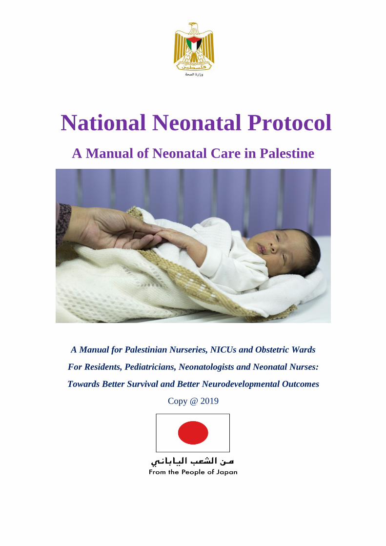


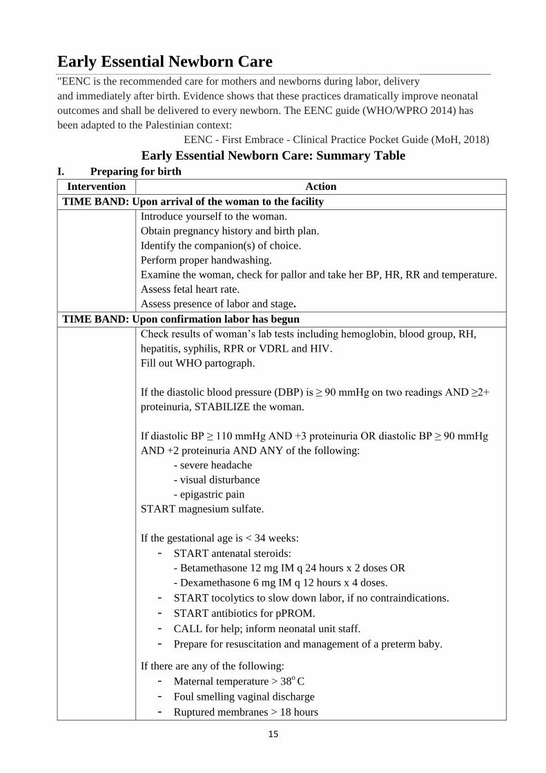
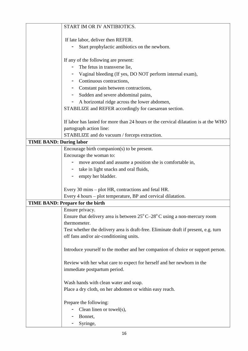
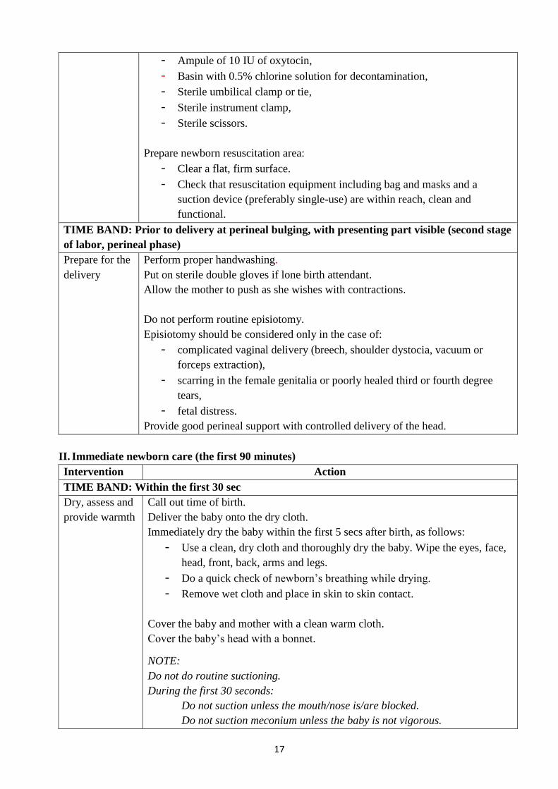
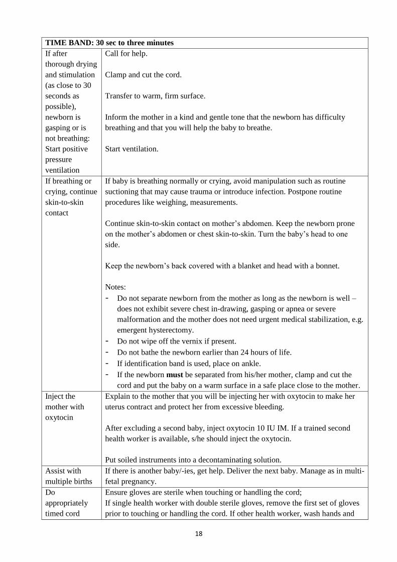
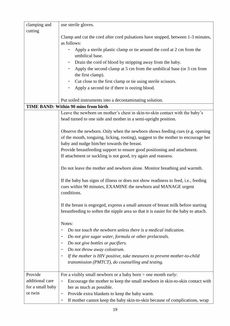
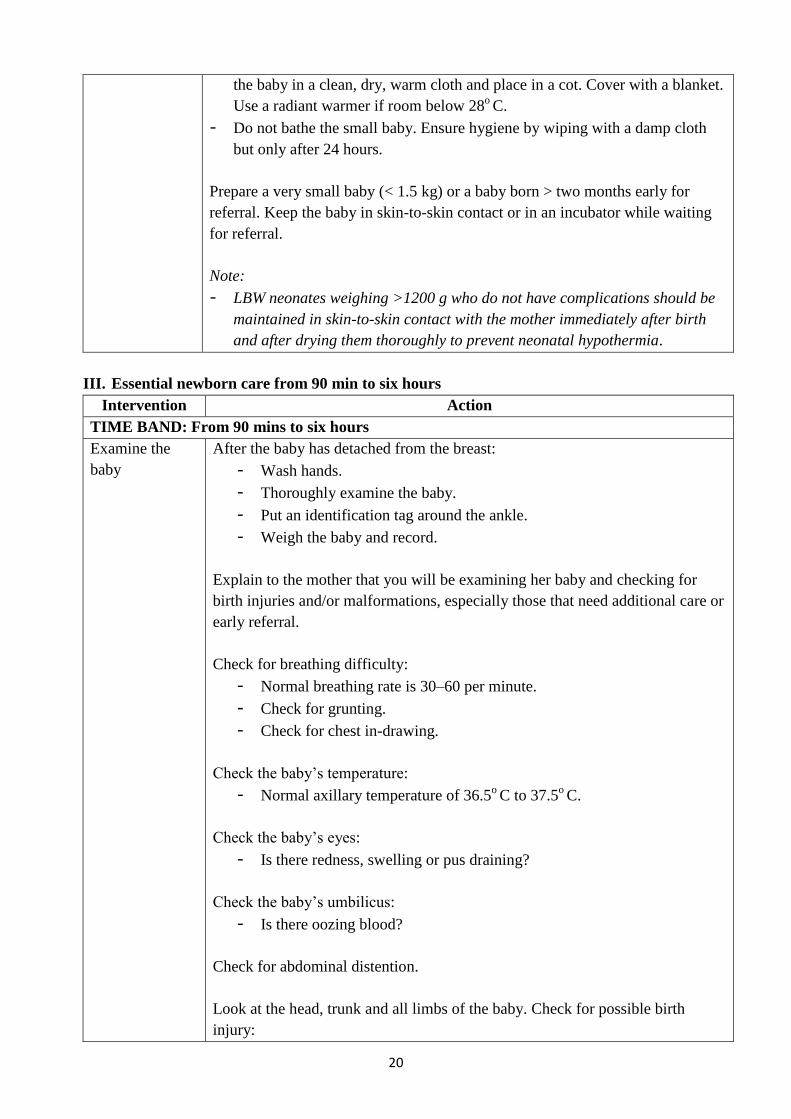
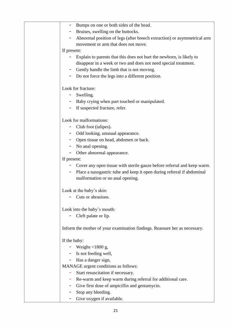

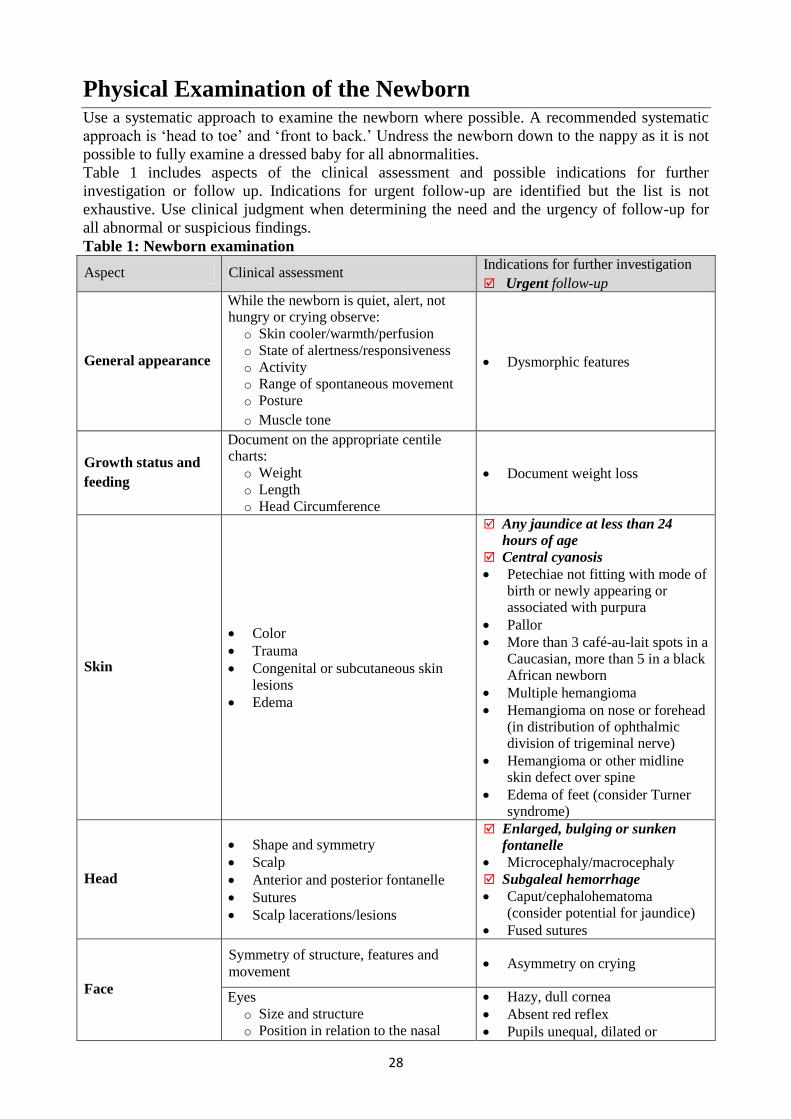
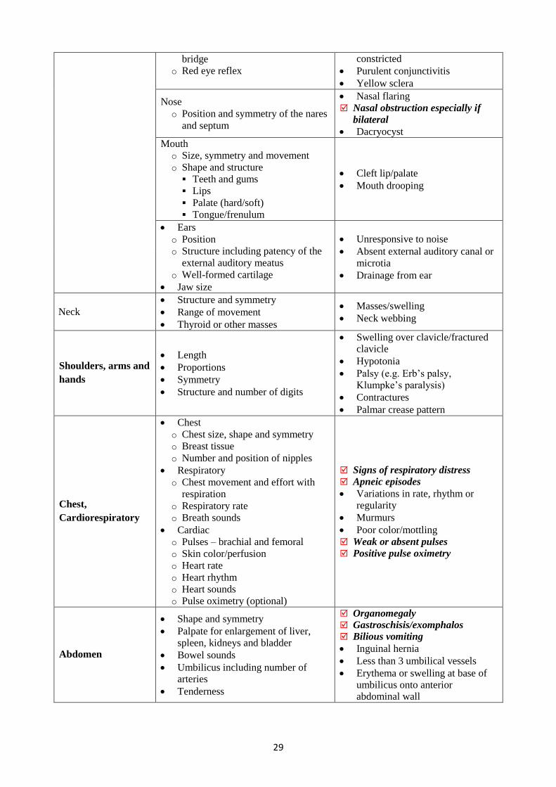
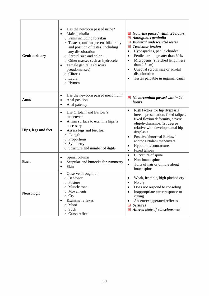
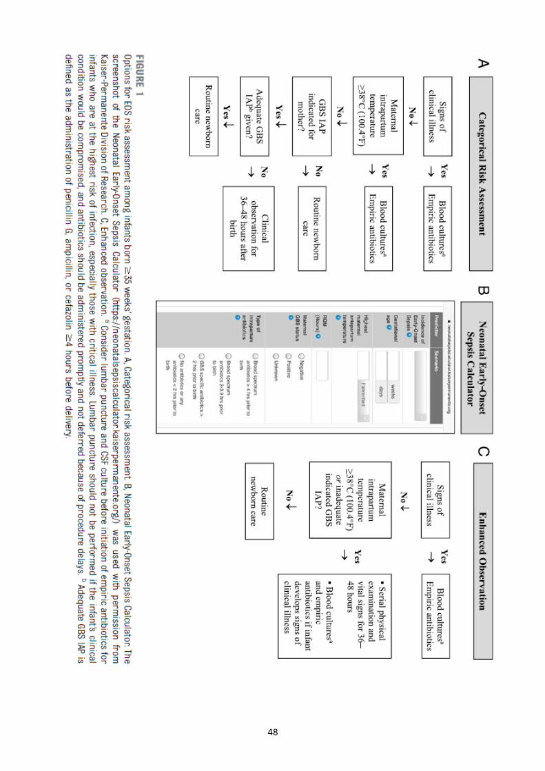
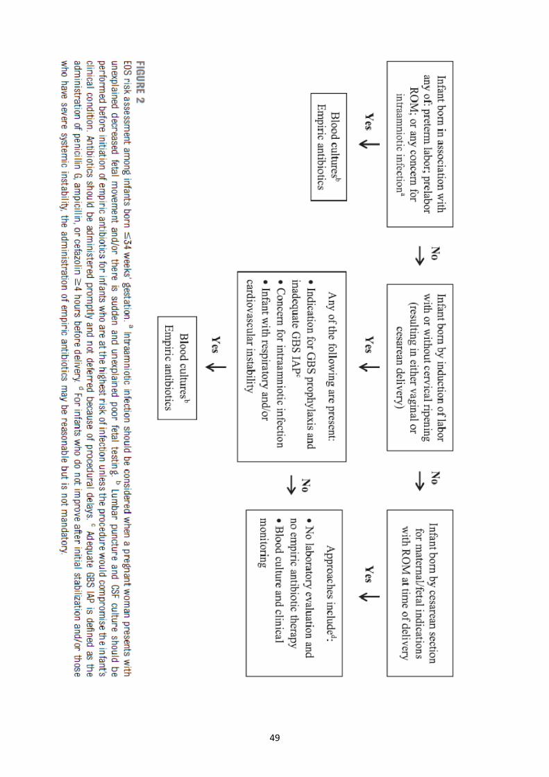
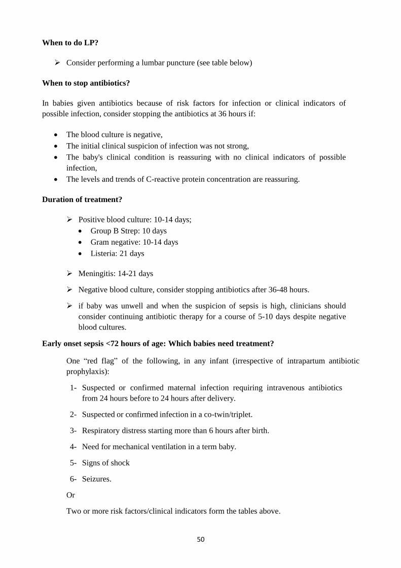
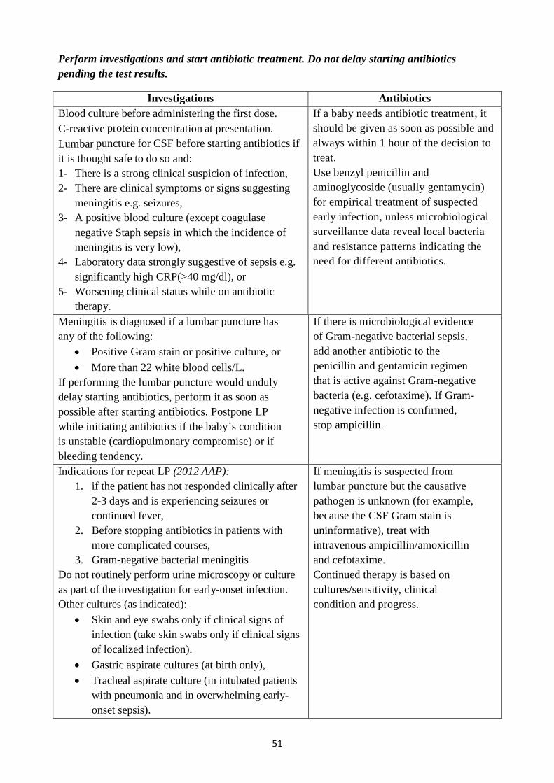
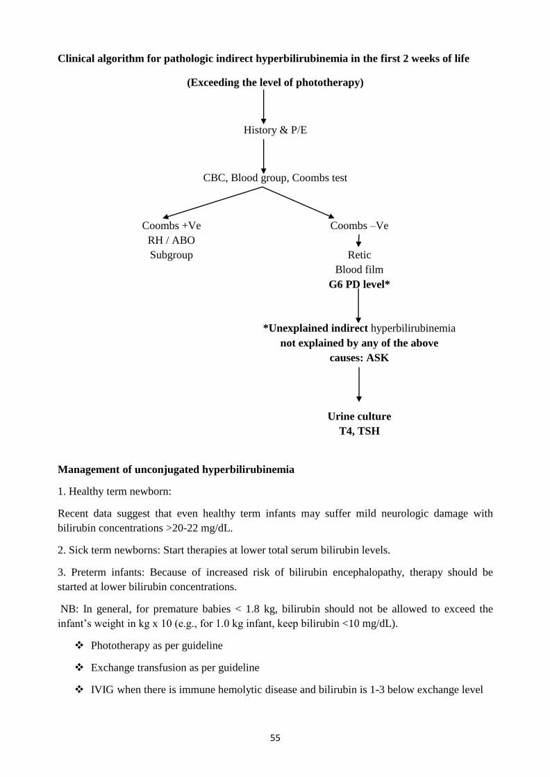
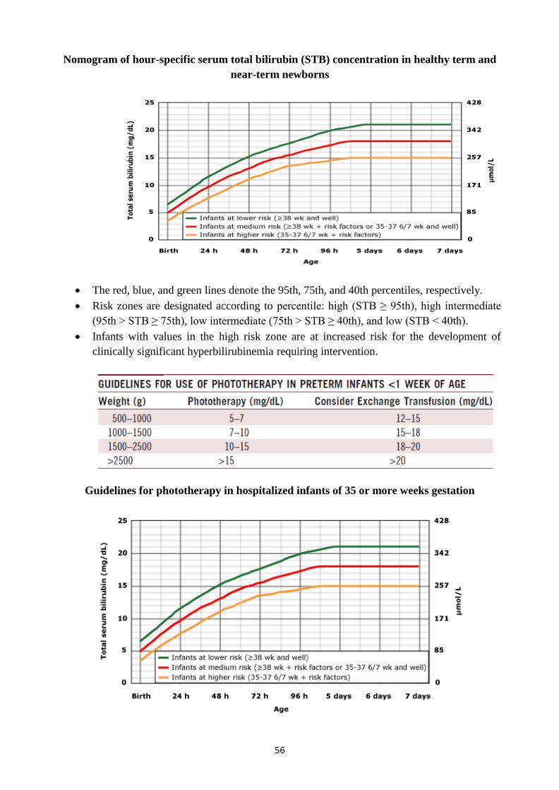
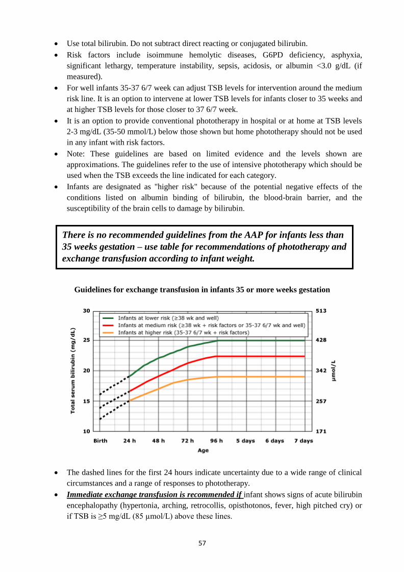
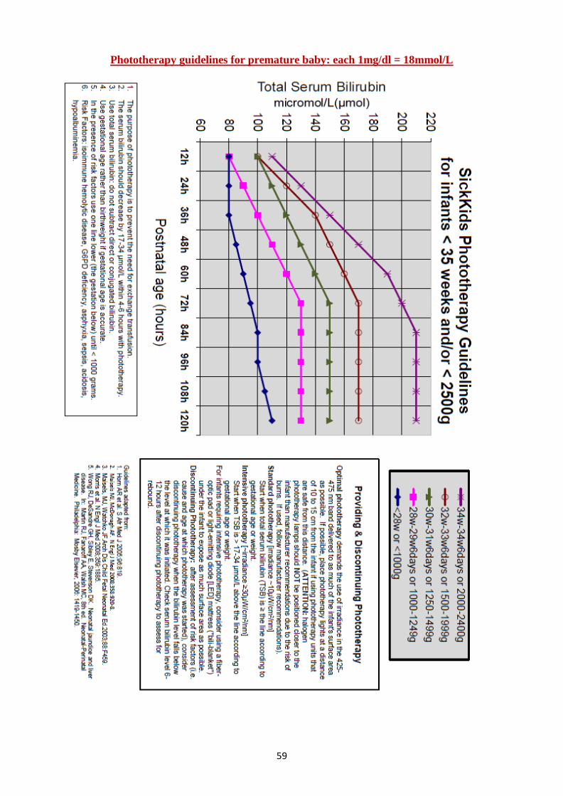
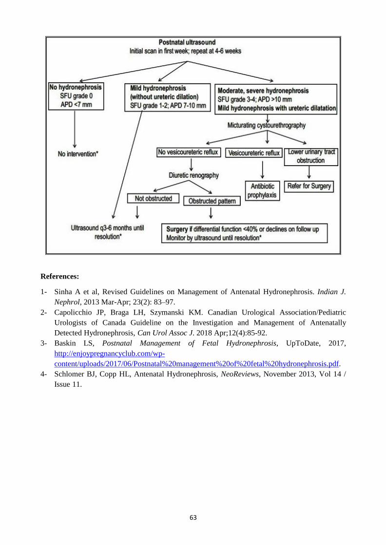
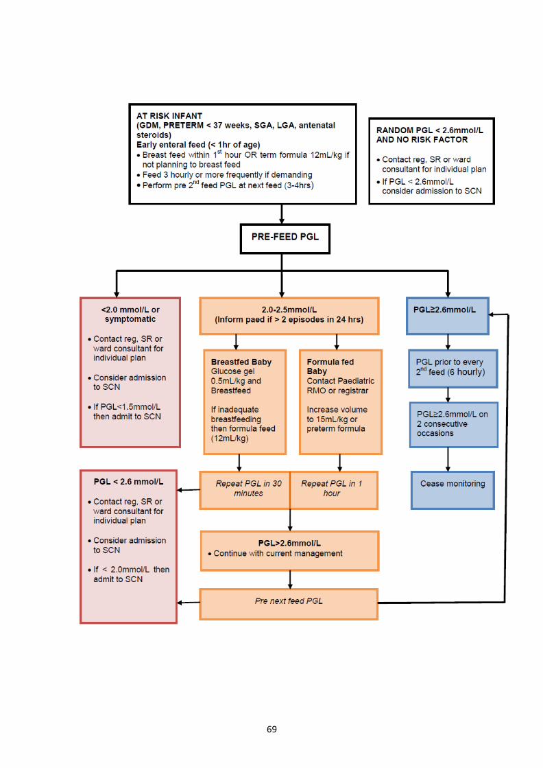

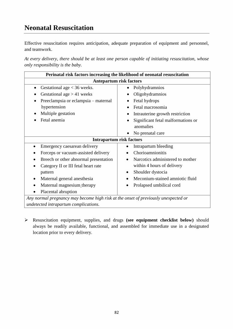
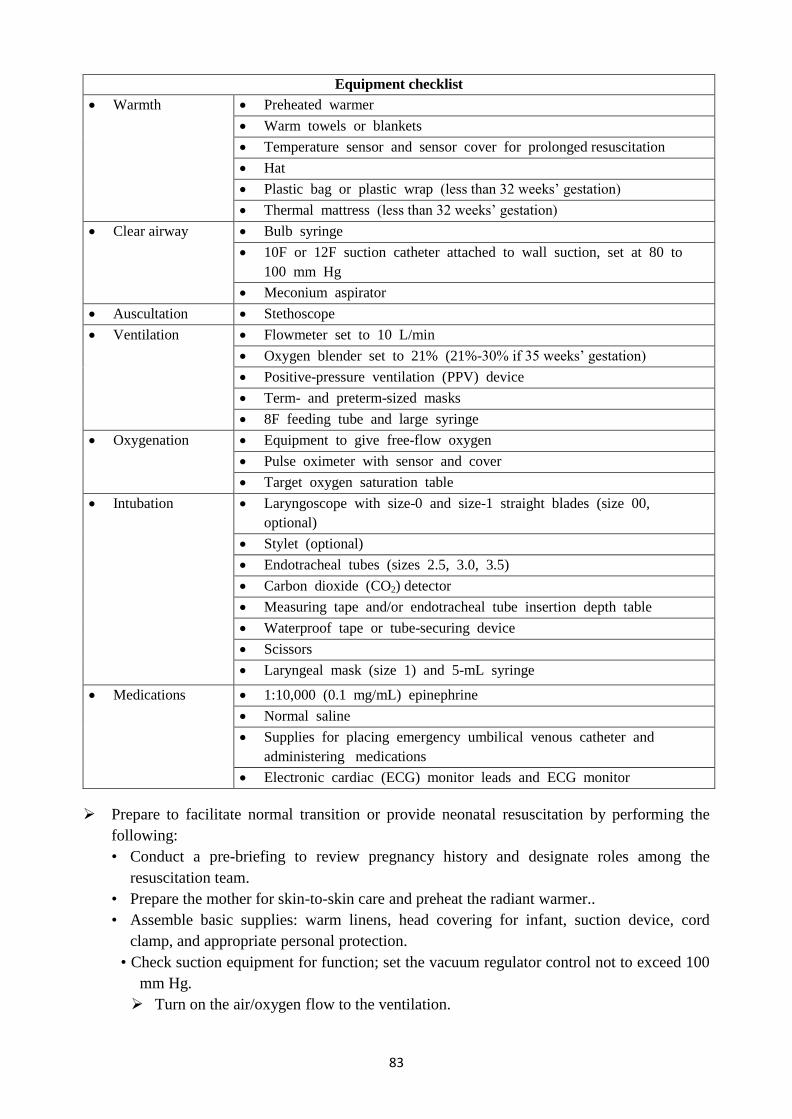
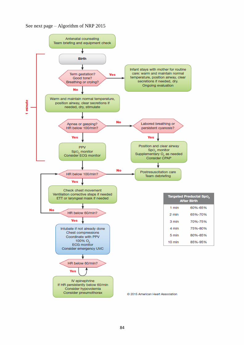
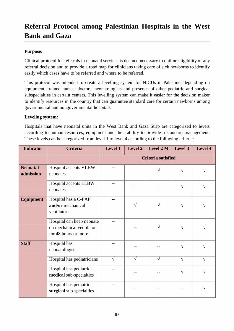
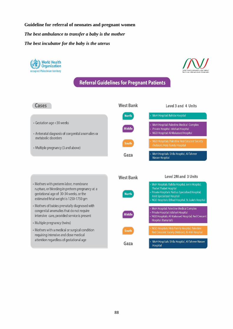
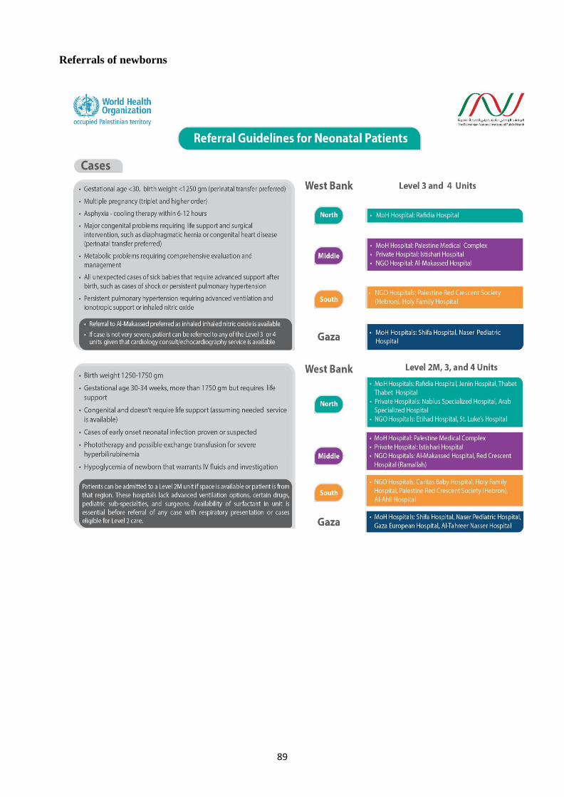
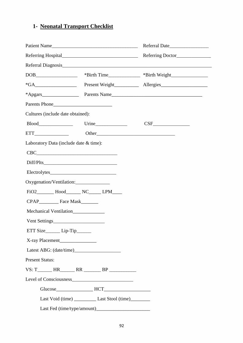
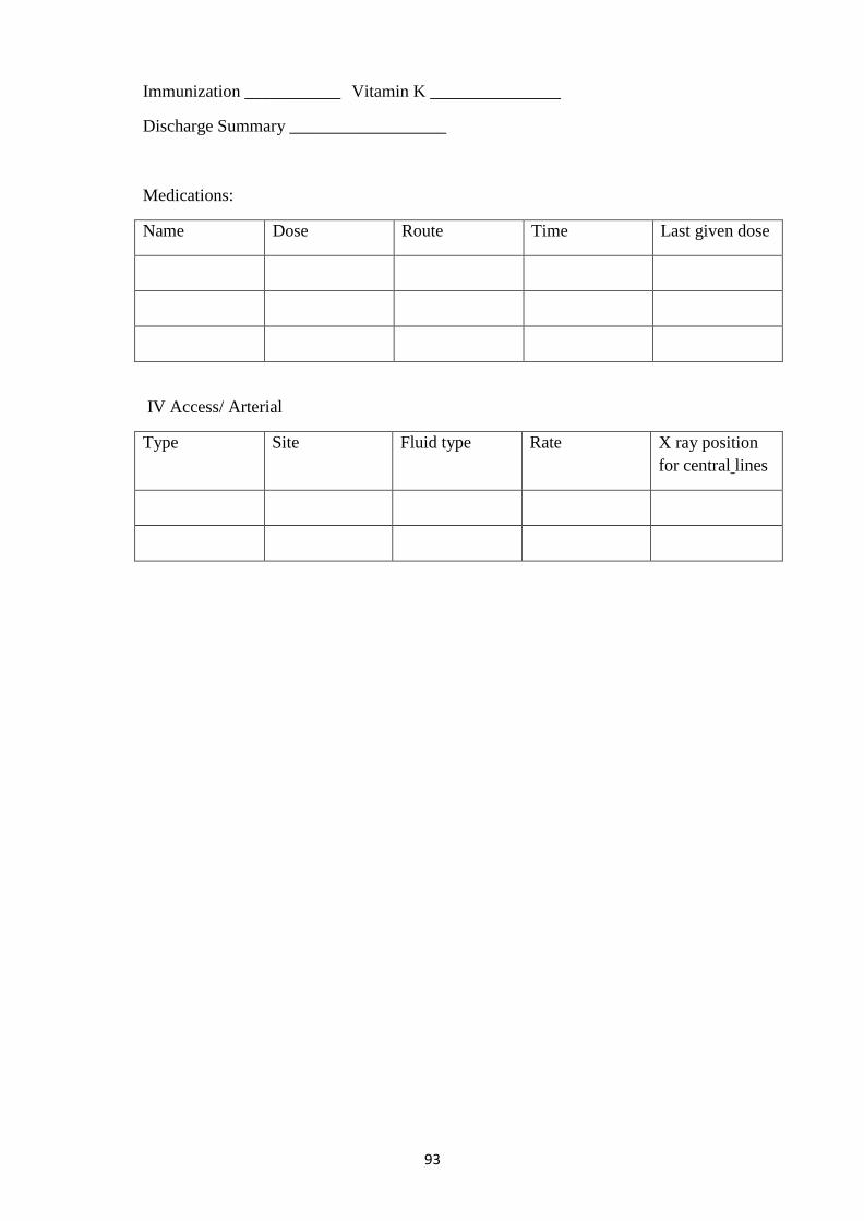
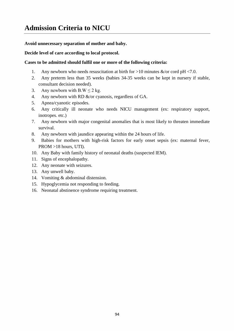
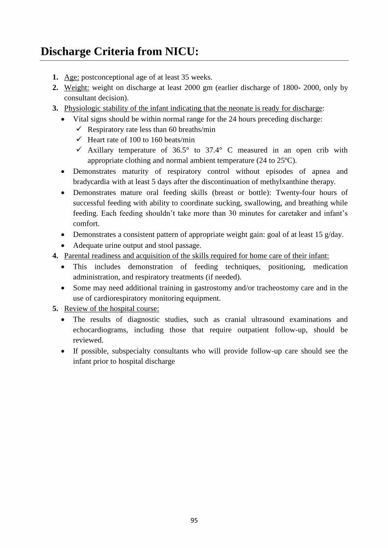

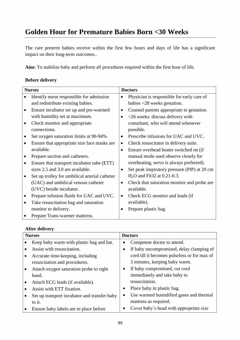
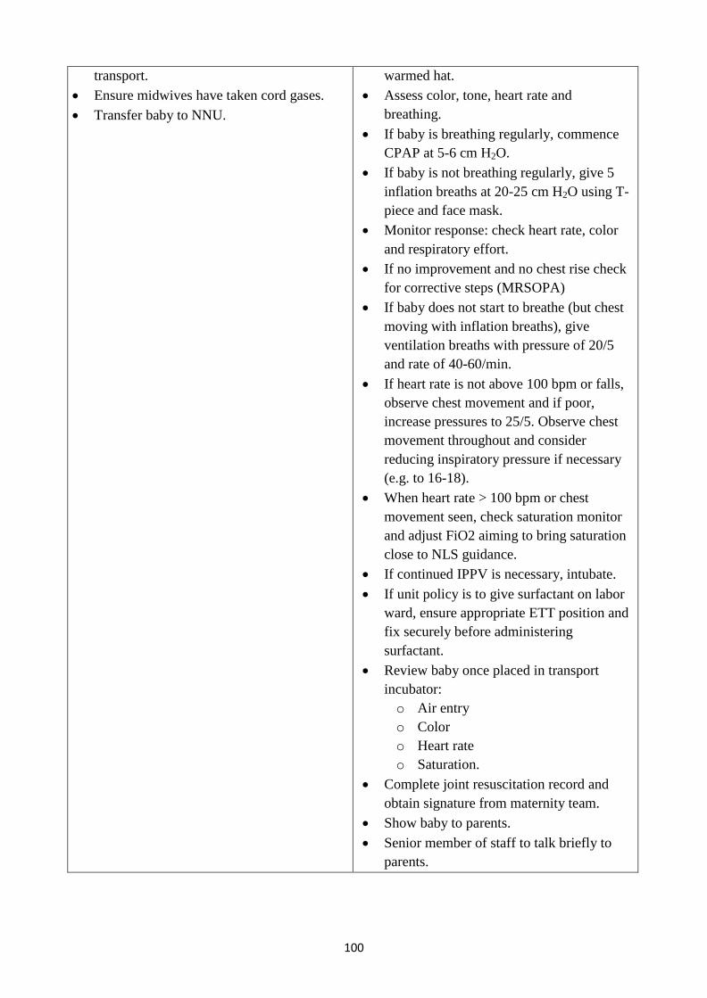
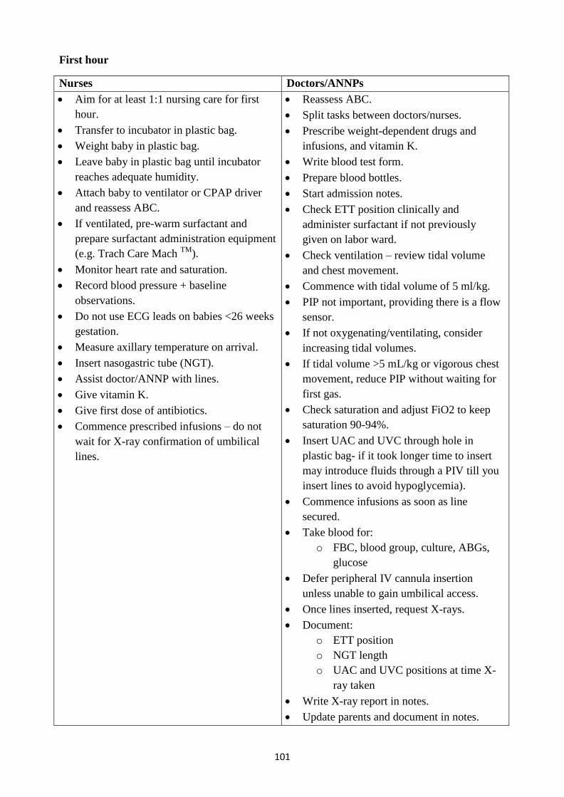
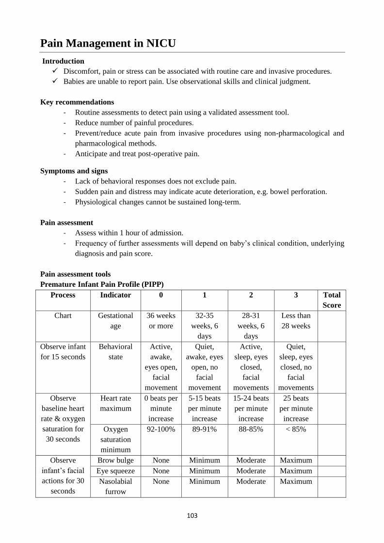
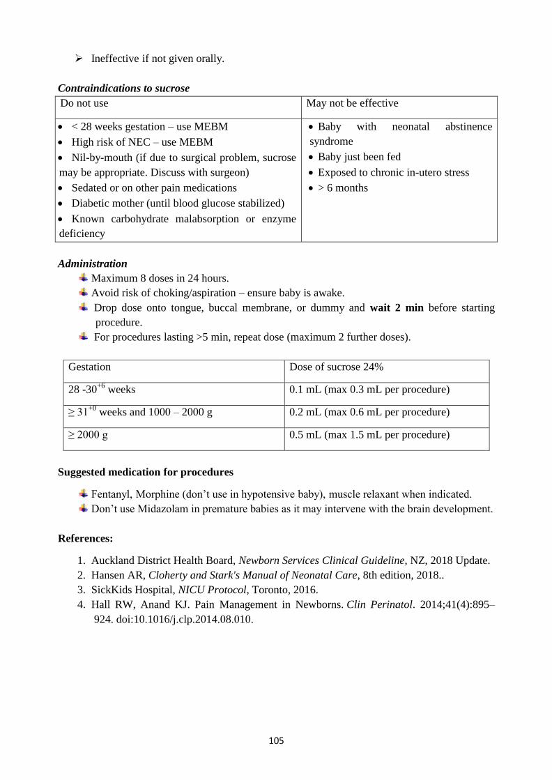
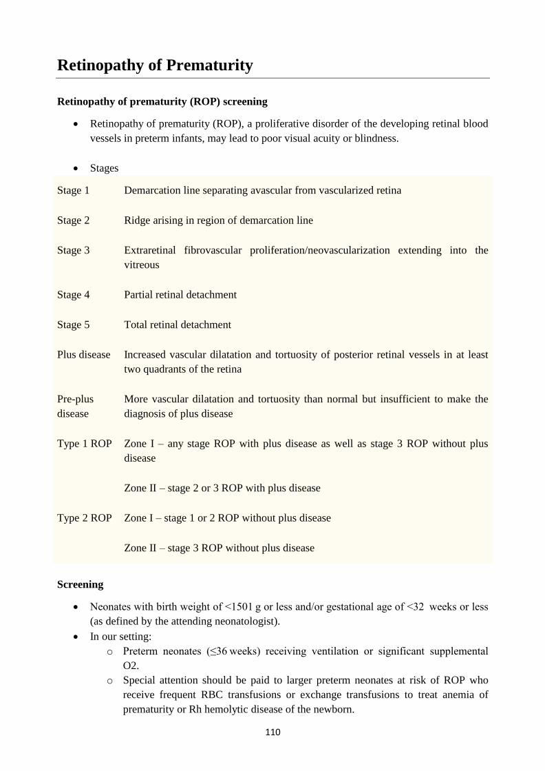
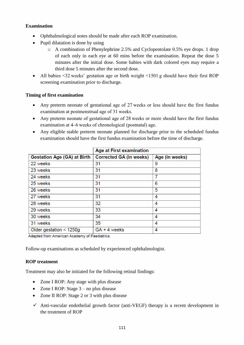
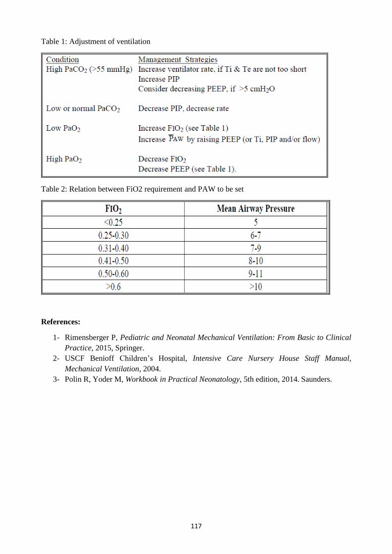

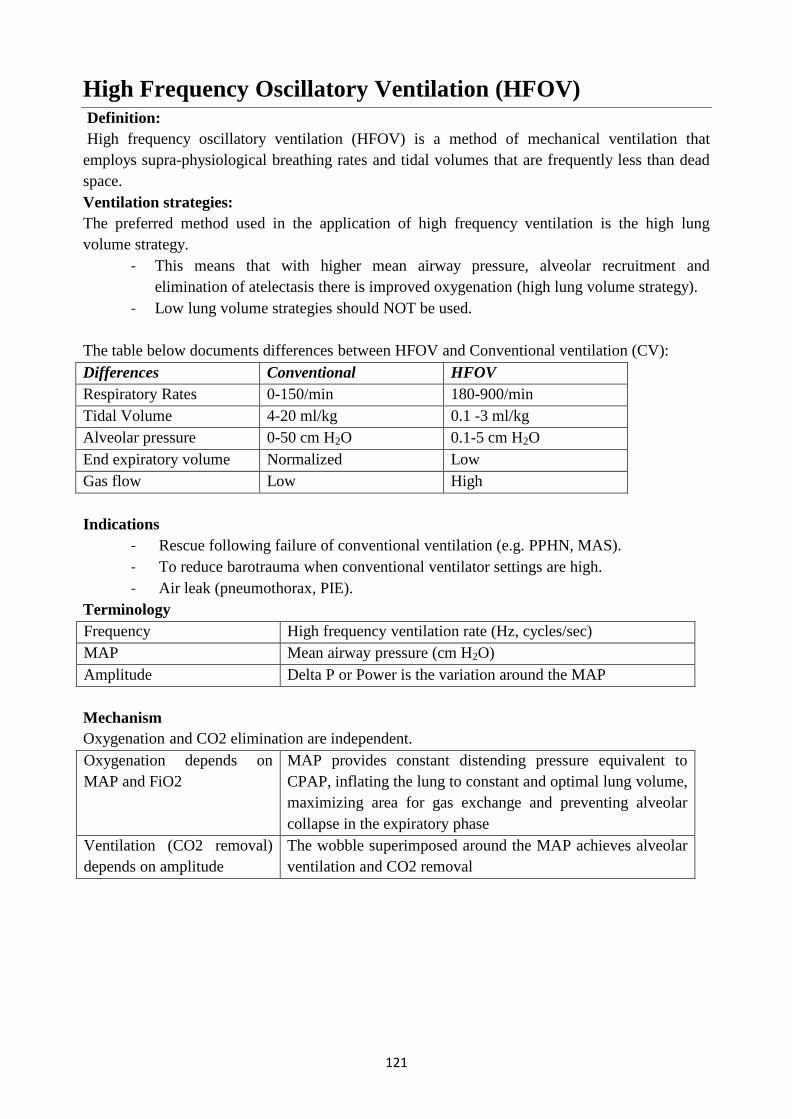
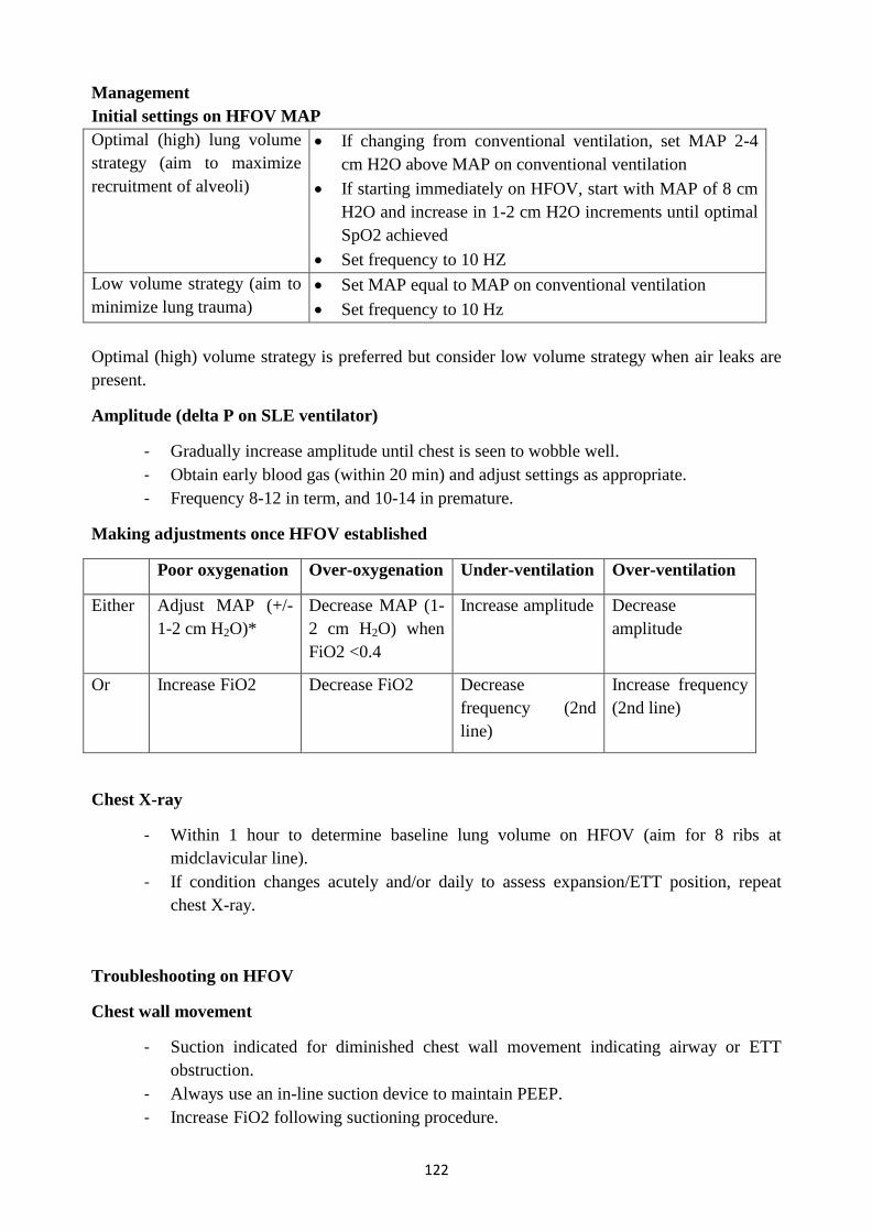

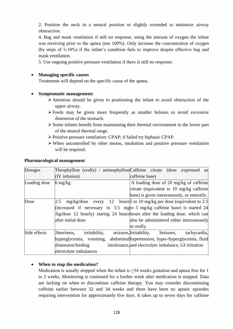
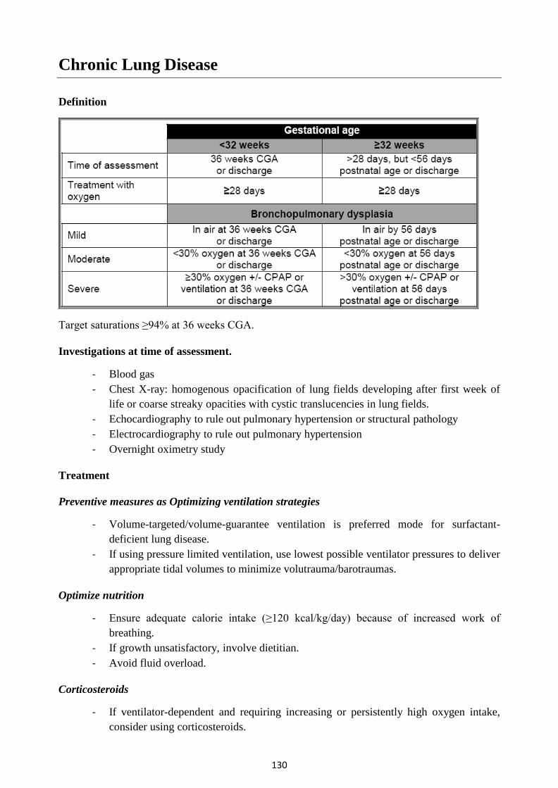
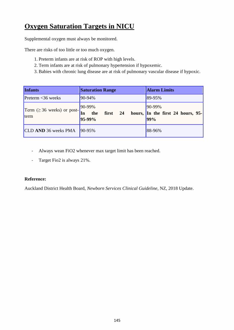

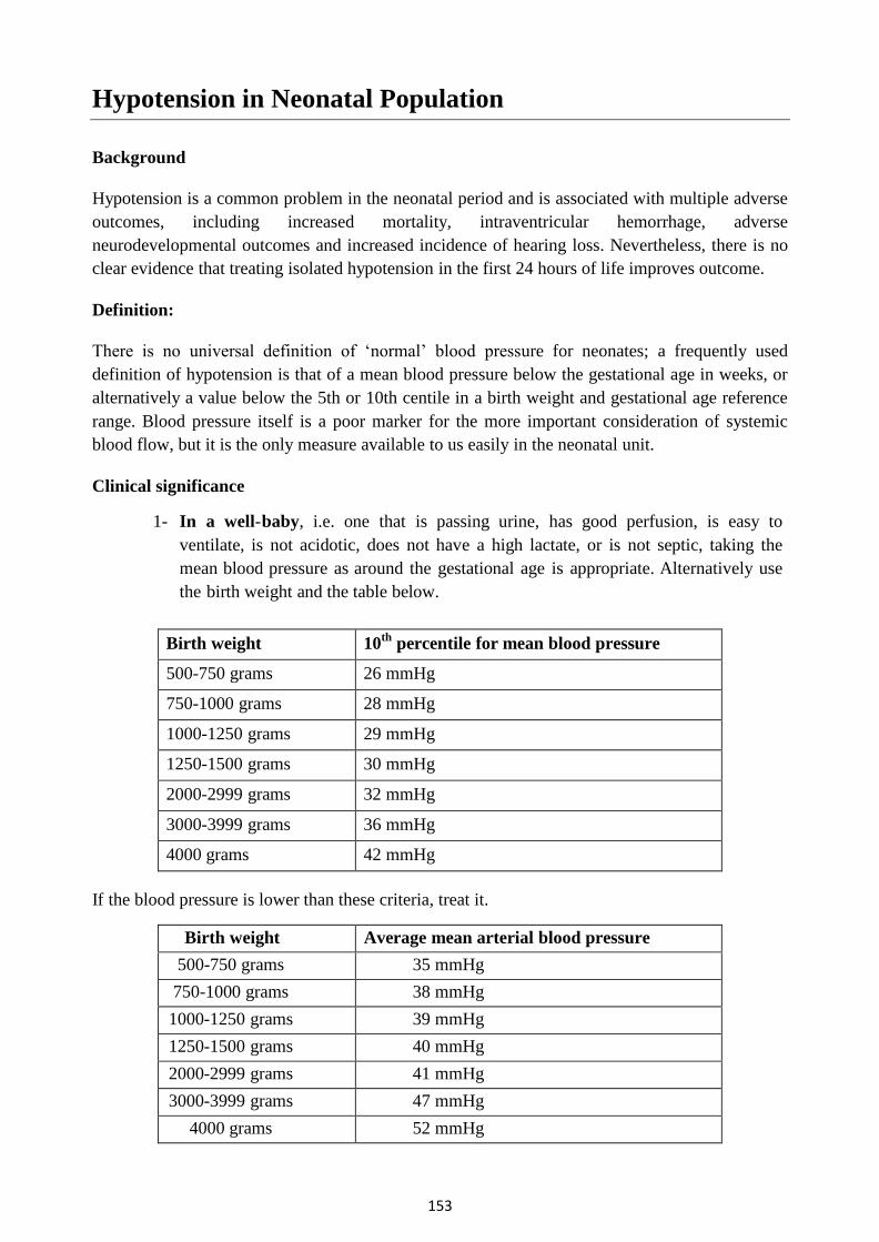
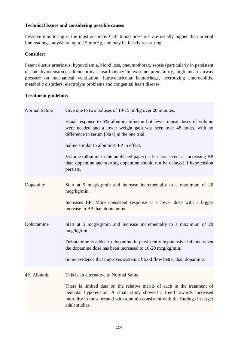
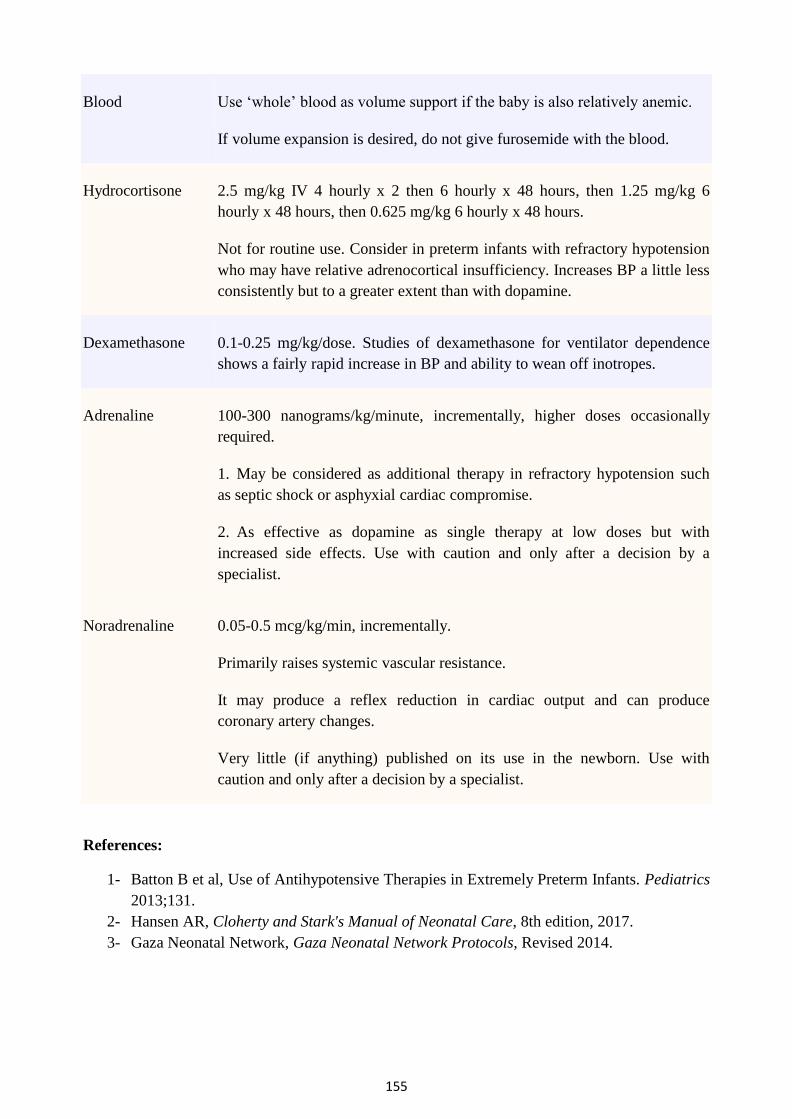
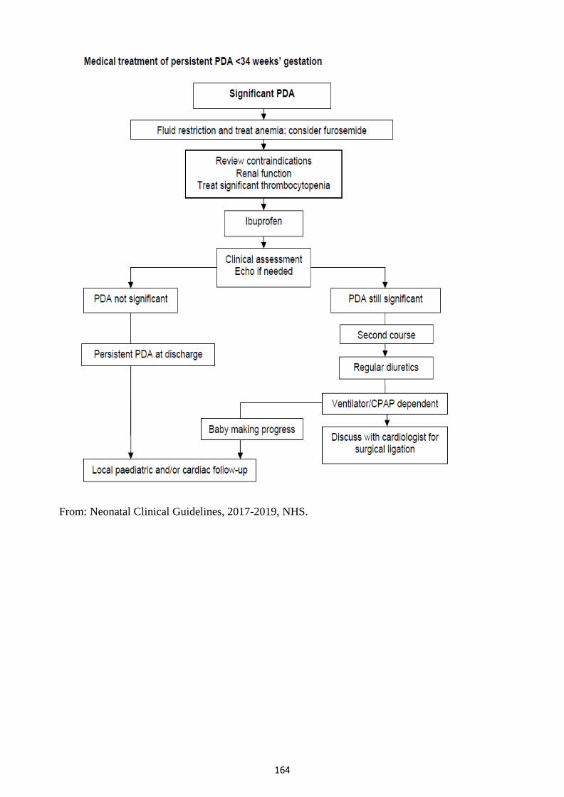
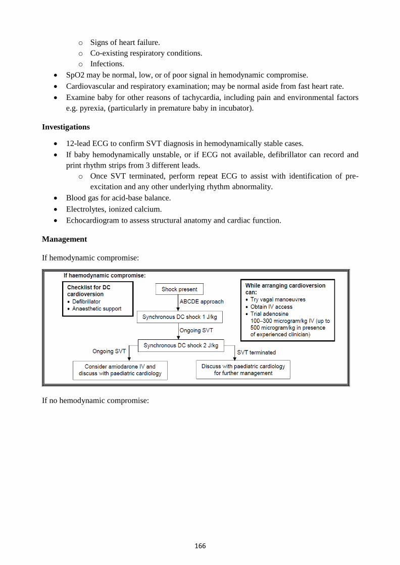
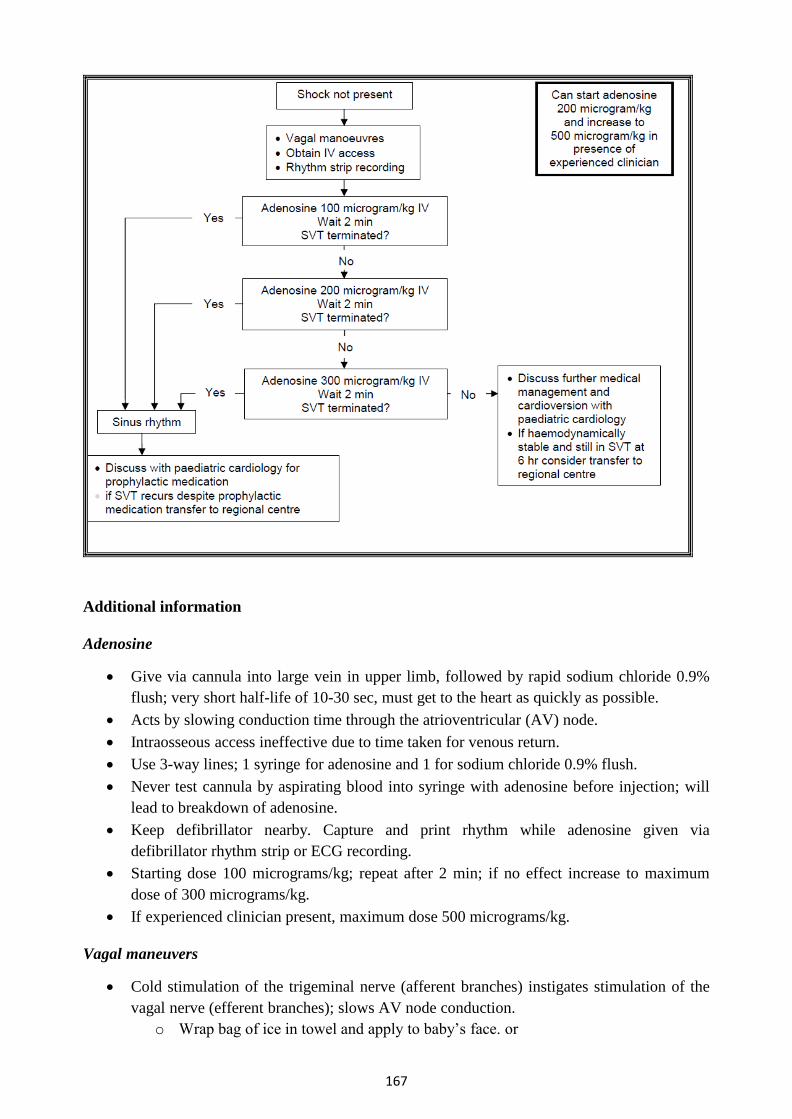
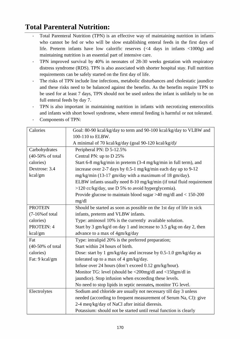
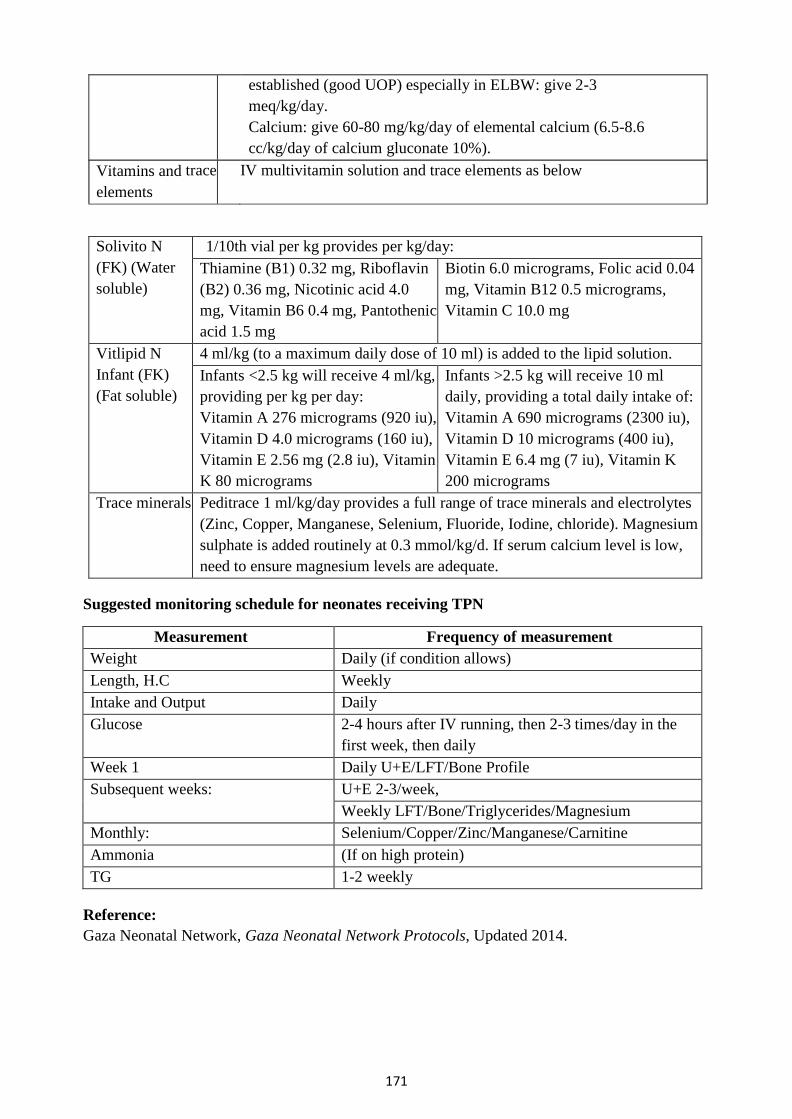
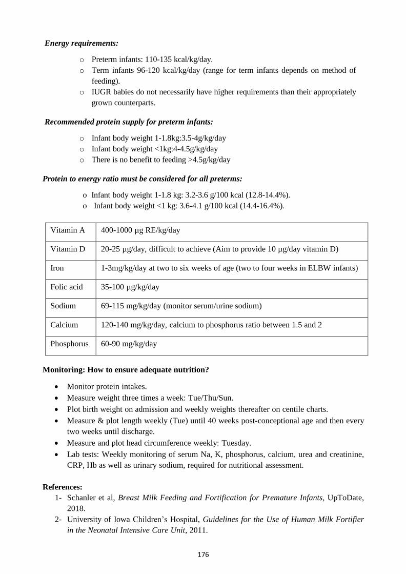
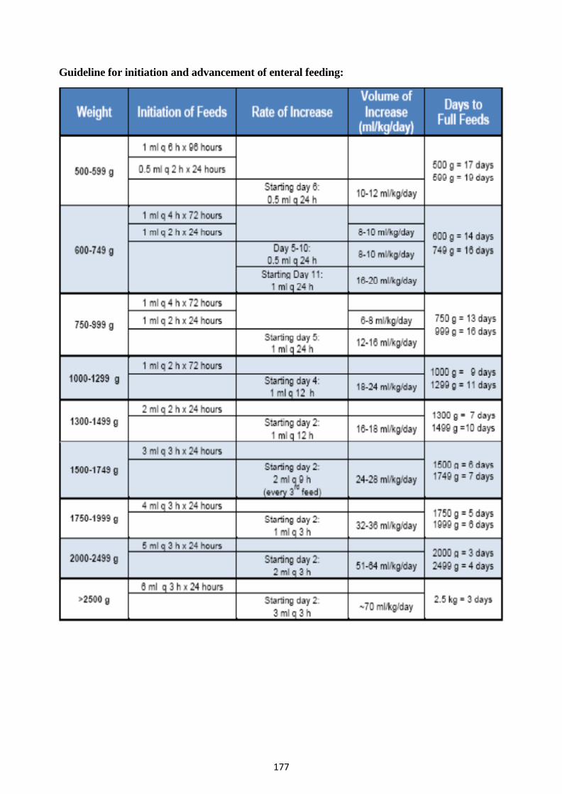
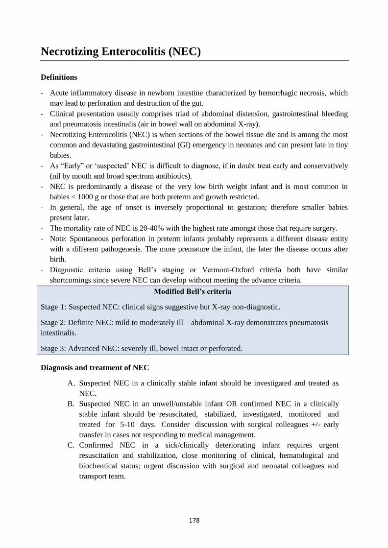

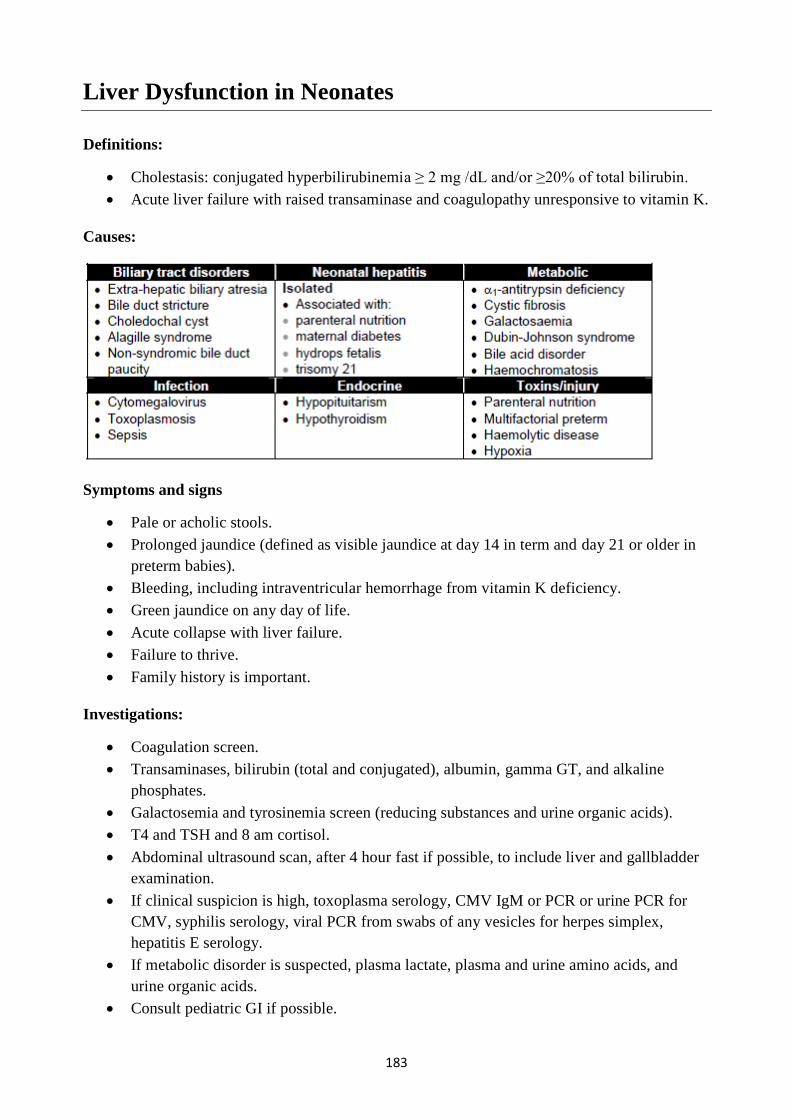
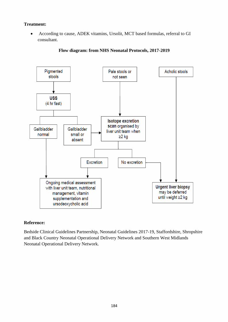
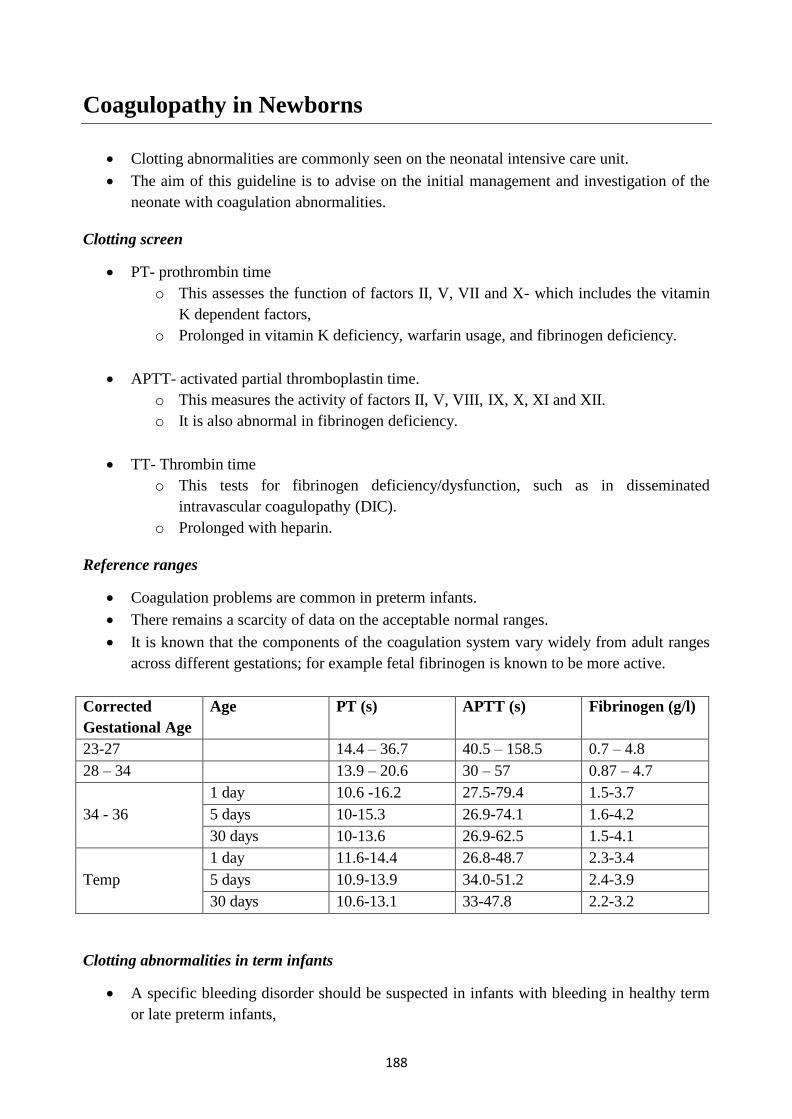
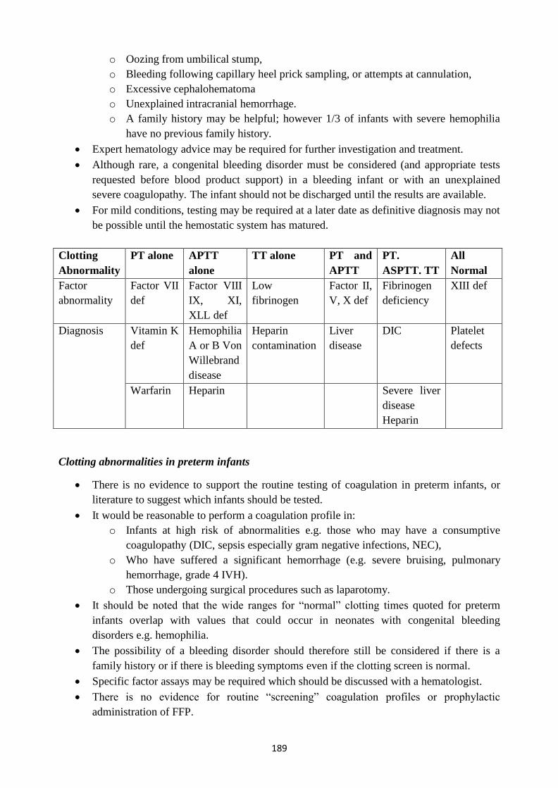
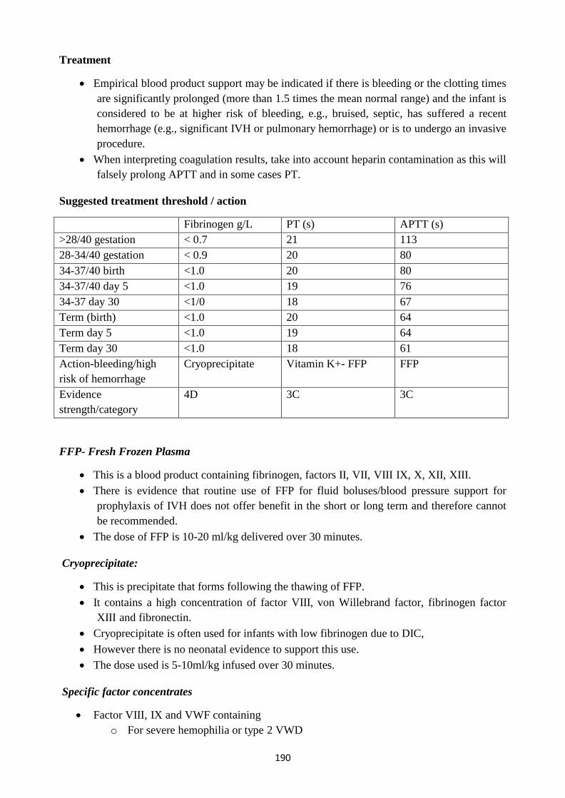
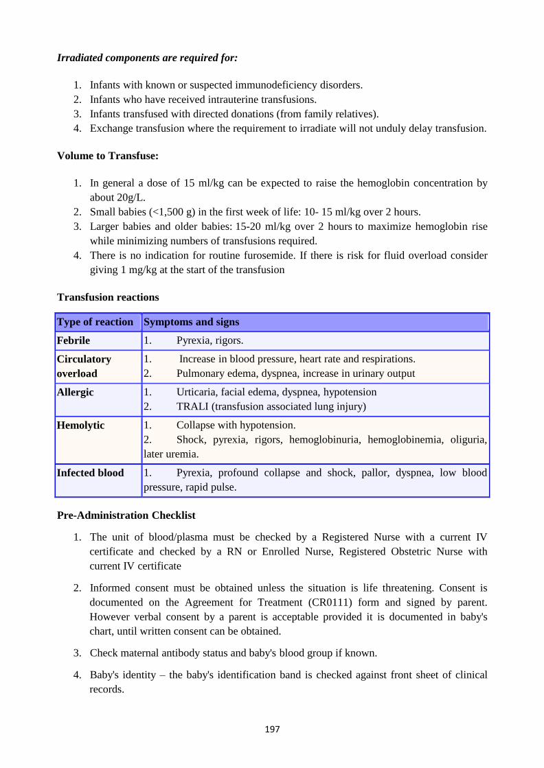
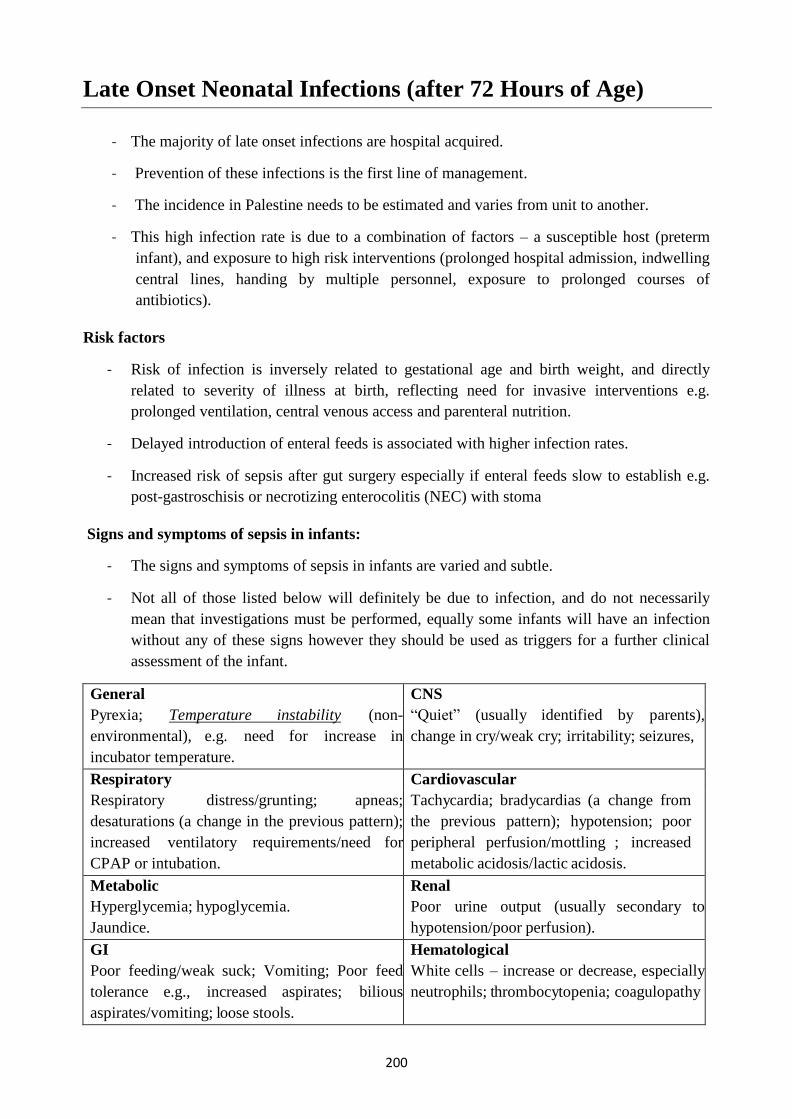
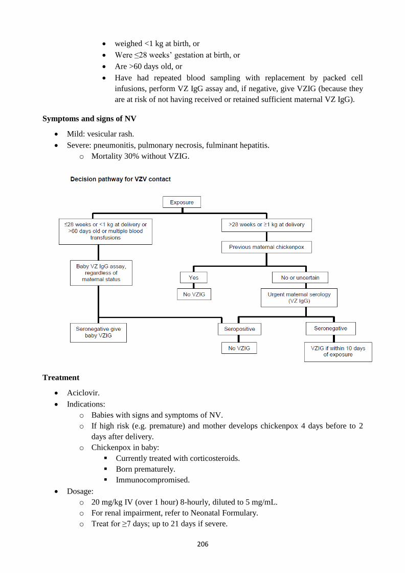

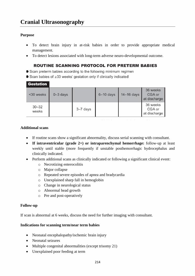
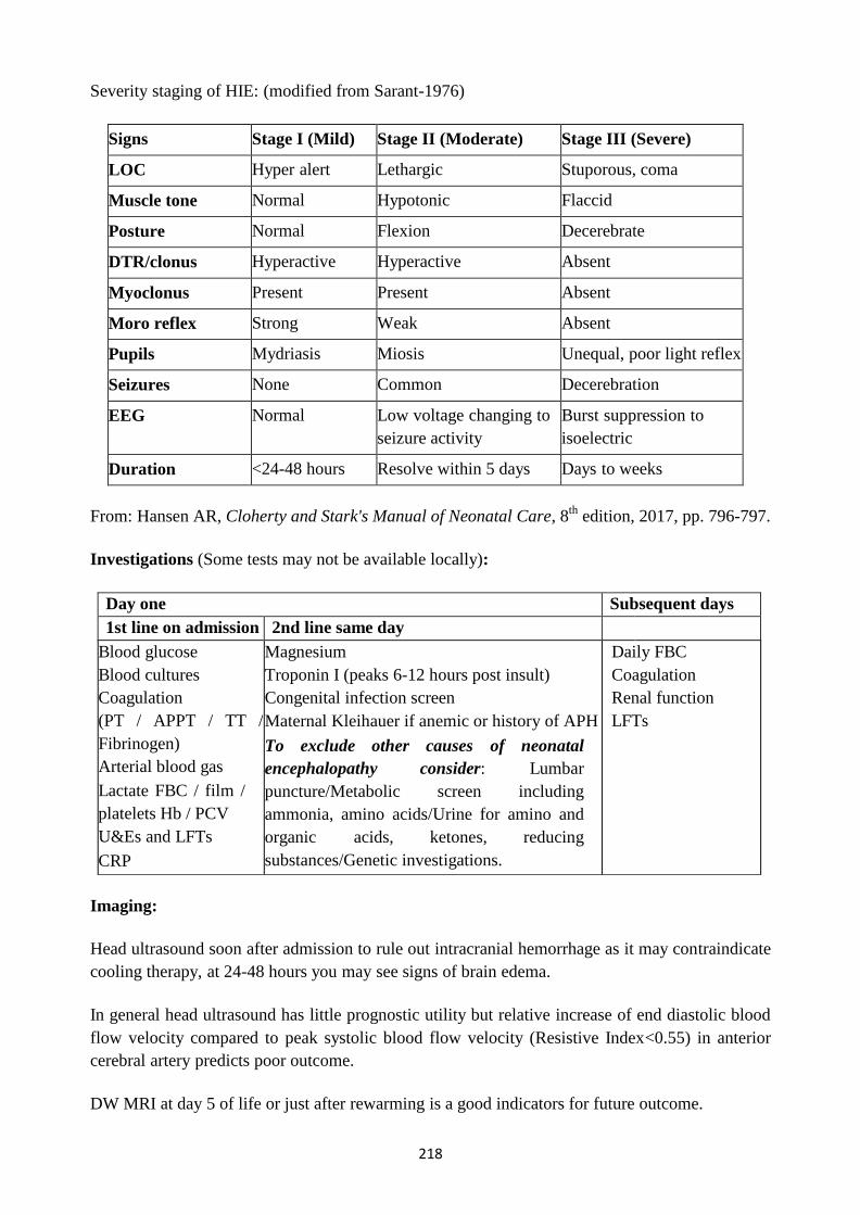
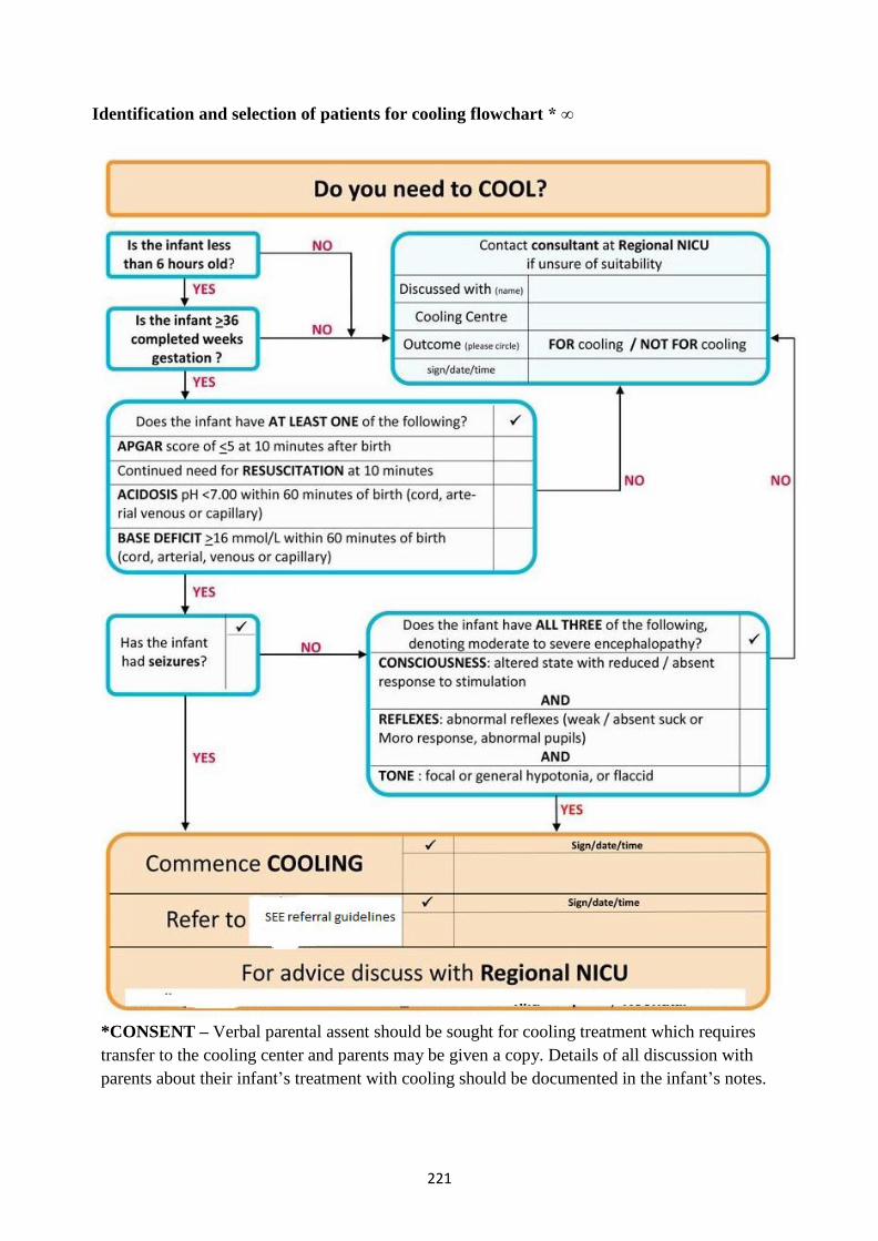
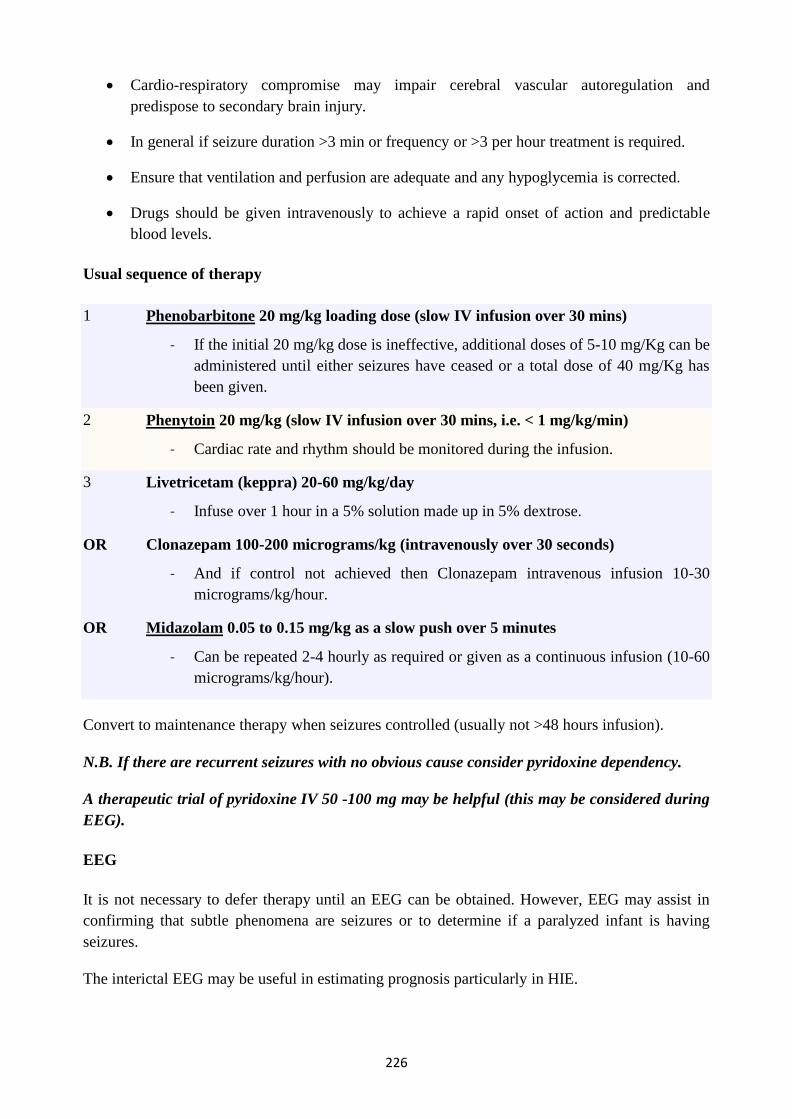
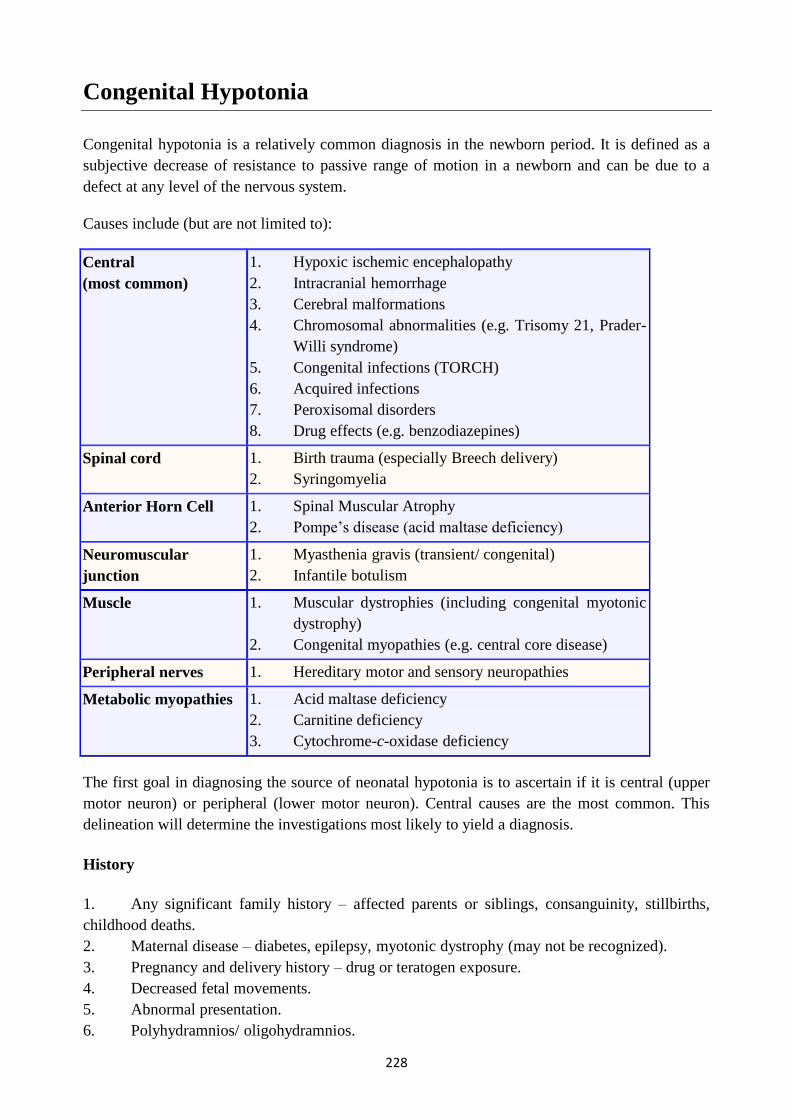
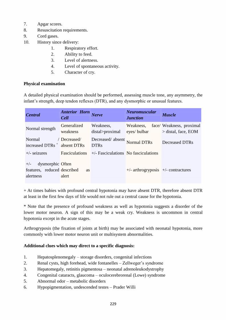

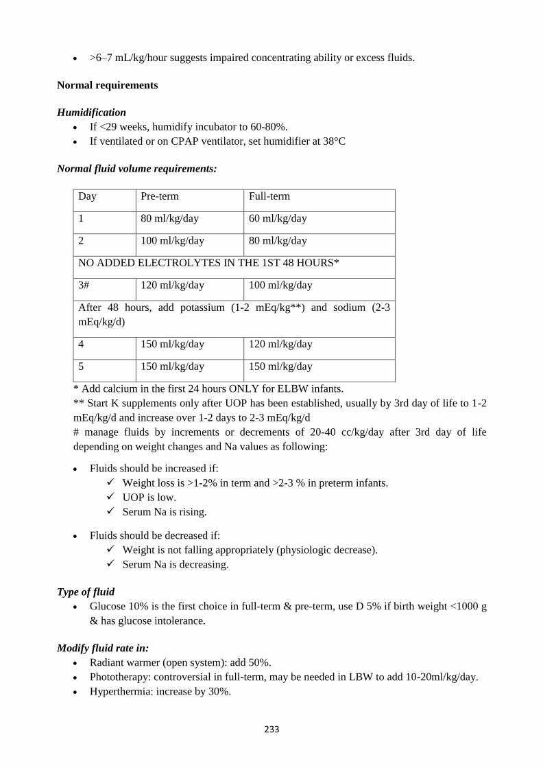
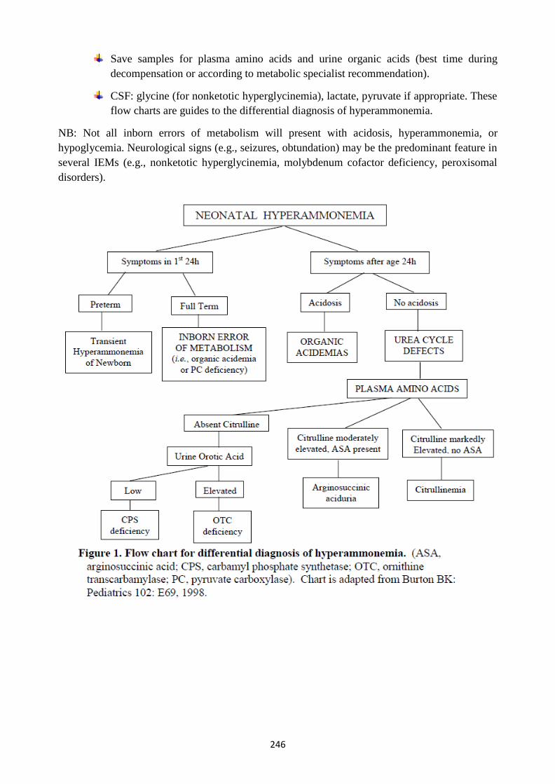
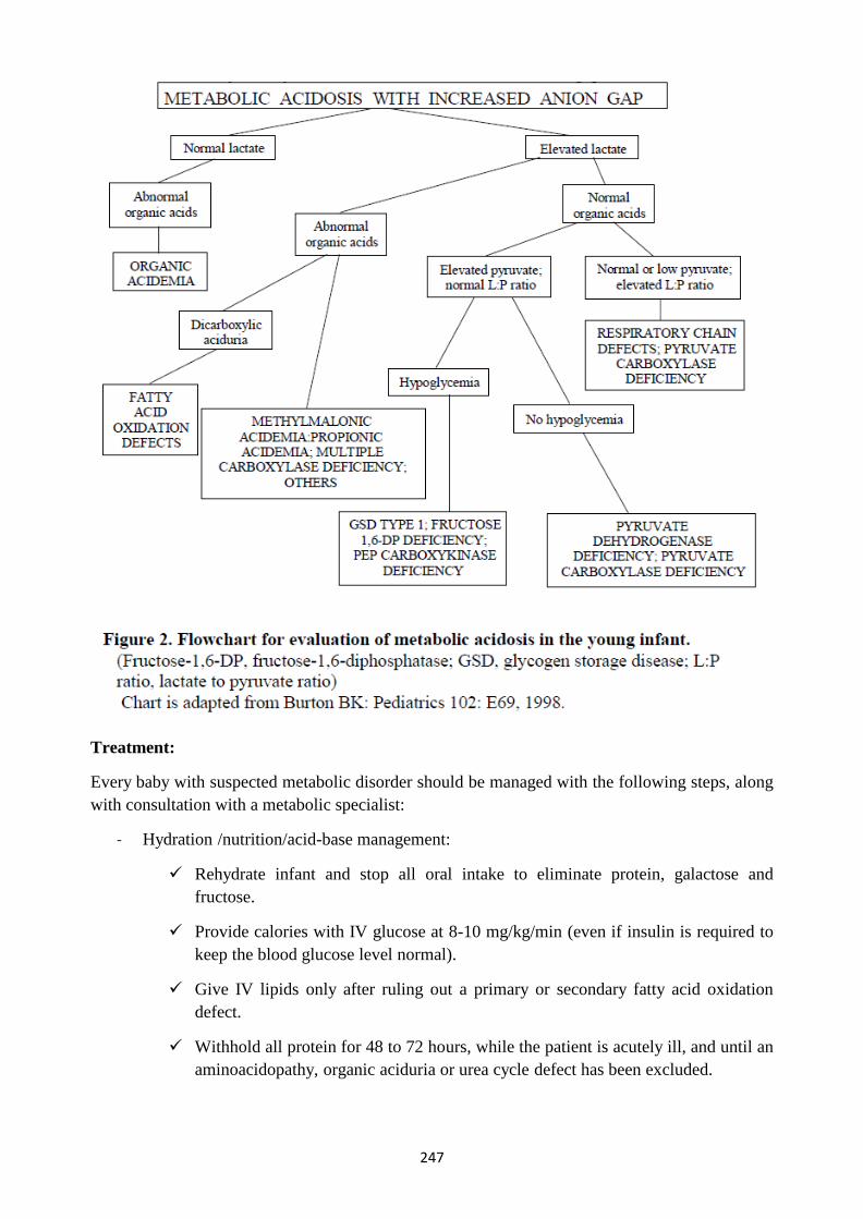
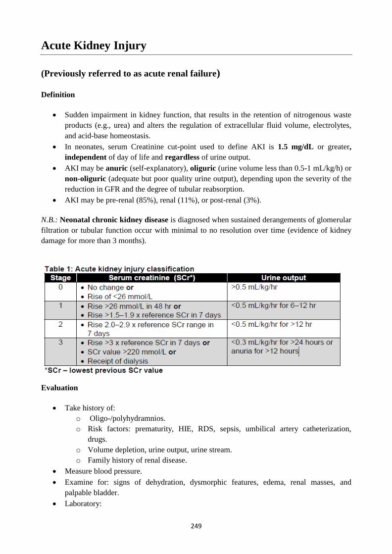
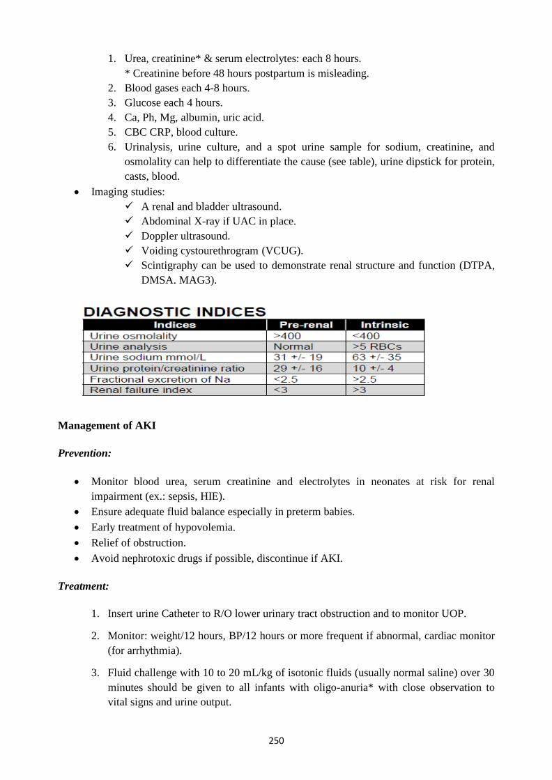


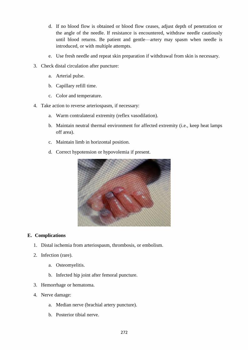
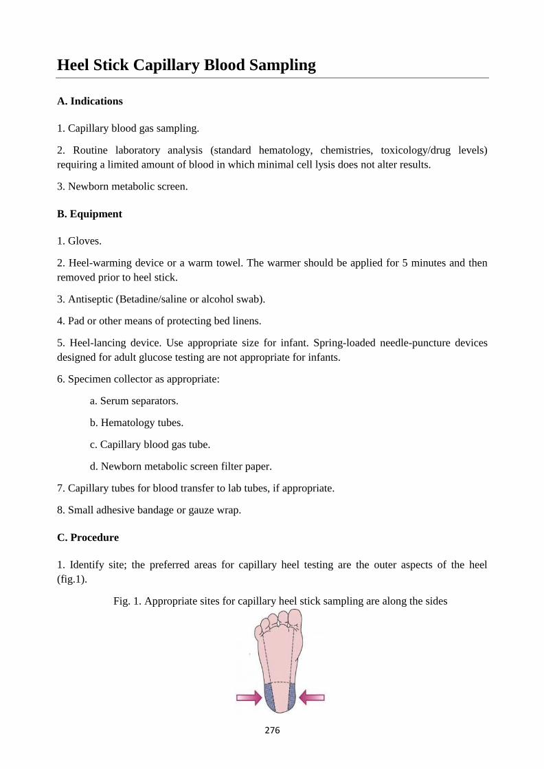
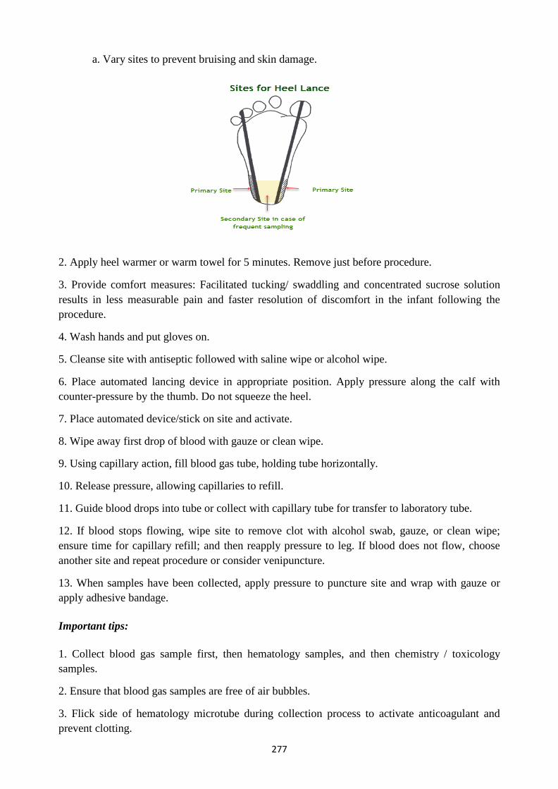
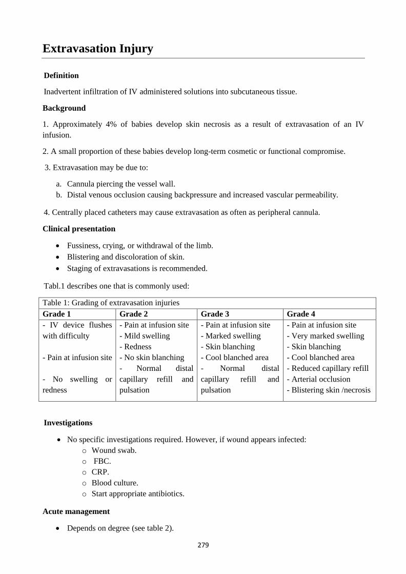
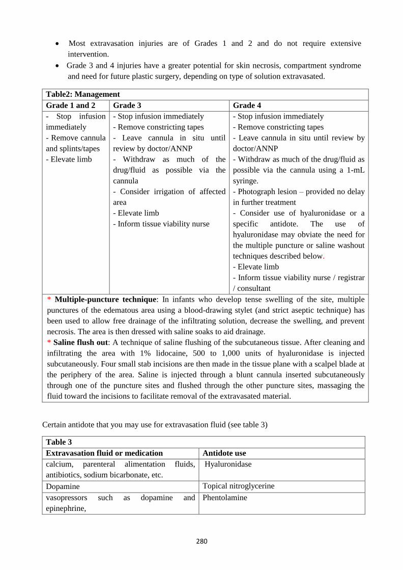
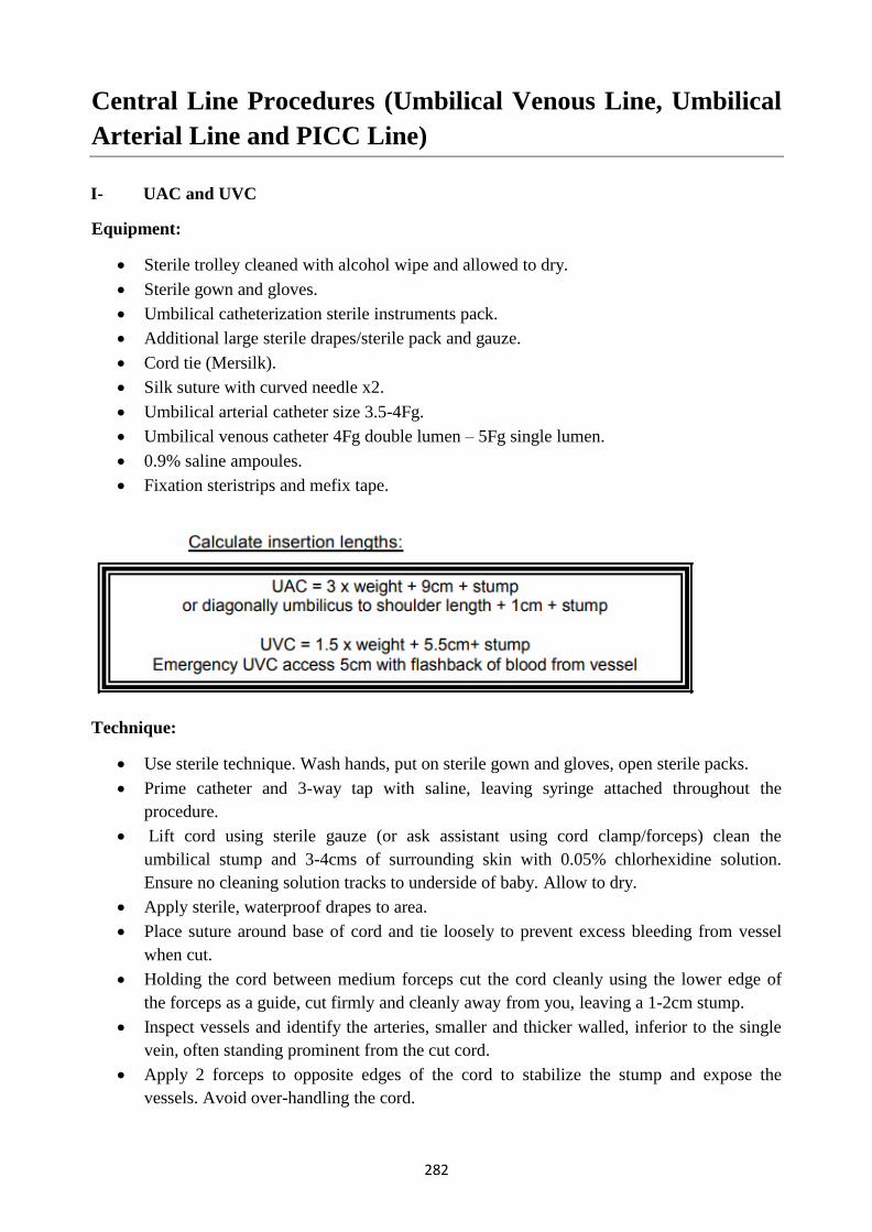
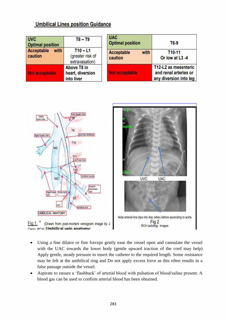

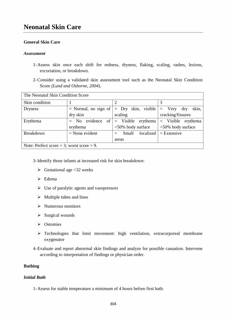
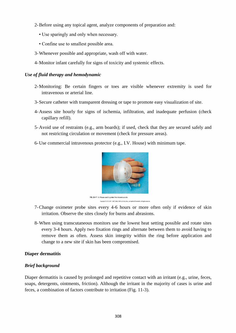
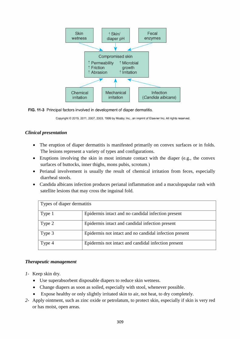
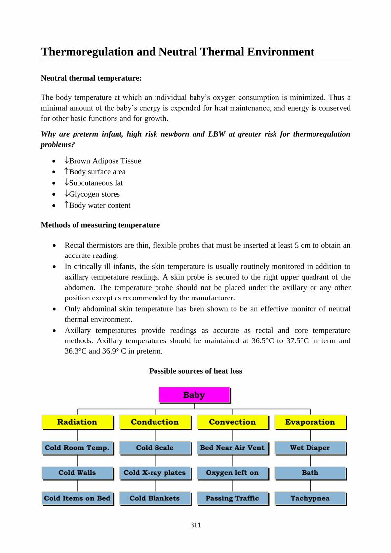
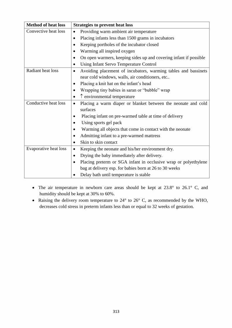
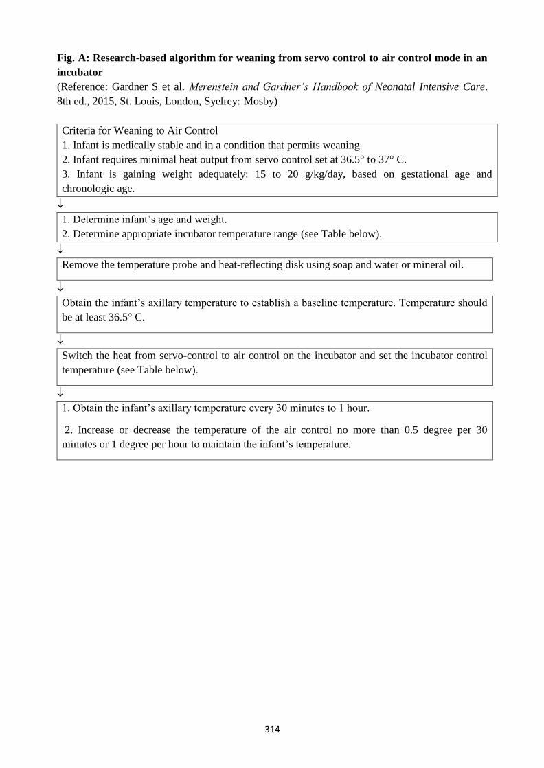
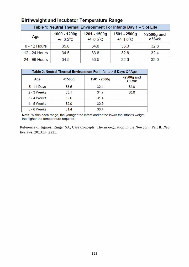
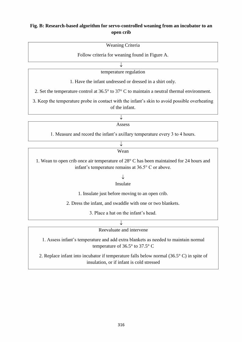
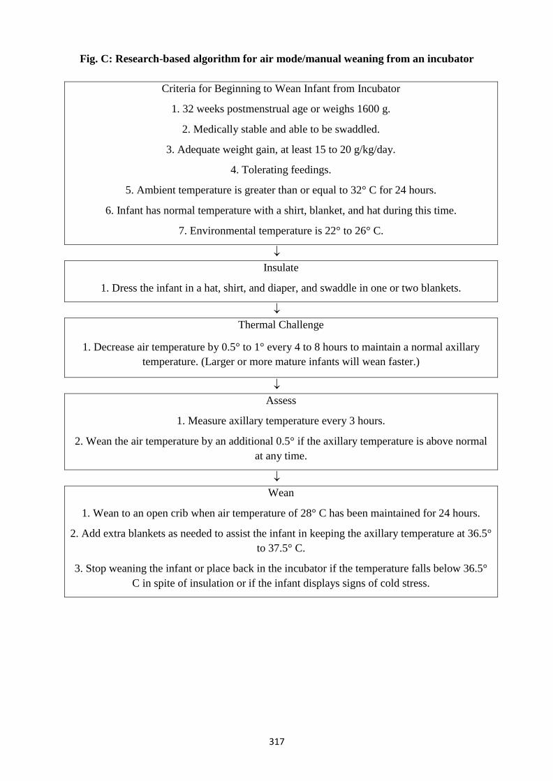
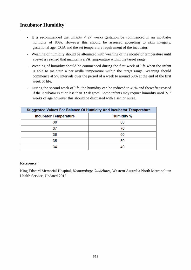
![Neonatal Standard concentration/Dilution Medication chart Dr… · Windsor Regional Hospital [NEONATAL STANDARD CONCENTRATION/DILUTION MEDICATION CHART]-updated 3.23.2020 1 Reference:](https://static.documents.pub/doc/80x56/5ea028ffa86e5a7fc4270000/neonatal-standard-concentrationdilution-medication-chart-dr-windsor-regional.jpg)


