Page 1
NATURAL PRODUCTS AS POSSIBLE TREATMENTS OF TYPE II DIABETES MELLITUS AND ITS COMPLICATIONS
by
JOHNETTA L. FARRAR
(Under the Guidance of Phillip Greenspan)
Abstract
Diabetes mellitus is a chronic disease caused by inherited and/or acquired deficiency in
production of insulin by the pancreas, or by the ineffectiveness of the insulin that is
produced. Such a deficiency results in increased concentrations of glucose in the blood,
which in turn damages several physiological systems, in particular blood vessels and
nerves. Recently compiled data from the World Health Organization (WHO) show that
approximately 150 million people have diabetes mellitus worldwide, and that this number
may well double by the year 2025. Protein glycation, or the reaction of biological amines
with reducing sugars to form a complex family of rearranged and dehydrated covalent
adducts, is implicated in the formation of diabetic complications. Although some plant
extracts have been shown to inhibit glycation, the effect of extracts of food products on
protein glycation has not received significant attention. In this dissertation, the effect of
ethanolic extracts of muscadine grapes, sorghum bran, and Japanese knotweed on protein
glycation are investigated. A very high antioxidant capacity is common to these three
products. These studies show that each of these strongly inhibit protein glycation.
Possible mechanisms for this inhibition are scavenging of free radicals that are produced
Page 2
in abundance in a hyperglycemic state during protein glycation or the complexing of
metal ions that mediate the glycation reaction. This research, therefore, supports the
rationale to incorporate muscadine grapes, sorghum bran, and Japanese knotweed into
“functional foods” as a preventive of the complications of diabetes.
INDEX WORDS: muscadine grape, sorghum bran, Japanese knotweed, protein
glycation, diabetes mellitus, Advanced Glycation Endproducts (AGEs)
Page 3
iii iii
NATURAL PRODUCTS AS POSSIBLE TREATMENTS OF TYPE II DIABETES
MELLITUS AND ITS COMPLICATIONS
by
JOHNETTA L. FARRAR
B.S. Chemistry, Fort Valley State University, 2002
A Dissertation submitted to the Graduate Faculty of The University of Georgia in Partial Fulfillment of the Requirements for the Degree
Doctor of Philosophy
ATHENS, GEORGIA
2006
Page 4
iv iv
© 2006
Johnetta L. Farrar
All Rights Reserved
Page 5
v v
Natural Products as Possible Treatments for Type II Diabetes Mellitus
by
Johnetta L. Farrar
Major Professor: Dr. Phillip Greenspan
Committee: Dr. Anthony Capomacchia
Dr. James Hargrove Dr. Diane Hartle
Dr. Cecil Jennings
Electronic Version Approved: Maureen Grasso Dean of the Graduate School The University of Georgia December 2006
Page 6
vi vi
DEDICATION
To Mama – I wish you were here to see your dream materialize…
Page 7
v
ACKNOWLEDGEMENTS
First and foremost, giving honor, glory and praise to my Lord and Savior Jesus Christ –
without whom none of this would even be imaginable. I would like to thank my major
professor, Dr. Phillip Greenspan, for endless weekends spent in the lab nurturing and
guiding me throughout life as a graduate student. Without you, I would have long given
up on this goal of being a “scientist.” I wish you and your family nothing but success and
happiness in the future.
To my committee, Drs. Capomacchia, Hargrove, Hartle, Jennings and Taylor - thanks so
much for your time and guidance.
Michael – without you I am pretty sure I would still be enrolled as a graduate student.
From your persistence to your contagious smile, I could not have done it without you.
Linda: “Quick, is Dr. Greenspan looking?? No – well give me a hug!” I don’t know what
I’ll do when I’m unable to get those anymore. Thanks for your time and dedication in
taking care of my “babies.” You really improved their quality of life and made me feel a
little less horrible. Thank you.
To my other family – Mr. Vernus, Mrs. Millie and Danielle: the way you accepted me as
your own truly irradiates your Christian spirit. I know you were all meant to be a part of
my life and can only hope that I have enhanced yours at least half as much as you have
mine.
Page 8
vi
To the many friends that I have met along this journey – thank you for all of your various
contributions of support.
Last but certainly not least, my family. Jay and Dee – I genuinely appreciate you
allowing me to run to your family and home as a refuge. I may have completely lost it if
I was not able to get away and visit you guys. Dee, it takes a Godly woman to be able to
create an oasis, as you have, in the midst of chaos (and deal with my brother ;~). Daddy,
you are truly one to be admired. You have shown the value of hard work and led by
example versus “do as I say, not as I do.” I could not have even dreamed this had it not
been for you and your confidence in your baby girl…
Page 9
vii
Table of Contents
Acknowledgements……………………………………………………………………..v
Chapter
I Introduction and Literature Review …………………………………........1
Diabetes Mellitus………………………………………………….1
Pathogenesis of Type II Diabetes Mellitus ………………….........2
Diabetic Complications ……………………………………….......4
History of the Maillard Reaction …………………………………4
Role of Oxidation in the Glycation Process ………………………6
Role of Methylglyoxal in Glycation ……………………………...7
The Polyol Pathway ………………………………………………8
Role of Oxidative Stress in the Pathogenesis of Diabetic
Complications …………………………………………...10
Current Treatment of Type II Diabetes ………………………….11
Current Non-Pharmacological Management of Type II
Diabetes Mellitus ……………………………………….13
Glycation Inhibitors ……………………………………………..14
America’s Premier Grape: The Muscadine ……………………...15
Sorghum Bran …………………………………………………...16
Polygonum cuspidatum ……………………………………….....17
Literature Cited ………………………………………………….19
Page 10
viii
II Inhibition of Protein Glycation by Skins and Seeds of the Muscadine
Grape …………………………………………………………….24
Abstract ………………………………………………………….25
Introduction ……………………………………………………...26
Material and Methods …………………………………………...27
Results …………………………………………………………...30
Discussion ……………………………………………………….32
Literature Cited ………………………………………………….36
Figures …………………………………………………………...39
III Novel Nutraceutical Property of Select Sorghum Brans: Inhibition of
Protein Glycation
Glycation ………………………………………………………...44
Abstract ………………………………………………………….45
Introduction ……………………………………………………...46
Material and Methods …………………………………………...47
Results …………………………………………………………...49
Discussion ……………………………………………………….52
Literature Cited ………………………………………………….56
Tables …………………………………………………………....60
Figures …………………………………………………………...62
Page 11
ix
IV Resveratrol, a Major Constituent of Polygonum
cuspidatum, is an Inhibitor of Protein Glycation ………………..68
Abstract ………………………………………………………….69
Introduction ……………………………………………………...70
Material and Methods …………………………………………...70
Results …………………………………………………………...72
Discussion ……………………………………………………….73
Literature Cited ………………………………………………….76
Figures …………………………………………………………..78
V Conclusions………………………………………………………………81
Page 12
1 1
Chapter I – Introduction and Literature Review
Diabetes Mellitus.
Diabetes mellitus is a group of diseases characterized by high blood
glucose levels resulting from defects in insulin secretion, insulin action, or both.
Abnormalities in the metabolism of carbohydrate, protein, and fat are also present.
Diabetics do not adequately produce or respond to insulin, a hormone produced by the β-
cells of the pancreas; insulin is necessary for the use or storage of carbohydrates and fats.
Without effective insulin action, hyperglycemia occurs and can lead to both the short-
term and long-term complications of diabetes mellitus [1, 2].
There are two major types of this disease – Type I or insulin dependent diabetes
mellitus (IDDM) and Type II or non-insulin dependent diabetes mellitus (NIDDM).
Type I diabetes is an autoimmune disease that results in the destruction of pancreatic β-
cells and insulin deficiency in the patient. Insulin is the storage and anabolic hormone of
the body that is responsible for allowing target tissues to take up glucose [3]. Type I
diabetes usually develops during childhood and only accounts for 5-10% of all diagnosed
cases of diabetes [4]. The cause of Type I diabetes is not completely understood; however,
environmental factors that could trigger the initiation of pancreatic β-cell destruction are
thought to play a role. A genetic predisposition for the occurrence of this disease has
been documented. Type II diabetes, conversely, is the most common form of the two
types of diabetes. It accounts for about 90-95% of all diagnosed cases [4]. Type II
diabetes is a progressive disease in which a person gradually forms a resistance to insulin
[5]. In Type II diabetes, insulin is produced by the β-cells, but there is a lack of functional
receptors to take up glucose for use by the cells.
Page 13
2
Risk factors for Type II Diabetes include older age, obesity, a family history of diabetes,
a prior history of gestational diabetes, impaired glucose homeostasis, physical inactivity,
and race or ethnicity. Although approximately 80% of these with Type II Diabetes
people are obese or have a history of obesity at the time of diagnosis, the disease can
occur in individuals who are not obese, especially the elderly [6]. People with Type II
diabetes can range from being predominantly insulin-resistant to predominantly deficient
in insulin secretion with insulin resistance. Endogenous insulin levels may be normal,
depressed, or elevated, but they are inadequate to maintain normal blood glucose levels.
The actual cause of NIDDM is unknown, but diet, lifestyle and genetic factors are
thought to play a role. This disease may develop at any age and obesity is common in
patients with diabetes of this type. Although the direct cause remains unknown, there are
several symptoms that provide a clinical progression of the disease. These symptoms
include frequent urination, excessive thirst, extreme hunger, unusual weight loss,
increased fatigue, irritability, and vision problems [3].
Pathogenesis of Type II Diabetes Mellitus.
A large body of work done by many investigators over several decades has
documented that Type II diabetes develops in obese persons when resistance to insulin
action can no longer be compensated by insulin secretion. Hence, insulin resistance
alone will not result in Type II diabetes without at least some degree of impaired ß-cell
function. The relationship between insulin resistance and the amount of insulin needed to
overcome this resistance is hyperbolic. This hyperbolic relationship is probably why
people who are genetically predisposed to develop Type II diabetes maintain normal
Page 14
3
blood glucose levels for many years. During the early prediabetic years, their insulin
resistance is only mildly to moderately elevated and requires only modest increases in
insulin secretion for compensation. Only when these patients enter the steep part of the
insulin resistance – insulin secretion curve are they no longer able to recompense. This
situation usually happens when they become older, physically inactive, or overweight [8].
Figure 1. Cooper et. al., 2001
Page 15
4
Diabetic Complications
Diabetes mellitus is a widespread disease and is one of the leading causes of
blindness, kidney failure, heart attack, stroke, and amputation [9, 10]. Organs such as the
lens, retina, and nerves are target organs for these diabetic complications [11]. These
complications arise from chronic hyperglycemia that causes oxidative stress in tissues
and results in damage to blood vessels and peripheral nerves [12].
Diabetes mellitus and its complications – cardiovascular disease, nephropathy,
neuropathy, and retinopathy – are major public health problems that will assume
epidemic proportions, as the population grows older. These disease processes can be
slowed by early diagnosis and treatment [5]. Diabetes imposes a substantial cost burden to
society and, in particular, to those individuals with diabetes and their families.
Eliminating or reducing the health problems caused by diabetes through factors such as
better access to preventive care, expanding diagnosis, increased intensive disease
management, and the advent of new medical technologies could significantly improve the
quality of life for people with diabetes and their families, while potentially reducing
national expenditures for health care services [8]. In 2002 in the United States alone, with
18 million diagnosed with Type II diabetes, $132 billion was spent annually as health
care costs or loss of revenue due to disability and low productivity [12].
History of the Maillard Reaction.
The Maillard reaction is a chemical reaction between an amino acid and a
reducing sugar, that has been implicated in the progression of diabetic complications [14].
Described at the beginning of the 20th century by Louis Camille Maillard, a French
Page 16
5
chemist, this reaction was first demonstrated when meat (protein) and sugar-containing
(glucose) preparations where heated together. This reaction, “the preparation of
melanoidins”, occurs in ordinary cooking and was forgotten for a long time until it was
rediscovered by food chemists during the 1950s because of its importance in food
deterioration. In 1968, glycated hemoglobin A1C, a protein of the Maillard reaction
became recognized as a reliable marker of long-term elevated glycemia in diabetic
patients. This important discovery was followed by an avalanche of ever increasing
output of research reports on the various aspects of the Maillard reaction [14]. Among
other important results, the Maillard reaction was proposed as the molecular explanation
of the Verzar phenomenon, the age-dependent increase of the cross-linking of collagen
[15].
In this reaction, free amino groups of protein react slowly with the carbonyl
groups of reducing sugars to produce Schiff base intermediates (Figure 3), which undergo
spontaneous Amadori rearrangement to stable ketoamine derivatives [16]. The Amadori
products then degrade into α-dicarbonyl compounds and deoxyglucosones. Schiff bases
may also be fragmented to glyoxal species. These compounds are more reactive than the
parent sugars with respect to their ability to react with amino groups of proteins.
Thus, the α-dicarbonyl compounds or α-ketoaldehydes are mainly responsible for
forming inter- and intramolecular cross-links of proteins, known as advanced glycation
end products (AGEs). The AGEs, which are irreversibly formed, accumulate with aging,
atherosclerosis, and diabetes mellitus, especially associated with proteins, which are not
subject to rapid turnover.
Page 17
6
The α-dicarbonyl compounds are produced in a variety of ways. Fenton reaction-
mediated oxidation of sugars, lipids, and proteins produces various α-dicarbonyl
compounds. Accordingly, the transition metal ion-catalyzed oxidation of glucose is
suggested to be a more important factor in glycation than the formation of the Amadori
product of glucose itself. Another possible mechanism for the formation of these
compounds is via α-ketoaldehydes, such as methylglyoxal, which are intermediates in
biochemical pathways in both microorganisms and mammals.
Role of Oxidation in the Glycation Process.
Wolff and Dean (1987) demonstrated the importance of glucose autoxidation in
glycation. They demonstrated that glycation, as determined both by fluorescence at the
350 and 415 nm wavelength pair and measuring the covalent attachment to glucose to
Figure 3. Schematic of
the Maillard reaction.
Page 18
7
albumin is a function of autooxidation. In the presence of a metal-chelating agent,
diethylenetriaminepenta-acetic acid, keto-aldehyde production and glycation were
inhibited by approximately 50%. There is an abundance of literature examining
antioxidants, plant extracts and specific phenolic compounds inhibiting glycation.
Vitamin E, a model antioxidant, inhibits both LDL and albumin glycation [17]. Another
natural compound, turmeric, isolated from curcumin, is a potent inhibitor of AGE
formation [18]. Flavonoids possess both antioxidant and AGE inhibitor properties; their
potency functioning according to a structure-activity relationship [19].
The effects of antioxidants on AGE- related diabetic complications also have been
studied in both animals and humans. Flavonoids decrease skin collagen-linked glycation-
induced fluorescence in diabetic rats [20]). Vitamin E (800 mg/day) was reported to
reduce AGE accumulation in arterial walls of diabetic patients [1]. A multi-center double
blind study in 300 type II diabetic patients showed improvement of some clinical features
of neuropathy after 3-week treatment of the antioxidant, lipoic acid [18].
Role of Methylglyoxal in Glycation.
Methylglyoxal (MG) also has received considerable attention as a common mediator to
form AGEs. Methylglyoxal has been identified as a major intracellular reactive
dicarbonyl intermediate originating from glycolysis; spontaneous
dephosphorylation results in methylglyoxal formation. Methylglyoxal reacts with free
lysine groups to form specific AGEs. It also reacts with free arginine groups to form
hydroimidazolones. MG also has been found to be the most active dicarbonyl AGE-
intermediate in the cross-linking of proteins, as well as generating reactive oxygen
Page 19
8
species in the course of glycation reactions [21]. In patients with both Type I and Type II
diabetes, the concentration of MG was increased at least two-fold [22].
The Polyol Pathway.
The polyol pathway was first discovered in seminal vesicles by Hers (1956) who
demonstrated the conversion of blood glucose to fructose as an energy source of sperm
cells [23]. In 1959, Van Heyningen proved that sorbitol could be found in the diabetic rat
lens [24]. Van Heyningen’s research lead to the recognition of the importance of the
polyol pathway in the development of diabetic complications [23]. The polyol pathway
(Figure 4) is an alternate pathway for glucose metabolism and converts glucose to
fructose using two enzymes, aldose reductase and sorbitol dehydrogenase. Aldose
reductase is the rate-limiting enzyme in this pathway and it catalyzes the conversion of
glucose to sorbitol using NADPH as its cofactor [23]. Sorbitol is then oxidized to fructose
by sorbitol dehydrogenase (SDH), which utilizes NAD+ as its cofactor [25].
Page 20
9
Polyol Pathway
Glucose
NADPH NADP+
Sorbitol
NAD+ NADH
Fructose
Aldose Reductase
Sorbitol Dehydrogenase
Figure 4. The Polyol Pathway
Page 21
10
SDH activity was elevated in diabetic rats, which lead to increased fructose availability
[26], with fructose being a ten-fold better substrate than glucose for glycosylation [27].
Amano, et. al. [28] suggests that SDH-mediated conversion of sorbitol to fructose and the
resultant ROS generation may play an active role in the pathogenesis of diabetic
retinopathy. Blockage of sorbitol formation by aldose reductase inhibitors is a therapeutic
strategy for the treatment of early phases of diabetic retinopathies [28].
Role of Oxidative Stress in the Pathogenesis of Diabetic Complications.
Elevated levels of glucose in the blood or other body fluids cause oxidative
damage, followed by an imbalance between the productions of reactive oxygen species
and the antioxidant defense mechanisms present in biological systems [29]. Reactive
oxygen species encompass a variety of diverse chemical species including superoxide
anions, hydrogen peroxide, alkoxyl, peroxyl, hydroxyl radicals, and hypochlorous acid
[28]. This presence of these species results in damage to various cell components and
triggers activation of specific signalling pathways. AGE deposited in the arterial wall
could generate free radicals capable of oxidizing vascular wall lipids and accelerate
atherogenesis in hyperglycemic diabetic patients. These findings were confirmed
recently in a study investigating the reaction of methylglyoxal (MGO) and amino acids
such as alanine. In addition to the yellow fluorescent products formed, an indication of
glycation, three types of free radicals species were generated. Their structures were
identified using spectroscopy to be cross-linked radical cations, the methylglyoxal radical
anions and superoxide radical anions. These reactions may contribute to the increased
peroxidation of lipids in the arterial vessel wall and the pro-atherogenic state in diabetes.
Page 22
11
In addition, structural and functional alterations in plasma and extracellular matrix
(ECM) proteins occur in the diabetic state. Cross-linking of proteins and interaction of
AGE with their receptors and/or binding proteins also can lead to enhanced formation of
reactive oxygen species, which leads to activation of nuclear factor-κB (NF-κB) and
release of pro-inflammatory cytokines, growth factors, and adhesion molecules.
Collagen, a long-lived structural protein found in the ECM section of the kidney,
experiences changes in elasticity, ionic charge and thickness when there is an AGE
buildup [30].
Current Treatment of Type II Diabetes.
Exercise and dietary management for blood glucose control and weight loss often
are first treatments for Type II Diabetes. These measures frequently are insufficient to
bring blood glucose levels back to normal range. Administration of an oral medication to
help decrease insulin requirements or cause an increase in the production of insulin is the
next treatment option. Obese patients started on biguanides (for example, metformin),
while nonobese patients are started on sulfonylureas (for example, glyburide or
glipizide). If blood glucose levels can't be controlled with one medication, combination
therapy such as a sulfonylurea and biguanide can be instituted. Other medications such
as thiazolidinediones (for example, Actos or Avandia) or an alpha-glucosidase inhibitor
(for example, Precose or Glyset) are also available.
Many individuals with type II diabetes will eventually require more than one
medication to control their blood sugar levels. There are products on the market that
combine both medications into a single tablet, which enables the patient to ingest one
Page 23
12
tablet while getting the effects from both medications. These single tablet combinations
are available for a sulfonylurea/biguanide and a biguanide/thiazolidineodione. If diabetes
control cannot be maintained on oral medication, then insulin therapy may be started
either alone or in combination with oral therapy. Medication works best when used in
combination with appropriate meal planning and exercise. An optimum treatment for
Type II diabetes does not exist.
Recently, the U.S. Food and Drug Administration (FDA) has approved two new
medications (Symlin® and Byetta®) for the treatment of diabetes. Symlin® (pramlintide
acetate) injection has been approved by the FDA and is structurally similar to the human
hormone amylin, Symlin® is used in addition to insulin to help control blood sugar levels.
By slowing down the movement of food through the stomach, sugar absorption into the
blood is delayed, thus allowing for better blood glucose control.
Byetta® (exenatide) injection is a medication used to control blood sugar levels in
Type II diabetics. Recently approved by the FDA in May 2005, Byetta is the first in a
new class of drugs called incretin mimetics. When used together with insulin, Byetta®
enhances glucose dependent insulin secretion from the beta cells in the pancreas, thus
mimicking the body’s natural response to glucose. As a result, more insulin is available in
the body to help control blood sugar levels, especially after meals. Interestingly, current
therapies of diabetes are directed to normalizing blood glucose concentrations. These
remedies are not designed to block the complications of diabetes thought to arise from
glycation and cross-linking of proteins.
Page 24
13
Current Non-Pharmacological Management of Type II Diabetes Mellitus.
Diet has been recognized as a cornerstone in the management of diabetes
mellitus. Currently, diet rich in fiber and low in fat, particularly saturated fatty acids, is
recommended for the treatment of Type II diabetes to achieve better glycemic control
and for lowering plasma LDL cholesterol. Further, there is growing interest in herbal
remedies because of the side effects associated with the therapeutic agents (oral
hypoglycemic agents and insulin) for the treatment of diabetes mellitus. Many
traditional folk medicinal herb extracts have been used for the treatment of diabetes
mellitus. However, most of them have exerted little or no effect on glycemic control in
experimental studies, although some herbs possess hypoglycemic properties.
Cinnamon is one of the traditional folk herbs used in Korea, China, and Russia
for diabetes mellitus. Cinnamon is the name given to the bark of the Cinnamomi
cassiae (Lauraceae). Cinnamic aldehyde, cinnamic acid, tannin and
methylhydroxychalcone polymer (MHCP) are its main components. Cinnamon extract
decreases blood glucose in Wistar rats and can increase the insulin sensitivity and
glucose uptake in adipocytes [31]. Turmeric, isolated from curcumin, was administered
to diabetic animals and both blood glucose levels and blood cholesterol levels were
significantly decreased [32]. An apparent blocking of enzymes, (i.e. sucrase and maltase)
that convert dietary carbohydrates into sugar may be involved in lowering of blood
sugar, whereas the decrease in cholesterol may be related to accelerated cholesterol
turnover [3]. Recently, Banini et. al. (2006) reported a lowering of blood glucose,
insulin, and glycated hemoglobin concentrations after diabetic patients drank
muscadine wine for a four-week period [13].
Page 25
14
Glycation Inhibitors.
In addition to the aforementioned natural products, other glycation inhibitors have
been identified. Compounds such as aminoguanidine and taurine have shown to inhibit
protein glycation both in vivo and in vitro [33]. Aminoguanidine (AG) is a prototype
therapeutic agent for the prevention of formation of advanced glycation endproducts. It
reacts rapidly with α, β-dicarbonyl compounds such as methylglyoxal, glyoxal, and 3-
deoxyglucosone to prevent the formation of advanced glycation endproducts (AGEs).
Most importantly, AG has been shown to inhibit the production AGEs and decrease the
incidence of diabetic complications (such as retinopathy) in experimental animals.
Aminoguanidine has not been employed in the treatment of diabetes; the safety of the
drug has been questioned since it also inhibits catalase and increases the production of
hydrogen peroxide in experimental systems [34].
Until now, research has shown that either specific compounds isolated from
natural substances or plant extracts have been shown to inhibit protein glycation [35].
There has been a scarcity of data on the effect of ordinary foods or extracts of these foods
on protein glycation. A water soluble and low molecular weight fraction of tomato paste
has been shown to inhibit protein glycation [42]. Methanolic extracts of Finger millet and
Kodo millet has also been shown to significantly lower the rate of collagen glycation [43].
In this dissertation, the effect of ethanolic extracts of muscadine grapes and sorghum bran
on protein glycation is investigated. Both the muscadine grape and sorghum can be
grown in hot, arid conditions and both contain extremely high content of antioxidants
needed to survive the environment. Polygonum cuspidatum, (i.e. Japanese knotweed) is
Page 26
15
also investigated in this dissertation for its ability to inhibit glycation; this specialty crop,
currently grown in China contains extremely high amounts of resveratrol.
America’s Premier Grape: The Muscadine
The muscadine grape (Vitis rotundifolia, bullace, scuppernong, southern fox
grape) is native to the southeastern United States; it is found wild as far north as
Delaware and as far west as Texas, but commercial production is limited to the Southeast
(38). Muscadines are very well adapted to the extreme heat of the southeast US and are
have been cultivated since the 16th century. Muscadines are of importance to the
economy of the southeastern US because the plant thrives in both the climate and soil
conditions as well as maintaining a resistance to Pierce disease [36]. The vines may grow
as tall as 100 feet in the wild, producing round fruits that range from one to one and a half
inches in diameter. The fruit has a characteristic thick skin and may contain several seeds
[37]. Muscadines range from bronze to dark purple to black in color when ripe. If the fruit
is eaten raw, people often eat the fruit , but discard the skins and seeds; however, the
highest medicinal content is found in the seeds and skins. Muscadines are also used in
making juice, wine, and jelly.
Although sharing the genus Vitis with the other grapevine species, muscadines
belong to a separate subgenus, Muscadinia (the other belonging to Euvitis). Because
muscadines have 40 chromosomes, rather than 38, the grape is generally not cross-
compatibile with other “Vitis” species, and most hybrids between the subgenera are
sterile [38].
Page 27
16
Muscadines contain vitamin C, one of the most potent antioxidant compounds, as
well as a plethora of other compounds. These include catechin, epicatechin and gallic
acid in the seeds. In the skins, ellagic acid, myricetin, quercetin and kaemferol are the
predominant phenolic compounds. Ellagic acid is a phenolic compound that is unique to
the muscadine. Anthocyanins are the major class of flavonoids found in the skin. The
phenolics found in the seed, however, have a higher antioxidant capacity than other parts
of the fruit. Muscadines rank extremely high in antioxidant capacity and compare
favorably with raspberries, blackberries, and blueberries. The pulp has a high
concentration of vitamin C [39]. The phenolics in the seeds have a higher antioxidant
capacity than other parts of the fruit. [37].
Because the skins and seeds are not used in the juicing process, many vineyards
have these leftover products that could be marketed. Many commercial vineyards have
taken interest in developing both muscadine food supplements and nutraceutical
products. Dried, powdered muscadine skin, seed, and pomace can be encapsulated and
taken as a food supplement. Since most of the antioxidants are found in the skins and
seeds, these capsules have a high nutraceutical value. These products have a long shelf
life and obviously have a high concentration of naturally occurring antioxidant species.
Sorghum Bran.
Sorghum [Sorghum bicolor (L.) Moench] is the fifth leading cereal crop in the
world and is used primarily in Asia and Africa as a food crop. The United States,
however, uses sorghum mainly as a feed grain. Although all sorghums contain phenolic
compounds, its genotype and the environment in which it is grown influence the amount
present in any particular cultivar. In addition, these same factors affect the color,
Page 28
17
appearance, and nutritional quality of the grain and its products. Experimental
manipulation has led to sorghum brans that are either high in tannins or very low in these
compounds. While tannins protect the plant against diseases, they also reduce
digestibility of the plant’s nutrients. Awika et. al. (2004) found that high tannin and
sumac sorghum brans have a higher oxygen radical absorbance capacity, an antioxidant
index, than common fruits [40]. In sorghum, the most common anthocyanin types are the
3-deoxyanthocyanidins and their derivatives. These anthocyanins, which include
luteolinidin and apigeninidin, are not commonly found in higher plants. They lack a
hydroxyl group at the C-3 position and are more stable in acidic solutions than the
anthocyanins found in most food plants. This suggests that sorghum is potentially
valuable source of natural food pigments. Although the benefits of dietary fiber and
nutraceuticals consumption have been documented (decreased cardiovascular and cancer
risk), the role of phytochemicals in the health benefits of whole grain has not been widely
appreciated.
Polygonum cuspidatum.
Polygonum cuspidatum, otherwise known as Japanese knotweed, Mexican bamboo, etc.
is a large, herbaceous, perennial plant, native to eastern Asia in Japan, China and Korea.
A member of the familyPolygonaceae, Japanese knotweed has hollow stems with distinct
raised nodes that give it the appearance of bamboo, though it is not related to that.
Japanese knotweed was first introduced to Europe and North America in the late
nineteenth century for ornamental use, for planting to prevent soil erosion, and sometimes
as a forage crop for grazing animals. Now, however, it is typically considered an invasive
weed where it has been introduced, and is a frequent colonizer of roadsides and waste
Page 29
18
places. It can be found in 39 of the 50 United States (PUSDA). The rapid growth of new
shoots and leaves in the spring shades out any vegetation below, suppressing the growth
of other plants, including established native species. Both Japanese knotweed and giant
knotweed are sources of resveratrol.
Resveratrol (3,4′, 5-trihydroxystilbene) is a phytochemical found in many plants,
mainly in grapes and in numerous types of wine as a trans- or cis-isoform. Resveratrol is
a secondary plant metabolite belonging to the class of stilbenes and is found in relatively
high concentrations in mulberries, grapes, peanuts, pine, and grapevine. [41]. Resveratrol
inhibits lipid peroxidation mainly by scavenging peroxyl radicals and has a significant
inhibitory effect on the NF-kB signaling pathway after cellular exposure to metal-induced
radicals. The health benefits of red wine have been attributed, in part, to the presence of
resveratrol in the product.
Page 30
19
Literature Cited
1. Bonnefont-Rousselot, D., [Antioxidant and anti-AGE therapeutics: evaluation
and perspectives]. J Soc Biol, 2001. 195(4): p. 391-8.
2. Buchanan, T.A., et al., Preservation of pancreatic beta-cell function and
prevention of type 2 diabetes by pharmacological treatment of insulin resistance
in high-risk Hispanic women. Diabetes, 2002. 51(9): p. 2796-803.
3. Seely, R., Anatomy & Physiology. 6th ed. 2003, Boston: McGraw-Hill.
4. Basics About Diabetes. (2005) [cited; Available from: www.cdc.gov/diabetes.
5. Garber, A., Endocrinology & Metabolism Clinics of North America: Type 2
Diabetes. 2001, Philadelphia: W.B. Saunders Company.
6. Inzucchi, S.E., Oral Antihyperglycemic Therapy for Type 2 Diabetes. Journal of
the American Medical Association, 2002. 287(3): p. 360-72.
7. Hansen, B., et. al., Insulin Resistance and Insulin Resistance Syndrome. 2002,
New York: Taylor & Francis.
8. Buchanan, T.A., et. al. , Preservation of Pancreatic Beta-cell Function and
Prevention of Type 2 Diabetes by Pharmacological Treatment of Insulin
Resistance in High Risk Hispanic Women. Diabetes 2002. 51: p. 2796-2803.
9. Leslie, R., et. al., Diabetic Complications. 2004, London: Martin, Dunitz, Taylor
& Francis.
10. Suzen, S. and E. Buyukbingol, Recent studies of aldose reductase enzyme
inhibition for diabetic complications. Curr Med Chem, 2003. 10(15): p. 1329-52.
Page 31
20
11. Ueda, H., K. Kawanishi, and M. Moriyasu, Effects of ellagic acid and 2-(2,3,6-
trihydroxy-4-carboxyphenyl)ellagic acid on sorbitol accumulation in vitro and in
vivo. Biol Pharm Bull, 2004. 27(10): p. 1584-7.
12. Sheetz, M.J. and G.L. King, Molecular understanding of hyperglycemia's adverse
effects for diabetic complications. Jama, 2002. 288(20): p. 2579-88.
13. Banini, A.E., et al., Muscadine grape products intake, diet and blood constituents
of non-diabetic and type 2 diabetic subjects. Nutrition, 2006.
14. Monnier, V.M., et al., Maillard reaction-mediated molecular damage to
extracellular matrix and other tissue proteins in diabetes, aging, and uremia.
Diabetes, 1992. 41 Suppl 2: p. 36-41.
15. Robert, L. and J. Labat-Robert, The metabolic syndrome and the Maillard
reaction. An introduction. Pathol Biol (Paris), 2006. 54(7): p. 371-4.
16. Lee, C., et. al., Oxidation-Reduction Properties of Methylglyoxal-modified
Protein in Relation to Free Radical Generation. The Journal of Biological
Chemistry, 1998. 273(39): p. 25272-78.
17. Haidara, M.A., et al., Role of oxidative stress in development of cardiovascular
complications in diabetes mellitus. Curr Vasc Pharmacol, 2006. 4(3): p. 215-27.
18. Rahbar, S. and J.L. Figarola, Novel inhibitors of advanced glycation endproducts.
Arch Biochem Biophys, 2003. 419(1): p. 63-79.
19. Verzijl, N., et al., Age-related accumulation of the advanced glycation endproduct
pentosidine in human articular cartilage aggrecan: the use of pentosidine levels
as a quantitative measure of protein turnover. Matrix Biol, 2001. 20(7): p. 409-
17.
Page 32
21
20. Odetti, P.R., et al., Prevention of diabetes-increased aging effect on rat collagen-
linked fluorescence by aminoguanidine and rutin. Diabetes, 1990. 39(7): p. 796-
801.
21. Rahbar, S., et al., Evidence that pioglitazone, metformin and pentoxifylline are
inhibitors of glycation. Clin Chim Acta, 2000. 301(1-2): p. 65-77.
22. Yim, H.S., et al., Free radicals generated during the glycation reaction of amino
acids by methylglyoxal. A model study of protein-cross-linked free radicals. J Biol
Chem, 1995. 270(47): p. 28228-33.
23. Yabe-Nishimura, C., Aldose reductase in glucose toxicity: a potential target for
the prevention of diabetic complications. Pharmacol Rev, 1998. 50(1): p. 21-33.
24. Van Heyningen, R., Metabolism of xylose by the lens, 2. Rat lens in vivo and in
vitro. Biochem J, 1959. 73: p. 197-207.
25. Vander Jagt, D.L., L.M. Deck, and R.E. Royer, Gossypol: prototype of inhibitors
targeted to dinucleotide folds. Curr Med Chem, 2000. 7(4): p. 479-98.
26. Latha, M. and L. Pari, Effect of an aqueous extract of Scoparia dulcis on blood
glucose, plasma insulin and some polyol pathway enzymes in experimental rat
diabetes. Braz J Med Biol Res, 2004. 37(4): p. 577-86.
27. Hamada, Y., et al., Role of polyol pathway in nonenzymatic glycation. Nephrol
Dial Transplant, 1996. 11 Suppl 5: p. 95-8.
28. Amano, S., et al., Sorbitol dehydrogenase overexpression potentiates glucose
toxicity to cultured retinal pericytes. Biochem Biophys Res Commun, 2002.
299(2): p. 183-8.
Page 33
22
29. Wu, C.H. and G.C. Yen, Inhibitory effect of naturally occurring flavonoids on the
formation of advanced glycation endproducts. J Agric Food Chem, 2005. 53(8):
p. 3167-73.
30. Rahbar, S., et. al., Novel Inhibitors of Advanced Glycation Endproducts. Archives
and Biochemistry and Biophysics, 2003. 419: p. 63-79.
31. Kim, S.H., S.H. Hyun, and S.Y. Choung, Anti-diabetic effect of cinnamon extract
on blood glucose in db/db mice. J Ethnopharmacol, 2006. 104(1-2): p. 119-23.
32. Qin, B., et al., Cinnamon extract (traditional herb) potentiates in vivo insulin-
regulated glucose utilization via enhancing insulin signaling in rats. Diabetes Res
Clin Pract, 2003. 62(3): p. 139-48.
33. Nandhini, A.T., V. Thirunavukkarasu, and C.V. Anuradha, Stimulation of glucose
utilization and inhibition of protein glycation and AGE products by taurine. Acta
Physiol Scand, 2004. 181(3): p. 297-303.
34. Chang, K.C., et al., Aminoguanidine prevents arterial stiffening in a new rat
model of type 2 diabetes. Eur J Clin Invest, 2006. 36(8): p. 528-35.
35. Kim, H.Y. and K. Kim, Protein glycation inhibitory and antioxidative activities of
some plant extracts in vitro. J Agric Food Chem, 2003. 51(6): p. 1586-91.
36. Talcott, S.T. and J.H. Lee, Ellagic acid and flavonoid antioxidant content of
muscadine wine and juice. J Agric Food Chem, 2002. 50(11): p. 3186-92.
37. Pastrana-Bonilla, E., et al., Phenolic content and antioxidant capacity of
muscadine grapes. J Agric Food Chem, 2003. 51(18): p. 5497-503.
38. Hartle, D.K., Greenspan, P, Hargrove, JL, Muscadine Medicine. 2005, St
George's Island: Blue Heron Nutraceuticals, LLC.
Page 34
23
39. Yi, W., J. Fischer, and C.C. Akoh, Study of anticancer activities of muscadine
grape phenolics in vitro. J Agric Food Chem, 2005. 53(22): p. 8804-12.
40. Awika, J.M., L.W. Rooney, and R.D. Waniska, Properties of 3-
deoxyanthocyanins from sorghum. J Agric Food Chem, 2004. 52(14): p. 4388-94.
41. Kim, M.J., et al., Protective effects of epicatechin against the toxic effects of
streptozotocin on rat pancreatic islets: in vivo and in vitro. Pancreas, 2003. 26(3):
p. 292-9.
42. Kiho, T., et al., Tomato paste fraction inhibiting the formation of advanced
glycation end-products. Biosci Biotechnol Biochem, 2004. 68(1): p. 200-5.
43. Hegde, P., G. Chandrakasan, and T. Chandra, Inhibition of collagen glycation and
crosslinking in vitro by methanolic extracts of Finger millet (Eleusine coracana)
and Kodo millet (Paspalum scrobiculatum). J Nutr Biochem, 2002. 13(9): p. 517.
Page 35
24
Chapter II - Inhibition of Protein Glycation by Skins and Seeds of the Muscadine
Grape
1Farrar, JL, Hartle, DK, Hargrove, JL, Greenspan, P*, to be submitted to the
Journal of Agricultural and Food Chemistry
Page 36
25
Abstract.
The formation of advanced glycation end (AGE) products, mediated in part by
oxidative processes, has been linked to the pathogenesis of diabetic complications. In
this study, the effects of seed and skin extracts of the muscadine grape on AGE product
formation were examined. Seeds and skins were extracted (10% w/v) with 50% ethanol
and incubated at 37ºC with a solution containing 250 mM fructose and 10mg/ml albumin
in 200 mM potassium phosphate buffer, pH 7.4. After 72 hours, fluorescence was
measured at the wavelength pair of 370 and 440 nm as an index of the formation of AGE
products. Both seed and skin extracts were found to be efficacious inhibitors of AGE
product formation. A 1:300 dilution of the seed extract decreased fluorescence by
approximately 65%, whereas muscadine grape skin extract produced a 40% lowering.
This difference correlates with the greater antioxidant activity found in muscadine seeds
in comparison to skins. Gallic acid, catechin and epicatechin, the three major
polyphenols in the seeds, all decreased the AGE product related fluorescence by 40% at a
concentration of 100 µM. Neither muscadine seed extract nor skin extract inhibited the
methylglyoxal-mediated glycation of albumin. These results suggest that consumption of
the muscadine grape may provide protective effects against the development of diabetic
complications.
KEYWORDS: muscadine grape; advanced glycation end products (AGEs);
diabetic complications; antioxidant activity, oxidative processes
Page 37
26
Introduction.
Among the major risks of type II diabetes mellitus are chronic problems affecting
multiple organ systems that eventually affect patients with poor glycemic control. Many
problems in the kidneys, eyes and nervous system arise from damage to blood vessels [1].
These angiopathies may be divided into those arising from large and small blood vessels.
Interestingly enough, small vessel disease is minimized by tight blood glucose control,
but large vessel disease remains unaffected [2].
Glycation is a spontaneous non-enzymatic aminocarbonyl reaction between
reducing sugars and long-lived proteins and lipids. These chemical damages are
detectable in the form of advanced glycation endproducts (AGE) [3, 4]. Both reactive
oxygen species and reactive α-dicarbonyl intermediates are generated during the
glycation process. In this reaction, free amino groups of protein react slowly with the
carbonyl groups of reducing sugars to produce Schiff base intermediates, which undergo
spontaneous Amadori rearrangement to stable ketoamine derivatives [5]. The Amadori
products then degrade into α-dicarbonyl compounds and deoxyglucosones. These
compounds are more reactive than the parent sugars with respect to their ability to react
with amino groups of proteins. Thus, the α-dicarbonyl compounds or α-ketoaldehydes
are mainly responsible for forming inter- and intramolecular cross-links of proteins,
known as advanced glycation end products (AGEs) [3]. These processes produce
structural and functional alterations in plasma and extracellular matrix (ECM) proteins,
specifically, from cross-linking of proteins and interaction of AGE with their receptors
Page 38
27
and/or binding proteins. Such interactions lead to an enhanced formation of reactive
oxygen species with successive activation of the nuclear factor-κB (NF-κB) cascade and
release of pro-inflammatory cytokines, growth factors, and adhesion molecules [6]. These
processes increase the rate at which atherosclerosis develops.
Inhibition of the glycation process both in vitro and in vivo has been observed
with antioxidants such as vitamin E [7], plant extracts [8] and isolated phenolic compounds
[9]. In this communication, the effect of ethanolic extracts of the muscadine grape on the
glycation of albumin is examined. The muscadine grape (Vitis rotundifolia) is native to
the southeastern United States [10]. Having been cultivated since the 16th century, the
grapes are very well adapted to the heat of the southeast and are more prevalent than any
other grape variety in this climate. The grape possesses one of the highest antioxidant
capacities of any fruit [11]. The seeds of the muscadine have a greater antioxidant
capacity than the skins. The results presented demonstrate that both muscadine skins and
seeds can significantly inhibit the non-enzymatic glycation of albumin.
Materials and Methods.
Chemicals.
Bovine serum albumin (BSA) (Fraction V, Essentially Fatty Acid Free, D- (-)
fructose, Chelex 100 (sodium form), Folin-Ciocalteu reagent, methylglyoxal solution,
gallic acid, epicatechin and catechin, and TPTZ (2,4,6-tri[2-pyridyl]-s-triazine) were
Page 39
28
purchased from Sigma Chemical Company (St Louis, MO). Muscadine grape seed and
skin were a gift of Muscadine Product Corporation, LLC (Wray, GA).
Preparation of Muscadine Extracts.
Muscadine grapes, from the Ison variety, were pressed for juice production and
deseeded [11]. The grape skins were dried at 50°C for 12 hours in a forced-air pan dryer
manufactured by Powell Manufacturing Company (Bennettsville, SC). The dried skins
were ground in a Fitz Mill Comminutor Hammermill manufactured by the Fitzpatrick
Company (Elmhurst, IL). To prepare the muscadine seed extract, dried muscadine seed
was made into a powder using a commercial coffee grinder. The seeds and skins were
extracted 1:10 (w/v) with 50% ethanol for 2 hours at room temperature with periodic
vortexing. The extract was then centrifuged to remove the precipitate and filter sterilized
to obtain the final extract.
Total Phenolic Content.
Phenolic content of samples was determined with the Folin-Ciocalteu method as
described by Singleton [12]. Gallic acid was employed as the standard. Absorbance was
measured at 765 nm in a Beckman DU 600 series spectrophotometer. Results are
expressed as milligrams of gallic acid per gram of pulverized extract.
Ferric Reducing Antioxidant Protein (FRAP).
FRAP values were determined with a modified version of the Benzie and Strain
method [13], with ferrous sulfate as the reference standard. Absorption was measured at
593 nm in a Beckman DU 600 series spectrophotometer. The FRAP assay is based on
the reduction of a ferric 2,4,6-tripyridyl-s-triazine complex to the ferrous form. The
Page 40
29
results are expressed as millimoles of ferrous sulfate formed per 100 grams of dry weight
of grape skin or seed.
Albumin Glycation.
The fluorescence assay to determine the glycation of albumin was performed as
described by McPherson, et. al. [4] Bovine serum albumin (BSA; 10 mg/mL) was
incubated with D- (-) fructose (250 mM) in potassium phosphate buffer (200 mM; pH
7.4; 0.02% sodium azide) in a 5% carbon dioxide incubator at 37ºC for 72 hours. The
buffer was treated with Chelex 100 prior to use. Various concentrations of the extracts
were added to the 3-ml incubation mixture. To control for the ethanol present in the
extract, control incubations were incubated in the presence of the appropriate
concentration of ethanol. The fluorescence intensity was measured at an
excitation/emission wavelength pair of 370/440 nm using a Perkin-Elmer LS 55
Luminesence Spectrometer. Slit widths were set at three nanometers. Values were
corrected for the intrinsic fluorescence of muscadine seed and skin extracts.
Modification of Albumin by Methylglyoxal.
Bovine serum albumin (100 µM) was incubated with 1 mM methylglyoxal in 0.1
M sodium phosphate, pH 7.0 according to the method of Lee [5]. The buffer was treated
with Chelex prior to use. After 96 hours, the fluorescence was measured using the
wavelength pair of 350 and 409 nm. Values were corrected for the intrinsic fluorescence
of muscadine seed and skins extracts.
Page 41
30
Statistical Analysis.
Experiments were performed in triplicate. Values are expressed as mean ± SEM.
Data within skin and seed groups were analyzed using one-way analysis of variance
(ANOVA) and multiple comparisons were performed employing the Duncan’s Multiple
Range test. P < 0.05 was considered statistically significant.
Results.
Effect of Muscadine Grape Skin and Seed Extracts on Albumin Glycation.
Both muscadine seeds and skins were extracted with 50% ethanol (10% w/v) and
the phenolic content of the extracts was ascertained. Muscadine seeds had a phenolic
content of (38.7 mg/g), which was significantly greater than muscadine skins (20.0
mg/g), in agreement with the results of Pastrana-Bonilla and colleagues [11]. The effect of
muscadine seeds and skin extracts on the glycation of albumin was examined at three
different dilutions of the extract. Control incubations of fructose and albumin resulted in
significant albumin glycation; the relative fluorescence intensity was found to be
approximately 180 units. When fructose and albumin were incubated with 1:300 and
1:600 dilutions of muscadine seeds and skins, a significant concentration dependent
decrease in fluorescence intensity was observed. Conversely, the 1:1200 dilution was not
significantly different from the fluorescence intensity observed from the control
incubation. A significant concentration-dependent decrease in fluorescence intensity was
also observed with muscadine seed extracts (Figure 2). In contrast to that observed with
muscadine grape skins, the muscadine grape seed extract at a dilution of 1:1200 produced
a significant inhibition in albumin glycation.
Page 42
31
Effect of Muscadine Grape Seed Phenolic Compounds on Albumin Glycation.
Three of the major monomeric phenolics present in the muscadine seed fractions
are catechin, epicatechin and gallic acid [14]. The effect of these three compounds at two
different concentrations on the glycation of albumin was examined. As seen in Figure 3,
all three phenolic compounds inhibited the glycation of albumin. Catechin and
epicatechin produced the greatest inhibition, an approximate 65% inhibition of glycation
was observed at a concentration of 100 µM. In contrast gallic acid was the weakest
inhibitor of glycation; 100 µM gallic acid produced an approximate 40% decrease in the
extent of albumin glycation.
Effect of Water and Ethanolic Extracts of Muscadine Grape Seeds on Albumin Glycation.
In this experiment, muscadine grape seeds were extracted with 50% ethanol or
distilled water. After the two-hour incubation with periodic vortexing, the phenolic
content of the extracts was determined. The ethanolic extract had a phenolic content of
2.4 mg/mL while the content of the aqueous extract was 1.6 mg/ml. The antioxidant
property of the extracts was determined employing the FRAP assay. In agreement with
phenolic content, the ethanolic extract has a greater FRAP value (27.8 mmoles/100 g vs.
23.5 mmoles/100 g, respectively). The fluorescence intensity of the glycation of albumin
was measured in the presence of two dilutions (1:300 and 1:600) of the muscadine grape
seed extracts. As seen in Figure 4, both ethanolic and aqueous extracts at both dilutions
inhibited the glycation of albumin as evidenced by a significant decrease in fluorescence.
The ethanolic extract inhibited glycation to a greater extent than the corresponding
Page 43
32
aqueous extract, in agreement with the greater phenolic concentration found in the
ethanolic extracts.
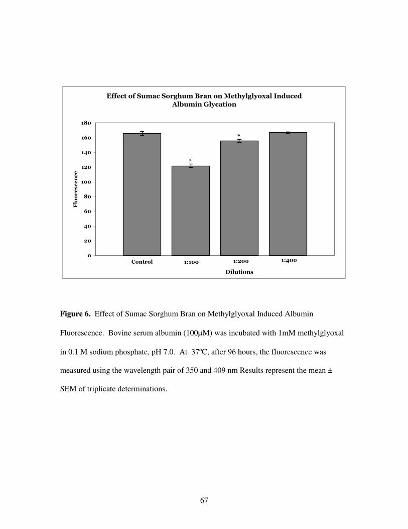
Effect of Muscadine Seeds and Skins on Methylglyoxal Induced Albumin Fluorescence.
Methylglyoxal, an important intermediate in the autooxidation of reducing sugars,
can readily glycate proteins [16]. As seen in Figure 5, methylglyoxal, when incubated
with albumin, produced significance fluorescence at the wavelength pair of 350 and 409
nm, indicative of albumin glycation. When two dilutions (1:100 and 1:200) of muscadine
seed and skin ethanolic extracts were incubated with methylglyoxal, a significant
decrease in fluorescence intensity was not observed. These results indicate that
muscadine seed and skin extracts do not inhibit all pathways of protein glycation.
Discussion.
Results presented in this communication suggest that phenolic compounds in both
the seeds and skins of the muscadine grape inhibit the protein glycation, a reaction
thought to be responsible for diabetic complications [18]. This reaction has been shown to
be inhibited by a variety of antioxidant compounds [9] and therefore it was not unexpected
that high in phenolic muscadine extracts inhibit the formation of glycated albumin.
Hyperglycemia induced oxidative damage may be prevented by the antioxidant defense
mechanisms present in biological systems [15, 19].
Page 44
33
The total phenolics content of the muscadine extracts differ between the skins and
seeds. The phenolics present in seeds, catechin, epicatechin and gallic acid, all inhibited
albumin glycation by fructose (Figure 2). The results agree with those reported by Wu
and Yen [19] for the inhibition of catechin and epicatechin of glucose mediated albumin
glycation. However, the phenolics present in the skin are different than those in the seed.
The skins have high levels of ellagic acid, myricetin, quercetin, and kaempferol [11].
Although not performed in this study, the previous report of Wu and Yen showed that
quercetin and kaempferol were potent inhibitors of glucose mediated albumin glycation.
These results shown in this study demonstrate that individual phenolics have distinct
inhibitory properties on protein glycation; this agrees with previous published findings
indicating that phenolic-induced inhibition correlated with antioxidant properties [15].
Methylglyoxal has been shown to be an important intermediate in the
autooxidation of reducing sugars and can readily glycate proteins [16]. Protein glycation
by methylglyoxal is a nonenzymatic modification whereby arginine and lysine side
chains of proteins participate in forming a heterogeneous group of advanced glycation
end-products [17]. It is interesting to note that while muscadine grape seed extracts
strongly inhibited fructose mediated albumin glycation, the extract did not significantly
inhibit methylglyoxal mediated albumin glycation. This result suggests that the
antioxidants present in the muscadine seed extract do not possess significant inhibitory
activity of ketoaldehyde-induced glycation. Wu and Yen demonstrated similar results
with both catechins; this flavonoid significantly inhibited glucose mediated glycation, but
have no effect on methylglyoxal mediated albumin glycation. This suggests that
Page 45
34
muscadine seed extracts were inhibiting the glycation process prior to the non-enzymatic
formation of methylglyoxal.
The glycation of albumin mediated by the autooxidation of reducing sugars is
dependent on the presence of metal ions in the incubation solution [20]; chelating metal
ions results in significant decrease in glycation. Flavonoids possess free radical-
scavenging activity and this may be the mechanism by which the muscadine grape skins
and seeds inhibit protein glycation. Certain flavonoids, such as quercetin, are capable of
complexing metal ions directly [21].
Many studies in the literature demonstrating that certain plant extracts [22] and
spice extracts (garlic, Cassia tora [23]) and individual phenolic compounds inhibit protein
glycation [15]. This report is the first to demonstrate that an extract from a commonly
edible grape or berry can significantly inhibit this important reaction that has been
implicated in the pathogenesis of diabetic complications. Previous reports have showed
that a water-soluble extract of tomato paste [25] and methanolic extracts of Finger and
Kodo millet can inhibit protein glycation [26].
The possibility that these muscadine products may have a pharmacological effect
is dependent on the absorption of phenolic compounds in the gastrointestinal tract. As
recently reviewed, bioavailability of certain phenolic compounds is significant, with
approximately 40% of the dose being excreted in the urine [24].
Since both muscadine seed and skin fractions come from a generally recognized
as safe (GRAS) fruit, there is no toxicity associated with these products. These fractions
may prove useful for nutraceutical and functional food/beverage products to treated type
Page 46
35
II diabetic patients. In the future, these nutraceutical products may be employed to
prevent or delay the onset of diabetic complications.
Page 47
36
Literature Cited
1. Jawa, A. and V. Fonseca, Cardiovascular effects of insulin sensitizers in diabetes.
Curr Opin Investig Drugs, 2006. 7(9): p. 806-14.
2. Haidara, M.A., et al., Role of oxidative stress in development of cardiovascular
complications in diabetes mellitus. Curr Vasc Pharmacol, 2006. 4(3): p. 215-27.
3. Ahmed, N., Advanced glycation endproducts--role in pathology of diabetic
complications. Diabetes Res Clin Pract, 2005. 67(1): p. 3-21.
4. McPherson, J.D., B.H. Shilton, and D.J. Walton, Role of fructose in glycation and
cross-linking of proteins. Biochemistry, 1988. 27(6): p. 1901-7.
5. Lee, C., et al., Oxidation-reduction properties of methylglyoxal-modified protein
in relation to free radical generation. J Biol Chem, 1998. 273(39): p. 25272-8.
6. Iwashima, Y., et al., Advanced glycation end product-induced peroxisome
proliferator-activated receptor gamma gene expression in the cultured mesangial
cells. Biochem Biophys Res Commun, 1999. 264(2): p. 441-8.
7. Packer, L., E.H. Witt, and H.J. Tritschler, alpha-Lipoic acid as a biological
antioxidant. Free Radic Biol Med, 1995. 19(2): p. 227-50.
8. Kim, H.Y. and K. Kim, Protein glycation inhibitory and antioxidative activities of
some plant extracts in vitro. J Agric Food Chem, 2003. 51(6): p. 1586-91.
9. Bousova, I., et al., Evaluation of in vitro effects of natural substances of plant
origin using a model of protein glycoxidation. J Pharm Biomed Anal, 2005. 37(5):
p. 957-62.
Page 48
37
10. Hartle DK, G.P., Hargrove JL, Muscadine Medicine. 1st ed. 2005, St George's
Island: Blue Heron Nutraceuticals, LLC. 132.
11. Pastrana-Bonilla, E., et al., Phenolic content and antioxidant capacity of
muscadine grapes. J Agric Food Chem, 2003. 51(18): p. 5497-503.
12. Singleton, V.L.R. Colorimetry of total phenolic with phosphomolybdic-
phosphotungstic acid agents. Am. J. Enol. Vitic., 1965. 16: p. 144-158.
13. Benzie, I.F. and J.J. Strain, The ferric reducing ability of plasma (FRAP) as a
measure of "antioxidant power": the FRAP assay. Anal Biochem, 1996. 239(1):
p. 70-6.
14. Yilmaz, Y. and R.T. Toledo, Major flavonoids in grape seeds and skins:
antioxidant capacity of catechin, epicatechin, and gallic acid. J Agric Food
Chem, 2004. 52(2): p. 255-60.
15. Wu, J.H., et al., Phenolic antioxidants from the heartwood of Acacia confusa. J
Agric Food Chem, 2005. 53(15): p. 5917-21.
16. Nagaraj, R.H., et al., Effect of pyridoxamine on chemical modification of proteins
by carbonyls in diabetic rats: characterization of a major product from the
reaction of pyridoxamine and methylglyoxal. Arch Biochem Biophys, 2002.
402(1): p. 110-9.
17. Gomes, R.A., et al., Yeast protein glycation in vivo by methylglyoxal. Febs J,
2006.
18. Wu, X. and V.M. Monnier, Enzymatic deglycation of proteins. Arch Biochem
Biophys, 2003. 419(1): p. 16-24.
Page 49
38
19. Wu, C.H. and G.C. Yen, Inhibitory effect of naturally occurring flavonoids on the
formation of advanced glycation endproducts. J Agric Food Chem, 2005. 53(8):
p. 3167-73.
20. Ou, P., et al., Activation of aldose reductase in rat lens and metal-ion chelation by
aldose reductase inhibitors and lipoic acid. Free Radic Res, 1996. 25(4): p. 337-
46.
21. Leopoldini, M., et al., Iron chelation by the powerful antioxidant flavonoid
quercetin. J Agric Food Chem, 2006. 54(17): p. 6343-51.
22. Babu, P.V., K.E. Sabitha, and C.S. Shyamaladevi, Therapeutic effect of green tea
extract on oxidative stress in aorta and heart of streptozotocin diabetic rats.
Chem Biol Interact, 2006. 162(2): p. 114-20.
23. Lee, G.Y., et al., Naphthopyrone glucosides from the seeds of Cassia tora with
inhibitory activity on advanced glycation end products (AGEs) formation. Arch
Pharm Res, 2006. 29(7): p. 587-90.
24. Scalbert, A., et al., Absorption and metabolism of polyphenols in the gut and
impact on health. Biomed Pharmacother, 2002. 56(6): p. 276-82.
25. Kiho, T., et al., Tomato paste fraction inhibiting the formation of advanced
glycation end-products. Biosci Biotechnol Biochem, 2004. 68(1): p. 200-5.
26. Hegde, P., G. Chandrakasan, and T. Chandra, Inhibition of collagen glycation and
crosslinking in vitro by methanolic extracts of Finger millet (Eleusine coracana)
and Kodo millet (Paspalum scrobiculatum). J Nutr Biochem, 2002. 13(9): p. 517
Page 50
39
Effect of Muscadine Grape Skin Extract on Albumin Glycation
Control 1:300 1:600 1:1200
0
20
40
60
80
100
120
140
160
180
200
Fluorescence
*
*
Dilutions
Figure 1. Effect of Muscadine Grape Skin Extract on Albumin Glycation. Briefly,
bovine serum albumin (10 mg/mL) was incubated with fructose (250 mM) in potassium
phosphate buffer (200 mM; pH 7.4; 0.02% sodium azide) and treated with varying
concentrations of the extract, as shown, for 72 hours at 37°C. The fluorescence intensity
was measured at an excitation/emission wavelength pair of 370/440 nm. Results
represent the mean ± SEM of triplicate determinations. *P < 0.05 when compared to
controls.
Page 51
40
Effect of Muscadine Seed Extract on Albumin Glycation
Control 1:300 1:600 1:12000
20
40
60
80
100
120
140
160
180
200
Fluorescence
*
*
*
Dilutions
Figure 2. Effect of Muscadine Grape Seed Extract on Albumin Glycation. Bovine
serum albumin (10 mg/mL) was incubated with fructose (250 mM) in potassium
phosphate buffer (200 mM; pH 7.4; 0.02% sodium azide) and treated with varying
concentrations of the extract, as shown, for 72 hours at 37°C. The fluorescence intensity
was measured at an excitation/emission wavelength pair of 370/440 nm. Results
represent the mean ± SEM of triplicate determinations. *P < 0.05 when compared to
controls.
Page 52
41
Effect of Muscadine Seed Phenolic Compounds on Albumin
Glycation
Control0
20
40
60
80
100
120
140
160
180
Fluorescence
*
*
*
*
*
*
Catechin Gallic AcidEpicatechin
50µM50µM50µM 100µM 100µM100µM
Figure 3. Effect of Muscadine Seed Phenolic Compounds on Albumin Glycation.
Bovine serum albumin (10 mg/mL) was incubated with fructose (250 mM) in potassium
phosphate buffer (200 mM; pH 7.4; 0.02% sodium azide) and treated with varying
concentrations of phenolic compounds found in seed extracts, as shown, for 72 hours at
37°C. The fluorescence intensity was measured at an excitation/emission wavelength
pair of 370/440 nm. Results represent the mean ± SEM of triplicate determinations. *P <
0.05 when compared to controls.
Page 53
42
Effect of Water and Ethanolic Extracts of Muscadine Seed on
Albumin Glycation
Control 1:300 1:600 1:300 1:6000
20
40
60
80
100
120
140
160
Fluorescence
Water Extract
**
Ethanol Extract
*
**
*
Figure 4. Effect of Water and Ethanolic Extracts of Muscadine Seed on Albumin
Glycation. Bovine serum albumin (10 mg/mL) was incubated with fructose (250 mM) in
potassium phosphate buffer (200 mM; pH 7.4; 0.02% sodium azide) and treated with
varying concentrations of both water and ethanolic derived extracts of muscadine seeds,
as shown, for 72 hours at 37°C. The fluorescence intensity was measured at an
excitation/emission wavelength pair of 370/440 nm. Results represent the mean ± SEM
of triplicate determinations. *P < 0.05 when compared to controls, **P < 0.05 when
compared to the corresponding dilution of the water extract.
*
*
Page 54
43
Effect of Muscadine Seed and Skin Extracts on
Methylglyoxal Induced Albumin Fluorescence
Control
1:100 1:200 1:100 1:2000
20
40
60
80
100
Fluorescence
*
Seeds Skins
Figure 5. Effect of Muscadine Seed and Skin Extracts on Methylglyoxal Induced
Albumin Fluorescence. Bovine serum albumin (100 µM) was incubated with 1 mM
methylglyoxal in 0.1 M sodium phosphate, pH 7.0. After 96 hours, the fluorescence was
measured using the wavelength pair of 350 and 409 nm. Results represent the mean ±
SEM of triplicate determinations.
Page 55
44
Chapter III - Novel Nutraceutical Property of Select Sorghum Brans: Inhibition of
Protein Glycation
________________________________________________________________________
1Farrar, JL, Hartle, DK, Hargrove, JL, Greenspan, P*, to be submitted to the Journal of
Agricultural and Food Chemistry
Page 56
45
Abstract.
Despite the high levels of phytochemicals in grain sorghum and its position as a
major food staple, there has been a lack of research on its effects on both animal and
human health and disease prevention. These phenolic compounds, mainly located in the
bran fraction, result in the plant having substantial antioxidant properties. In this study,
we examined the effect of ethanolic extracts of several varieties of sorghum bran on
albumin glycation, a non-enzymatic process thought to be important in the pathogenesis
of many diabetic complications. Sorghum brans with high phenolic content and high
antioxidant properties inhibited protein glycation; whereas sorghum brans that are low in
these properties did not inhibit this process. Ethanolic extracts of wheat, rice or oat bran
did not inhibit protein glycation. Although one high phenolic sorghum bran variety
(sumac) inhibited protein glycation mediated by the auto-oxidation of fructose by
approximately 60%, the inhibition of methyglyoxal mediated albumin glycation was
minimized. These results suggest that certain varieties of sorghum bran may affect
important biological processes that are important in diabetes and insulin resistance.
These results distinguish select sorghum brans from the common food brans and suggest
a nutraceutical rationale for its human consumption.
KEYWORDS: sorghum bran, diabetes mellitus, protein glycation, advanced
glycation endproducts (AGEs)
Page 57
46
Introduction.
Consistent consumption of foods that contain significant levels of phytochemicals
and dietary fiber correlates with tangible disease prevention. For example, whole grain
consumption is known to help in reducing cases of heart disease, diabetes, and other
chronic diseases partly due to components in their brans, especially dietary fiber and
phytochemicals [1,2,4]. This has led to the dietary guideline that increased consumption of
whole grains. Cereals are the most widely and consistently consumed food staples of
diets all over the world [2].
Sorghum grain has been a dietary staple for millenia in parts of India, Africa, and
China [3]. Much of the growth in the world’s population will be in the semiarid,
developing countries where drought-tolerant sorghum and millet varieties are major crops
[4] that contribute to the protein and energy requirements. Some sorghum varieties have
extremely high contents of phenolic compounds that aid in the natural defense of plants
against pests and diseases. These phenolic compounds, mainly located in the bran
fraction, result in the plant having significant antioxidant properties [1].
Sorghum phenolic compounds fall into two major categories; phenolic acids and
flavonoids. The phenolic acids are benzoic or cinnamic acid derivatives, whereas the
flavonoids are largely tannins and anthocyanins. Limited data exists on the levels of
anthocyanins found in cereals and grains; however, anthocyanins are a significant
component of sorghum bran [5, 26].
Despite the high levels of phytochemicals in sorghum and its position as a major
grain, there has been a paucity of medical research on its effects on human health. The
effect of sorghum on pathological processes, such as inflammation, has not been
Page 58
47
examined. Researchers have tested the hypothesis that ethanolic extracts of sorghum
brans will inhibit protein glycation, and present the results in this communication. This
non-enzymatic reaction between reducing sugars and proteins is inhibited by antioxidants
such as flavonoids [6] and is thought to be extremely important in the pathogenesis of
diabetic complications. The results presented suggest that ingestion of sorghum bran
could have previously unrecognized health benefits especially important to metabolic
syndrome and diabetes patients.
Materials and Methods.
Chemicals.
Bovine serum albumin (BSA) (Fraction V, Essentially Fatty Acid Free, D- (-)
fructose, Chelex 100 (sodium form), Folin-Ciocalteu reagent, methylglyoxal solution,
and TPTZ (2,4,6-tri[2-pyridyl]-s-triazine) were purchased from Sigma Chemical
Company, (St Louis, MO). Sorghum brans were gifts from Dr. Lloyd Rooney of Texas
A& M University and Dr Scott Bean, USDA, Manhattan, KS. Rice and wheat brans
were purchased from Bob’s Red Mill Natural Foods, Inc (Milwaukee, OR). Oat bran was
obtained from a health food store in Georgia.
Preparation of Sorghum Extracts.
To prepare the bran extracts, dried bran was made into a powder with a
commercial coffee grinder. The bran was extracted with a 1:10 (w/v) with 50% ethanol
for 2 hours at room temperature with periodic vortexing. The extract was then
centrifuged to remove the precipitate and filter sterilized to obtain the final extract..
Page 59
48
Total Phenolic Content.
Phenolic content of samples was determined with the Folin-Ciocalteu method as
described by Singleton [7]. Gallic acid was employed as the standard. Absorbance was
measured at 765 nm in a Beckman DU 600 series spectrophotometer. Results are
expressed as milligrams of gallic acid per gram of bran.
Ferric Reducing Antioxidant Protein (FRAP).
FRAP values were determined with a modified version of the Benzie and Strain
method [8], with ferrous sulfate as the reference standards. Absorption was measured at
593 nm in a Beckman DU 600 series spectrophotometer. The FRAP assay is based on
the reduction of a ferric 2,4,6-tripyridyl-s-triazine complex to the ferrous form. The
results are expressed as millimoles of ferrous sulfate formed per 100 grams of dry weight
of bran.
Albumin Glycation.
The fluorescence assay to determine the glycation of albumin was performed as
essentially described by McPherson, et. al. [9] Bovine serum albumin (BSA; 10 mg/mL)
was incubated with D- (-) fructose (250 mM) in potassium phosphate buffer (200 mM;
pH 7.4; 0.02% sodium azide) in a 5% carbon dioxide incubator at 37°C for 72 hours.
The buffer was treated with Chelex 100 prior to use. Various concentrations of the
extracts were added to the 3-ml incubation mixture. To control for the ethanol present in
the extract, control incubations were incubated in the presence of the appropriate
concentration of ethanol. The fluorescence intensity was measured at an
Page 60
49
excitation/emission wavelength pair of 370/440 nm using a Perkin-Elmer LS 55
Luminescence Spectrometer. Slit widths were set a 3 nm. Values were corrected for the
intrinsic fluorescence of bran extracts.
Modification of Albumin by Methylglyoxal.
Bovine serum albumin (100µM) was incubated with 1mM methylglyoxal in 0.1
M sodium phosphate, pH 7.0 according to the method of Packer and colleagues[10]. The
buffer was treated with Chelex 100 prior to use. After 96 hours, the fluorescence was
measured using the wavelength pair of 350 and 409 nm. Values were corrected for the
intrinisic fluorescence of bran extracts.
Statistical Analysis.
Experiments were performed in triplicate. Values are expressed as mean ± SEM..
Data within groups were analyzed using one-way analysis of variance (ANOVA) and
multiple comparisons were performed employing the Duncan’s Multiple Range test. P <
0.05 was considered statistically significant.
Results.
Effect of Sumac Sorghum Bran Extract on Albumin Glycation.
Sorghum bran (sumac variety) was extracted with 50% ethanol (10% w/v) and the
phenolic content of the extract was determined. Sumac bran had a phenolic content of
52.8 mg/g and a FRAP value of 47.2 mmoles/100g, in agreement with high antioxidant
capacity of this tannin-rich sorghum [15]. The effect of sumac sorghum bran extract on
Page 61
50
the glycation of albumin was initially examined at four different dilutions of the extract
(Figure 1). Control incubations of fructose and albumin resulted in significant albumin
glycation; the relative fluorescence intensity was found to be approximately 135 units.
When fructose and albumin were incubated in the presence 1:300 and 1:600 dilutions of
the sorghum extract, a significant concentration dependent decrease in fluorescence
intensity was observed. When the 1:1200 and 1:2400 dilutions were examined, a
significant difference from the control incubation was not observed.
Since sumac sorghum bran significantly inhibited protein glycation, five other
sorghum brans were examined to determine their effect on this non-enzymatic reaction.
Four sorghum brans, sumac, shanqui, black and mycogen varieties at dilutions of 1:300
significantly inhibited the glycation of albumin. Fontanelle and white sorghum bran
varieties, however, did not significantly produce a decreased fluorescence intensity at this
dilution (Figure 2). The phenolic content and the FRAP values for the extracts of these
sorghum brans were also determined (Table 1). Fontanelle and white were found to have
a very low content of phenolic compounds and low FRAP values, while the sorghum
brans that inhibited glycation (sumac, shanqui, black and mycogen) had much greater
phenolic contents and antioxidant FRAP values. From these data, it is apparent that the
degree of glycation inhibition correlates with the phenolic content/FRAP values.
Effect of Various Bran Extracts on Albumin Glycation.
Since sorghum bran was found to significantly inhibit albumin glycation, the
effect of other cereal brans was also examined. As seen in Figure 3, neither rice, wheat
nor oat bran significantly inhibited the glycation of albumin. The phenolic content and
Page 62
51
FRAP values of the bran extracts were examined (Table 2). In contrast to the high
phenolic content and FRAP values in certain sorghum varieties, these non-sorghum brans
possessed a very low content of phenolic compounds and were found to have a negligible
antioxidant capacity.
Effect of Sumac Sorghum Bran and Muscadine Grape Seed Extracts on Albumin
Glycation.
In our laboratory, the muscadine grape seed has previously been shown to
significantly inhibit protein glycation [12]. This grape, native to the southeastern United
States has one of the highest antioxidant capacities of any fruit. In this experiment,
sumac sorghum bran was and muscadine grape seeds were extracted with 50% ethanol
and their inhibitory properties on albumin glycation determined. As shown in figure 4,
both concentrations (1:300 and 1:600 dilutions) of both the sumac sorghum and
muscadine seeds extracts significantly inhibited protein glycation These results
demonstrate that both sumac sorghum and muscadine seed extracts are efficacious
inhibitors of protein glycation..
Comparison of Water and Ethanolic Extracts of Sumac Sorghum Bran on Protein
Glycation.
In this experiment, water and ethanolic extracts of sumac sorghum bran were
prepared with distilled water or 50% ethanol. After the 2 hours of incubation with
periodic vortexing, the phenolic content of the extracts was determined. The ethanolic
extract had a phenolic content of 5.6 mg/ml while the content of the aqueous extract was
2.1 mg/ml. The antioxidant property of the extracts was determined employing the
FRAP assay. In agreement with phenolic content, the ethanolic extract has a greater
Page 63
52
FRAP values (48.6 mmoles/100 grams vs 20.6 mmoles/100 grams, respectively). The
fluorescence intensity of the glycation of albumin was measured in the presence of two
dilutions (1:300 and 1:600) of the sorghum bran extracts. As seen in figure 5, both
ethanolic and aqueous extracts at both dilutions inhibited the glycation of albumin as
evidenced by a significant decrease in fluorescence. The ethanolic extract inhibited
glycation to a greater extent than the corresponding aqueous extract, in agreement with
the greater phenolic concentration found in the ethanolic extracts.
Effect of Sumac Sorghum Bran on Methylglyoxal Induced Albumin Fluorescence.
Methylglyoxal has been shown to be an important intermediate in the
autooxidation of reducing sugars and can readily glycate proteins [13]. Protein glycation
by methylglyoxal is a nonenzymatic modification whereby arginine and lysine side
chains of proteins participate in forming a heterogeneous group of advanced glycation
end-products [14]. As seen in Figure 6, methylglyoxal, when incubated with albumin
produced significance fluorescence at the wavelength pair of 350 and 409 nm, indicative
of albumin glycation. When two dilutions (1:100 and 1:200) of ethanolic extracts of
sumac sorghum bran were incubated with methylglyoxal, a significant decrease in
fluorescence intensity was observed. However, a dilution of 1:400 did not produce a
significant decrease in glycation
Discussion.
The data presented here support the hypothesis that phenolic compounds in
certain sorghum bran varieties significantly inhibit protein glycation. The inhibitory
Page 64
53
sorghum brans were those with high phenolic content and high antioxidant FRAP values.
The high levels of antioxidants present in sumac bran and black brans and the relatively
low levels present in white bran are in agreement with previous reports [1, 11]. Numerous
specialty sorghum varieties are now being cultivated. Experimental manipulation has led
to the development of sorghum brans that are either high in tannins (sumac, black) or
very low in these compounds (white, fonatelle). These high tannin sorghum bran
varieties contain anthocyanins such as luteolinidin and apigeninidin [15] and are the
varieties that strongly inhibit the glycation reaction. These results are in agreement with
previous findings that demonstrated that antioxidants [16] and plant extracts with high
contents of flavonoids [17] can significantly inhibit protein glycation
In comparison to sorghum, other cereal brans, such as oat, rice and wheat had low
phenolic contents and low antioxidant FRAP values. The FRAP values of these brans
were approximately 1-2% of that found for the sumac sorghum bran. These findings on
the antioxidant properties of the brans are in agreement with the findings of Awika et.
al.[11]. Sumac sorghum bran was found to have twenty-five times higher antioxidant
capacity than red wheat bran as measured by the antioxidant ABTS (2,2′-azinobis (3-
ethylbenzothiazoline-6-sulfonic acid) diammonium salt) assay [1]. Using another
antioxidant assay, the TEAC (Trolox Equivalent Antioxidant Capacity) assay, wheat bran
was shown to have three times the antioxidant capacity than oat bran [18]. Finally, rice
flour was found to have a similar antioxidant capacity to that of wheat flour [19].
Therefore, the antioxidant properties observed here are consistent with those measured by
other investigators.
Page 65
54
Although sumac sorghum bran was a potent inhibitor of fructose mediated protein
glycation , its affect on methylglyoxal-induced glycation was not as great. For instance at
1:200, an extract of sorghum bran sumac produced an 8% inhibition in methylglyoxal
mediated albumin glycation whereas a 1:300 dilution produced a 60% reduction in
fructose mediated glycation. The failure to block the methylglyoxal reaction suggests
that oxidative processes may not participate in the glycation of albumin mediated by
methylglyoxal to same extent as it does in fructose-induced glycation. A similar profile
of strong inhibition on glucose mediated glycation and weak, but significant, inhibition
on methylglyoxal inhibition was observed for epicatechin and epigallocatechin [20].
The inhibition of protein glycation by high tannin sorghum bran varieties was
found to be quite similar to that produced by muscadine grape seeds. These two crops
have several commonalities. Both have extremely high phenolic content and antioxidant
capacities. Both the muscadine grapes and high tannin sorghum brans can be grown in
arid environments; the high content of antioxidants found in these plant parts protects
them from pests and diseases and allows them to flourish in such environments [11, 21, 22].
This study illustrates that select sorghum brans have potential to modulate
processes involved in inflammatory diseases. The present results show that phenolic-rich
sorghum brans inhibit a non-enzymatic process thought to be important in the
pathogenesis of diabetic complications, such as neuropathy and cardiovascular disease.
To date, most of the substances known to inhibit protein glycation are either antioxidants
[16], spices [23] or extracts of plant substances [20]. One study has documented that an
extract of tomato paste can inhibit protein glycation [24]. Clearly, effects of food staples
on protein glycation deserves study. Millets, from the Kodo and Finger varieties, have
Page 66
55
been shown to inhibit protein glycation [25]. The results of this study confirm that
specialty sorghum brans are far superior to impact human health when compared with
other brans commonly used in the diet [26]. The high antioxidant and anti-glycation
properties provide a rationale for nutraceutical and food ingredient use of select sorghum
brans and their extracts.
Page 67
56
Literature Cited
1. Awika, J.M., C.M. McDonough, and L.W. Rooney, Decorticating sorghum to
concentrate healthy phytochemicals. J Agric Food Chem, 2005. 53(16): p. 6230-
4.
2. Kushi, L.H., K.A. Meyer, and D.R. Jacobs, Jr., Cereals, legumes, and chronic
disease risk reduction: evidence from epidemiologic studies. Am J Clin Nutr,
1999. 70(3 Suppl): p. 451S-458S.
3. Zhao, Z.Y., Sorghum (Sorghum bicolor L.). Methods Mol Biol, 2006. 343: p. 233-
44.
4. Klopfenstein, C.H., RC, Nutritional Properties of Sorghum and the Millets, in
Sorghum and Millets, Chemistry and Technology, D. Dendy, Editor. 1995,
American Association of Cereal Chemists, Inc.: Upton.
5. Lietti, A., A. Cristoni, and M. Picci, Studies on Vaccinium myrtillus
anthocyanosides. I. Vasoprotective and antiinflammatory activity.
Arzneimittelforschung, 1976. 26(5): p. 829-32.
6. Cervantes-Laurean, D., et al., Inhibition of advanced glycation end product
formation on collagen by rutin and its metabolites. J Nutr Biochem, 2006. 17(8):
p. 531-40.
7. Singleton, V.L.R. Colorimetry of total phenolic with phosphomolybdic-
phosphotungstic acid agents. Am. J. Enol. Vitic., 1965. 16: p. p. 144-158.
Page 68
57
8. Benzie, I.F.a.J.J.S., The ferric reducing ability of plasma (FRAP) as a measure of
"antioxidant power": the FRAP assay. Anal Biochem, 1996.
239(1): p. p. 70-6.
9. McPherson, J.D., B.H. Shilton, and D.J. Walton, Role of fructose in glycation and
cross-linking of proteins. Biochemistry, 1988. 27(6): p. p. 1901-7.
10. Packer, L., E.H. Witt, and H.J. Tritschler, alpha-Lipoic acid as a biological
antioxidant. Free Radic Biol Med, 1995. 19(2): p. p. 227-50.
11. Awika, J.M., et al., Screening methods to measure antioxidant activity of sorghum
(sorghum bicolor) and sorghum products. J Agric Food Chem, 2003. 51(23): p.
6657-62.
12. Farrar, J., Hartle, DK, Hargrove, JL, Greenspan, P (2006) Inhibition of protein
glycation by skins and seeds of the muscadine grape. . FASEB J. Volume, A1002
13. Lee, C., et al. , Oxidation-reduction properties of methylglyoxal-modified protein
in relation to free radical generation. J Biol Chem, 1998. 273(39): p. p. 25272-8.
14. Wu, X.a.V.M.M., Enzymatic deglycation of proteins. Arch Biochem Biophys,
2003. 419(1): p. p. 16-24.
15. Dykes, L., et al., Phenolic compounds and antioxidant activity of sorghum grains
of varying genotypes. J Agric Food Chem, 2005. 53(17): p. 6813-8.
16. Selvaraj, N., Z. Bobby, and V. Sathiyapriya, Effect of lipid peroxides and
antioxidants on glycation of hemoglobin: an in vitro study on human erythrocytes.
Clin Chim Acta, 2006. 366(1-2): p. 190-5.
Page 69
58
17. Hsieh, C.L., et al., Inhibitory effect of some selected nutraceutic herbs on LDL
glycation induced by glucose and glyoxal. J Ethnopharmacol, 2005. 102(3): p.
357-63.
18. Martinez-Tome, M., et al., Evaluation of antioxidant capacity of cereal brans. J
Agric Food Chem, 2004. 52(15): p. 4690-9.
19. Perez-Jimenez, J. and F. Saura-Calixto, Literature data may underestimate the
actual antioxidant capacity of cereals. J Agric Food Chem, 2005. 53(12): p. 5036-
40.
20. Wu, C.H. and G.C. Yen, Inhibitory effect of naturally occurring flavonoids on the
formation of advanced glycation endproducts. J Agric Food Chem, 2005. 53(8):
p. 3167-73.
21. Greenspan, P., et al., Antiinflammatory properties of the muscadine grape (Vitis
rotundifolia). J Agric Food Chem, 2005. 53(22): p. 8481-4.
22. Hartle DK, Greenspan, P., Hargrove JL, Muscadine Medicine. 1st ed. ed. 2005, St
George's Island: Lulu.
23. Ahmad, M.S. and N. Ahmed, Antiglycation properties of aged garlic extract:
possible role in prevention of diabetic complications. J Nutr, 2006. 136(3 Suppl):
p. 796S-799S.
24. Kiho, T., et al., Tomato paste fraction inhibiting the formation of advanced
glycation end-products. Biosci Biotechnol Biochem, 2004. 68(1): p. 200-5.
Page 70
59
25. Hegde, P., G. Chandrakasan, and T. Chandra, Inhibition of collagen glycation and
crosslinking in vitro by methanolic extracts of Finger millet (Eleusine coracana)
and Kodo millet (Paspalum scrobiculatum). J Nutr Biochem, 2002. 13(9): p. 517.
26. Awika, J.M. and L.W. Rooney, Sorghum phytochemicals and their potential
impact on human health. Phytochemistry, 2004. 65(9): p. 1199-221.
Page 71
60
Table 1. Phenolic content and FRAP values of various sorghum bran extracts.
Extract Phenolic Value (mg/g) FRAP Value
(mmoles/100g )
Sumac Sorghum Bran 62.4 ± 0.9 28.4 ± 0.1
Shanqui Sorghum Bran 47.7 ± 0.7 15.3 ± 0.1
Black Sorghum Bran 22.7 ± 0.5 7.9 ± 0.2
Mycogen Sorghum Bran 5.6 ± 0.1 2.2 ± 0.1
Fontanelle Sorghum Bran 2.5 ± 0.1 1.0 ± 0.1 White Sorghum Bran 3.9 ± 0.1 1.2 ± 0.1
Data represent mean ± SEM of triplicate determinations.
Page 72
61
Table 2. Phenolic content and FRAP values of various bran extracts.
Extract Phenolic Value (mg/g) FRAP Value
(mmoles/100 g)
Sorghum Bran 52.8 ± 0.5 47.3 ± 0.8
Wheat Bran 2.0 ± 0.1 0.7 ± 0.1
Oat Bran 0.6 ± 0.1 0.4 ± 0.1
Rice bran 6.0 ± 0.3 5.3 ± 0.6
Data represent mean ± SEM of triplicate determinations.
Page 73
62
Effect of Sumac Sorghum Bran Extract on Albumin Glycation
Control 1:300 1:600
1:1200 1:24000
20
40
60
80
100
120
140
160
Fluorescence
Dilutions
*
*
Figure 1. Effect of Sumac Sorghum Bran Extract on Albumin Glycation. Briefly, bovine
serum albumin (10mg/mL) was incubated with fructose (250 mM) in potassium
phosphate buffer (200mM; pH 7.4) and treated with varying concentrations of the extract
for 72 hours at 37°C. The fluorescence intensity was measured at an excitation/emission
wavelength pair of 370/440 nm. Results represent the mean ± SEM of triplicate
determination. *P < 0.05 when compared to controls.
Page 74
63
Effect of Various Sorghum Bran Extracts on Albumin
Glycation
0
20
40
60
80
100
120
Fluorescence
Control FontanelleBlack MycogenShanquiSumac White
*
*
*
*
Figure 2. Effect of Various Sorghum Bran Extracts on Albumin Glycation. Bovine
serum albumin 10 mg/mL) was incubated with fructose (250 mM) in potassium
phosphate buffer (200 mM; pH 7.4) and incubated with a 1:300 dilution of various
extracts for 72 hours at 37°C. The fluorescence intensity was measured at an
excitation/emission wavelength pair of 370/440 nm. Results represent the mean ± SEM
of triplicate determination. *P < 0.05 when compared to controls.
Page 75
64
Effect of Various Bran Extracts on Albumin Glycation
Control Rice Bran Wheat Bran Oat Bran
0
20
40
60
80
100
120
140
160
180
200
Fluorescence
Figure 3. Effect of Various Bran Extracts on Albumin Glycation. Bovine serum albumin
(10 mg/mL) was incubated with fructose (250 mM) in potassium phosphate buffer (200
mM; pH 7.4) and treated at a dilution of 1:300 with the various bran extracts for 72 hours
at 37°C. The fluorescence intensity was measured at an excitation/emission wavelength
pair of 370/440 nm. Results represent the mean ± SEM of triplicate determination. *P <
0.05 when compared to controls.
Page 76
65
Effect of Sorghum Bran and Muscadine Seed Extract on Protein Glycation
Control 1:300 1:3001:600 1:600
0
20
40
60
80
100
120
Fluorescence
Bran Seed
*
*
*
*
Figure 4. Effect of Sorghum Bran and Muscadine Grape Seed Extracts on Albumin
Glycation. Bovine serum albumin (10 mg/mL) was incubated with fructose (250 mM) in
potassium phosphate buffer (200 mM; pH 7.4) and treated with varying concentrations of
different sorghum bran and muscadine seed extracts for 72 hours at 37°C. The
fluorescence intensity was measured at an excitation/emission wavelength pair of
370/440 nm. Results represent the mean ± SEM of triplicate determination. *P < 0.05
when compared to controls.
Page 77
66
Effect of Water and Ethanolic Extracts of Sumac Sorghum
Bran on Albumin Glycation
Control 1:300 1:600 1:300 1:600
0
20
40
60
80
100
120
Fluorescence *
*
**
**
Water Extract Ethanol Extract
Figure 5. Effect of Water and Ethanolic Extracts of Sumac Sorghum Bran on Albumin
Glycation. Bovine serum albumin (10 mg/mL) was incubated with fructose (250 mM) in
potassium phosphate buffer (200mM; pH 7.4) and treated with varying concentrations of
both water and ethanolic extracts of sumac sorghum bran for 72 hours at 37°C. The
fluorescence intensity was measured at an excitation/emission wavelength pair of
370/440 nm. Results represent the mean ± SEM of triplicate determinations. *P < 0.05
when compared to controls. **P< 0.05 when compared to the same dilution of the water
extract.
*
*
Page 78
67
Effect of Sumac Sorghum Bran on Methylglyoxal Induced
Albumin Glycation
Control 1:100 1:200 1:4000
20
40
60
80
100
120
140
160
180
Fluorescence
*
*
Dilutions
Figure 6. Effect of Sumac Sorghum Bran on Methylglyoxal Induced Albumin
Fluorescence. Bovine serum albumin (100µM) was incubated with 1mM methylglyoxal
in 0.1 M sodium phosphate, pH 7.0. At 37ºC, after 96 hours, the fluorescence was
measured using the wavelength pair of 350 and 409 nm Results represent the mean ±
SEM of triplicate determinations.
Page 79
68
Chapter IV - Resveratrol, a Major Constituent of Polygonum cuspidatum, is an
Inhibitor of Protein Glycation
1Farrar, JL, Hartle, DK, Hargrove, JL, Greenspan, P*, to be submitted to
Phytochemistry
Page 80
69
Abstract.
Polygonum cuspidatum (Japanese knotweed, Mexican bamboo, P. cuspidatum,
etc.) is a traditional herb used in China and Japan to treat skin burns, gallstones, hepatitis,
and inflammation. Resveratrol, a phenolic compound present in this herb, has been
shown to have significant pharmacological action in numerous biological pathways. This
communication, tested the hypothesis that ethanolic extracts of Polygonum cuspidatum
and resveratrol would inhibit protein glycation. Both the ethanolic extracts of Polygonum
cuspidatum and resveratrol were found to significantly inhibit this non-enzymatic
process. The results show that resveratrol may inhibit protein glycation, a biological
process thought to be important in aging and diabetes.
KEYWORDS: polygonum cuspidatum; Japanese knotweed; Mexican bamboo; advanced
glycation end products (AGEs); diabetic complications; antioxidant activity, oxidative
processe
Page 81
70
Introduction.
Polygonum cuspidatum is a perennial plant with spreading rhizomes and
numerous reddish-brown stems that are traditionally used in East Asia to treat skin burns,
gallstones, hepatitis and inflammation. It has also been used in Korea to maintain oral
hygiene [1]. This plant contains a large number of flavonoids, which are frequently found
as glucosides, and other constituents including phenyl alcohols, sterols, essential oils and
amino acids [2]. Yang, et. al. identified resveratrol as a major phenolic constituent of P.
cuspidatum [3]. Resveratrol, a polyphenolic constituent of grapes, berries, juices and
wines, has gained significant attention for its apparent therapeutic value [4]. Resveratrol
has been shown to exert multifaceted antioxidant and anti-inflammatory effects in various
disease models [5]. Experimentally, resveratrol has been shown to possess both anti-
atherosclerotic and anti-cancer activities [6]. The biological activity of resveratrol has
been studied in aging; this compound has been shown to increase longevity in mice fed a
high fat diet [6].
The effect of resveratrol on protein glycation, a spontaneous non-enzymatic
aminocarbonyl reaction between reducing sugars and proteins [7, 8], has not been
previously examined. In this communication, the inhibitory effects of ethanolic extracts
of Polygonum cuspidatum and resveratrol on the glycation of albumin are reported.
Materials and Methods.
Chemicals.
Bovine serum albumin (BSA) (Fraction V, Essentially Fatty Acid Free, D- (-)
fructose, Chelex 100 (sodium form), and resveratrol were purchased from Sigma
Chemical Company, (St Louis, MO). Polygonum cuspidatum was obtained from
Page 82
71
Supplemental Health Formulations, LLC, Mayer, AZ. This commercial product is a
proprietary extract of the plant and is sold in powder form.
Preparation of the P. cuspidatum Ethanolic Extracts.
The P. cuspidatum product obtained from Supplemental Health Formulations was
extracted with a 1:10 (w/v) with 50% ethanol for 2 hours at room temperature with
periodic vortexing. The extract was then centrifuged to remove the precipitate and filter
sterilized to obtain the final extract.
Analysis of the Concentration of Resveratrol in the Polygonum cuspidatum commercial
product.
The ethanolic extract was diluted 400-fold and subjected to HPLC analysis using
an ESA (Chelmsford, MA) system consisting of a Model 582 Solvent Delivery Module, a
Model 542 autosampler maintained at 6ºC and a Model 5600A CoulArray detector at 250
mV. The column was an MCM C18 (4.6 X 150 mm, 5-120A) from MC Medical, Japan.
Mobile phase A was 75 mM citric acid, 25 mM ammonium acetate and 10% aceto nitrile;
Mobile phase B was similar to A but with 50% acetonitrile. The gradient was linear from
0-17 minutes from 10%A to 80%B. Flow rate 1.0 ml/min and 20 µl of sample was
injected. Resveratrol eluted between 16.2 and 16.8 minutes.
Albumin Glycation.
The fluorescence assay to determine the glycation of albumin was performed as
essentially described by McPherson, et. al. [9] Bovine serum albumin (BSA; 10 mg/mL)
was incubated with D- (-) fructose (250 mM) in potassium phosphate buffer (200mM; pH
Page 83
72
7.4; 0.02% sodium azide) in a 5% carbon dioxide incubator at 37°C for 72 hours. The
buffer was treated with Chelex 100 prior to use. Various concentrations of the extracts or
concentrations of resveratrol were added to the 3-ml incubation mixture. To control for
the ethanol present in the extract, control incubations were incubated in the presence of
the appropriate concentration of ethanol. The fluorescence intensity was measured at an
excitation/emission wavelength pair of 370/440 nm. Slit widths were set at 3 nm.
Values were corrected for the intrinsic fluorescence of P. cuspidatum extracts.
Statistical Analysis.
Experiments were performed in triplicate. Values are expressed as mean ± SEM.
Data within groups were analyzed using one-way analysis of variance (ANOVA) and
multiple comparisons were performed employing the Duncan’s Multiple Range test. P <
0.05 was considered statistically significant.
Results.
Analysis of the Resveratrol Content of Polygonum cuspidatum Extract.
The ethanolic extract of Polygonum cuspidatum was analyzed by HPLC to
determine the concentration of resveratrol in the commercial product. As seen in Figure
1, resveratrol was the major phenolic compound detected in the ethanolic extract of
Polygonum cuspidatum. Other peaks were eluted from the column however their peak
height was significantly smaller than that of resveratrol. Analysis revealed that the
concentration of resveratrol in the commercial product, based on extraction with 50%
Page 84
73
ethanol, was 3.8% (w/w); the 10% (w/v) ethanolic extract prepared from this product
represents a concentration of resveratrol of 3.8 mg/mL.
Effect of P. cuspidatum Extract on Albumin Glycation.
The effect of Polygonum cuspidatum on the glycation of albumin was examined
at four different dilutions of the extract. Control incubations of fructose and albumin
resulted in significant albumin glycation; the relative fluorescence intensity was found to
be approximately 115 units. When fructose and albumin were incubated with 1:300,
1:600, 1:1200 and 1:2400 dilutions of the P. cuspidatum extract, a significant
concentration dependent decrease in fluorescence intensity was observed at all four
dilutions. At a 1:2400.dilution, the extent of albumin glycation, as determined by
fluorescence was inhibited by approximately 35%.
Effect of Resveratrol on Albumin Glycation.
The effect of four different concentrations of resveratrol, ranging from 50 to 500
µM, on the glycation of albumin was examined. As seen in Figure 2, a dose dependent
inhibition of albumin glycation was observed; 50 µM resveratrol inhibited protein
glycation by approximately 30% while 500 µM resveratrol produced a 50% reduction in
this process.
Discussion.
The results presented here show that an ethanolic extract of Polygonum
cuspidatum and resveratrol both inhibit the glycation of albumin by fructose.
Page 85
74
Based on the concentration of resveratrol that was extracted with 50% ethanol, the
concentration in the 1:300 dilution of the extract is approximately 50 µM resveratrol.
Resveratrol, at this concentration, inhibited protein glycation by 27% while the 1:300
dilution of the extract was a stronger inhibitor of glycation, producing a 58% inhibition in
this process. These results suggest that the phenolic compounds present in Polygonum
cuspidatum, which also include catechin, contribute to inhibition observed with the
extract. The degree of inhibition observed with the extract of Polygonum cuspidatum
was greater than that observed with extracts of sorghum brans or muscadine skins and
seeds. A 1:2400 dilution of the Polygonum cuspidatum extract significantly inhibited
protein glycation while this concentration of sumac sorghum bran or muscadine seeds did
not produce a significant effect. The probable reason for this difference is not that the
extract of Polygonum cuspidatum is a more powerful inhibitor, but rather, the Polygonum
cuspidatum material employed in these experiments was purchased as an already
concentrated product.
Resveratrol, in other studies, has been shown to increase the uptake of glucose
by tissues and increase the synthesis of glycogen in the streptozotocin diabetic rat [2]. In
a recent study, resveratrol, when given to mice fed a high caloric diet, decreased fasting
blood glucose concentrations and also decreased insulin levels in both the fed and fasted
state. When a glucose bolus was given to these animals, an increase in insulin sensitivity
was observed, thought to be a result of an increase in AMPK kinase [6]. Resveratrol has
also been shown to attenuate diabetic nephropathy in in steptozotocin-treated rats; it
decreased both the excretion of albumin in the urine and the blood urea nitrogen
concentration. While resveratrol has not been shown to inhibit the formation of age-
Page 86
75
related glycation products (AGE), the prototype inhibitor of protein glycation,
aminoguanidine, has been shown to decrease fluorescence in the streptozotocin diabetic
rat and attenuate the diabetic nephropathy [10]. In this manner, the effect of resveratrol in
improving renal function in the streptozotocin-treated rat [11] may well be a result of
inhibition of protein glycation.
Page 87
76
Literature Cited
1. Song, J.H., et al., In vitro inhibitory effects of Polygonum cuspidatum on bacterial
viability and virulence factors of Streptococcus mutans and Streptococcus
sobrinus. Arch Oral Biol, 2006.
2. Su, H.C., L.M. Hung, and J.K. Chen, Resveratrol, a red wine antioxidant,
possesses an insulin-like effect in streptozotocin-induced diabetic rats. Am J
Physiol Endocrinol Metab, 2006. 290(6): p. E1339-46.
3. Yang, F., T. Zhang, and Y. Ito, Large-scale separation of resveratrol,
anthraglycoside A and anthraglycoside B from Polygonum cuspidatum Sieb. et
Zucc by high-speed counter-current chromatography. J Chromatogr A, 2001.
919(2): p. 443-8.
4. Bakhtiarova, A., et al., Resveratrol inhibits firefly luciferase. Biochem Biophys
Res Commun, 2006.
5. Labinskyy, N., et al., Vascular dysfunction in aging: potential effects of
resveratrol, an anti-inflammatory phytoestrogen. Curr Med Chem, 2006. 13(9): p.
989-96.
6. Baur, J.A., et al., Resveratrol improves health and survival of mice on a high-
calorie diet. Nature, 2006.
7. Ahmed, N., Advanced glycation endproducts--role in pathology of diabetic
complications. Diabetes Res Clin Pract, 2005. 67(1): p. 3-21.
Page 88
77
8. Iwashima, Y., et al., Advanced glycation end product-induced peroxisome
proliferator-activated receptor gamma gene expression in the cultured mesangial
cells. Biochem Biophys Res Commun, 1999. 264(2): p. 441-8.
9. McPherson, J.D., B.H. Shilton, and D.J. Walton, Role of fructose in glycation and
cross-linking of proteins. Biochemistry, 1988. 27(6): p. 1901-7.
10. Soulis-Liparota, T., et al., The relative roles of advanced glycation, oxidation and
aldose reductase inhibition in the development of experimental diabetic
nephropathy in the Sprague-Dawley rat. Diabetologia, 1995. 38(4): p. 387-94.
11. Sharma, S., et al., Resveratrol, a polyphenolic phytoalexin, attenuates diabetic
nephropathy in rats. Pharmacology, 2006. 76(2): p. 69-75.
Page 89
78
0.0 5.0 10.0 15.0 20.0
0.0
5.0
10.0
15.0
Retention time (minutes)
Res
pons
e (µ
A)
16
15
14
13
12
11
10
9
8
7
6
5
4
3
2
1
[250 mV]
*16.
3m 1
0.7µ
A
Figure 1. Chromatogram of the Ethanolic Extract of the Commerical Polygonum
cuspidatum Extract.
Page 90
79
Effect of Polygonum cuspidatum Extract on Albumin Glycation
Control 1:300 1:600 1:1200 1:24000
20
40
60
80
100
120
140
Dilutions
Fluorescence
*
*
*
*
Figure 2. Effect of Polygonum cuspidatum Extract on Albumin Glycation. Bovine serum
albumin (BSA) was incubated with fructose (250 mM) in potassium phosphate buffer
(200 mM; pH 7.4) and treated with four concentrations of the extracts for 72 hours at
37°C. The fluorescence intensity was measured at an excitation/emission wavelength
pair of 370/440 nm. Results represent the mean ± SEM of triplicate determination. *P <
0.05 when compared to controls.
Page 91
80
Effect of Resveratrol on Albumin Glycation
Control 50 uM 100 uM 250 uM 500 uM0
20
40
60
80
100
120
140
Concentrations
Fluorescence
*
*
*
*
Figure 3. Effect of Resveratrol on Albumin Glycation. Bovine serum albumin (10
mg/mL) was incubated with fructose (250 mM) in potassium phosphate buffer (200 mM;
pH 7.4) and treated with four concentrations of resveratrol for 72 hours at 37°C. The
fluorescence intensity was measured at an excitation/emission wavelength pair of
370/440 nm. Results represent the mean ± SEM of triplicate determination. *P < 0.05
when compared to controls.
Page 92
81
Chapter V - Conclusions.
The results presented in this dissertation demonstrate that certain food products, high
in antioxidant capacity, can inhibit protein glycation. Ethanolic extracts of muscadine
skins and seeds and special varieties of sorghum bran were found to contain a high
concentration of phenolic compounds, high antioxidant capacity and strongly inhibited
the fructose mediated non-enzymatic glycation of albumin. This inhibitory activity was
also seen with Polygonum cuspidatum and its major phenolic constituent, resveratrol. The
mechanism of action of these products is probably a result of their antioxidant properties;
inhibition of protein glycation has been observed by antioxidants such as vitamins A, C
and E.
The glycation of proteins and subsequently cross-linking of proteins in the diabetic
state is thought to participate in pathogenesis of diabetic complicatons, such as diabetic
nephropathy. In this manner, resveratrol has been shown to attenuate the progression of
this nephropathy in a diabetic rat model. The results of these studies provide a rationale
to study the effect of high tannin sorghum brans and muscadine seeds and skins on the
progression of diabetic nephropathy in experimental animals. The outcome of this
research approach is to incorporate high antioxidant foods, such as specialty sorghum
brans and muscadine products into functional foods and nutraceuticals in an attempt to
prevent diabetic complications.
According to the American Diabetes Association, this disease and its complications
affected approximately 21 million Americans – or 7% of the population. It is the third
Page 93
82
leading cause of death in the United States, only after heart disease and cancer.
Economically speaking, the disease accounts for nearly $132 billion spent in both direct
and indirect expenditures. Globally, the statistics are staggering. Diabetes and diabetic
complication inflict a significant cost burden to humanity. Eliminating or reducing the
health problems caused by diabetes through nutritional intervention could significantly
improve the quality of life for people with diabetes while simultaneously potentially
reducing expenditures for health care services and increasing productivity in the global
economies.