*Steven M. Horwitz, MD/Chair † Þ Memorial Sloan Kettering Cancer Center
*Stephen Ansell, MD, PhD/Vice-Chair ‡ Mayo Clinic Cancer Center
Weiyun Z. Ai, MD, PhD † ‡ UCSF Helen Diller Family Comprehensive Cancer Center
Jeffrey Barnes, MD, PhD † Massachusetts General Hospital Cancer Center
Stefan K. Barta, MD, MRCP, MS † ‡ Þ Fox Chase Cancer Center
Mark W. Clemens, MD ʘ The University of Texas MD Anderson Cancer Center
Ahmet Dogan, MD, PhD ≠ Memorial Sloan Kettering Cancer Center
Francine M. Foss, MD † ‡ ξ Yale Cancer Center/Smilow Cancer Hospital
Aaron M. Goodman, MD ‡ ξ UC San Diego Moores Cancer Center
John P. Greer, MD ‡ ξ Vanderbilt-Ingram Cancer Center
Joan Guitart, MD ≠ ϖ Robert H. Lurie Comprehensive Cancer Center of Northwestern University Ahmad Halwani, MD ‡ Huntsman Cancer Institute at the University of Utah
Clinical Trials: NCCN believes that the best management for any patient with cancer is in a clinical trial. Participation in clinical trials is especially encouraged. To find clinical trials online at NCCN Member Institutions, click here:nccn.org/clinical_trials/physician.html.NCCN Categories of Evidence and Consensus: All recommendations are category 2A unless otherwise indicated. See NCCN Categories of Evidence and Consensus.
NCCN Categories of Preference:All recommendations are consideredappropriate.See NCCN Categories of Preference
NCCN T-Cell Lymphomas Panel MembersSummary of the Guidelines Updates
• Peripheral T-Cell Lymphomas (TCEL-1)• Breast Implant-Associated ALCL (BIAA-1)• T-Cell Large Granular Lymphocytic Leukemia (LGLL-1)• Adult T-Cell Leukemia/Lymphoma (ATLL-1)• T-Cell Prolymphocytic Leukemia (TPLL-1)• Extranodal NK/T-Cell Lymphoma, nasal type (NKTL-1)• Hepatosplenic Gamma-Delta T-Cell Lymphoma (HGTL-1)• Principles of Molecular Analysis in T-Cell Lymphomas (LYMP-A)• Supportive Care (LYMP-B)• Lugano Response Criteria for Non-Hodgkin’s Lymphoma (LYMP-C)• Principles of Radiation Therapy (LYMP-D)
Classification and Staging (ST-1)Use of Immunophenotyping/Genetic Testing in Differential Diagnosis of Mature B-Cell and NK/T-Cell Neoplasms (See NCCN Guidelines for B-Cell Lymphomas - NHODG-A)
Peripheral T-Cell LymphomasTCEL-B 1 of 5• First-line therapy for ALCL�The qualifier "ALK+ histology" was removed. �"Brentuximab vedotin + CHP (cyclophosphamide, doxorubicin, and prednisone)" was added as a prefrred refimen with a
category 1 designation.• First-line therapy for other histologies (PTCL, NOS; AITL; EATL; MEITL; nodal PTCL, TFH; and FTCL)�Brentuximab vedotin + CHP (cyclophosphamide, doxorubicin, and prednisone) for CD30+ histologies was added as a preferred
regimen with a category 2A designation.
Breast Implant-Associated ALCLBIAA-2• Extended disease (stage II-IV)�"Brentuximab vedotin + CHP (cyclophosphamide, doxorubicin, and prednisone)" was added as a systemic therapy option with a
category 2A designation.
Adult T-Cell Leukemia/LymphomaATLL-B 1 of 2• Initial chemotherapy�"Brentuximab vedotin + CHP (cyclophosphamide, doxorubicin, and prednisone) for CD30+ cases" was added as an option with a
category 2A designation.
Hepatosplenic T-Cell LymphomaHGTL-4• Primary treatment�"Brentuximab vedotin + CHP (cyclophosphamide, doxorubicin, and prednisone) for CD30+ cases" was added with a category
2A designation. A corresponding footnote was added, "Patients with HGDTL were eligible for the ECHELON-2 study (Horwitz SM, Connor OA, Pro B, et al. The ECHELON-2 trial: results of a randomized, double-blind, active-controlled phase 3 study of brentuximab vedotin and CHP (A+CHP) versus CHOP in the frontline treatment of patients with CD30+ peripheral T-cell lymphomas [abstract]. Blood 2018;132:Abstract 997) but no patients were enrolled."
Updates in Version 2.2019 of the NCCN Guidelines for T-Cell Lymphomas from Version 1.2019 include:
Peripheral T-Cell LymphomasTCEL-1• Diagnosis, Useful�3rd bullet was revised, "Additional immunohistochemical studies to
characterize subsets of PTCL including markers of T-follicular helper [TFH] cell origin): ßF1, CXCL13, ICOS, BCL6, TCRdelta, cytotoxic T-cell markers (TIA-1, granzyme B, perforin)."
�Bullet was removed, "Karyotype to establish clonality."
TCEL-2• Workup, Useful�"Hepatitis C testing" was added.
TCEL-3• ALCL, ALK positive, Stage I, II�ISRT dosing was removed from the page and a footnote with a link to
the Principles of Radiation Therapy was added. (Also for TCEL-4)�Footnote n was added, "Interim restaging after 3–4 cycles." (Also for
TCEL-4)
TCEL-4• Footnote r was revised, "If PET/CT scan positive, rebiopsy is strongly
recommended before changing course of treatment."
TCEL-5• Relapsed/refractory disease, no intention to transplant, �"Preferred" was added to "Clinical trial."�After second-line therapy, see suggested regimens, "or clinical
benefit" was added to "complete or partial response."
TCEL-B 2 of 5• Footnote h was added "See Supportive Care (LYMP-B)." Also for other
TCEL-B pages.• Footnote l was added, "See Principles of Radiation Therapy (LYMP-D)."
Also for other TCEL-B pages.
New algorithm• Diagnosis, workup, and treatment recommendations for "Hepatosplenic
T-cell lymphoma (HSTCL)" were added to the Guidelines. (HGTL-1)
Global changes• Suggested treatment regimen references were updated throughout the
guidelines.• A footnote to the new "See Principles of Molecular Analysis in T-Cell
Lymphomas (LYMP-A)" was added to the Diagnosis heading for all subtypes.• A footnote was added to PET/CT scan as appropriate throughout the
guidelines: "Patients with T-cell lymphomas often have extranodal disease, which may be inadequately imaged by CT. PET scan may be preferred in these instances."
• Diagnosis, Useful Under Certain Circumstances, the following bullet and corresponding footnote were made consistent throughout: "Molecular analysis to detect clonal T-cell antigen receptor (TCR) gene rearrangements or other assessment of clonality (karyotype, array-CGH, or FISH analysis to detect somatic mutations or genetic alterations)." Footnote: "Clonal TCR gene rearrangements can be assessed by PCR or by high-throughput sequencing (HTS) techniques. Results should be interpreted with caution since clonal TCR gene rearrangements can also be seen in patients with non-malignant conditions. See Principles of Molecular Analysis in T-Cell Lymphomas (LYMP-A)."
Updates in Version 1.2019 of the NCCN Guidelines for T-Cell Lymphomas from Version 5.2018 include:
Updates in Version 1.2019 of the NCCN Guidelines for T-Cell Lymphomas from Version 5.2018 include:
Breast Implant-Associated ALCLBIAA-1• Pathologic workup, Essential�1st bullet was revised, "Cytology with cell block preparation."�2nd bullet was revised, "IHC and/or flow cytometry for CD2, CD3, CD4,
CD5, CD7, CD8, CD30, CD45, and ALK."• Pathologic workup, Useful�Bullet was added, "If there is solid mass associated with the implant,
biopsy (excisional or incisional or core needle) may be required for diagnosis."
• Footnote d was revised by adding, "If possible, obtain >50 mL for cytology and cell block; >10 mL for flow cytometry immunophenotype."
BIAA-2• Treatment�3rd bullet was revised, "Consultation with surgical oncologist
recommended for patients with preoperative tumor mass."• Localized disease to capsule/implant/breast�Qualifier, "Incomplete excision or partial capsulectomy with residual
disease" was revised by adding, "± regional lymph node involvement." ◊ Algorithm after qualifier was then revised to, "Discuss adjuvant treatment options with multidisciplinary team, RT (24–36 Gy) for local residual disease ± systemic therapy (as listed below), if node positive or RT not feasible."
Brentuximab vedotin or See first-line systemic ALCL regimens (TCEL-B 1 of 5)" to "Consider systemic therapy (alphabetical order): Brentuximab vedotin, CHOP, CHOEP, Dose-adjusted EPOCH."
• Footnote l was added, "Brentuximab vedotin may be appropriate for low burden disease in selected patients."
BIAA-B• Footnote 2 was added, "Bilateral breast implantation for ALCL is not
considered in this staging system. Complete excision of bilateral disease may be recommended if it is determined that 2 independent primaries are present (one on each side). Pathologic staging should be assessed in both sides. Identification of clonal abnormalities in bilateral cases is desirable and may help in determining if the disease represents metastasis."
T-Cell Large Granular Lymphocytic LeukemiaLGLL-1• Diagnosis, Useful �1st bullet, 1st sub-bullet was revised, "IHC panel (on bone marrow
biopsy):.."• Workup, Useful�Bullet was added, "Discussion of fertility and sperm banking, if fertility-
impacting therapy is planned." (Similar bullet also for ATLL-1, TPLL-1)
LGLL-2• Footnote m was added, "See Supportive Care for Purine Analogs and
Alemtuzumab (LYMP-B)."
Adult T-Cell Leukemia/LymphomaATLL-1• Workup, Useful�7th bullet was added, "HLA typing."
• Footnote b was revised, "The diagnosis of ATLL requires peripheral blood cytology or tissue histopathology and immunophenotyping of tumor lesion, or morphology and immunophenotyping of peripheral blood, and HTLV-1 serology."
ATLL-B• Heading was revised, "Second-line Therapy (with intention to proceed to
HDT/ASCR) or Subsequent Therapy to HDT/ASCR."• Footnote b was added, "See Supportive Care for Brentuximab Vedotin,
Lenalidomide, Alemtuzumab, and Bendamustine (LYMP-B)."
T-Cell Prolymphocytic LeukemiaTPLL-1• Diagnosis, Essential �3rd bullet, 1st sub-bullet was revised by adding, "TCL1."�4th bullet was revised as, "Cytogenetics (FISH and karyotype):..."
• Diagnosis, Useful�"IHC: TCL1" was removed.
TPLL-2• Footnote h was revised from, "Monitor for CMV reactivation; anti-infective
prophylaxis for herpes virus and PCP is recommended when treating with alemtuzumab ± purine analogs" to "See Supportive Care (LYMP-B)."
Updates in Version 1.2019 of the NCCN Guidelines for T-Cell Lymphomas from Version 5.2018 include:
Principles of Radiation TherapyLYMP-D 1 of 4• Bullet was removed, "Treatment with photons, electrons, or protons may all
be appropriate, depending on clinical circumstances."
LYMP-D 2 of 4• Target volumes�ISRT for nodal disease, bullet was removed, "For indolent NHL, often
treated with RT alone, larger fields should be considered. For example, the CTV definition for treating follicular lymphoma with radiation therapy alone will be greater than that employed for DLBCL with similar disease distribution being treated with combined modality therapy."
�ISRT for extranodal disease "(excluding NK/T-cell lymphoma)" was added. ◊ 2nd sub-bullet was revised, "For most organs and particularly for indolent disease, the whole organ comprises the CTV (eg, stomach, salivary gland, thyroid). For other organs, including orbit, breast, lung, bone, and localized skin, and in some cases when RT is consolidation after chemotherapy, partial organ RT may be appropriate."
◊ 3rd sub-bullet was revised, "For most NHL subtypes no Prophylactic irradiation is not required for uninvolved lymph nodes."
LYMP-D 3 of 4• ISRT for extranodal NK/T-cell lymphoma recommendations were added. • General Dose Guidelines, �2nd bullet,
◊ 1st sub-bullet for NK-T cell was revised, "RT alone as primary treatment (if unfit for chemotherapy): 50–55 Gy."
◊ 2nd sub-bullet, "RT in combined modality therapy: 45–50.4 45-56 Gy ◊ 3rd sub-bullet, combined modality therapy dosing was added.
aSee Principles of Molecular Analysis in T-Cell Lymphomas (LYMP-A).bSee Use of Immunophenotyping/Genetic Testing in Differential Diagnosis of Mature B-Cell and NK/T-Cell Neoplasms (See NCCN Guidelines for B-Cell Lymphomas).cClonal TCR gene rearrangements alone are not sufficient for diagnosis, as these are often seen with reactive/inflammatory processes. can be assessed by PCR or by
high-throughput sequencing (HTS) techniques. Results should be interpreted with caution since clonal TCR gene rearrangements can also be seen in patients with non-malignant conditions. See Principles of Molecular Analysis in T-Cell Lymphomas (LYMP-A).
dSee map for prevalence of HTLV-1 by geographic region.ePrimary cutaneous peripheral T-cell lymphomas with limited skin involvement may have an indolent disease course are very heterogeneous and the optimal
management may not be along these guidelines.fAITL may occasionally present with concurrent DLBCL. EBV and appropriate immunohistochemistry should be performed. Clonal hematopoesis in AITL is considered
as a risk factor for cardiovascular disease.gMEITL has only recently been separated as its own entity and optimal treatment has not been defined.
DIAGNOSISaSUBTYPES
ESSENTIAL:• Review of all slides with at least one paraffin block representative of
the tumor should be done by a hematopathologist with expertise in the diagnosis of PTCL. Rebiopsy if consult material is nondiagnostic.
• Excisional or incisional biopsy is preferred over core needle biopsy. An FNA alone is not sufficient for the initial diagnosis of lymphoma. A core needle biopsy is not optimal but can be used under certain circumstances. In certain circumstances, when a lymph node is not easily accessible for excisional or incisional biopsy, a combination of core needle biopsy and FNA in conjunction with appropriate ancillary techniques may be sufficient for diagnosis.
• Adequate immunophenotyping to establish diagnosisb�IHC panel may include CD20, CD3, CD10, BCL6, Ki-67, CD5, CD30,
CD2, CD4, CD8, CD7, CD56, CD21, CD23, EBER-ISH, TCRβ, TCRẟ, PD1/CD279, ALK with or without�Cell surface marker analysis by flow cytometry may include kappa/
Note: All recommendations are category 2A unless otherwise indicated.Clinical Trials: NCCN believes that the best management of any patient with cancer is in a clinical trial. Participation in clinical trials is especially encouraged.
ESSENTIAL:h• History and physical (H&P) exam; full skin exam; attention to node-bearing
areas, including Waldeyer's ring; evaluation of size of liver and spleen, nasopharynx
• Performance status• B symptoms• CBC with differential• Bone marrow biopsy ± aspirate• LDH• Comprehensive metabolic panel• Uric acid• PET/CT scani and/or chest/abdominal/pelvic (C/A/P) CT with contrast of
diagnostic quality• Calculation of International Prognostic Index (IPI)j• Echocardiogram or MUGA scan if anthracycline or anthracenedione-based
regimen is indicated• Pregnancy testing in women of child-bearing age (if chemotherapy or RT
planned)
USEFUL IN SELECTED CASES:• Neck CT with contrast• Head CT or MRI with contrast• Skin biopsy• HIV testing• Hepatitis B and C testing• Consider quantitative EBV PCR• Consider celiac disease in newly diagnosed EATL• Discussion of fertility issues and sperm banking
See TCEL-3
hThe role of intrathecal prophylaxis in PTCL is largely unknown.iPatients with T-cell lymphomas often have extranodal disease, which may be inadequately imaged by CT. PET scan may be preferred in these instances.jSee International Prognostic Index (TCEL-A).
Note: All recommendations are category 2A unless otherwise indicated.Clinical Trials: NCCN believes that the best management of any patient with cancer is in a clinical trial. Participation in clinical trials is especially encouraged.
iPatients with T-cell lymphomas often have extranodal disease, which may be inadequately imaged by CT. PET scan may be preferred in these instances.kConsider consolidative HDT/ASCR for high-risk IPI patients in CR1.lSee Suggested Treatment Regimens (TCEL-B).mSee Principles of Radiation Therapy (LYMP-D).nInterim restaging after 3–4 cycles.oSee Lugano Response Criteria for Non-Hodgkin’s Lymphoma (LYMP-C).
Consider prophylaxis for tumor lysis syndrome (See LYMPH-B)STAGE FIRST-LINE THERAPY
ALCL, ALK positive
Stage I, II
Stage III, IVk
Multiagent chemotherapyl x 6 cycles ± ISRTmorMultiagent chemotherapyl x 3–4 cycles + ISRTm (category 2B)
Multiagent chemotherapyl x 6 cycles
See Relapsed/Refractory Disease (TCEL-5)
Complete or partial responseo
No reponse or Progressive diseaseo
Interim restagingn with PET/CTi or C/A/P CT scan with contrast
Complete planned course of treatmentand Observe for recurrence
Note: All recommendations are category 2A unless otherwise indicated.Clinical Trials: NCCN believes that the best management of any patient with cancer is in a clinical trial. Participation in clinical trials is especially encouraged.
gMEITL has only recently been separated as its own entity and optimal treatment has not been defined.iPatients with T-cell lymphomas often have extranodal disease, which may be inadequately imaged by CT. PET scan may be preferred in these instances.lSee Suggested Treatment Regimens (TCEL-B).mSee Principles of Radiation Therapy (LYMP-D).nInterim restaging after 3–4 cycles.oSee Lugano Response Criteria for Non-Hodgkin’s Lymphoma (LYMP-C).pALCL-, ALK-negative with a DUSP22 rearrangement has been associated with a prognosis more similar to ALK-positive disease and could be treated according to the
ALCL-, ALK-positive algorithm. (Parrilla Castellar ER, Jaffe ES, Said JW, et al. ALK-negative anaplastic large cell lymphoma is a genetically heterogeneous disease with widely disparate clinical outcomes. Blood 2014;124:1473-1480.)
qFor selected patients (elderly, comorbid conditions), a trial of single-agent corticosteroid may be considered for symptom management.rIf PET/CT scan positive, rebiopsy is strongly recommended before changing course of treatment.sLocalized areas can be irradiated before or after high-dose therapy. See Principles of Radiation Therapy (LYMP-D).
Consider prophylaxis for tumor lysis syndrome (See LYMPH-B)
Note: All recommendations are category 2A unless otherwise indicated.Clinical Trials: NCCN believes that the best management of any patient with cancer is in a clinical trial. Participation in clinical trials is especially encouraged.
mSee Principles of Radiation Therapy (LYMP-D).oSee Lugano Response Criteria for Non-Hodgkin’s Lymphoma (LYMP-C).tLocalized areas can be irradiated before or after high-dose therapy. See Principles of Radiation Therapy (LYMP-D).
Consider prophylaxis for tumor lysis syndrome (See LYMPH-B)
Note: All recommendations are category 2A unless otherwise indicated.Clinical Trials: NCCN believes that the best management of any patient with cancer is in a clinical trial. Participation in clinical trials is especially encouraged.
aThe International Non-Hodgkin’s Lymphoma Prognostic Factors Project. A predictive model for aggressive non-hodgkin’s lymphoma. N Engl J Med 1993;329:987-994.bGallamini A, Stelitano C, Calvi R, et al. Peripheral T-cell lymphoma unspecified (PTCL-U): A new prognostic model from a retrospective multicentric clinical study. Blood
2004;103:2474-2479.cWent P, Agostinelli C, Gallamini A, et al. Marker expression in peripheral T-cell lymphoma: a proposed clinical-pathologic prognostic score. J Clin Oncol 2006;24:2472-
2479.dVose JM. International peripheral T-cell lymphoma (PTCL) clinical and pathologic review project: poor outcome by prognostic indices and lack of efficacy with
Note: All recommendations are category 2A unless otherwise indicated.Clinical Trials: NCCN believes that the best management of any patient with cancer is in a clinical trial. Participation in clinical trials is especially encouraged.
aSee references for regimens on TCEL-B 5 of 5. bWhile anthracycline-based regimens confer a favorable prognosis in ALCL, ALK +, these regimens have not provided the same favorable results for other PTCL
histologies; clinical trial is therefore preferred for the management of these other histologies.cALCL, ALK-negative with a DUSP22 rearrangement has been associated with a prognosis more similar to ALK-positive disease and could be treated according to the
ALCL, ALK-positive algorithm. (Parrilla Castellar ER, Jaffe ES, Said JW, et al. ALK-negative anaplastic large cell lymphoma is a genetically heterogeneous disease with widely disparate clinical outcomes. Blood 2014;124:1473-1480.)
dSee Supportive Care (LYMP-B).eOther histologies include PTCL, NOS; AITL; EATL; MEITL; nodal PTCL, TFH; and FTCL. fMEITL has only recently been separated as its own entity and optimal treatment has not been defined.gCHOP followed by IVE regimen includes HCT.
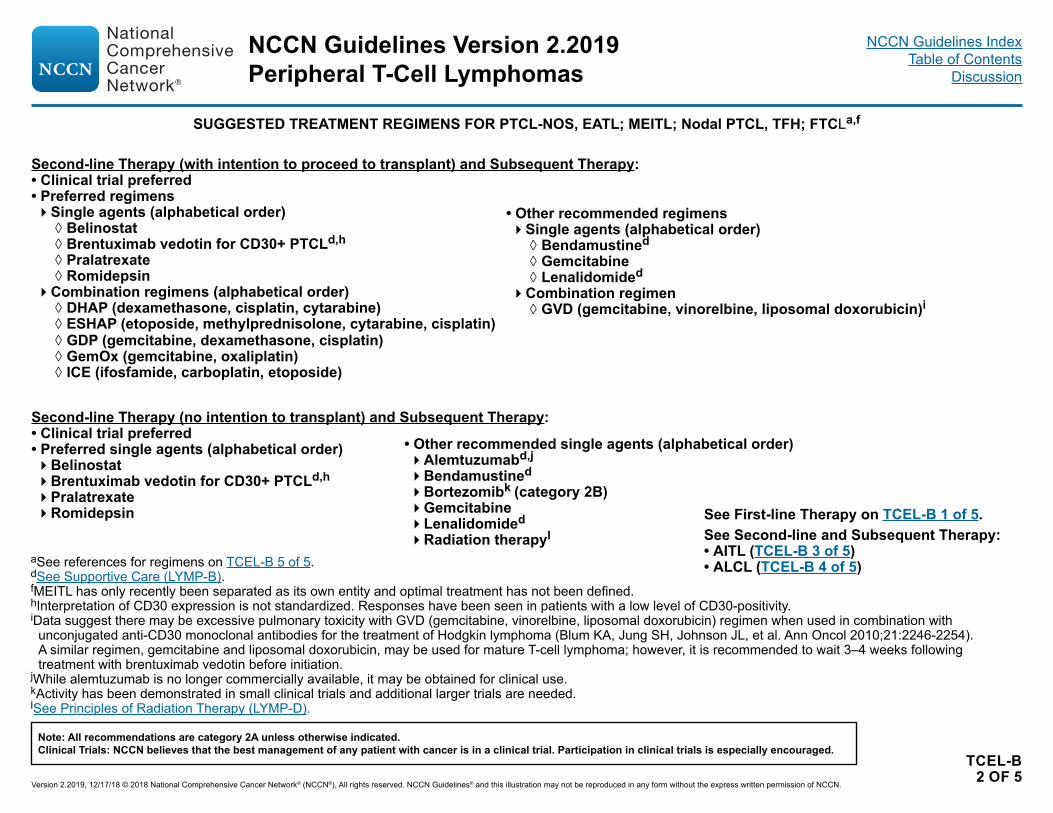
See Second-line and Subsequent Therapy:• PTCL-NOS; EATL; MEITL; nodal PTCL, TFH; FTCL (TCEL-B 2 of 5)• AITL (TCEL-B 3 of 5)• ALCL (TCEL-B 4 of 5)
• Other histologies:e,f�Preferred regimens (in alphabetical order)
◊ Brentuximab vedotin + CHP (cyclophosphamide, doxorubicin, and prednisone)d for CD30+ histologies ◊ CHOEP ◊ CHOP ◊ Dose-adjusted EPOCH (etoposide, prednisone, vincristine, cyclophosphamide, doxorubicin)
�Other recommended regimens (in alphabetical order) ◊ CHOP followed by IVE (ifosfamide, etoposide, epirubicin) alternating with intermediate-dose methotrexate [Newcastle Regimen] [studied only in patients with EATL]g
◊ HyperCVAD (cyclophosphamide, vincristine, doxorubicin, dexamethasone) alternating with high-dose methotrexate and cytarabine (category 3)
First-line Consolidation:• Consider consolidation with high-dose therapy and stem cell rescue.
Note: All recommendations are category 2A unless otherwise indicated.Clinical Trials: NCCN believes that the best management of any patient with cancer is in a clinical trial. Participation in clinical trials is especially encouraged.
aSee references for regimens on TCEL-B 5 of 5.dSee Supportive Care (LYMP-B).fMEITL has only recently been separated as its own entity and optimal treatment has not been defined.hInterpretation of CD30 expression is not standardized. Responses have been seen in patients with a low level of CD30-positivity.iData suggest there may be excessive pulmonary toxicity with GVD (gemcitabine, vinorelbine, liposomal doxorubicin) regimen when used in combination with
unconjugated anti-CD30 monoclonal antibodies for the treatment of Hodgkin lymphoma (Blum KA, Jung SH, Johnson JL, et al. Ann Oncol 2010;21:2246-2254). A similar regimen, gemcitabine and liposomal doxorubicin, may be used for mature T-cell lymphoma; however, it is recommended to wait 3–4 weeks following treatment with brentuximab vedotin before initiation.
jWhile alemtuzumab is no longer commercially available, it may be obtained for clinical use.kActivity has been demonstrated in small clinical trials and additional larger trials are needed.lSee Principles of Radiation Therapy (LYMP-D).
Second-line Therapy (no intention to transplant) and Subsequent Therapy:• Clinical trial preferred• Preferred single agents (alphabetical order)�Belinostat�Brentuximab vedotin for CD30+ PTCLd,h�Pralatrexate�Romidepsin
Note: All recommendations are category 2A unless otherwise indicated.Clinical Trials: NCCN believes that the best management of any patient with cancer is in a clinical trial. Participation in clinical trials is especially encouraged.
See First-line Therapy on TCEL-B 1 of 5.aSee references for regimens on TCEL-B 5 of 5.dSee Supportive Care (LYMP-B)hInterpretation of CD30 expression is not standardized. Responses have been seen in
patients with a low level of CD30-positivity..jWhile alemtuzumab is no longer commercially available, it may be obtained for clinical use.kActivity has been demonstrated in small clinical trials and additional larger trials are needed.lSee Principles of Radiation Therapy (LYMP-D).mIn AITL, pralatrexate has limited activity. nWith close follow-up of renal function.
SUGGESTED TREATMENT REGIMENS FOR AITLa
Second-line Therapy (no intention to transplant) and Subsequent Therapy:• Clinical trial preferred• Preferred single agents (alphabetical order)�Belinostat�Brentuximab vedotin for CD30+ AITLd,h�Romidepsin
Second-line Therapy (with intention to proceed to transplant) and Subsequent Therapy:• Clinical trial preferred• Preferred regimens�Single agents (alphabetical order)
◊ Belinostat ◊ Brentuximab vedotin for CD30+ AITLd,h ◊ Romidepsin
Note: All recommendations are category 2A unless otherwise indicated.Clinical Trials: NCCN believes that the best management of any patient with cancer is in a clinical trial. Participation in clinical trials is especially encouraged.
aSee references for regimens on TCEL-B 5 of 5.dSee Supportive Care (LYMP-B).kActivity has been demonstrated in small clinical trials and additional larger trials are needed.lSee Principles of Radiation Therapy (LYMP-D).
SUGGESTED TREATMENT REGIMENS FOR ALCLa
Second-line Therapy (no intention to transplant) and Subsequent Therapy:• Clinical trial preferred• Preferred regimen�Brentuximab vedotind
• Other recommended single agents (alphabetical order)�Belinostat�Bendamustined �Bortezomibk (category 2B)�Crizotinib (ALK+ ALCL only)�Gemcitabine �Pralatrexate�Radiation therapyl�Romidepsin
Second-line Therapy (with intention to proceed to transplant) and Subsequent Therapy:• Clinical trial preferred• Preferred regimen�Brentuximab vedotind
• Other recommended regimens �Single agents (alphabetical order)
Note: All recommendations are category 2A unless otherwise indicated.Clinical Trials: NCCN believes that the best management of any patient with cancer is in a clinical trial. Participation in clinical trials is especially encouraged.
Brentuximab vedotin + CHP (cyclophosphamide, doxorubicin, and prednisone)Horwitz SM, Connor OA, Pro B, et al. The ECHELON-2 trial: results of a randomized, double-blind, active-controlled phase 3 study of brentuximab vedotin and CHP (A+CHP) versus CHOP in the frontline treatment of patients with CD30+ peripheral T-cell lymphomas [abstract]. Blood 2018;132:Abstract 997.CHOPSavage KJ, Chhanabhai M, Gascoyne RD, Connors JM. Characterization of peripheral T-cell lymphomas in a single North American institution by the WHO classification. Ann Oncol 2004;15:1467-1475. CHOEPPfreundschuh M, Trümper L, Kloess M, et al. German High-Grade Non-Hodgkin's Lymphoma Study Group. Two-weekly or 3-weekly CHOP chemotherapy with or without etoposide for the treatment of young patients with good-prognosis (normal LDH) aggressive lymphomas: results of the NHL-B1 trial of the DSHNHL. Blood 2004;104:626-33. Pfreundschuh M, Trümper L, Kloess M, et al. German High-Grade Non-Hodgkin's Lymphoma Study Group. Two-weekly or 3-weekly CHOP chemotherapy with or without etoposide for the treatment of elderly patients with aggressive lymphomas: Results of the NHL-B2 trial of the DSHNHL. Blood 2004;104:634-41. Schmitz N, Trumper L, Ziepert M, et al. Treatment and prognosis of mature T-cell and NK-cell lymphoma: an analysis of patients with T-cell lymphoma treated in studies of the German High-Grade Non-Hodgkin Lymphoma Study Group. Blood 2010;116:3418-3425.Dose-adjusted EPOCH Dunleavy K, Pittaluga S, Shovlin M, et al. Phase II trial of dose-adjusted EPOCH in untreated systemic anaplastic large cell lymphoma. Haematologica 2016;101:e27–e29.CHOP followed by IVESieniawski M, Angamuthu N, Boyd K, et al. Evaluation of enteropathy-associated T-cell lymphoma comparing standard therapies with a novel regimen including autologous stem cell transplantation. Blood 2010;115:3664-3670.HyperCVAD alternating with high-dose methotrexate and cytarabineEscalon MP, Liu NS, Yang Y, et al. Prognostic factors and treatment of patients with T-cell non-Hodgkin lymphoma: the M. D. Anderson Cancer Center experience. Cancer 2005;103:2091-2098.Pozadzides JV, Perini G, Hess M, et al. Prognosis and treatment of patients with peripheral T-cell lymphoma: The M. D. Anderson Cancer Center experience [abstract]. J Clin Oncol 2010;28: Abstract 8051.Second-line TherapyAlemtuzumabEnblad G, Hagberg H, Erlanson M, et al. A pilot study of alemtuzumab (anti-CD52 monoclonal antibody) therapy for patients with relapsed or chemotherapy-refractory peripheral T-cell lymphomas. Blood 2004;103:2920-2924.BelinostatO'Connor OA, Horwitz S, Masszi T, et al. Belinostat in patients with relapsed or refractory peripheral T-cell lymphoma: Results of the pivotal phase II BELIEF (CLN-19) study. J Clin Oncol 2015;33:2492-2499.BendamustineDamaj G, Gressin R, Bouabdallah K, et al. Results from a prospective, open-label, phase II trial of bendamustine in refractory or relapsed T-cell lymphomas: the BENTLY trial. J Clin Oncol 2013;31:104-110.Bortezomib Zinzani P, Musuraca G, Tani M, et al. Phase II trial of proteasome inhibitor bortezomib in patients with relapsed or refractory cutaneous T-cell lymphoma. J Clin Oncol 2007;25:4293-4297. Brentuximab vedotinPro B, Advani R, Brice P, et al. Brentuximab vedotin (SGN-35) in patients with relapsed or refractory systemic anaplastic large-cell lymphoma: Results of a phase II study. J Clin Oncol 2012;30:2190-2196.Horwitz SM, Advani RH, Bartlett NL, et al. Objective responses in relapsed T-cell lymphomas with single agent brentuximab vedotin. Blood 2014;123 3095-3100.Pro B, Advani R, Brice P, et al. Five-year results of brentuximab vedotin in patients with relapsed or refractory systemic anaplastic large cell lymphoma. Blood 2017;130:2709-2717.
CrizotinibGambacorti Passerini C, Farina F, Stasia A, et al. Crizotinib in advanced, chemoresistant anaplastic lymphoma kinase-positive lymphoma patients. J Natl Cancer Inst 2014;106:djt378.Cyclosporine for AITLAdvani R, Horwitz S, Zelenetz A, Horning SJ. Angioimmunoblastic T cell lymphoma: treatment experience with cyclosporine. Leuk Lymphoma 2007;48:521-525.DHAP (dexamethasone, cisplatin, cytarabine)Velasquez WS, Cabanillas F, Salvador P, et al. Effective salvage therapy for lymphoma with cisplatin in combination with high-dose Ara-C and dexamethasone (DHAP). Blood 1988;71:117-122.Mey UJ, Orlopp KS, Flieger D, et al. Dexamethasone, high-dose cytarabine, and cisplatin in combination with rituximab as salvage treatment for patients with relapsed or refractory aggressive non-Hodgkin's lymphoma. Cancer Invest 2006;24:593-600.ESHAP (etoposide, methylprednisolone, cytarabine, cisplatin)Velasquez WS, McLaughlin P, Tucker S, et al. ESHAP - an effective chemotherapy regimen in refractory and relapsing lymphoma: a 4-year follow-up study. J Clin Oncol 1994;12:1169-1176.GemcitabineZinzani PL, Baliva G, Magagnoli M, et al. Gemcitabine treatment in pretreated cutaneous T-cell lymphoma: Experience in 44 patients. J Clin Oncol 2000;18:2603-2606.Zinzani PL, Magagnoli M, Bendandi M, et al. Therapy with gemcitabine in pretreated peripheral T-cell lymphoma patients. Ann Oncol 1998;9:1351-1353.GDP (gemcitabine, dexamethasone, cisplatin) Connors JM, Sehn LH, Villa D, et al. Gemcitabine, dexamethasone, and cisplatin (GDP) as secondary chemotherapy in relapsed/refractory peripheral T-cell lymphoma [abstract]. Blood 2013;122:Abstract 4345. Park BB, Kim WS, Suh C, et al. Salvage chemotherapy of gemcitabine, dexamethasone, and cisplatin (GDP) for patients with relapsed or refractory peripheral T-cell lymphomas: a consortium for improving survival of lymphoma (CISL) trial. Ann Hematol 2015;94:1845-1851.GND (gemcitabine, vinorelbine, liposomal doxorubicin)Qian Z, Song Z, Zhang H, et al. Gemcitabine, navelbine, and doxorubicin as treatment for patients with refractory or relapsed T-cell lymphoma. Biomed Res Int 2015;2015:606752. GemOX (gemcitabine, oxaliplatin)Lopez A, Gutierrez A, Palacios A, et al. GEMOX-R regimen is a highly effective salvage regimen in patients with refractory/relapsing diffuse large-cell lymphoma: A phase II study. Eur J Haematol 2008;80:127-132.ICE (ifosfamide, carboplatin, etoposide)Horwitz S, Moskowitz C, Kewalramani T, et al. Second-line therapy with ICE followed by high dose therapy and autologous stem cell transplantation for relapsed/refractory peripheral T-cell lymphomas: minimal benefit when analyzed by intent to treat [abstract]. Blood 2005;106:Abstract 2679. LenalidomideMorschhauser, Fitoussi O, Haioun C, et al. A phase 2, multicentre, single-arm, open-label study to evaluate the safety and efficacy of single-agent lenalidomide (Revlimid) in subjects with relapsed or refractory peripheral T-cell non-Hodgkin lymphoma: the EXPECT trial. Eur J Cancer 2013;49:2869-2876. Toumishey E, Prasad A, Dueck G, et al. Final report of a phase 2 clinical trial of lenalidomide monotherapy for patients with T-cell lymphoma. Cancer 2015;121:716-723. PralatrexateO'Connor OA, Pro B, Pinter-Brown L, et al. Pralatrexate in patients with relapsed or refractory peripheral T-cell lymphoma: Results from the pivotal PROPEL study. J Clin Oncol 2011;29:1182-1189.RomidepsinCoiffier B, Pro B, Prince HM, et al. Results from a pivotal, open-label, phase II study of romidepsin in relapsed or refractory peripheral T-cell lymphoma after prior systemic therapy. J Clin Oncol 2012;30:631-636. Coiffier B, Pro B, Prince HM, et al. Romidepsin for the treatment of relapsed/refractory peripheral T-cell lymphoma: pivotal study update demonstrates durable responses. J Hematol Oncol 2014;7:11.
Note: All recommendations are category 2A unless otherwise indicated.Clinical Trials: NCCN believes that the best management of any patient with cancer is in a clinical trial. Participation in clinical trials is especially encouraged.
Physical signsb (effusion, enlargement, mass, ulceration) >1 year post implantation (Average 8–10 years post-implantation)
Ultrasound of breastor Breast MRI in selected casesor PET/CT scanc in selected cases
aRare cases with parenchymal breast or nodal involvement may have an aggressive course more in line with systemic ALK-positive ALCL (See TCEL-3). Optimal treatment of these cases is not well defined and management should be individualized.
bA majority of cases have been seen in textured implants (Miranda RN, et al. J Clin Oncol 2014;32:114-120).cPatients with T-cell lymphomas often have extranodal disease, which may be inadequately imaged by CT. PET scan may be preferred in these instances.dLarger volume of fluid yields a more accurate diagnosis. If possible, obtain >50 mL for cytology and cell block; >10 mL for flow cytometry immunophenotype.eSee Principles of Molecular Analysis in T-Cell Lymphomas (LYMP-A).fBreast implant-associated ALCL (BIA-ALCL) is usually ALK-negative but has a good prognosis.gThe FDA recommends reporting all BIA-ALCL cases to the PROFILE Registry: www.thepsf.org/PROFILE.
Ultrasound inconclusive
Breast MRI, if not previously done
Any effusion
Mass
FNA of fluidd around breast implant
Biopsy of mass
ESSENTIAL:• Cytology with cell
block preparationd • IHC and/or flow
cytometryd for CD2, CD3, CD4, CD5, CD7, CD8, CD30, CD45, and ALKf
USEFUL UNDER CERTAIN CIRCUMSTANCES:• If there is solid
mass associated with the implant, biopsy (excisional or incisional or core needle) may be required for diagnosis
If indeterminate of lymphoma
Second pathology consultation by tertiary cancer center
Histologic confirmation or suspicious of BIA-ALCLg
Note: All recommendations are category 2A unless otherwise indicated.Clinical Trials: NCCN believes that the best management of any patient with cancer is in a clinical trial. Participation in clinical trials is especially encouraged.
NCCN Guidelines Version 2.2019Breast Implant-Associated ALCL
• Total capsulectomy and excision of associated mass with biopsy of supicous node(s), explantation
• Consider removal of contralateral implantk
• Consultation with surgical oncologist recommended for patients with preoperative tumor mass
Extended disease (stage II–IV)
Observation • H&P for every 3–6
mo for 2 y and then as clinically indicated
• ± C/A/P CT with contrast or PET/CT scanc no more often than every 6 mo for 2 y then only as clinically indicated
Histologic confirmation or suspicious of BIA-ALCLg
cPatients with T-cell lymphomas often have extranodal disease, which may be inadequately imaged by CT. PET scan may be preferred in these instances.
gThe FDA recommends reporting all BIA-ALCL cases to the PROFILE Registry: www.thepsf.org/PROFILE.hSee Proposed TNM Staging for Breast Implant–Associated Anaplastic Large-Cell Lymphoma (BIAA-B).iFor BIA-ALCL, bone marrow biopsy is only needed in selected cases (eg, extensive disease or unexplained cytopenia).jEg, oncologist, surgical oncologist, plastic surgeon, hematopathologist. kIn approximately 4.6% of cases, lymphoma was found in the contralateral breast (Clemens MW, Medeiros LJ, Butler CE, et al.
J Clin Oncol 2016;34:160-168).lBrentuximab vedotin may be appropriate for low burden disease in selected patients. mSee Supportive Care (LYMP-B).
• Recommend discussion of treatment options with multidisciplinary teamj
• H&P exam, including complete skin exam
• CBC with differential• Comprehensive metabolic panel• LDH• PET/CT scanc• Echocardiogram or MUGA
scan if anthracycline or anthracenedione-based regimen is indicated
• Pregnancy testing in women of child-bearing age (if chemotherapy or RT planned)
Note: All recommendations are category 2A unless otherwise indicated.Clinical Trials: NCCN believes that the best management of any patient with cancer is in a clinical trial. Participation in clinical trials is especially encouraged.
NCCN Guidelines Version 2.2019Breast Implant-Associated ALCL
ReferencesAdrada BE, Miranda RN, Rauch GM, et al. Breast implant-associated anaplastic large cell lymphoma: sensitivity, specificity, and findings of imaging studies in 44
patients. Breast Cancer Res Treat 2014;147:1-14.Miranda RN, Aladily TN, Prince HM, et al. Breast implant-associated anaplastic large-cell lymphoma: long-term follow-up of 60 patients. J Clin Oncol 2014;32:114-120.Clemens MW, Medeiros LJ, Butler CE, et al. Complete surgical excision is essential for the management of patients with breast implant-associated anaplastic large-cell
lymphoma. J Clin Oncol 2016;34:160-168.Pro B, Advani R, Brice P, et al. Brentuximab vedotin (SGN-35) in patients with relapsed or refractory systemic anaplastic large-cell lymphoma: results of a phase II study.
J Clin Oncol 2012;30:2190-2196. Pro B, Advani R, Brice P, et al. Four-year survival data from an ongoing pivotal phase 2 study of brentuximab vedotin in patients with relapsed or refractory systemic
anaplastic large cell lymphoma [abstract]. Blood 2014 124:Abstract 3095.Horwitz SM, Connor OA, Pro B, et al. The ECHELON-2 trial: results of a randomized, double-blind, active-controlled phase 3 study of brentuximab vedotin and CHP
(A+CHP) versus CHOP in the frontline treatment of patients with CD30+ peripheral T-cell lymphomas [abstract]. Blood 2018;132:Abstract 997.
Note: All recommendations are category 2A unless otherwise indicated.Clinical Trials: NCCN believes that the best management of any patient with cancer is in a clinical trial. Participation in clinical trials is especially encouraged.
NCCN Guidelines Version 2.2019Breast Implant-Associated ALCL
Proposed TNM Staging for Breast Implant–Associated Anaplastic Large-Cell Lymphoma1,2
TNM DescriptionT: tumor extent
T1 Confined to effusion or a layer on luminal side of capsule T2 Early capsule infiltration T3 Cell aggregates or sheets infiltrating the capsule T4 Lymphoma infiltrates beyond the capsuleN: lymph node N0 No lymph node involvement N1 One regional lymph node (+) N2 Multiple regional lymph nodes (+)M: metastasis M0 No distant spread M1 Spread to other organs/distant sites
Stage Designation DescriptionIA T1 N0 M0IB T2 N0 M0IC T3 N0 M0IIA T4 N0 M0IIB T1-3 N1 M0III T4 N1-2 M0IV T any N any M1
1Clemens MW, Medeiros LJ, Butler CE, et al. Complete surgical excision is essential for the management of patients with breast implant-associated anaplastic large-cell lymphoma. J Clin Oncol 2016;34:160-168.
2Bilateral breast implantation for ALCL is not considered in this staging system. Complete excision of bilateral disease may be recommended if it is determined that 2 independent primaries are present (one on each side). Pathologic staging should be assessed in both sides. Identification of clonal abnormalities in bilateral cases is desirable and may help in determining if the disease represents metastasis.
Note: All recommendations are category 2A unless otherwise indicated.Clinical Trials: NCCN believes that the best management of any patient with cancer is in a clinical trial. Participation in clinical trials is especially encouraged.
NCCN Guidelines Version 2.2019Breast Implant-Associated ALCL
aApproximately 10% of LGLL will be of the NK, provisional type called chronic lymphoproliferative disorder of NK cells. This is treated with a similar approach to T-LGL.
bSee Principles of Molecular Analysis in T-Cell Lymphomas (LYMP-A).cAutoimmune disorders such as rheumatoid arthritis can occur in patients with
T-cell large granular lymphocytic (LGL) leukemia. Small, clinically non-significant clones of T-cell LGLs can be detected concurrently in patients with bone marrow failure disorders.
dRule out reactive LGL lymphocytosis. Repeat peripheral blood flow cytometry and clonal TCR gene rearrangement studies in 6 months in asymptomatic patients with small clonal LGL populations (<0.5 × 10⁹/L) or polyclonal LGL lymphocytosis.
fTypically needed to confirm diagnosis; essential for cases with low T-LGL counts (<0.5 × 10⁹/L) and cases suspicious for concurrent bone marrow failure disorders.
gClonal TCR gene rearrangement can be assessed by PCR or by HTS techniques. Results should be interpreted with caution since clonal TCR gene rearrangements can also be seen in patients with non-malignant conditions. See Principles of Molecular Analysis in T-Cell Lymphomas (LYMP-A).
hIn patients with unexplained shortness of breath and/or right heart failure.
DIAGNOSISa,b WORKUPESSENTIAL:c,d• Peripheral blood smear analysis for cytology; presence
of larger lymphocytes characterized by reniform or round nucleus and abundant cytoplasm containing azurophilic granules
• Flow cytometry on peripheral blood• Adequate immunophenotyping to establish diagnosise�Cell surface marker analysis by flow cytometry: CD3, CD4,
Note: All recommendations are category 2A unless otherwise indicated.Clinical Trials: NCCN believes that the best management of any patient with cancer is in a clinical trial. Participation in clinical trials is especially encouraged.
T-Cell Large Granular Lymphocytic LeukemiaNCCN Guidelines Index
iMethotrexate with or without steroids may be beneficial in patients with autoimmune disease; cyclophosphamide or cyclosporine may be used as a first- or second-line option in patients with anemia. Lamy T, Loughran TP Jr. How I treat LGL leukemia. Blood 2011;117(10):2764-74.
jComplete response is defined as: recovery of blood counts to Hgb >12 g/dL, ANC >1.5 x 10⁹/L, platelet >150 x 109/L, resolution of lymphocytosis (<4 x 10⁹/L), and circulating LGL counts within normal range (<0.5 x 10⁹/L). Partial response is defined as: recovery of hematologic parameters to Hgb >8 g/dL, ANC >0.5 x 10⁹/L, platelet >50 x 109/L, and absence of transfusions. Bareau B, Rey J, Hamidou M, et al. Analysis of a French cohort of patients with large granular lymphocyte leukemia: a report on 229 cases. Hematologica 2010;95:1534-1541.
kLimit therapy with cyclophosphamide to 4 mo if no response and to ≤12 mo if PR observed at 4 mo due to increased risk of leukemogenesis.lPentostatin, cladribine, and fludarabine have been used in LGL.mSee Supportive Care for Purine Analogs and Alemtuzumab (LYMP-B).nWhile alemtuzumab is no longer commercially available, it may be obtained for clinical use.
INDICATION FOR TREATMENT
FIRST-LINE THERAPY
RESPONSE(at 4 mo)
FOLLOW-UP SECOND-LINE THERAPY
Consider prophylaxis for tumor lysis syndrome (See LYMP-B)
• ANC <0.5 x 10⁹/L• Hemoglobin <10 g/dL or
need for RBC transfusion• Platelets <50 x 109/L• Associated autoimmune
Note: All recommendations are category 2A unless otherwise indicated.Clinical Trials: NCCN believes that the best management of any patient with cancer is in a clinical trial. Participation in clinical trials is especially encouraged.
T-Cell Large Granular Lymphocytic LeukemiaNCCN Guidelines Index
aSee Principles of Molecular Analysis in T-Cell Lymphomas (LYMP-A).bThe diagnosis of ATLL requires peripheral blood cytology or tissue histopathology
and immunophenotyping of tumor lesion, or morphology and immunophenotyping of peripheral blood and HTLV-1 serology.
cTypical ATLL cells (“flower cells”) have distinctly polylobated nuclei with homogeneous and condensed chromatin, small or absent nucleoli, and agranular and basophilic cytoplasm, but multiple morphologic variations can be encountered. Presence of ≥5% atypical cells by morphology in peripheral blood is required for diagnosis in the absence of other criteria.
dShimoyama M and members of The Lymphoma Study Group. Diagnostic criteria and classification of clinical subtypes of adult T-cell leukaemia-lymphoma. A report from the Lymphoma Study Group (1984-87). Br J Haematol 1991;79:428-437.
eTypical immunophenotype: CD2+ CD3+ CD4+ CD5+ CD7- CD8- CD25+ CD30-/+ TCRαβ+. Presence of ≥5% T-lymphocytes with an abnormal immunophenotype in peripheral blood is required for diagnosis.
fSee map for prevalence of HTLV-1 by geographic region.gBone marrow involvement is an independent poor prognostic factor.hSee Use of Immunophenotyping/Genetic Testing in Differential Diagnosis of
Mature B-Cell and NK/T-Cell Neoplasms (See B-Cell Lymphomas Guidelines).iUsually CD4+ T-cells with expression of CD2, CD5, CD25, CD45RO, CD29,
T-cell receptor αβ, and HLA-DR. Most cases are CD7- and CD26- with low CD3 expression. Rare cases are CD8+ or CD4/CD8 double positive or double negative.
jPatients with T-cell lymphomas often have extranodal disease, which may be inadequately imaged by CT. PET scan may be preferred in these instances.
DIAGNOSISa WORKUP DIAGNOSTIC CATEGORYd
ESSENTIAL:b• CBC with differential and peripheral blood smear
for atypical cells:c lymphocytosis (ALC >4000/µL in adults) in acute and chronic subtypesd
• Flow cytometry on peripheral bloode• HTLV-1 serology:f ELISA and confirmatory
western blot if ELISA is positive. If western blot is indeterminate, then HTLV-1 PCR can be performed
USEFUL IN CERTAIN CIRCUMSTANCES:• Biopsy of lymph nodes (excisional), skin biopsy, GI
tract, or bone marrow biopsyg is required if:�Diagnosis is not established on peripheral blood, or �Ruling out an underlying infection (eg, tuberculosis,
histoplasmosis, toxoplasmosis)• If biopsy performed, the recommended panel
for paraffin section immunohistochemistry is as follows:h,i CD3, CD4, CD5, CD7, CD8, CD25, CD30
ESSENTIAL:• H&P examination, including complete skin exam• Comprehensive metabolic panel• LDH• Chest/abdominal/pelvic/neck CT with contrast• Pregnancy testing in women of child-bearing age
(if chemotherapy or RT planned)
USEFUL IN SELECTED CASES:• Upper gastrointestinal endoscopy• Skeletal survey in symptomatic patients• Stool examination for parasites (strongyloides is
most likely)• PET/CT scanj• Central nervous system (CNS) evaluation: Head
CT or MRI with contrast and/or lumbar puncture in all patients with acute or lymphoma subtypes or in patients with neurologic manifestations
• Uric acid• HLA typing• Discussion of fertility and sperm banking
See First-Line Therapy for Chronic/Smoldering Subtype (ATLL-2)
Note: All recommendations are category 2A unless otherwise indicated.Clinical Trials: NCCN believes that the best management of any patient with cancer is in a clinical trial. Participation in clinical trials is especially encouraged.
Adult T-Cell Leukemia/LymphomaNCCN Guidelines Index
dShimoyama M and members of The Lymphoma Study Group. Diagnostic criteria and classification of clinical subtypes of adult T-cell leukaemia-lymphoma. A report from the Lymphoma Study Group (1984-87). Br J Haematol 1991;79:428-437.
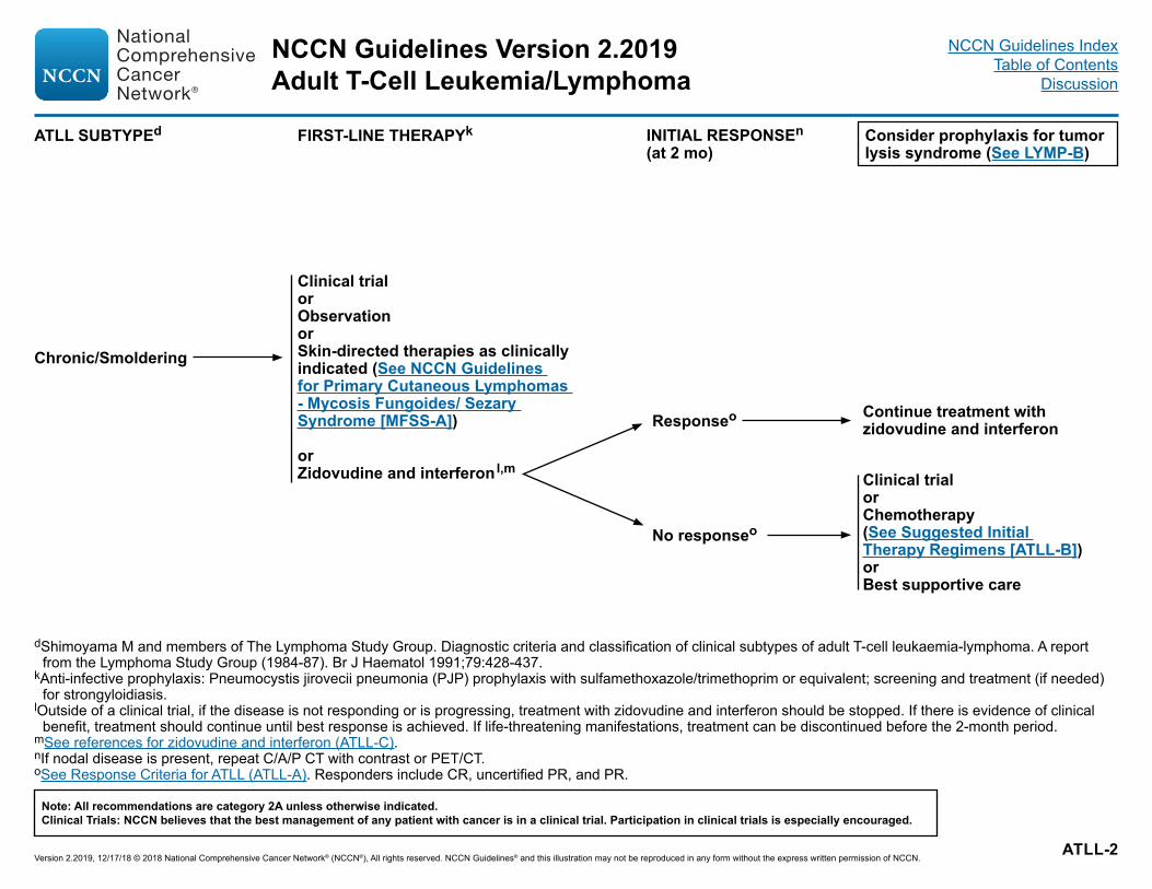
kAnti-infective prophylaxis: Pneumocystis jirovecii pneumonia (PJP) prophylaxis with sulfamethoxazole/trimethoprim or equivalent; screening and treatment (if needed) for strongyloidiasis.
lOutside of a clinical trial, if the disease is not responding or is progressing, treatment with zidovudine and interferon should be stopped. If there is evidence of clinical benefit, treatment should continue until best response is achieved. If life-threatening manifestations, treatment can be discontinued before the 2-month period.
mSee references for zidovudine and interferon (ATLL-C).nIf nodal disease is present, repeat C/A/P CT with contrast or PET/CT.oSee Response Criteria for ATLL (ATLL-A). Responders include CR, uncertified PR, and PR.
Note: All recommendations are category 2A unless otherwise indicated.Clinical Trials: NCCN believes that the best management of any patient with cancer is in a clinical trial. Participation in clinical trials is especially encouraged.
Adult T-Cell Leukemia/LymphomaNCCN Guidelines Index
Consider prophylaxis for tumor lysis syndrome (See LYMPH-B)
Acutep,q
Lymphomap,q,r
dShimoyama M and members of The Lymphoma Study Group. Diagnostic criteria and classification of clinical subtypes of adult T-cell leukaemia-lymphoma. A report from the Lymphoma Study Group (1984-87). Br J Haematol 1991;79:428-437.
kAnti-infective prophylaxis: Pneumocystis jirovecii pneumonia (PJP) prophylaxis with sulfamethoxazole/trimethoprim or equivalent; screening and treatment (if needed) for strongyloidiasis.
lOutside of a clinical trial, if the disease is not responding or is progressing, treatment with zidovudine and interferon should be stopped. If there is evidence of clinical benefit, treatment should continue until best response is achieved. If life-threatening manifestations, treatment can be discontinued before the 2-month period.
mSee references for zidovudine and interferon (ATLL-B 2 of 2).
Clinical trial orZidovudine and interferonl,morChemotherapy (See Suggested Initial Therapy Regimens [ATLL-B])
Responseo
No responseo
Clinical trialorChemotherapy (See Suggested Initial Therapy Regimens [ATLL-B])
Responseo
No responseo
Continue prior therapyor Consider allogeneic HCT
Clinical trial orBest supportive care or Alternate therapy not previously treated with:• Second-line therapy
(ATLL-B) or• Zidovudine and interferon
Responsen,o
Continue chemotherapy orConsider allogeneic HCT
Clinical trial orBest supportive care or Chemotherapy (See Second-line therapy (ATLL-B)
Responsen,o
Consider allogeneic HCT
Consider allogeneic HCT
RESPONSE ASSESSMENT
nIf nodal disease is present, repeat C/A/P CT with contrast or PET/CT.oSee Response Criteria for ATLL (ATLL-A). Responders include CR, uncertified
PR, and PR.pEfficacy of long-term treatment is limited. There are small series where
transplant is beneficial. There is no defined treatment.qCNS prophylaxis is strongly recommended.rAntiviral therapy is not effective.
Note: All recommendations are category 2A unless otherwise indicated.Clinical Trials: NCCN believes that the best management of any patient with cancer is in a clinical trial. Participation in clinical trials is especially encouraged.
Adult T-Cell Leukemia/LymphomaNCCN Guidelines Index
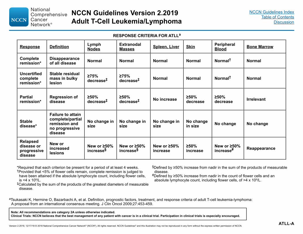
*Required that each criterion be present for a period of at least 4 weeks.†Provided that <5% of flower cells remain, complete remission is judged to
have been attained if the absolute lymphocyte count, including flower cells, is <4 x 10⁹/L.
‡Calculated by the sum of the products of the greatest diameters of measurable disease.
§Defined by ≥50% increase from nadir in the sum of the products of measurable disease.
#Defined by ≥50% increase from nadir in the count of flower cells and an absolute lymphocyte count, including flower cells, of >4 x 10⁹/L.
aTsukasaki K, Hermine O, Bazarbachi A, et al. Definition, prognostic factors, treatment, and response criteria of adult T-cell leukemia-lymphoma: A proposal from an international consensus meeting. J Clin Oncol 2009;27:453-459.
RESPONSE CRITERIA FOR ATLLa
Response Definition Lymph Nodes
Extranodal Masses Spleen, Liver Skin Peripheral
Blood Bone Marrow
Complete remission*
Disappearance of all disease Normal Normal Normal Normal Normal† Normal
Uncertified complete remission*
Stable residual mass in bulky lesion
≥75% decrease‡
≥75% decrease‡ Normal Normal Normal† Normal
Partial remission*
Regression of disease
≥50% decrease‡
≥50% decrease‡ No increase ≥50%
decrease≥50% decrease Irrelevant
Stable disease*
Failure to attain complete/partial remission and no progressive disease
Note: All recommendations are category 2A unless otherwise indicated.Clinical Trials: NCCN believes that the best management of any patient with cancer is in a clinical trial. Participation in clinical trials is especially encouraged.
Adult T-Cell Leukemia/LymphomaNCCN Guidelines Index
Alternative Regimens• Single agents (alphabetical order)�Alemtuzumabb�Arsenic trioxide/interferon alpha�Belinostat�Bendamustineb �Bortezomib �Gemcitabine �Pralatrexate �Radiation therapy in selected cases with localized, symptomatic disease
aSee References for Regimens (ATLL-B 2 of 2).bSee Supportive Care for Brentuximab Vedotin, Lenalidomide, Alemtuzumab, and Bendamustine (LYMP-B).cHigher responses have been observed in patients with leukemic disease. A retrospective study showed a particularly high risk of developing GVHD in patients
proceeding to allogenic transplant within 50 days of mogamulizumab (Fuji S, Inoue Y, Utsunomiya A, et al. Pretransplantation anti-CCR4 antibody mogamulizumab against adult t-cell leukemia/lymphoma is associated with significantly increased risks of severe and corticosteroid-refractory graft-versus-host disease, nonrelapse mortality, and overall mortality. J Clin Oncol 2016;34:3426-3433).
Note: All recommendations are category 2A unless otherwise indicated.Clinical Trials: NCCN believes that the best management of any patient with cancer is in a clinical trial. Participation in clinical trials is especially encouraged.
Adult T-Cell Leukemia/LymphomaNCCN Guidelines Index
Bazarbachi A, Plumelle Y, Carlos Ramos J, et al. Meta-analysis on the use of zidovudine and interferon-alfa in adult T-cell leukemia/lymphoma showing improved survival in the leukemic subtypes. J Clin Oncol 2010;28:4177-4183.
Hermine O, Allard I, Levy V, Arnulf B, Gessain A, Bazarbachi A. A prospective phase II clinical trial with the use of zidovudine and interferon-alpha in the acute and lymphoma forms of adult T-cell leukemia/lymphoma. Hematol J 2002;3:276-282.
Hodson A, Crichton S, Montoto S, et al. Use of zidovudine and interferon alfa with chemotherapy improves survival in both acute and lymphoma subtypes of adult T-cell leukemia/lymphoma. J Clin Oncol 2011;29:4696-4701.
White JD, Wharfe G, Stewart DM, et al. The combination of zidovudine and interferon alpha-2B in the treatment of adult T-cell leukemia/lymphoma. Leuk Lymphoma 2001;40:287-294.
Initial TherapyBrentuximab vedotin + CHP (cyclophosphamide, doxorubicin, and prednisone)Horwitz SM, Connor OA, Pro B, et al. The ECHELON-2 trial: results of a randomized,
double-blind, active-controlled phase 3 study of brentuximab vedotin and CHP (A+CHP) versus CHOP in the frontline treatment of patients with CD30+ peripheral T-cell lymphomas [abstract]. Blood 2018;132:Abstract 997.
CHOPTaguchi H, Kinoshita KI, Takatsuki K, et al. An intensive chemotherapy of adult T-cell leukemia/
lymphoma: CHOP followed by etoposide, vindesine, ranimustine,and mitoxantrone with granulocyte colony-stimulating factor support. J Acquir Immune Defic Syndr Hum Retrovirol 1996;12:182-186
Tsukasaki K, Utsunomiya A, Fukuda H, et al. VCAP-AMP-VECP compared with biweekly CHOP for adult T-cell leukemia-lymphoma: Japan Clinical Oncology
Group Study JCOG9801. J Clin Oncol 2007;25:5458-5464.Dose-adjusted EPOCHRatner L, Harrington W, Feng X, et al. Human T-cell leukemia virus reactivation with
progression of adult T-cell leukemia-lymphoma. PLoS ONE 2009;4:e4420Ratner L, Rauch D, Abel H, et al. Dose-adjusted EPOCH chemotherapy with bortezomib and
raltegravir for human T-cell leukemia virus-associated adult T-cell leukemia lymphoma. Blood Cancer J 2016;6:e408.
HyperCVADAlduaij A, Butera JN, Treaba D, Castillo J. Complete remission in two cases ofadult T-cell leukemia/lymphoma treated with hyper-CVAD: a case report andreview of the literature. Clin Lymphoma Myeloma Leuk 2010;10:480-483.
Second-line Therapy or Subsequent TherapyAlemtuzumabSharma K, Janik JE, O'Mahony D, et al. Phase II study of alemtuzumab
(CAMPATH-1) in patients with HTLV-1-associated adult T-cell leukemia/lymphoma. Clin Cancer Res 2017;23:35-42.
Arsenic trioxide and interferon alfaHermine O, Dombret H, Poupon J, et al. Phase II trial of arsenic trioxide and alpha
interferon in patients with relapsed/refractory adult T-cell leukemia/lymphoma. Hematol J 2004;5:130-134.
Ishitsuka K, Suzumiya J, Aoki M, et al. Therapeutic potential of arsenic trioxide with or without interferon-alpha for relapsed/refractory adult T-cell leukemia/lymphoma. Haematologica 2007;92:719-720.
BortezomibIshitsuka K, Utsunomiya A, Katsuya H, et al. A phase II study of bortezomib in
patients with relapsed or refractory aggressive adult T-cell leukemia/lymphoma. Cancer Sci 2015;106:1219-1223.
Brentuximab vedotinHorwitz SM, Advani RH, Bartlett NL, et al. Objective responses in
relapsed T-cell lymphomas with single-agent brentuximab vedotin. Blood 2014;123:3095-3100.
LenalidomideIshida T, Fujiwara H, Nosaka K, et al. Multicenter phase II study of lenalidomide
in relapsed or recurrent adult T-cell leukemia/lymphoma: ATLL-002. J Clin Oncol 2016;34:4086-4093.
MogamulizumabIshida T, Utsunomiya A, Jo T, et al. Mogamulizumab for relapsed adult T-cell
leukemialymphoma: Updated follow-up analysis of phase I and II studies. Cancer Sci 2017;108:2022-2029.
Phillips AA, Fields P, Hermine O, et al. A prospective, multicenter, randomized study of anti-CCR4 monoclonal antibody mogamulizumab (moga) vs investigator's choice (IC) in the treatment of patients (pts) with relapsed/refractory (R/R) adult T-cell leukemia-lymphoma (ATL) [abstract]. J Clin Oncol 2016;34 (15_suppl):Abstract 7501.
PralatrexateLunning MA, Gonsky J, Ruan J, et al. Pralatrexate in Relapsed/Refractory HTLV-1
Associated Adult T-Cell Lymphoma/Leukemia [abstract]: A New York City Multi-Institutional Experience. Blood 2012;120:Abstract 2735.
Note: All recommendations are category 2A unless otherwise indicated.Clinical Trials: NCCN believes that the best management of any patient with cancer is in a clinical trial. Participation in clinical trials is especially encouraged.
Adult T-Cell Leukemia/LymphomaNCCN Guidelines Index
aSee Principles of Molecular Analysis in T-Cell Lymphomas (LYMP-A).bTypical immunophenotype: CD1a-, TdT-, CD2+, sCD3+/-, cCD3+/-, CD5+, CD7++, CD52++, TCRαß+, CD4+/CD8- (65%), CD4+/CD8+ (21%), CD4-/CD8+ (13%).cClonal TCR gene rearrangement can be assessed by PCR or by HTS techniques. Results should be interpreted with caution since clonal TCR gene rearrangements
can also be seen in patients with non-malignant conditions. See Principles of Molecular Analysis in T-Cell Lymphomas (LYMP-A).dPatients with T-cell lymphomas often have extranodal disease, which may be inadequately imaged by CT. PET scan may be preferred in these instances.eIn a minority of patients, the disease may be asymptomatic and can follow an indolent course of variable duration. In these selected cases expectant observation is a
reasonable option.
DIAGNOSISa WORKUP
ESSENTIAL:• Tissue histology not essential for diagnosis• Peripheral blood smear analysis for morphology• Peripheral blood flow cytometry to establish
diagnosisb�TdT, CD 1a, CD2, CD3, CD4, CD5, CD7, CD8,
CD52, TCRαβ, TCL1• Cytogenetics (FISH and karyotype): inv(14)
Note: All recommendations are category 2A unless otherwise indicated.Clinical Trials: NCCN believes that the best management of any patient with cancer is in a clinical trial. Participation in clinical trials is especially encouraged.
T-Cell Prolymphocytic LeukemiaNCCN Guidelines Index
fSee Treatment References (TPLL-A).gIV alemtuzumab is preferred over subcutaneous based on data showing inferior activity with subcutaneous delivery in patients with T-PLL (Dearden CE, Khot A, Else
M, et al. Alemtuzumab therapy in T-cell prolymphocytic leukemia: comparing efficacy in a series treated intravenously and a study piloting the subcutaneous route. Blood 2011;118:5799-5802).
hSee Supportive Care (LYMP-B).iWhile alemtuzumab is no longer commercially available, it may be obtained for clinical use.jConsider HDT/ASCR if a suitable donor is not available.
SYMPTOMATIC DISEASE
PRIMARY TREATMENTf
INITIAL RESPONSE
CONSOLIDATIONf SECOND-LINE THERAPYf
Symptomatic disease
Consider prophylaxis for tumor lysis syndrome (See LYMP-B)
Note: All recommendations are category 2A unless otherwise indicated.Clinical Trials: NCCN believes that the best management of any patient with cancer is in a clinical trial. Participation in clinical trials is especially encouraged.
T-Cell Prolymphocytic LeukemiaNCCN Guidelines Index
AlemtuzumabDearden CE, Matutes E, Cazin B, et al. High remission rate in T-cell prolymphocytic leukemia with CAMPATH-1H. Blood 2001;98:1721-1726.Keating MJ, Cazin B, Coutre S, et al. Campath-1H treatment of T-cell prolymphocytic leukemia in patients for whom at least one prior chemotherapy regimen has failed. J Clin Oncol 2002;20:205-213.Dearden CE, Khot A, Else M, et al. Alemtuzumab therapy in T-cell prolymphocytic leukaemia: Comparing efficacy in a series treated intravenously and a study piloting the subcutaneous route. Blood 2011;118:5799-5802.
Alemtuzumab + pentostatinRavandi F, Aribi A, O'Brien S, et al. Phase II study of alemtuzumab in combination with pentostatin in patients with T-cell neoplasms. J Clin Oncol 2009;27:5425-5430.
FMC (fludarabine, mitoxantrone, cyclophosphamide) followed by alemtuzumab Hopfinger G, Busch R, Pflug N, et al. Sequential chemoimmunotherapy of fludarabine, mitoxantrone, and cyclophosphamide induction followed by alemtuzumab consolidation is effective in T-cell prolymphocytic leukemia. Cancer 2013;119:2258-2267.
Allogeneic hematopoietic cell transplantCastagna L, Nozza A, Bertuzzi A, et al. Allogeneic peripheral blood stem cell transplantation with reduced intensity conditioning in primary refractory prolymphocytic leukemia: graft-versus-leukemia effect without graft-versus-host disease. Bone Marrow Transplant 2001;28:1155-1156.Kalaycio ME, Kukreja M, Woolfrey AE, et al. Allogeneic hematopoietic cell transplant for prolymphocytic leukemia. Biol Blood Marrow Transplant. 2010;16:543-547.Murase K, Matsunaga T, Sato T, et al. Allogeneic bone marrow transplantation in a patient with T-prolymphocytic leukemia with small-intestinal involvement. Int J Clin Oncol 2003;8:391-394.Wiktor-Jedrzejczak W, Dearden C, de Wreede L, et al. Hematopoietic stem cell transplantation in T-prolymphocytic leukemia: A retrospective study from the European Group for Blood and Marrow Transplantation and the Royal Marsden Consortium. Leukemia 2012;26:972-972. Krishnan B, Else M, Tjonnfjord G, et al. Stem cell transplantation after alemtuzumab in T-cell prolymphocytic leukaemia results in longer survival than after alemtuzumab alone: a multicentre retrospective study. Br J Haematol 2010;149: 907–910.
Note: All recommendations are category 2A unless otherwise indicated.Clinical Trials: NCCN believes that the best management of any patient with cancer is in a clinical trial. Participation in clinical trials is especially encouraged.
T-Cell Prolymphocytic LeukemiaNCCN Guidelines Index
DIAGNOSISa,b SUBTYPESESSENTIAL: • Hematopathology review of all slides with at least one paraffin block
representative of the tumor. Rebiopsy if consult material is nondiagnostic.• Excisional or incisional biopsy is preferred over core needle biopsy. An FNA
alone is not sufficient for the initial diagnosis of lymphoma.c A core needle biopsy is not optimal but can be used under certain circumstances. In certain circumstances, when a lymph node is not easily accessible for excisional or incisional biopsy, a combination of core needle biopsy and FNA in conjunction with appropriate ancillary techniques may be sufficient for diagnosis.
• Adequate immunophenotyping to establish diagnosisd,e
�IHC panel: For high clinical suspicion of NKTL, first panel should include: CD2, cCD3ɛ, CD56, EBER-ISH,f CD5, CD56, TIA1�Flow panel: CD2, CD3, CD4, CD5, CD7, CD8, CD56, TCRα/β, TCRɣ/ẟ
USEFUL UNDER CERTAIN CIRCUMSTANCES: • Molecular analysis to detect clonal TCR gene rearrangements or other
assessment of clonality (karyotype, array-CGH, or FISH analysis to detect somatic mutations or genetic alterations)g
Subtypes not included:• NK-cell leukemias • Precursor NK-cell
neoplasm
See Workup (NKTL-2)
aIt is preferred that treatment occur at centers with expertise in the management of this disease.bPrinciples of Molecular Analysis in T-Cell Lymphomas (LYMP-A).cNecrosis is very common in diagnostic biopsies and may delay diagnosis significantly. Biopsy
should include the edges of lesions to increase the odds of having viable tissue. Useful to perform multiple nasopharyngeal biopsies even in areas not clearly involved.
dSee Use of Immunophenotyping/Genetic Testing in Differential Diagnosis of Mature B-Cell and NK/T-Cell Neoplasms (See B-Cell Lymphomas Guidelines).
eTypical NK-cell immunophenotype: CD20-, CD2+, cCD3ɛ+ (surface CD3-), CD4-, CD5-, CD7-/+, CD8-/+, CD43+, CD45RO+, CD56+, T-cell receptor (TCR)αß-, TCRγδ-, EBV- EBER+. TCR and Ig genes are germline (NK lineage). Cytotoxic granule proteins (TIA1, perforin, granzyme B) are usually expressed. Typical T-cell immunophenotype: CD2+ sCD3+ cCD3e+, CD4, CD5, CD7, CD8 variable, CD56+/-, EBV-EBER+, TCRαß or γδ+, cytotoxic granule proteins +. TCR genes are clonally rearranged.
fNegative result should prompt pathology review for alternative diagnosis.
gClonal TCR gene rearrangement can be assessed by PCR or by HTS techniques. Results should be interpreted with caution since clonal TCR gene rearrangements can also be seen in patients with non-malignant conditions. See Principles of Molecular Analysis in T-Cell Lymphomas (LYMP-A).
Note: All recommendations are category 2A unless otherwise indicated.Clinical Trials: NCCN believes that the best management of any patient with cancer is in a clinical trial. Participation in clinical trials is especially encouraged.
Extranodal NK/T-Cell Lymphoma, nasal typeNCCN Guidelines Index
WORKUPESSENTIAL:• H&P exam with attention to node-bearing areas (including Waldeyer's ring),
testicles, and skin• ENT evaluation of nasopharynx• Performance status• B symptoms• CBC with differential• LDH• Comprehensive metabolic panel• Uric acid• Bone marrow biopsy + aspirateh
• PET/CT scani and/or C/A/P CT with contrast of diagnostic quality • MRI ± CT pretreatment for RT planning of the nasal cavity, hard palate, anterior
fossa, and nasopharynx• Calculation of Prognostic Index of Natural Killer Lymphoma (PINK)j• Echocardiogram or MUGA scan if treatment includes regimens containing
anthracyclines or anthracenedione• EBV viral loadk by quantitative PCR• Concurrent referral to RT for pretreatment evaluation
USEFUL IN SELECTED CASES:• Pregnancy testing in women of child-bearing age (if chemotherapy or RT
planned)• Discussion of fertility and sperm banking • HIV testing
See Induction Therapy (NKTL-3)
hBM aspirate - lymphoid aggregates are rare, and are considered involved if EBER-1 positive; hemophagocytosis may be present.iPatients with T-cell lymphomas often have extranodal disease, which may be inadequately imaged by CT. PET scan may be preferred in these instances.jSee Prognostic Index of Natural Killer Lymphoma (PINK) (NKTL-A).kEBV viral load is important in diagnosis and possibly in monitoring of disease. A positive result is consistent with NK/T-cell, nasal type. Lack of normalization of EBV
viremia should be considered indirect evidence of persistent disease.
Note: All recommendations are category 2A unless otherwise indicated.Clinical Trials: NCCN believes that the best management of any patient with cancer is in a clinical trial. Participation in clinical trials is especially encouraged.
Extranodal NK/T-Cell Lymphoma, nasal typeNCCN Guidelines Index
aIt is preferred that treatment occur at centers with expertise in the management of this disease.lIn rare circumstances of stage IE primary cutaneous NK/T-cell lymphoma, involved-field RT for solitary lesions can be considered.mSee Suggested Treatment Regimens (NKTL-B).
Consider prophylaxis for tumor lysis syndrome (See LYMP-B)
Note: All recommendations are category 2A unless otherwise indicated.Clinical Trials: NCCN believes that the best management of any patient with cancer is in a clinical trial. Participation in clinical trials is especially encouraged.
Extranodal NK/T-Cell Lymphoma, nasal typeNCCN Guidelines Index
aIt is preferred that treatment occur at centers with expertise in the management of this disease.iPatients with T-cell lymphomas often have extranodal disease, which may be inadequately imaged by CT.
PET scan may be preferred in these instances.mSee Suggested Treatment Regimens (NKTL-B).nSee Lugano Response Criteria for Non-Hodgkin’s Lymphoma (LYMP-C).oIncludes a negative ENT evaluation.pMay include H&P, ENT evaluation, PET/CT scan, and EBV viral load by quantitative PCR. qThere are no clear data to suggest whether allogeneic or autologous HCT is preferred and treatment should be individualized.rClinical trial is the preferred relapsed/refractory option. In the absence of a clinical trial, pembrolizumab is an appropriate option.sReports of EBV reactivation have been seen with HDAC inhibitors; consider monitoring or prophylaxis.
END-OF-TREATMENT EVALUATIONa
RESPONSE TO THERAPYn
ADDITIONAL THERAPY
• Repeat initial imaging of CT, MRI, or PET/CT scani
• Endoscopy with visual inspection and repeat biopsies
• EBV viral load
Nasal
Extranasal
Stage I, II
Stage IV
Stage I–IV
CRo
PR
No response
Clinical trialorAlternate chemotherapy regimen (asparaginase-based)m if not previously usedor Best supportive care
CR
No response
Consider HCTq
Clinical trialorAlternate chemotherapy regimen (asparaginase-based)m if not previously usedor Best supportive care
Clinical trial(preferred)orPembrolizumabrorNivolumab (category 3)orSee Preferred regimens for Second-line Therapy for PTCL-NOS (TCEL-B)sorHCT, if eligible
PR Biopsy
Biopsy Negative
Negative
Positive
Positive Clinical trial(preferred)orPembrolizumabrorNivolumab (category 3)orSee Preferred regimens for Second-line Therapy for PTCL-NOS (TCEL-B)sorHCT, if eligible
Note: All recommendations are category 2A unless otherwise indicated.Clinical Trials: NCCN believes that the best management of any patient with cancer is in a clinical trial. Participation in clinical trials is especially encouraged.
Extranodal NK/T-Cell Lymphoma, nasal typeNCCN Guidelines Index
PROGNOSTIC INDEX OF NATURAL KILLER LYMPHOMA (PINK)a
aKim SJ, Yoon DH, Jaccard A, et al. A prognostic index for natural killer cell lymphoma after non-anthracycline-based treatment: a multicentre, retrospective analysis. Lancet Oncol 2018;17:389-400.
RISK FACTORS
Age >60 yStage III or IV diseaseDistant lymph-node involvementNon-nasal type disease
Number of risk factorsLow 0Intermediate 1High ≥2
PROGNOSTIC INDEX OF NATURAL KILLER CELL LYMPHOMA WITH EPSTEIN-BARR VIRUS DNA (PINK-E)a
RISK FACTORS
Age >60 yStage III or IV diseaseDistant lymph-node involvementNon-nasal type diseaseEpstein-Barr virus DNA
Number of risk factorsLow 0–1Intermediate 2High ≥3
Note: All recommendations are category 2A unless otherwise indicated.Clinical Trials: NCCN believes that the best management of any patient with cancer is in a clinical trial. Participation in clinical trials is especially encouraged.
Extranodal NK/T-Cell Lymphoma, nasal typeNCCN Guidelines Index
aSee references for regimens NKTL-B 2 of 2.bSee Asparaginase Toxicity Management in the NCCN Guidelines for Acute Lymphoblastic Leukemia.cPegaspargase-based regimens are preferred. However, there are no data to recommend one particular regimen over another. Treatment should be individualized
based on patient's tolerance and comorbidities. P-GEMOX is an option for selected patients who cannot tolerate intense chemotherapy.dSee Principles of Radiation Therapy (LYMP-D).
SUGGESTED TREATMENT REGIMENSa
(in alphabetical order)Combination chemotherapy regimen (asparaginase-based)b,c• AspaMetDex (pegaspargase, methotrexate, and dexamethasone) • Modified-SMILE (steroid [dexamethasone], methotrexate, ifosfamide, pegaspargase, and etoposide) x 4–6 cycles for advanced stage• P-GEMOX (gemcitabine, pegaspargase, and oxaliplatin)
Combined modality therapy (non-asparaginase-based) • Concurrent chemoradiation therapy (CCRT) �RTd and 3 courses of DeVIC (dexamethasone, etoposide, ifosfamide, and carboplatin)�RTd and cisplatin followed by 3 cycles of VIPD (etoposide, ifosfamide, cisplatin, and dexamethasone)
• Sequential chemoradiation�For stage I, II, modified-SMILE x 2–4 cycles followed by RTd
• Sandwich chemoradiationc�P-GEMOX x 2 cycles followed by RTd followed by P-GEMOX x 2–4 cycles
Radiation therapy alone (if unfit for chemotherapy)d• Early or upfront RT had an essential role in improved overall survival and disease-free survival in patients with localized extranodal
NK/T-cell lymphoma, nasal-type, in the upper aerodigestive tract.• Upfront RT may yield more benefits on survival in patients with stage I disease.
Note: All recommendations are category 2A unless otherwise indicated.Clinical Trials: NCCN believes that the best management of any patient with cancer is in a clinical trial. Participation in clinical trials is especially encouraged.
Extranodal NK/T-Cell Lymphoma, nasal typeNCCN Guidelines Index
Combination Chemotherapy RegimenYamaguchi M, Kwong YL, Kim WS, et al. Phase II study of SMILE chemotherapy for newly diagnosed stage IV, relapsed, or refractory extranodal natural killer (NK)/T-cell lymphoma, nasal type: The NK-Cell Tumor Study Group Study. J Clin Oncol 2011;29:4410-4416.Lunning M, Pamer E, Maragulia J, et al. Modified SMILE (mSMILE) is Active in the Treatment of Extranodal Natural Killer/T-Cell Lymphoma: A Single Center US Experience. Clinical Lymphoma, Myeloma and Leukemia 2014;14:S143-S144. Jaccard A, Gachard N, Marin B, et al. Efficacy of L-asparaginase with methotrexate and dexamethasone (AspaMetDex regimen) in patients with refractory or relapsing extranodal NK/T-cell lymphoma, a phase 2 study. Blood 2011;117:1834-1839.Wang JH, Wang H, Wang YJ, et al. Analysis of the efficacy and safety of a combined gemcitabine, oxaliplatin and pegaspargase regimen for NK/T-cell lymphoma. Oncotarget 2018;7:35412-35422. Qi S, Yahalom J, Hsu M, et al. Encouraging experience in the treatment of nasal type extra-nodal NK/T-cell lymphoma in a non-Asian population. Leuk Lymphoma 2018;57:2575-2583.
Concurrent ChemoradiationYamaguchi M, Tobinai K, Oguchi M, et al. Concurrent chemoradiotherapy for localized nasal natural killer/T-cell lymphoma: an updated analysis of the Japan clinical oncology group study JCOG0211. J Clin Oncol 2012;30:4044-4046.Kim SJ, Kim K, Kim BS, et al. Phase II trial of concurrent radiation and weekly cisplatin followed by VIPD chemotherapy in newly diagnosed, stage IE to IIE, nasal, extranodal NK/T-cell lymphoma: Consortium for Improving Survival of Lymphoma study. J Clin Oncol 2009;27:6027-6032. Yamaguchi M, Suzuki R, Oguchi M, et al. Treatments and Outcomes of Patients With Extranodal Natural Killer/T-Cell Lymphoma Diagnosed Between 2000 and 2013: A Cooperative Study in Japan. J Clin Oncol 2018;35:32-39.
Sequential ChemoradiationLunning M, Pamer E, Maragulia J, et al. Modified SMILE (mSMILE) is Active in the Treatment of Extranodal Natural Killer/T-Cell Lymphoma: A Single Center US Experience. Clinical Lymphoma, Myeloma and Leukemia 2014;14:S143-S144.
Sandwich ChemoradiationTse E, Kwong YL. The diagnosis and management of NK/T-cell lymphomas. J Hematol Oncol 2018;10:85. Wang L, Wang ZH, Chen XQ, et al. First-line combination of GELOX followed by radiation therapy for patients with stage IE/IIE ENKTL: An updated analysis with long-term follow-up. Oncol Lett 2015;10:1036-1040. Bi XW, Xia Y, Zhang WW, et al. Radiotherapy and PGEMOX/GELOX regimen improved prognosis in elderly patients with early-stage extranodal NK/T-cell lymphoma. Ann Hematol 2015;94:1525-1533.
Radiation Therapy AloneHuang MJ, Jiang Y, Liu WP, et al. Early or up-front radiotherapy improved survival of localized extranodal NK/T-cell lymphoma, nasal-type in the upper aerodigestive tract. Int J Radiat Oncol Biol Phys 2008;70:166-174.
Relapsed/Refractory TherapyKwong YL, Chan TSY, Tan D, et al. PD1 blockade with pembrolizumab is highly effective in relapsed or refractory NK/T-cell lymphoma failing l-asparaginase. Blood 2018;129:2437-2442. Chan TSY, Li J, Loong F2 PD blockade with low-dose nivolumab in NK/T cell lymphoma failing L-asparaginase: efficacy and safety. Ann Hematol 2018;97:193-196.
Note: All recommendations are category 2A unless otherwise indicated.Clinical Trials: NCCN believes that the best management of any patient with cancer is in a clinical trial. Participation in clinical trials is especially encouraged.
Extranodal NK/T-Cell Lymphoma, nasal typeNCCN Guidelines Index
• Hepatosplenic T-cell lymphoma (HSTCL) is a rare, systemic, mature T-cell malignancy most often characterized by spleen, liver, and bone marrow involvement and an aggressive clinical course. Bulky lymphadenopathy is uncommon.
• The disease predominantly affects males with a median age of 35 years. Up to 20% of cases arise in the setting of chronic immune suppression. Patients frequently present with systemic symptoms, hepatosplenomegaly, cytopenias, and sometimes hemophagocytic lymphohistiocytosis (HLH).
• The diagnosis is most frequently reached by histologic examination of a bone marrow biopsy, and/or a liver biopsy or splenectomy or spleen biopsy. On bone marrow histology the neoplastic T cells may be difficult to identify, and immunohistochemistry is required for the diagnosis.
• The neoplastic cells are cytotoxic T cells, frequently with surface expression of gamma/delta T-cell receptor, and typically show the following phenotype: CD2+, CD3+, CD4-, CD5-, CD8-, CD56+, TIA1+, granzyme B-. A small subset has alpha-beta TCR expression, which is a described variant of HSTCL.
• Studies of clonality by fragment analysis or next-generation sequencing will usually identify clonal rearrangement of T-cell receptor genes.�A T-cell receptor gamma chain gene rearrangement on molecular analysis reflects clonality of the T-cell, but may be seen in alpha/beta
or gamma/delta-expressing T cells and is NOT necessarily synonymous with a gamma/delta T-cell lymphoma.• Characteristic genetic features include isochromosome 7q, trisomy 8, activating mutations of JAK/STAT pathway (STAT5b, STAT3), and
chromatin-modifying genes (SETD2, INO80, ARID1B).• Main differential diagnosis includes gamma/delta-expressing T-cell large granular lymphocytic leukemia (T-LGLL), reactive gamma/delta
T-cell proliferations, aggressive NK-cell leukemia, EBV-positive T-cell and NK-cell lymphoproliferative diseases of childhood, and, rarely, other T-cell lymphomas that may have gamma/delta expression.
Note: All recommendations are category 2A unless otherwise indicated.Clinical Trials: NCCN believes that the best management of any patient with cancer is in a clinical trial. Participation in clinical trials is especially encouraged.
Hepatosplenic Gamma-Delta T-Cell LymphomaNCCN Guidelines Index
ESSENTIAL:• Review of all slides with at least one paraffin block representative of the tumor should be done
by a hematopathologist with expertise in the diagnosis of T-cell lymphomas. Rebiopsy if consult material is nondiagnostic.
• A core or incisional biopsy of bone marrow, liver, or spleen is required for diagnosis. Bone marrow aspirate, FNA of liver or spleen, or peripheral blood studies may be helpful but are not alone sufficient for diagnosis.
• Adequate immunophenotyping to establish diagnosisc,d�IHC panel may include CD20, CD3, CD10, Ki-67, CD5, CD30, CD2, CD4, CD8, CD7, CD56, EBER-ISH,
TCRβ, TCRẟ, TIA-1, or granzyme B with�Cell surface marker analysis by flow cytometry may include kappa/lambda, CD45, CD3, CD5,
CD19, CD10, CD20, CD30, CD4, CD8,CD7, CD2; TCRα/β, or TCRɣ/ẟ
USEFUL UNDER CERTAIN CIRCUMSTANCES:• Molecular analysis to detectc clonal TCR gene rearrangements or other assessment of clonality
(karyotype, array-CGH, or FISH analysis to detect somatic mutations or genetic alterations)e• Karyotype to establish clonality and investigate the presence of isochromosome 7q and trisomy 8.• FISH analysis for isochromosome 7q and trisomy 8.• Genomic analysis for STAT3, STAT5B, PIK3CD, SETD2, INO80, TET3, and SMARCA2.
aIt is preferred that treatment occur at centers with expertise in the management of this disease.bSee Principles of Molecular Analysis in T-Cell Lymphomas (LYMP-A).cSee Use of Immunophenotyping/Genetic Testing in Differential Diagnosis of Mature B-Cell and NK/T-Cell Neoplasms (See B-Cell Lymphomas Guidelines).dTypical immunophenotype: CD3+, generally TCRδ + and TCRβ – (GM3 positive, βF-1 negative), CD4 -, CD8-/+, CD56 +/-, CD5-.eClonal TCR gene rearrangement can be assessed by PCR or by HTS techniques. Results should be interpreted with caution since clonal TCR gene
rearrangements can also be seen in patients with non-malignant conditions. See Principles of Molecular Analysis in T-Cell Lymphomas (LYMP-A).
Note: All recommendations are category 2A unless otherwise indicated.Clinical Trials: NCCN believes that the best management of any patient with cancer is in a clinical trial. Participation in clinical trials is especially encouraged.
Hepatosplenic Gamma-Delta T-Cell LymphomaNCCN Guidelines Index
WORKUPESSENTIAL:• H&P exam; full skin exam; attention to node-bearing areas, including Waldeyer's
ring; evaluation of size of liver and spleen, nasopharynx • Performance status • B symptoms • CBC with differential • Bone marrow biopsy ± aspirate • LDH • Comprehensive metabolic panel • HLH workup• Uric acid • PET/CT scanf and/or C/A/P CT with contrast of diagnostic quality • Echocardiogram or MUGA scan if anthracycline or anthracenedione-based
regimen is indicated • Pregnancy testing in women of child-bearing age (if chemotherapy or RT planned) • HLA typing
USEFUL IN SELECTED CASES:• Neck CT with contrast • Head CT or MRI with contrast • HIV testing • Hepatitis B and C testing • Consider quantitative EBV PCR • Discussion of fertility issues and sperm banking
See Primary Treatment (HGDTL-4)
fPatients with T-cell lymphomas often have extranodal disease, which may be inadequately imaged by CT. PET scan may be preferred in these instances.
Note: All recommendations are category 2A unless otherwise indicated.Clinical Trials: NCCN believes that the best management of any patient with cancer is in a clinical trial. Participation in clinical trials is especially encouraged.
Hepatosplenic Gamma-Delta T-Cell LymphomaNCCN Guidelines Index
vedotin + CHP (cyclophosphamide, doxorubicin, and prednisone) for CD30+ casesh
Complete or partial responsei,j
No response or progressive disease
Allogeneic HCTk (preferred)
• Clinical trial (preferred)• Consider alternate
induction regimens not used in primary treatment
gCHOP is not adequate therapy.hPatients with HGDTL were eligible for the ECHELON-2 study (Horwitz SM, Connor OA, Pro B, et al. The ECHELON-2 trial: results
of a randomized, double-blind, active-controlled phase 3 study of brentuximab vedotin and CHP (A+CHP) versus CHOP in the frontline treatment of patients with CD30+ peripheral T-cell lymphomas [abstract]. Blood 2018;132:Abstract 997) but no patients were enrolled. Also see Supportive Care (LYMP-B)
iPatients should have very low tumor burden at the time of HCT. The goal of therapy is to induce complete or near complete response before proceeding to HCT. Full-course chemotherapy may not be needed to achieve adequate response to allow HCT.
jPET scan alone is inadequate for response assessment. PET-negative response should be confirmed by bone marrow biopsy. HGTL is non-nodal and Lugano response criteria do not apply.
kConsider HDT/ASCR if unfit or lacking a suitable donor.lResponses have been observed with alemtuzumab, pralatrexate, and ESHAP.
• Clinical trial (preferred)• Second-line
therapy regimens recommended for PTCL-NOS (See TCEL-B)l
Refractorydiseaseafter 2primarytreatmentregimens
Complete or partial responsej
No response or progressive disease
Alternativesecond-line therapy(See TCEL-B)and/orBest supportive care
Note: All recommendations are category 2A unless otherwise indicated.Clinical Trials: NCCN believes that the best management of any patient with cancer is in a clinical trial. Participation in clinical trials is especially encouraged.
Hepatosplenic Gamma-Delta T-Cell LymphomaNCCN Guidelines Index
PRINCIPLES OF MOLECULAR ANALYSIS IN T-CELL LYMPHOMASa
Continued
• Molecular technologies, including HTS technologies, that detect gene signatures are often tremendously informative and in some cases essential for an accurate and precise diagnostic and prognostic assessment of T-cell lymphomas.
T-cell Antigen Receptor (TCR) Gene Rearrangements• TCR gene rearrangement testing is recommended to confirm a diagnosis of T-cell lymphoma. • Diseases: �PTCLs; mycosis fungoides/Sezary syndrome; primary cutaneous CD30+ T-cell lymphoproliferative disorders (CD30+ T-cell LPD); T-cell
LGLL; T-cell prolymphocytic leukemia; extranodal NK/T-cell lymphoma, nasal type; and hepatosplenic gamma-delta T-cell lymphoma • Description: �TCR gene rearrangement is indicative of T-cell clonal expansion. The test targets the gamma and/or beta TCR genes using PCR methods
with capillaroscopy or gel electrophoresis detection methods. Alternatively, HTS methods are increasingly utilized. HTS methods are more sensitive, precise, and capable of providing a unique sequence of the T-cell clone, which allows for comparison and confirmation of disease evolution and monitoring during remission. Clonal T-cell expansions can also be detected using V beta families in blood or tissue with flow cytometry methods.
• Diagnostic value: �Clonal TCR gene rearrangements without cytologic and immunophenotypic evidence of abnormal T-cell population does not constitute
a diagnosis of T-cell lymphoma since it can be identified in patients with non-malignant conditions. Conversely, a negative result does not exclude the diagnosis of T-cell lymphoma, which occasionally may fail TCR amplification. Nonetheless, it often provides essential information and increased precision for many of these complex diagnoses.
• Prognostic value: �Determination of clonal TCR gene rearrangement is an ancillary confirmatory test without prognostic value, except when used to assess
relapse or residual disease.
ALK Gene Rearrangement• A subset of CD30-positive ALCLs expresses anaplastic lymphoma kinase (ALK) by immunohistochemistry. ALK expression is often
associated with t(2;5)(p23;q35), leading to the fusion of nucleophosmin (NPM1) to ALK and resulting in a chimeric protein. • Detection: �FISH using probes to ALK (2p23)
• Diagnostic value: �The present WHO classification of ALCLs includes two entities distinguishing ALK-positive and ALK-negative variants.
• Prognostic value: �Systemic ALK-positive ALCL with t(2;5) and ALK-negative ALCL with DUSP22 rearrangement (to a lesser extent) have been associated with
a favorable prognosis. ALK inhibition can be an effective therapeutic strategy.
Note: All recommendations are category 2A unless otherwise indicated.Clinical Trials: NCCN believes that the best management of any patient with cancer is in a clinical trial. Participation in clinical trials is especially encouraged.
PRINCIPLES OF MOLECULAR ANALYSIS IN T-CELL LYMPHOMASa
Continued
DUSP22-IRF4 Gene Rearrangement• Testing for DUSP22 rearrangement is considered if CD30-positive ALCL, ALK negative is diagnosed, and considered useful under certain
circumstances for the diagnosis of primary cutaneous CD30+ T-cell lymphoproliferative disorders.• Diseases: �PTCLs, primary cutaneous CD30+ T-cell lymphoproliferative disorders
• Description: �DUSP22 (dual-specificity phosphatase 22) is a tyrosine/threonine/serine phosphatase that may function as a tumor suppressor. DUSP22
inactivation contributes to the development of PTCLs. • Detection: �FISH using probes to DUPS22-IRF4 gene region at 6p25.3
• Diagnostic value: • DUSP22 rearrangements are associated with a newly recognized variant of ALK-negative ALCL and a newly reported subtype of
lymphomatoid papulosis. • Prognostic value: �ALCL, ALK negative with DUSP22 rearrangement has preliminarily been associated with a favorable prognosis; however, the impact of
this on choice of therapy is not currently known.
TP63 Rearrangement• TP63 gene rearrangements encoding p63 fusion proteins define a subset of ALK-negative ALCL cases and are associated with aggressive
course.• Detection: �FISH using probes to TP63 (3q28) and TBL1XR1/TP63
TCL1 and TRA Translocation• Most T-cell prolymphocytic leukemias have an inversion or translocation of chromosome 14 with breakpoints in the long arm at q11 and
q32 [inv(14)(q11q32) and t(14;14)(q11;q32)]. These translocations and inversions cause gene overexpression due to juxtaposition with TCR-alpha or TCR-beta regulatory elements and activate the oncogenes TCL1A and MTCP1-B1.
Note: All recommendations are category 2A unless otherwise indicated.Clinical Trials: NCCN believes that the best management of any patient with cancer is in a clinical trial. Participation in clinical trials is especially encouraged.
PRINCIPLES OF MOLECULAR ANALYSIS IN T-CELL LYMPHOMASa
TET2/IDH1/IDH2/RHOA/DNMT3A mutations• High incidence of somatic mutations in IDH2 and TET2 genes has been identified in AITLs. IDH2 and TET2 encode for proteins involved
in epigenetic regulation, suggesting that disruption of gene expression regulation by methylation and acetylation may be involved in AITL development and/or progression. Additional genetic findings include the presence of mutations affecting ROHA small GTPase gene (RHOA G17V) and DNMT3A.
• Disease: �Suspected AITL versus other PTCL
• Detection method: �Bi-directional sequencing of the entire coding or select exons in the genes IDH1, IDH2, DNMT3A, TET2, and RHOA.
• Diagnostic value: �Diagnosis of AITL versus other PTCLs. This pathway has been preliminarily associated with higher rates of response to histone
deacetylase inhibitors and other epigenetic modifiers. Clinical trials of this approach are currently ongoing.
STAT3/STAT5B mutations• STAT3 mutation testing is recommended under certain circumstances for diagnosis of LGLL and natural killer (NK) leukemias. STAT5B
mutations are less common but associated with aggressive subtypes. • Diseases: �LGLL and NK leukemia. Similar mutations are also reported in hepatosplenic gamma-delta T-cell lymphomas.
• Description: �STAT3 mutations have been identified in approximately 50% of LGLL and NK leukemias, including Y640F, N647I, E638Q, I659L, and
Note: All recommendations are category 2A unless otherwise indicated.Clinical Trials: NCCN believes that the best management of any patient with cancer is in a clinical trial. Participation in clinical trials is especially encouraged.
PRINCIPLES OF MOLECULAR ANALYSIS IN T-CELL LYMPHOMAS
Chiarle R, Voena C, Ambrogio C, Piva R, Inghirami G. The anaplastic lymphoma kinase in the pathogenesis of cancer. Nat Rev Cancer 2008;8:11-23.
De Schouwer P, Dyer M, Brito-Babapulle V, et al. T-cell prolymphocytic leukemia: antigen receptor gene rearrangement and a novel mode of MTCP1-B1 activation. Br J Haematol 2000;110:831-838.
Hu Z, Medeiros L, Fang L, et al. Prognostic significance of cytogenetic abnormalities in T-cell prolymphocytic leukemia. Am J Hematol 2017;92:441-447.
Morris SW, Kirstein M, Valentine M, et al. Fusion of a kinase gene, ALK, to a nucleolar protein gene, NPM, in non-Hodgkin’s Lymphoma. Science 1994;263:1281-1284.
Odejide O, Weigert O, Lane A, et al. A targeted mutational landscape of angioimmunoblastic T-cell lymphoma. Blood 2014;123:1293-1296.
Pedersen M, Hamilton-Dutoit S, Bendix K, et al. DUSP22 and TP63 rearrangements predict outcome of ALK-negative anaplastic large cell lymphoma: a Danish cohort study. Blood 2017;130:554-557.
Wada D, Law M, Hsi E, et al. Specificity of IRF4 translocations for primary cutaneous anaplastic large cell lymphoma: a multicenter study of 204 skin biopsies. Mod Pathol 2011;24:596-605.
Note: All recommendations are category 2A unless otherwise indicated.Clinical Trials: NCCN believes that the best management of any patient with cancer is in a clinical trial. Participation in clinical trials is especially encouraged.
• Symptoms of TLS:�Nausea and vomiting, shortness of breath, irregular heartbeat,
clouding of urine, lethargy, and/or joint discomfort.
• TLS features:�Consider TLS prophylaxis for patients with the following risk
factors: ◊ Spontaneous TLS ◊ Elevated WBC count ◊ Bone marrow involvement ◊ Pre-existing elevated uric acid ◊ Renal disease or renal involvement by tumor
aThere are data to support that fixed-dose rasburicase is very effective in adult patients.
• Treatment of TLS:�TLS is best managed if anticipated and treatment is started
prior to chemotherapy.�Centerpiece of treatment includes:
◊ Rigorous hydration ◊ Management of hyperuricemia ◊ Frequent monitoring of electrolytes and aggressive correction (essential)
�First-line and at retreatment for hyperuricemia ◊ Allopurinol beginning 2–3 days prior to chemotherapy and continued for 10–14 days or Rasburicase (Doses of 3 to 6 mg are usually effective.a One dose of rasburicase is frequently adequate. Re-dosing should be individualized.) is indicated for patients with any of the following risk factors:
- Urgent need to initiate therapy in a high-bulk patient - Situations where adequate hydration may be difficult
or impossible - Acute renal failure �If TLS is untreated, its progression may cause acute kidney
failure, cardiac arrhythmias, seizures, loss of muscle control, and death.
Note: All recommendations are category 2A unless otherwise indicated.Clinical Trials: NCCN believes that the best management of any patient with cancer is in a clinical trial. Participation in clinical trials is especially encouraged.
SUPPORTIVE CAREFor other immunosuppressive situations, see NCCN Guidelines for Prevention and Treatment of Cancer-Related Infections.Monoclonal Antibody Therapy and Viral Reactivation Brentuximab Vedotin (anti-CD30 antibody-drug conjugate)Progressive multifocal leukoencephalopathy (PML):• Caused by the JC virus and is usually fatal.�Diagnosis made by polymerase chain reaction (PCR) of cerebrospinal fluid (CSF) and in some cases brain biopsy.
• No known effective treatment. • Clinical indications may include changes in behavior such as confusion, dizziness or loss of balance, difficulty talking or walking, and vision
problems.Anti-CD52 Antibody Therapy: AlemtuzumabCytomegalovirus (CMV) reactivation:• The current appropriate management is controversial; some NCCN Member Institutions use ganciclovir (oral or IV) preemptively if viremia is
present, others only if viral load is rising. • Herpes virus prophylaxis with acyclovir or equivalent• PJP prophylaxis with sulfamethoxazole/trimethoprim or equivalent• Consider antifungal prophylaxis• CMV viremia should be measured by quantitative PCR at least every 2 to 3 weeks.• Consultation with an infectious disease expert may be necessary. See NCCN Guidelines for Prevention and Treatment of Cancer-Related
Infections.
Renal Dysfunction Associated with Methotrexate• Consider use of glucarpidase if significant renal dysfunction and methotrexate levels are >10 microM beyond 42 to 48 hours. Leucovorin
remains a component in the treatment of methotrexate toxicity and should be continued for at least 2 days following glucarpidase administration. However, be aware that leucovorin is a substrate for glucarpidase, and therefore should not be administered within two hours prior to or following glucarpidase.
Management of Tumor Flare Recommended for Patients Receiving Lenalidomide• Tumor flare reactions: Painful lymph node enlargement or lymph node enlargement with evidence of local inflammation, occurring with
treatment initiation; may also be associated with spleen enlargement, low-grade fever, and/or rash• Treatment: Steroids (eg, prednisone 25–50 mg PO for 5–10 days); antihistamines for rash and pruritus (cetirizine 10 mg PO once daily or
loratadine 10 mg PO daily)• Prophylaxis: Consider in patients with bulky lymph nodes (>5 cm); administer steroids (eg, prednisone 20 mg PO for 5–7 days followed by
Note: All recommendations are category 2A unless otherwise indicated.Clinical Trials: NCCN believes that the best management of any patient with cancer is in a clinical trial. Participation in clinical trials is especially encouraged.
LUGANO RESPONSE CRITERIA FOR NON-HODGKIN’S LYMPHOMAPET should be done with contrast-enhanced diagnostic CT and can be done simultaneously or at separate procedures.
Response Site PET-CT (Metabolic response) CT (Radiologic response)d
Complete response
Lymph nodes and extralymphatic sites
Score 1, 2, or 3a with or without a residual mass on 5 point scale (5-PS)b,c
All of the following: Target nodes/nodal masses must regress to ≤1.5 cm in longest transverse diameter of a lesion (LDi) No extralymphatic sites of disease
Non-measured lesion Not applicable Absent
Organ enlargement Not applicable Regress to normal New Lesions None None
Bone Marrow No evidence of FDG-avid disease in marrow Normal by morphology; if indeterminate, and flow cytometry IHC negative
Partial response
Lymph nodes and extralymphatic sites
Score 4 or 5b with reduced uptake compared with baseline. No new or progressive lesions. At interim these findings suggest responding disease. At end of treatment these findings may indicate residual disease.
All of the following: ≥50% decrease in SPD of up to 6 target measurable nodes and extranodal sites When a lesion is too small to measure on CT, assign 5mm x 5mm as the default value. When no longer visible, 0x0 mm For a node >5mm x 5mm, but smaller than normal, use actual measurement for calculation
Non-measured lesion Not applicable Absent/normal, regressed, but no increase
Organ enlargement Not applicable Spleen must have regressed by >50% in length beyond normal
New Lesions None None
Bone Marrow
Residual uptake higher than uptake in normal marrow but reduced compared with baseline (diffuse uptake compatible with reactive changes from chemotherapy allowed). If there are persistent focal changes in the marrow in the context of a nodal response, consider further evaluation with biopsy, or an interval scan.
Note: All recommendations are category 2A unless otherwise indicated.Clinical Trials: NCCN believes that the best management of any patient with cancer is in a clinical trial. Participation in clinical trials is especially encouraged.
LUGANO RESPONSE CRITERIA FOR NON-HODGKIN’S LYMPHOMAPET should be done with contrast-enhanced diagnostic CT and can be done simultaneously or at separate procedures.
Response Site PET-CT (Metabolic response) CT (Radiologic response) d
No response or stable disease
Target nodes/nodal masses, extranodal lesions
Score 4 or 5b with no significant change in FDG uptake from baseline at interim or end of treatment. No new or progressive lesions
<50% decrease from baseline in SPD of up to 6 dominant, measurable nodes and extranodal sites; no criteria for progressive disease are met
Non-measured lesion Not applicable No increase consistent with progression
Organ enlargement Not applicable No increase consistent with progression New Lesions None None Bone Marrow No change from baseline Not applicable
Score 4 or 5 b with an increase in intensity of uptake from baseline and/or New FDG-avid foci consistent with lymphoma at interim or end-of-treatment assessmente
Requires at least one of the following PPD progression: An individual node/lesion must be abnormal with: LDi >1.5 cm and Increase by >50% from PPD nadir and An increase in LDi or SDi from nadir 0.5 cm for lesions <2 cm 1.0 cm for lesions >2 cm In the setting of splenomegaly, the splenic length must increase by >50% of the extent of its prior increase beyond baseline. If no prior splenomegaly, must increase by at least 2 cm from baseline New or recurrent splenomegaly
Non-measured lesion None New or clear progression of preexisting nonmeasured
lesions
New Lesions
New FDG-avid foci consistent with lymphoma rather than another etiology (eg, infection, inflammation). If uncertain regarding etiology of new lesions, biopsy or interval scan may be considerede
Regrowth of previously resolved lesions A new node >1.5 cm in any axis A new extranodal site >1.0 cm in any axis; if <1.0 cm in any axis, its presence must be unequivocal and must be attributable to lymphoma Assessable disease of any size unequivocally attributable to lymphoma
Bone Marrow New or recurrent FDG-avid foci New or recurrent involvement
Note: All recommendations are category 2A unless otherwise indicated.Clinical Trials: NCCN believes that the best management of any patient with cancer is in a clinical trial. Participation in clinical trials is especially encouraged.
aScore 3 in many patients indicates a good prognosis with standard treatment, especially if at the time of an interim scan. However, in trials involving PET where de-escalation is investigated, it may be preferable to consider score 3 as an inadequate response (to avoid under-treatment).
bSee PET Five Point Scale (5-PS).cIt is recognized that in Waldeyer’s ring or extranodal sites with high physiological uptake or with activation within spleen or marrow, e.g. with chemotherapy or
myeloid colony stimulating factors, uptake may be greater than normal mediastinum and/or liver. In this circumstance, CMR may be inferred if uptake at sites of initial involvement is no greater than surrounding normal tissue even if the tissue has high physiological uptake.
dFDG-avid lymphomas should have response assessed by PET-CT. Diseases that can typically be followed with CT alone include CLL/SLL and marginal zone lymphomas.
eFalse-positive PET scans may be observed related to infectious or inflammatory conditions. Biopsy of affected sites remains the gold standard for confirming new or persistent disease at end of therapy.
PET Five Point Scale (5-PS) 1 No uptake above background 2 Uptake ≤ mediastinum 3 Uptake > mediastinum but ≤ liver 4 Uptake moderately > liver 5 Uptake markedly higher than liver and/or new lesions X New areas of uptake unlikely to be related to lymphoma
SPD – sum of the product of the perpendicular diameters for multiple lesionsLDi – Longest transverse diameter of a lesionSDi – Shortest axis perpendicular to the LDiPPD – Cross product of the LDi and perpendicular diameter
Measured dominant lesions – Up to 6 of the largest dominant nodes, nodal masses and extranodal lesions selected to be clearly measurable in 2 diameters. Nodes should preferably be from disparate regions of the body, and should include, where applicable, mediastinal and retroperitoneal areas. Non-nodal lesions include those in solid organs, e.g., liver, spleen, kidneys, lungs, etc, gastrointestinal involvement, cutaneous lesions of those noted on palpation.Non-measured lesions – Any disease not selected as measured, dominant disease and truly assessable disease should be considered not measured. These sites include any nodes, nodal masses, and extranodal sites not selected as dominant, measurable or which do not meet the requirements for measurability, but are still considered abnormal. As well as truly assessable disease which is any site of suspected disease that would be difficult to follow quantitiatively with measurement, including pleural effusions, ascites, bone lesions, leptomeningeal disease, abdominal masses and other lesions that cannot be confirmed and followed by imaging.
Note: All recommendations are category 2A unless otherwise indicated.Clinical Trials: NCCN believes that the best management of any patient with cancer is in a clinical trial. Participation in clinical trials is especially encouraged.
General Principles • Advanced radiation therapy technologies such as IMRT, breath hold or respiratory gating, image-guided RT (IGRT), or proton therapy may
offer significant and clinically relevant advantages in specific instances to spare important organs at risk (OARs) such as the heart (including coronary arteries and valves), lungs, kidneys, spinal cord, esophagus, bone marrow, breasts, stomach, muscle/soft tissue, and salivary glands and decrease the risk for late, normal tissue damage while still achieving the primary goal of local tumor control. Achieving highly conformal dose distributions is especially important for patients who are being treated with curative intent or who have long life expectancies following therapy.
• The demonstration of significant dose-sparing for these OARs reflects best clinical practice. • In mediastinal lymphoma, the use of 4D-CT for simulation and the adoption of strategies to deal with respiratory motion such as inspiration
breath-hold techniques and IGRT during treatment delivery is also important. • Since the advantages of these techniques include tightly conformal doses and steep gradients next to normal tissues, target definition
and delineation and treatment delivery verification require careful monitoring to avoid the risk of tumor geographic miss and subsequent decrease in tumor control. Image guidance may be required to provide this assurance.
• Randomized studies to test these concepts are unlikely to be done since these techniques are designed to decrease late effects, which take 10+ years to evolve. In light of that, the modalities and techniques that are found to best reduce the doses to the OARs in a clinically meaningful way without compromising target coverage should be considered.
Note: All recommendations are category 2A unless otherwise indicated.Clinical Trials: NCCN believes that the best management of any patient with cancer is in a clinical trial. Participation in clinical trials is especially encouraged.
Target Volumes:• Involved-site radiation therapy (ISRT) for nodal disease�ISRT is recommended as the appropriate field for NHL. Planning for ISRT requires modern CT-based simulation and planning capabilities.
Incorporating other modern imaging like PET and MRI often enhances treatment volume determination.�ISRT targets the site of the originally involved lymph node(s). The volume encompasses the original suspicious volume prior to
chemotherapy or surgery. Yet, it spares adjacent uninvolved organs (eg, lungs, bone, muscle, kidney) when lymphadenopathy regresses following chemotherapy. �The pre-chemotherapy or pre-biopsy gross tumor volume (GTV) provides the basis for determining the clinical target volume (CTV).
Concerns for questionable subclinical disease and uncertainties in original imaging accuracy or localization may lead to expansion of the CTV and are determined individually using clinical judgment. �Possible movement of the target by respiration as determined by 4D-CT or fluoroscopy (internal target volume [ITV]) should also influence
the final CTV.�The planning target volume (PTV) is an additional expansion of the CTV that accounts only for setup variations (see ICRU definitions).�The OARs should be outlined for optimizing treatment plan decisions.�The treatment plan is designed using conventional, 3-D conformal, or IMRT techniques using clinical treatment planning considerations of
coverage and dose reductions for OARs.
• ISRT for extranodal disease (excluding NK/T-cell lymphoma)�Similar principles as for ISRT nodal sites (see above).�For most organs, the whole organ comprises the CTV (eg, stomach, salivary gland, thyroid). For other organs, including orbit, breast, lung,
bone, and localized skin, partial organ RT may be appropriate.�Prophylactic irradiation is not required for uninvolved lymph nodes.
Note: All recommendations are category 2A unless otherwise indicated.Clinical Trials: NCCN believes that the best management of any patient with cancer is in a clinical trial. Participation in clinical trials is especially encouraged.
General Dose Guidelines: (RT in conventional fraction sizes)• PTCL�Consolidation after chemotherapy CR: 30–36 Gy�Complementary after PR: 40–50 Gy�RT as primary treatment for refractory or non-candidates for chemotherapy: 40–55 Gy�In combination with HCT: 20–36 Gy, depending on sites of disease and prior RT exposure
• NK/T-cell lymphoma�RT alone as primary treatment (if unfit for chemotherapy): 50–55 Gy�RT in combined modality therapy: 45-56 Gy�Combined modality therapy (non-asparaginase-based):
◊ Concurrent chemoradiation therapy (CCRT): – 50 Gy in combination with DeVIC (dexamethasone, etoposide, ifosfamide, and carboplatin) – 50-54 Gy in combination with cisplatin followed by VIPD (etoposide, ifosfamide, cisplatin, and dexamethasone)
◊ Sequential chemoradiation: Modified SMILE regimen followed by RT 45-50.4 Gy for stage I-II disease ◊ Sandwich chemoradiation: P-GEMOX (2 cycles) followed by RT 56 Gy followed by P-GEMOX (2–4 cycles)
Treatment Modalities• Treatment with photons, electrons, or protons may all be appropriate, depending on clinical circumstances.
• ISRT for extranodal NK/T-cell lymphoma �For optimal treatment planning, both contrast-enhanced CT and contrast-enhanced MRI are essential. A PET/CT scan is necessary for
defining the presence of nodal disease.�The GTV is defined based on combined abnormalities identified on endoscopy, CT, and MRI.�The ISRT CTV should include the entire involved cavity and adjacent structures due to the high risk for submucosal spread.
◊ For unilateral anterior or mid-nasal cavity, the CTV should include the bilateral nasal cavities, ipsilateral maxillary sinus, and bilateral anterior ethmoids.
◊ For bilateral nasal cavity involvement the CTV should include both maxillary sinuses. ◊ If there is posterior nasal cavity involvement, the nasopharynx should be included in the CTV. ◊ If there is anterior ethmoid involvement, the posterior ethmoids should be included in the CTV. ◊ All involved paranasal sinuses should be included in the CTV. ◊ Any areas of soft tissue extension should be included in the CTV. ◊ Prophylactic irradiation is not required for uninvolved lymph nodes. ◊ Experience combining newer chemotherapy regimens with smaller ISRT fields (ie, GTV with minimal expansion to define the CTV) is limited and the likelihood of local failure with these smaller fields is not known.
�The PTV is an additional expansion of the CTV that accounts only for setup variations (see ICRU definitions). �The OARs should be outlined for optimizing treatment plan decisions. �The treatment plan is designed using conventional, 3-D conformal, or IMRT techniques using clinical treatment planning considerations of
Note: All recommendations are category 2A unless otherwise indicated.Clinical Trials: NCCN believes that the best management of any patient with cancer is in a clinical trial. Participation in clinical trials is especially encouraged.
Girinsky T, Pichenot C, Beaudre A, et al. Is intensity-modulated radiotherapy better than conventional radiation treatment and three-dimensional conformal radiotherapy for mediastinal masses in patients with Hodgkin's disease, and is there a role for beam orientation optimization and dose constraints assigned to virtual volumes? Int J Radiat Oncol Biol Phys 2006;64:218-226.
Hoskin PJ, Díez P, Williams M, et al. Recommendations for the use of radiotherapy in nodal lymphoma. Clin Oncol (R Coll Radiol) 2013;25:49-58.
Illidge T, Specht L, Yahalom J, et al. Modern radiation therapy for nodal non-Hodgkin lymphoma-target definition and dose guidelines from the International Lymphoma Radiation Oncology Group. Int J Radiat Oncol Biol Phys 2014;89:49-58.
Li YX, Wang H, Jin J, et al. Radiotherapy alone with curative intent in patients with stage I extranodal nasal-type NK/T-cell lymphoma. Int J Radiat Oncol Biol Phys 2012;82:1809-1815.
Nieder C, Schill S, Kneschaurek P, Molls M. Influence of different treatment techniques on radiation dose to the LAD coronary artery. Radiat Oncol 2007;2:20.
Wang H, Li YX, Wang WH, et al. Mild toxicity and favorable prognosis of high-dose and extended involved-field intensity-modulated radiotherapy for patients with early-stage nasal NK/T-cell lymphoma. Int J Radiat Oncol Biol Phys 2012;82:1115-1121.
Yahalom J, Illidge T, Specht L, et al. Modern radiation therapy for extranodal lymphomas: field and dose guidelines from the International Lymphoma Radiation Oncology Group. Int J Radiat Oncol Biol Phys 2015;92:11-31.
Note: All recommendations are category 2A unless otherwise indicated.Clinical Trials: NCCN believes that the best management of any patient with cancer is in a clinical trial. Participation in clinical trials is especially encouraged.
WHO Classification of the Mature B-Cell, T-Cell, and NK-Cell Neoplasms (2016)
Swerdlow SH, Campo E, Pileri SA, Harris NL, Stein H, Siebert R, Advani R, Ghielmini M, Salles GA, Zelenetz AD, Jaffe ES. The 2016 revision of the World Health Organization classification of lymphoid neoplasms. Blood 2016;127:2375-2390.**
**For an updated version, see Swerdlow SH CE, Harris NL, Jaffe ES, Pileri SA, Stein H, Thiele J, ed. WHO Classification of Tumours of Haematopoietic and Lym-phoid Tissues. Revised 4th ed. Lyon: IARC; 2017.
*Extent of disease is determined by PET/CT for avid lymphomas, and CT for non-avid histologies. Note: Tonsils, Waldeyer’s ring, and spleen are considered nodal tissue.
**Whether II bulky is treated as limited or advanced disease may be determined by histology and a number of prognostic factors. Categorization of A versus B has been removed from the Lugano Modification of Ann Arbor Staging.
Principles of Radiation Therapy ........................................................................................................................................................................ MS-3
Supportive Care .................................................................................................................................................................................................. MS-4
Breast Implant-Associated ALCL..................................................................................................................................................................... MS-32
T-cell Large Granular Lymphocytic Leukemia ................................................................................................................................................ MS-38
Discussion NCCN Guidelines Version 2.2019 T-cell Lymphomas
MS-2
Overview Non-Hodgkin’s lymphomas (NHL) are a heterogeneous group of lymphoproliferative disorders originating in B lymphocytes, T lymphocytes, or natural killer (NK) cells. NK/T-cell lymphomas are very rare. In 2018, an estimated 74,680 people will be diagnosed with NHL and there will be approximately 19,910 deaths due to the disease.1 NHL is the seventh leading site of new cancer cases among men and women in the United States, accounting for 4% to 5% of new cancer cases and 3% to 4% of cancer-related deaths.1 In a prospectively collected data from the National Cancer Data Base, diffuse large B-cell lymphoma (DLBCL; 32%), chronic lymphocytic leukemia/small lymphocytic lymphoma (CLL/SLL; 19%), follicular lymphoma (FL; 17%), marginal zone lymphoma (MZL; 8%), mantle cell lymphoma (MCL; 4%), and peripheral T-cell lymphoma not-otherwise-specified (PTCL-NOS; 2%) were the major subtypes of NHL diagnosed in the United States between 1998 and 2011.2
The incidence of NHL has increased from 2001 to 2012, particularly for B-cell neoplasms.3 This increase has been attributed partly to the human immunodeficiency virus (HIV) epidemic and the development of AIDS-related NHL. However, much of the increase in incidence has been observed in patients in their sixth and seventh decades; a large part of this increase incidence has paralleled a major decrease in mortality from other causes. The median age of individuals with NHL has risen in the last two decades.4 As a result, patients with NHL may also have significant comorbid conditions, which complicate treatment options.
The National Comprehensive Cancer Network (NCCN®) Clinical Practice Guidelines in Oncology (NCCN Guidelines®) were developed as a result of meetings convened by a multidisciplinary panel of NHL experts, with the aim to provide recommendations for diagnostic workup, treatment, supportive care, and surveillance strategies for the most common subtypes of NHL.
The most common T-cell lymphoma subtypes that are covered in these NCCN Guidelines are listed below:
• Peripheral T-cell lymphomas (PTCL) • Breast implant-associated anaplastic large cell lymphoma
Response Assessment The International Working Group (IWG) first published the guidelines for response criteria for lymphoma in 1999 based on the reduction in the size of the enlarged lymph node as measured by CT scan and the extent of bone marrow involvement that is determined by bone marrow aspirate and biopsy.5 These response criteria were revised in 2007 by the International Harmonization Project to incorporate immunohistochemistry (IHC), flow cytometry, and PET scans in the definition of response for lymphoma.6 In the revised guidelines, the response category of complete response uncertain (CRu) was essentially eliminated because residual masses were defined as a partial response (PR) or a complete response (CR) based on the result of a PET scan. The response is categorized as CR, PR, stable disease (SD), relapsed disease, or progressive disease (PD).
In 2014, revised response criteria, known as the Lugano response criteria, were introduced for staging and response assessment using PET/CT scans.7 PET/CT is recommended for initial staging of all 18F-fluorodeoxyglucose (FDG)-avid lymphomas. The use of a 5-point scale (5-PS) is recommended for the interpretation and reporting of PET/CT scans. The 5-PS is based on the visual assessment of FDG uptake in the involved sites relative to that of the mediastinum and the
Discussion NCCN Guidelines Version 2.2019 T-cell Lymphomas
MS-3
liver.8-10 A score of 1 denotes no abnormal FDG avidity, while a score of 2 represents uptake less than the mediastinum. A score of 3 denotes uptake greater than the mediastinum but less than the liver, while scores of 4 and 5 denote uptake greater than the liver, and greater than the liver with new sites of disease, respectively. Different clinical trials have considered scores of either 1 to 2 or 1 to 3 to be PET negative, while scores of 4 to 5 are universally considered PE positive. A score of 4 on an interim or end-of-treatment restaging scan may be consistent with a PR if the FDG avidity has declined from initial staging, while a score of 5 denotes PD. However, the application of PET/CT to response assessment is limited to histologies where there is reliable FDG uptake in active tumor and the revised response criteria have thus far only been validated for DLBCL and Hodgkin lymphoma.
Staging PET/CT scans are now employed for initial staging, restaging, and end-of-treatment response assessment in the majority of patients with NHL. PET is positive at diagnosis in 90% of patients with T-cell lymphoma.11 However, a number of benign conditions including sarcoid, infection, and inflammation can result in false-positive PET scans, complicating the interpretation. Lesions smaller than 1 cm are not reliably visualized with PET scans. Although PET scans may detect additional disease sites at diagnosis, the clinical stage is modified in only 15% to 20% of patients and a change in treatment in only 8% of patients. PET scans are now virtually always performed as combined PET/CT scans.
PET/CT has distinct advantages in both staging and restaging compared to full-dose diagnostic CT or PET alone.12,13 In a retrospective study, PET/CT performed with low-dose non-enhanced CT was found to be more sensitive and specific than the routine contrast-enhanced CT in the evaluation of lymph node and organ involvement in patients with Hodgkin disease or high-grade NHL.12 Preliminary results of another recent
prospective study (47 patients; patients who had undergone prior diagnostic CT were excluded) showed a good correlation between low-dose unenhanced PET/CT and full-dose enhanced PET/CT in the evaluation of lymph nodes and extranodal disease in lymphomas.13 PET/CT is particularly important for staging before consideration of RT and baseline PET/CT will aid in the interpretation of post-treatment response evaluation based on the 5-PS as described above.14
PET/CT is recommended for initial staging of FDG-avid lymphomas. PET should be done with contrast-enhanced diagnostic CT. FDG-avid lymphomas should have response assessed by PET/CT using the 5-PS. False-positive PET scans may be observed related to infectious or inflammatory conditions. Biopsy of affected sites remains the gold standard for confirming new or persistent disease at end of therapy.
Principles of Radiation Therapy Radiation therapy (RT) can be delivered with photons, electrons, or protons depending on clinical circumstances. Advanced RT techniques emphasize tightly conformal doses and steep gradients next to normal tissues. Therefore, target definition and delineation and treatment delivery verification require careful monitoring to avoid the risk of missing geographic location of the tumor and subsequent decrease in tumor control. Image guidance may be required to facilitate target definition. Significant dose reduction to organs at risk (OAR; eg, lungs, heart, breasts, kidneys, spinal cord, esophagus, carotid artery, bone marrow, stomach, muscle, soft tissue and salivary glands) can be achieved with advanced RT planning and delivery techniques such as 4D-CT simulation, intensity-modulated RT (IMRT), image-guided RT (IGRT), respiratory gating, or deep inspiration breath hold.15,16 These techniques offer significant and clinically relevant advantages in specific instances to spare OAR and decrease the risk for normal tissue damage and late effects without compromising the primary goal of local tumor control.15-18
Discussion NCCN Guidelines Version 2.2019 T-cell Lymphomas
MS-4
Randomized prospective studies to test these concepts are unlikely to be done since these techniques are designed to decrease late effects, which usually develop ≥10 years after completion of treatment. Therefore, the guidelines recommend that RT delivery techniques that are found to best reduce the doses to the OAR in a clinically meaningful manner without compromising target coverage should be considered.
Involved-site RT (ISRT) is intended to limit radiation exposure to adjacent uninvolved organs (eg, lungs, bone, muscle, kidney) when lymphadenopathy regresses following chemotherapy, thus minimizing the potential long-term complications. Extended-field RT (EFRT) and involved-field RT (IFRT) techniques have now been replaced by ISRT in an effort to restrict the size of the RT fields to smaller volumes.15,16 ISRT targets the initially involved nodal and extranodal sites detectable at presentation.15,16 Larger RT fields should be considered for limited-stage indolent NHL, often treated with RT alone.15
Treatment planning for ISRT requires the use of CT-based simulation. The incorporation of additional imaging techniques such as PET and MRI often enhances the treatment planning. The OAR should be outlined for optimizing treatment plan decisions. The treatment plan is designed using conventional, 3D conformal, or IMRT techniques using clinical treatment planning considerations of coverage and dose reductions for OAR.15
The principles of ISRT are similar for both nodal and extranodal disease. The gross tumor volume (GTV) defined by radiologic imaging prior to biopsy, chemotherapy, or surgery provides the basis for determining the clinical target volume (CTV).19 Possible movement of the target by respiration as determined by 4D-CT or fluoroscopy should also influence the final CTV. The presence of suspected subclinical disease and uncertainties in original imaging accuracy or localization may lead to the expansion of the CTV. The planning treatment volume (PTV) is an additional expansion of the CTV that accounts only for setup variations.
In the case of extranodal disease, the whole organ (eg, stomach, salivary gland, thyroid) comprises the CTV in most cases. For other organs, including orbit, breast, lung, bone, and localized skin, and in some cases when RT is consolidation after chemotherapy, partial organ RT may be appropriate. No radiation is required for uninvolved lymph nodes for most NHL subtypes.
The treatment planning recommendations and general dose guidelines for individual subtypes of T-cell lymphomas are outlined in the “Principles of RT” section of the guidelines.
Supportive Care Tumor Lysis Syndrome Tumor lysis syndrome (TLS) is a potentially serious complication of anticancer therapy characterized by metabolic and electrolyte abnormalities caused by the abrupt release of intracellular contents into the peripheral blood resulting from cellular disintegration induced by anticancer therapy. It is usually observed within 12 to 72 hours after start of chemotherapy.20 Untreated TLS can induce profound metabolic changes resulting in cardiac arrhythmias, seizures, loss of muscle control, acute renal failure, and even death.
Laboratory TLS is defined as a 25% increase in the levels of serum uric acid, potassium, or phosphorus or a 25% decrease in calcium levels.21 Clinical TLS refers to laboratory TLS with clinical toxicity that requires intervention. Clinical complications may include renal insufficiency, cardiac arrhythmia, or seizures. The four primary electrolyte abnormalities of TLS are hyperkalemia, hyperuricemia, hyperphosphatemia, hypocalcemia, and elevated creatinine. Symptoms associated with TLS may include nausea and vomiting, diarrhea, seizures, shortness of breath, or cardiac arrhythmias.
Discussion NCCN Guidelines Version 2.2019 T-cell Lymphomas
MS-5
The risk factors for TLS include bone marrow involvement, rapidly proliferative or aggressive hematologic malignancies, spontaneous TLS, elevated while blood cell count, bone marrow involvement, pre-existing elevated uric acid, ineffectiveness of allopurinol and renal disease, or renal involvement by tumor.
Allopurinol and rasburicase are highly effective for the management of hyperuricemia. Allopurinol is a xanthine analog and a competitive inhibitor of xanthine oxidase, thereby blocking the conversion of purine metabolites to uric acid and decreasing the formation of uric acid production.22 Since the drug inhibits new uric acid formation rather than reduce existing uric acid, it can take several days for elevated levels of uric acid to normalize after the initiation of treatment, which may delay the start of chemotherapy. Furthermore, allopurinol may lead to the accumulation of xanthine crystals in renal tubules leading to acute obstructive uropathy. Allopurinol will also reduce clearance of 6-mercaptopurine and high-dose methotrexate. Rasburicase is a recombinant urate oxidase, which catalyzes the oxidation of uric acid to a highly soluble non-toxic metabolite that is readily excreted. It has been shown to be safe and highly effective in the prevention and treatment of chemotherapy-induced hyperuricemia in both children and adults with hematologic malignancies.23,24
A prospective, multicenter randomized phase III trial compared the efficacy and safety of rasburicase and allopurinol in 275 adult patients with hematological malignancies at high or potential risk for TLS.25 Patients were randomized to receive treatment with rasburicase alone (0.20 mg/kg/day IV for days 1–5; n=92), rasburicase combined with allopurinol (rasburicase 0.20 mg/kg/day IV for days 1–3; allopurinol 300 mg/day PO for days 3–5; n=92) or allopurinol alone (300 mg/day PO for days 1–5; n=91). The rate of uric acid response (defined as plasma uric acid levels ≤7.5 mg/dL for all measurements from days 3–5) was 87% for rasburicase, 78% for rasburicase combined with allopurinol and 66% for
allopurinol.25 The incidence of clinical TLS was similar across treatment arms, occurring in 3%, 3% and 4% of patients, respectively. The incidence of laboratory TLS was 21%, 27%, and 41%, respectively, with significantly lower incidence observed in the rasburicase arm compared with allopurinol (P =.003). The response rate with rasburicase was superior to allopurinol in the overall study population (87% vs. 66%, as above; P =.001) as well as in patients with high risk TLS (89% vs. 68%; P =.001) and in patients with baseline hyperuricemia (90% vs. 53%; P =.015). The median time to control for serum uric acid in hyperuricemic patients was 4 hours for rasburicase, 4 hours for rasburicase combined with allopurinol and 27 hours for allopurinol.25 Potential hypersensitivity to study regimen was reported in 4% of patients in the rasburicase arm and 1% in the combination arm; no anaphylaxis or grade 4 hypersensitivity reactions were reported in this trial.25 However, rasburicase can induce anaphylactic reactions. Other adverse reactions include methemoglobinemia and severe hemolysis in patients with glucose-6-phosphate dehydrogenase (G6PD) deficiency.
There are data to support that single fixed dose (6 mg) or single weight-based dose of rasburicase (0.05–0.15 mg/kg) in adult patients with hyperuricemia or with high-risk factors for TLS.26-30 In the phase II randomized trial that compared the efficacy of rasburicase administered as a single dose (0.15 mg/kg, followed by additional days of dosing as needed) versus rasburicase (0.15 mg/kg/day) given for 5 days in 80 adult patients at high risk or potential risk for TLS, nearly all treated patients (99%) showed normalization of uric acid levels within 4 hours after the first dose of rasburicase; levels of uric acid were undetectable (<0.7 mg/dL) in 84% of patients.30 The median pretreatment uric acid level was 8.5 mg/dL for high-risk patients (n=40) and 5.6 mg/dL for potential risk patients (n=40). In the single-dose rasburicase arm, 85% of patients had sustained uric acid response compared with 98% of patients in the 5-day rasburicase arm. Among high-risk patients within the single-dose arm, 6
Discussion NCCN Guidelines Version 2.2019 T-cell Lymphomas
MS-6
patients received a second dose of rasburicase to achieve uric acid response.
TLS is best managed if anticipated and when treatment is started prior to chemotherapy. The cornerstone of TLS management is hydration and the management of hyperuricemia. Frequent monitoring of electrolytes and aggressive correction is essential. The NCCN Guidelines recommend allopurinol or rasburicase as first-line and at retreatment of hyperuricemia. Allopurinol be started 2−3 days prior to the initiation of chemotherapy and continued for 10−14 days. Rasburicase is recommended for patients with any of the following risk factors: presence of any high risk feature (i.e., Burkitt lymphoma or lymphoblastic lymphomas; spontaneous TLS; elevated WBC count; elevated uric acid levels; bone marrow involvement; renal disease or renal involvement by tumor); bulky disease requiring immediate therapy; patients in whom adequate hydration is not possible; allopurinol is ineffective; or acute renal failure. A single dose is adequate in most cases; repeat dosing should be given on an individual basis.
Viral Reactivation and Infections Cytomegalovirus Reactivation Cytomegalovirus (CMV) reactivation may occur among patients with lymphoproliferative malignancies receiving alemtuzumab therapy, and occurs most frequently between 3 to 6 weeks after initiation of therapy when T-cell counts reach a nadir. CMV reactivation is a well-documented infectious complication in patients receiving treatment with alemtuzumab, occurring in up to 25% of treated patients. Current management practices for prevention of CMV reactivation include the use of prophylactic ganciclovir (oral or IV) if CMV viremia is present prior to alemtuzumab therapy, or preemptive use of these drugs when the viral load is found to be increasing during therapy.
Patients with hematologic malignancies treated with alemtuzumab-containing regimens should be closely monitored and
managed for potential development of CMV reactivation. To this end, periodic monitoring for the presence of CMV antigens using quantitative polymerase chain reaction (PCR) assays is an effective management approach. The panel recommends routine surveillance for CMV viremia (every 2–3 weeks) during the treatment course with alemtuzumab and for 2 months following completion of alemtuzumab treatment. Herpes virus prophylaxis with acyclovir or equivalent and pneumocystis jirovecii pneumonia (PJP) prophylaxis with sulfamethoxazole/trimethoprim or equivalent is recommended for patients receiving alemtuzumab-based regimens. Antifungal prophylaxis should be considered.
Progressive Multifocal Leukoencephalopathy Progressive multifocal leukoencephalopathy (PML) is a rare but serious and usually fatal central nervous system (CNS) infection caused by reactivation of the latent JC polyomavirus. Patients with NHL receiving treatment with the anti-CD30 antibody-drug conjugate brentuximab vedotin may be at potential risk for PML.31 Cases of PML generally occur in severely immunocompromised individuals, as in the case of patients with AIDS. Patients with hematologic malignancies who have profound immunosuppression (due to the underlying disease and/or immunosuppressive therapies) are also at risk of developing PML. Development of PML is clinically suspected based on neurologic signs and symptoms that may include confusion, motor weakness or poor motor coordination, visual changes, and/or speech changes.31 PML is usually diagnosed with PCR of cerebrospinal fluid (CSF) or, in some cases, by analysis of brain biopsy material. There is no effective treatment for PML. Patients should be carefully monitored for the development of any neurologic symptoms. There is currently no consensus on pretreatment evaluations that can be undertaken to predict for the subsequent development of PML.
Discussion NCCN Guidelines Version 2.2019 T-cell Lymphomas
MS-7
References
1. Siegel RL, Miller KD, Jemal A. Cancer statistics, 2018. CA Cancer J Clin 2018;68:7-30. Available at: https://www.ncbi.nlm.nih.gov/pubmed/29313949.
2. Al-Hamadani M, Habermann TM, Cerhan JR, et al. Non-Hodgkin lymphoma subtype distribution, geodemographic patterns, and survival in the US: A longitudinal analysis of the National Cancer Data Base from 1998 to 2011. Am J Hematol 2015;90:790-795. Available at: http://www.ncbi.nlm.nih.gov/pubmed/26096944.
3. Teras LR, DeSantis CE, Cerhan JR, et al. 2016 US lymphoid malignancy statistics by World Health Organization subtypes. CA Cancer J Clin 2016;66:443-459. Available at: https://www.ncbi.nlm.nih.gov/pubmed/27618563.
4. Groves FD, Linet MS, Travis LB, Devesa SS. Cancer surveillance series: non-Hodgkin's lymphoma incidence by histologic subtype in the United States from 1978 through 1995. J Natl Cancer Inst 2000;92:1240-1251. Available at: http://www.ncbi.nlm.nih.gov/pubmed/10922409.
5. Cheson BD, Horning SJ, Coiffier B, et al. Report of an International Workshop to standardize response criteria for Non-Hodgkin's Lymphomas. J Clin Oncol 1999;17:1244-1253. Available at: http://www.ncbi.nlm.nih.gov/pubmed/10561185.
6. Cheson BD, Pfistner B, Juweid ME, et al. Revised response criteria for malignant lymphoma. J Clin Oncol 2007;25:579-586. Available at: http://www.ncbi.nlm.nih.gov/pubmed/17242396.
7. Barrington SF, Mikhaeel NG, Kostakoglu L, et al. Role of imaging in the staging and response assessment of lymphoma: Consensus of the International Conference on Malignant Lymphomas Imaging Working Group. J Clin Oncol 2014. Available at: http://www.ncbi.nlm.nih.gov/pubmed/25113771.
8. Barrington SF, Qian W, Somer EJ, et al. Concordance between four European centres of PET reporting criteria designed for use in multicentre
trials in Hodgkin lymphoma. Eur J Nucl Med Mol Imaging 2010;37:1824-1833. Available at: http://www.ncbi.nlm.nih.gov/pubmed/20505930.
9. Meignan M, Gallamini A, Haioun C, Polliack A. Report on the Second International Workshop on interim positron emission tomography in lymphoma held in Menton, France, 8-9 April 2010. Leuk Lymphoma 2010;51:2171-2180. Available at: http://www.ncbi.nlm.nih.gov/pubmed/21077737.
10. Meignan M, Gallamini A, Itti E, et al. Report on the Third International Workshop on Interim Positron Emission Tomography in Lymphoma held in Menton, France, 26-27 September 2011 and Menton 2011 consensus. Leuk Lymphoma 2012;53:1876-1881. Available at: http://www.ncbi.nlm.nih.gov/pubmed/22432519.
11. Feeney J, Horwitz S, Gonen M, Schoder H. Characterization of T-cell lymphomas by FDG PET/CT. AJR Am J Roentgenol 2010;195:333-340. Available at: http://www.ncbi.nlm.nih.gov/entrez/20651187.
12. Schaefer NG, Hany TF, Taverna C, et al. Non-Hodgkin lymphoma and Hodgkin disease: coregistered FDG PET and CT at staging and restaging--do we need contrast-enhanced CT? Radiology 2004;232:823-829. Available at: http://www.ncbi.nlm.nih.gov/pubmed/15273335.
13. Rodriguez-Vigil B, Gomez-Leon N, Pinilla I, et al. PET/CT in lymphoma: prospective study of enhanced full-dose PET/CT versus unenhanced low-dose PET/CT. J Nucl Med 2006;47:1643-1648. Available at: http://www.ncbi.nlm.nih.gov/pubmed/17015900.
14. Cheson BD, Fisher RI, Barrington SF, et al. Recommendations for initial evaluation, staging, and response assessment of Hodgkin and non-Hodgkin lymphoma: the Lugano classification. J Clin Oncol 2014;32:3059-3068. Available at: http://www.ncbi.nlm.nih.gov/pubmed/25113753.
15. Illidge T, Specht L, Yahalom J, et al. Modern radiation therapy for nodal non-Hodgkin lymphoma-target definition and dose guidelines from the International Lymphoma Radiation Oncology Group. Int J Radiat Oncol Biol Phys 2014;89:49-58. Available at: http://www.ncbi.nlm.nih.gov/pubmed/24725689.
Discussion NCCN Guidelines Version 2.2019 T-cell Lymphomas
MS-8
16. Yahalom J, Illidge T, Specht L, et al. Modern radiation therapy for extranodal lymphomas: field and dose guidelines from the International Lymphoma Radiation Oncology Group. Int J Radiat Oncol Biol Phys 2015;92:11-31. Available at: https://www.ncbi.nlm.nih.gov/pubmed/25863750.
17. Nieder C, Schill S, Kneschaurek P, Molls M. Influence of different treatment techniques on radiation dose to the LAD coronary artery. Radiat Oncol 2007;2:20. Available at: http://www.ncbi.nlm.nih.gov/pubmed/17547777.
18. Charpentier AM, Conrad T, Sykes J, et al. Active breathing control for patients receiving mediastinal radiation therapy for lymphoma: Impact on normal tissue dose. Pract Radiat Oncol 2014;4:174-180. Available at: http://www.ncbi.nlm.nih.gov/pubmed/24766684.
19. Hoskin PJ, Diez P, Williams M, et al. Recommendations for the use of radiotherapy in nodal lymphoma. Clin Oncol (R Coll Radiol) 2013;25:49-58. Available at: http://www.ncbi.nlm.nih.gov/pubmed/22889569.
20. Coiffier B, Altman A, Pui C, et al. Guidelines for the management of pediatric and adult tumor lysis syndrome: an evidence-based review. J Clin Oncol 2008;26:2767-2778. Available at: http://www.ncbi.nlm.nih.gov/pubmed/18509186.
21. Cairo MS, Bishop M. Tumour lysis syndrome: new therapeutic strategies and classification. Br J Haematol 2004;127:3-11. Available at: http://www.ncbi.nlm.nih.gov/pubmed/15384972.
22. Krakoff IH, Meyer RL. Prevention of hyperuricemia in leukemia and lymphoma: use of alopurinol, a xanthine oxidase inhibitor. . JAMA 1965;193:1-6. Available at: http://www.ncbi.nlm.nih.gov/pubmed/14297704.
23. Bosly A, Sonet A, Pinkerton CR, et al. Rasburicase (recombinant urate oxidase) for the management of hyperuricemia in patients with cancer: report of an international compassionate use study. Cancer 2003;98:1048-1054. Available at: http://www.ncbi.nlm.nih.gov/pubmed/12942574.
24. Coiffier B, Mounier N, Bologna S, et al. Efficacy and safety of rasburicase (recombinant urate oxidase) for the prevention and treatment of hyperuricemia during induction chemotherapy of aggressive non-Hodgkin's lymphoma: results of the GRAAL1 (Groupe d'Etude des Lymphomes de l'Adulte Trial on Rasburicase Activity in Adult Lymphoma) study. J Clin Oncol 2003;21:4402-4406. Available at: http://www.ncbi.nlm.nih.gov/pubmed/14581437.
25. Cortes J, Moore JO, Maziarz RT, et al. Control of plasma uric acid in adults at risk for tumor lysis syndrome: efficacy and safety of rasburicase alone and rasburicase followed by allopurinol compared with allopurinol alone-results of a multicenter phase III study. J Clin Oncol 2010;28:4207-4213. Available at: http://www.ncbi.nlm.nih.gov/pubmed/20713865.
26. McDonnell AM, Lenz KL, Frei-Lahr DA, et al. Single-dose rasburicase 6 mg in the management of tumor lysis syndrome in adults. Pharmacotherapy 2006;26:806-812. Available at: http://www.ncbi.nlm.nih.gov/pubmed/16716134.
27. Campara M, Shord SS, Haaf CM. Single-dose rasburicase for tumour lysis syndrome in adults: weight-based approach. J Clin Pharm Ther 2009;34:207-213. Available at: http://www.ncbi.nlm.nih.gov/pubmed/19250141.
28. Vines AN, Shanholtz CB, Thompson JL. Fixed-dose rasburicase 6 mg for hyperuricemia and tumor lysis syndrome in high-risk cancer patients. Ann Pharmacother 2010;44:1529-1537. Available at: http://www.ncbi.nlm.nih.gov/pubmed/20841516.
29. Knoebel RW, Lo M, Crank CW. Evaluation of a low, weight-based dose of rasburicase in adult patients for the treatment or prophylaxis of tumor lysis syndrome. J Oncol Pharm Pract 2011;17:147-154. Available at: https://www.ncbi.nlm.nih.gov/pubmed/20332174.
30. Vadhan-Raj S, Fayad LE, Fanale MA, et al. A randomized trial of a single-dose rasburicase versus five-daily doses in patients at risk for tumor lysis syndrome. Ann Oncol 2012;23:1640-1645. Available at: http://www.ncbi.nlm.nih.gov/pubmed/22015451.
Discussion NCCN Guidelines Version 2.2019 T-cell Lymphomas
MS-9
31. Carson KR, Newsome SD, Kim EJ, et al. Progressive multifocal leukoencephalopathy associated with brentuximab vedotin therapy: a report of 5 cases from the Southern Network on Adverse Reactions (SONAR) project. Cancer 2014;120:2464-2471. Available at: http://www.ncbi.nlm.nih.gov/pubmed/24771533.
Discussion NCCN Guidelines Version 2.2019 T-cell Lymphomas
MS-10
Peripheral T-Cell Lymphomas Peripheral T-cell lymphomas (PTCL) are a heterogeneous group of lymphoproliferative disorders arising from mature T-cells, accounting for about 10% of non-Hodgkin’s lymphomas (NHL).1 PTCL-not otherwise specified (PTCL-NOS; 26%) is the most common subtype, followed by angioimmunoblastic T-cell lymphoma (AITL; 19%), anaplastic large cell lymphoma (ALCL), anaplastic lymphoma kinase (ALK)-positive (7%), ALCL, ALK-negative (6%), and enteropathy-associated T-cell lymphoma (EATL; <5%).2 In the 2016 WHO classification, nodal T-cell lymphoma with T-cell lymphoma of T-follicular helper (TFH) phenotype (PTCL,TFH) and follicular T-cell lymphoma (FTCL) are also included as provisional entities of TFH origin (which were previously classified as PTCL-NOS).3
PTCL-NOS most often involves nodal sites; however, many patients present with extranodal involvement, including the liver, bone marrow, GI tract, and skin. PTCL-NOS is associated with poorer overall survival (OS) and event-free survival (EFS) rates compared to B-cell lymphomas.4,5 AITL is the classic form of TFH phenotype, usually presents with generalized lymphadenopathy, and is often with associated hypergammaglobulinemia, hepatomegaly or splenomegaly, eosinophilia, skin rash, and fever. It occurs mainly in older patients. Prognosis is similar to PTCL-NOS. In a single institution study, which reviewed the data from 199 patients with PTCLs, the 5-year OS and progression-free survival (PFS) rates were 36% and 13%, respectively, for the subgroup of patients with AITL.5 In the more recent report from the GELA study, which included the largest series of patients with AITL (n = 157), 5- and 7-year OS rates were 33% and 29%, respectively, reaching an apparent plateau around 6 years.6 The corresponding EFS rates were 29% and 23%, respectively.
ALCL is a CD30-expressing subtype that accounts for less than 5% of all cases of NHL. There are now 3 distinctly recognized subtypes of ALCL: systemic ALCL, ALK-positive; systemic ALCL, ALK-1 negative; and
primary cutaneous ALCL. ALCL, ALK-positive is most common in children and young adults and is characterized by the overexpression of ALK-1 protein, resulting from a chromosomal translocation [t(2;5)] in 40% to 60% of patients.7 The majority of patients with ALCL present with advanced stage III or IV disease (65% for ALK-positive and 58% for ALK-negative) frequently associated with systemic symptoms and extra nodal involvement.8
Recent molecular and gene expression profiling (GEP) studies have identified distinct subsets of ALCL, ALK-negative and PTCL-NOS.9-11 In a series of 105 patients with ALCL, ALCL, ALK-negative with dual-specificity phosphatase 22 (DUSP22) rearrangements by FISH had clinical outcomes similar to that of ALCL, ALK-positive. The 5-year OS rates were 85% and 90%, respectively, for ALCL, ALK-positive and ALCL, ALK-negative with DUSP22 rearrangement.9 These findings were also confirmed in a more recent Danish cohort study that evaluated the prognostic significance of DUSP22 rearrangements in 138 patients with nodal PTCL.11 The 5-year OS rates were similar for patients with DUSP22-rearranged ALCL, ALK-negative and ALCL, ALK-positive (80% and 85%, respectively; P = .85). In another series of 372 patients, GEP identified 2 major molecular subgroups of PTCL-NOS, characterized by high expression of either GATA3 or TBX21. High expression of GATA3 was significantly associated with poor OS.10 In the 2016 WHO classification, ALCL, ALK-negative is listed as a definite entity and the updated classification also recognizes the clinical significance of GATA3 and TBX21 expression in PTCL-NOS subtypes.3
EATL is a rare T-cell lymphoma of the small intestine, accounting for <1% of all NHLs, and is associated with a very poor prognosis.12-15 The median age of diagnosis is 60 years. In the analysis from the International T-Cell Lymphoma Project, EATL comprised 5% of all PTCL and NK-cell lymphomas included in the study; EATL type 1 was more common (66%)
Discussion NCCN Guidelines Version 2.2019 T-cell Lymphomas
MS-11
than type 2 (34%).15 With a median follow-up of 11 months, the median OS and failure-free survival (FFS) were 10 months and 6 months, respectively. The 5-year OS and FFS rates were 20% and 4%, respectively. In the previous WHO classifications, EATLs were classified as EATL type I and EATL type II, but only EATL type I was truly associated with enteropathy (celiac disease). In the 2016 classification, EATL refers only to EATL type I whereas EATL type II is renamed as monomorphic epitheliotropic intestinal T-cell lymphoma (MEITL) and is listed as a separate entity.3 The optimal treatment for MEITL has not yet been defined.
Literature Search Criteria and Guidelines Update Methodology Prior to the update of this version of the NCCN Guidelines® for T-Cell Lymphomas an electronic search of the PubMed database was performed to obtain key literature in “Peripheral T-cell lymphomas” published between May 2016 and December 2017 using the following search terms: peripheral T-cell lymphoma, anaplastic large cell lymphoma, angioimmunoblastic T-cell lymphoma, and enteropathy-associated T-cell lymphoma. The PubMed database was chosen as it remains the most widely used resource for medical literature and indexes only peer-reviewed biomedical literature.16
The search results were narrowed by selecting studies in humans published in English. Results were confined to the following article types: Clinical Trial, Phase II; Clinical Trial, Phase III; Clinical Trial, Phase IV; Guideline; Randomized Controlled Trial; Meta-Analysis; Systematic Reviews; and Validation Studies.
The PubMed search resulted in 134 citations and their potential relevance was examined. The data from key PubMed articles as well as articles from additional sources deemed as relevant to these Guidelines have been included in this version of the Discussion section. Recommendations for
which high-level evidence is lacking are based on the panel’s review of lower-level evidence and expert opinion.
The complete details of the Development and Update of the NCCN Guidelines are available on the NCCN webpage.
Prognosis PTCLs carry a poorer prognosis since they are less responsive to and have less frequent durable remissions with standard anthracycline-based chemotherapy regimens. Progress has been further hampered by the relative rarity and the biological heterogeneity. In general ALCL, ALK-positive is associated with better clinical outcomes than ALCL, ALK-negative, PTCL-NOS, or AITL, although the favorable prognosis of ALK-1 positivity is diminished with older age and higher prognostic risk scores.17-
21
In the survival analysis from the International T-Cell Lymphoma Project, ALCL, ALK-positive was associated with significantly better prognosis with anthracycline-containing regimens compared with ALCL, ALK-negative, both in terms of the 5-year FFS rate (60% vs. 36%; P = .015) and OS rate (70% vs. 49%; P = .016). The differences in prognosis were most pronounced for younger patients with favorable prognostic factors.18 The 5-year FFS and OS rates for patients with PTCL-NOS were 20% and 32%, respectively. The 5-year FFS and OS rates for patients with AITL were 18% and 32%, respectively. ALCL, ALK-negative was associated with superior survival rates when compared with PTCL-NOS.18 An analysis from the GELA study found that age and beta-2 microglobulin, not ALK-1 expression, were the most significant prognostic factors of OS for patients with ALCL; however, age was very closely associated with ALK-1 expression.19
In an analysis of 341 patients with newly diagnosed PTCL, the overall response rate (ORR) to anthracycline-based chemotherapy was 73%
Discussion NCCN Guidelines Version 2.2019 T-cell Lymphomas
MS-12
(61% complete); with a median follow-up of 39 months, 3-year PFS and OS were 32% and 52%. PFS and OS for PTCL patients were significantly inferior to matched patients with diffuse large B-cell lymphoma (DLBCL). The 3-year PFS and OS rates for patients with newly diagnosed PTCL (32% and 52%, respectively) were significantly inferior to the matched cohort of patients with DLBCL and there was no clear benefit for patients undergoing consolidative hematopoietic cell transplant (HCT).20 Stage I–II disease was the only significant pretreatment prognostic factor in the multivariate analysis. ALK positivity was a prognostic factor on univariate analysis, but lost its significance on multivariate analysis.
Historically, the International Prognostic Index (IPI) developed for DLBCL has also been used for the risk stratification of patients with PTCL. IPI was predictive of both OS and PFS; it also maintained independent predictive value in multivariate analysis.4,8,22 A new prognostic index for PTCL-NOS, known as the Prognostic Index for PTCL-U (PIT), has been proposed for the risk stratification of patients with PTCL based on the following risk factors: age >60 years, elevated lactate dehydrogenase (LDH) levels, performance status of 2 or more, and bone marrow involvement.23 The 5-year OS rate was 33% for patients with 2 risk factors and 18% for those with 3 or 4 risk factors. This prognostic index also identified a subset of patients with relatively favorable prognosis who had no adverse risk factors.23 This group represented 20% of patients and had a 5-year OS rate of 62%. Both IPI and PIT can be used to stratify for prognosis and under certain circumstances may aid in guiding treatment decisions for patients with PTCL.
In a large multinational cohort study of 775 patients with newly diagnosed PTCL, disease progression was 64% within the first 24 months after primary treatment and was associated with an inferior OS (5 months vs. not reached in achieving EFS at 24 months).24 The corresponding 5-year OS rates were 11% and 78%, respectively. These results suggest that
patients with primary refractory disease or early relapse have extremely poor survival and that EFS at 24 months could be used for risk stratification of patients with PTCL.
Diagnosis Excisional or incisional biopsy is preferred over core needle biopsy if possible for initial diagnosis. If only cores are feasible due to the sites of disease, multiple cores should be obtained to allow adequate work-up. Adequate immunophenotyping is essential to distinguish PTCL subtypes from B-cell lymphomas. PTCL-NOS has variable T-cell–associated antigens and usually lacks B-cell–associated antigens (although aberrant CD20 expression in T-cell lymphomas is infrequently encountered). With the exception of CD30 expression in ALCL, antigen expression is variable across the aggressive T-cell lymphomas. The majority of the nodal cases express CD4+ and lack CD8-; however, CD4-/CD8+, CD4-/CD8-, and CD4+/CD8+ cases are seen.25 While CD30 expression can be found at times in many T-cell lymphomas, systemic ALCL has uniform strong expression of CD30. In ALCL cases only, evaluation of ALK-1 status, either based on immunophenotyping or genetic analysis of the t(2;5) or variant chromosomal rearrangements, is important to identify the ALK-1–positive tumors that have a better prognosis. AITL cells express T-cell–associated antigens and are usually CD4+. Expression of CXCL13 has been identified as a useful marker that may help distinguish AITL from PTCL-NOS.26,27 It is also characterized by the frequent presence of Epstein-Barr virus (EBV)-positive B-cells and cases of co-existent EBV+DLBCL are reported. EBER (EBV-encoded RNA) is positive in about 40% of PTCL and some case series have reported that EBER-positive tumors have a worse prognosis.
The initial paraffin panel for immunohistochemistry (IHC) studies may only include pan–T-cell markers and can be expanded to include antibodies of T-cell lymphoma, if suspected. The following markers should be
Discussion NCCN Guidelines Version 2.2019 T-cell Lymphomas
MS-13
considered for the IHC analysis: CD20, CD3, CD10, BCL6, Ki-67, CD5, CD30, CD2, CD4, CD8, CD7, CD56, CD21, CD23, EBER-ISH, TCRbeta, TCRdelta, PD1/CD279, and ALK. Alternatively, the following markers can be analyzed by flow cytometry: CD45, CD3, CD5, CD19, CD10, CD20, CD30, CD4, CD8, CD7, and CD2; and TCRalpha, TCRbeta, and TCRgamma. Additional IHC studies to evaluate ßF1, CXCL13, ICOS, BCL6, TCRdelta, and cytotoxic T-cell markers may be useful to characterize subsets of PTCL with TFH origin.28
PTCL is often associated with clonal T-cell antigen receptor (TCR) gene rearrangements that are less frequently seen in non-cancer T-cell diseases, although false-positive results or non-malignant clones can at times be identified. Under certain circumstances, molecular analysis to detect clonal TCR gene rearrangements and translocations involving the ALK gene (ie, t(2;5) or variant) may be useful. Molecular analysis to detect DUSP22 rearrangement may be useful for patients with ALCL, ALK-negative. ALCL, ALK-negative with DUSP22 rearrangement has been associated with a favorable prognosis more similar to ALK-positive disease and preliminary data suggests that dose intensification and/or autologous HCT may not be needed to achieve these results.9,10
Workup The workup for PTCL is similar to the workup for other lymphoid neoplasms, focusing on the determination of stage, routine laboratory studies (bone marrow biopsy ± aspirate, complete blood count [CBC] with differential, comprehensive metabolic panel), physical examination including a full skin exam, and imaging studies, as indicated. PET/CT scan and/or chest/abdominal/pelvic (C/A/P) CT with contrast of diagnostic quality are essential during workup. In some cases, CT scan of the neck and CT or MRI of the head may be useful. Multigated acquisition (MUGA) scan or echocardiogram is also recommended, since chemotherapy is usually anthracycline based. In selected cases, serology testing for the
human immunodeficiency virus (HIV) and human T-cell lymphotropic virus (HTLV-1) may be useful. HTLV-1 positivity, in particular, can lead to the alternate diagnosis and alternate management of adult T-cell leukemia/lymphoma (ATLL) for cases that would otherwise be classified as PTCL-NOS by the pathologist if positive HTLV-1 serology was not known.
First-line Therapy In prospective randomized studies, PTCLs have been included with aggressive B-cell lymphomas. However, it has not been possible to assess the impact of chemotherapy in this subgroup of patients with PTCLs due to small sample size. Only limited data exist from randomized trials comparing the efficacy of chemotherapy regimens used exclusively in patients with PTCL.29
Although CHOP (cyclophosphamide, doxorubicin, vincristine and prednisone) is the most commonly used first-line regimen for patients with PTCL, with the exception of ALK+ ALCL, outcomes are disappointing in other subtypes (PTCL-NOS and AITL) and the use of more intensive chemotherapy regimens has not resulted in favorable outcomes in patients with PTCL, with the exception of ALCL.
In a retrospective study conducted by the British Columbia Cancer Agency, the 5-year OS rate for patients with PTCL-NOS primarily treated with CHOP or CHOP-like regimens was only 35%; among these patients, the 5-year OS rates were higher in patients with low-risk IPI scores compared with those with high-risk IPI scores (64% vs. 22%, respectively).5 In addition, patients with ALCL, ALK-positive had superior clinical outcome compared to those with ALCL, ALK-negative (5-year OS, 58% vs. 34%, respectively).
In a randomized study by the German High-grade NHL Study Group (DSHNHL), the addition of etoposide to CHOP (CHOEP) resulted in
Discussion NCCN Guidelines Version 2.2019 T-cell Lymphomas
MS-14
significantly higher complete response (CR) rate (88% vs. 79% for CHOP; P = .003) and 5-year EFS rate (69% vs. 58% for CHOP; P = .004) with no difference in OS outcomes between the regimens.30 It should also be noted that in this study, the majority of patients had aggressive B-cell NHL and were relatively young with favorable prognosis (age ≤60 years; normal LDH levels), with only 14% diagnosed with T-cell NHL (ALCL, 9%; PTCL-NOS, 3%; and AITL, <1%). CHOEP has been shown to be an effective treatment option for ALCL, ALK-positive in younger patients <60 to 65 years.31,32 In an analysis of 289 patients with PTCL treated within the DSHNHL trials, the 3-year EFS and OS rates were 76% and 90%, respectively, for patients with ALCL, ALK-positive treated with CHOEP.31 The corresponding survival rates were 50% and 67.5%, respectively, for AITL; 46% and 62%, respectively, for ALCL, ALK-negative; and 41% and 54%, respectively, for PTCL-NOS. CHOEP was associated with a trend for improved EFS among patients <60 years. CHOP-21 appeared to be the standard regimen for patients age >60 years, given that CHOEP did not provide an advantage in these older patients due to increased toxicity. Among patients with ALCL, ALK-negative; AITL; and PTCL-NOS, those with low-risk IPI scores (IPI <1) had a relatively favorable prognosis; contrastingly, patients with higher risk IPI scores had low rates of EFS and OS with CHOP or CHOEP.31 The results of a more recent analysis from the Nordic Lymphoma Group also reported similar findings among 122 patients with ALCL, ALK-positive treated with the CHOEP regimen.32 The 5-year OS and PFS rates were 78% and 64%, respectively. The CHOEP regimen was associated with an improved OS in patients aged 41 to 65 years, even after adjusting for risk factors (P = .05). Bone marrow involvement was independently associated with poorer PFS in a multivariate analysis.
In a retrospective analysis of data from patients with T-cell malignancies treated at the MD Anderson Cancer Center (N = 135; PTCL-NOS, n = 50; ALCL, n = 40; AITL, n = 14), CHOP was compared with more intensive
chemotherapy regimens, including hyper-CVAD (hyper-fractionated cyclophosphamide, vincristine, doxorubicin, and prednisone).33 The estimated median OS was 46 months for all patients. The 3-year OS rate with CHOP and intensive therapies was 62% and 56%, respectively. Within the subgroup of patients with ALCL, those with ALK-positive disease showed a trend for a higher 3-year OS rate compared with those with ALCL, ALK-negative (100% vs. 70%, respectively).33 When the subgroup with ALCL was excluded from the analysis, the median OS was 21 months; the 3-year OS rate with CHOP and intensive therapies was 43% and 49%, respectively.33
Dose-adjusted EPOCH (etoposide, prednisone, vincristine, cyclophosphamide, and doxorubicin) has been evaluated in prospective phase II studies in patients with previously untreated PTCL.34,35 In a prospective study of 24 patients with previously untreated ALCL (ALCL, ALK-positive, n = 15; ALCL, ALK-negative, n = 9), with a median follow-up of 14 years, the EFS rates were 72% and 63% (P = .54), respectively, for patients with ALCL, ALK-positive and ALCL, ALK-negative and the OS rates were 78.0% and 88% (P = .83), respectively.34 However, definitive conclusions from these findings are limited by the small number of patients and possible selection bias (24 patients recruited over 16 years; median patient age was 36 years for ALCL, ALK-positive and 43 years for ALCL, ALK-negative). In another prospective study from Japan that evaluated dose-adjusted EPOCH as initial therapy in 41 patients with PTCL (PTCL-NOS was the predominant subtype [ n= 21, 51%] followed by AITL [n = 17, 42%]), the ORR and CR rates were 78% and 61%, respectively.35 At a median follow-up of 24 months, the 2-year PFS and OS rates were 53% and 73%, respectively. The ORR, CR, PFS, and OS rates were higher among patients ≤60 years (94%, 71%, 63%, and 82%, respectively).
The final analysis of the ACT-2 trial that evaluated CHOP plus alemtuzumab compared to CHOP alone in elderly patients with previously
Discussion NCCN Guidelines Version 2.2019 T-cell Lymphomas
MS-15
untreated PTCL (n = 116) showed that the addition of alemtuzumab to CHOP resulted in increased CR rates (60% vs. 43% for CHOP), but there was no improvement in PFS and OS rates mostly due to treatment-related toxicity.36 The 3-year PFS and OS rates were 26% and 38%, respectively, for CHOP plus alemtuzumab compared to 29% and 56% for CHOP. Grade 3 or 4 hematologic toxicities (70% vs. 54%) and grade ≥3 infections were more frequent with CHOP plus alemtuzumab (40% vs. 21%).
ALCL, ALK-positive Multiagent chemotherapy for 6 cycles with or without involved-site radiation therapy (ISRT) (30–40 Gy) or for 3 to 4 cycles with ISRT (30–40 Gy) is considered the standard first-line therapy for patients with stage I,II ALCL, ALK-positive. Multiagent chemotherapy alone for 6 cycles is recommended for patients with stage III-IV ALCL, ALK-positive. CHOP, CHOEP, or dose-adjusted EPOCH are included as options for multiagent chemotherapy.
Other Subtypes Participation in clinical trials is the preferred management approach for patients with other subtypes (PTCL, NOS, ALCL, ALK-negative, AITL and EATL, MEITL, nodal PTCL, TFH, and FTCL). In the absence of suitable clinical trials, multiagent chemotherapy (6 cycles) with or without ISRT (30–40 Gy) is recommended for all patients (stage I–IV disease). ALK-negative with a DUSP22 rearrangement has been shown to have a prognosis more similar to ALK-positive disease and could be treated according to the algorithm for ALCL, ALK-positive.9,10
CHOP, CHOEP, or dose-adjusted EPOCH are the preferred regimens. CHOP followed by IVE (ifosfamide, etoposide, and epirubicin) alternating with intermediate-dose methotrexate (MTX) (evaluated only in patients with EATL) and hyper-CVAD are included as other recommended regimens.
AITL is a highly heterogeneous disease and at times in selected situations may be treated solely with corticosteroids or other immunosuppressive agents. Most patients with AITL are managed similarly to other forms of PTCL as described above; however, the NCCN Guidelines Panel suggests a trial of singe-agent corticosteroid for symptom management in elderly patients or in patients with comorbid conditions in whom the risks of combination chemotherapy are excessive.
Response Assessment and First-line Consolidation Therapy Recent studies that have evaluated the utility of PET scans for assessment of response to therapy suggest that a positive PET scan after first-line therapy or second-line therapy for relapsed/refractory disease is predictive of worse outcomes and the use of interim PET scans may be helpful in determining the prognosis and refine response assessments.37-41 However, the optimal of use PET scans for the evaluation of response to treatment has not yet been established.
The guidelines recommend interim restaging with PET/CT or CT scan for all patients. Completion of planned course of treatment and observation for recurrence is recommended for patients with ALCL, ALK-positive achieving CR or partial response (PR) to first-line therapy. Patients with progressive or refractory disease after initial therapy are treated similarly to patients with relapsed or refractory disease.
Completion of planned course of treatment and end-of treatment restaging after completion of treatment are recommended for patients with other subtypes (PTCL, NOS, ALCL, ALK-negative, AITL and EATL, MEITL, nodal PTCL, TFH, and FTCL) achieving response to first-line therapy. Patients with a CR can either be observed or treated with consolidation therapy with high-dose therapy following by autologous stem cell rescue (HDT/ASCR). Localized areas can be irradiated before or after HDT. Patients with PR or progressive disease after initial therapy are treated
Discussion NCCN Guidelines Version 2.2019 T-cell Lymphomas
MS-16
similarly to patients with relapsed or refractory disease. Rebiopsy is recommended before changing course of treatment for patients with PR (PET-positive) at end-of-treatment restaging.
The generally poor results with conventional chemotherapy have led many to explore the role of HDT/ASCR as first-line consolidation therapy in non-randomized prospective studies.42-46
Long-term follow-up data from two prospective studies showed that up-front HDT followed by ASCR could induce a high rate of long-term CR in patients with ALCL, ALK-positive.42 In a multivariate analysis, the achievement of CR before transplant was a strong predictor of survival benefit. At a median follow-up of 76 months, the estimated 12-year OS and EFS rates were 34 and 30%, respectively, for the whole study cohort. The 10-year OS and EFS rates were significantly higher among the patients with ALCL, ALK-positive (63% and 54%, respectively) compared with patients with other PTCL subtypes (21% and 19%, respectively). In the subgroup of patients with PTCL-NOS, the corresponding survival rates were 37% and 25%, respectively.42 Overall treatment-related mortality (TRM) rate was 5%. The projected 10-year OS and EFS rates for patients in CR before transplant were 48% and 47%, respectively, compared with 22% and 11%, respectively, for those who were not in CR prior to transplant.42
In the prospective study conducted by the Spanish Group for Lymphoma and Autologous Transplantation (GELTAMO) (n = 26), patients with CR or PR to induction therapy with MegaCHOP were planned for ASCR.43 The 3-year OS and PFS rates on an intent-to-treat basis were 73% and 53%, respectively. At 2-year post-transplant follow-up, OS and PFS rates were 84% and 56%, respectively, among the patients who proceeded to ASCR consolidation (n = 19). In a phase II study (n = 41), high-dose CHOP regimen alternating with etoposide, cisplatin, cytarabine, and prednisone followed by ASCR, resulted in a CR rate of 51%. With a median follow-up
of 3 years, the 4-year OS and PFS rates were 39% and 30%, respectively.44
Reimer et al reported the final analysis of the first prospective PTCL-restricted multicenter study on upfront HDT/ASCR in 83 patients.45 The treatment regimen consisted of 4 to 6 cycles of CHOP followed by HDT/ASCR. The ORR following CHOP chemotherapy was 79% (39% CR). Fifty-five of the 83 patients (66%) received transplantation; the remaining 34% of patients were unable to proceed to transplant, primarily due to progressive disease. After HDT/ASCR, 48 of the 55 patients achieved a CR, and 7 patients achieved a PR. In an intent-to-treat analysis, the ORR after myeloablative therapy was 66% (56% CR). The estimated 3-year OS and PFS rates were 48% and 36%, respectively.
In the largest prospective trial of HDT/ASCR as part of initial therapy in PTCL (NLG-T-01 study), the Nordic Lymphoma Group evaluated dose-dense induction therapy with CHOEP followed by HDT/ASCR in patients with previously untreated PTCL.46,47 Patients with ALCL, ALK-positive were excluded from this study. Among 160 patients enrolled with histopathologically confirmed PTCL (PTCL-NOS, 39%; ALCL, ALK-negative, 19%; AITL, 19%; EATL, 13%) who had achieved CR/PR to induction therapy, 115 patients (72%) underwent HDT/ASCR.46 At a median follow-up of 61 months, the 5-year OS and PFS rates were 51% and 44%, respectively. TRM rate was 4%. Patients with ALCL, ALK-negative had the highest 5-year OS and PFS survival rates (70% and 61%, respectively); the 5-year OS and PFS rates were 47% and 38%, respectively, for the subgroup of patients with PTCL-NOS; 52% and 49% respectively, for patients with AITL; and 48% and 38%, respectively, for patients with EATL. Long-term follow-up results also confirmed the efficacy of CHOEP followed by HDT/ASCR as an upfront treatment for patients with previously untreated PTCL.47 At a median follow-up of 10 years, the 10-year OS and PFS rates for the whole intent-to-treat
Discussion NCCN Guidelines Version 2.2019 T-cell Lymphomas
MS-17
population were 41% and 38%, respectively. The 10-year OS and PFS rates were both 48% for patients with ALCL, ALK-negative and the survival rates for patients with PTCL-NOS, AITL and EATL did not differ substantially from the 5-year follow-up analysis.
In another phase II study, CHOP plus alemtuzumab followed by HDT/ASCR or allogeneic HCT as initial therapy effectively prolonged disease-free survival (DFS) in younger patients with PTCL (≤60 years of age).48 At a median follow-up of 40 months, the 4-year OS, PFS, and DFS rates were 49%, 44%, and 65%, respectively. At a median follow-up of 48 months, the corresponding survival rates for older patients were 31%, 26%, and 44%, respectively, after initial therapy with CHOP plus alemtuzumab.
An ongoing international randomized phase III trial (ACT-1) is evaluating CHOP plus alemtuzumab compared to CHOP alone followed by consolidation with HDT/ASCR in patients ≤60 years with previously untreated PTCL.49 Patients with ALCL were excluded regardless of ALK status. Results from the planned interim analysis based on 68 patients reported 1-year overall non-arm–specific EFS of 55%. The corresponding 1-year OS and PFS rates were 78% and 54%, respectively. Viral infectious events were more frequent in the alemtuzumab arm (28% vs. 10%), primarily due to asymptomatic cytomegalovirus (CMV) reactivations. The frequency of grade 3 or higher bacterial and fungal infections were similar between treatment arms.
HDT/ASCR as a first-line consolidation therapy may also improve outcomes in patients with AITL and EATL.50-56 In an analysis of data from a large cohort of 146 patients with AITL from the EBMT Lymphoma Registry, the 2-year and 4-year OS rates were 67% and 59%, respectively, for the overall group of patients undergoing HDT/ASCR; the 2-year and 4-year OS rates were 81% and 78%, respectively, for the subgroup of patients who underwent HDT/ASCR in first CR.51 In a
prospective study of 54 patients with EATL, CHOP followed by IVE/MTX and HDT/ASCR as initial therapy resulted in a median PFS and OS of 3 months and 7 months, respectively.54 The 5-year PFS and OS rates (52% and 60%, respectively) were significantly higher in historical comparison with the corresponding survival rates reported with conventional anthracycline-based chemotherapy regimens (the 5-year PFS and OS rates were 22%). In an intent-to-treat analysis of 252 patients with nodal PTCL (excluding ALCL, ALK-positive ) and EATL from the Swedish Lymphoma Registry, CHOEP followed by upfront consolidation with HDT/ASCR resulted in superior OS (HR, 0.58; P = .004) and PFS (HR, 0.56; P = .002) rates compared to those treated without HDT/ASCR.55
Several retrospective studies have also reported favorable outcomes in patients with PTCL undergoing HDT/ASCR as first-line consolidation therapy with the 3-year and 5-year OS rates of 39% to 58% and 54% to 58%, respectively. The OS rates were higher for patients achieving CR or PR to HDT/ASCR and the outcomes were also superior for patients with ALCL compared to those with other PTCL subtypes.57-60 Results from recent retrospective studies, however, suggest that consolidation with HDT/ASCR may not offer any survival advantage for patients PTCL-NOS, AITL, or ALCL, ALK-negative achieving CR or PR to induction therapy.61-63
Longer follow-up and preferably a prospective randomized trial are necessary to evaluate the impact of first-line consolidation therapy on time-to-treatment failure and OS outcomes. In the absence of data from randomized controlled trials, available evidence (as discussed above) from non-randomized prospective studies suggest that HDT/ASCR is a reasonable treatment option only in patients with disease responding to induction therapy.
Discussion NCCN Guidelines Version 2.2019 T-cell Lymphomas
MS-18
Treatment for Relapsed or Refractory Disease Role of Transplant HDT/ASCR in patients with relapsed or refractory PTCL-NOS has been evaluated in several retrospective studies.64-67 In a retrospective analysis of data from the GELTAMO registry (n = 115), the 5-year OS rate was 45% for the group of patients with PTCL treated with HDT/ASCR in the second-line setting (n = 78) compared with 80% for those who were transplanted in first CR (n = 37) (P = .007).64 Within the group of patients in the second-line setting, the 5-year OS rates for patients who underwent HDT/ASCR in first PR, CR at second-line or later lines of therapy, or with refractory disease, were 46%, 54%, and 0%, respectively.64 In a retrospective analysis of 36 patients with relapsed or primary refractory PTCL undergoing HDT/ASCR, the 3-year OS and EFS rates were 48% and 37%, respectively, which in historical comparison appeared similar to the outcomes of patients with relapsed DLBCL who received HDT/ASCR (53% and 42%, respectively).65
In another retrospective study of patients with relapsed or primary refractory PTCL (n = 24; excluding patients with ALCL, ALK-positive) who received HDT/ASCR after responding to second-line therapy, the 5-year PFS and OS rates were 24% and 33%, respectively; these outcomes also appeared similar to outcomes in patients with relapsed DLBCL (34% and 39%, respectively).66 In another retrospective review of patients with PTCL who underwent HDT/ASCR at Stanford University (n = 53), the disease status and the number of regimens received prior to transplant were significant prognostic factors. The 5-year PFS rates for patients in first CR/PR, CR/PR after second-line therapy, and those with refractory disease were 51%, 12%, and 0%, respectively; the 5-year OS rates were 76%, 40%, and 30%, respectively.67
Nevertheless, second-line therapy for patients with relapsed/refractory PTCL remains suboptimal, even with the incorporation of HDT/ASCR.
While HDT/ASCR has been reported to result in survival rates comparable to DLBCL in patients with relapsed/refractory PTCL, in one retrospective analysis when the outcomes were analyzed by major PTCL subtypes, the EFS rates were inferior in patients with PTCL-NOS (23%, P = .028) and patients with ALCL had a non-significant trend towards improved EFS rates (67%, P = .41).65
Allogeneic HCT using myeloablative conditioning or reduced-intensity conditioning (RIC) may also provide an option for patients with relapsed or refractory PTCL.68-71 In a phase II study, Corradini et al investigated the role of RIC allogeneic HCT in patients with relapsed or refractory PTCL (N = 17).68 The estimated 3-year PFS and OS rates were 64% and 81%, respectively. Donor lymphocyte infusion induced responses in some patients progressing after allografting. The estimated probability of non-relapse mortality (NRM) at 2 years was 6%.68 In a retrospective analysis of data from the French registry for patients who received allogeneic HCT with myeloablative conditioning (N = 77; PTCL-NOS 35%; ALCL 35%; AITL 14%), the 5-year EFS and OS rates were 53% and 57%, respectively.69 The TRM rates at 100 days and at 5 years were 21% and 34%, respectively. Patients had previously received a median of 2 prior therapies (range, 1–5), and 74% had received myeloablative conditioning prior to transplantation.69 Patients who received ≤2 lines of prior chemotherapy had a significantly higher 5-year OS rate compared with those who received >2 lines of chemotherapy (73% vs. 39%; P = .003). The 5-year OS rate was also significantly higher among patients transplanted in remission (CR or PR) compared with those who were transplanted with less than a PR (69% vs. 29%; P = .0003). No significant differences in outcomes (OS, EFS, or TRM) were observed between types of conditioning regimen. Based on multivariate analysis, resistant disease (less than PR) at the time of transplantation and severe acute graft-versus-host disease (GVHD) were significant independent predictors for worse survival outcomes.
Discussion NCCN Guidelines Version 2.2019 T-cell Lymphomas
MS-19
A retrospective study of data from the EBMT database demonstrated that allogeneic HCT induced long-term remissions in patients with AITL (N = 45; 62% of patients had ≥2 lines of therapy prior to transplantation).70 Myeloablative conditioning was employed in 56% of patients while the remaining patients received RIC. The cumulative NRM rate at 1 year was 25%; these rates were similar between myeloablative conditioning (29%) and RIC (24%). The estimated 3-year relapse rate was 20%. The 3-year PFS and OS rates were 54% and 64%, respectively. These outcomes were not significantly different between conditioning regimens.70 Patients with chemotherapy-sensitive disease had a significantly higher rate of PFS compared with those with refractory disease (66% vs. 33%, respectively).
A retrospective analysis of long-term data from patients with relapsed/refractory PTCL treated with RIC allogeneic HCT (N = 52; PTCL-NOS, n = 23; ALCL, n = 11; AITL, n = 9) showed 5-year PFS and OS rates of 40% and 50%, respectively.71 The 5-year NRM rate was 12%, and extensive chronic GVHD was associated with increased risks for NRM. The 5-year cumulative relapse rate was 49%; worse disease status at the time of transplantation and greater lines of prior therapy were associated with higher relapse risks.71 Further prospective data are needed to determine the role of allogeneic HCT (either with myeloablative conditioning or RIC) in patients with relapsed/refractory PTCL.
In an analysis of data from CIBMTR that evaluated outcomes with HDT/ASCR and allogeneic HCT in patients with T-cell lymphomas (n = 241; 112 patients with ALCL; 102 patients with PTCL; and 27 patients with AITL), HDT/ASCR resulted in improved outcomes compared with allogeneic HCT for the subgroup of patients with ALCL but not for other subtypes.72 Among patients with ALCL (n = 111), HDT/ASCR resulted in significantly higher 3-year PFS (55% vs. 35%; P = .03) and OS (68% vs. 41%; P= .003) with significantly reduced NRM and overall mortality compared with allogeneic HCT. Survival outcomes with HDT/ASCR
appeared less favorable for patients with PTCL-NOS (n = 102), and no significant differences in outcomes were observed between HDT/ASCR and allogeneic HCT with regard to 3-year PFS (29% vs. 33%) or OS (45% vs. 42%) in this subgroup. The overall NRM rate for all patients at 100 days was 2% for the HDT/ASCR group compared with 19% for the myeloablative allogeneic HCT group and 18% for the RIC allogeneic HCT. A higher percentage of patients undergoing HDT/ASCR had ALCL histology, chemosensitive disease, and were transplanted in first CR. Allogeneic HCT recipients had more bone marrow involvement, more lines of chemotherapy prior to transplant, extranodal disease at diagnosis, and higher second-line prognostic index at transplantation. For the group of patients who were transplanted in the second-line setting (ie, less than first CR), the corresponding 3-year OS rates were 53%, 31%, and 50% respectively. For patients who received transplantation beyond first CR, HDT/ASCR resulted in numerically higher 3-year PFS (41% vs. 33%) and OS (53% vs. 41%) compared with allogeneic HCT, but these differences were not statistically significant; cumulative incidence of NRM was higher with allogeneic HCT compared with HDT/ASCR in patients transplanted beyond first CR (P < .001).
In a recent analysis of single-institution data from the MD Anderson Cancer Center, outcomes were reported for 134 patients with T-cell lymphomas who underwent HDT/ASCR and allogeneic HCT either as frontline consolidation (n = 58) or for relapsed disease (n = 76).73 PTCL-NOS and AITL were the dominant histologic types. Among patients who were underwent HDT/ASCR (n = 41) or allogeneic HCT (n = 35) for relapsed disease, the 4-year OS rates for HDT/ASCR and allogeneic HCT were 50% and 36%, respectively (P < .05). The 4-year PFS rates were not statistically significantly different between the 2 groups (38% and 28%). The 4-year OS rates were of 59% and 53%, respectively, for patients who were in CR2 and CR3 at the time of transplant. The corresponding survival rates for those who were in PR were 55% and 22%, respectively. Patients
Discussion NCCN Guidelines Version 2.2019 T-cell Lymphomas
MS-20
with chemorefractory disease had inferior outcomes compared to those with chemosensitive disease; however, the results were not significantly different between HDT/ASCR and allogeneic HCT. The 4-year OS rates were 29% and 35%, respectively (P = .6) and the 4-year PFS rates were 25% and 18%, respectively (P = .4). The 4-year NRM rate was significantly higher with allogeneic HCT (40% vs. 17% for HDT/ASCR; P < .001).
Thus, these findings suggest that HDT/ASCR less frequently results in durable benefit in patients with relapsed or refractory disease as compared to allogeneic HCT. However, this conclusion is not universal in the literature and those with relapsed ALCL and more chemosensitive relapsed disease appear to benefit from HDT/ASCR more often than those with non-ALCL subtypes and less chemosensitive disease. Allogeneic HCT using RIC may provide a more reliably curative option for the majority of patients with relapsed or refractory PTCL, based on the patient’s eligibility for transplant.68-71
Second-line Systemic Therapy Participation in a clinical trial is strongly preferred for patients with relapsed/refractory disease. In the absence of a suitable clinical trial, the initial treatment for relapse/refractory disease depends largely on the patient’s eligibility for transplant. Second-line systemic therapy followed by consolidation with HDT/ASCR or allogeneic HCT for those with a CR or PR is recommended for patients who are candidates for transplant. Localized relapse (limited to one or two sites) may be treated with ISRT before or after HDT/ASCR. Allogeneic HCT, when feasible, should be considered as a more reliably curative therapy for the majority of patients with relapsed/refractory disease. HDT/ASCR may be an appropriate option for patients, particularly those with ALCL and for selected patients with other subtypes with chemosensitive relapsed disease. Patients who
are not candidates for transplant should be treated with second-line systemic therapy or palliative radiation therapy (RT).
Patients who are not candidates for transplant should be treated with second-line systemic therapy or palliative RT. See Suggested Treatment Regimens in the PTCL section of the algorithm for the list recommended treatment options for relapsed/refractory disease.
Brentuximab Vedotin The safety and efficacy of brentuximab vedotin (an antibody-drug conjugate that targets CD30-expressing malignant cells) in patients with relapsed or refractory systemic ALCL was established in a multicenter phase II study (n = 58).74 Patients had received a median of 2 prior systemic therapies and 62% were considered to have primary refractory disease. In addition, 50% of patients were refractory to their most recent prior therapy and 22% had never responded to any therapy. In August 2011, based on the results from this study, brentuximab vedotin was approved by the FDA for the treatment of patients with systemic ALCL after failure of at least one prior multiagent chemotherapy regimen.
Long-term follow-up results confirmed the durability of clinical benefit of brentuximab vedotin in patients with relapsed or refractory systemic ALCL.75 After a median follow-up of approximately 6 years, the ORR of 86% (66% CR and 21% PR) was similar to the previously reported ORR of 86% (59% CR) evaluated by an independent review committee. The estimated 5-year OS and PFS rates were 60% and 39%, respectively. The 5-year OS rate was higher for patients who achieved a CR (79% compared to 25% for those who did not achieve a CR). The median duration of objective response for all patients was 26 months (the median duration of response was not reached for patients with a CR). The ORRs were similar for patients with ALK-negative disease (88%; 52% CR) and those with ALK-positive disease (81%; 69% CR). The estimated 5-year OS and PFS rates were 61% and 39%, respectively, for patients with ALK-
Discussion NCCN Guidelines Version 2.2019 T-cell Lymphomas
MS-21
negative disease. The corresponding survival rates were 56% and 37%, respectively, for those with ALK-positive disease. Among patients who achieved a CR, the 5-year PFS rate was 60% for patients with ALK-negative disease and 50% for those with ALK-positive disease.
The planned subset analysis of a phase II multicenter study that evaluated the efficacy and safety of brentuximab vedotin in relapsed/refractory CD30-positive NHL showed that it was also effective in other subtypes of relapsed PTCL, particularly AITL.76 This analysis included 35 patients with PTCL (22 patients with PTCL-NOS and 13 patients with AITL); the ORR, median duration of response, and median PFS for all T-cell lymphoma patients were 41%, 8 months, and 3 months, respectively. The ORR (54% vs. 33%) and the median PFS (7 months vs. 2 months) were better for patients with AITL than those with PTCL-NOS.
Histone Deacetylase Inhibitors Histone deacetylase (HDAC) inhibitors such as romidepsin and belinostat have shown single-agent activity in patients with relapsed or refractory PTCL.77-79
Romidepsin was approved by the FDA in June 2011 for the treatment of patients with relapsed PTCL based on the results of the pivotal multicenter phase II study that evaluated romidepsin in 130 patients with relapsed/refractory PTCL (PTCL-NOS, n = 69 [53%]; AITL, n = 27 [21%]; ALCL, ALK-negative, n = 21 [16%]).77 Patients had received a median of 2 prior systemic therapies and 16% had failed prior autologous HCT. Updated results from this study confirmed that responses were durable across all 3 subtypes of PTCL.78 At a median follow-up of 22 months, there were no significant differences in ORR or rates of CR between the 3 most common subtypes of PTCL. The ORRs were 29%, 30%, and 24%, respectively, for patients with PTCL-NOS, AITL, and ALCL, ALK-negative. The corresponding CR rates were 14%, 19%, and 19%, respectively. The median PFS was 20 months for all responders and it was significantly
longer for patients who achieved CR for ≥12 months compared to those who achieved CR for <12 months or PR (29 months, 13 months, and 7 months, respectively). The median OS was not reached for patients who achieved CR and 18 months for those who achieved PR.78 The most common grade ≥3 adverse events included thrombocytopenia (24%), neutropenia (20%), and infections (19%).77
The BELIEF trial evaluated belinostat in 129 patients with relapsed or refractory PTCL (pretreated with more than one prior systemic therapy).79 The ORR in 120 evaluable patients was 25.8% (CR rate of 11% and PR rate of 15%). The median duration of response, median PFS, and median OS were 14 months, 2 months, and 8 months, respectively. The 1-year PFS rate was 19%.79 The ORR was higher for AITL compared to other subtypes (45% compared to 23% and 15%, respectively, for patients with PTCL-NOS and ALCL, ALK-negative). Anemia (11%), thrombocytopenia (7%), dyspnea (6%), and neutropenia (6%) were the most common grade 3 or 4 adverse events. Belinostat was approved by the FDA in July 2014 for the treatment of relapsed or refractory PTCL. Belinostat induced responses across all types of PTCL (with the exception of ALCL, ALK-positive) and response rates were significantly higher for AITL than other subtypes.79
Bendamustine Bendamustine was evaluated in a multicenter phase II study (BENTLEY trial) in patients with relapsed or refractory PTCL (n = 60; AITL, 53%; PTCL-NOS, 38%).80 Patients had received a median of 1 prior therapy (range, 1–3) and 45% were considered refractory to their last therapy; 92% had received prior CHOP or CHOP-like regimens. Forty patients (67%) had completed 3 or more cycles of bendamustine; 25% received all 6 cycles of therapy. The ORR after 3 cycles of bendamustine was 50% with CR in 28% of patients. The median duration of response was short, at only 3.5 months. Response rates were higher in patients with AITL
Discussion NCCN Guidelines Version 2.2019 T-cell Lymphomas
MS-22
compared to those with other subtypes. The ORR for AITL and PTCL-NOS was 69% and 41%, respectively (P = .47). However, this study was not powered to show differences in response rates between the different histologic subtypes. The median PFS and OS for all patients were 3.6 months and 6 months, respectively. The most common grade 3 or 4 toxicity included neutropenia (30%), thrombocytopenia (24%), and infectious events (20%).
Pralatrexate The pivotal, international, phase II study (PROPEL) evaluated pralatrexate, an antifolate with a high affinity for reduced folate carrier type 1 (RFC-1), in heavily pretreated patients with relapsed or refractory PTCL (n = 109; 59 patients with PTCL-NOS; 13 patients with AITL and 17 patients with ALCL).81 Patients in this study had received a median of 3 prior systemic therapies; 63% were refractory to their most recent prior therapy, 24% had never responded to any prior therapy, and 16% had received prior autologous HCT. Pralatrexate resulted in an ORR of 29% (CR 11%; response assessed by an independent central review). While the study was not statistically designed to analyze the ORR in specific subsets, response analyses by key subsets indicated that the ORR was lower in AITL (8%) than in the other 2 subtypes (32% and 35%, respectively, for PTCL-NOS and ALCL).81 The median duration of response was 10 months. For all patients, the median PFS and OS were 4 months and 15 months, respectively. The most common grade 3–4 adverse events included thrombocytopenia (32%), neutropenia (22%), anemia (18%), and mucositis (22%).
Other Single Agents Data to support the use of monotherapy with other single agents such as alemtuzumab, bortezomib, crizotinib, gemcitabine, and lenalidomide is mainly from small single-institution series.
In a pilot study of 14 patients with relapsed or chemotherapy-refractory PTCLs, alemtuzumab at standard-dose schedule produced an ORR of 36% (CR 21%).82 However, alemtuzumab was associated with significant hematologic toxicity and infectious complications, including 5 deaths due to opportunistic infections. The preliminary results of another phase II study showed that in patients with pretreated T-cell lymphoma (n = 10; PTCL, n = 6), alemtuzumab at a reduced dose was less toxic and equally effective as the standard dose used in the prior pilot study.83 In the subset of patients with PTCL-NOS, ORR was 50% (CR 33%). The median duration of response was 7 months. CMV reactivation was observed only in 10% of patients, as compared with 42% of the patients reported by Enblad et al.82
Long-term follow-up data from a small series of 39 patients with pretreated relapsed/refractory T-cell lymphoma showed that single-agent gemcitabine resulted in an ORR of 55% (CR 30%) in a subgroup of 20 patients with PTCL-NOS; 5 of these patients achieved continuous CR with a median response duration of 34 months (range, 15–60 months).84
Bortezomib also has demonstrated activity in patients with relapsed or refractory cutaneous T-cell lymphoma (CTCL) (10 patients with MF and 2 patients with PTCL-NOS with isolated skin involvement), resulting in an ORR of 67% (17% CR and 50% PR).85 Histologically, responses were observed in 7 patients with CTCL and one patient with PTCL-NOS with isolated skin involvement. All responses were durable, lasting from 7 to 14 or more months.
Lenalidomide monotherapy has also been effective in the treatment of relapsed or refractory PTCL resulting in an ORR of 24%. The median OS and PFS were 12 months and 4 months, respectively, with a median duration of response of 5 months.86 The results of a multicenter, single-arm, phase II trial (EXPECT) that evaluated the efficacy of lenalidomide monotherapy in patients with relapsed or refractory PTCL (n = 54),
Discussion NCCN Guidelines Version 2.2019 T-cell Lymphomas
MS-23
showed that lenalidomide was particularly active in patients with relapsed or refractory AITL. The ORR was 22% (11% CR) for the entire study population.87 The median PFS and median duration of response were 3 months and 4 months, respectively, in the intent-to-treat population. Among patients with AITL, the ORR, median PFS, and median duration of response were 31% (15% CR), 5 months, and 4 months, respectively.
Cyclosporine has been effective in patients with relapsed AITL following treatment with steroid or multiagent chemotherapy or HDT/ASCR.88,89 In a small series of 12 patients with relapsed/refractory AITL that had failed prior therapy with steroid or multiagent chemotherapy, cyclosporine, at fairly high doses, induced CRs and PRs in 3 and 5 patients, respectively.88 A more recent case report also demonstrated that cyclosporine is an effective treatment for AITL relapsing after HDT/ASCR.89
Crizotinib has demonstrated activity in small series of 11 patients with advanced, heavily pretreated, ALK-positive lymphoma, resulting in an ORR of 91%; the estimated 2-year OS and PFS rates were 73% and 64%, respectively.90
Combination Chemotherapy There are very limited data available for the specific use of combination chemotherapy regimens in patients with relapsed or refractory PTCL (as discussed below).91-94
Aggressive second-line chemotherapy with ICE (ifosfamide, carboplatin, and etoposide) followed by HDT/ASCR was evaluated in patients with relapsed/refractory PTCL.91 Among 40 patients treated with ICE, 27 (68%) underwent HDT/ASCR. Based on intent-to-treat analysis, median PFS was 6 months from the time of last ICE therapy; 70% of patients relapsed within 1 year. Patients with relapsed disease had a significantly higher 3-year PFS rate compared with those who were primary refractory (20% vs. 6%; P = .0005).
Gemcitabine, dexamethasone, and cisplatin (GDP) has also been shown to be effective for the treatment of patients with relapsed or refractory PTCL.92,93 In a retrospective analysis of 51 patients with relapsed (n = 31) or primary refractory (n = 20) PTCL identified in the BCCA Lymphoid Cancer database, GDP resulted in an ORR of 80% (CR 47%).92 The 2-year PFS and OS rates were 25% and 43%, respectively, with no differences among the histologic subtypes. The median follow-up was 10 months. Among patients who were treated subsequently with HDT/ASCR, the 2-year post-transplant OS was 53% with no difference in survival rates between patients with relapsed and refractory disease (P = .23). For all non-transplanted patients, the median PFS and OS after treatment with GDP were 4 months and 7 months, respectively. In another trial that evaluated GDP followed by HDT/ASCR in 25 patients with relapsed/refractory PTCL (14 patients with PTCL-NOS and 4 patients with AITL), the ORR was 72% (48% CR and 24% PR) after a median of 4 cycles of GDP and the median PFS was 9 months.93 The results of a recent retrospective analysis showed that the gemcitabine, vinorelbine, and doxorubicin (GND) was effective and well tolerated by patients with refractory or relapsed T-cell lymphomas (n = 49; 28 patients with PTCL-NOS), with an ORR of 65% and a median OS of 36 months. The 5-year estimated OS rate was 32%.94
The inclusion of other combination chemotherapy regimens (eg, DHAP and ESHAP) for the treatment of relapsed/refractory PTCL are derived from aggressive lymphoma clinical trials that have also included a limited number of patients with PTCL.
Selection of Second-line Systemic Therapy There are not enough data to support the use of a particular regimen for second-line therapy based on the subtype, with the exception of ALCL. The selection of second-line chemotherapy regimen (single agent vs.
Discussion NCCN Guidelines Version 2.2019 T-cell Lymphomas
MS-24
combination regimen) should be based on the patient’s age, performance status, donor availability, agent’s side effect profile, and goals of therapy.
For instance, if the intent is to transplant, ORR or CR rate may be more important than the ability to give a treatment in an ongoing or maintenance fashion without cumulative toxicity. For patients who are intended for transplant soon, combination chemotherapy prior to transplant is often preferred if HDT/ASCR is being considered. Combination chemotherapy may also be preferred for patients who are ready to proceed to allogeneic HCT when a suitable donor has already been identified. However, if there is no donor available, the use of intensive combination chemotherapy is not recommended due to the inability to maintain a response for longer periods with the continuous treatment.
Thus, for many patients with an intent to proceed to allogeneic HCT, the use of single agents as a bridge to transplant may be more appropriate because it is necessary to sustain response until a suitable donor is identified and worked up. Single agents may also be more appropriate for older patients with a limited performance status or for those patients who are unable to tolerate combination chemotherapy.
Brentuximab vedotin should be the preferred choice for second-line therapy for relapsed/refractory ALCL.74-76 Belinostat induced responses across all types of PTCL (with the exception of ALK-positive ALCL) and response rates were significantly higher for AITL than other subtypes.79 Bendamustine also induced higher response rates in patients with AITL compared to those with other subtypes.80 Pralatrexate has very limited activity in AITL compared to other subtypes.81 However, the aforementioned studies were not sufficiently powered to evaluate the response rates in specific subtypes.79-81 Cyclosporine may be appropriate for patients with relapsed AITL following treatment with steroids or multiagent chemotherapy or HDT/ASCR.88,89
Discussion NCCN Guidelines Version 2.2019 T-cell Lymphomas
MS-25
References 1. A clinical evaluation of the International Lymphoma Study Group classification of non-Hodgkin's lymphoma. The Non-Hodgkin's Lymphoma Classification Project. Blood 1997;89:3909-3918. Available at: http://www.ncbi.nlm.nih.gov/pubmed/9166827.
2. Vose J, Armitage J, Weisenburger D. International peripheral T-cell and natural killer/T-cell lymphoma study: pathology findings and clinical outcomes. J Clin Oncol 2008;26:4124-4130. Available at: http://www.ncbi.nlm.nih.gov/pubmed/18626005.
3. Swerdlow SH, Campo E, Pileri SA, et al. The 2016 revision of the World Health Organization classification of lymphoid neoplasms. Blood 2016;127:2375-2390. Available at: https://www.ncbi.nlm.nih.gov/pubmed/26980727.
4. Gisselbrecht C, Gaulard P, Lepage E, et al. Prognostic significance of T-cell phenotype in aggressive non-Hodgkin's lymphomas. Groupe d'Etudes des Lymphomes de l'Adulte (GELA). Blood 1998;92:76-82. Available at: http://www.ncbi.nlm.nih.gov/pubmed/9639502.
5. Savage KJ, Chhanabhai M, Gascoyne RD, Connors JM. Characterization of peripheral T-cell lymphomas in a single North American institution by the WHO classification. Ann Oncol 2004;15:1467-1475. Available at: http://www.ncbi.nlm.nih.gov/pubmed/15367405.
6. Mourad N, Mounier N, Briere J, et al. Clinical, biologic, and pathologic features in 157 patients with angioimmunoblastic T-cell lymphoma treated within the Groupe d'Etude des Lymphomes de l'Adulte (GELA) trials. Blood 2008;111:4463-4470. Available at: http://www.ncbi.nlm.nih.gov/pubmed/18292286.
7. Falini B, Pileri S, Zinzani PL, et al. ALK+ lymphoma: clinico-pathological findings and outcome. Blood 1999;93:2697-2706. Available at: http://www.ncbi.nlm.nih.gov/pubmed/10194450.
8. Weisenburger DD, Savage KJ, Harris NL, et al. Peripheral T-cell lymphoma, not otherwise specified: a report of 340 cases from the
International Peripheral T-cell Lymphoma Project. Blood 2011;117:3402-3408. Available at: http://www.ncbi.nlm.nih.gov/pubmed/21270441.
9. Parrilla Castellar ER, Jaffe ES, Said JW, et al. ALK-negative anaplastic large cell lymphoma is a genetically heterogeneous disease with widely disparate clinical outcomes. Blood 2014;124:1473-1480. Available at: http://www.ncbi.nlm.nih.gov/pubmed/24894770.
10. Iqbal J, Wright G, Wang C, et al. Gene expression signatures delineate biological and prognostic subgroups in peripheral T-cell lymphoma. Blood 2014;123:2915-2923. Available at: http://www.ncbi.nlm.nih.gov/pubmed/24632715.
11. Pedersen MB, Hamilton-Dutoit SJ, Bendix K, et al. DUSP22 and TP63 rearrangements predict outcome of ALK-negative anaplastic large cell lymphoma: a Danish cohort study. Blood 2017;130:554-557. Available at: https://www.ncbi.nlm.nih.gov/pubmed/28522440.
12. Gale J, Simmonds PD, Mead GM, et al. Enteropathy-type intestinal T-cell lymphoma: clinical features and treatment of 31 patients in a single center. J Clin Oncol 2000;18:795-803. Available at: http://www.ncbi.nlm.nih.gov/pubmed/10673521.
13. Wohrer S, Chott A, Drach J, et al. Chemotherapy with cyclophosphamide, doxorubicin, etoposide, vincristine and prednisone (CHOEP) is not effective in patients with enteropathy-type intestinal T-cell lymphoma. Ann Oncol 2004;15:1680-1683. Available at: http://www.ncbi.nlm.nih.gov/pubmed/15520071.
14. Babel N, Paragi P, Chamberlain RS. Management of enteropathy-associated T-cell lymphoma: an algorithmic approach. Case Rep Oncol 2009;2:36-43. Available at: http://www.ncbi.nlm.nih.gov/pubmed/20740143.
15. Delabie J, Holte H, Vose JM, et al. Enteropathy-associated T-cell lymphoma: clinical and histological findings from the international peripheral T-cell lymphoma project. Blood 2011;118:148-155. Available at: https://www.ncbi.nlm.nih.gov/pubmed/21566094.
Discussion NCCN Guidelines Version 2.2019 T-cell Lymphomas
MS-26
16. U.S. National Library of Medicine Key MEDLINE® Indicators Available at: http://www.nlm.nih.gov/bsd/bsd_key.html.
17. Gascoyne RD, Aoun P, Wu D, et al. Prognostic significance of anaplastic lymphoma kinase (ALK) protein expression in adults with anaplastic large cell lymphoma. Blood 1999;93:3913-3921. Available at: http://www.ncbi.nlm.nih.gov/pubmed/10339500.
18. Savage KJ, Harris NL, Vose JM, et al. ALK- anaplastic large-cell lymphoma is clinically and immunophenotypically different from both ALK+ ALCL and peripheral T-cell lymphoma, not otherwise specified: report from the International Peripheral T-Cell Lymphoma Project. Blood 2008;111:5496-5504. Available at: http://www.ncbi.nlm.nih.gov/pubmed/18385450.
19. Sibon D, Fournier M, Briere J, et al. Long-term outcome of adults with systemic anaplastic large-cell lymphoma treated within the Groupe d'Etude des Lymphomes de l'Adulte trials. J Clin Oncol 2012;30:3939-3946. Available at: http://www.ncbi.nlm.nih.gov/pubmed/23045585.
20. Abramson JS, Feldman T, Kroll-Desrosiers AR, et al. Peripheral T-cell lymphomas in a large US multicenter cohort: prognostication in the modern era including impact of frontline therapy. Ann Oncol 2014;25:2211-2217. Available at: http://www.ncbi.nlm.nih.gov/pubmed/25193992.
21. Gleeson M, Peckitt C, Cunningham D, et al. Outcomes following front-line chemotherapy in peripheral T-cell lymphoma: 10-year experience at The Royal Marsden and The Christie Hospital. Leuk Lymphoma 2017:1-10. Available at: https://www.ncbi.nlm.nih.gov/pubmed/29119842.
22. Lopez-Guillermo A, Cid J, Salar A, et al. Peripheral T-cell lymphomas: initial features, natural history, and prognostic factors in a series of 174 patients diagnosed according to the R.E.A.L. Classification. Ann Oncol 1998;9:849-855. Available at: http://www.ncbi.nlm.nih.gov/pubmed/9789607.
23. Gallamini A, Stelitano C, Calvi R, et al. Peripheral T-cell lymphoma unspecified (PTCL-U): a new prognostic model from a retrospective
multicentric clinical study. Blood 2004;103:2474-2479. Available at: http://www.ncbi.nlm.nih.gov/pubmed/14645001.
24. Maurer MJ, Ellin F, Srour L, et al. International Assessment of Event-Free Survival at 24 Months and Subsequent Survival in Peripheral T-Cell Lymphoma. J Clin Oncol 2017;35:4019-4026. Available at: https://www.ncbi.nlm.nih.gov/pubmed/29072976.
25. Jaffe ES. Pathobiology of Peripheral T-cell Lymphomas. Hematology 2006;2006:317-322. Available at: http://www.ncbi.nlm.nih.gov/pubmed/17124078.
26. Dupuis J, Boye K, Martin N, et al. Expression of CXCL13 by neoplastic cells in angioimmunoblastic T-cell lymphoma (AITL): a new diagnostic marker providing evidence that AITL derives from follicular helper T cells. Am J Surg Pathol 2006;30:490-494. Available at: http://www.ncbi.nlm.nih.gov/pubmed/16625095.
27. Grogg KL, Attygalle AD, Macon WR, et al. Expression of CXCL13, a chemokine highly upregulated in germinal center T-helper cells, distinguishes angioimmunoblastic T-cell lymphoma from peripheral T-cell lymphoma, unspecified. Mod Pathol 2006;19:1101-1107. Available at: http://www.ncbi.nlm.nih.gov/pubmed/16680156.
28. Dobay MP, Lemonnier F, Missiaglia E, et al. Integrative clinicopathological and molecular analyses of angioimmunoblastic T-cell lymphoma and other nodal lymphomas of follicular helper T-cell origin. Haematologica 2017;102:e148-e151. Available at: https://www.ncbi.nlm.nih.gov/pubmed/28082343.
29. Simon A, Peoch M, Casassus P, et al. Upfront VIP-reinforced-ABVD (VIP-rABVD) is not superior to CHOP/21 in newly diagnosed peripheral T cell lymphoma. Results of the randomized phase III trial GOELAMS-LTP95. Br J Haematol 2010;151:159-166. Available at: http://www.ncbi.nlm.nih.gov/pubmed/20738307.
30. Pfreundschuh M, Trumper L, Kloess M, et al. Two-weekly or 3-weekly CHOP chemotherapy with or without etoposide for the treatment of young patients with good-prognosis (normal LDH) aggressive lymphomas: results
Discussion NCCN Guidelines Version 2.2019 T-cell Lymphomas
MS-27
of the NHL-B1 trial of the DSHNHL. Blood 2004;104:626-633. Available at: http://www.ncbi.nlm.nih.gov/pubmed/14982884.
31. Schmitz N, Trumper L, Ziepert M, et al. Treatment and prognosis of mature T-cell and NK-cell lymphoma: an analysis of patients with T-cell lymphoma treated in studies of the German High-Grade Non-Hodgkin Lymphoma Study Group. Blood 2010;116:3418-3425. Available at: http://www.ncbi.nlm.nih.gov/pubmed/20660290.
32. Cederleuf H, Bjerregard Pedersen M, Jerkeman M, et al. The addition of etoposide to CHOP is associated with improved outcome in ALK+ adult anaplastic large cell lymphoma: A Nordic Lymphoma Group study. Br J Haematol 2017;178:739-746. Available at: https://www.ncbi.nlm.nih.gov/pubmed/28485010.
33. Escalon MP, Liu NS, Yang Y, et al. Prognostic factors and treatment of patients with T-cell non-Hodgkin lymphoma: the M. D. Anderson Cancer Center experience. Cancer 2005;103:2091-2098. Available at: http://www.ncbi.nlm.nih.gov/pubmed/15816054.
34. Dunleavy K, Pittaluga S, Shovlin M, et al. Phase II trial of dose-adjusted EPOCH in untreated systemic anaplastic large cell lymphoma. Haematologica 2016;101:e27-29. Available at: http://www.ncbi.nlm.nih.gov/pubmed/26518748.
35. Maeda Y, Nishimori H, Yoshida I, et al. Dose-adjusted EPOCH chemotherapy for untreated peripheral T-cell lymphomas: a multicenter phase II trial of West-JHOG PTCL0707. Haematologica 2017;102:2097-2103. Available at: https://www.ncbi.nlm.nih.gov/pubmed/28971899.
36. Trumper LH, Wulf G, Ziepert M, et al. Alemtuzumab added to CHOP for treatment of peripheral T-cell lymphoma (pTNHL) of the elderly: Final results of 116 patients treated in the international ACT-2 phase III trial. J Clin Oncol 2016;34:7500. Available at: http://meeting.ascopubs.org/cgi/content/abstract/34/15_suppl/7500.
37. Pellegrini C, Argnani L, Broccoli A, et al. Prognostic value of interim positron emission tomography in patients with peripheral T-cell lymphoma.
Oncologist 2014;19:746-750. Available at: http://www.ncbi.nlm.nih.gov/pubmed/24869930.
38. El-Galaly TC, Pedersen MB, Hutchings M, et al. Utility of interim and end-of-treatment PET/CT in peripheral T-cell lymphomas: A review of 124 patients. Am J Hematol 2015;90:975-980. Available at: http://www.ncbi.nlm.nih.gov/pubmed/26201505.
39. Horwitz S, Coiffier B, Foss F, et al. Utility of (1)(8)fluoro-deoxyglucose positron emission tomography for prognosis and response assessments in a phase 2 study of romidepsin in patients with relapsed or refractory peripheral T-cell lymphoma. Ann Oncol 2015;26:774-779. Available at: http://www.ncbi.nlm.nih.gov/pubmed/25605745.
40. Tomita N, Hattori Y, Fujisawa S, et al. Post-therapy (1)(8)F-fluorodeoxyglucose positron emission tomography for predicting outcome in patients with peripheral T cell lymphoma. Ann Hematol 2015;94:431-436. Available at: http://www.ncbi.nlm.nih.gov/pubmed/25338967.
41. Cottereau AS, El-Galaly TC, Becker S, et al. Predictive value of PET response combined with baseline metabolic tumor volume in peripheral T-cell lymphoma patients. J Nucl Med 2017. Available at: https://www.ncbi.nlm.nih.gov/pubmed/28864629.
42. Corradini P, Tarella C, Zallio F, et al. Long-term follow-up of patients with peripheral T-cell lymphomas treated up-front with high-dose chemotherapy followed by autologous stem cell transplantation. Leukemia 2006;20:1533-1538. Available at: http://www.ncbi.nlm.nih.gov/pubmed/16871285.
43. Rodriguez J, Conde E, Gutierrez A, et al. Frontline autologous stem cell transplantation in high-risk peripheral T-cell lymphoma: a prospective study from The Gel-Tamo Study Group. Eur J Haematol 2007;79:32-38. Available at: http://www.ncbi.nlm.nih.gov/pubmed/17598836.
44. Mercadal S, Briones J, Xicoy B, et al. Intensive chemotherapy (high-dose CHOP/ESHAP regimen) followed by autologous stem-cell transplantation in previously untreated patients with peripheral T-cell
Discussion NCCN Guidelines Version 2.2019 T-cell Lymphomas
MS-28
lymphoma. Ann Oncol 2008;19:958-963. Available at: http://www.ncbi.nlm.nih.gov/pubmed/18303032.
45. Reimer P, Rudiger T, Geissinger E, et al. Autologous stem-cell transplantation as first-line therapy in peripheral T-cell lymphomas: results of a prospective multicenter study. J Clin Oncol 2009;27:106-113. Available at: http://www.ncbi.nlm.nih.gov/pubmed/19029417.
46. d'Amore F, Relander T, Lauritzsen GF, et al. Up-front autologous stem-cell transplantation in peripheral T-cell lymphoma: NLG-T-01. J Clin Oncol 2012;30:3093-3099. Available at: http://www.ncbi.nlm.nih.gov/pubmed/22851556.
47. d'Amore F, Relander T, Lauritzsen GF, et al. Ten years median follow-up of the NORDIC NLG-T-01 trial on CHOEP and upfront autologous transplantation in peripheral T-cell lymphomas [abstract]. Hematological Oncology 2015;33 (Suppl S1):Abstract 074. Available at: http://onlinelibrary.wiley.com/doi/10.1002/hon.2227/epdf.
48. Corradini P, Vitolo U, Rambaldi A, et al. Intensified chemo-immunotherapy with or without stem cell transplantation in newly diagnosed patients with peripheral T-cell lymphoma. Leukemia 2014;28:1885-1891. Available at: http://www.ncbi.nlm.nih.gov/pubmed/24662801.
49. d'Amore F, Leppa S, da Silva MG, et al. First Interim Efficacy and Safety Analysis of an International Phase III Randomized Trial in Newly Diagnosed Systemic Peripheral T-Cell Lymphoma Treated with Chemotherapy with or without Alemtuzumab and Consolidated by High Dose Therapy [abstract]. Blood 2012;120:Abstract 57. Available at: http://www.bloodjournal.org/content/120/21/57.
50. Schetelig J, Fetscher S, Reichle A, et al. Long-term disease-free survival in patients with angioimmunoblastic T-cell lymphoma after high-dose chemotherapy and autologous stem cell transplantation. Haematologica 2003;88:1272-1278. Available at: http://www.ncbi.nlm.nih.gov/pubmed/14607756.
51. Kyriakou C, Canals C, Goldstone A, et al. High-dose therapy and autologous stem-cell transplantation in angioimmunoblastic lymphoma: complete remission at transplantation is the major determinant of Outcome-Lymphoma Working Party of the European Group for Blood and Marrow Transplantation. J Clin Oncol 2008;26:218-224. Available at: http://www.ncbi.nlm.nih.gov/pubmed/18182664.
52. Rodriguez J, Conde E, Gutierrez A, et al. Prolonged survival of patients with angioimmunoblastic T-cell lymphoma after high-dose chemotherapy and autologous stem cell transplantation: the GELTAMO experience. Eur J Haematol 2007;78:290-296. Available at: http://www.ncbi.nlm.nih.gov/pubmed/17378891.
53. Bishton MJ, Haynes AP. Combination chemotherapy followed by autologous stem cell transplant for enteropathy-associated T cell lymphoma. Br J Haematol 2007;136:111-113. Available at: http://www.ncbi.nlm.nih.gov/pubmed/17116129.
54. Sieniawski M, Angamuthu N, Boyd K, et al. Evaluation of enteropathy-associated T-cell lymphoma comparing standard therapies with a novel regimen including autologous stem cell transplantation. Blood 2010;115:3664-3670. Available at: http://www.ncbi.nlm.nih.gov/pubmed/20197551.
55. Ellin F, Landstrom J, Jerkeman M, Relander T. Real-world data on prognostic factors and treatment in peripheral T-cell lymphomas: a study from the Swedish Lymphoma Registry. Blood 2014;124:1570-1577. Available at: http://www.ncbi.nlm.nih.gov/pubmed/25006130.
56. Nijeboer P, de Baaij LR, Visser O, et al. Treatment response in enteropathy associated T-cell lymphoma; survival in a large multicenter cohort. Am J Hematol 2015;90:493-498. Available at: http://www.ncbi.nlm.nih.gov/pubmed/25716069.
57. Blystad AK, Enblad G, Kvaloy S, et al. High-dose therapy with autologous stem cell transplantation in patients with peripheral T cell lymphomas. Bone Marrow Transplant 2001;27:711-716. Available at: http://www.ncbi.nlm.nih.gov/pubmed/11360110.
Discussion NCCN Guidelines Version 2.2019 T-cell Lymphomas
MS-29
58. Jantunen E, Wiklund T, Juvonen E, et al. Autologous stem cell transplantation in adult patients with peripheral T-cell lymphoma: a nation-wide survey. Bone Marrow Transplant 2004;33:405-410. Available at: http://www.ncbi.nlm.nih.gov/pubmed/14676776.
59. Feyler S, Prince HM, Pearce R, et al. The role of high-dose therapy and stem cell rescue in the management of T-cell malignant lymphomas: a BSBMT and ABMTRR study. Bone Marrow Transplant 2007;40:443-450. Available at: http://www.ncbi.nlm.nih.gov/pubmed/17589529.
60. Rodriguez J, Conde E, Gutierrez A, et al. The results of consolidation with autologous stem-cell transplantation in patients with peripheral T-cell lymphoma (PTCL) in first complete remission: the Spanish Lymphoma and Autologous Transplantation Group experience. Ann Oncol 2007;18:652-657. Available at: http://www.ncbi.nlm.nih.gov/pubmed/17229774.
61. Yam C, Landsburg DJ, Nead KT, et al. Autologous stem cell transplantation in first complete remission may not extend progression-free survival in patients with peripheral T cell lymphomas. Am J Hematol 2016;91:672-676. Available at: https://www.ncbi.nlm.nih.gov/pubmed/27012928.
62. Fossard G, Broussais F, Coelho I, et al. Role of up-front autologous stem cell transplantation in peripheral T-cell lymphoma for patients in response after induction: An analysis of patients from LYSA centers. Ann Oncol 2017. Available at: https://www.ncbi.nlm.nih.gov/pubmed/29253087.
63. Kitahara H, Maruyama D, Maeshima AM, et al. Prognosis of patients with peripheral T cell lymphoma who achieve complete response after CHOP/CHOP-like chemotherapy without autologous stem cell transplantation as an initial treatment. Ann Hematol 2017;96:411-420. Available at: https://www.ncbi.nlm.nih.gov/pubmed/27928587.
64. Rodriguez J, Caballero MD, Gutierrez A, et al. High-dose chemotherapy and autologous stem cell transplantation in peripheral T-cell lymphoma: the GEL-TAMO experience. Ann Oncol 2003;14:1768-1775. Available at: http://www.ncbi.nlm.nih.gov/pubmed/14630683.
65. Song KW, Mollee P, Keating A, Crump M. Autologous stem cell transplant for relapsed and refractory peripheral T-cell lymphoma: variable outcome according to pathological subtype. Br J Haematol 2003;120:978-985. Available at: http://www.ncbi.nlm.nih.gov/pubmed/12648067.
66. Kewalramani T, Zelenetz AD, Teruya-Feldstein J, et al. Autologous transplantation for relapsed or primary refractory peripheral T-cell lymphoma. Br J Haematol 2006;134:202-207. Available at: http://www.ncbi.nlm.nih.gov/pubmed/16759221.
67. Chen AI, McMillan A, Negrin RS, et al. Long-term results of autologous hematopoietic cell transplantation for peripheral T cell lymphoma: the Stanford experience. Biol Blood Marrow Transplant 2008;14:741-747. Available at: http://www.ncbi.nlm.nih.gov/pubmed/18541192.
68. Corradini P, Dodero A, Zallio F, et al. Graft-versus-lymphoma effect in relapsed peripheral T-cell non-Hodgkin's lymphomas after reduced-intensity conditioning followed by allogeneic transplantation of hematopoietic cells. J Clin Oncol 2004;22:2172-2176. Available at: http://www.ncbi.nlm.nih.gov/pubmed/15169805.
69. Le Gouill S, Milpied N, Buzyn A, et al. Graft-versus-lymphoma effect for aggressive T-cell lymphomas in adults: a study by the Societe Francaise de Greffe de Moelle et de Therapie Cellulaire. J Clin Oncol 2008;26:2264-2271. Available at: http://www.ncbi.nlm.nih.gov/pubmed/18390969.
70. Kyriakou C, Canals C, Finke J, et al. Allogeneic stem cell transplantation is able to induce long-term remissions in angioimmunoblastic T-cell lymphoma: a retrospective study from the lymphoma working party of the European group for blood and marrow transplantation. J Clin Oncol 2009;27:3951-3958. Available at: http://www.ncbi.nlm.nih.gov/pubmed/19620487.
71. Dodero A, Spina F, Narni F, et al. Allogeneic transplantation following a reduced-intensity conditioning regimen in relapsed/refractory peripheral T-cell lymphomas: long-term remissions and response to donor lymphocyte infusions support the role of a graft-versus-lymphoma effect.
Discussion NCCN Guidelines Version 2.2019 T-cell Lymphomas
MS-30
Leukemia 2012;26:520-526. Available at: http://www.ncbi.nlm.nih.gov/pubmed/21904377.
72. Smith SM, Burns LJ, van Besien K, et al. Hematopoietic cell transplantation for systemic mature T-cell non-Hodgkin lymphoma. J Clin Oncol 2013;31:3100-3109. Available at: http://www.ncbi.nlm.nih.gov/pubmed/23897963.
73. Beitinjaneh A, Saliba RM, Medeiros LJ, et al. Comparison of survival in patients with T cell lymphoma after autologous and allogeneic stem cell transplantation as a frontline strategy or in relapsed disease. Biol Blood Marrow Transplant 2015;21:855-859. Available at: http://www.ncbi.nlm.nih.gov/pubmed/25652691.
74. Pro B, Advani R, Brice P, et al. Brentuximab Vedotin (SGN-35) in Patients With Relapsed or Refractory Systemic Anaplastic Large-Cell Lymphoma: Results of a Phase II Study. J Clin Oncol 2012;30:2190-2196. Available at: http://www.ncbi.nlm.nih.gov/pubmed/22614995.
75. Pro B, Advani R, Brice P, et al. Five-year results of brentuximab vedotin in patients with relapsed or refractory systemic anaplastic large cell lymphoma. Blood 2017;130:2709-2717. Available at: https://www.ncbi.nlm.nih.gov/pubmed/28974506.
76. Horwitz SM, Advani RH, Bartlett NL, et al. Objective responses in relapsed T-cell lymphomas with single-agent brentuximab vedotin. Blood 2014;123:3095-3100. Available at: http://www.ncbi.nlm.nih.gov/pubmed/24652992.
77. Coiffier B, Pro B, Prince HM, et al. Results From a Pivotal, Open-Label, Phase II Study of Romidepsin in Relapsed or Refractory Peripheral T-Cell Lymphoma After Prior Systemic Therapy. J Clin Oncol 2012;30:631-636. Available at: http://www.ncbi.nlm.nih.gov/pubmed/22271479.
78. Coiffier B, Pro B, Prince HM, et al. Romidepsin for the treatment of relapsed/refractory peripheral T-cell lymphoma: pivotal study update demonstrates durable responses. J Hematol Oncol 2014;7:11. Available at: http://www.ncbi.nlm.nih.gov/pubmed/24456586.
79. O'Connor OA, Horwitz S, Masszi T, et al. Belinostat in Patients With Relapsed or Refractory Peripheral T-Cell Lymphoma: Results of the Pivotal Phase II BELIEF (CLN-19) Study. J Clin Oncol 2015;33:2492-2499. Available at: http://www.ncbi.nlm.nih.gov/pubmed/26101246.
80. Damaj G, Gressin R, Bouabdallah K, et al. Results from a prospective, open-label, phase II trial of bendamustine in refractory or relapsed T-cell lymphomas: the BENTLY trial. J Clin Oncol 2013;31:104-110. Available at: http://www.ncbi.nlm.nih.gov/pubmed/23109692.
81. O'Connor OA, Pro B, Pinter-Brown L, et al. Pralatrexate in Patients With Relapsed or Refractory Peripheral T-Cell Lymphoma: Results From the Pivotal PROPEL Study. J Clin Oncol 2011;29:1182-1189. Available at: http://www.ncbi.nlm.nih.gov/pubmed/21245435.
82. Enblad G, Hagberg H, Erlanson M, et al. A pilot study of alemtuzumab (anti-CD52 monoclonal antibody) therapy for patients with relapsed or chemotherapy-refractory peripheral T-cell lymphomas. Blood 2004;103:2920-2924. Available at: http://www.ncbi.nlm.nih.gov/pubmed/15070664.
83. Zinzani PL, Alinari L, Tani M, et al. Preliminary observations of a phase II study of reduced-dose alemtuzumab treatment in patients with pretreated T-cell lymphoma. Haematologica 2005;90:702-703. Available at: http://www.ncbi.nlm.nih.gov/pubmed/15921394.
84. Zinzani PL, Venturini F, Stefoni V, et al. Gemcitabine as single agent in pretreated T-cell lymphoma patients: evaluation of the long-term outcome. Ann Oncol 2010;21:860-863. Available at:
85. Zinzani PL, Musuraca G, Tani M, et al. Phase II trial of proteasome inhibitor bortezomib in patients with relapsed or refractory cutaneous T-cell lymphoma. J Clin Oncol 2007;25:4293-4297. Available at: http://www.ncbi.nlm.nih.gov/pubmed/17709797.
86. Toumishey E, Prasad A, Dueck G, et al. Final report of a phase 2 clinical trial of lenalidomide monotherapy for patients with T-cell lymphoma. Cancer 2015;121:716-723. Available at: http://www.ncbi.nlm.nih.gov/pubmed/25355245.
Discussion NCCN Guidelines Version 2.2019 T-cell Lymphomas
MS-31
87. Morschhauser F, Fitoussi O, Haioun C, et al. A phase 2, multicentre, single-arm, open-label study to evaluate the safety and efficacy of single-agent lenalidomide (Revlimid) in subjects with relapsed or refractory peripheral T-cell non-Hodgkin lymphoma: the EXPECT trial. Eur J Cancer 2013;49:2869-2876. Available at: http://www.ncbi.nlm.nih.gov/pubmed/23731832.
88. Advani R, Horwitz S, Zelenetz A, Horning SJ. Angioimmunoblastic T cell lymphoma: treatment experience with cyclosporine. Leuk Lymphoma 2007;48:521-525. Available at: http://www.ncbi.nlm.nih.gov/pubmed/17454592.
89. Wang X, Zhang D, Wang L, et al. Cyclosporine treatment of angioimmunoblastic T-cell lymphoma relapsed after an autologous hematopoietic stem cell transplant. Exp Clin Transplant 2015;13:203-205. Available at: http://www.ncbi.nlm.nih.gov/pubmed/24918480.
90. Gambacorti Passerini C, Farina F, Stasia A, et al. Crizotinib in advanced, chemoresistant anaplastic lymphoma kinase-positive lymphoma patients. J Natl Cancer Inst 2014;106:djt378. Available at: https://www.ncbi.nlm.nih.gov/pubmed/24491302.
91. Horwitz S, Moskowitz C, Kewalramani T, et al. Second-line therapy with ICE followed by high dose therapy and autologous stem cell transplantation for relapsed/refractory peripheral T-cell lymphomas: minimal benefit when analyzed by intent to treat [abstract]. Blood 2005;106:Abstract 2679. Available at: http://www.bloodjournal.org/content/106/11/2679.
92. Connors JM, Sehn LH, Villa D, et al. Gemcitabine, Dexamethasone, and Cisplatin (GDP) As Secondary Chemotherapy In Relapsed/Refractory Peripheral T-Cell Lymphoma. Blood 2013;122:4345-4345. Available at: http://www.bloodjournal.org/content/122/21/4345.abstract.
93. Park BB, Kim WS, Suh C, et al. Salvage chemotherapy of gemcitabine, dexamethasone, and cisplatin (GDP) for patients with relapsed or refractory peripheral T-cell lymphomas: a consortium for improving survival of lymphoma (CISL) trial. Ann Hematol 2015;94:1845-1851. Available at: http://www.ncbi.nlm.nih.gov/pubmed/26251158.
94. Qian Z, Song Z, Zhang H, et al. Gemcitabine, navelbine, and doxorubicin as treatment for patients with refractory or relapsed T-cell lymphoma. Biomed Res Int 2015;2015:606752. Available at: http://www.ncbi.nlm.nih.gov/pubmed/25866797.
Discussion NCCN Guidelines Version 2.2019 T-cell Lymphomas
MS-32
Breast Implant-Associated ALCL Primary breast lymphomas (PBLs) are very rare, comprising only <0.5% of malignant breast tumors and about 2% of extranodal lymphomas. The majority of PBLs are of B-cell origin.1,2 The incidence of PBL has increased over the last four decades and continues to increase in women younger than 50 years.3 In recent years, numerous instances of breast implant-associated ALCL (BIA-ALCL) have been reported.4-9 In 2011, the FDA identified a possible association between breast implants and ALCL, indicating that women with breast implants may have an increased risk of developing BIA-ALCL in an effusion or scar tissue adjacent to the implant.10 According to updated information issued by the FDA in March 2018, a total of 414 medical device adverse event reports of BIA-ALCL, including 9 deaths, have been reported as of September 30, 2017.11 In 2012, the FDA and the American Society of Plastic Surgeons formed PROFILE, a prospective patient registry, which to date has received 194 unique confirmed U.S. cases and is aware of 518 unique cases and 16 disease-related deaths across 25 countries.12 However, the exact number of cases and risk remains difficult to determine since the disease is emerging and federal reporting of BIA-ALCL has several limitations.13
Clinical and histologic findings suggest that BIA-ALCL represents a distinct entity from systemic ALCL. The majority of patients present with localized disease and systemic involvement has also been less commonly reported.14-16 BIA-ALCL is ALK-negative in all reports to date but is associated with a good prognosis, and the majority of cases have been reported in women with textured-surface implant without any documented cases in a person only receiving a smooth surfaced implant s.9,17-21 BIA-ALCL is included as a provisional entity in the 2017 WHO classification.22
It is also becoming recognized that BIA-ALCL is characterized by a spectrum of clinicopathologic presentations associated with different outcomes: effusion-limited BIA-ALCL followed by anaplastic cell
proliferation into the fibrous scar capsule; pleomorphic cells aggregating into a mass progressing with adjacent tissue infiltration and chest wall invasion; regional lymph node involvement; and, rarely, organ and bone metastasis.23 BIA-ALCL presenting in an effusion or confined by the fibrous capsule can be adequately treated with surgery alone with an excellent long-term survival. Unresectable disease and lymph node metastasis have higher rates of relapse.18,23-25 The EFS and OS rates were better for patients with surgically resectable BIA-ALCL confined to the fibrous capsule surrounding the implant compared to patients with invasive BIA-ALCL that had spread beyond the capsule.18
Literature Search Criteria and Guidelines Update Methodology Prior to the update of this version of the NCCN Guidelines® for T-Cell Lymphomas, a literature search of the PubMed database was performed to obtain key literature in BIA-ALCL published between May 2016 and December 2017. The PubMed database was chosen as it remains the most widely used resource for medical literature and indexes only peer-reviewed biomedical literature.26
The search results were narrowed by selecting studies in humans published in English. Results were confined to the following article types: Clinical Trial, Phase II; Clinical Trial, Phase III; Clinical Trial, Phase IV; Guideline; Randomized Controlled Trial; Meta-Analysis; Systematic Reviews; and Validation Studies.
The PubMed search resulted in 70 citations and their potential relevance was examined. The data from key PubMed articles selected by the panel for review during the Guidelines update meeting as well as articles from additional sources deemed as relevant to these Guidelines have been included in this version of the Discussion section. Recommendations for which high-level evidence is lacking are based on the panel’s review of lower-level evidence and expert opinion.
Discussion NCCN Guidelines Version 2.2019 T-cell Lymphomas
MS-33
The complete details of the Development and Update of the NCCN Guidelines are available at NCCN.org.
Diagnosis and Pathologic Workup Patients with BIA-ALCL present with physical signs (periprosthetic effusion, breast enlargement, tumor mass, rash, lymphadenopathy, and skin ulceration) more than 1 year after receiving a textured-surface breast implant (mean time of presentation is 8–10 years post-implantation). The majority of patients present with periprosthetic effusion, whereas a tumor mass or lymph node involvement has been reported in some patients.18,25
Parenchymal breast or lymph node involvement, although less common, may have an aggressive clinical course more in line with systemic ALK-positive ALCL. A recent study that assessed the clinical and histopathologic features of lymph nodes in 70 patients with BIA-ALCL reported lymph node involvement in 20% of patients (regional axillary lymph node involvement was the most frequently observed location in 93% of patients followed by clavicular and internal mammary lymph node basins).25 BIA-ALCL beyond the capsule was associated with higher risk of lymph node involvement (38% compared to 12% in patients with tumor confined by the capsule).The 5-year OS rates were 75% and 98%, respectively, for patients with and without lymph node involvement at presentation.
Initial workup should include ultrasound of breast or breast MRI in selected cases or PET/CT scan in selected cases. In patients with BIA-ALCL, the sensitivity of ultrasound for detecting an effusion (84%) or a mass (46%) was similar to that of MRI (82% and 50%, respectively).27 If ultrasound is inconclusive, breast MRI should be performed if not done previously. Cytologic evaluation and biopsy (fine-needle aspiration [FNA] biopsy of periprosthetic effusion and/or biopsy of the tumor mass) with adequate immunophenotyping (IHC and flow cytometry) are essential to confirm the diagnosis of BIA-ALCL.28,29 Biopsy specimens show large pleomorphic
tumor cells of T-cell lineage with a strong and uniform expression of CD30 with variable CD3-, CD5-, CD4+, and CD43+.6,30 IHC and flow cytometry should include CD2, CD3, CD4, CD5, CD7, CD8, CD30, CD45, and ALK.
Referral to a plastic surgeon for appropriate management of an implant seroma is recommended if the pathologic diagnosis is negative for BIA-ALCL. A second pathology consultation in a tertiary cancer center is recommended if the pathologic diagnosis is indeterminate of BIA-ALCL. Histologically confirmed BIA-ALCL requires individualized management by a multidisciplinary team including a medical oncologist, surgical oncologist, plastic surgeon, and hematopathologist. In accordance with the FDA recommendation, all cases of histologically confirmed BIA-ALCL should be reported to the BIA-ALCL PROFILE Registry (http://www.thepsf.org).
Lymphoma Workup and Staging The workup should include history and physical examination, routine laboratory studies (ie, CBC with differential, comprehensive metabolic panel, serum LDH), and PET/CT scan. MUGA scan or echocardiogram is also recommended, if anthracycline- or anthracenedione-based chemotherapy is indicated. Bone marrow biopsy is only needed in selected patients with extensive disease or unexplained cytopenia.
The Lugano modification of the Ann Arbor staging system used for primary nodal lymphomas was not adequate for the staging of BIA-ALCL since this staging system divided patients into only 2 prognostic groups and 86% of patients were diagnosed as having stage I disease. A new TNM staging system has been proposed to better stratify and predict prognosis.18 This new staging system divided patients with BIA-ALCL into a spectrum of multiple prognostic groups: stage IA (36%); stage IB (12%); stage IC (14%); stage IIA (25%); stage IIB (5%); stage III (9%); and stage IV (0%). The EFS was significantly higher for patients with stage I disease than for
Discussion NCCN Guidelines Version 2.2019 T-cell Lymphomas
MS-34
those with higher stage disease (P = .003), and the rate of events was 3-fold higher stage II or III compared with stage I disease.
Treatment Total capsulectomy with removal of the breast implant and excision of any associated mass with a biopsy of suspicious lymph nodes is recommended for all patients. In a retrospective study of 87 patients with BIA-ALCL (52 patients [60%] presented with effusion only; 15 patients [17%] presented with a mass only, and 17 patients [20%] had effusion and mass), 74 patients underwent a complete surgical excision (total capsulectomy with breast implant removal and complete removal of any disease or mass with negative margins).18 The 3-year OS and EFS rates were 94% and 49%, respectively, for the entire study group. The 5-year OS and EFS rates were 91% and 49%, respectively. The OS rates were significantly better (P < .001) for patients who underwent complete surgical excision. Removal of the contralateral implant can be considered since simultaneous or subsequent bilateral breast involvement has been reported in approximately 5% of patients with BIA-ALCL, and somatic and germ-line mutations in JAK1 and STAT3 may predispose patients to the development of the disease.18,31,32
Consultation with a surgical oncologist may be considered since complete surgical excision alone is the optimal treatment for patients with localized disease who present with effusion without a distinct breast mass. As BIA-ALCL is not a disease of the breast parenchyma, there is no role for mastectomy or sentinel lymph node biopsy. In contrast, patients presenting with an unresectable mass have higher relapse rates and may require additional therapy (systemic therapy and/or RT). However, there are very limited data to recommend an optimal approach for patients who undergo incomplete surgical excision or those who present with extended disease. Treatment options should be discussed with a multidisciplinary team.
RT for local residual disease may be beneficial, if complete surgical excision is not possible.18,33 Although data are limited, systemic therapy with first-line chemotherapy regimens recommended for systemic ALCL (eg, CHOP or CHOEP) or brentuximab vedotin could be considered following incomplete excision and for those who present with extended disease.18,33 Brentuximab vedotin has shown promising clinical activity in anecdotal reports.33,34
Follow-up History and physical examination (every 3–6 months for 2 years and then as clinically indicated) with or without contrast-enhanced CT or PET/CT (not more often than every 6 months for 2 years and then only as clinically indicated) is recommended for all patients after completion of treatment.
Discussion NCCN Guidelines Version 2.2019 T-cell Lymphomas
MS-35
References
1. Talwalkar SS, Miranda RN, Valbuena JR, et al. Lymphomas involving the breast: a study of 106 cases comparing localized and disseminated neoplasms. Am J Surg Pathol 2008;32:1299-1309. Available at: http://www.ncbi.nlm.nih.gov/pubmed/18636016.
2. Validire P, Capovilla M, Asselain B, et al. Primary breast non-Hodgkin's lymphoma: a large single center study of initial characteristics, natural history, and prognostic factors. Am J Hematol 2009;84:133-139. Available at: http://www.ncbi.nlm.nih.gov/pubmed/19199367.
3. Thomas A, Link BK, Altekruse S, et al. Primary Breast Lymphoma in the United States: 1975-2013. J Natl Cancer Inst 2017;109:Epub ahead of Print. Available at: https://www.ncbi.nlm.nih.gov/pubmed/28376147.
4. Roden AC, Macon WR, Keeney GL, et al. Seroma-associated primary anaplastic large-cell lymphoma adjacent to breast implants: an indolent T-cell lymphoproliferative disorder. Mod Pathol 2008;21:455-463. Available at: https://www.ncbi.nlm.nih.gov/pubmed/18223553.
5. Popplewell L, Thomas SH, Huang Q, et al. Primary anaplastic large-cell lymphoma associated with breast implants. Leuk Lymphoma 2011;52:1481-1487. Available at: http://www.ncbi.nlm.nih.gov/pubmed/21699454.
6. Miranda RN, Aladily TN, Prince HM, et al. Breast implant-associated anaplastic large-cell lymphoma: long-term follow-up of 60 patients. J Clin Oncol 2014;32:114-120. Available at: http://www.ncbi.nlm.nih.gov/pubmed/24323027.
7. Brody GS, Deapen D, Taylor CR, et al. Anaplastic large cell lymphoma occurring in women with breast implants: analysis of 173 cases. Plast Reconstr Surg 2015;135:695-705. Available at: http://www.ncbi.nlm.nih.gov/pubmed/25490535.
8. Leberfinger AN, Behar BJ, Williams NC, et al. Breast Implant-Associated Anaplastic Large Cell Lymphoma: A Systematic Review. JAMA
Surg 2017;152:1161-1168. Available at: https://www.ncbi.nlm.nih.gov/pubmed/29049466.
9. de Boer M, van Leeuwen FE, Hauptmann M, et al. Breast Implants and the Risk of Anaplastic Large-Cell Lymphoma in the Breast. JAMA Oncol 2018;4:335-341. Available at: https://www.ncbi.nlm.nih.gov/pubmed/29302687.
10. U.S. Food and Drug Administration Anaplastic Large Cell Lymphoma (ALCL) In Women with Breast Implants: Preliminary FDA Findings and Analyses. 2011. Available at: http://www.fda.gov/MedicalDevices/ProductsandMedicalProcedures/ImplantsandProsthetics/BreastImplants/ucm239996.htm. Accessed November 2011.
11. Breast Implant-associated anaplastic large cell lymphoma (BIA-ALCL). 2018. Available at: https://www.fda.gov/MedicalDevices/ProductsandMedicalProcedures/ImplantsandProsthetics/BreastImplants/ucm239995.htm. Accessed March 23rd, 2018.
12. Clemens MW, Brody GS, Mahabir RC, Miranda RN. How to Diagnose and Treat Breast Implant-Associated Anaplastic Large Cell Lymphoma. Plast Reconstr Surg 2018;141:586e-599e. Available at: https://www.ncbi.nlm.nih.gov/pubmed/29595739.
13. Srinivasa DR, Miranda RN, Kaura A, et al. Global Adverse Event Reports of Breast Implant-Associated ALCL: An International Review of 40 Government Authority Databases. Plast Reconstr Surg 2017;139:1029-1039. Available at: https://www.ncbi.nlm.nih.gov/pubmed/28157770.
14. Alobeid B, Sevilla DW, El-Tamer MB, et al. Aggressive presentation of breast implant-associated ALK-1 negative anaplastic large cell lymphoma with bilateral axillary lymph node involvement. Leuk Lymphoma 2009;50:831-833. Available at: https://www.ncbi.nlm.nih.gov/pubmed/19330656.
15. Carty MJ, Pribaz JJ, Antin JH, et al. A patient death attributable to implant-related primary anaplastic large cell lymphoma of the breast. Plast
Discussion NCCN Guidelines Version 2.2019 T-cell Lymphomas
MS-36
Reconstr Surg 2011;128:112e-118e. Available at: https://www.ncbi.nlm.nih.gov/pubmed/21775924.
16. Loghavi S, Medeiros LJ, Javadi S, et al. Breast Implant-Associated Anaplastic Large Cell Lymphoma With Bone Marrow Involvement. Aesthet Surg J 2018. Available at: https://www.ncbi.nlm.nih.gov/pubmed/29635424.
17. Lazzeri D, Agostini T, Bocci G, et al. ALK-1-negative anaplastic large cell lymphoma associated with breast implants: a new clinical entity. Clin Breast Cancer 2011;11:283-296. Available at: http://www.ncbi.nlm.nih.gov/pubmed/21729665.
18. Clemens MW, Medeiros LJ, Butler CE, et al. Complete Surgical Excision Is Essential for the Management of Patients With Breast Implant-Associated Anaplastic Large-Cell Lymphoma. J Clin Oncol 2016;34:160-168. Available at: http://www.ncbi.nlm.nih.gov/pubmed/26628470.
19. Ramos-Gallardo G, Cuenca-Pardo J, Rodriguez-Olivares E, et al. Breast Implant and Anaplastic Large Cell Lymphoma Meta-Analysis. J Invest Surg 2017;30:56-65. Available at: https://www.ncbi.nlm.nih.gov/pubmed/27537783.
20. Doren EL, Miranda RN, Selber JC, et al. U.S. Epidemiology of Breast Implant-Associated Anaplastic Large Cell Lymphoma. Plast Reconstr Surg 2017;139:1042-1050. Available at: https://www.ncbi.nlm.nih.gov/pubmed/28157769.
21. Loch-Wilkinson A, Beath KJ, Knight RJW, et al. Breast Implant-Associated Anaplastic Large Cell Lymphoma in Australia and New Zealand: High-Surface-Area Textured Implants Are Associated with Increased Risk. Plast Reconstr Surg 2017;140:645-654. Available at: https://www.ncbi.nlm.nih.gov/pubmed/28481803.
22. Swerdlow SH, Campo E, Pileri SA, et al. The 2016 revision of the World Health Organization classification of lymphoid neoplasms. Blood 2016;127:2375-2390. Available at: https://www.ncbi.nlm.nih.gov/pubmed/26980727.
23. Laurent C, Delas A, Gaulard P, et al. Breast implant-associated anaplastic large cell lymphoma: two distinct clinicopathological variants with different outcomes. Ann Oncol 2016;27:306-314. Available at: http://www.ncbi.nlm.nih.gov/pubmed/26598546.
24. Thompson PA, Prince HM. Breast implant-associated anaplastic large cell lymphoma: a systematic review of the literature and mini-meta analysis. Curr Hematol Malig Rep 2013;8:196-210. Available at: http://www.ncbi.nlm.nih.gov/pubmed/23765424.
25. Ferrufino-Schmidt MC, Medeiros LJ, Liu H, et al. Clinicopathologic Features and Prognostic Impact of Lymph Node Involvement in Patients With Breast Implant-associated Anaplastic Large Cell Lymphoma. Am J Surg Pathol 2018;42:293-305. Available at: https://www.ncbi.nlm.nih.gov/pubmed/29194092.
26. U.S. National Library of Medicine Key MEDLINE® Indicators Available at: http://www.nlm.nih.gov/bsd/bsd_key.html.
27. Adrada BE, Miranda RN, Rauch GM, et al. Breast implant-associated anaplastic large cell lymphoma: sensitivity, specificity, and findings of imaging studies in 44 patients. Breast Cancer Res Treat 2014;147:1-14. Available at: https://www.ncbi.nlm.nih.gov/pubmed/25073777.
28. Granados R, Lumbreras EM, Delgado M, et al. Cytological Diagnosis of Bilateral Breast Implant-Associated Lymphoma of the ALK-Negative Anaplastic Large-Cell Type. Clinical Implications of Peri-Implant Breast Seroma Cytological Reporting. Diagn Cytopathol 2016;44:623-627. Available at: https://www.ncbi.nlm.nih.gov/pubmed/27079579.
29. Di Napoli A, Pepe G, Giarnieri E, et al. Cytological diagnostic features of late breast implant seromas: From reactive to anaplastic large cell lymphoma. PLoS One 2017;12:e0181097. Available at: https://www.ncbi.nlm.nih.gov/pubmed/28715445.
30. Aladily TN, Medeiros LJ, Amin MB, et al. Anaplastic large cell lymphoma associated with breast implants: a report of 13 cases. Am J Surg Pathol 2012;36:1000-1008. Available at: http://www.ncbi.nlm.nih.gov/pubmed/22613996.
Discussion NCCN Guidelines Version 2.2019 T-cell Lymphomas
MS-37
31. Bautista-Quach MA, Nademanee A, Weisenburger DD, et al. Implant-associated primary anaplastic large-cell lymphoma with simultaneous involvement of bilateral breast capsules. Clin Breast Cancer 2013;13:492-495. Available at: https://www.ncbi.nlm.nih.gov/pubmed/24267734.
32. Blombery P, Thompson ER, Jones K, et al. Whole exome sequencing reveals activating JAK1 and STAT3 mutations in breast implant-associated anaplastic large cell lymphoma anaplastic large cell lymphoma. Haematologica 2016;101:e387-390. Available at: https://www.ncbi.nlm.nih.gov/pubmed/27198716.
33. Johnson L, O'Donoghue JM, McLean N, et al. Breast implant associated anaplastic large cell lymphoma: The UK experience. Recommendations on its management and implications for informed consent. Eur J Surg Oncol 2017;43:1393-1401. Available at: https://www.ncbi.nlm.nih.gov/pubmed/28596034.
34. Alderuccio JP, Desai A, Yepes MM, et al. Frontline brentuximab vedotin in breast implant-associated anaplastic large-cell lymphoma. Clin Case Rep 2018;6:634-637. Available at: https://www.ncbi.nlm.nih.gov/pubmed/29636930.
Discussion NCCN Guidelines Version 2.2019 T-cell Lymphomas
MS-38
T-cell Large Granular Lymphocytic Leukemia Large granular lymphocyte leukemia (LGLL) is a rare chronic lymphoproliferative disorder originating in the mature T-cells and natural killer (NK) cells, accounting for 2% to 5% of all the chronic lymphoproliferative disorders in North America and Europe. T-cell LGLL (T-LGLL) is the most common subtype representing about 85% of LGLL and NK-cell LGLL (chronic NK-cell lymphocytosis; included as a provisional entity in the WHO classification) represents about 10% of LGLL.1 Most of T-LGLL are of αβ T-cell origin, but some also have γδ T-cell phenotype. Autoimmune disorders and immune-mediated cytopenias can occur in patients with T-LGLL, with rheumatoid arthritis being the most common autoimmune disorder associated with T-LGLL.2,3 NK-cell LGLL has clinical and biologic features similar to T-LGLL and is managed similar to T-LGLL.4-6 Aggressive NK-LGL leukemia represents about 5% of LGL leukemia. It is associated with EBV infection and is mainly diagnosed in Asia. The prognosis is very poor since it is refractory to chemotherapy.7
Diagnosis The diagnosis of LGLL requires the finding of an expanded clonal T- or NK-cell large granular lymphocytes. Typically, morphology comprises the presence of larger lymphocytes characterized by reniform or round nucleus and abundant cytoplasm containing azurophilic granules. Morphologic examinations of peripheral blood smear, as well as flow cytometry with adequate immunophenotyping are essential to confirm the diagnosis of T-LGLL. Bone marrow aspirate and biopsy is not essential for initial evaluation. However, bone marrow biopsy with immunophenotyping is useful for patients with low large granular lymphocyte count (<0.5 × 10⁹/L) and is also useful when considering the differential diagnosis of concurrent bone marrow failure disorders.8-10
Typical immunophenotype for T-LGLL is consistent with that of mature post-thymic phenotype and in the vast majority of cases. T-LGLL is CD3+, CD8+, CD16+, CD57+, CD56-, CD28-, CD5 dim and/or CD7 dim, CD45RA+, CD62L-, TCRαβ+, TIA1+ and granzyme B+, and granzyme M+.8-10 Peripheral blood flow cytometry analysis should include the following markers: CD3, CD4, CD5, CD7, CD8, CD16, CD56, CD57, CD28, TCRαß, TCRγδ, CD45RA, and CD62L. The IHC panel should include CD3, CD4, CD5, CD7, CD8, CD56, CD57, TCRbeta, TCRgamma, TIA1, perforin, and granzyme B. Granzyme M is expressed in LGLL of both T-cell and NK-cell lineage, and IHC for granzyme M may be useful in selected circumstances.11
Assessment of T-cell clonality either by molecular analysis for the detection of TCR gene rearrangements or other assessment of clonality is useful under selected circumstances.12-16 However, TCR gene rearrangement results should be interpreted with caution, since TCR gene rearrangement without cytologic and immunophenotypic evidence of abnormal T-cell population can also be seen in healthy subjects. Small, clinically non-significant clones of large granular lymphocytes can be detected concurrently in patients with bone marrow failure disorders. Therefore, it is essential to rule out reactive large granular lymphocyte lymphocytosis in patients with autoimmune or bone marrow failure disorders. Peripheral blood flow cytometry and TCR gene rearrangement studies should be repeated in 6 months in asymptomatic patients with small clonal large granular lymphocyte populations (<0.5 × 10⁹/L) or polyclonal large granular lymphocyte lymphocytosis.
Somatic mutations in the STAT3 gene are more common in patients with T-LGLL and NK-LGL leukemia.17-22 STAT5B mutations have also been identified in a smaller proportion of patients and are more common in patients with CD4+ T-LGLL.23,24 A recent report from a cohort analysis of 101 patients with T-LGLL also identified specific molecular subtypes of T-LGLL based on the STAT3 or STAT5B mutation status
Discussion NCCN Guidelines Version 2.2019 T-cell Lymphomas
MS-39
(CD8+/CD16+/CD56- T-LGLL characterized by the presence of STAT3 mutations and neutropenia; CD4+/CD8 +/- T-LGLL are devoid of STAT3 mutations but characterized by STAT5B mutations).25 Mutational analysis for STAT3 and STAT5B is useful under certain selected circumstances.
Workup The initial workup for T-LGLL should include comprehensive medical history and physical examination, including careful evaluation of lymph nodes, spleen, and liver, in addition to evaluation of performance status and the presence of autoimmune disorders. Laboratory assessments should include CBC with differential, comprehensive metabolic panel, serology studies for the detection of antibodies against HIV (type 1 and type 2) and HTLV (type 1 and type 2) as well as PCR for viral DNA or RNA.
Evaluation of serological markers such as rheumatoid factor (RF), antinuclear antibodies (ANA), and erythrocyte sedimentation rate (ESR) is useful in patients with autoimmune disease. In addition, imaging studies, including ultrasound of liver/spleen and chest/abdomen/pelvic CT scan with contrast of diagnostic quality echocardiography (for patients with unexplained shortness of breath and/or right heart failure) may also be useful under selected circumstances.
Treatment Options First-line Therapy Treatment should be initiated in symptomatic patients in the presence of indications for treatment, which include absolute neutrophil count (ANC) <0.5 x 10⁹/L, hemoglobin <10 g/dL, or the need for red blood cell (RBC) transfusion, platelet count <50 x 109/L, autoimmune diseases associated with T-LGLL requiring treatment, symptomatic splenomegaly, and pulmonary artery hypertension. Because LGLL is relatively rare, few clinical trials have been conducted26,27 and treatment recommendations
are based on the evidence mainly from retrospective studies. Methotrexate,26-32 cyclophosphamide,32-34 and cyclosporine are most commonly for first-line therapy.29,32,33,35-38
In the first prospective phase II trial of 59 patients with T-LGLL, 55 eligible patients received first-line therapy with low-dose methotrexate (10 mg/m2) and prednisone (1 mg/kg orally for 30 days and then tapered off in the subsequent 24 days) resulting in an ORR rate of 38% (5% CR and 33% PR).27 The ORRs were 42%, 34%, and 29%, respectively, for patients with neutropenia, anemia, and rheumatoid arthritis.
In a single-center series of 39 patients with T-LGLL (15 patients never required treatment), among the 24 patients requiring treatment, 9 patients received low-dose methotrexate as first-line therapy, resulting in an ORR of 89% and the median duration of response was 133 months.30 Among 5 patients treated with methotrexate after prednisolone failure, the ORR was 100% and the median duration of response was14 months.
In a more recent single-center cohort study of 204 patients with LGLL (90% had T-LGLL and 10% had NK-LGLL), cyclosporine, methotrexate, and cyclophosphamide were given as first-line therapy in 37%, 29%, and 19% of patients, respectively.32 Initial response rates were 45%, 47%, and 44%, respectively, for cyclosporine, cyclophosphamide, and methotrexate. Many patients received multiple therapies due to lack of initial response and/or toxicity. The combined ORRs was 48%, 53%, and 43%, respectively. Methotrexate resulted in more durable responses (36 months) than cyclosporine (21 months) or cyclophosphamide (14 months). STAT3 mutations were associated with significantly longer median OS. After a median follow-up of 36 months, the median survival was 118 months in patients without a STAT3 mutation and the median survival was not reached in those with a STAT3 mutation.
Discussion NCCN Guidelines Version 2.2019 T-cell Lymphomas
MS-40
Another series of 23 patients with T-LGLL reported ORR and CR rates of 78% and 30%, respectively, with cyclosporine as first-line therapy.29 In a series of 45 patients with LGLL, cyclophosphamide (with or without prednisone) as a first-line therapy resulted in an ORR of 71% (47% CR and 24% PR).34 The ORR was 72% and 68%, respectively, for patients with T-LGLL and NK-LGLL, and 72% and 67%, respectively, for patients with neutropenia and anemia.
NCCN Recommendations Low-dose methotrexate or cyclophosphamide (with or without corticosteroids) or cyclosporine are included as options for first-line therapy. Low-dose methotrexate may be beneficial for patients with autoimmune disease. Cyclophosphamide or cyclosporine may be used in patients with anemia or neutropenia.
Continuation of initial treatment is recommended for patients achieving CR or PR after 4 months. The guidelines recommend that treatment with cyclophosphamide should be limited due to increased risk of leukemogenesis (4 months if there is no response and up to ≤12 months if PR is achieved at 4 months). Alternate first-line therapy is recommended for patients with disease not responding to initial treatment. Cyclophosphamide and cyclosporine have also been shown to be effective in patients with disease not responding to initial treatment with methotrexate.27,39,40 In the first prospective phase II trial that evaluated methotrexate with prednisone as first-line therapy, patients with disease not responding to methotrexate were treated with cyclophosphamide, resulting in an ORR of 64%.27
Patients with progressive disease or refractory disease to all first-line therapies should be managed with second-line therapy as described below.
Second-line Therapy Clinical trial, purine analogues, alemtuzumab, and splenectomy are included as options for relapsed or refractory to disease. Purine analogues, including pentostatin, cladribine, and fludarabine have shown activity (mostly in small series or case reports) in refractory LGLL.29,35,36,41-43 The results of a prospective phase II study (n = 25) confirmed that alemtuzumab is active in patients with relapsed and refractory disease, resulting in an ORR of 56%.44 While alemtuzumab is no longer commercially available, it may be obtained for clinical use. Routine monitoring for CMV reactivation and the use of anti-infective prophylaxis for herpes virus and Pneumocystis jiroveci pneumonia (PCP) is recommended for all patients receiving alemtuzumab-based regimens. See Supportive Care: Monoclonal Antibody Therapy and Viral Reactivation in the algorithm. Splenectomy has been shown to be a safe treatment option for patients with splenomegaly and refractory cytopenia.45
Discussion NCCN Guidelines Version 2.2019 T-cell Lymphomas
MS-41
References 1. Swerdlow SH, Campo E, Pileri SA, et al. The 2016 revision of the World Health Organization classification of lymphoid neoplasms. Blood 2016;127:2375-2390. Available at: https://www.ncbi.nlm.nih.gov/pubmed/26980727.
2. Zhang R, Shah MV, Loughran TP, Jr. The root of many evils: indolent large granular lymphocyte leukaemia and associated disorders. Hematol Oncol 2010;28:105-117. Available at: https://www.ncbi.nlm.nih.gov/pubmed/19645074.
3. Bockorny B, Dasanu CA. Autoimmune manifestations in large granular lymphocyte leukemia. Clin Lymphoma Myeloma Leuk 2012;12:400-405. Available at: https://www.ncbi.nlm.nih.gov/pubmed/22999943.
4. Bareau B, Rey J, Hamidou M, et al. Analysis of a French cohort of patients with large granular lymphocyte leukemia: a report on 229 cases. Haematologica 2010;95:1534-1541. Available at: http://www.ncbi.nlm.nih.gov/pubmed/20378561.
5. Poullot E, Zambello R, Leblanc F, et al. Chronic natural killer lymphoproliferative disorders: characteristics of an international cohort of 70 patients. Ann Oncol 2014;25:2030-2035. Available at: https://www.ncbi.nlm.nih.gov/pubmed/25096606.
6. Kurt H, Jorgensen JL, Amin HM, et al. Chronic lymphoproliferative disorder of NK-cells: A single-institution review with emphasis on relative utility of multimodality diagnostic tools. Eur J Haematol 2018. Available at: https://www.ncbi.nlm.nih.gov/pubmed/29385279.
7. Suzuki R, Suzumiya J, Nakamura S, et al. Aggressive natural killer-cell leukemia revisited: large granular lymphocyte leukemia of cytotoxic NK cells. Leukemia 2004;18:763-770. Available at: https://www.ncbi.nlm.nih.gov/pubmed/14961041.
8. Evans HL, Burks E, Viswanatha D, Larson RS. Utility of immunohistochemistry in bone marrow evaluation of T-lineage large granular lymphocyte leukemia. Hum Pathol 2000;31:1266-1273. Available at: https://www.ncbi.nlm.nih.gov/pubmed/11070120.
9. Morice WG, Kurtin PJ, Tefferi A, Hanson CA. Distinct bone marrow findings in T-cell granular lymphocytic leukemia revealed by paraffin section immunoperoxidase stains for CD8, TIA-1, and granzyme B. Blood 2002;99:268-274. Available at: https://www.ncbi.nlm.nih.gov/pubmed/11756181.
10. Osuji N, Beiske K, Randen U, et al. Characteristic appearances of the bone marrow in T-cell large granular lymphocyte leukaemia. Histopathology 2007;50:547-554. Available at: https://www.ncbi.nlm.nih.gov/pubmed/17394489.
11. Morice WG, Jevremovic D, Hanson CA. The expression of the novel cytotoxic protein granzyme M by large granular lymphocytic leukaemias of both T-cell and NK-cell lineage: an unexpected finding with implications regarding the pathobiology of these disorders. Br J Haematol 2007;137:237-239. Available at: https://www.ncbi.nlm.nih.gov/pubmed/17408463.
12. Ryan DK, Alexander HD, Morris TC. Routine diagnosis of large granular lymphocytic leukaemia by Southern blot and polymerase chain reaction analysis of clonal T cell receptor gene rearrangement. Mol Pathol 1997;50:77-81. Available at: https://www.ncbi.nlm.nih.gov/pubmed/9231154.
13. Langerak AW, van Den Beemd R, Wolvers-Tettero IL, et al. Molecular and flow cytometric analysis of the Vbeta repertoire for clonality assessment in mature TCRalphabeta T-cell proliferations. Blood 2001;98:165-173. Available at: https://www.ncbi.nlm.nih.gov/pubmed/11418476.
14. Lima M, Almeida J, Santos AH, et al. Immunophenotypic analysis of the TCR-Vbeta repertoire in 98 persistent expansions of CD3(+)/TCR-alphabeta(+) large granular lymphocytes: utility in assessing clonality and insights into the pathogenesis of the disease. Am J Pathol 2001;159:1861-1868. Available at: https://www.ncbi.nlm.nih.gov/pubmed/11696446.
15. Feng B, Jorgensen JL, Hu Y, et al. TCR-Vbeta flow cytometric analysis of peripheral blood for assessing clonality and disease burden in patients with T cell large granular lymphocyte leukaemia. J Clin Pathol
Discussion NCCN Guidelines Version 2.2019 T-cell Lymphomas
MS-42
2010;63:141-146. Available at: https://www.ncbi.nlm.nih.gov/pubmed/20154036.
16. Clemente MJ, Wlodarski MW, Makishima H, et al. Clonal drift demonstrates unexpected dynamics of the T-cell repertoire in T-large granular lymphocyte leukemia. Blood 2011;118:4384-4393. Available at: https://www.ncbi.nlm.nih.gov/pubmed/21865345.
17. Koskela HL, Eldfors S, Ellonen P, et al. Somatic STAT3 mutations in large granular lymphocytic leukemia. N Engl J Med 2012;366:1905-1913. Available at: https://www.ncbi.nlm.nih.gov/pubmed/22591296.
18. Jerez A, Clemente MJ, Makishima H, et al. STAT3 mutations unify the pathogenesis of chronic lymphoproliferative disorders of NK cells and T-cell large granular lymphocyte leukemia. Blood 2012;120:3048-3057. Available at: http://www.ncbi.nlm.nih.gov/pubmed/22859607.
19. Andersson EI, Rajala HL, Eldfors S, et al. Novel somatic mutations in large granular lymphocytic leukemia affecting the STAT-pathway and T-cell activation. Blood Cancer J 2013;3:e168. Available at: https://www.ncbi.nlm.nih.gov/pubmed/24317090.
20. Ohgami RS, Ma L, Merker JD, et al. STAT3 mutations are frequent in CD30+ T-cell lymphomas and T-cell large granular lymphocytic leukemia. Leukemia 2013;27:2244-2247. Available at: https://www.ncbi.nlm.nih.gov/pubmed/23563237.
21. Rajala HL, Olson T, Clemente MJ, et al. The analysis of clonal diversity and therapy responses using STAT3 mutations as a molecular marker in large granular lymphocytic leukemia. Haematologica 2015;100:91-99. Available at: https://www.ncbi.nlm.nih.gov/pubmed/25281507.
22. Shi M, He R, Feldman AL, et al. STAT3 mutation and its clinical and histopathologic correlation in T-cell large granular lymphocytic leukemia. Hum Pathol 2018;73:74-81. Available at: https://www.ncbi.nlm.nih.gov/pubmed/29288042.
23. Rajala HL, Eldfors S, Kuusanmaki H, et al. Discovery of somatic STAT5b mutations in large granular lymphocytic leukemia. Blood 2013;121:4541-4550. Available at: https://www.ncbi.nlm.nih.gov/pubmed/23596048.
24. Andersson EI, Tanahashi T, Sekiguchi N, et al. High incidence of activating STAT5B mutations in CD4-positive T-cell large granular lymphocyte leukemia. Blood 2016;128:2465-2468. Available at: https://www.ncbi.nlm.nih.gov/pubmed/27697773.
25. Teramo A, Barila G, Calabretto G, et al. STAT3 mutation impacts biological and clinical features of T-LGL leukemia. Oncotarget 2017;8:61876-61889. Available at: https://www.ncbi.nlm.nih.gov/pubmed/28977911.
26. Loughran TP, Jr., Kidd PG, Starkebaum G. Treatment of large granular lymphocyte leukemia with oral low-dose methotrexate. Blood 1994;84:2164-2170. Available at: https://www.ncbi.nlm.nih.gov/pubmed/7919331.
27. Loughran TP, Jr., Zickl L, Olson TL, et al. Immunosuppressive therapy of LGL leukemia: prospective multicenter phase II study by the Eastern Cooperative Oncology Group (E5998). Leukemia 2015;29:886-894. Available at: http://www.ncbi.nlm.nih.gov/pubmed/25306898.
28. Hamidou MA, Sadr FB, Lamy T, et al. Low-dose methotrexate for the treatment of patients with large granular lymphocyte leukemia associated with rheumatoid arthritis. Am J Med 2000;108:730-732. Available at: https://www.ncbi.nlm.nih.gov/pubmed/10924650.
29. Osuji N, Matutes E, Tjonnfjord G, et al. T-cell large granular lymphocyte leukemia: A report on the treatment of 29 patients and a review of the literature. Cancer 2006;107:570-578. Available at: https://www.ncbi.nlm.nih.gov/pubmed/16795070.
30. Munir T, Bishton MJ, Carter I, et al. Single-center Series of Bone Marrow Biopsy-Defined Large Granular Lymphocyte Leukemia: High Rates of Sustained Response to Oral Methotrexate. Clin Lymphoma
Discussion NCCN Guidelines Version 2.2019 T-cell Lymphomas
MS-43
Myeloma Leuk 2016;16:705-712. Available at: https://www.ncbi.nlm.nih.gov/pubmed/27640075.
31. Qiu ZY, Fan L, Wang R, et al. Methotrexate therapy of T-cell large granular lymphocytic leukemia impact of STAT3 mutation. Oncotarget 2016;7:61419-61425. Available at: https://www.ncbi.nlm.nih.gov/pubmed/27542218.
32. Sanikommu SR, Clemente MJ, Chomczynski P, et al. Clinical features and treatment outcomes in large granular lymphocytic leukemia (LGLL). Leuk Lymphoma 2018;59:416-422. Available at: https://www.ncbi.nlm.nih.gov/pubmed/28633612.
33. Fujishima N, Sawada K, Hirokawa M, et al. Long-term responses and outcomes following immunosuppressive therapy in large granular lymphocyte leukemia-associated pure red cell aplasia: a Nationwide Cohort Study in Japan for the PRCA Collaborative Study Group. Haematologica 2008;93:1555-1559. Available at: https://www.ncbi.nlm.nih.gov/pubmed/18641028.
34. Moignet A, Hasanali Z, Zambello R, et al. Cyclophosphamide as a first-line therapy in LGL leukemia. Leukemia 2014;28:1134-1136. Available at: https://www.ncbi.nlm.nih.gov/pubmed/24280867.
35. Aribi A, Huh Y, Keating M, et al. T-cell large granular lymphocytic (T-LGL) leukemia: experience in a single institution over 8 years. Leuk Res 2007;31:939-945. Available at: https://www.ncbi.nlm.nih.gov/pubmed/17045649.
36. Fortune AF, Kelly K, Sargent J, et al. Large granular lymphocyte leukemia: natural history and response to treatment. Leuk Lymphoma 2010;51:839-845. Available at: https://www.ncbi.nlm.nih.gov/pubmed/20367569.
37. Pawarode A, Wallace PK, Ford LA, et al. Long-term safety and efficacy of cyclosporin A therapy for T-cell large granular lymphocyte leukemia. Leuk Lymphoma 2010;51:338-341. Available at: https://www.ncbi.nlm.nih.gov/pubmed/20038217.
38. Zhao X, Zhou K, Jing L, et al. Treatment of T-cell large granular lymphocyte leukemia with cyclosporine A: experience in a Chinese single institution. Leuk Res 2013;37:547-551. Available at: https://www.ncbi.nlm.nih.gov/pubmed/23395383.
39. Bible KC, Tefferi A. Cyclosporine A alleviates severe anaemia associated with refractory large granular lymphocytic leukaemia and chronic natural killer cell lymphocytosis. Br J Haematol 1996;93:406-408. Available at: https://www.ncbi.nlm.nih.gov/pubmed/8639439.
40. Peng G, Yang W, Zhang L, et al. Moderate-dose cyclophosphamide in the treatment of relapsed/refractory T-cell large granular lymphocytic leukemia-associated pure red cell aplasia. Hematology 2016;21:138-143. Available at: https://www.ncbi.nlm.nih.gov/pubmed/27077768.
41. Edelman MJ, O'Donnell RT, Meadows I. Treatment of refractory large granular lymphocytic leukemia with 2-chlorodeoxyadenosine. Am J Hematol 1997;54:329-331. Available at: https://www.ncbi.nlm.nih.gov/pubmed/9092691.
42. Tse E, Chan JC, Pang A, et al. Fludarabine, mitoxantrone and dexamethasone as first-line treatment for T-cell large granular lymphocyte leukemia. Leukemia 2007;21:2225-2226. Available at: https://www.ncbi.nlm.nih.gov/pubmed/17525720.
43. Costa RO, Bellesso M, Chamone DA, et al. T-cell large granular lymphocytic leukemia: treatment experience with fludarabine. Clinics (Sao Paulo) 2012;67:745-748. Available at: https://www.ncbi.nlm.nih.gov/pubmed/22892917.
44. Dumitriu B, Ito S, Feng X, et al. Alemtuzumab in T-cell large granular lymphocytic leukaemia: interim results from a single-arm, open-label, phase 2 study. Lancet Haematol 2016;3:e22-29. Available at: https://www.ncbi.nlm.nih.gov/pubmed/26765645.
45. Subbiah V, Viny AD, Rosenblatt S, et al. Outcomes of splenectomy in T-cell large granular lymphocyte leukemia with splenomegaly and cytopenia. Exp Hematol 2008;36:1078-1083. Available at: https://www.ncbi.nlm.nih.gov/pubmed/18550263.
Discussion NCCN Guidelines Version 2.2019 T-cell Lymphomas
MS-44
Adult T-Cell Leukemia/Lymphoma Overview Adult T Cell Leukemia/Lymphoma (ATLL) is malignancy of peripheral T-lymphocytes caused by the human T-cell lymphotropic virus type I (HTLV-1), and is associated with a long period of latency (often manifesting several decades after exposure).1,2 ATLL is endemic to several regions, including southwest regions in Japan, the Caribbean, and parts of central Africa, owing to the distribution of HTLV-1.1 In the International Peripheral T-cell Lymphoma (PTCL) Project, ATLL comprised about 10% of the diagnosis for confirmed cases of PTCL or NK/T-cell lymphomas (n = 1153).3 ATLL was rare in North America or Europe (≤2%), but prevalent in Asia (25%), with all cases from Asia originating in Japan.
The Lymphoma Study Group of the Japan Clinical Oncology Group (JCOG) have classified ATLL into four subtypes (smoldering, chronic, acute, or lymphoma) based on laboratory evaluations (eg, serum LDH, calcemia, lymphocytosis) and clinical features (eg, lymphadenopathy, hepatosplenomegaly, skin involvement).4 The smoldering and chronic subtypes are considered indolent, usually characterized by ≥5% abnormal T-lymphocytes in the peripheral blood and may have skin or pulmonary lesions (but no ascites or pleural effusion). In addition, the smoldering subtype is also associated with a normal lymphocyte count, normal serum calcium level, LDH levels within 1.5 times upper normal limit, and no involvement of liver, spleen, CNS, bone, or gastrointestinal (GI) tract.4 The chronic subtype is characterized by absolute lymphocytosis (≥4 x 10⁹/L) with T-lymphocytes ≥3.5 x 10⁹/L, normal calcium level, LDH levels within 2 times upper limit of normal, and no involvement of CNS, bone, or GI tract; lymphadenopathy and involvement of liver and spleen may be present.4 The lymphoma subtype is characterized by the absence of lymphocytosis, ≤1% abnormal T-lymphocytes, and histologically proven lymphadenopathy with or without extranodal lesions. The acute subtype is associated with a rapidly PD course and usually presents with leukemic
manifestation and tumor lesions, and represents cases that are not classified as any of the other 3 subtypes above.4 The acute subtype is characterized by elevated LDH levels, hypercalcemia (with or without lytic bone lesions), B symptoms, generalized lymphadenopathy, splenomegaly, hepatomegaly, skin involvement, and organ infiltration.5
Literature Search Criteria and Guidelines Update Methodology Prior to the update of this version of the NCCN Guidelines® for T-Cell Lymphomas, a literature search of the PubMed database was performed to obtain key literature in ATLL published between May 2016 and December 2017. The PubMed database was chosen as it remains the most widely used resource for medical literature and indexes only peer-reviewed biomedical literature.6
The search results were narrowed by selecting studies in humans published in English. Results were confined to the following article types: Clinical Trial, Phase II; Clinical Trial, Phase III; Clinical Trial, Phase IV; Guideline; Randomized Controlled Trial; Meta-Analysis; Systematic Reviews; and Validation Studies.
The literature search resulted in 90 citations and their potential relevance was examined. The data from key PubMed articles as well as articles from additional sources deemed as relevant to these Guidelines have been included in this version of the Discussion section. Recommendations for which high-level evidence is lacking are based on the panel’s review of lower-level evidence and expert opinion.
The complete details of the Development and Update of the NCCN Guidelines are available on the NCCN website.
Prognosis The smoldering and chronic subtypes have a more favorable prognosis compared with the acute or the lymphoma subtypes. In the analysis of 818
Discussion NCCN Guidelines Version 2.2019 T-cell Lymphomas
MS-45
patients with ATLL (median age 57 years) from the Lymphoma Study Group of JCOG, the estimated 4-year OS rates for patients with acute, lymphoma, chronic, and smoldering subtypes were 5%, 6%, 27%, and 63%, respectively.4 The median OS was 6, 10, 24 months, and not yet reached, respectively. The maximum duration of follow-up was 7 years in this study.4 In a report from a long-term follow-up of 90 patients with newly diagnosed indolent ATLL, the median OS was 4 years and the estimated 5-, 10-, and 15-year survival rates were 47%, 25%, and 14%, respectively.7 In the subgroup analysis, the 15-year OS rate and median OS tended to be higher for the chronic subtype (15% and 5 years, respectively) than the smoldering subtype (13% and 3 years, respectively). The heterogeneity in outcomes among patients with even the indolent subtype of the disease may be explained, in part, by differences in patient- and disease-related factors. The acute and lymphoma subtypes can be associated with an aggressive disease course, with a median OS of 6 to 10 months.3,8,9 The recent report from the ATL–Prognostic Index (ATL-PI) Project from Japan also confirmed the poor prognosis of acute and lymphoma subtypes with a median survival of 8 and 11 months, respectively, compared to 32 months and 55 months for chronic and smoldering subtypes.9
Poor performance status, elevated LDH level, ≥4 total involved lesions, hypercalcemia, and age ≥40 years have been identified as major adverse prognostic factors based on data from a large number of patients.10 Among patients with the chronic subtype, poor performance status, ≥4 total involved lesions, bone marrow involvement, elevated LDH, elevated blood urea nitrogen, and low albumin levels have been identified as potential prognostic factors for decreased survival.7 Further studies with a larger number of patients are needed to elucidate prognostic factors that may help to further risk stratify patients with indolent ATLL. For patients with aggressive subtypes of ATLL, the International PTCL Project recently reported that the International Prognostic Index (IPI) was a useful model
for predicting outcomes.3 Based on univariate analysis, presence of B symptoms, platelet count <150 × 109/L, and high IPI score (≥3) were found to be associated with decreased OS. However, in a multivariate analysis, IPI score was the only independent predictor for OS outcomes.3
New prognostic models have been proposed for patients since IPI scores are not always predictive of ATLL outcomes.8,11 In a study based on the data from 89 patients with ATLL in North America (acute or lymphoma subtypes in 79%), the investigators proposed a new prognostic model that identified 3 prognostic categories based on ECOG performance status, Ann Arbor stage, age, and serum calcium level at diagnosis.8 In a retrospective analysis of 807 patients newly diagnosed with acute- and lymphoma-type, Ann Arbor stage, ECOG performance status, and three continuous variables (age, serum albumin, and soluble interleukin-2 receptor [sIL-2R]) were identified as independent prognostic factors.11 A prognostic model based on these variables stratified patients into 3 risk categories (low, intermediate, and high) with a median survival of 16.2 months, 7.3 months, and 3.6 months. A prognostic index for indolent ATLL (iATL-PI) has also been developed based on the sIL-2R levels.11 In a retrospective analysis of 248 patients with chronic or smoldering ATLL, this prognostic index stratified patients into 3 risk groups (low risk, sIL-2R ≤1000 U/mL; intermediate risk, sIL-2R >1000 U/mL and ≤6000 U/mL; and high risk, sIL-2R >6000 U/mL). The median survival was not reached for patients with a low-risk score, whereas the median survival was 5.5 years and 1.6 years, respectively, for patients with an intermediate- or high-risk score. This prognostic index has to be validated in prospective trials.
Diagnosis The clinical features of ATLL differ by subtype and disease stage, but patients with the most common acute or lymphoma subtypes may frequently present with lymphadenopathy (77%), fatigue (32%), anorexia (26%), skin eruptions (23%), abdominal pain (23%), pulmonary
Discussion NCCN Guidelines Version 2.2019 T-cell Lymphomas
MS-46
complications (18%; due to leukemic infiltration and/or infections), splenomegaly (13%), and hepatomegaly (10%).3 Bone marrow involvement (28%) and CNS involvement (10%) are also not uncommon.3
The diagnosis of ATLL requires histopathology and immunophenotyping of tumor lesion, peripheral blood smear analysis for atypical cells, flow cytometry on peripheral blood, and HTLV-1 serology.12,13 The presence of ≥5% T-lymphocytes with an abnormal immunophenotype in the peripheral blood is required for the diagnosis of ATLL in patients without histologically proven tumor lesions.4 The cytologic features of ATLL may be broad, but typical ATLL cells are characterized by so-called “flower cells,” which show distinct polylobate nuclei with homogeneous and condensed chromatin, small or absent nucleoli, and agranular and basophilic cytoplasm.5,13 These cytologic characteristics are most evident in the acute subtype of the disease. HTLV-1 integration patterns have been reported to have clinical and prognostic implications for ATLL.14 HTLV-1 serology should be assessed by ELISA and, if positive, confirmed by western blot. If the result from western blot is indeterminate, then PCR analysis for HTLV-1 can be performed. Monoclonal integration of HTLV-1 proviral DNA occurs in all cases of ATLL.
If the diagnosis of ATLL is not established on peripheral blood examination, bone marrow biopsy or biopsy of the lymph nodes or lesions in skin or the GI tract should be performed. Excisional biopsy is recommended instead of core needle biopsy for the lymph nodes.13 Biopsy of the suspicious lesion may also help to rule out certain underlying infections (eg, tuberculosis, histoplasmosis, toxoplasmosis). Bone marrow biopsy or aspiration is generally not required to establish the diagnosis of ATLL. However, bone marrow evaluation may be useful as bone marrow involvement has been reported as an independent predictor of poor prognosis in ATLL.15
If a biopsy is performed, the immunophenotyping panel should at minimum include the following markers: CD3, CD4, CD5, CD7, CD8, CD25, and CD30. The typical immunophenotype in most patients with ATLL involves mature CD4-positive T-cells with expression of CD2, CD5, CD25, CD45RO, CD29, T-cell receptor αβ, and HLA-DR.5,13 Most ATLL cells lack CD7 and CD26 and have a dim CD3 expression.13 Rare cases are CD8+ or CD4/CD8 double positive or double negative. In the Guidelines, the following is included as representative of a typical immunophenotype for ATLL: CD2+, CD3+, CD4+, CD7-, CD8-, CD25+, CD30-/+, TCR αβ+.
Workup The initial workup for ATLL should include a complete history and physical examination with complete skin examination, and CT scans of the chest, abdomen, and pelvis. Most patients with acute ATLL have elevated LDH levels, and lymphocytosis is found in patients with the acute or chronic type at presentation. Laboratory evaluations should include a CBC with differential and complete metabolic panel (serum electrolyte levels, calcium, creatinine and blood urea nitrogen) and measurement of serum LDH levels. Measurement of serum uric acid levels should be considered for patients with acute or lymphoma subtype since these are associated with a higher risk of developing spontaneous tumor lysis syndrome (TLS). See Supportive Care: Tumor Lysis Syndrome in the algorithm.
Upper GI tract endoscopy should be considered in selected cases since GI tract involvement is frequently observed in patients with aggressive ATLL.16 CNS evaluation using CT scan, MRI, and/or lumbar puncture may also be useful for all patients with acute or lymphoma subtypes or in patients with neurologic manifestations.17
Response Criteria The current response criteria used for ATLL are based on modifications to the original 1991 JCOG response criteria as suggested at the international
Discussion NCCN Guidelines Version 2.2019 T-cell Lymphomas
MS-47
consensus meeting.13 These response criteria are based on the normalization or reduction in the size of enlarged lymph nodes and extranodal masses (as calculated by the sum of the products of the greatest diameters of measurable disease), reduction in the size of spleen or liver, and decrease in the involvement of peripheral blood, bone marrow, and skin.13
The response is categorized as a CR (defined as complete disappearance of all clinical, microscopic, and radiographic evidence of disease and absolute lymphocyte count, including flower cells, <4 x 10⁹/L in the peripheral blood), PR (defined as ≥50% reduction in the sum of the products of the greatest diameters of measurable disease without the appearance of new lesions, no increase in spleen or liver size, ≥50% reduction in skin involvement, and ≥50% reduction in absolute lymphocyte counts in peripheral blood), SD (failure to achieve CR or PR with no PD), and relapsed disease or PD (new or ≥50% increase in lymph node lesions, extranodal mass, or splenomegaly/hepatomegaly; ≥50% increase in skin involvement; 50% increase from nadir in the count of flower cells; and an increase in absolute lymphocyte count, including flower cells, of >4 x 10⁹/L).13 Each criterion for the response categories should be observed for a minimal period of 4 weeks to qualify for the response (eg, CR, PR, SD). The response criteria also include a category for CRu, defined as ≥75% reduction in tumor size but with a residual mass after treatment, with an absolute lymphocyte count, including flower cells, of <4 x 10⁹/L. The usefulness of PET or PET/CT has not been evaluated in the response assessment of patients with ATLL.
Treatment Options First-line Therapy The ATLL subtype is an important factor for deciding appropriate treatment strategies. Smoldering and chronic subtypes are usually
managed with watchful waiting until symptomatic disease. In contrast, the acute and lymphoma subtypes typically require immediate therapy.
The activity of zidovudine in combination with IFN-alfa has been reported in a number of small studies and case reports.18-22 Among patients with primarily treatment-naïve aggressive ATLL, zidovudine in combination with IFN-alfa resulted in ORR of 58% to 80% and CR rates of 20% to 50%.18,19,22 Outcomes with this therapy were poorer for patients with previously treated relapsed/refractory disease, with ORR 17% to 67% (nearly all PRs).20,21
In a meta-analysis of 254 patients with ATLL, first-line therapy was composed of antiviral therapy (n = 75; comprising a combination of zidovudine and IFN-alfa in 97% of cases), chemotherapy alone (n = 77; CHOP in 86% of cases), or chemotherapy followed by maintenance antiviral therapy (n = 55).23 Most of the patients (n = 207 evaluable) had acute (47%) or lymphoma (41%) subtypes, with the remaining patients presenting with indolent disease. Among the patients who received first-line antiviral therapy alone, 60% had the acute subtype; in contrast, among the patients who received chemotherapy alone, 62% had the lymphoma subtype. In patients with available survival data and recorded first-line therapy (n = 207), the 5-year OS rates were 46%, 20%, and 12%, respectively, for patients who received first-line antiviral therapy alone, chemotherapy alone, and chemotherapy followed by antiviral therapy.23 The ORR was 66% (CR in 35%) among patients who received first-line antiviral therapy (n = 62 evaluable) and 88% (CR in 25%) among those who received first-line chemotherapy alone (n = 48 evaluable). Among patients who received chemotherapy followed by antiviral therapy (n = 14 evaluable), the ORR was 93% (CR in 50%).23 For all patients with follow-up survival data (n = 238), the median OS was 12 months and the 5-year OS rate was 23%. In the subgroup analysis by ATLL subtype, median OS was 6 months, 13 months, and not reached, respectively, in
Discussion NCCN Guidelines Version 2.2019 T-cell Lymphomas
MS-48
patients with acute lymphoma and indolent (chronic or smoldering) subtypes; the 5-year OS rate was 15%, 16%, and 76%, respectively.23 In the subgroup analysis by first-line treatment regimen, antiviral therapy resulted in significantly longer median OS (17 months vs. 12 months) and higher 5-year OS rate (46% vs. 14%) compared with chemotherapy (with or without maintenance antiviral therapy). Interestingly, only the patients with the acute and indolent subtype benefited significantly from first-line antiviral therapy, whereas patients with the lymphoma subtype had worse survival with antiviral therapy and better outcomes with first-line chemotherapy (with or without maintenance antiviral treatment). Multivariate analysis showed that only the ATLL subtype and type of first-line treatment were significant independent predictors for poorer OS.23 These data suggest that zidovudine in combination with IFN-alfa is effective in patients with leukemic ATLL, but not in the lymphoma subtype. A retrospective analysis evaluated outcomes in patients with aggressive ATLL (n = 73; 60% had lymphoma subtype) treated with chemotherapy alone (n = 39; primarily with CHOP-containing regimens) or combined therapy with chemotherapy and antiviral agents (zidovudine and IFN-alfa; given concurrent or sequential to chemotherapy or deferred).24 The median OS among patients with the acute and lymphoma subtypes was 7.5 months and 10 months, respectively. The use of antiviral treatments (at any point on the study) was associated with significant OS benefit for both the subgroups with acute and lymphoma ATLL.24 Among patients with the lymphoma subtype (n = 32), treatment with first-line combination therapy (with chemotherapy and antiviral agents) or chemotherapy with deferred antivirals resulted in significant OS benefits compared with chemotherapy alone.24
Combination chemotherapy with CHOP has resulted in an ORR of 64% to 88% (CR rates of 18% to 25%) with median OS ranging from about 8 to 12 months.8,23,25 In a meta-analysis of patients with ATLL treated with first-line therapies, chemotherapy (primarily CHOP) alone resulted in median OS of
10 months and chemotherapy with or without maintenance antiviral therapy resulted in median OS of 12 months.23 Patients with the lymphoma subtype appeared to benefit more from first-line therapy with CHOP or CHOP-like chemotherapy (with or without maintenance antivirals) than with antivirals alone. In the subgroup of patients with the lymphoma subtype, OS was significantly improved with first-line chemotherapy (n = 72; median OS 16 months; 5-year OS 18%) compared with first-line antiviral treatment alone (n = 13; median OS 7 months; 5-year OS 0%; P = .009).23
Several prospective studies have evaluated the role of more intensive combination chemotherapy regimens.26-28
A phase II multicenter study investigated the activity of CHOP followed by a regimen with vincristine, doxorubicin, cyclophosphamide, prednisolone, etoposide, vindesine, ranimustine, mitoxantrone, and G-CSF (ATL-G-CSF) in patients with ATLL (n = 81).26 The ORR was 74% (CR in 36%) and the median duration of response was 8 months. The median OS for all patients remained rather short, at 8.5 months; the 3-year OS rate was 13.5%.26 A randomized phase III trial conducted by JCOG compared VCAP-AMP-VECP with biweekly CHOP (CHOP-14) as first-line therapy for patients with aggressive ATLL (n = 118).27 The CR rate was significantly higher with VCAP-AMP-VECP compared with CHOP-14 (40% vs. 25%; P = .02) but the 1-year progression-free survival (PFS) rate (28% vs. 16%) and 3-year OS rate (24% vs. 13%) were not significantly different. Median PFS (7 months vs. 5 months, respectively) and median OS (13 months vs. 11 months, respectively) were not different between treatment arms.27 The VCAP-AMP-VECP regimen was associated with higher incidence of toxicities compared with CHOP-14, including grade 4 neutropenia (98% vs. 83%), grade 4 thrombocytopenia (74% vs. 17%) and grade 3-4 infections (32% vs. 15%).
Discussion NCCN Guidelines Version 2.2019 T-cell Lymphomas
MS-49
In a small phase II trial conducted by the AIDS Malignancy Consortium in 19 patients with aggressive ATLL, EPOCH followed by antiretroviral therapy (zidovudine, lamivudine, IFN-alfa up to 1 year) resulted in an ORR of 58% (CR in 10.5%) and a median duration of response of 13 months.28 Although this regimen appeared to be active in this patient population, viral reactivation during therapy coincided with disease progression, which likely contributed to treatment failure. In a more recent report, the use of dose-adjusted EPOCH in combination with bortezomib and antiviral therapy (raltegravir) resulted in an ORR of 67% in patients acute and lymphoma subtypes.29 After a follow-up of >2 years, the median PFS and OS were both 6 months. In this study, no patients had dose-limiting toxicity, most likely due to the lower dose of cyclophosphamide at treatment initiation.
Hyper-CVAD (hyperfractionated cyclophosphamide, vincristine, doxorubicin, and dexamethasone) has also been reported to be an active regimen resulting in durable CRs in two patients with ATLL;30 however, prospective evaluations are needed.
Relapsed/Refractory Disease There are no effective treatment options since patients with ATLL have either been underrepresented or excluded from many clinical trials evaluating treatment options for relapsed/refractory T-cell lymphomas. Arsenic trioxide in combination with IFN-alfa has been shown to be an effective treatment option for relapsed or refractory disease despite significant toxicity.31,32 Alemtuzumab, lenalidomide, bortezomib, and pralatrexate also have demonstrated activity as single agents in a small number of patients with relapsed/refractory ATLL.
In a phase II trial of 29 patients with chronic, acute, and lymphoma subtypes, alemtuzumab resulted in an objective ORR of 52% with a median response duration of 14.5 months among responders.33 The
median PFS and OS were 2 months and 6 months, respectively. CMV reactivation (responding to antiviral therapy) was observed in all patients.
In a phase II study that evaluated the efficacy and safety of lenalidomide in 26 patients with relapsed or refractory ATLL, lenalidomide resulted in an ORR of 42% and a tumor control rate of 73%.34 The median PFS and OS were 4 months and 20 months, respectively. Neutropenia, leukopenia, lymphopenia, and thrombocytopenia were the most common grade ≥ 3 adverse events occurring in 65%, 38%, 38%, and 23% of patients, respectively.
Pralatrexate and bortezomib have limited activity in patients with relapsed/refractory ATLL resulting in an ORR of 19% and 7%, respectively.35,36 The risk of Stevens-Johnson syndrome may be higher in patients with ATLL compared to those with PTCL.35 There are no data from prospective clinical trials on the use of HDAC inhibitors, belinostat and romidepsin) for the treatment of relapsed/refractory ATLL. In a small case series of patients with relapsed/refractory ATLL, romidepsin resulted in modest response rates and was also associated with higher rate of cytopenias.37
Mogamulizumab, a humanized anti-CCR4 monoclonal antibody, recently approved for the treatment of relapsed or refractory MFSS, has also demonstrated activity in relapsed or refractory CCR4-positive ATLL.38,39 In a multicenter phase II study in Japan for 28 patients with relapsed, aggressive CCR4-positive ATLL, mogamulizumab resulted in an ORR of 50%. The median PFS and OS were approximately 5 months and 14 months, respectively.38 Skin rashes and infusion reactions were the most common adverse events reported in 63% and 89% of patients respectively. The 3-year OS rate was 23% (7%, 17% and 67%, respectively for patients with acute, lymphoma, and chronic subtypes) and the development of a moderate immune-related adverse event in the form of skin rash was associated with significantly improved PFS and OS.39 The
Discussion NCCN Guidelines Version 2.2019 T-cell Lymphomas
MS-50
median PFS, median OS and the 3-year OS rate were was 12 months, 26 months and 36% respectively, for patients who developed ≥grade 2 rash compared to 0.8 months, 6 months and 8%, respectively, for those without a rash or who developed grade 1 rash. Based on these results, mogamulizumab was approved for the treatment of patients with relapsed or refractory CCR4-positive ATLL in Japan. The safety and efficacy of mogamulizumab for the patients with relapsed/refractory ATLL outside of Japan was demonstrated in another prospective randomized study.40 In this study, 71 patients with relapsed or refractory ATLL (acute, chronic and lymphomas subtypes) were randomized to either mogamulizumab (n =47) or an investigator choice (IC) regimen (n = 24; GEMOX, DHAP or pralatrexate). Patients in the IC arm were permitted crossover to mogamulizumab upon disease progression. The ORRs as assessed by the investigator and independent review were higher for patients treated with mogamulizumab (34% and 23% respectively) than for those treated with IC regimen (0% and 8%, respectively). After crossover to the mogamulizumab arm, responses were achieved in 3/18 patients. Infusion reactions (47%), rash/drug eruption (25%) and infections (15%) were the most common adverse events in the mogamulizumab arm. Survival data are not available at the time of this report. Mogamulizumab is not approved by the FDA for the treatment of relapsed or refractory ATLL. Based on the results from the prospective randomized study (outside of Japan), the panel has included mogamulizumab (off-label use) as a preferred single agent second-line therapy option for relapsed or refractory ATLL.40
Allogeneic Hematopoietic Cell Transplantation HCT has been shown to improve survival for some patients with ATLL,41-46 suggesting a contribution of graft-versus-leukemia/lymphoma (GVL) effect.47-49
In a multicenter retrospective analysis that evaluated outcomes in patients with aggressive ATLL who received myeloablative allogeneic HCT (n = 40), the median OS for all patients following transplant was about 10 months.43 Acute GVHD developed in 67% of patients. The estimated 3-year RFS and OS rates were 34% and 45%, respectively. The incidence of TRM was 42.5%, with early TRM (within 6 months of transplant) occurring in 13 patients (32.5%).43 A large retrospective analysis was conducted in patients with ATLL who underwent allogeneic HCT (related or unrelated) (n = 386).46 After a median follow-up of 41 months, the 3-year OS rate for this patient cohort was 33%. Overall, the incidence of TRM was 43%, which was mainly due to infectious complications and organ failure. Based on multivariate analysis, patient age (>50 years), male sex, lack of a CR at the time of transplant, and the use of unrelated or cord blood were identified as adverse prognostic factors for OS outcomes.46
Allogeneic HCT using reduced-intensity conditioning (RIC) regimens has also been evaluated in an effort to reduce the high rate of TRM.44,45,50 In a combined analysis from two prospective clinical trials (n = 29), the 5-year OS rate with RIC allogeneic HCT was 34%.45 The NRM rate was 27.5%; 11 patients died due to disease progression. Ten patients were alive at a median follow-up of 82 months following transplant.45
In a retrospective study of 586 patients with ATLL (majority of patients had either acute [57%] or lymphoma [28%] subtypes), the use of myeloablative conditioning or RIC regimens resulted in similar outcomes with allogeneic HCT.50 Patients who received RIC regimens were older than those who received myeloablative conditioning regimens (median age 57 years vs. 49 years). The median OS (survival measured from time of HCT) and 3-year OS rate was 9.5 months and 39%, respectively, among patients who received myeloablative conditioning. The median OS and 3-year OS rate was 10 months and 34%, respectively, for
Discussion NCCN Guidelines Version 2.2019 T-cell Lymphomas
MS-51
patients who received RIC regimens. The 3-year cumulative incidence of TRM and ATLL-related death were 38% and 22.5%, respectively, for myeloablative conditioning regimens. The corresponding 3-year cumulative incidence rates for TRM and ATLL-related death were both 33% for RIC regimens. In the multivariate analysis, older age (>55 years), male sex, lack of CR at time of HCT, poorer performance status (PS ≥1), and unrelated donor HCT were significant independent factors for decreased OS outcomes. Male sex, poorer performance status (PS ≥1), and unrelated donor HCT were significant independent factors for risk of TRM.50 Older age (>55 years) was a significant independent factor for poorer OS among patients who received myeloablative conditioning, but not for those who received RIC regimens. This analysis suggested that allogeneic HCT may offer long-term survival in some patients with ATLL.
Donor lymphocyte infusion (DLI) has been shown to induce long-term remissions in a few patients with PD or disease relapse after allogeneic HCT.51 In a retrospective analysis of 35 patients with disease progression or disease relapse after first allogeneic HCT, among the patients who subsequently received DLI (n = 9), the median OS after relapse or progression was 17 months; the 3-year OS was 33%. Debulking of tumors (with dose-reduced CHOP or RT) prior to DLI seemed to be associated with improved outcomes; response was achieved in 5 of 6 patients who underwent pre-DLI cytoreductive therapy. DLI resulted in remission lasting more than 3 years in 3 of the patients.51 Among the patients who did not receive DLI (n = 26), the median OS was 4 months and the 3-year OS was 14%. The majority of these patients were treated with chemotherapy regimens following initial withdrawal of immunosuppression.51 This analysis showed that induction of GVL effect via DLI may provide long-lasting remission in selected patients with relapsed ATLL. However, prospective clinical trials are needed to confirm these findings.
HCT-specific comorbidity index (HCT-CI) and EBMT risk score have been considered as prognostic factors in patients with ATLL receiving allogeneic HCT.52 An optimized prognostic index (ATL-HCT-PI; based on age, HCT-CI, and donor-recipient sex) has been recently developed for predicting NRM in patients receiving HCT.53
Prospective studies in larger groups of patients are warranted to further evaluate the role of allogeneic HCT and validate the use of ATL-HCT-PI in the management of patients with ATLL.
NCCN Recommendations In the NCCN Guidelines, patients with ATLL are classified into 4 subtypes (chronic, smoldering, acute, and lymphoma) according to the Shimoyama criteria.4 There are no optimal standard treatment regimens for the management of ATLL. Thus, the NCCN Guidelines panel recommends enrollment in clinical trials as one of the options for all patients with ATLL. PJP prophylaxis with sulfamethoxazole/trimethoprim or equivalent and screening and treatment (if needed) for strongyloidiasis are recommended for all patients.13
First-line Therapy Observation is appropriate for patients with asymptomatic chronic or smoldering ATLL since both of these subtypes are considered indolent. Alternatively, if symptoms are present, these patients can be managed with skin-directed therapies for skin lesions (as recommend for patients with MF or SS within these NCCN Guidelines for T-Cell Lymphomas) as clinically indicated, or zidovudine in combination with IFN-alfa. Combination chemotherapy or zidovudine in combination with IFN-alfa are included as treatment options for patients with acute ATLL. Combination chemotherapy (as mentioned above for acute ATLL) is recommended for patients with the lymphoma subtype. Zidovudine in combination with IFN-alfa is not considered effective for this group of patients.23 CNS
Discussion NCCN Guidelines Version 2.2019 T-cell Lymphomas
MS-52
prophylaxis (with intrathecal methotrexate and cytarabine and corticosteroids) is recommended in patients with lymphoma subtype.
The duration of initial therapy is usually 2 months. If life-threatening manifestations occur, however, treatment can be discontinued before this period. Outside of a clinical trial, treatment with zidovudine and IFN-alfa should be continued until best response is achieved, if there is evidence of clinical benefit. If the disease is not responding or is progressing on zidovudine and IFN-alfa, treatment should be stopped.
No standard treatment has been defined for patients with acute or lymphoma subtype and the efficacy of long-term treatment is limited. In the recent report from the ATL-PI Project from Japan that included 1250 patients with acute or lymphoma subtype, CHOP-like regimen (CHOP-21 or CHOP-14) was the most commonly used treatment (n = 579; 50%) followed by VCAP-AMP-VECP (n = 365; 31%), ATL-G-CSF (n = 56; 5%), and modified EPOCH (n = 42; 4%).9 The chemotherapy regimens listed in the NCCN Guidelines (CHOP, CHOEP, dose-adjusted EPOCH, or hyper-CVAD) are based on institutional preferences and limited available data (mostly from retrospective analyses) as discussed above.8,23,25,28-30 VCAP-AMP-VECP and ATL-G-CSF are not included since vindesine and ranimustine are not available in the United States.
Response Assessment and Additional Therapy Continuation of the prior therapy is recommended for all patients who achieve an initial response to first-line systemic therapy (CR, uncertified PR, or PR at 2 months following start of treatment). Allogeneic HCT should be considered for patients with acute or lymphoma subtype, if donor is available.
For patients with chronic or smoldering subtype that is not responding to initial therapy (persistent disease or has disease progression at 2 months from start of treatment), options for additional therapy include combination
chemotherapy regimens (as recommended for primary therapy for acute or lymphoma subtypes) or best supportive care. Patients with acute ATLL that is not responding to initial therapy should be treated with an alternate regimen not previously used for first-line therapy for ATLL or best supportive care. Second-line therapy or best supportive care are included as options for patients with lymphoma subtype that is not responding to initial therapy. In patients with acute or lymphoma subtypes who achieve a response to second-therapy, allogeneic HSCT should be considered if a donor is available.
The optimal second-line chemotherapy regimen is not yet established. Clinical trial is the preferred treatment option for all patients with relapsed/refractory disease. The regimens listed in the NCCN Guidelines are based on institutional preferences. Regimens that are used for the treatment of relapsed/refractory PTCL are often applied to the treatment of relapsed or refractory ATLL, as there are limited data for this subtype. Lenalidomide, alemtuzumab, bortezomib, and pralatrexate are included as monotherapy options based on limited available data as discussed above. Patients receiving alemtuzumab should be closely monitored and managed for potential development of CMV reactivation. See Supportive Care: Monoclonal Antibody Therapy and Viral Reactivation in the algorithm. CD30 expression has been reported at variable frequencies in ATLL subtypes with a trend towards a higher frequency of CD30 expression in lymphoma subtype compared to acute subtype.54 Brentuximab vedotin in included as an option for patients with CD30-positive relapsed/refractory disease. Based the results of the prospective randomized study (outside of Japan), the panel has included mogamulizumab (off-label use) as a preferred single agent second-line therapy option for relapsed or refractory ATLL.40 Mogamulizumab therapy for ATLL prior to allogeneic HCT has been significantly associated with an increased risk of GVHD-related mortality and should be used with caution
Discussion NCCN Guidelines Version 2.2019 T-cell Lymphomas
MS-54
References
1. Goncalves DU, Proietti FA, Ribas JG, et al. Epidemiology, treatment, and prevention of human T-cell leukemia virus type 1-associated diseases. Clin Microbiol Rev 2010;23:577-589. Available at: http://www.ncbi.nlm.nih.gov/pubmed/20610824.
2. Ishitsuka K, Tamura K. Human T-cell leukaemia virus type I and adult T-cell leukaemia-lymphoma. Lancet Oncol 2014;15:e517-526. Available at: https://www.ncbi.nlm.nih.gov/pubmed/25281470.
3. Suzumiya J, Ohshima K, Tamura K, et al. The International Prognostic Index predicts outcome in aggressive adult T-cell leukemia/lymphoma: analysis of 126 patients from the International Peripheral T-Cell Lymphoma Project. Ann Oncol 2009;20:715-721. Available at: http://www.ncbi.nlm.nih.gov/pubmed/19150954.
4. Shimoyama M. Diagnostic criteria and classification of clinical subtypes of adult T-cell leukaemia-lymphoma. A report from the Lymphoma Study Group (1984-87). Br J Haematol 1991;79:428-437. Available at: http://www.ncbi.nlm.nih.gov/pubmed/1751370.
5. Ohshima K, Jaffe ES, Kikuchi M. Adult T-cell leukemia/lymphoma. In: Swerdlow SH, Campo E, Harris NL, et al., eds. WHO classification of tumours of haematopoietic and lymphoid tissues (ed 4th). Lyon: IARC; 2008:281-284.
6. U.S. National library of medicine key medline® indicators Available at: http://www.nlm.nih.gov/bsd/bsd_key.html.
7. Takasaki Y, Iwanaga M, Imaizumi Y, et al. Long-term study of indolent adult T-cell leukemia-lymphoma. Blood 2010;115:4337-4343. Available at: http://www.ncbi.nlm.nih.gov/pubmed/20348391.
8. Phillips AA, Shapira I, Willim RD, et al. A critical analysis of prognostic factors in North American patients with human T-cell lymphotropic virus type-1-associated adult T-cell leukemia/lymphoma: a multicenter clinicopathologic experience and new prognostic score. Cancer
2010;116:3438-3446. Available at: http://www.ncbi.nlm.nih.gov/pubmed/20564100.
9. Katsuya H, Ishitsuka K, Utsunomiya A, et al. Treatment and survival among 1594 patients with ATL. Blood 2015;126:2570-2577. Available at: https://www.ncbi.nlm.nih.gov/pubmed/26361794.
10. Major prognostic factors of patients with adult T-cell leukemia-lymphoma: a cooperative study. Lymphoma Study Group (1984-1987). Leuk Res 1991;15:81-90. Available at: http://www.ncbi.nlm.nih.gov/pubmed/2016910.
11. Katsuya H, Yamanaka T, Ishitsuka K, et al. Prognostic index for acute- and lymphoma-type adult T-cell leukemia/lymphoma. J Clin Oncol 2012;30:1635-1640. Available at: https://www.ncbi.nlm.nih.gov/pubmed/22473153.
12. Tsukasaki K, Imaizumi Y, Tawara M, et al. Diversity of leukaemic cell morphology in ATL correlates with prognostic factors, aberrant immunophenotype and defective HTLV-1 genotype. Br J Haematol 1999;105:369-375. Available at: http://www.ncbi.nlm.nih.gov/pubmed/10233406.
13. Tsukasaki K, Hermine O, Bazarbachi A, et al. Definition, prognostic factors, treatment, and response criteria of adult T-cell leukemia-lymphoma: a proposal from an international consensus meeting. J Clin Oncol 2009;27:453-459. Available at: http://www.ncbi.nlm.nih.gov/pubmed/19064971.
14. Tsukasaki K, Tsushima H, Yamamura M, et al. Integration patterns of HTLV-I provirus in relation to the clinical course of ATL: frequent clonal change at crisis from indolent disease. Blood 1997;89:948-956. Available at: https://www.ncbi.nlm.nih.gov/pubmed/9028326.
15. Takasaki Y, Iwanaga M, Tsukasaki K, et al. Impact of visceral involvements and blood cell count abnormalities on survival in adult T-cell leukemia/lymphoma (ATLL). Leuk Res 2007;31:751-757. Available at: http://www.ncbi.nlm.nih.gov/pubmed/17188352.
Discussion NCCN Guidelines Version 2.2019 T-cell Lymphomas
MS-55
16. Utsunomiya A, Hanada S, Terada A, et al. Adult T-cell leukemia with leukemia cell infiltration into the gastrointestinal tract. Cancer 1988;61:824-828. Available at: http://www.ncbi.nlm.nih.gov/pubmed/3257406.
17. Teshima T, Akashi K, Shibuya T, et al. Central nervous system involvement in adult T-cell leukemia/lymphoma. Cancer 1990;65:327-332. Available at: http://www.ncbi.nlm.nih.gov/pubmed/2295055.
18. Gill PS, Harrington W, Kaplan MH, et al. Treatment of adult T-cell leukemia-lymphoma with a combination of interferon alfa and zidovudine. N Engl J Med 1995;332:1744-1748. Available at: http://www.ncbi.nlm.nih.gov/pubmed/7760890.
19. Bazarbachi A, Hermine O. Treatment with a combination of zidovudine and alpha-interferon in naive and pretreated adult T-cell leukemia/lymphoma patients. J Acquir Immune Defic Syndr Hum Retrovirol 1996;13 Suppl 1:186-190. Available at: http://www.ncbi.nlm.nih.gov/pubmed/8797722.
20. Matutes E, Taylor GP, Cavenagh J, et al. Interferon alpha and zidovudine therapy in adult T-cell leukaemia lymphoma: response and outcome in 15 patients. Br J Haematol 2001;113:779-784. Available at: http://www.ncbi.nlm.nih.gov/pubmed/11380470.
21. White JD, Wharfe G, Stewart DM, et al. The combination of zidovudine and interferon alpha-2B in the treatment of adult T-cell leukemia/lymphoma. Leuk Lymphoma 2001;40:287-294. Available at: http://www.ncbi.nlm.nih.gov/pubmed/11426550.
22. Hermine O, Allard I, Levy V, et al. A prospective phase II clinical trial with the use of zidovudine and interferon-alpha in the acute and lymphoma forms of adult T-cell leukemia/lymphoma. Hematol J 2002;3:276-282. Available at: http://www.ncbi.nlm.nih.gov/PubMed/12522449.
23. Bazarbachi A, Plumelle Y, Carlos Ramos J, et al. Meta-analysis on the use of Zidovudine and interferon-alfa in adult T-cell leukemia/lymphoma showing improved survival in the leukemic subtypes. J Clin Oncol
2010;28:4177-4183. Available at: http://www.ncbi.nlm.nih.gov/pubmed/20585095.
24. Hodson A, Crichton S, Montoto S, et al. Use of Zidovudine and Interferon Alfa With Chemotherapy Improves Survival in Both Acute and Lymphoma Subtypes of Adult T-Cell Leukemia/Lymphoma. J Clin Oncol 2011;29:4696-4701. Available at: http://www.ncbi.nlm.nih.gov/pubmed/22042945.
25. Besson C, Panelatti G, Delaunay C, et al. Treatment of adult T-cell leukemia-lymphoma by CHOP followed by therapy with antinucleosides, alpha interferon and oral etoposide. Leuk Lymphoma 2002;43:2275-2279. Available at: http://www.ncbi.nlm.nih.gov/pubmed/12613513.
26. Taguchi H, Kinoshita KI, Takatsuki K, et al. An intensive chemotherapy of adult T-cell leukemia/lymphoma: CHOP followed by etoposide, vindesine, ranimustine, and mitoxantrone with granulocyte colony-stimulating factor support. J Acquir Immune Defic Syndr Hum Retrovirol 1996;12:182-186. Available at: http://www.ncbi.nlm.nih.gov/pubmed/8680890.
27. Tsukasaki K, Utsunomiya A, Fukuda H, et al. VCAP-AMP-VECP compared with biweekly CHOP for adult T-cell leukemia-lymphoma: Japan Clinical Oncology Group Study JCOG9801. J Clin Oncol 2007;25:5458-5464. Available at: http://www.ncbi.nlm.nih.gov/pubmed/17968021.
28. Ratner L, Harrington W, Feng X, et al. Human T-cell leukemia virus reactivation with progression of adult T-cell leukemia-lymphoma. PLoS ONE 2009;4:e4420. Available at: http://www.ncbi.nlm.nih.gov/pubmed/19204798.
29. Ratner L, Rauch D, Abel H, et al. Dose-adjusted EPOCH chemotherapy with bortezomib and raltegravir for human T-cell leukemia virus-associated adult T-cell leukemia lymphoma. Blood Cancer J 2016;6:e408. Available at: https://www.ncbi.nlm.nih.gov/pubmed/27015285.
Discussion NCCN Guidelines Version 2.2019 T-cell Lymphomas
MS-56
30. Alduaij A, Butera JN, Treaba D, Castillo J. Complete remission in two cases of adult T-cell leukemia/lymphoma treated with hyper-CVAD: a case report and review of the literature. Clin Lymphoma Myeloma Leuk 2010;10:480-483. Available at: http://www.ncbi.nlm.nih.gov/pubmed/21156467.
31. Hermine O, Dombret H, Poupon J, et al. Phase II trial of arsenic trioxide and alpha interferon in patients with relapsed/refractory adult T-cell leukemia/lymphoma. Hematol J 2004;5:130-134. Available at: https://www.ncbi.nlm.nih.gov/pubmed/15048063.
32. Ishitsuka K, Suzumiya J, Aoki M, et al. Therapeutic potential of arsenic trioxide with or without interferon-alpha for relapsed/refractory adult T-cell leukemia/lymphoma. Haematologica 2007;92:719-720. Available at: https://www.ncbi.nlm.nih.gov/pubmed/17488707.
33. Sharma K, Janik JE, O'Mahony D, et al. Phase II Study of Alemtuzumab (CAMPATH-1) in Patients with HTLV-1-Associated Adult T-cell Leukemia/lymphoma. Clin Cancer Res 2017;23:35-42. Available at: https://www.ncbi.nlm.nih.gov/pubmed/27486175.
34. Ishida T, Fujiwara H, Nosaka K, et al. Multicenter Phase II Study of Lenalidomide in Relapsed or Recurrent Adult T-Cell Leukemia/Lymphoma: ATLL-002. J Clin Oncol 2016;34:4086-4093. Available at: https://www.ncbi.nlm.nih.gov/pubmed/27621400.
35. Lunning MA, Gonsky J, Ruan J, et al. Pralatrexate in Relapsed/Refractory HTLV-1 Associated Adult T-Cell Lymphoma/Leukemia: A New York City Multi-Institutional Experience. Blood 2012;120:2735. Available at: http://www.bloodjournal.org/content/120/21/2735.abstract.
36. Ishitsuka K, Utsunomiya A, Katsuya H, et al. A phase II study of bortezomib in patients with relapsed or refractory aggressive adult T-cell leukemia/lymphoma. Cancer Sci 2015;106:1219-1223. Available at: https://www.ncbi.nlm.nih.gov/pubmed/26179770.
37. Mukhi N, Verma V, Ahmed A, et al. Romidepsin in Relapsed/Refractory HTLV-1 Associated Adult T-Cell
Lymphoma/Leukemia: A Case Series. Blood 2015;126:5113. Available at: http://www.bloodjournal.org/content/126/23/5113.abstract.
38. Ishida T, Joh T, Uike N, et al. Defucosylated anti-CCR4 monoclonal antibody (KW-0761) for relapsed adult T-cell leukemia-lymphoma: a multicenter phase II study. J Clin Oncol 2012;30:837-842. Available at: http://www.ncbi.nlm.nih.gov/pubmed/22312108.
39. Ishida T, Utsunomiya A, Jo T, et al. Mogamulizumab for relapsed adult T-cell leukemia-lymphoma: Updated follow-up analysis of phase I and II studies. Cancer Sci 2017;108:2022-2029. Available at: https://www.ncbi.nlm.nih.gov/pubmed/28776876.
40. Phillips AA, Fields P, Hermine O, et al. A prospective, multicenter, randomized study of anti-CCR4 monoclonal antibody mogamulizumab (moga) vs investigator's choice (IC) in the treatment of patients (pts) with relapsed/refractory (R/R) adult T-cell leukemia-lymphoma (ATL) [abstract]. J Clin Oncol 2016;34 (15_suppl):Abstract 7501. Available at: http://ascopubs.org/doi/abs/10.1200/JCO.2016.34.15_suppl.7501.
41. Utsunomiya A, Miyazaki Y, Takatsuka Y, et al. Improved outcome of adult T cell leukemia/lymphoma with allogeneic hematopoietic stem cell transplantation. Bone Marrow Transplant 2001;27:15-20. Available at: http://www.ncbi.nlm.nih.gov/pubmed/11244433.
42. Kami M, Hamaki T, Miyakoshi S, et al. Allogeneic haematopoietic stem cell transplantation for the treatment of adult T-cell leukaemia/lymphoma. Br J Haematol 2003;120:304-309. Available at: http://www.ncbi.nlm.nih.gov/pubmed/12542491.
43. Fukushima T, Miyazaki Y, Honda S, et al. Allogeneic hematopoietic stem cell transplantation provides sustained long-term survival for patients with adult T-cell leukemia/lymphoma. Leukemia 2005;19:829-834. Available at: http://www.ncbi.nlm.nih.gov/pubmed/15744352.
44. Okamura J, Uike N, Utsunomiya A, Tanosaki R. Allogeneic stem cell transplantation for adult T-cell leukemia/lymphoma. Int J Hematol 2007;86:118-125. Available at: http://www.ncbi.nlm.nih.gov/pubmed/17875524.
Discussion NCCN Guidelines Version 2.2019 T-cell Lymphomas
MS-57
45. Choi I, Tanosaki R, Uike N, et al. Long-term outcomes after hematopoietic SCT for adult T-cell leukemia/lymphoma: results of prospective trials. Bone Marrow Transplant 2010;46:116-118. Available at: http://www.ncbi.nlm.nih.gov/pubmed/20400987.
46. Hishizawa M, Kanda J, Utsunomiya A, et al. Transplantation of allogeneic hematopoietic stem cells for adult T-cell leukemia: a nationwide retrospective study. Blood 2010;116:1369-1376. Available at: http://www.ncbi.nlm.nih.gov/pubmed/20479287.
47. Shiratori S, Yasumoto A, Tanaka J, et al. A retrospective analysis of allogeneic hematopoietic stem cell transplantation for adult T cell leukemia/lymphoma (ATL): clinical impact of graft-versus-leukemia/lymphoma effect. Biol Blood Marrow Transplant 2008;14:817-823. Available at: http://www.ncbi.nlm.nih.gov/pubmed/18541202.
48. Yonekura K, Utsunomiya A, Takatsuka Y, et al. Graft-versus-adult T-cell leukemia/lymphoma effect following allogeneic hematopoietic stem cell transplantation. Bone Marrow Transplant 2008;41:1029-1035. Available at: http://www.ncbi.nlm.nih.gov/pubmed/18332910.
49. Ishida T, Hishizawa M, Kato K, et al. Impact of graft-versus-host disease on allogeneic hematopoietic cell transplantation for adult T cell leukemia-lymphoma focusing on preconditioning regimens: nationwide retrospective study. Biol Blood Marrow Transplant 2013;19:1731-1739. Available at: https://www.ncbi.nlm.nih.gov/pubmed/24090597.
50. Ishida T, Hishizawa M, Kato K, et al. Allogeneic hematopoietic stem cell transplantation for adult T-cell leukemia-lymphoma with special emphasis on preconditioning regimen: a nationwide retrospective study. Blood 2012;120:1734-1741. Available at: http://www.ncbi.nlm.nih.gov/pubmed/22689862.
51. Itonaga H, Tsushima H, Taguchi J, et al. Treatment of relapsed adult T-cell leukemia/lymphoma after allogeneic hematopoietic stem cell transplantation: the Nagasaki Transplant Group experience. Blood 2013;121:219-225. Available at: http://www.ncbi.nlm.nih.gov/pubmed/23100309.
52. Tokunaga M, Uto H, Takeuchi S, et al. Newly identified poor prognostic factors for adult T-cell leukemia-lymphoma treated with allogeneic hematopoietic stem cell transplantation. Leuk Lymphoma 2017;58:37-44. Available at: https://www.ncbi.nlm.nih.gov/pubmed/27654808.
53. Yoshimitsu M, Tanosaki R, Kato K, et al. Risk Assessment in Adult T Cell Leukemia/Lymphoma Treated with Allogeneic Hematopoietic Stem Cell Transplantation. Biol Blood Marrow Transplant 2017. Available at: https://www.ncbi.nlm.nih.gov/pubmed/29155320.
54. Malpica Castillo LE, Campuzano-Zuluaga G, Toomey NL, et al. Targeting CD30 Expression in Adult T-Cell Leukemia-Lymphoma (ATLL) [abstract]. Blood 2017;130:Abstract 374. Available at: http://www.bloodjournal.org/content/130/Suppl_1/374.abstract.
55. Fuji S, Inoue Y, Utsunomiya A, et al. Pretransplantation Anti-CCR4 Antibody Mogamulizumab Against Adult T-Cell Leukemia/Lymphoma Is Associated With Significantly Increased Risks of Severe and Corticosteroid-Refractory Graft-Versus-Host Disease, Nonrelapse Mortality, and Overall Mortality. J Clin Oncol 2016;34:3426-3433. Available at: https://www.ncbi.nlm.nih.gov/pubmed/27507878.
56. Sugio T, Kato K, Aoki T, et al. Mogamulizumab Treatment Prior to Allogeneic Hematopoietic Stem Cell Transplantation Induces Severe Acute Graft-versus-Host Disease. Biol Blood Marrow Transplant 2016;22:1608-1614. Available at: https://www.ncbi.nlm.nih.gov/pubmed/27220263.
Discussion NCCN Guidelines Version 2.2019 T-cell Lymphomas
MS-58
T-Cell Prolymphocytic Leukemia Overview T-cell prolymphocytic leukemia (T-PLL) is a rare malignancy, comprising approximately 2% of all mature lymphoid malignancies. Clinically, patients frequently present with lymphadenopathy, hepatomegaly, splenomegaly, and elevated WBC counts.1 Skin lesions can also be present in about 30% of patients, although the cutaneous presentation is not well characterized.2,3
Recurrent inversions or translocations involving chromosome 14, inv(14)(q11;q32) or t(14;14)(q11;q32), resulting in the overexpression of TCL-1 oncogene are the most common cytogenetic abnormalities observed in T-PLL.4-7 Although less frequent, the translocation t(X;14)(q28;q11), leading to overexpression of the MTCP-1 oncogene, may also occur.8,9 Deletions or mutations to the tumor suppressor gene ATM, which localizes to the chromosome region 11q22-23, have also been detected in patients with T-PLL.10,11 ATM gene is mutated in patients with ataxia telangiectasia, and these patients appear to be predisposed to developing T-cell malignancies, including T-PLL. Thus, it is postulated that abnormalities in the ATM gene may also be one of the key events in the pathogenesis of T-PLL.10,11 Abnormalities in chromosome 8, mainly trisomy 8q, are also frequently observed.4,5 More recently gene sequencing studies have identified a high frequency of mutations in genes in the JAK-STAT pathway that could contribute to the pathogenesis of T-PLL.12-14 The presence of complex karyotype (≥5 cytogenetic abnormalities) has also been reported as a poor prognostic factor in patients with T-PLL.15
Literature Search Criteria and Guidelines Update Methodology Prior to the update of this version of the NCCN Guidelines® for T-Cell Lymphomas, a literature search of the PubMed database was performed to obtain key literature in T-PLL published between May 2016 and
December 2017. The PubMed database was chosen as it remains the most widely used resource for medical literature and indexes only peer-reviewed biomedical literature.16
The search results were narrowed by selecting studies in humans published in English. Results were confined to the following article types: Clinical Trial, Phase II; Clinical Trial, Phase III; Clinical Trial, Phase IV; Guideline; Randomized Controlled Trial; Meta-Analysis; Systematic Reviews; and Validation Studies.
The PubMed search resulted in 18 citations and their potential relevance was examined. The data from key PubMed articles selected by the panel for review during the Guidelines update meeting as well as articles from additional sources deemed as relevant to these Guidelines have been included in this version of the Discussion section. Recommendations for which high-level evidence is lacking are based on the panel’s review of lower-level evidence and expert opinion.
The complete details of the Development and Update of the NCCN Guidelines are available on the NCCN website.
Diagnosis Morphologic examinations of peripheral blood smear, as well as adequate immunophenotyping by flow cytometry, are essential to establish the diagnosis of T-PLL. Peripheral blood smears show prolymphocytes with round or oval nuclei in about half of the cases, and irregular nuclei (often with convolutions) in the remaining cases. In most cases (about 75%), the typical morphology comprises medium-sized prolymphocytes with agranular basophilic cytoplasm and a single visible nucleolus, while in about 20% to 25% of cases, the cell is small and the nucleolus may not be readily visible.17 Diffuse infiltration in the bone marrow is typically observed with T-PLL, but diagnosis is difficult to
Discussion NCCN Guidelines Version 2.2019 T-cell Lymphomas
MS-59
establish based on bone marrow evaluation alone. Tissue histology is not considered essential to establish the diagnosis.
The immunophenotype of T-PLL is consistent with a mature post-thymic T-cell phenotype, with a typical immunophenotype that is TdT-, CD1a-, CD2+, CD5+, and CD7+.17 CD3 expression may be weak on the cell surface but is usually expressed in the cytoplasm. In 65% of cases, the cells are CD4+/CD8- but cases with CD4+/CD8+ (21%) and CD4-/CD8+ (13%) can also be seen.1 CD52 is often highly expressed.18 Peripheral blood flow cytometry analysis should include the following markers: TdT, CD1a, CD2, CD3, CD4, CD5, CD7, CD8, CD52, and TCRαβ. In general, bone marrow biopsy is not essential for establishing a diagnosis of T-PLL. Under certain circumstances, IHC analysis on bone marrow biopsy samples may be useful. In such cases, the IHC panel should include TdT, CD1a, CD2, CD3, CD5, and TCL-1.
Cytogenetics by conventional karyotyping and/or FISH to detect chromosome 14 abnormalities and trisomy 8 should be performed at the time of diagnostic workup. Under certain circumstances, molecular genetics to detect clonal TCR gene rearrangements and IHC for TCL-1 overexpression may be useful.
Workup The initial workup for T-PLL should comprise a comprehensive medical history and physical examination, including careful evaluation of lymph nodes, spleen, and liver, in addition to a complete skin examination and evaluation of performance status. Laboratory assessments should include standard blood work including CBC with differential, and a comprehensive metabolic panel, as well as measurements of serum LDH. Bone marrow evaluation is generally unnecessary, as evaluation of peripheral blood smears and immunophenotyping are sufficient to establish the diagnosis of T-PLL, as discussed above; however, bone marrow assessments may be useful in some cases. CT scans of the
chest, abdomen, and pelvis should also be performed at the time of initial workup. PET/CT scans may also be useful in selected cases. If treatment regimens containing anthracyclines or anthracenediones are being considered, a MUGA scan or echocardiogram may be useful, particularly for older patients or for patients with a prior history of cardiac disease. Serology for detection of antibodies against the human T-lymphotropic leukemia virus type 1 (HTLV-1) may be useful, especially to distinguish adult T-cell leukemia/lymphoma from T-PLL (HTLV-1 should be negative in the latter). If serology shows positivity for HTLV-1 by ELISA, a confirmatory Western blot should be performed. Screening for active infections and CMV serology should be strongly considered prior to initiation of treatment with alemtuzumab-containing regimens.
Treatment Options First-line Therapy T-PLL is an aggressive malignancy associated with rapid disease progression, and in most cases of T-PLL patients are symptomatic at the time of presentation. In the minority of patients who are asymptomatic with a more indolent course of disease, observation is a reasonable approach until symptoms develop.
In an early study of 78 patients with T-PLL treated with alkylating agents, pentostatin, or CHOP, the median OS was only 7.5 months; among the subgroup of patients who responded to pentostatin (n= 15), the median OS was 16 months.1 In a retrospective analysis of patients (both previously untreated and treated) with post-thymic T-cell malignancies treated with pentostatin, the ORR was 45% (CR rate of 9%) for patients with T-PLL (n = 55).19 The median duration of response was short, however, at 6 months (range, 3-16 months). The median OS from treatment initiation was 17.5 months for responding disease and 9 months for non-responding disease.19
Discussion NCCN Guidelines Version 2.2019 T-cell Lymphomas
MS-60
Treatment with the anti-CD52 monoclonal antibody alemtuzumab results in high response rates in both previously treated and untreated T-PLL.20-23 In a study that primarily included patients with pretreated T-PLL, intravenous (IV) alemtuzumab resulted in an ORR of 76% (60% CR rate).21 The median disease-free interval was 7 months. Among the patients with pretreated T-PLL (n = 37), none had achieved a CR to previous therapy and 61.5% were resistant to prior treatments.21 The median OS for all patients was 10 months, and was 16 months for patients with a CR. Following alemtuzumab, 11 patients HCT (autologous HCT, n = 7; allogeneic HCT, n = 4). Similar outcomes were reported in a subsequent report, in which IV alemtuzumab induced an ORR of 74% (CR rare of 60%) in patients with relapsed/refractory T-PLL (n = 45); the 4-year OS rate in this patient group was 18%.23 In a larger study in patients with T-PLL (N = 76; previously treated, n = 72), treatment with IV alemtuzumab induced an ORR of 51% (CR rate of 39.5%); among the 4 patients who received alemtuzumab as first-line therapy, 3 achieved a CR.22 The TTP for all patients was 4.5 months, and the median OS was 7.5 months. Among the patients who achieved a CR, the median response duration and OS was 9 months and 15 months, respectively.22 The most common toxicities reported with alemtuzumab in patients with T-PLL included infusion-related reactions, prolonged lymphocytopenia, and infectious events, including opportunistic infections.21,22
Alemtuzumab has also been evaluated as part of combination regimens in patients with T-PLL. A prospective multicenter phase II study conducted by the German CLL Study Group evaluated the safety and efficacy of induction chemotherapy with FCM (fludarabine, cyclophosphamide, mitoxantrone) followed by consolidation with alemtuzumab in patients who were previously treated (n = 9) and treatment-naive (n = 16). Patients with SD or progression after 2 courses of FCM were also eligible to received alemtuzumab.24 Following FCM
chemotherapy, 21 patients subsequently received consolidation with IV alemtuzumab. The ORR after FCM was 68% with a CR rate of 24%. After consolidation with alemtuzumab, the ORR increased to 92% with a CR rate of 48% (intent-to-treat population). The median PFS and OS were 12 months and 17 months, respectively. PFS was shorter among patients with higher TCL-1 expression levels. Among the patients who received consolidation with alemtuzumab (n = 21), CMV reactivation occurred in 13 patients (62%). Outcomes with this treatment approach appear promising; however, the high rate of CMV reactivation warrants careful monitoring (and preemptive antiviral therapy upon increasing viral load) to prevent the development of infectious complications.
In a phase II study that evaluated the combination of alemtuzumab and pentostatin in patients with T-cell malignancies, this regimen resulted in an ORR of 69% (CR rate of 62%) in the subgroup of patients with T-PLL (n = 13). The median PFS and OS for this subgroup of patients were 8 months and 10 months, respectively.25 The study included both patients with previously treated and untreated disease. In a more recent study that analyzed the characteristics and clinical outcome of 119 patients with T-PLL, 55 patients with previously untreated T-PLL received treatment with an alemtuzumab-based regimen (42 patients received alemtuzumab monotherapy and 13 patients received alemtuzumab combination with pentostatin).26 The ORR and CR rate for alemtuzumab monotherapy were 83% and 66%, respectively. The corresponding response rates were 82% and 73% respectively, for alemtuzumab in combination with pentostatin. In this study, the presence of pleural effusion, high LDH, and low hemoglobin were associated with shorter OS.
Hematopoietic Cell Transplant The potential utility of HCT in patients with T-PLL has been reported in a number of individual case studies and retrospective analyses.27-34
Discussion NCCN Guidelines Version 2.2019 T-cell Lymphomas
MS-61
A retrospective study reviewed the outcomes of 28 patients with T-PLL treated with either allogeneic (n = 13) or autologous HCT (n = 15) after alemtuzumab.31 The clinical outcomes were compared against a retrospective cohort of 23 patients with T-PLL who achieved a CR and survived >6 months after alemtuzumab but did not undergo HCT. Among the 13 patients who received allogeneic HCT after alemtuzumab (9 patients had a CR and 4 patients had a PR at the time of transplant), all patients achieved a CR following allogeneic HCT (except one patient who was not evaluable), and 5 were alive with a CR after a median follow-up of 28 months after transplant.31 The median OS for all patients who underwent allogeneic HCT was 33 months, which was more favorable compared to the median OS of 20 months for the retrospective cohort of patients treated with alemtuzumab alone. However, allogeneic HCT was associated with a TRM rate of 31%. Among the 15 patients who received autologous HCT after alemtuzumab (11 patients had a first CR, 2 patients had a second CR, and 2 patients had a PR at the time of transplant), all of the 15 patients achieved a CR following autologous HCT and 5 patients were alive with a CR at 8, 45, 81, 107, and 115 months after transplant. Nine patients had relapsed at a median of 15 months from transplant, and all died. The median OS (from start of alemtuzumab therapy) for all patients who underwent autologous HCT was 52 months, which appeared to compare favorably to that of a retrospective cohort of patients who received alemtuzumab alone (20 months). No statistically significant difference in OS was observed between autologous versus allogeneic HCT (52 months vs. 33 months).
In a review of data from the CIBMTR database (47 patients with PLL treated with allogeneic HCT), the 1-year PFS and OS rates were 33% and 48%, respectively.32 The median OS was 11 months. For the subgroup of patients with T-PLL (n = 21), the median PFS with allogeneic HCT was 5 months. The 1-year cumulative incidence of TRM and the incidence of relapse or disease progression were 28% and 39%,
respectively.32 In another study that evaluated the outcome of allogeneic HCT in 41 patients with T-PLL from the EBMT database, the median PFS, OS, and 3-year RFS and OS rates were 10 months, 12 months, 19%, and 21%, respectively.33 The 3-year TRM and relapse rates were 41% for both endpoints; most relapses (71% of cases) occurred within the first year following transplant.33 Patients who underwent HCT in first remission (CR or PR) tended to have a lower relapse rate (2-year rate: 30% vs. 46%) and higher EFS rate (2-year rate: 39% vs. 15%) compared with those transplanted with advanced disease. Based upon multivariate analysis, the use of total body irradiation (TBI) conditioning and a shorter interval between diagnosis and transplant were significant independent predictors of longer RFS with allogeneic HCT. None of the variables evaluated were independent predictors of OS outcomes.33
A more recent retrospective study reported the outcomes of allogeneic HCT in 27 patients with T-PLL identified in the registry for French Society for stem cell transplantation.34 The majority of these patients (85%) had received alemtuzumab prior to HCT (14 patients had a CR and 10 patients had a PR). Following HCT, 21 patients achieved a CR as the best response (CR rate of 78% after HCT). After a median follow-up of 33 months, 10 patients were still alive with a continuous CR. TRM occurred in 6 patients (30%), with early TRM in 2 of the patients. Four deaths occurred due to disease progression. The estimated 3-year OS and PFS rates were 36% and 26%, respectively. The relapse incidence after HCT was 47% occurring at a median of 12 months and the overall cumulative incidence of TRM at 3 years was 31%.
These data from retrospective studies suggest that allogeneic HCT may offer the best chance for long-term disease control in a subgroup of patients with T-PLL.
Discussion NCCN Guidelines Version 2.2019 T-cell Lymphomas
MS-62
NCCN Recommendations Given the poor prognosis associated with T-PLL, the NCCN Guidelines panel recommends that patients be managed in a clinical trial. Monotherapy with IV alemtuzumab is the preferred primary treatment option for patients with symptomatic disease.23 Sequential therapy with FCM followed by IV alemtuzumab24 or pentostatin in combination with alemtuzumab25,26 are included as alternate treatment options for selected patients with bulky disease, splenomegaly, and hepatic involvement who may not respond well to alemtuzumab monotherapy. In the minority of patients who are asymptomatic with a more indolent course of disease, observation is a reasonable approach until symptoms develop.
SC alemtuzumab is associated with inferior response rates and survival than IV alemtuzumab.23,35 In the small number of patients who were treated with SC alemtuzumab (n = 9), the ORR was 33% with no CR rate; moreover, 2 of the patients (22%) died of progression of disease during therapy. In contrast, IV alemtuzumab (n = 32) induced an ORR of 91% with a CR in 81% of patients. In a retrospective analysis that included 41 patients with T-PLL, there was a significant survival difference among patients treated with IV and SC alemtuzumab (41 months vs.14 months; P = .0014).35 Based on these data showing inferior response rates with the SC alemtuzumab, the panel recommends the use of IV alemtuzumab.
Given the potential risks for viral reactivation and opportunistic infections associated with alemtuzumab, routine monitoring for CMV reactivation and the use of anti-infective prophylaxis for herpes virus and Pneumocystis jiroveci pneumonia (PCP) is recommended for all patients receiving alemtuzumab-based regimens. See Supportive Care: Monoclonal Antibody Therapy and Viral Reactivation in the algorithm.
In patients who achieve a CR or PR following initial therapy, consolidation with allogeneic HCT should be considered.31-34 Autologous HCT may be
considered, if a donor is not available and if the patient is not physically fit enough to undergo allogeneic HCT.31
Disease relapse following an initial response to therapy, disease not responding to initial therapy, or disease progression during initial therapy should be managed with alternate regimens not used during first-line therapy. At this time, the limited availability of data precludes any definitive recommendations for the management of relapsed/refractory disease.36-38
Discussion NCCN Guidelines Version 2.2019 T-cell Lymphomas
MS-63
References
1. Matutes E, Brito-Babapulle V, Swansbury J, et al. Clinical and laboratory features of 78 cases of T-prolymphocytic leukemia. Blood 1991;78:3269-3274. Available at: http://www.ncbi.nlm.nih.gov/pubmed/1742486.
2. Magro CM, Morrison CD, Heerema N, et al. T-cell prolymphocytic leukemia: an aggressive T cell malignancy with frequent cutaneous tropism. J Am Acad Dermatol 2006;55:467-477. Available at: https://www.ncbi.nlm.nih.gov/pubmed/16908353.
3. Hsi AC, Robirds DH, Luo J, et al. T-cell prolymphocytic leukemia frequently shows cutaneous involvement and is associated with gains of MYC, loss of ATM, and TCL1A rearrangement. Am J Surg Pathol 2014;38:1468-1483. Available at: https://www.ncbi.nlm.nih.gov/pubmed/25310835.
4. Brito-Babapulle V, Catovsky D. Inversions and tandem translocations involving chromosome 14q11 and 14q32 in T-prolymphocytic leukemia and T-cell leukemias in patients with ataxia telangiectasia. Cancer Genet Cytogenet 1991;55:1-9. Available at: http://www.ncbi.nlm.nih.gov/pubmed/1913594.
5. Maljaei SH, Brito-Babapulle V, Hiorns LR, Catovsky D. Abnormalities of chromosomes 8, 11, 14, and X in T-prolymphocytic leukemia studied by fluorescence in situ hybridization. Cancer Genet Cytogenet 1998;103:110-116. Available at: http://www.ncbi.nlm.nih.gov/pubmed/9614908.
6. Virgilio L, Lazzeri C, Bichi R, et al. Deregulated expression of TCL1 causes T cell leukemia in mice. Proc Natl Acad Sci U S A 1998;95:3885-3889. Available at: http://www.ncbi.nlm.nih.gov/pubmed/9520462.
7. Herling M, Patel KA, Teitell MA, et al. High TCL1 expression and intact T-cell receptor signaling define a hyperproliferative subset of T-cell prolymphocytic leukemia. Blood 2008;111:328-337. Available at: http://www.ncbi.nlm.nih.gov/pubmed/17890451.
8. Stern MH, Soulier J, Rosenzwajg M, et al. MTCP-1: a novel gene on the human chromosome Xq28 translocated to the T cell receptor alpha/delta locus in mature T cell proliferations. Oncogene 1993;8:2475-2483. Available at: https://www.ncbi.nlm.nih.gov/pubmed/8361760.
9. de Oliveira FM, Tone LG, Simoes BP, et al. Translocations t(X;14)(q28;q11) and t(Y;14)(q12;q11) in T-cell prolymphocytic leukemia. Int J Lab Hematol 2009;31:453-456. Available at: https://www.ncbi.nlm.nih.gov/pubmed/18294235.
10. Stilgenbauer S, Schaffner C, Litterst A, et al. Biallelic mutations in the ATM gene in T-prolymphocytic leukemia. Nat Med 1997;3:1155-1159. Available at: http://www.ncbi.nlm.nih.gov/pubmed/9334731.
11. Stoppa-Lyonnet D, Soulier J, Lauge A, et al. Inactivation of the ATM gene in T-cell prolymphocytic leukemias. Blood 1998;91:3920-3926. Available at: http://www.ncbi.nlm.nih.gov/pubmed/9573030.
12. Bergmann AK, Schneppenheim S, Seifert M, et al. Recurrent mutation of JAK3 in T-cell prolymphocytic leukemia. Genes Chromosomes Cancer 2014;53:309-316. Available at: https://www.ncbi.nlm.nih.gov/pubmed/24446122.
13. Kiel MJ, Velusamy T, Rolland D, et al. Integrated genomic sequencing reveals mutational landscape of T-cell prolymphocytic leukemia. Blood 2014;124:1460-1472. Available at: https://www.ncbi.nlm.nih.gov/pubmed/24825865.
14. Lopez C, Bergmann AK, Paul U, et al. Genes encoding members of the JAK-STAT pathway or epigenetic regulators are recurrently mutated in T-cell prolymphocytic leukaemia. Br J Haematol 2016;173:265-273. Available at: https://www.ncbi.nlm.nih.gov/pubmed/26917488.
15. Hu Z, Medeiros LJ, Fang L, et al. Prognostic significance of cytogenetic abnormalities in T-cell prolymphocytic leukemia. Am J Hematol 2017;92:441-447. Available at: https://www.ncbi.nlm.nih.gov/pubmed/28194886.
Discussion NCCN Guidelines Version 2.2019 T-cell Lymphomas
MS-64
16. U.S. National Library of Medicine Key MEDLINE® Indicators Available at: http://www.nlm.nih.gov/bsd/bsd_key.html.
17. Catovsky D, Muller-Hermelink HK, Ralfkiaer E, eds. T-cell prolymphocytic leukaemia. In: Swerdlow SH, Campo E, Harris NL, et al., eds. WHO Classification of Tumours or Haematopoietic and Lymphoid Tissues (ed 4th). Lyon 2008.
18. Ginaldi L, De Martinis M, Matutes E, et al. Levels of expression of CD52 in normal and leukemic B and T cells: correlation with in vivo therapeutic responses to Campath-1H. Leuk Res 1998;22:185-191. Available at: http://www.ncbi.nlm.nih.gov/pubmed/9593475.
19. Mercieca J, Matutes E, Dearden C, et al. The role of pentostatin in the treatment of T-cell malignancies: analysis of response rate in 145 patients according to disease subtype. J Clin Oncol 1994;12:2588-2593. Available at: http://www.ncbi.nlm.nih.gov/pubmed/7989933.
20. Pawson R, Dyer MJ, Barge R, et al. Treatment of T-cell prolymphocytic leukemia with human CD52 antibody. J Clin Oncol 1997;15:2667-2672. Available at: http://www.ncbi.nlm.nih.gov/pubmed/9215839.
21. Dearden CE, Matutes E, Cazin B, et al. High remission rate in T-cell prolymphocytic leukemia with CAMPATH-1H. Blood 2001;98:1721-1726. Available at: http://www.ncbi.nlm.nih.gov/pubmed/11535503.
22. Keating MJ, Cazin B, Coutre S, et al. Campath-1H treatment of T-cell prolymphocytic leukemia in patients for whom at least one prior chemotherapy regimen has failed. J Clin Oncol 2002;20:205-213. Available at: http://www.ncbi.nlm.nih.gov/pubmed/11773171.
23. Dearden CE, Khot A, Else M, et al. Alemtuzumab therapy in T-cell prolymphocytic leukaemia: comparing efficacy in a series treated intravenously and a study piloting the subcutaneous route. Blood 2011;118:5799-5802. Available at: http://www.ncbi.nlm.nih.gov/pubmed/21948296.
24. Hopfinger G, Busch R, Pflug N, et al. Sequential chemoimmunotherapy of fludarabine, mitoxantrone, and cyclophosphamide induction followed by alemtuzumab consolidation is effective in T-cell prolymphocytic leukemia. Cancer 2013;119:2258-2267. Available at: http://www.ncbi.nlm.nih.gov/pubmed/23512246.
25. Ravandi F, Aribi A, O'Brien S, et al. Phase II study of alemtuzumab in combination with pentostatin in patients with T-cell neoplasms. J Clin Oncol 2009;27:5425-5430. Available at: https://www.ncbi.nlm.nih.gov/pubmed/19805674.
26. Jain P, Aoki E, Keating M, et al. Characteristics, outcomes, prognostic factors and treatment of patients with T-cell prolymphocytic leukemia (T-PLL). Ann Oncol 2017;28:1554-1559. Available at: https://www.ncbi.nlm.nih.gov/pubmed/28379307.
27. Collins RH, Pineiro LA, Agura ED, Fay JW. Treatment of T prolymphocytic leukemia with allogeneic bone marrow transplantation. Bone Marrow Transplant 1998;21:627-628. Available at: http://www.ncbi.nlm.nih.gov/pubmed/9580345.
28. Garderet L, Bittencourt H, Kaliski A, et al. Treatment of T-prolymphocytic leukemia with nonmyeloablative allogeneic stem cell transplantation. Eur J Haematol 2001;66:137-139. Available at: http://www.ncbi.nlm.nih.gov/pubmed/11168523.
29. Murase K, Matsunaga T, Sato T, et al. Allogeneic bone marrow transplantation in a patient with T-prolymphocytic leukemia with small-intestinal involvement. Int J Clin Oncol 2003;8:391-394. Available at: http://www.ncbi.nlm.nih.gov/pubmed/14663643.
30. de Lavallade H, Faucher C, Furst S, et al. Allogeneic stem cell transplantation after reduced-intensity conditioning in a patient with T-cell prolymphocytic leukemia: graft-versus-tumor effect and long-term remission. Bone Marrow Transplant 2006;37:709-710. Available at: http://www.ncbi.nlm.nih.gov/pubmed/16474410.
31. Krishnan B, Else M, Tjonnfjord GE, et al. Stem cell transplantation after alemtuzumab in T-cell prolymphocytic leukaemia results in longer
Discussion NCCN Guidelines Version 2.2019 T-cell Lymphomas
MS-65
survival than after alemtuzumab alone: a multicentre retrospective study. Br J Haematol 2010;149:907-910. Available at: http://www.ncbi.nlm.nih.gov/pubmed/20201944.
32. Kalaycio ME, Kukreja M, Woolfrey AE, et al. Allogeneic hematopoietic cell transplant for prolymphocytic leukemia. Biol Blood Marrow Transplant 2010;16:543-547. Available at: http://www.ncbi.nlm.nih.gov/pubmed/19961946.
33. Wiktor-Jedrzejczak W, Dearden C, de Wreede L, et al. Hematopoietic stem cell transplantation in T-prolymphocytic leukemia: a retrospective study from the European Group for Blood and Marrow Transplantation and the Royal Marsden Consortium. Leukemia 2011;26:972-976. Available at: http://www.ncbi.nlm.nih.gov/pubmed/22116553.
34. Guillaume T, Beguin Y, Tabrizi R, et al. Allogeneic hematopoietic stem cell transplantation for T-prolymphocytic leukemia: a report from the French society for stem cell transplantation (SFGM-TC). Eur J Haematol 2015;94:265-269. Available at: https://www.ncbi.nlm.nih.gov/pubmed/25130897.
35. Damlaj M, Sulai NH, Oliveira JL, et al. Impact of Alemtuzumab Therapy and Route of Administration in T-Prolymphocytic Leukemia: A Single-Center Experience. Clin Lymphoma Myeloma Leuk 2015;15:699-704. Available at: https://www.ncbi.nlm.nih.gov/pubmed/26422251.
36. Herbaux C, Genet P, Bouabdallah K, et al. Bendamustine is effective in T-cell prolymphocytic leukaemia. Br J Haematol 2015;168:916-919. Available at: https://www.ncbi.nlm.nih.gov/pubmed/25316212.
37. Hasanali ZS, Saroya BS, Stuart A, et al. Epigenetic therapy overcomes treatment resistance in T cell prolymphocytic leukemia. Sci Transl Med 2015;7:293ra102. Available at: https://www.ncbi.nlm.nih.gov/pubmed/26109102.
38. Boidol B, Kornauth C, van der Kouwe E, et al. First-in-human response of BCL-2 inhibitor venetoclax in T-cell prolymphocytic leukemia. Blood
2017;130:2499-2503. Available at: https://www.ncbi.nlm.nih.gov/pubmed/28972014.
Discussion NCCN Guidelines Version 2.2019 T-cell Lymphomas
MS-66
Extranodal NK/T-Cell Lymphomas, Nasal Type Overview NK/T-cell lymphomas are a rare and distinct subtype NHL.1 NK/T-cell lymphomas are predominantly extranodal and the majority of these are of nasal type, often localized to the upper aerodigestive tract including the nasal cavity, nasopharynx, paranasal sinuses, tonsils, hypopharynx, and larynx.2,3 The most common clinical features of extranodal NK/T-Cell lymphomas (ENKL), nasal type include nasal obstruction or nasal bleeding.2,3 However, ENKL can have an extranasal presentation, with skin, testis, and gastrointestinal tract being the most common sites of extranasal involvement or metastatic disease.2,4,5
In an analysis of 1153 patients with a confirmed diagnosis of T-cell or NK-cell lymphomas from the International T-cell Lymphoma Project, 136 patients (12%) had ENKL (nasal 68%, extranasal 26%, aggressive or unclassifiable 6%) and the frequency was higher in Asia than in Western countries (22% vs. 5%).4 A greater proportion of the patients with extranasal disease present with advanced-stage disease (68% vs. 27%), mass >5 cm (68% vs. 12%), >2 extranodal sites (55% vs. 16%), elevated LDH levels (60% vs. 45%), and B symptoms (54% vs. 39%) than those with ENKL, nasal type.4 The median OS and failure-free survival (FFS) for the entire cohort were only 8 months and 6 months, respectively. ENKL, nasal type was associated with longer median OS (19 months vs. 4 months) and higher 5-year OS rate (42% vs. 9%).
Literature Search Criteria and Guidelines Update Methodology Prior to the update of this version of the NCCN Guidelines® for T-Cell Lymphomas, a literature search of the PubMed database was performed to obtain key literature in ENKL published between May 2016 and September 2017. The PubMed database was chosen as it remains the most widely used resource for medical literature and indexes only peer-reviewed biomedical literature.6
The search results were narrowed by selecting studies in humans published in English. Results were confined to the following article types: Clinical Trial, Phase II; Clinical Trial, Phase III; Clinical Trial, Phase IV; Guideline; Randomized Controlled Trial; Meta-Analysis; Systematic Reviews; and Validation Studies.
The literature search resulted in 123 citations and their potential relevance was examined. The data from key PubMed articles as well as articles from additional sources deemed as relevant to these Guidelines have been included in this version of the Discussion section. Recommendations for which high-level evidence is lacking are based on the panel’s review of lower-level evidence and expert opinion.
The complete details of the Development and Update of the NCCN Guidelines are available on the NCCN website.
Diagnosis Histopathologic features in most cases of ENKL are characterized by diffuse lymphomatous infiltrates, angiocentricity, angiodestructive growth patterns resulting in tissue ischemia and necrosis, and ulceration of mucosal sites.2 Lymphoma cells can be variable, but are usually medium sized or a mixture of small and large cells. Necrosis is very common in diagnostic biopsies and may delay diagnosis. Biopsy specimen should include edges of the lesions to increase the odds of having a viable tissue. It may also be useful to perform multiple nasopharyngeal biopsies for the evaluation of occult disease even in areas that are not clearly involved on endoscopic examination.
Adequate immunophenotyping is essential to confirm the diagnosis. The initial IHC panel should include cytoplasmic CD3ε (cCD3ε), CD56. Additional recommended markers for the IHC panel include CD20 for B-cell lineage; CD2, CD4, CD5, CD7, and CD8 for T-cell lineage; CD30; and Ki-67. EBV infection is always present in ENKL and should be
Discussion NCCN Guidelines Version 2.2019 T-cell Lymphomas
MS-67
determined by EBV-encoded RNA in situ hybridization (EBER-ISH). A negative EBER-ISH result should prompt hematopathology review for an alternative diagnosis. Clonal T-cell receptor (TCR) gene rearrangements have been found in up to a third of cases with ENKL, nasal type.4 Molecular analysis to detect clonal TCR gene rearrangements may be useful under certain circumstances.
The typical immunophenotype for NK-cell ENKL is CD20-, CD2+, cCD3ε+ (surface CD3-), CD4-, CD5-, CD7-/+, CD8-/+, CD43+, CD45RO+, CD56+, TCRαβ-, TCRδγ-, EBV-EBER+, and cytotoxic granule proteins positive (eg, TIA-1+, granzyme B+).4,7 For NK-cell lineage, TCR and immunoglobulin gene represent germline sequences. The typical immunophenotype for T-cell lineage is CD2+, cCD3ε+, surface CD3+, variable CD4/CD5/CD7/CD8, TCRαβ+ or TCRδγ+, EBV-EBER+, and cytotoxic granule proteins positive. For T-cell lineage, clonal rearrangements of TCR genes are observed. Ki-67 expression has been reported to be prognostic in patients with stage I/II ENKL, nasal type.8,9 High Ki-67 expression (65% or more) was associated with a shorter OS and DFS. In multivariate analysis, Ki-67 expression and primary site of involvement were found to be independent prognostic factors for both OS and DFS.8
Workup The initial workup should include a history and physical examination with attention to node-bearing areas (including Waldeyer’s ring), testicles and skin, complete ENT evaluation of nasopharynx, as well as evaluation of B-symptoms and performance status. Laboratory tests should include a CBC with differential, comprehensive metabolic panel, measurement of serum uric acid, and LDH. CT scans of chest, abdomen, and pelvis, with contrast of diagnostic quality and/or PET/CT should be performed. CT scan or MRI of the nasal cavity, hard palate, anterior fossa, and nasopharynx is also essential for initial workup. A MUGA scan or
echocardiogram should be performed if treatment with anthracycline or anthracenedione is being considered. Bone marrow biopsy and aspirate is recommended. Bone marrow involvement is uncommon at diagnosis and occurs in less than 10% of patients.10 Morphologically negative biopsies should be evaluated by EBER-ISH and, if positive, should be considered involved.10-13
Measurement of EBV-DNA viral load by quantitative PCR is useful in the diagnosis and often in the monitoring of the disease. EB- DNA viral load correlates well with clinical stage, response to therapy, and survival.14,15 EBV-DNA ≥6.1×10⁷ copies/mL at presentation has been shown to be associated with an inferior DFS.14 Pretreatment EBV-DNA level in whole blood and plasma has been shown to be a good predictor of response and survival after treatment with asparaginase-based chemotherapy in patients with ENKL, nasal type.16-19 In the phase II study from the NK-Cell Tumor Study Group, the ORR was significantly higher in patients with <105 copies/mL of EBV-DNA in whole blood prior to initiation of asparaginase-based chemotherapy (90% vs. 20%, P = .007) and in patients with <104 copies/mL of EBV-DNA in plasma (95% vs. 29%, P = .002).18 In addition, the incidence of grade 4 non-hematologic toxicity was significantly higher among patients with ≥105 copies/mL of EBV-DNA in whole blood (100% vs. 29%, P = .007) and in patients with ≥104
copies/mL of EBV-DNA in plasma (86% vs. 26%, P = .002).
The use of IPI, most commonly used for patients with aggressive lymphomas, is limited in patients with ENKL because most patients present with localized disease, rare involvement of bone marrow, and the presence of constitutional symptoms even with localized disease. Lee et al have proposed a prognostic model specifically for patients with ENKL, nasal type, that stratifies patients into 4 risk groups (low risk, low-intermediate risk, intermediate-high risk, and high risk) with different survival outcomes based on the presence or absence of 4 prognostic
Discussion NCCN Guidelines Version 2.2019 T-cell Lymphomas
MS-68
factors (B symptoms, stage of the disease, LDH levels, and regional lymph node involvement).20 Most patients had received anthracycline-based chemotherapy regimens with or without RT. Kim et al have proposed a prognostic index of natural killer lymphoma (PINK) for ENKL treated with non-anthracycline-based chemotherapy.21 In a retrospective analysis of 527 patients, age >60 years, stage III or IV disease, distant lymph-node involvement and non-nasal type disease were identified as predictors of OS and PFS. Among the 328 patients with data for EBV-DNA, detectable EBV-DNA measured by quantitative PCR was a significant predictor of OS. Based on these risk factors, PINK stratified patients into 3 risk groups (low-risk, no risk factors; intermediate-risk, one risk factor; and high-risk, ≥2 risk factors) with 3-year OS rates of 81%, 62%, and 25%, respectively. PINK-E (for patients with data for EBV-DNA) also stratified patients into 3 risk groups (low-risk; 0 or 1 risk factor, intermediate-risk; 2 risk factors and high-risk; ≥3 risk factors) with 3-year OS rates of 81%, 55%, and 28%, respectively.
The NCCN Guidelines recommend measurement of EBV-DNA load and calculation prognostic index (PINK or PINK-E) as part of initial workup.
Treatment Options Radiation Therapy With or Without Chemotherapy RT is an important component of initial treatment and RT alone has also been effective in achieving favorable CR rates compared to chemotherapy alone in patients with localized ENKL.4,22-28
In the analysis of the International T-cell Lymphoma Project, which retrospectively reviewed the clinical outcome of 136 patients with ENKL, more patients with ENKL, nasal type received RT with or without anthracycline-based chemotherapy compared with patients with extranasal ENKL (52% vs. 24%); the remainder of patients received chemotherapy alone.4 In the subgroup of patients with early-stage ENKL,
nasal type (n = 57), the addition of RT to chemotherapy resulted in significantly improved 3-year OS rate compared with chemotherapy alone (57% vs. 30%; P = .045).4
In a retrospective review of 105 patients with localized stage I/II ENKL, nasal type, RT alone resulted in higher CR rates than with chemotherapy alone (83% vs. 20%); CR rates improved to 81% among patients who received RT following chemotherapy.24 The 5-year OS rates were similar among the patient groups that received RT alone (66%; n = 31), RT followed by chemotherapy (77%; n = 34), and chemotherapy followed by RT (74%; n = 37). Notably, in this study, the addition of chemotherapy to RT did not appear to improve OS outcomes.24
Early or up-front RT at doses of ≥54 Gy (alone or in combination with chemotherapy) was associated with better survival outcomes in patients with localized ENKL, nasal type in the upper aerodigestive tract.25 Among 74 patients who received RT as a component of initial therapy, the 5-year OS and DFS rates were 76% and 60%, respectively, for patients treated with RT doses of ≥54 Gy, compared with 46% and 33%, respectively, for patients treated with RT doses of <54 Gy. Among patients with stage I disease, up-front RT was associated with higher survival rates than early RT following initial chemotherapy (5-year OS rates were 90% vs. 49%, P = .012; 5-year DFS rates were 79% vs. 40%, P = .021).
RT following chemotherapy also resulted in significantly higher response rates and prolonged survival in patients with advanced stage disease.27 In a retrospective analysis of 73 patients with stage III-IV disease, the ORR was significantly higher in patients treated with chemotherapy followed by RT than those treated with chemotherapy alone (82% vs. 29%; P < .001). The 2-year OS rates were 58% versus 15%, (P < .001) and the 2-year PFS rates were 46% versus 8%, (P < .001). RT significantly improved the prognosis of patients who achieved a CR or PR after initial chemotherapy (2-year OS rates were 82% vs. 40%, P = .002; 2-year PFS rates were 66%
Discussion NCCN Guidelines Version 2.2019 T-cell Lymphomas
MS-69
vs. 23%, P = .008) but failed to provide a significant survival advantage among those with stable or PD after initial chemotherapy.
Concurrent Chemoradiation Concurrent chemoradiation (with or without consolidation chemotherapy) is a feasible and effective treatment for localized ENKL. In the phase I/II study conducted by the Japanese Clinical Oncology Group (JCOG0211 study), high-risk patients with stage I/II nasal disease (n = 33; with lymph node involvement, B symptoms and elevated LDH) were treated with concurrent chemoradiation (RT 50 Gy and 3 courses of chemotherapy with dexamethasone, etoposide, ifosfamide, and carboplatin [DeVIC]).29 With a median follow-up of 32 months, the 2-year OS was 78% and the CR rate was 77%. Long-term follow-up from this study (median follow-up of 68 months) reported 5-year PFS and OS rates of 67% and 73%, respectively.30 Late toxicities were manageable with few grade 3 or 4 events, which included only one grade 3 event (irregular menstruation) and one grade 4 event (perforation of nasal skin). The results of a more recent retrospective analysis (358 patients; 257 patients had localized disease) also reported favorable response and survival rates for patients treated with concurrent RT-DeVIC regimen.31 After a median follow-up of 5.6 years, the 5-year OS and PFS rates were 72% and 61%, respectively. In this analysis, only 4% of patients with localized disease were classified as high risk according to PINK. In multivariate analysis, elevated soluble interleukin-2 receptor was an independent predictive factor for worse OS and PFS among patients treated with RT-DeVIC.
Another phase II study also reported promising results with concurrent chemoradiation (cisplatin and 40–52.8 Gy RT) followed by 3 cycles of etoposide, ifosfamide, cisplatin, and dexamethasone (VIPD) in patients with ENKL, nasal type (n = 30; 21 patients had stage I/II disease and 9 patients had stage III/IV disease).32 The CR rate was 73% after initial chemoradiation and increased to 80% after VIPD chemotherapy. The
estimated 3-year PFS and OS rates were 85% and 86%, respectively.32 The safety and efficacy of concurrent chemoradiation followed by consolidation chemotherapy in patients with localized ENKL, nasal type has also been confirmed in more recent studies.33,34
Asparaginase-based or Pegaspargase-based Chemotherapy or Chemoradiation ENKL cells are associated with a high expression of P-glycoprotein leading to multidrug resistance that is likely responsible for the poor response to conventional anthracycline-based chemotherapy.35 Asparaginase-based or pegaspargase-based regimens have been evaluated to improve response rates.
The SMILE regimen (dexamethasone, methotrexate, ifosfamide, L-asparaginase, and etoposide) has been evaluated in patients with newly diagnosed and relapsed/refractory ENKL, nasal type.36,37 A phase II study from the NK-Cell Tumor Study Group evaluated the safety and efficacy of the SMILE regimen in patients with newly diagnosed stage IV, and relapsed or refractory ENKL, nasal type (n = 38). A total of 28 patients (74%) completed the planned treatment in the phase II study, with an ORR and CR rate of 79% and 45%, respectively.36 The response rates were not different between previously untreated patients and patients with relapsed disease. The 1-year PFS and OS rates were 53% and 55%, respectively.36 Another phase II study from the Asia Lymphoma Study Group (n = 87) also reported favorable outcomes with the SMILE regimen in patients with newly diagnosed or relapsed/refractory ENKL, nasal type.37 The ORR was 81% (CR in 66%), with similar response rates between newly diagnosed and relapsed/refractory patients. At a median follow-up of 31 months, the 4-year DFS was 64% and the 5-year OS was 50%.
The modified SMILE regimen (a single dose of pegaspargase is substituted for 7 doses of L-asparaginase per cycle) was also shown to be active for the treatment of ENKL.38,39 In a retrospective analysis of 43
Discussion NCCN Guidelines Version 2.2019 T-cell Lymphomas
MS-70
patients with ENKL, nasal type was treated at a single institution (26 patients with early-stage disease received 2 cycles of chemotherapy followed by 45 Gy ISRT; 17 patients with advanced-stage disease received 3 cycles of chemotherapy alone and ISRT to bulky disease sites).The modified SMILE regimen resulted in a significantly higher CR rate than the accelerated-CHOP regimen (80% vs. 30%; P = .015), and the 2-year OS (87% vs. 21%) and PFS (56% vs.18%) rates were significantly higher for patients with early stage disease than advanced-stage disease (P < .001) for the total cohort of patients.39 Among 11 patients with early-stage disease treated with the modified SMILE regimen and 45 Gy of ISRT, the estimated 2-year PFS rate was 83% and all patients were alive with no evidence of disease at the time of publication.
Pegaspargase in combination with gemcitabine and oxaliplatin (P-GEMOX) with or without RT is also an effective treatment option for newly diagnosed as well as relapsed/refractory disease.40,41 In a retrospective analysis of 117 patients with ENKTL (96 patients with newly diagnosed ENKL and 21 patients with relapsed/refractory disease), the P-GEMOX regimen resulted in an ORR of 88% and responses were similar for patients with newly diagnosed and relapsed/refractory ENKL.40 After a median follow-up of 17 months, the 3-year OS and PFS rates were 73% and 58%, respectively. In a subgroup analysis, PFS was significantly better for patients with newly diagnosed ENKL than relapsed/refractory disease, but there were no differences in OS. AspaMetDex regimen (L-asparaginase, methotrexate and dexamethasone) was evaluated in a phase II intergroup study in 19 patients with refractory or relapsed ENKL.16 After 3 cycles, patients with localized disease were treated with consolidative RT, if not received previously; those with disseminated disease received high-dose therapy with peripheral blood stem cell infusion. The ORR and CR rates after 3 cycles of AspaMetDex were 78% and 61%, respectively. The median PFS and OS were both 1 year; the
absence of anti-asparaginase antibodies and the disappearance of serum EBV-DNA were significantly associated with a better outcome.16
Sandwich chemoradiation (2 cycles of chemotherapy followed by IFRT [56 Gy] followed by 2–4 cycles of chemotherapy within 7 days of completion of IFRT) with asparaginase-based or pegaspargase-based chemotherapy has been shown to be effective for the treatment of newly diagnosed stage I-II ENKL, nasal type.42-44 In a phase II study of 27 patients with newly diagnosed stage I-II ENKL, nasal type, sandwich chemoradiation with GELOX regimen (L-asparaginase, gemcitabine, and oxaliplatin) resulted in an ORR of 96% (CR in 74%). After a median follow-up of 63 months, the 5-year OS and PFS rates were 85% and 74%, respectively. Grade 3 or 4 toxicities were infrequent, and no treatment-related deaths were reported.42 In another phase II study of 26 patients with newly diagnosed stage I-II ENKL, nasal type, sandwich chemoradiation with LVP regimen (L-asparaginase, vincristine, and prednisone; 2 or 3 cycles) resulted in an ORR of 89% (CR in 81%). After a median follow-up of 67 months, the 5-year PFS and OS rates were both 64%.43 In a subgroup analysis, the 5-year OS rates were higher for patients who achieved a CR (76% compared to 0% for those without a CR). Grade 3 leukocytopenia occurred in 2 patients (8%), and no grade 4 toxicities or treatment-related deaths were reported. Sandwich chemoradiation with the P-GEMOX regimen is also effective for the treatment of patients with newly diagnosed ENKL (n = 38) resulting in an ORR of 92% (87% CR). At a median follow-up of 15.5 months, the 1-year PFS and OS were both 87%.44 Long-term benefit of this approach needs to be confirmed in larger prospective randomized clinical trials.
RT is also an independent prognostic factor for OS and PFS in ENKL in patients with stage I-II ENKL treated with asparaginase-based chemotherapy.45 In a retrospective analysis of 143 patients with stage I-II ENKL treated with asparaginase-based chemotherapy with or without RT,
Discussion NCCN Guidelines Version 2.2019 T-cell Lymphomas
MS-71
the 2-year OS rates (90% vs. 49%; P < .001) and PFS rates (87% vs. 37%; P < .001) were significantly higher for patients who received RT. The survival benefit was also seen in patients who achieved CR after chemotherapy. The 2-year OS and PFS rates were 91% and 86% for patients treated with RT, compared to 60% (both OS and PFS) for those who did not receive RT.
Hematopoietic Cell Transplant Autologous HCT has been evaluated as a consolidation therapy for patients with early- and advanced-stage ENKL responding to primary therapy. In retrospective analyses, disease status at the time of transplant was the most important prognostic factor for OS and RFS.46-49 A retrospective analysis of 47 patients that evaluated the survival benefits of autologous HCT showed that among patients with CR at the time of transplant, the 5-year disease-specific survival rates were significantly higher in the transplant group compared with the historical non-transplant control group (87% and 68%, respectively).48 When stratified by risk based on NK/T-cell prognostic index, there was no significant difference in disease-specific survival rates between the transplant and non-transplant control groups for patients with low risk (87% vs. 69%), whereas the survival benefit with transplant was significantly greater (100% vs. 52%) for patients in the high-risk group.48 In a retrospective analysis of 62 patients with newly diagnosed ENKL who underwent autologous HCT after primary therapy, patients with early-stage disease had significantly better 3-year PFS (64% vs. 40%, P = .017) and OS (68% vs. 52%, P = .048) than those with advanced disease.49 In the multivariate analysis, NK/T-cell prognostic index (for limited disease) and pretransplant response (for advanced-stage disease) were independent prognostic factors for survival. In addition, RT was an independent prognostic factor for reduced progression and survival in patients with limited disease, and anthracycline-based chemotherapy was a poor prognostic factor for progression in patients with advanced disease. In a more recent report,
pre-transplant response status assessed by Deauville 5-PS and the presence of detectable EBV-DNA were identified as independent predictors of OS following autologous HCT.50
Allogeneic HCT has also been evaluated in retrospective studies predominantly in Asian patients.47,51,52 In a retrospective, questionnaire-based study that included 22 patients with ENKL who underwent allogeneic HCT with primarily myeloablative regimens, the 2-year PFS and OS rates were 34% and 40%, respectively.51 In another retrospective analysis that evaluated the role of allogeneic HCT in 18 patients with stage IV ENKL at first CR or chemotherapy-sensitive relapsed/refractory disease, the 5-year OS and EFS rates were 57% and 51%, respectively.52 The use of the SMILE regimen prior to HCT was the most important positive prognostic indicator for superior OS and EFS (P < .01). In a more recent retrospective analysis from CIBMTR that evaluated the allogeneic HCT in a predominantly Caucasian patient cohort, the 3-year PFS and OS rates were 28% and 34% respectively.53 The survival rates were similar regardless of the remission status prior to allogeneic HCT suggesting that allogeneic HCT may be associated with a survival benefit even in the subset of patients with chemorefractory disease at the time of transplant. Several small case reports have also reported favorable long-term outcomes after allogeneic HCT in patients with relapsed/refractory ENKL.54-56
In a retrospective analysis from the Lymphoma Working Group of the Japan Society for Hematopoietic Cell Transplantation (JSHCT), outcomes were compared between treatment with autologous (n = 60) and allogeneic (n = 74) HCT in patients with ENKL.57 A greater proportion of patients had stage IV disease in the allogeneic HCT group compared with the autologous HCT group (64% vs. 33%), and a smaller proportion in the allogeneic HCT group had low-risk IPI scores (34% vs. 62%). Thus, patients who underwent autologous HCT in this series appeared to have
Discussion NCCN Guidelines Version 2.2019 T-cell Lymphomas
MS-72
better prognostic features. The 2-year OS rate was significantly higher with autologous HCT compared with allogeneic HCT (69% vs. 41%). However, the type of transplant was not a significant prognostic factor in multivariate analysis, and when controlling for other factors that were significant (ie, stage IV disease, non-CR and performance status at transplant).57
NCCN Recommendations Participation in a clinical trial is the preferred option for all patients with ENKL with any stage of disease. It is recommended that patients with ENKL be treated at centers with expertise in the management of this disease and, when possible, enrolled in clinical trials. Because ENKL are rare malignancies, randomized trials comparing different regimens have not been conducted to date. Most of the available data are from retrospective analyses and small prospective series. Therefore, standard therapy has not yet been established for patients with ENKL. Retrospective comparative studies have shown that asparaginase-based or pegaspargase-based regimens are associated with superior efficacy than the conventional anthracycline-based regimens for the treatment of stage I-II disease.58,59 Pegaspargase-based regimens are preferred. However, there are no data to recommend one particular regimen over another. Treatment should be individualized based on patient's tolerance and comorbidities.
Induction Therapy In the NCCN Guidelines, patients with ENKL are stratified by nasal versus extranasal disease at presentation and then by the stage of the disease. Patients with stage I or II nasal disease are further stratified based on their performance status and ability to tolerate chemotherapy.
RT alone is recommended for patients with stage I or II nasal disease who are unfit to receive chemotherapy. Patients with stage I or II nasal disease
who are fit to receive chemotherapy can be treated with concurrent chemoradiation [RT (50 Gy) and 3 courses of DeVIC or RT (40–52.8 Gy) and cisplatin followed by 3 cycles of VIPD] or sequential chemoradiation [modified SMILE followed by RT (45–50.4 Gy)] or sandwich chemoradiation (2 cycles of P-GEMOX followed by RT 56 Gy followed by 2–4 cycles of P-GEMOX).
ISRT is recommended as the appropriate field as it limits the volume of RT to the region of involvement only.60 An ISRT dose of 50 to 55 Gy is recommended when used alone as primary treatment and 45 to 50.4 Gy is recommended when used in combination with chemotherapy. When ISRT is used alone, the CTV should encompass the involved region as defined by MRI and CT scan, with expansions to include any of the sinuses that were initially partially involved, all adjacent paranasal sinuses, as well as a 0.5 to 1 cm expansion into soft tissue.60 In instances when chemotherapy was given prior to ISRT and has produced a CR, the CTV should include at least the prechemotherapy GTV with appropriate margins (0.5–1 cm). Recommendations for planning and treatment with ISRT are outlined in the Principles of Radiation Therapy section of the algorithm.
Patients with stage IV nasal disease and patients with extranasal disease (stage I-IV) can be treated with pegaspargase-based combination chemotherapy (AspaMetDex, modified SMILE or P-GEMOX regimen) with or without RT, or concurrent chemoradiation [RT (50 Gy) and 3 courses of DeVIC or concurrent RT(40–52.8 Gy) and cisplatin followed by 3 cycles of VIPD]. Pegaspargase-based combination chemotherapy alone may be appropriate for selected patients who are not eligible to receive RT. The P-GEMOX regimen is an option for patients who cannot tolerate intense chemotherapy.
Response Assessment and Additional Therapy End-of-treatment evaluation after induction therapy should include appropriate imaging studies (CT, MRI, or PET/CT) based on the type of
Discussion NCCN Guidelines Version 2.2019 T-cell Lymphomas
MS-73
imaging performed at the initial workup, endoscopy with visual inspection, repeat biopsies, and measurement of EBV DNA. Recent reports from retrospective studies suggest that post-treatment PET/CT using the Deauville 5PS may be a valuable tool for response assessment in patients with newly diagnosed and relapsed/refractory disease.61-63 In a retrospective analysis of 102 patients with newly diagnosed ENKL, Deauville 5PS and EBV DNA after completion of initial treatment were independently associated with PFS and OS in the multivariable analysis.62 Given the primarily extranodal sites of involvement often outside of the chest, abdomen, and pelvis, PET/CT is also preferred for follow-up to better assess these sites.
Patients with stage I or II nasal disease achieving a CR to induction therapy may be observed without further treatment. A CR should also include a negative ENT evaluation. Biopsy is recommended for patients with a PR after induction therapy and those with a negative biopsy may be observed without further treatment. Patients with a positive biopsy should be managed as described below for refractory disease.
Patients with stage IV nasal disease or extranasal disease (stage I-IV) achieving a CR to induction therapy should be considered for HCT. There are no clear data to suggest whether allogeneic or autologous HCT is preferred and treatment should be individualized.57 Biopsy is recommended for patients with a PR after induction therapy and those with a negative biopsy should be considered for HCT. Patients with a positive biopsy should be managed as described below for refractory disease.
Relapsed/Refractory Disease Second-line therapy with pegaspargase-based combination chemotherapy, as described for induction therapy, may offer benefit for patients with refractory disease (nasal or extranasal, and regardless of
disease stage). Clinical trial or best supportive care are also included as options for refractory disease with no response to induction therapy.
Clinical trial is the preferred treatment option for relapsed/refractory disease following treatment with pegaspargase-based regimens. Pembrolizumab, anti-programmed death 1 antibody has been shown to induce high response rates in patients with relapsed/refractory ENKL following treatment with asparaginase-based regimens.64 Pembrolizumab is an appropriate option in the absence of a clinical trial. Only limited data exist regarding the role of HCT in this patient population. Allogeneic HCT is preferred, if a donor available.
Discussion NCCN Guidelines Version 2.2019 T-cell Lymphomas
MS-74
References
1. Teras LR, DeSantis CE, Cerhan JR, et al. 2016 US lymphoid malignancy statistics by World Health Organization subtypes. CA Cancer J Clin 2016;66:443-459. Available at: https://www.ncbi.nlm.nih.gov/pubmed/27618563.
2. Chan JKC, Quintanilla-Martinez L, Ferry JA, Peh S-C. Extranodal NK/T-cell lymphoma, nasal type. In: Swerdlow SH, Campo E, Harris NL, et al., eds. WHO classification of tumours of haematopoietic and lymphoid tissues (ed 4th ). Lyon: IARC; 2008:285-288.
3. Liang R. Advances in the management and monitoring of extranodal NK/T-cell lymphoma, nasal type. Br J Haematol 2009;147:13-21. Available at: http://www.ncbi.nlm.nih.gov/pubmed/19604234.
4. Au W-y, Weisenburger DD, Intragumtornchai T, et al. Clinical differences between nasal and extranasal natural killer/T-cell lymphoma: a study of 136 cases from the International Peripheral T-Cell Lymphoma Project. Blood 2009;113:3931-3937. Available at: http://www.ncbi.nlm.nih.gov/pubmed/19029440.
5. Kim SJ, Jung HA, Chuang SS, et al. Extranodal natural killer/T-cell lymphoma involving the gastrointestinal tract: analysis of clinical features and outcomes from the Asia Lymphoma Study Group. J Hematol Oncol 2013;6:86. Available at: https://www.ncbi.nlm.nih.gov/pubmed/24238138.
6. U.S. National Library of Medicine Key MEDLINE® Indicators Available at: http://www.nlm.nih.gov/bsd/bsd_key.html.
7. Kwong YL. Natural killer-cell malignancies: diagnosis and treatment. Leukemia 2005;19:2186-2194. Available at: http://www.ncbi.nlm.nih.gov/pubmed/16179910.
8. Kim SJ, Kim BS, Choi CW, et al. Ki-67 expression is predictive of prognosis in patients with stage I/II extranodal NK/T-cell lymphoma, nasal type. Ann Oncol 2007;18:1382-1387. Available at: http://www.ncbi.nlm.nih.gov/pubmed/17693651.
9. Yasuda H, Sugimoto K, Imai H, et al. Expression levels of apoptosis-related proteins and Ki-67 in nasal NK / T-cell lymphoma. Eur J Haematol 2009;82:39-45. Available at: http://www.ncbi.nlm.nih.gov/pubmed/18778369.
10. Wong KF, Chan JK, Cheung MM, So JC. Bone marrow involvement by nasal NK cell lymphoma at diagnosis is uncommon. Am J Clin Pathol 2001;115:266-270. Available at: http://www.ncbi.nlm.nih.gov/pubmed/11211616.
11. Chim CS, Ma ESK, Loong F, Kwong YL. Diagnostic cues for natural killer cell lymphoma: primary nodal presentation and the role of in situ hybridisation for Epstein-Barr virus encoded early small RNA in detecting occult bone marrow involvement. J Clin Pathol 2005;58:443-445. Available at: http://www.ncbi.nlm.nih.gov/pubmed/15790718.
12. Huang W-T, Chang K-C, Huang G-C, et al. Bone marrow that is positive for Epstein-Barr virus encoded RNA-1 by in situ hybridization is related with a poor prognosis in patients with extranodal natural killer/T-cell lymphoma, nasal type. Haematologica 2005;90:1063-1069. Available at: http://www.ncbi.nlm.nih.gov/pubmed/16079105.
13. Lee J, Suh C, Huh J, et al. Effect of positive bone marrow EBV in situ hybridization in staging and survival of localized extranodal natural killer/T-cell lymphoma, nasal-type. Clin Cancer Res 2007;13:3250-3254. Available at: http://www.ncbi.nlm.nih.gov/pubmed/17545530.
14. Au W-Y, Pang A, Choy C, et al. Quantification of circulating Epstein-Barr virus (EBV) DNA in the diagnosis and monitoring of natural killer cell and EBV-positive lymphomas in immunocompetent patients. Blood 2004;104:243-249. Available at: http://www.ncbi.nlm.nih.gov/pubmed/15031209.
15. Kim HS, Kim KH, Kim KH, et al. Whole blood Epstein-Barr virus DNA load as a diagnostic and prognostic surrogate: extranodal natural killer/T-cell lymphoma. Leuk Lymphoma 2009;50:757-763. Available at: http://www.ncbi.nlm.nih.gov/pubmed/19330658.
Discussion NCCN Guidelines Version 2.2019 T-cell Lymphomas
MS-75
16. Jaccard A, Gachard N, Marin B, et al. Efficacy of L-asparaginase with methotrexate and dexamethasone (AspaMetDex regimen) in patients with refractory or relapsing extranodal NK/T-cell lymphoma, a phase 2 study. Blood 2011;117:1834-1839. Available at: http://www.ncbi.nlm.nih.gov/pubmed/21123825.
17. Suzuki R, Yamaguchi M, Izutsu K, et al. Prospective measurement of Epstein-Barr virus-DNA in plasma and peripheral blood mononuclear cells of extranodal NK/T-cell lymphoma, nasal type. Blood 2011;118:6018-6022. Available at: https://www.ncbi.nlm.nih.gov/pubmed/21984805.
18. Ito Y, Kimura H, Maeda Y, et al. Pretreatment EBV-DNA copy number is predictive of response and toxicities to SMILE chemotherapy for extranodal NK/T-cell lymphoma, nasal type. Clin Cancer Res 2012;18:4183-4190. Available at: https://www.ncbi.nlm.nih.gov/pubmed/22675173.
19. Liang JH, Wang L, Peter Gale R, et al. Efficacy of pegaspargase, etoposide, methotrexate and dexamethasone in newly diagnosed advanced-stage extra-nodal natural killer/T-cell lymphoma with the analysis of the prognosis of whole blood EBV-DNA. Blood Cancer J 2017;7:e608. Available at: https://www.ncbi.nlm.nih.gov/pubmed/29016569.
20. Lee J, Suh C, Park YH, et al. Extranodal natural killer T-cell lymphoma, nasal-type: a prognostic model from a retrospective multicenter study. J Clin Oncol 2006;24:612-618. Available at: http://www.ncbi.nlm.nih.gov/PubMed/16380410.
21. Kim SJ, Yoon DH, Jaccard A, et al. A prognostic index for natural killer cell lymphoma after non-anthracycline-based treatment: a multicentre, retrospective analysis. Lancet Oncol 2016;17:389-400. Available at: https://www.ncbi.nlm.nih.gov/pubmed/26873565.
22. You JY, Chi KH, Yang MH, et al. Radiation therapy versus chemotherapy as initial treatment for localized nasal natural killer (NK)/T-cell lymphoma: a single institute survey in Taiwan. Ann Oncol 2004;15:618-625. Available at: http://www.ncbi.nlm.nih.gov/pubmed/15033670.
23. Kim K, Chie EK, Kim CW, et al. Treatment outcome of angiocentric T-cell and NK/T-cell lymphoma, nasal type: radiotherapy versus chemoradiotherapy. Jpn J Clin Oncol 2005;35:1-5. Available at: http://www.ncbi.nlm.nih.gov/pubmed/15681596.
24. Li Y-X, Yao B, Jin J, et al. Radiotherapy as primary treatment for stage IE and IIE nasal natural killer/T-cell lymphoma. J Clin Oncol 2006;24:181-189. Available at: http://www.ncbi.nlm.nih.gov/pubmed/16382127.
25. Huang MJ, Jiang Y, Liu WP, et al. Early or up-front radiotherapy improved survival of localized extranodal NK/T-cell lymphoma, nasal-type in the upper aerodigestive tract. Int J Radiat Oncol Biol Phys 2008;70:166-174. Available at: http://www.ncbi.nlm.nih.gov/PubMed/17919841.
26. Chauchet A, Michallet AS, Berger F, et al. Complete remission after first-line radio-chemotherapy as predictor of survival in extranodal NK/T cell lymphoma. J Hematol Oncol 2012;5:27. Available at: http://www.ncbi.nlm.nih.gov/pubmed/22682004.
27. Bi XW, Jiang WQ, Zhang WW, et al. Treatment outcome of patients with advanced stage natural killer/T-cell lymphoma: elucidating the effects of asparaginase and postchemotherapeutic radiotherapy. Ann Hematol 2015;94:1175-1184. Available at: https://www.ncbi.nlm.nih.gov/pubmed/25687842.
28. Bi XW, Xia Y, Zhang WW, et al. Radiotherapy and PGEMOX/GELOX regimen improved prognosis in elderly patients with early-stage extranodal NK/T-cell lymphoma. Ann Hematol 2015;94:1525-1533. Available at: http://www.ncbi.nlm.nih.gov/pubmed/25957850.
29. Yamaguchi M, Tobinai K, Oguchi M, et al. Phase I/II study of concurrent chemoradiotherapy for localized nasal natural killer/T-cell lymphoma: Japan Clinical Oncology Group Study JCOG0211. J Clin Oncol 2009;27:5594-5600. Available at: http://www.ncbi.nlm.nih.gov/pubmed/19805668.
30. Yamaguchi M, Tobinai K, Oguchi M, et al. Concurrent chemoradiotherapy for localized nasal natural killer/T-cell lymphoma: an updated analysis of the Japan clinical oncology group study JCOG0211. J
Discussion NCCN Guidelines Version 2.2019 T-cell Lymphomas
MS-76
Clin Oncol 2012;30:4044-4046. Available at: http://www.ncbi.nlm.nih.gov/pubmed/23045573.
31. Yamaguchi M, Suzuki R, Oguchi M, et al. Treatments and Outcomes of Patients With Extranodal Natural Killer/T-Cell Lymphoma Diagnosed Between 2000 and 2013: A Cooperative Study in Japan. J Clin Oncol 2017;35:32-39. Available at: https://www.ncbi.nlm.nih.gov/pubmed/28034070.
32. Kim SJ, Kim K, Kim BS, et al. Phase II trial of concurrent radiation and weekly cisplatin followed by VIPD chemotherapy in newly diagnosed, stage IE to IIE, nasal, extranodal NK/T-Cell Lymphoma: Consortium for Improving Survival of Lymphoma study. J Clin Oncol 2009;27:6027-6032. Available at: http://www.ncbi.nlm.nih.gov/pubmed/19884539.
33. Oh D, Ahn YC, Kim SJ, et al. Concurrent Chemoradiation Therapy Followed by Consolidation Chemotherapy for Localized Extranodal Natural Killer/T-Cell Lymphoma, Nasal Type. Int J Radiat Oncol Biol Phys 2015;93:677-683. Available at: https://www.ncbi.nlm.nih.gov/pubmed/26461010.
34. Tsai HJ, Lin SF, Chen CC, et al. Long-term results of a phase II trial with frontline concurrent chemoradiotherapy followed by consolidation chemotherapy for localized nasal natural killer/T-cell lymphoma. Eur J Haematol 2015;94:130-137. Available at: https://www.ncbi.nlm.nih.gov/pubmed/24957163.
35. Yamaguchi M, Kita K, Miwa H, et al. Frequent expression of P-glycoprotein/MDR1 by nasal T-cell lymphoma cells. Cancer 1995;76:2351-2356. Available at: http://www.ncbi.nlm.nih.gov/pubmed/8635042.
36. Yamaguchi M, Kwong YL, Kim WS, et al. Phase II Study of SMILE Chemotherapy for Newly Diagnosed Stage IV, Relapsed, or Refractory Extranodal Natural Killer (NK)/T-Cell Lymphoma, Nasal Type: The NK-Cell Tumor Study Group Study. J Clin Oncol 2011;29:4410-4416. Available at: http://www.ncbi.nlm.nih.gov/pubmed/21990393.
37. Kwong YL, Kim WS, Lim ST, et al. SMILE for natural killer/T-cell lymphoma: analysis of safety and efficacy from the Asia Lymphoma Study
Group. Blood 2012;120:2973-2980. Available at: http://www.ncbi.nlm.nih.gov/pubmed/22919026.
38. Lunning M, Pamer E, Maragulia J, et al. Modified SMILE (mSMILE) is active in the treatment of extranodal natural killer/t-cell lymphoma: a single center US experience. Clinical Lymphoma Myeloma and Leukemia 2014;14:S143-S144. Available at: http://dx.doi.org/10.1016/j.clml.2014.06.080.
39. Qi S, Yahalom J, Hsu M, et al. Encouraging experience in the treatment of nasal type extra-nodal NK/T-cell lymphoma in a non-Asian population. Leuk Lymphoma 2016;57:2575-2583. Available at: https://www.ncbi.nlm.nih.gov/pubmed/27183991.
40. Wang JH, Wang H, Wang YJ, et al. Analysis of the efficacy and safety of a combined gemcitabine, oxaliplatin and pegaspargase regimen for NK/T-cell lymphoma. Oncotarget 2016;7:35412-35422. Available at: https://www.ncbi.nlm.nih.gov/pubmed/27072578.
41. Wei W, Wu P, Li L, Zhang ZH. Effectiveness of pegaspargase, gemcitabine, and oxaliplatin (P-GEMOX) chemotherapy combined with radiotherapy in newly diagnosed, stage IE to IIE, nasal-type, extranodal natural killer/T-cell lymphoma. Hematology 2017;22:320-329. Available at: https://www.ncbi.nlm.nih.gov/pubmed/27917702.
42. Wang L, Wang ZH, Chen XQ, et al. First-line combination of GELOX followed by radiation therapy for patients with stage IE/IIE ENKTL: An updated analysis with long-term follow-up. Oncol Lett 2015;10:1036-1040. Available at: https://www.ncbi.nlm.nih.gov/pubmed/26622621.
43. Zhang L, Jiang M, Xie L, et al. Five-year analysis from phase 2 trial of "sandwich" chemoradiotherapy in newly diagnosed, stage IE to IIE, nasal type, extranodal natural killer/T-cell lymphoma. Cancer Med 2016;5:33-40. Available at: https://www.ncbi.nlm.nih.gov/pubmed/26633585.
44. Jing XM, Zhang ZH, Wu P, et al. Efficacy and tolerance of pegaspargase, gemcitabine and oxaliplatin with sandwiched radiotherapy in the treatment of newly-diagnosed extranodal nature killer (NK)/T cell
Discussion NCCN Guidelines Version 2.2019 T-cell Lymphomas
MS-77
lymphoma. Leuk Res 2016;47:26-31. Available at: https://www.ncbi.nlm.nih.gov/pubmed/27239738.
45. Li YY, Feng LL, Niu SQ, et al. Radiotherapy improves survival in early stage extranodal natural killer/T cell lymphoma patients receiving asparaginase-based chemotherapy. Oncotarget 2017;8:11480-11488. Available at: https://www.ncbi.nlm.nih.gov/pubmed/28002792.
46. Au WY, Lie AKW, Liang R, et al. Autologous stem cell transplantation for nasal NK/T-cell lymphoma: a progress report on its value. Ann Oncol 2003;14:1673-1676. Available at: http://www.ncbi.nlm.nih.gov/pubmed/14581277.
47. Suzuki R, Suzumiya J, Nakamura S, et al. Hematopoietic stem cell transplantation for natural killer-cell lineage neoplasms. Bone Marrow Transplant 2006;37:425-431. Available at: http://www.ncbi.nlm.nih.gov/pubmed/16400344.
48. Lee J, Au W-Y, Park MJ, et al. Autologous hematopoietic stem cell transplantation in extranodal natural killer/T cell lymphoma: a multinational, multicenter, matched controlled study. Biol Blood Marrow Transplant 2008;14:1356-1364. Available at: http://www.ncbi.nlm.nih.gov/pubmed/19041057.
49. Yhim HY, Kim JS, Mun YC, et al. Clinical Outcomes and Prognostic Factors of Up-Front Autologous Stem Cell Transplantation in Patients with Extranodal Natural Killer/T Cell Lymphoma. Biol Blood Marrow Transplant 2015;21:1597-1604. Available at: https://www.ncbi.nlm.nih.gov/pubmed/25963920.
50. Lim SH, Hyun SH, Kim HS, et al. Prognostic relevance of pretransplant Deauville score on PET-CT and presence of EBV DNA in patients who underwent autologous stem cell transplantation for ENKTL. Bone Marrow Transplant 2016;51:807-812. Available at: https://www.ncbi.nlm.nih.gov/pubmed/26855154.
51. Murashige N, Kami M, Kishi Y, et al. Allogeneic haematopoietic stem cell transplantation as a promising treatment for natural killer-cell
neoplasms. Br J Haematol 2005;130:561-567. Available at: http://www.ncbi.nlm.nih.gov/pubmed/16098071.
52. Tse E, Chan TS, Koh LP, et al. Allogeneic haematopoietic SCT for natural killer/T-cell lymphoma: a multicentre analysis from the Asia Lymphoma Study Group. Bone Marrow Transplant 2014;49:902-906. Available at: https://www.ncbi.nlm.nih.gov/pubmed/24777195.
53. Kanate AS, DiGilio A, Ahn KW, et al. Allogeneic haematopoietic cell transplantation for extranodal natural killer/T-cell lymphoma, nasal type: a CIBMTR analysis. Br J Haematol 2017. Available at: https://www.ncbi.nlm.nih.gov/pubmed/28771676.
54. Yokoyama H, Yamamoto J, Tohmiya Y, et al. Allogeneic hematopoietic stem cell transplant following chemotherapy containing l-asparaginase as a promising treatment for patients with relapsed or refractory extranodal natural killer/T cell lymphoma, nasal type. Leuk Lymphoma 2010;51:1509-1512. Available at: http://www.ncbi.nlm.nih.gov/pubmed/20496989.
55. Ennishi D, Maeda Y, Fujii N, et al. Allogeneic hematopoietic stem cell transplantation for advanced extranodal natural killer/T-cell lymphoma, nasal type. Leuk Lymphoma 2011;52:1255-1261. Available at: http://www.ncbi.nlm.nih.gov/pubmed/21599584.
56. Li M, Gao C, Li H, et al. Allogeneic haematopoietic stem cell transplantation as a salvage strategy for relapsed or refractory nasal NK/T-cell lymphoma. Med Oncol 2011;28:840-845. Available at: http://www.ncbi.nlm.nih.gov/pubmed/20414818.
57. Suzuki R, Kako S, Hyo R, et al. Comparison of Autologous and Allogeneic Hematopoietic Stem Cell Transplantation for Extranodal NK/T-Cell Lymphoma, Nasal Type: Analysis of the Japan Society for Hematopoietic Cell Transplantation (JSHCT) Lymphoma Working Group [abstract]. Blood 2011;118:Abstract 503. Available at: http://www.bloodjournal.org/content/118/21/503.
58. Wang L, Wang WD, Xia ZJ, et al. Combination of gemcitabine, L-asparaginase, and oxaliplatin (GELOX) is superior to EPOCH or CHOP in the treatment of patients with stage IE/IIE extranodal natural killer/T cell
Discussion NCCN Guidelines Version 2.2019 T-cell Lymphomas
MS-78
lymphoma: a retrospective study in a cohort of 227 patients with long-term follow-up. Med Oncol 2014;31:860. Available at: https://www.ncbi.nlm.nih.gov/pubmed/24481637.
59. Wang H, Wuxiao ZJ, Zhu J, et al. Comparison of gemcitabine, oxaliplatin and L-asparaginase and etoposide, vincristine, doxorubicin, cyclophosphamide and prednisone as first-line chemotherapy in patients with stage IE to IIE extranodal natural killer/T-cell lymphoma: a multicenter retrospective study. Leuk Lymphoma 2015;56:971-977. Available at: https://www.ncbi.nlm.nih.gov/pubmed/24991715.
60. Yahalom J, Illidge T, Specht L, et al. Modern radiation therapy for extranodal lymphomas: field and dose guidelines from the International Lymphoma Radiation Oncology Group. Int J Radiat Oncol Biol Phys 2015;92:11-31. Available at: https://www.ncbi.nlm.nih.gov/pubmed/25863750.
61. Khong PL, Huang B, Lee EY, et al. Midtreatment (1)(8)F-FDG PET/CT Scan for Early Response Assessment of SMILE Therapy in Natural Killer/T-Cell Lymphoma: A Prospective Study from a Single Center. J Nucl Med 2014;55:911-916. Available at: https://www.ncbi.nlm.nih.gov/pubmed/24819420.
62. Kim SJ, Choi JY, Hyun SH, et al. Risk stratification on the basis of Deauville score on PET-CT and the presence of Epstein-Barr virus DNA after completion of primary treatment for extranodal natural killer/T-cell lymphoma, nasal type: a multicentre, retrospective analysis. Lancet Haematol 2015;2:e66-74. Available at: https://www.ncbi.nlm.nih.gov/pubmed/26687611.
63. Jiang C, Liu J, Li L, et al. Predictive approaches for post-therapy PET/CT in patients with extranodal natural killer/T-cell lymphoma: a retrospective study. Nucl Med Commun 2017;38:937-947. Available at: https://www.ncbi.nlm.nih.gov/pubmed/28858180.
64. Kwong YL, Chan TSY, Tan D, et al. PD1 blockade with pembrolizumab is highly effective in relapsed or refractory NK/T-cell lymphoma failing l-asparaginase. Blood 2017;129:2437-2442. Available at: https://www.ncbi.nlm.nih.gov/pubmed/28188133.