Localized surface plasmon resonance (LSPR), the collective electron oscilla-tion excited by light, was fi rst discov-ered in 1902 [ 1 ] , and then recognized as a universal characteristic of various metallic nanoparticles. [ 2–4 ] From then on, plasmonics has evoked considerable attentions due to its rich scientifi c con-notation as well as its potential appli-cations in various fi elds. [ 5–7 ] Especially, LSPRs exhibit enhanced near-fi eld ampli-tude with high localization and spatial resolution at the resonant wavelength, which is widely used to enhance the per-formance of biological imaging, [ 8 ] photo-thermal therapy, [ 9 ] and optoelectronic devices, [ 10,11 ] etc.
However, the practical applications to the biological and photoelectric fi elds have been limited to the utilization of expen-sive noble metals including Ag, [ 12 ] Au, [ 13 ] etc., for decades. The LSPRs of colloidal suspension of Ag and Au nanopsheres in water locate approximately at 400 and
500 nm. For both materials, the resonances are diffi cult to redshift to the biological (650–1350 nm) and communication (1260–1675 nm) windows. [ 14,15 ] The current strategy to tune the resonant wavelength relies on complicated and elaborate adjustments on microstructures, such as nanorods, [ 9,16 ] nano-cages, [ 17 ] and nanoshells. [ 18 ] Even though, the tunability is still very limited, e.g., the LSPRs wavelength of Au can only be tuned near 700–1000 nm using such complicated manipulation of particle shape.
Here, for the fi rst time, we report the discovery of biological and communication windows-matched LSPRs of semimetal TiS 2 nanosheets (NSs), and their robust plasmonic effects on temperature rise of photothermal therapy and detection enhancement of NIR photodetectors. The TiS 2 NSs were found to exhibit a novel and intensive absorption peak in the range of 1000–1400 nm, besides the inherent interband absorption peak near 700 nm. The experimental observation of its dependence on particle shape and refractive index of the solvent, as well as fi nite difference time domain (FDTD) simulations confi rmed that the newly observed NIR absorption peak originates from the LSPR of TiS 2 NSs. The superiority of such NIR LSPRs was demonstrated by ≈50 °C photothermal temperature rise under excitations with wavelength 808 and 980 nm (in biological
Near-Infrared Plasmonic 2D Semimetals for Applications in Communication and Biology
Zhengfeng Zhu , Yousheng Zou , Weida Hu , Yuebin Li , Yu Gu , Bingqiang Cao , Nan Guo , Lin Wang , Jizhong Song , Shengli Zhang , Haoshuang Gu , and Haibo Zeng *
Localized surface plasmon resonance (LSPR) has many applications which require meeting specifi c wavelength windows. The most prominent examples are photothermal therapy in biology, matched to the biological window (650–1350 nm), and communication relying on photodetection in optoelectronics, matched to the communication window (1260–1675 nm). However, for the classic noble metals (Au, Ag), tuning LSPRs from visible region to these two windows is still a demanding task due to their intrinsic limitations on charge density and dielectric function. Here, the discovery of near-infrared biological and communication window-matched plasmonic properties of semimetal TiS 2 nanosheets (NSs) is reported for the fi rst time. Developed synthesis procedures allow fi ne-tuning width and thickness of single-crystal TiS 2 NSs. During characterization a new and intensive absorp-tion peak in the 1000–1400 nm range is observed from both TiS 2 NS colloid solutions and fi lms. This peak is attributed to LSPR due to its dependence on particle shape and on the refractive index of solvents. The superiority of such LSPRs is demonstrated in both, biological and optical applications: excitation at 808 and 980 nm generates a ≈50 °C photothermal temperature rise, while excitation at 1310 nm results in two-times enhanced photocur-rents of PbS photodetectors compared to untreated devices.
DOI: 10.1002/adfm.201504884
Dr. Z. Zhu, Prof. Y. Zou, Prof. Y. Gu, Prof. J. Song, Prof. S. Zhang, Prof. H. Zeng Institute of Optoelectronics and Nanomaterials Jiangsu Key Laboratory of Advanced Micro and Nano Materials and Technology College of Material Science and Engineering Nanjing University of Science and Technology Nanjing 210094 , China E-mail: [email protected] Prof. W. Hu, Dr. N. Guo National Laboratory for Infrared Physics Shanghai Institute of Technical Physics Chinese Academy of Sciences 500 Yu Tian Road , Shanghai 200083 , China Prof. Y. Li, Dr. L. Wang, Prof. H. Gu Collaborative Innovation Center for Advanced Organic Chemical Materials and Faculty of Physics and Electronic Science Hubei University Wuhan 430062 , China Prof. B. Cao School of Material Science and Engineering University of Jinan Jinan 250022 , China
window), and by the more than two times enhancement of pho-tocurrents of PbS photodetectors under excitations of 1310 nm (in communication window). Such NIR LSPRs from semimetal nanomaterials could open up a new group of materials toward biological and communication-functional plasmonics.
2. Results and Discussion
2.1. Concept of Near-Infrared Plasmon from 2D Semimetal
LSPRs are collective electron charge oscillations in metallic nanoparticles that are excited by incident light. According to its basic defi nitions, several factors are necessary to activate LSPR from a nanoparticle, especially the suffi ciently high concentration of free charges as well as suffi ciently small size of particles compared to the resonant wavelength to provide a strong charge confi nement. The resonant wavelength for a given material is mainly determined by its dielectric function ε ( ω ), its shape, and the permittivity of medium ε m . If we apply a simple Drude model for ε ( ω ) without considering damping effects and assume a spherical shape of the particle, the reso-nant frequency can be described by Equation ( 1) , where N is the concentration of free electrons, m e is the effective mass of free carrier, e is the charge of one electron, and ε ∞ is the high-frequency permittivity of the material
/ 2sp
20 e mNe mω ε ε ε( )( )= +∞
(1)
Based on equation ( 1) , the resonant wavelength λ sp = 2 πc / ω sp ( c : speed of light in vacuum) is inversely proportional to N 1/2 . As a result, materials with free electron concentration more than 10 22 cm −3 is required to have the resonance in the visible range ( λ sp < 700 nm). This is why the noble metal nanoparticles have been the focus of nanoplasmonics for a long time. [ 3,4 ] For a typical spherical Au nanoparticle in water, its resonant wavelength locates at 500 nm as shown in Figure 1 . Recently, LSPRs were also observed in the heavily doped semiconductor nano crystals with their charge density as high as 10 21 cm −3 . [ 19 ]
However, none of the resonant wavelengths fi t the Near-Infrared (NIR) biological and communication windows. [ 20–23 ] According to equation ( 1) , if N = 10 21 cm −3 and ε ∞ + 2 ε m = 5, the resonant wavelength is about 2 µm. Therefore, a new group of materials with LSPR located in NIR waveband covering the biological and communication windows is yet to be found. Besides noble metals and heavily doped semiconductors, semi-metals are also able to provide suffi ciently high-charge concen-trations for LSPRs. Moreover, their charge densities can be in the range ≈10 21 –10 22 cm −3 , between those of metals and semi-conductors. Based on equation ( 1) , such carrier density could induce LSPRs located right in the biological and communica-tion windows. [ 24 ] The experimental absorption spectrum of titanium disulfi de NS (a typical semimetal) also confi rms this statement as shown in Figure 1 .
From a structure perspective, the semimetal TiS 2 has several advantages to be a promising candidates for NIR LSPR in the biological and communication windows. First, TiS 2 has a typical layered structure with weak van der Waals interactions between (001) planes as shown in Figure 2 a, rendering the c dimension reducible from a bulk size into nanometer even atomic scale through bottom up or top down method. Such shrink in c axis could provide strong confi nement of charge to induce longitu-dinal LSPRs. Second, as shown by the energy band structure calculated by density functional theory (DFT) in Figure 2 b, there is an apparent overlap of valence band and conduction band, indicating its semimetallic characteristic of TiS 2 which possesses suffi cient free carrier concentrations (≈10 20 cm −3 ) in bulk TiS 2 [ 25 ] and the concentration can be as high as 10 23 cm −3 for the ultrathin 2D layered structure. [ 26 ]
2.2. Elaborate Controls on Width and Thickness of 2D TiS 2 Nanosheets
The single-crystal TiS 2 nanosheets were prepared via modi-fi ed colloidal chemistry method, which is similar to previous reports. [ 27 ] Briefl y, TiCl 4 and CS 2 were selected as Ti and S precursors, mixed, and then heated in the mixed solvent of
oleylamine (OAm) and octadecen (ODE) at 300 °C for 3 h. OAm acted as the growth solvent, while ODE as the protecting agent. Here, the width and thickness are fi nely con-trolled according to the adjustment of tem-perature and ODE concentration.
As the scanning electron microscope (SEM) image shown in Figure 3 a, the prod-ucts are TiS 2 NSs with very uniform mor-phology and narrow size distribution, the large production and corresponding size distribution can be seen from the full SEM image presented in Figure S1 in the Sup-porting Information. Moreover, Figure 3 b,c are the low-resolution transmission elec-tron microscope (TEM) images both from top and side-view, confi rming their uniform hexagonal morphology and equal thick-ness of obtained TiS 2 NSs, which is neces-sary for the study of the LSPR characteristic
Adv. Funct. Mater. 2016, 26, 1793–1802
www.afm-journal.dewww.MaterialsViews.com
Figure 1. Characteristics of NIR LSPR from semimetal. General dependence of the LSPR wavelength on free carrier concentrations ( N ) for metal, semimetal, and heavily doped semiconductors.
of a nanomaterial. Then, the highly hexagonal crystal facet in Figure 3 d is its (001) plane of TiS 2 with the corresponding (110), (100) interplanar spacings (as shown in Figure 2 d) of
1.7 and 2.9 Å, respectively. Figure 3 e exhibits its layered struc-ture of a NS with an interplanar spacing of 0.57 nm, consistent with its theoretical interlayer spacing, and the corresponding
Adv. Funct. Mater. 2016, 26, 1793–1802
www.afm-journal.dewww.MaterialsViews.com
Figure 2. Crystal and band structure of TiS 2 . a) Visualization of TiS 2 using a ball and stick model to illustrate the 2D layered structure. b) Calculated energy band structure of 2D TiS 2 by density function theory, indicating the semimetal characteristics.
Figure 3. a) SEM images of TiS 2 NSs, inset shows the 3D morphology of a single TiS 2 NS, the size and thickness of which are defi ned as L and H, respectively. b) Top-view and c) side-view low-resolution TEM images of TiS 2 NSs. d,e) Corresponding top-view and side-view high-resolution TEM images. f) Detailed selected area electron diffraction (SAED) of a single TiS 2 NS.
s elected area electron diffraction of a single TiS 2 NS is pre-sented in Figure 3 f, indicating its hexagonal phase of TiS 2 NS. More importantly, as their X-ray diffraction (XRD) pat-tern ( Figure 4 b) and EDXS spectrum (Figure S2, Supporting Information) of obtained TiS 2 NSs show, they are very pure in phase without any oxide and particle by-products, which fre-quently appeared in previous reports. [ 28 ] All these features con-fi rm the single-crystal characteristics of the obtained TiS 2 NSs with a typical lateral size ( L ) of ≈300 nm and thickness ( H ) of ≈16 nm (the defi nition of L and H are presented in the inset of Figure 3 a). Hence, such ultrathin NSs provide strong confi ne-ment of the charges and single crystal structures ensure the high mobility which is required for the strong collective oscil-lation of LSPRs.
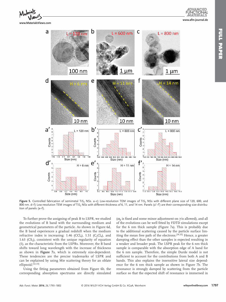
Varying reactant concentration, nucleation rates and growth rates, the structural parameters, H and L , can be well con-trolled. For examples, both L and H will decrease with the increase of reactant concentration, while only L deceases when prolonging nucleation time. Therefore, a series of TiS 2 NSs with different H and L can be obtained as shown in Figure 5 , and their detailed correspondences of H and L are presented in Figure S3 in the Supporting Information. Meanwhile, the crys-tallinity of obtained TiS 2 NSs with short reaction time for 1 h is poor than that with longer time, as observed from the XRD pattern in Figure 4 c, and the TEM of inset can also confi rm its amorphous edge of TiS 2 NS obtained with short time. Subse-quently, the capability of accurate shape and size control allows us to do the subsequent study of the geometrical dependence of LSPRs.
2.3. Characteristics of LSPRs from TiS 2 Nanosheets
The UV–vis–NIR absorption spectra of semimetal TiS 2 NSs are studied in Figure 6 . The distinct spectra difference of bulk TiS 2 from its nanostructured counterpoints can be observed from Figure 6 a. The absorption of bulk TiS 2 or poorly crystalline TiS 2 NSs only shows a sharp peak at 570 nm, marked as “A” band, which comes from the interband transitions from chalcogen p states to Ti 3d states according to our DFT calculations and previous reports [ 29–31 ] However, for the single-crystal TiS 2 NSs, a new absorption peak at 1380 nm emerges, marked as “B”, which has never been observed before. Meanwhile, B band is so strong that it distorts A band signifi cantly and results in a shoulder over the whole range of ≈600–1600 nm, perfectly covering the optical windows for both biological and communication applications.
Such newly observed B band can be attributed to LSPR of semimetal TiS 2 NSs according to the following facts. First, the measured B band can be well fi tted by the simulated spectrum using FDTD calculations as shown in Figure 6 a, and the detailed fi tting process is described as follows. In the simulation, the permittivity of TiS 2 is described by equation ( 1) by using the free electron Drude model, and the simula-tions are restricted to be starting from 1200 nm to avoid large disturbance from the interband transition
( ) 1
( )p2
iε ω
ωω ω γ
= −+
(2)
To simulate the full spectrum (from 400 to 2400 nm), addi-tional terms correspond to Lorentz oscillators should be added to equation ( 1) to account for the interband transition. Since our main focus is to uncover the underlying physics of peak B, we would like to stick to the much simpler Durde model and only study the long wavelength band (>1200 nm) where the contribution of the interband transition is weak.
For the permittivity of the material described by equation ( 1) , both plasma frequency ω p and collision frequency γ are unknowns and are taken as adjustable parameters. Peak posi-tion is mainly determined by ω p and peak width is determined by γ . Since the nanosheets are oriented randomly in the solu-tion, we considered three different cases for each simulation, case I: k ⊥ n , E // n , case II: k ⊥ n , E ⊥ n , case III: k // n , E ⊥ n (Figure 6 b). k is the wave vector of the plane wave, E is the electric component of the electromagnetic wave, and n is the normal vector of the par-ticle surface. For each case, the extinction cross section is calcu-lated and denoted as σ I , σ II , σ III , respectively. Figure 6 b indicates that the peak only appears in case I where the E-fi eld is perpendic-ular to the particle surface. The extinction cross section to compare with experimental data is the average of these three different cases
( )/3I II IIIσ σ σ σ= + + (3)
By adjusting both ω p and γ , the averaged extinction cross section can be matched to the experimental data as shown in Figure 6 c, the peak profi le at the long wavelength side show excellent agreement with experimental result. It is a direct evi-dence that a simple Drude model is suffi cient to describe the optical properties of TiS 2 in this waveband and peak B can be attributed to LSPR.
Adv. Funct. Mater. 2016, 26, 1793–1802
www.afm-journal.dewww.MaterialsViews.com
Figure 4. X-ray diffraction (XRD) powder pattern for obtained TiS 2 NSs. a) Joint Committee on Powder Diffraction Standards powder pattern no. 15-0853 for hexagonal (P-3 mL) TiS 2 . b,c) XRD powder pattern of TiS 2 NSs with well and poor crystallinity, reacted for 3 and 1 h when prepared, respectively, insets show the corresponding TEM images. TiS 2 NSs with poor crystallinity are obtained when the reaction time is relatively short, refl ected in the amorphous edges, mainly corresponding to the lattice plane of (100) and (002).
To further prove the assigning of peak B to LSPR, we studied the evolutions of B band with the surrounding medium and geometrical parameters of the particle. As shown in Figure 6 d, the B band experiences a gradual redshift when the medium refractive index is increasing: 1.46 (CCl 4 ), 1.51 (C 2 Cl 4 ), and 1.63 (CS 2 ), consistent with the unique regularity of equation ( 1) , as the characteristic from the LSPRs. Moreover, the B band shifts toward long wavelength with the increase of thickness as shown in Figure 7 a, which is extremely size-dependent. These tendencies are the precise trademarks of LSPR and can be explained by using Mie scattering theory for an oblate ellipsoid. [ 32,33 ]
Using the fi tting parameters obtained from Figure 6 b, the corresponding absorption spectrums are directly simulated
( ω p is fi xed and some minor adjustment on γ is allowed), and all of the evolutions can be well fi tted by FDTD simulations except for the 6 nm thick sample (Figure 7 a). This is probably due to the additional scattering caused by the particle surface lim-iting the mean free path of the electrons. [ 34,35 ] Hence, a greater damping effect than the other samples is expected resulting in a weaker and broader peak. The LSPR peak for the 6 nm thick sample is comparable with the absorption edge of A band for the 6 nm sample. Therefore, the simple Durde model is not suffi cient to account for the contributions from both A and B bands. This also explains the insensitive lateral size depend-ence for the 6 nm thick sample as shown in Figure 7 b. The resonance is strongly damped by scattering from the particle surface so that the expected shift of resonance is immersed in
Adv. Funct. Mater. 2016, 26, 1793–1802
www.afm-journal.dewww.MaterialsViews.com
Figure 5. Controlled fabrication of semimetal TiS 2 NSs. a–c) Low-resolution TEM images of TiS 2 NSs with different plane size of 120, 600, and 800 nm. d–f) Low-resolution TEM images of TiS 2 NSs with different thickness of 6, 11, and 14 nm. Panels (a′–f′) are their corresponding size distribu-tion of panels (a–f).
the absorption edge of A band and the LSPR peak cannot be observed apparently.
2.4. Application of LSPRs on Photodetectors at Communication Window
Obviously, one of the merits of LSPR is the near-fi eld enhance-ment effect at the resonant wavelength, which has been widely applied to enhance the performances of bioimaging,
photocatalysis, photodetection, etc. [ 36–38 ] Here, the natural NIR LSPR of semimetal TiS 2 NSs offers great opportunities for the applications in both biological technology and optic communi-cation as demonstrated subsequently.
Figure 8 a presents the device confi guration of a NIR pho-todetector with PbS quantum dots (QDs) as photoresponsive medium and inserted TiS 2 NSs as plasmonic enhancement layer, which works at the 1310 nm communication wavelength. To spin coat a homogeneous TiS 2 layer loading PbS QDs, the TiS 2 NSs were composited with polymethyl methacrylate
Adv. Funct. Mater. 2016, 26, 1793–1802
www.afm-journal.dewww.MaterialsViews.com
Figure 7. Size dependence of LSPRs from TiS 2 NSs. a) Absorption spectra of TiS 2 NSs with different thickness and fi xed planar sizes of 800 nm. b) Absorption spectra of TiS 2 with different planar sizes and fi xed thickness of about 6 nm. Solid lines are experimental data and dotted lines are FDTD simulations.
Figure 6. Characteristics of the LSPRs from TiS 2 NSs. a) UV–vis–NIR absorption spectra of bulk and TiS 2 NSs. b) FDTD simulation extinction spectrum for three different cases. In case I, the E -vector is perpendicular to the surface of TiS 2 nanosheet. In Case II and III, the E -vector is parallel to the surface of TiS 2 nanosheet. c) Fitting of the simulating results with the experimental data. In this simulation, the size L = 300 nm, height H = 16 nm. Plasma frequency ω p = 1.56 × 10 15 rad s −1 , collision frequency γ = 7 × 10 14 rad s −1 . After ω p and γ are found for one particular simulation, ω p will be fi xed for the later simulation and only some minor adjustments of γ will be allowed to get better fi t. d) Absorption spectra of TiS 2 NSs in different surrounding media. The refractive indices of CCl 4 , C 2 Cl 4 , and CS 2 are 1.46, 1.51, and 1.63, respectively. Solid lines are experimental data and dotted lines are FDTD simulations.
(PMMA), but not weaken their inherent LSPRs characteristics too much (as presented in Figure S6 in the Supporting Informa-tion). Figure 8 b shows the absorption spectrum of the adopted QDs, exhibiting a characteristic excitonic peak at 1350 nm, the corresponding size of which is 4.6 nm showed in Figure S4 in the Supporting Information. The comparison clearly demon-strates the perfect matching between excitonic absorption and LSPR of TiS 2 NS, which ensures the strong near fi eld enhance-ment inside the photodetector devices. The near fi eld enhance-ment is clearly depicted by the FDTD simulation results in Figure 8 c, where the E-fi eld enhancement effect is very strong when excited by ≈1200–1500 nm light, while relatively weak under irradiation of 1800 nm light.
Such near fi eld enhancement will lead to the elevation of photo current as shown in Figure 9 a, which is a net cur-rent resulted from the illumination. The excitation source is a
1310 nm laser, and the powers are 2.5, 4.5, 6, 8, and 10 mW, respectively. In comparison with the simple device, the photo-currents of photodetectors with TiS 2 layer are enhanced to 2.3, 2.4, 2.4, 2.6, and 2.7 times, respectively. The irradiance-dependent responsivity is also about twice as high as that of the simple device without TiS 2 as shown in Figure 9 b. The near fi eld enhancement demonstrated here could have signifi cant poten-tials in NIR optical communication and imaging applications.
2.5. Application of LSPRs on Photothermal Therapy at Biological Window
Furthermore, besides the photodetection in communication wavelength, the near-fi eld amplitude enhancement effect due to the LSPRs of TiS 2 NSs can also be applied to photothermal
Figure 8. a) Schematics of photodetector confi guration with TiS 2 NSs as plasmonic enhancing layer. b) Comparison of absorption spectrum of PbS photoconductive layer and TiS 2 NSs. c) Corresponding E-fi eld distribution for a TiS 2 NS ( L = 300 nm, H = 16 nm) at the wavelength of 1380 and 1800 nm, respectively.
Figure 9. Plasmonic enhancement on photodetection. a) Current–time ( I – t ) curves of photodetectors with and without TiS 2 NSs layer under illumina-tion with different power (2.5, 4.5, 6, 8, 10 mW). b) Dependence of the responsivity on illumination power for photodetectors with and without 2D TiS 2 layer, respectively.
therapy at wavelength within the biological window. Though relative work was reported that TiS 2 NSs were successfully applied for in vivo photoacoustic imaging and photothermal, [ 39 ] but the previous work cannot clearly explain the mechanism of the photothermal effect. Hence, it is necessary for us to dem-onstrate this effect precisely resulting from the LSPRs of TiS 2 NSs via a new experimental system different from the previous one in this work. Figure 10 a shows the schematic diagram of photothermal therapy using TiS 2 NSs. Under a low-energy NIR illumination, intense absorption of photons by the TiS 2 NSs arising from LSPR will increase the temperature of sur-rounding cancer cells and inactivate them gradually without damaging the non-cancer cells.
In order to adapt for the biological aqueous environment, the hydrophobic TiS 2 NSs were assembled with F127 to form hydrophilic F127/TiS 2 composite micelles for the photothermal application. The photographs of F127/TiS 2 composite micelles water solution with different concentration shown in Figure 10 b and the F127/TiS 2 composite micelles exhibit wonderful hydro-philicity, which can be steadily dispersed in water but not in either chloroform or n -hexane (Figure S7, Supporting Informa-tion). Meanwhile, the average hydraulic size of nanomicelles is 190 nm (Figure 10 c), slightly larger than the average size of pure TiS 2 NSs, which is attributed to the extension of polyeth-ylene oxide (PEO) block in water. As shown in Figure 10 d, a negative zeta potential of −10 mV could further provide static repulsion to enhance the aqueous stability of as-prepared TiS 2 /F127 composite nanomicelles.
Figure 11 exhibits their corresponding characteristic photo-thermal temperature rise behavior with different concentration of F127/TiS 2 under 808 and 980 nm laser irradiation, and the irradiation power are 0.5 and 1 W with corresponding energy
density of 0.64 and 1.27 W cm −2 , respectively. The laser power and wavelength are selected to optimize the photothermal heating effect. Similar temperature rise is obtained both under the 808 and 980 nm, and the maximum temperature rise can be as high as 50 °C. More importantly, the strong temperature rise clearly demonstrates that the photothermal effect is from TiS 2 NSs due to their NIR LSPRs, successfully confi rming the mechanism of photothermal attributed to the LSPRs of TiS 2 NSs. Moreover, with the low toxicity of TiS 2 NSs to cells, [ 39 ] they have signifi cant potentials in photothermal therapy of cancer cells.
TiS 2 NS obtained in this work can successfully apply in NIR wavelength due to its strong localized surface plasmon reso-nances (LSPSs), it can effectively replace noble metal Au with the advantages of low cost and easy synthesis when applied for photothermal therapy. Meanwhile, it is a rare material with NIR LSPRs successfully used for enhancing the NIR photodetec-tion. However, when used for these related applications, more other improvements can be done, such as treating the surface of NSs to heighten their dispersibility and optimizing the TiS 2 fi lm to improve the properties of E-fi eld enhancement.
3. Conclusion
High-quality hexagonal TiS 2 NSs with high crystallinity and purity are successfully synthesized. Characterization of the material reveals a hitherto elusive absorption peak in the 1000–1400 nm range. This peak exhibits a characteristic bathochromic shift that increases with both the refractive index of the surrounding solvent and also with the thickness of the nanosheet. Experimental evidence and FDTD simulations
Figure 10. Surface modifi cation of TiS 2 NSs. a) Schematic diagram of the heating effects of TiS 2 NSs aiming at cancer cells in biological tissue. b) Photographs of F127/TiS 2 composite micelles dispersed in water with different concentration under the sunlight. c) The hydraulic size of TiS 2 /F127 nanomicelles. d) The zeta potential of TiS 2 NSs of TiS 2 /F127 nanomicelles.
attribute this peak to LSPRs, rendering the TiS 2 NSs synthe-sized in this work the fi rst semimetal with NIR LSPR proper-ties. By using different irradiation wavelengths applications of this effect are successfully demonstrated in technical and biological settings. Under 1310 nm illumination, photocur-rents recorded by PbS photodetectors coated with TiS 2 NSs are enhanced by more than 100% compared to uncoated devices. Irradiation under 980 nm, however, causes an extraordinary temperature rise of about 50 °C due to LSPRs, which can be applied to kill cancer cells. In summary TiS 2 NSs show great potentia by constituting the fi rst semimetal material amenable to general plasmonics applications in the NIR regime.
4. Experimental Section Synthesis of TiS 2 NSs : In a typical procedure, 0.1 mmol titanium
chloride (TiCl 4 , 99.99%, Aladdin-reagent) was mixed with 11.2 mmol oleylamine (J&K chemical) and 15 mL octadecene (>90%(GC), Aladdin-reagent) in a 100 mL four-neck fl ask with strong magnetic stirring, heated to a low-boiling temperature and excluding air and impurities with high-purity argon for several times. After removing impurities, the reaction mixture was heated to 220 °C for 30 min under Ar conditions. Subsequently, carbon disulfi de (CS 2 , AR, Aladdin-reagent) was injected into the mixture and reaction temperature was heated to 300 °C ultimately. After 3 h, the reaction was quenched and cooled down to room temperature quickly. By addition of 10 mL butanol into the black solutions, TiS 2 NSs were precipitated via centrifugation and redispersed in hexane, then the sample was washed twice with ethanol by centrifugation.
Synthesis of the PbS Colloidal Quantum Dots (CQDs) : 0.9 g lead (II) oxide, 2.7 mL oleic acid, 1 mL oleylamine, and 15 mL 1-octadecene were added in a three-neck fl ask, evacuated for 10 min to remove air
and impurities with low boiling point, then heated to 160 °C keeping for 40 min. After 0.1 mL CS 2 was injected to the mixture and reacted for 5 s, the reaction was stopped and cooled down to the room temperature immediately. Products were precipitated by acetone and washed three times, then redispersed in toluene to prepare a 30 mg mL −1 solution ready for device fabrication.
Device Fabrication : First, TiS 2 /PMMA was prepared by TiS 2 NSs (100 mg) mixed with poly(methyl methacrylate) (3 g) in chloroform (10 mL) and stirred for 2 h. Then, the PbS fi lm was deposited by spin-coating the ready PbS CQDs toluene solution onto glass substrate with or without TiS 2 /PMMA fi lm covered at 2000 rpm for 30 s, respectively. The ligand exchange was then carried out by covering the former fi lm with three drops of 10% (v/v in methanol) mercaptopropionic acid (MPA), which was repeated two times every 5 s. Sequentially, the fi lm was rinsed three times with fi ve drops of methanol to remove the residual MPA. After the above sequence repeated another two times, the fi lm was heated at 90 °C for 10 min on the heating panel. Finally, the interdigitated Au electrodes were deposited on the fi lm by thermal evaporation utilizing a shadow mask.
Material Characterization : The optical extinction spectra of TiS 2 NS suspensions were measured by a Shimadzu UV-3600 UV–Vis–NIR spectrophotometer (in the 250–3300 nm spectral range). TEM images were taken on a TECNAI G2 20 LaB6 TEM instrument operated at an acceleration voltage of 200 kV. XRD patterns were acquired using a Bruker D8 Advance X-ray diffractometer operating with Cu Kα radiation ( λ = 1.5406 Å). Surface SEM images of the NSs were obtained on a Quant 250 FEG SEM instrument.
Photoelectronic Measurements : The measurement was carried out by a probe station connected to Keithley 4200-SCS semiconductor characterization system. Illumination was controlled at 1310 nm generated from laser diodes (Thorlabs SFL1550S).
Photothermal Measurements : The light source was provided by 808 and 980 nm wavelength semiconductor laser device (Beijing STONE Laser Co. LTD, P. R. of China) whose power could be adjusted externally (≈0–8 W). The temperature was recorded by an online type thermocouple thermometer (XMT 8008 Yuyao Tenghui Thermostat Factory, China) every 1 min.
Figure 11. Plasmonic photothermal effect. Temperature evolution curve of F127/TiS 2 composite micelles with varied concentration from 0.1, 0.2, 0.3, 0.4 to 0.5 mg mL −1 , under different laser irradiation in 30 min. The irradiation wavelength and power of the used laser are a) 808 nm, 0.5 W, 0.64 W cm −2 , b) 808 nm, 1 W,1.27 W cm −2 , c) 980 nm, 0.5 W, 0.64 W cm −2 , and d) 980 nm, 1 W, 1.27 W cm −2 .
Supporting Information Supporting Information is available from the Wiley Online Library or from the author.
Acknowledgements Z.Z., Y.Z., W.H., Y.L., Y.G., and B.C. contributed equally to this work. This work was fi nancially supported by the National Key Basic Research Program of China (Grant No. 2014CB931702), NSFC (Grant Nos. 51572128, 51173038, and 51303046), NSFC-RGC (Grant No. 5151101197), and the PAPD of Jiangsu Higher Education Institutions.
Received: November 14, 2015 Revised: December 2, 2015
Published online: January 22, 2016
[1] R. W. Wood , Proc. Phys. Soc., London 1902 , 18 , 269 . [2] J. A. Creighton , D. G. Eadon , J. Chem. Soc., Faraday Trans. 1991 , 87 ,
3881 . [3] P. K. Jain , X. H. Huang , I. H. El-Sayed , M. A. El-Sayed , Acc. Chem.
Res. 2008 , 41 , 1578 . [4] Y. K. Mishra , R. Adelung , G. Kumar , M. Elbahri , S. Mohapatra ,
R. Singhal , A. Tripathi , D. K. Avasthi , Plasmonics 2013 , 8 , 811 . [5] Y. K. Mishra , S. Mohapatra , R. Singhal , D. K. Avasthi , D. C. Agarwal ,
S. B. Ogale , Appl. Phys. Lett. 2008 , 92 , 043107 . [6] Z. T. Li , Z. N. Zhu , W. J. Liu , Y. L. Zhou , B. Han , Y. Gao , Z. Y. Tang ,
J. Am. Chem. Soc. 2012 , 134 , 3322 . [7] Y. T. Li , J. L. Tang , L. C. He , Y. Liu , Y. L. Liu , C. Y. Chen , Z. Y. Tang ,
Adv. Mater. 2015 , 27 , 4075 . [8] I. H. El-Sayed , X. H. Huang , M. A. El-Sayed , Nano Lett. 2005 , 5 , 829 . [9] X. H. Huang , I. H. El-Sayed , W. Qian , M. A. El-Sayed , J. Am. Chem.
Soc. 2006 , 128 , 2115 . [10] H. A. Atwater , A. Polman , Nat. Mater. 2010 , 9 , 205 . [11] X. Liu , L. C. He , J. Z. Zheng , J. Guo , F. Bi , X. Ma , K. Zhao , Y. L. Liu ,
R. Song , Z. Y. Tang , Adv. Mater. 2015 , 27 , 3273 . [12] S. D. Solomon , M. Bahadory , A. V. Jeyarajasingam , S. A. Rutkowsky ,
C. Boritz , L. Mulfi nger , J. Chem. Educ. 2007 , 84 , 322 . [13] S. Link , J. Phys. Chem. B 1999 , 103 , 4212 . [14] A. M. Smith , M. C. Mancini , S. M. Nie , Nat. Nanotechnol. 2009 , 4 ,
710 .
[15] E. H. Sargent , Adv. Mater. 2005 , 17 , 515 . [16] Z. N. Zhu , J. Guo , W. J. Liu , Z. T. Li , B. Han , W. Zhang , Z. Y. Tang ,
Angew. Chem., Int. Ed. 2013 , 52 , 13571 . [17] W. Hasan , C. L. Stender , M. H. Lee , C. L. Nehl , J. Lee , T. W. Odom ,
Nano Lett. 2009 , 9 , 1555 . [18] C. Loo , A. Lowery , N. J. Halas , J. West , R. Drezek , Nano Lett. 2005 ,
5 , 709 . [19] K. Manthiram , A. P. Alivisatos , J. Am. Chem. Soc. 2012 , 134 , 3995 . [20] M. Kanehara , H. Koike , T. Yoshinaga , T. Teranishi , J. Am. Chem. Soc.
2009 , 131 , 17736 . [21] J. A. Faucheaux , P. K. Jain , J. Phys. Chem. Lett. 2013 , 4 , 3024 . [22] I. Kriegel , C. Y. Jiang , J. Rodriguez-Fernandez , R. D. Schaller ,
D. V. Talapin , E. da Como , J. Feldmann , J. Am. Chem. Soc. 2012 , 134 , 1583 .
[23] T. R. Gordon , T. Paik , D. R. Klein , G. V. Naik , H. Caglayan , A. Boltasseva , C. B. Murray , Nano Lett. 2013 , 13 , 2857 .
[24] J. M. Luther , P. K. Jain , T. Ewers , A. P. Alivisatos , Nat. Mater. 2011 , 10 , 361 .
[25] E. Guilmeau , Y. Bré ard , A. Maignan , Appl. Phys. Lett. 2011 , 99 , 052107 .
[26] C. W. Lin , X. J. Zhu , J. Feng , C. Z. Wu , S. L. Hu , J. Peng , Y. Q. Guo , L. L. Peng , J. Y. Zhao , J. L. Huang , J. L. Yang , Y. Xie , J. Am. Chem. Soc. 2013 , 135 , 5144 .
[27] S. Jeong , D. Yoo , J. T. Jang , M. Kim , J. Cheon , J. Am. Chem. Soc. 2012 , 134 , 18233 .
[28] V. V. Plashnitsa , F. Vietmeyer , N. Petchsang , P. Tongying , T. H. Kosel , M. Kuno , J. Phys. Chem. Lett. 2012 , 3 , 1554 .
[29] F. R. Shepherd , P. M. Williams , J. Phys. C: Solid State Phys. 1974 , 7 , 4416 .
[30] S. C. Bayliss , W. Y. Liang , J. Phys. C: Solid State Phys. 1985 , 18 , 3327 . [31] A. H. Reshak , S. Auluck , Phys. Rev. B 2003 , 68 , 245113 . [32] G. Mie , Ann. Phys. 1908 , 25 , 337 . [33] K. L. Kelly , E. Coronado , L. L. Zhao , G. C. Schatz , J. Phys. Chem.
B 2003 , 107 , 668 . [34] S. Link , M. A. El-Sayed , Int. Rev. Phys. Chem. 2000 , 19 , 409 . [35] S. A. Maier , Plasmonics: Fundamentals and Applications , Springer
Science & Business Media , New York , 2007 . [36] B. Qin , H. Y. Chen , H. Liang , L. Fu , X. F. Liu , X. H. Qiu , S. Q. Liu ,
R. Song , Z. Y. Tang , J. Am. Chem. Soc. 2010 , 132 , 2886 . [37] P. K. Jain , K. Manthiram , J. H. Engel , S. L. White , J. A. Faucheaux ,
A. P. Alivisatos , Angew. Chem. Int. Ed. 2013 , 52 , 13671 . [38] L. B. Luo , L. H. Zeng , C. Xie , Y. Q. Yu , F. X. Liang , C. Y. Wu , L. Wang ,
J. G. Hu , Sci. Rep. 2014 , 4 , 3914 . [39] X. Qian , S. Shen , T. Liu , L. Cheng , Z. Liu , Nanoscale 2015 , 7 , 6380 .