Neck and Arm Pain and Related Symptoms: Cervical Disc Disease Discussion paper prepared for The Workplace Safety and Insurance Appeals Tribunal December 2002 Prepared by: Dr. J.F.R. Fleming Professor Emeritus, Division of Neurosurgery University of Toronto Division of Neurosurgery, Toronto Western Hospital University Health Network Revised February 2012 by: Dr. Joel Finkelstein, MD, MSc, FRCS(C) Associate Professor, Department of Surgery, University of Toronto Orthopaedic Surgeon, Sunnybrook Health Sciences Centre Dr. J.F. Ross Fleming graduated from the University of Toronto Medical School in 1947. He did post-graduate training in neurosurgery at the University of Toronto, at the University of Michigan and at Oxford, England, from 1947 to 1956. He became a Fellow in neurosurgery in 1956. He holds the rank of Professor Emeritus in the Division of Neurosurgery, Department of Surgery, at the University of Toronto. His clinical and research interests were in neurosurgery. He has published widely in that area. He practiced at the Toronto Western Hospital as the Head of the Division of Neurosurgery from 1965 to 1984 and as staff in the Division of Neurosurgery from 1956 to 1996. Dr. Fleming was involved at the Tribunal as an assessor from 1988 to 1992, as a counsellor from 1993 to 1997 and as Chair of the medical counsellors group from 1998 to 2006. Dr. Joel Finkelstein graduated from the University of Toronto Medical School in 1988. He did post-graduate training in Orthopaedic Surgery at the University of Toronto from 1989 to 1994. He has a subspecialty interest in adult spinal disorders and completed
Transcript
Neck and Arm Pain and Related Symptoms: Cervical Disc Disease
Discussion paper prepared for
The Workplace Safety and Insurance Appeals Tribunal
December 2002
Prepared by:
Dr. J.F.R. Fleming Professor Emeritus, Division of Neurosurgery
University of Toronto Division of Neurosurgery, Toronto Western Hospital
University Health Network
Revised February 2012 by:
Dr. Joel Finkelstein, MD, MSc, FRCS(C) Associate Professor, Department of Surgery, University of Toronto
Orthopaedic Surgeon, Sunnybrook Health Sciences Centre
Dr. J.F. Ross Fleming graduated from the University of Toronto Medical School in 1947. He did post-graduate training in neurosurgery at the University of Toronto, at the University of Michigan and at Oxford, England, from 1947 to 1956. He became a Fellow in neurosurgery in 1956. He holds the rank of Professor Emeritus in the Division of Neurosurgery, Department of Surgery, at the University of Toronto. His clinical and research interests were in neurosurgery. He has published widely in that area. He practiced at the Toronto Western Hospital as the Head of the Division of Neurosurgery from 1965 to 1984 and as staff in the Division of Neurosurgery from 1956 to 1996. Dr. Fleming was involved at the Tribunal as an assessor from 1988 to 1992, as a counsellor from 1993 to 1997 and as Chair of the medical counsellors group from 1998 to 2006.
Dr. Joel Finkelstein graduated from the University of Toronto Medical School in 1988. He did post-graduate training in Orthopaedic Surgery at the University of Toronto from 1989 to 1994. He has a subspecialty interest in adult spinal disorders and completed
Neck and Arm Pain and Related Symptoms: Cervical Disc Disease
fellowship training at Sunnybrook Health Science Center in Toronto and at Harborview Medical Center in Seattle Washington. He joined the faculty at the University of Toronto/Sunnybrook Health Science Center in 1996 and currently holds the rank of Associate Professor in the Department of Surgery (Orthopaedics). His clinical and research interests have been in spine trauma care and metastatic disease to the spine, focusing on clinical care and research into clinical outcome studies for those conditions. He is on the Active Staff in orthopaedic surgery at Sunnybrook Health Sciences Centre and currently serves as the spine section head for the Division of Orthopaedic Surgery.
This medical discussion paper will be useful to those seeking general information about the medical issue involved. It is intended to provide a broad and general overview of a medical topic that is frequently considered in Tribunal appeals.
Each medical discussion paper is written by a recognized expert in the field, who has been recommended by the Tribunal’s medical counsellors. Each author is asked to present a balanced view of the current medical knowledge on the topic. Discussion papers are not peer reviewed. They are written to be understood by lay individuals.
Discussion papers do not necessarily represent the views of the Tribunal. A vice-chair or panel may consider and rely on the medical information provided in the discussion paper, but the Tribunal is not bound by an opinion expressed in a discussion paper in any particular case. Every Tribunal decision must be based on the facts of the particular appeal. Tribunal adjudicators recognize that It is always open to the parties to an appeal to rely on or to distinguish a medical discussion paper, and to challenge it with alternative evidence: see Kamara v. Ontario (Workplace Safety and Insurance Appeals Tribunal) [2009] O.J. No. 2080 (Ont Div Court).
Neck and Arm Pain and Related Symptoms: Cervical Disc Disease
1
NECK AND ARM PAIN AND RELATED SYMPTOMS CERVICAL DISC DISEASE
Anatomical Considerations:
The cervical spine consists of 7 vertebrae however the upper cervical spine (Occiput to C2), is functionally different than the lower cervical spine, (C3 to C7). The upper cervical region is highly specialized to provide a large range of motion between the head and the torso. Approximately 50 percent of all motion occurs between the occiput and C2. Occiput to C1 is primarily for flexion/extension and between C1 and C2 for lateral rotation. The primary stabilizers in the upper cervical spine are the ligaments while stability in the lower cervical spine is more dependant on bony articulations involving the facet joints with their capsules posteriorly and the uncovertebral joints anteriorly on each side of the vertebral body.
The upper cervical spine has no discs while intervertebral discs are present in the lower cervical spine. These discs consist of an outer fibrous membrane called the “annulus fibrosis” and a semisolid core “nucleus pulposus” in its centre. The intervertebral disc is able to transmit compressive loads throughout a range of motion and prevent excessive stress concentration from occurring.
Neck and Arm Pain and Related Symptoms: Cervical Disc Disease
2
Figure 1: Front view of cervical spine
Neck and Arm Pain and Related Symptoms: Cervical Disc Disease
3
Figure 2: Side view of cervical vertebrae with side view cross section.
Neck and Arm Pain and Related Symptoms: Cervical Disc Disease
4
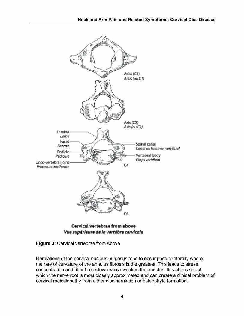
Figure 3: Cervical vertebrae from Above
Herniations of the cervical nucleus pulposus tend to occur posterolaterally where the rate of curvature of the annulus fibrosis is the greatest. This leads to stress concentration and fiber breakdown which weaken the annulus. It is at this site at which the nerve root is most closely approximated and can create a clinical problem of cervical radiculopathy from either disc herniation or osteophyte formation.
Neck and Arm Pain and Related Symptoms: Cervical Disc Disease
5
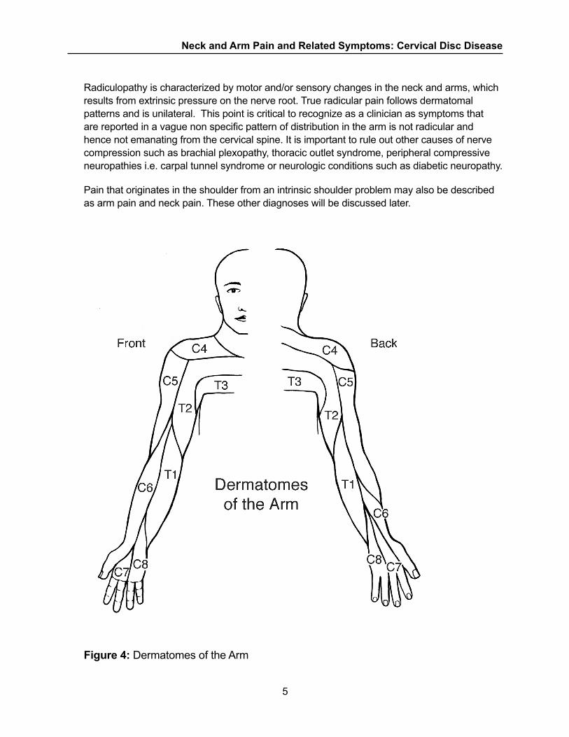
Radiculopathy is characterized by motor and/or sensory changes in the neck and arms, which results from extrinsic pressure on the nerve root. True radicular pain follows dermatomal patterns and is unilateral. This point is critical to recognize as a clinician as symptoms that are reported in a vague non specific pattern of distribution in the arm is not radicular and hence not emanating from the cervical spine. It is important to rule out other causes of nerve compression such as brachial plexopathy, thoracic outlet syndrome, peripheral compressive neuropathies i.e. carpal tunnel syndrome or neurologic conditions such as diabetic neuropathy.
Pain that originates in the shoulder from an intrinsic shoulder problem may also be described as arm pain and neck pain. These other diagnoses will be discussed later.
Figure 4: Dermatomes of the Arm
Neck and Arm Pain and Related Symptoms: Cervical Disc Disease
6
At each vertebral level a pair of spinal nerves (right and left “nerve roots”) exits from the spinal column through openings called foramina; these nerves supply sensation to the skin and power to the muscles of the arms and hands in defined patterns of innervation as depicted in Figure 4. The spinal cord itself carries the motor and sensory nerve pathways to the trunk and legs, including nerves that control bowel, bladder and sexual function. The spinal cord and nerve roots are enclosed in a tough membrane called the dura, inside of which is a flimsy membrane called the arachnoid, containing the clear colourless spinal fluid which bathes the spinal cord and nerves.
Large strong muscles run the length of the cervical spine, in front, beside and behind the vertebral column, maintaining and controlling head position and neck movement. Muscle bulk in the back of the spine is more massive than in the front. This is required due to the position of the head which is anterior to the center of gravity of the spinal column and thus requiring a larger lever arm to maintain the upright or neutral position of the head. The posterior musculature requires a constant degree of tonicity or contraction to counteract the effect of gravity on the head. Muscles which are weak, deconditioned or injured can fatigue more easily and be a source of pain. This is generally self limited pain which responds to rest and strengthening however barriers to recovery are not uncommon, which relate to misdiagnosis, fear perception of significant injury, over interpretation of incidental radiographic findings and non organic variables that can influence pain and recovery. It is incumbent upon the physician to rule out pathological processes and correctly identify anatomically inconsequential findings in order to minimize development of pain behavioiurs that can occur from soft tissue injuries of the neck, such as whiplash.
Degenerative or Aging Changes
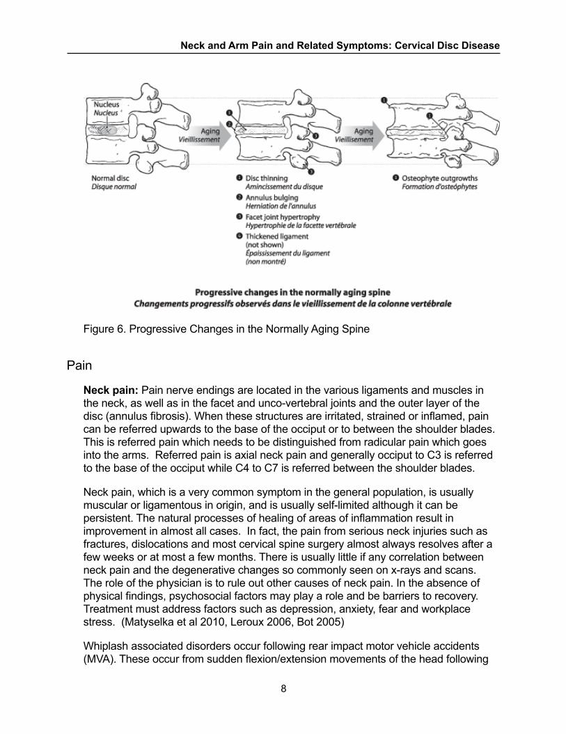
Progressive degenerative changes (aging changes) occur in the cervical spine of all adults. The nucleus portion of the discs gradually dries out and becomes thinner, allowing the adjacent vertebrae to become closer together. As a result, the annulus portion of the discs tends to “bulge”. Because the vertebral bodies come to lie closer together, there is increased wear and tear on the joints of the vertebral column, especially the unco-vertebral joints, the facet joints and disc margins, resulting in the gradual formation of bony overgrowths (“spurs”, “osteophytes”, “osteoarthritis”, “bone hypertrophy” - all synonyms in this context) at the disc margins, at the unco-vertebral joints and at the facet joints. This process is the normal aging process, and it begins in middle life. It is sometimes called “spondylosis”, and is present to a greater or lesser degree in all adults. The vast majority of individuals have these aging changes, even though the changes are quite advanced, are free of pain or any other symptoms. Various aging or degenerative changes such as bulging, degenerated or protruding discs, bony spurs or overgrowths, and facet joint hypertrophy are seen in X-rays, CT scans or MR scans of the cervical spine in over half the adult population. (Okada et al. 2009)
Neck and Arm Pain and Related Symptoms: Cervical Disc Disease
7
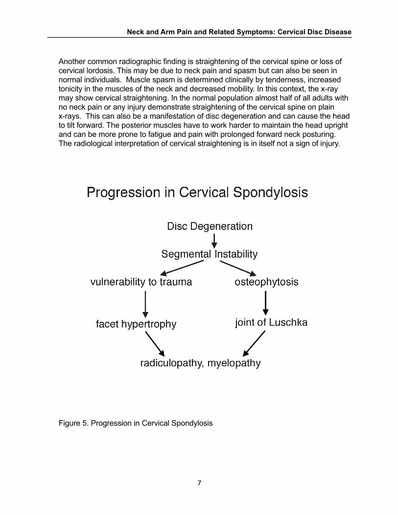
Another common radiographic finding is straightening of the cervical spine or loss of cervical lordosis. This may be due to neck pain and spasm but can also be seen in normal individuals. Muscle spasm is determined clinically by tenderness, increased tonicity in the muscles of the neck and decreased mobility. In this context, the x-ray may show cervical straightening. In the normal population almost half of all adults with no neck pain or any injury demonstrate straightening of the cervical spine on plain x-rays. This can also be a manifestation of disc degeneration and can cause the head to tilt forward. The posterior muscles have to work harder to maintain the head upright and can be more prone to fatigue and pain with prolonged forward neck posturing. The radiological interpretation of cervical straightening is in itself not a sign of injury.
Figure 5. Progression in Cervical Spondylosis
Neck and Arm Pain and Related Symptoms: Cervical Disc Disease
8
Figure 6. Progressive Changes in the Normally Aging Spine
Pain
Neck pain: Pain nerve endings are located in the various ligaments and muscles in the neck, as well as in the facet and unco-vertebral joints and the outer layer of the disc (annulus fibrosis). When these structures are irritated, strained or inflamed, pain can be referred upwards to the base of the occiput or to between the shoulder blades. This is referred pain which needs to be distinguished from radicular pain which goes into the arms. Referred pain is axial neck pain and generally occiput to C3 is referred to the base of the occiput while C4 to C7 is referred between the shoulder blades.
Neck pain, which is a very common symptom in the general population, is usually muscular or ligamentous in origin, and is usually self-limited although it can be persistent. The natural processes of healing of areas of inflammation result in improvement in almost all cases. In fact, the pain from serious neck injuries such as fractures, dislocations and most cervical spine surgery almost always resolves after a few weeks or at most a few months. There is usually little if any correlation between neck pain and the degenerative changes so commonly seen on x-rays and scans. The role of the physician is to rule out other causes of neck pain. In the absence of physical findings, psychosocial factors may play a role and be barriers to recovery. Treatment must address factors such as depression, anxiety, fear and workplace stress. (Matyselka et al 2010, Leroux 2006, Bot 2005)
Whiplash associated disorders occur following rear impact motor vehicle accidents (MVA). These occur from sudden flexion/extension movements of the head following
Neck and Arm Pain and Related Symptoms: Cervical Disc Disease
9
impact leading to soft tissue inflammation of the cervical spine. In the absence of any bony injury or consistent nerve compression, the natural history of this is similar to any soft tissue irritation or inflammation as described above with the normal healing time 8- 12 weeks. Malik et al 2004 found a preponderance of whiplash after relatively minor MVAs in comparison with patients with multiple trauma from high velocity MVAs with other injuries (ISS >16)* where only 2 /36 patients had neck pain at 8 weeks after accident. This suggests that other factors, such as psychosocial variables are important factors in determining the development of chronic neck pain.
Cervical nerve root pain. In the relatively uncommon situation in which a cervical nerve root is severely irritated or compressed, there is severe sharp pain radiating down the arm and into the forearm and hand. There may also be pain around the shoulder blades. Symptoms can be aggravated by neck movement or use of the arm.
A nerve root may be irritated or compressed by: (a) bone spurs or osteophytes growing into the exit foramen or canal through which the nerve travels, or (b) bulging of the part of the disc that lies in front of the nerve (the most lateral portion of the disc, not the central portion), or (c ) rupture or herniation of a piece of disc (nucleus pulposus) through the outer portion of the disc (annulus) into the nerve canal, or (d) fracture and/or dislocation injury causing bone fragments to narrow and/or impinge on the nerve canal (rare). In (a) (b) and (c ), a constant repair process is at work, and most symptoms subside over a period of time, usually a few weeks, almost regardless of treatment. Only a small percentage of patients with nerve root pain fail to recover, and require surgery.
There are a number of conditions with shoulder, arm and neck pain, weakness of arm and/or hand muscles, and/or numbness of the arm or hand, that must be differentiated from cervical disc and nerve root problems.
* ISS – Injury Severity Score is an anatomical scoring system that provides an overall score for patients with multiple injuries. Higher score indicates worse injury. (Baker et al. 1974)
Neck and Arm Pain and Related Symptoms: Cervical Disc Disease
10
Figure 7: Cross-sections of the cervical spine at the level of a herniated disc. Upper image shows a laterally located ruptured nucleus pulposus compressing the nerve root in its canal; bottom image shows a midline ruptured nucleus pulposus pressing on the spinal cord, but sparing the nerve root.
Neck and Arm Pain and Related Symptoms: Cervical Disc Disease
11
Figure 8: Cross-section of the cervical spine showing compression of the nerve root due to the narrowing of the nerve root canal by osteophyte outgrowths
Thoracic outlet syndrome (TOS) is a condition in which the nerves after exiting the spine through the foramina combine to form the brachial plexus and can become compressed dynamically by muscles, ligaments or an abnormal cervical rib on the C7vertebra causing arm and hand discomfort. Occasionally, symptoms can occur from poor posture. Exercise to elevate the shoulder girdle can relieve the symptoms to the hands.
Brachial Plexopathy can occur from a traction injury to the arm whereby the trunks or divisions of the brachial plexus are injured. This is differentiated from cervical nerve root radiculopathy as there is global weakness in the arm and shoulder girdle. Nerve conduction studies can help differentiate brachial plexus from cervical nerve root pathologies.
Peripheral nerves in the arm or hand may be entrapped or inflamed, giving rise to forearm and hand pain and numbness. Examples are entrapped median nerve at the wrist (carpal tunnel syndrome), entrapped ulnar nerve at the elbow (cubital tunnel syndrome), and peripheral neuropathy of these nerves due to diabetes.
Tumours or infections affecting the spinal column or the apex of the lung (Pancoast tumour), although rare, must also be considered in the differential diagnosis of a patient who complains of persisting neck or arm pain, weakness and/or numbness. The fact that a patient with these complaints may have had a neck injury does not
Neck and Arm Pain and Related Symptoms: Cervical Disc Disease
12
rule out the existence of a spinal tumour or infection as the cause of the symptoms. Therefore, all patients with persisting neck and arm pain, with or without weakness or numbness, require a thorough clinical history, physical examination and appropriate imaging.
Shoulder joint pain. Pain from a degenerated or injured shoulder joint often mimics and may be mistaken for nerve root pain, as the pain often spreads down to the top of the arm, but not below the elbow. Shoulder joint pain may inhibit the patient’s willingness to contract the arm or hand muscles strongly when these muscles are being tested for strength, thus leading to the erroneous conclusion that there is true muscle weakness caused by impairment of a nerve root.
The most common sources of shoulder pain are: 1) Rotator cuff tendonitis or tear; 2) osteoarthritis of the glenohumeral joint, and 3) Acromioclavicular joint (AC Joint) arthritis. Pain from a diseased or injured shoulder joint can be distinguished from nerve root pain by provocative testing: Rotator cuff tendonitis creates pain with active shoulder abduction above 30 degrees and impingement testing is positive. Osteoarthritis of the shoulder will have pain with active and passive movement of the shoulder usually in all planes; there may be crepitus and limited range of motion. AC joint arthritis will have tenderness directly over the joint and pain with forced adduction and rotation of the shoulder.
Headaches are a complaint that is frequently attributed to the cervical spine. It has been termed cervicogenic. They are a rare cause of chronic headache disorder.
The causes of headaches are numerous ranging from eye strain, sleep deprivation, migraine, elevated blood pressure and other causes. The C2 and C3 nerves: greater and lesser occipital nerves innervate the back of the occiput to behind the ears. Occipital neuralgia is a sharp stabbing pain caused by injury or pinching of the 2nd or 3rd cervical nerve roots. Ache or pain from sore neck muscles from C1 to C3 may be felt towards the back of the head or to between the shoulder blades when the irritation is from C4 to C7. Patients with cervicogenic headache will often have altered neck posture or restricted cervical range of motion. There are no neurologic findings of cervical radiculopathy with cervicogenic headache, though the patient might report scalp paresthesia or dysesthesia.
Diagnostic imaging such as radiography, magnetic resonance imaging (MRI), and computed tomography (CT) myelography are generally normal when there are complaints of cervicogenic headache. Diagnostic anesthetic blockade for the evaluation of cervicogenic headache is unreliable as the precise pain generator and the appropriate facet joint or osseous or muscular structure is unknown. Occipital neuralgia, mediated by C2 and 3 nerve roots, is an exception. Local injections may be helpful to alleviate this condition.
Neck and Arm Pain and Related Symptoms: Cervical Disc Disease
13
As noted above, muscle fatigue is a cause of sore neck muscles when the head is held in a static position for long periods of time. The posterior musculature is required to provide constant antigravity force to maintain the head posture optimally and can lead to pain in the neck and back of the head. When a person who has had a neck injury complains of headaches, other causes of the headaches must be sought before attributing the headache to injured or strained neck structures.
Spinal Canal Narrowing (Spinal Stenosis)
The spinal canal, through which the spinal cord travels, may become progressively narrow because degenerative or aging changes cause the discs and bony overgrowths to bulge into the spinal canal. If very severe spinal canal narrowing occurs, the spinal cord may be compressed, causing neurological symptoms. Abnormal functioning of the spinal cord is called “myelopathy”, and when it is due to aging changes or spondylosis, it is called “cervical spondylitic myelopathy”. Some individuals are born with an unusually narrow spinal canal (congenital spinal stenosis) which predisposes them to spinal cord compression as the normal aging changes progress. Cervical spondylitic myelopathy is usually a painless process, and the symptoms, which are caused by interference of spinal cord function, include numbness, weakness and awkwardness of the hands and stiffness (spasticity) of the legs with progressive difficulty walking (numb, clumsy hands and stiff legs). Due to chronic compression of the spinal cord there can be an abnormal signal within the spinal cord on MRI.
Injuries to the Neck
Injuries to the neck that are most commonly seen from workplace injuries are soft tissue in nature. Bony injuries with fractures or subluxations will be identified with x-ray and the severity of the injury will depend on the stability of the fracture. Most fractures are minor and have the same clinical consequence as a soft tissue injury. These fractures include spinous process, minor compression fractures and undisplaced lamina fractures. Osteophytes can also fracture from the endplate or anterior longitudinal ligament and again be of minimal clinical concern.
More severe fractures will lead to malalignment of the vertebrae, (subluxations) in the case of facet fractures. Fractures from higher energy forces to the head can lead to burst injuries and can be associated with spinal cord injury. In any neck injury, from a traumatic event, the neck should be immobilized and evaluated to ensure no severe injury is present. If there has been sufficient tearing or rupture of some of the ligaments that support the cervical vertebrae, instability at the injured level of the spinal column may result. Instability is detected when an abnormal amount of forward or backward movement, or slippage, of a vertebra in relation to its neighbour, is seen on “flexion-extension” x-rays of the neck taken with the head flexed forward and then with the head extended backward. Instability usually requires surgical treatment, although neck
Neck and Arm Pain and Related Symptoms: Cervical Disc Disease
14
immobilization by a special collar or a “halo-vest” for a few weeks or months may be sufficient to allow spontaneous healing. The usual outcome in such cases of instability, after treatment, is resolution of pain and other symptoms. A complete neurological examination is mandatory to ensure there is no spinal cord compromise. Specific fracture patterns are beyond the scope of this paper.
Soft tissue injuries can result from any sudden unexpected movement of the head which can wrench or strain structures such as muscles or ligaments in the cervical spinal column, and these injuries will normally heal within a few weeks. It is very rare for such an injury to cause rupture or herniation of an intervertebral disc, with compression of a nerve root causing nerve root pain.
Repetitive stress to the cervical spinal column may result from activities such as in carrying loads on the head (as in some societies), in football, or in high divers (such as in Acapulco). Although premature aging (degenerative) changes are seen in many of the spines of such individuals, pain or other symptoms are very uncommon. Repetitive stresses to the cervical spinal column also occur in individuals with neuro-degenerative disorders such as dystonia or torticollis, who suffer from repeated, sometimes quite violent, uncontrolled writhing and twisting movements of the neck, yet neck pain is remarkably uncommon in these individuals. Repetitive neck movements or prolonged awkward positioning of the neck in a workplace activity are usually well tolerated by most individuals, although they may be associated with muscular aches and pains.
Can a neck injury cause degenerative changes or premature aging in the cervical spine? This is certainly the case when there is a bony injury that involves the facet joint or endplate/disc junction. An injury to a disc or ligaments may be visible on MRI shortly after an injury, and will gradually heal. Localized bony overgrowth, hypertrophy and spurs at the site of injury may develop in a small percentage of individuals who have sustained a severe localized injury to the cervical spinal column; however, these “degenerative” localized bony changes take a long time (possibly a year or more) to develop. Thus, severe injury to ligaments and/or disc at a single vertebral level may result in delayed x-ray or scan evidence of localized degenerative changes at that level many months or years after the injury, however the acute injury is well noted at the time of insult. If such bony changes are seen soon after the injury, they must have been present before the injury and were not caused by the injury. Soft tissue injuries without any instability such as a whiplash associated disorder, (WAD) do not alter the natural aging progression of the cervical spine.
How are neck injuries diagnosed?
The task of the physician is to integrate the patient’s complaints and physical findings, together with appropriate imaging studies, into an accurate diagnosis. First and foremost is a history, noting the mechanism and forces of the injury, and the nature and location of the pain and other symptoms. This is followed by a physical examination, including palpation for tenderness in neck muscles, assessing the range
Neck and Arm Pain and Related Symptoms: Cervical Disc Disease
15
of neck movements, examination of the shoulders, chest and head, and a neurological examination of the arms and legs. Follow-up history taking and physical examination will record the progress and hopefully the resolution of symptoms in the weeks (or months) following the injury. Careful consideration of the reports of these early histories and examinations is probably the most important step when attempting to determine the underlying nature of the injury when evaluating an injured person a long time after the injury. For most soft tissue neck complaints, no imaging is necessary. In the absence of any traumatic event imaging will simply show incidental age commensurate changes. It is imperative to not over diagnose imaging results or place these into improper context. CT Axial imaging is not indicated in the absence of trauma or in the absence of any hard objective neurological findings as again the sensitivity of these tests for clinically insignificant findings increases and will result in false positive diagnoses.
Cervical spine x-rays are taken after a neck injury in order to rule out a fracture, dislocation, or instability. If the x-rays show degenerative changes right after the injury, then they were obviously present prior to the injury. Anterolisthesis (forward translation) of one vertebrae on the other is not uncommon and can be present normally up to 2 mm. Greater translations can occur with spondylosis and are not traumatic. X-rays taken with the neck in both flexion and extension will reveal whether instability is present.
CT scan of the cervical spine is valuable in assessing bone injury, such as fracture and/or dislocation. A herniated disc if present from a traumatic event can be detected and if the onset of symptoms is contemporaneous with the event and the dermatomal distribution is consistent with the imaging, then the disc can be considered acute. Bulging or herniated discs are commonly seen as incidental findings as are osteochondral bars and osteophytes. These “bone spurs” contribute to narrowing of the spinal canal and can create acquired spinal stenosis or cervical spondylitic myelopathy. Quite independent of acute or chronic neck pain a patient can have symptoms of radiculopathy or myelopathy on the basis of these findings and surgery may be required.
MR scanning (MRI) of the cervical spine is the best method of imaging the spinal cord and nerve roots, the intervertebral discs, and the ligaments. However it must be remembered that 50% of all adults have “abnormalities” in MR scans of the cervical spine. In the population over 40 years old, the frequency of these abnormalities was found by Boden et al to be as follows: bony spurs (70%), narrow discs (57%), degenerated discs (57%), herniated discs (13%), bulging discs (19%), and foraminal stenosis (48%). These findings have been confirmed by numerous other investigators. Therefore abnormal MRI findings can only be considered to be significant if a specific abnormality in the scan exactly matches the specific symptoms and signs of the patient. As an example, a patient complains of severe nerve root pain radiating all the way down the arm and forearm, numbness of the index and middle fingers, has a weak triceps muscle and absent triceps tendon reflex, with aggravation of the arm
Neck and Arm Pain and Related Symptoms: Cervical Disc Disease
16
pain during neck extension. In this case, the clinical diagnosis is clearly a 7th cervical nerve root compression. The only MRI abnormality that would be of significance in this case would be the finding of a herniated disc pressing on the 7th cervical nerve root as it is compressed by the C 6-7 intervertebral disc or as the nerve travels out its intervertebral foramen or canal. This same MRI finding would be of no significance in a patient whose only symptoms were vague diffuse neck pain. Thus MRI findings can only be of value when they are interpreted together with and in the light of the entire clinical picture, and exactly match the clinical findings.
Cervical myelography consists of neck x-rays taken after the injection of radio-opaque contrast material into the spinal fluid via a lumbar puncture, and is followed by post-myelogram CT scan of the cervical spine (myelo-CT). Myelography is used rarely today, as MRI has become more readily available. MRI provides superior images of the spinal cord, nerve roots and discs.
Cervical discography. X-rays taken after the injection of radio-opaque contrast material into one or more discs, through a needle inserted through the front of the neck, is of little if any value, and has been largely discontinued. Discs are best imaged by MRI. The finding of an abnormal disc on discography is of little clinical significance.
Electrodiagnostic studies (E.M.G. and nerve conduction velocities) are useful in evaluating weakness of hand and arm muscles, and can determine whether the weakness is due to abnormality or compression of a cervical nerve root, or to some other peripheral cause. EMG is useful in ruling out some of the other possible causes of numbness/weakness of the arm or hand, such as ulnar or median nerve entrapment at the elbow or wrist (cubital or carpal tunnel syndrome).
Issues:
1. How is a neck injury diagnosed?
A neck injury is diagnosed based on a history which will include an identification of the mechanism of injury. This is generally a traumatic event such as a fall or sudden acceleration/deceleration force as occurs in a motor vehicle accident. An examination of the patient will look for findings of muscle tenderness and possibly spasm. Most injuries are soft tissue and imaging is of no value, except to rule out a bony injury if there is a suspicion of this. Imaging needs to be evaluated in the context of incidental degenerative changes in the neck and not be over interpreted as injury findings. Prolonged static postures such as maintaining the head in a forward flexed position can lead to muscle strain which is not traumatic. It is self limiting in the vast majority of individuals. Over treatment or non efficacious treatment and immobilization can create barriers to recovery.
Neck and Arm Pain and Related Symptoms: Cervical Disc Disease
17
2. Are there any particular ergonomic risk factors, e.g. awkward positioning, repetitiveness or particular force in work activities that would create an increased risk (i.e. be likely to be compatible with) gradual onset or other neck injuries? Can you please explain when and how spontaneous neck pain occurs that is not related to an injury, and that arises in the absence of an external injuring process?
Deconditioned muscles and muscle fatigue can lead to muscular pain with prolonged static postures such as maintaining the head in a forward flexed position or with repetitive movement. A specific injury mechanism may not be present and there is no specific job duty that can be implicated. This is in contradistinction to other joints such as the shoulder where repetitive overhead work is a risk factor for rotator cuff tendonitis. The posterior cervical musculature requires a constant degree of tonicity or contraction to counteract the effect of gravity on the head. Muscles which are weak, deconditioned or injured can fatigue more easily and be a source of pain. This is generally self limited pain which responds to rest and strengthening however barriers to recovery are not uncommon which can be due to misdiagnosis, fear perception of significant injury, over interpretation of incidental radiographic findings and non organic variables that can influence pain and recovery. Ergonomically optimal head positions are ideal to prevent muscle fatigue. Cervical strain is generally not disabling and resolves with short periods of rest.
3. What is the relationship, if any, between spondylosis (DDD) or other neck conditions and repetitive activities?
Spondylosis can be a cause of neck pain with repetitive activities. The worker with spondylosis of the cervical spine may commonly experience pain symptoms during neck extension beyond what is ‘normal’ for him/her due to joint stiffness as in conditions such as advanced facet arthritis. – this is normal and not an injury”. It is most probable that the underlying DDD is just becoming symptomatic in the natural course
4. What type of frequency of activity would be considered repetitive in this context?
A degenerative joint can be painful with one neck rotation. Repetitive movements of a muscle can lead to fatigue pain which would have a variable frequency before fatigue depending on the intrinsic condition of the muscle. A sudden onset pain from a force such as overextension or flexion can lead to strain and short term inflammation of the muscle. This is self limiting and a short period of avoidance is indicated. Return to normal activities in 48-72 hours is the best treatment option as prolonged immobility leads to further deconditioning and potential for injury and pain.
Neck and Arm Pain and Related Symptoms: Cervical Disc Disease
18
5. What is the relationship, if any, between any particular neck condition and any other specific mechanisms of injury?
Spondylosis, particularly degenerative facet changes can predispose to limitation in extension of the neck. Forced extension such as overhead work will cause pain.
6. Can there be a delay in the onset of symptoms after the neck injury? If so, to what extent?
Muscle fatigue may come on in a delayed fashion by a few hours to a day. This is a strained muscle and is generally self limiting. This would be expected to resolve with short period of rest, anti inflammatory medication and muscle conditioning exercises.
7. Can neck pain radiate to the shoulder, arm or hand? Under what circumstances would that occur? Can hand, arm or shoulder pain radiate to the neck? Are there ways to distinguish when the pain is due to a neck injury as distinct from another condition?
Neck pain can radiate to the occiput and to the interscapular regions. The muscles that envelope the neck are contiguous with the shoulder. Muscular pain from the neck can be felt over the shoulder and these two areas may be difficult to distinguish. Specific tests for internal shoulder joint pathology will help identify the shoulder as a cause of pain. Pain from a degenerated or injured shoulder joint often mimics and may be mistaken for nerve root pain, as the pain often spreads down to the top of the arm, but not below the elbow. Shoulder joint pain may inhibit the patient’s willingness to contract the arm or hand muscles strongly when these muscles are being tested for strength, thus leading to the erroneous conclusion that there is true muscle weakness caused by impairment of a nerve root.
The most common sources of shoulder pain are: 1) Rotator cuff tendonitis or tear; 2) osteoarthritis of the glenohumeral joint, and 3) Acromioclavicular joint (AC Joint) arthritis. Pain from a diseased or injured shoulder joint can be distinguished from nerve root pain by provocative testing: Rotator cuff tendonitis creates pain with active shoulder abduction above 30 degrees and impingement testing is positive. Osteoarthritis of the shoulder will have pain with active and passive movement of the shoulder usually in all planes, there may be crepitus and limited range of motion. AC joint arthritis will have tenderness directly over the joint and pain with forced adduction and rotation of the shoulder.
Pain that is neuropathic is caused by a nerve root. This can be due to a disc herniation or chronic compression. This will radiate to a specific dermatome in the arm. Mechanical neck pain does not radiate below the shoulder.
Neck and Arm Pain and Related Symptoms: Cervical Disc Disease
19
8. What is the relationship, if any, between any particular neck condition and cervicogenic headaches? What is a cervicogenic headache? What are its causes and clinical significance, if any? How does one distinguish between cervicogenic headaches and primary headache disorders?
Ache or pain from sore neck muscles from C1 to C3 may be felt towards the back of the head or if the irritated structure is from C4 to C7 will be felt to between the shoulder blades. Patients with cervicogenic headache will often have altered neck posture or restricted cervical range of motion. There are no neurologic findings of cervical radiculopathy, though the patient might report scalp paresthesia or dysesthesia.
Diagnostic imaging such as radiography, magnetic resonance imaging (MRI), and computed tomography (CT) myelography are generally normal. Diagnostic anesthetic blockade for the evaluation of cervicogenic headache is unreliable as the precise pain generator and the appropriate facet joint or osseous or muscular structure is unknown. Occipital neuralgia, mediated by C2 and 3 nerve roots, is an exception. Local injections may be helpful to alleviate this condition.
Muscle fatigue is a cause of sore neck muscles when the head is held in a static position for long periods of time. The posterior musculature is required to provide constant antigravity force to maintain the head posture optimally and can lead to pain in the neck and back of the head. When a person who has had a neck injury complains of headaches, other causes of the headaches must be sought before attributing the headache to injured or strained neck structures.
9. Are there other conditions/diseases that would predispose someone to a neck injury?
Disorders such as ankylosing spondylitis can predispose somebody to fracture of the vertebrae as the spine is very rigid. Minimal trauma can cause this.
10. If someone has sustained a neck injury, are there any particular restrictions in work activities that would be appropriate?
In the absence of any fracture there are no restrictions. Soft tissue pain can be a cause of tolerance limitations. Certain activities may be an initiator of pain and these should be avoided for a short finite period of time. If a specific risk factor such as extreme loss of neck extension exists due to advanced spondylosis, then overhead work should be eliminated.
Neck and Arm Pain and Related Symptoms: Cervical Disc Disease
20
References:
Baker SP et al; “The Injury Severity Score: a method for describing patients with multiple injuries and evaluating emergency care”, J Trauma 14:187-196;1974
Boden SD, McCowin PR, Davis DO et al: Abnormal cervical spine MR scans in asymptomatic individuals: a prospective and blinded investigation, Journal of Bone Joint Surgery 72A: 1178-1184, 1990
Bot SD, van der Waal JM, Terwee CB, et al. Predictors of outcome in neck and shoulder symptoms: a coort study in general practice. Spine 2005;30(16): E459-70.
Leroux I, Brisson C, Monteuil S. Job strain and neck shoulder symptoms: a prevalence study of women and men white collar workers. Occup Med (Lond) 2006;56(2): 102-9.
Malik H, Lovell M. Soft tissue neck symptoms following high energy road traffic accidents. Spine 2004; 29(15): E315-7.
Matyselka P, Lupsakko T, Kautianinen H, Vanhala M. Neck-shoulder pain and depressive symptoms: a cohort study with a 7 year follow-up. Eur J Pain 2010 14 (2): 189-93.
Okada E, Matsumoto, M, Ichihara D et al. Aging of the cervical spine in healthy volunteers. A 10 year longitudinal magnetic resonance imaging study. Spine 2009; 34(7): 706-12.