Thorax (1970), 25, 691. Necrotizing pulmonary aspergillosis W. P. U. KENNEDY, D. N. MALONE, and W. BLYTH Respiratory Diseases Unit, Northern General Hospital, Edinburgh, and Departments of Respiratory Diseases, Medicine, and Botany, University of Edinburgh Necrotizing or invasive aspergillary infection of the lungs has previously been considered a rare condition affecting only the debilitated or seriously ill. Four patients with necrotizing pulmonary aspergillosis are described, and the mycological and histopathological findings are discussed. The diagnosis in the first patient was not made until necropsy, following death from fulminating pulmonary infection. Of three patients treated with natamycin, two made a satisfactory recovery ; the other died later from bronchial carcinoma. Previous multiple antibiotic therapy in each case may have been a contributory factor to infection with Aspergillus fumigatus. We suggest that fungal infection of the lungs should be actively sought in patients with pulmonary disease, especially in those receiving multiple antibiotic or corticosteroid therapy. The most important fungal disease of the lungs in the British Isles at the present time is broncho- pulmonary aspergillosis, a condition which is becoming more commonly recognized in a variety of clinical situations. Classifications have been suggested by various authors, such as Gowing and Hamlin (1960) and Campbell and Clayton (1964); here we propose the following simple terminology: 1. Allergic asthma 2. Pulmonary eosinophilia 3. Extrinsic allergic alveolitis 4. Intercavitary aspergilloma 5. Necrotizing pulmonary aspergillosis 6. Disseminated aspergillosis. This paper describes four adult male patients in whom bronchopulmonary aspergillosis was accom- panied by necrosis of lung tissue. CASE REPORTS PATIENT 1 A retired shipping manager aged 74 had been ill for four weeks before admission to hospital with a productive cough, increasing exertional dyspnoea, three episodes of right pleural pain, and a chest radiograph which showed only slight shadowing in the right mid zone. During the first four weeks in hospital, repeated culture of sputum yielded only upper respiratory tract organisms except on one occa- sion when a moderate growth of a coliform bacillus was obtained together with a scanty growth of Aspergillus fumigatus. Despite treatment with ampi- cillin, he became steadily worse. The chest radiograph showed progressive bronchopneumonic changes in both lungs, and eventual cavitation in both upper lobes (Fig. 1). A diagnosis of suppurative pneumonia was made, and physiotherapy and treatment with cephaloridine produced a slight improvement in his general condition. About 36 hours before death Pseudomonas aeruginosa or pyocyanea was isolated from the sputum, and antibiotic treatment was altered to gentamycin and erythromycin lactobionate. This did not prevent further deterioration and he died 47 days after admission to hospital. Subsequent myco- logical examination of the sputum specimens showed numerous colonies of A. fumigatus. A specimen of serum obtained after death was examined for pre- cipitating antibodies using extracts of A. fumigatus; multiple precipitin lines to all the antigen used were demonstrated. Necropsy findings The upper lobes of both lungs were lightly adherent to the chest wall by recently formed adhesions and there was a small amount of turbid fluid in both pleural cavities. Both upper lobes were occupied by multiloculated abscess cavities con- taining shreds of necrotic lung tissue; the abscess walls were lined in some places by a stringy whitish membrane (Fig. 2). Patches of suppurative broncho- pneumonia were present in the lower lobes. The trachea and major bronchi contained copious amounts of pus. Microscopical examination of the membrane lining parts of the larger abscesses revealed a mat of tangled hyphae, which were thick, imperfectly septate, and branched repeatedly in an arborescent pattern. The fungal layer also contained enormous numbers of Gram-negative rods. Other fungal masses, composed of rounded collections of radially orientated hyphae, lay in the pus within bronchioles and in the patches of suppurative bronchopneumonia. The apical situation of the large abscesses in each lung raised the possibility of colonization of pre- existing bronchiectatic spaces, but the necrosis of lung tissue was so extensive that it obscured any evidence 691 copyright. on December 26, 2019 by guest. Protected by http://thorax.bmj.com/ Thorax: first published as 10.1136/thx.25.6.691 on 1 November 1970. Downloaded from
Transcript
Thorax (1970), 25, 691.
Necrotizing pulmonary aspergillosisW. P. U. KENNEDY, D. N. MALONE, and W. BLYTH
Respiratory Diseases Unit, Northern General Hospital, Edinburgh, and Departments ofRespiratory Diseases, Medicine, and Botany, University of Edinburgh
Necrotizing or invasive aspergillary infection of the lungs has previously been considered a rare
condition affecting only the debilitated or seriously ill. Four patients with necrotizing pulmonaryaspergillosis are described, and the mycological and histopathological findings are discussed. Thediagnosis in the first patient was not made until necropsy, following death from fulminatingpulmonary infection. Of three patients treated with natamycin, two made a satisfactory recovery ;
the other died later from bronchial carcinoma. Previous multiple antibiotic therapy in each case
may have been a contributory factor to infection with Aspergillus fumigatus. We suggest thatfungal infection of the lungs should be actively sought in patients with pulmonary disease,especially in those receiving multiple antibiotic or corticosteroid therapy.
The most important fungal disease of the lungs inthe British Isles at the present time is broncho-pulmonary aspergillosis, a condition which isbecoming more commonly recognized in a varietyof clinical situations. Classifications have beensuggested by various authors, such as Gowing andHamlin (1960) and Campbell and Clayton (1964);here we propose the following simple terminology:
1. Allergic asthma2. Pulmonary eosinophilia3. Extrinsic allergic alveolitis4. Intercavitary aspergilloma5. Necrotizing pulmonary aspergillosis6. Disseminated aspergillosis.This paper describes four adult male patients in
whom bronchopulmonary aspergillosis was accom-panied by necrosis of lung tissue.
CASE REPORTS
PATIENT 1 A retired shipping manager aged 74 hadbeen ill for four weeks before admission to hospitalwith a productive cough, increasing exertionaldyspnoea, three episodes of right pleural pain, and a
chest radiograph which showed only slight shadowingin the right mid zone. During the first four weeks inhospital, repeated culture of sputum yielded onlyupper respiratory tract organisms except on one occa-sion when a moderate growth of a coliform bacilluswas obtained together with a scanty growth ofAspergillus fumigatus. Despite treatment with ampi-cillin, he became steadily worse. The chest radiographshowed progressive bronchopneumonic changes inboth lungs, and eventual cavitation in both upperlobes (Fig. 1). A diagnosis of suppurative pneumoniawas made, and physiotherapy and treatment with
cephaloridine produced a slight improvement in hisgeneral condition. About 36 hours before deathPseudomonas aeruginosa or pyocyanea was isolatedfrom the sputum, and antibiotic treatment was alteredto gentamycin and erythromycin lactobionate. Thisdid not prevent further deterioration and he died 47days after admission to hospital. Subsequent myco-logical examination of the sputum specimens showednumerous colonies of A. fumigatus. A specimen ofserum obtained after death was examined for pre-cipitating antibodies using extracts of A. fumigatus;multiple precipitin lines to all the antigen used weredemonstrated.
Necropsy findings The upper lobes of both lungswere lightly adherent to the chest wall by recentlyformed adhesions and there was a small amount ofturbid fluid in both pleural cavities. Both upper lobeswere occupied by multiloculated abscess cavities con-taining shreds of necrotic lung tissue; the abscesswalls were lined in some places by a stringy whitishmembrane (Fig. 2). Patches of suppurative broncho-pneumonia were present in the lower lobes. Thetrachea and major bronchi contained copious amountsof pus.
Microscopical examination of the membrane liningparts of the larger abscesses revealed a mat of tangledhyphae, which were thick, imperfectly septate, andbranched repeatedly in an arborescent pattern. Thefungal layer also contained enormous numbers ofGram-negative rods. Other fungal masses, composedof rounded collections of radially orientated hyphae,lay in the pus within bronchioles and in the patchesof suppurative bronchopneumonia.The apical situation of the large abscesses in each
lung raised the possibility of colonization of pre-existing bronchiectatic spaces, but the necrosis of lungtissue was so extensive that it obscured any evidence
691
copyright. on D
ecember 26, 2019 by guest. P
rotected byhttp://thorax.bm
j.com/
Thorax: first published as 10.1136/thx.25.6.691 on 1 N
FIG. 1. Patient 1. Chest radiograph shortly before death showingextensive cavitation in both upper lobes.
FIG. 2. Patient 1. Lateral view of right lung taken atnecropsy. Extensive abscess cavity in upper lobe, linedIn places by a whitish membrane offungus.
of previous lung damage which might have predis-posed to fungal infection. No evidence of fungal orbacterial infection was found in any organ outside therespiratory system.
PATIENT 2 A 59-year-old baker had a long history ofchronic bronchitis. During the four months precedinghis acute illness he had received one short course ofoxytetracycline for seven days, and two short coursesof prednisolone. He was admitted to hospital withacute dyspnoea and a pyrexia of 38.6° C., but at firstthe chest radiograph showed only an old calcifiedtuberculous lesion in the right upper lobe. The sputum,however, was purulent and yielded on culture a puregrowth of Streptococcus pneumoniae. He was treatedwith ampicillin for five days. There was temporaryimprovement, but on the ninth day he was ill onceagain, with fever and oral thrush. A pneumonia haddeveloped in the right upper lobe (Fig. 3), and he wasexpectorating purulent sputum, from which A. fumi-gatus was isolated. A diagnosis of suppurative ornecrotizing fungal pneumonia, perhaps secondary tobacterial infection, was made. The detection in theserum of precipitating antibodies against A. fumigatusprovided supporting evidence for the diagnosis.An aerosol suspension of natamycin in a dose of
2 5 mg. six-hourly was administered by intermittentpositive pressure with a Bennet respirator and con-tinued for 38 days. By the tenth day of treatment histemperature was normal, and his clinical conditionhad improved. The sputum remained purulent, andthe upper lobe of the right lung became cavitated
Amw
copyright. on D
ecember 26, 2019 by guest. P
rotected byhttp://thorax.bm
j.com/
Thorax: first published as 10.1136/thx.25.6.691 on 1 N
(Fig. 4). By the time that natamycin was discontinuedhe was feeling well, although the sputum still yieldedA. fumigatus on culture. He was discharged homewithout further antibacterial or antifungal treatment.When seen 10 weeks later his cough had lessened butthe sputum was still purulent and A. fumigatus wasobtained on culture. Serum precipitins were stillpresent. Four months after leaving hospital he waswell, with only slight cough, and the sputum was nowmucoid. The cavitated lesion in the right lung apexhad healed by fibrosis, leaving only a small residualopacity visible in the chest radiograph.
PATIENT 3 A hotel porter aged 48 had suffered fromchronic osteomyelitis of the left femur from the ageof 13 years. He also had a history of pulmonarytuberculosis with persistent cavitation for which theposterior segment of the right upper lobe had beenresected at the age of 36 years. For 10 months beforethe present illness he had been receiving lincomycinfollowing an episode of acute osteomyelitis associatedwith septicaemia and subacute bacterial endocarditis.He had now developed a fever, cough productive ofcopper-coloured sputum, and bilateral pleural pain.
He failed to improve despite treatment with ampicillinfor 10 days, and then for five days with trimethoprimand sulphamethoxazole, and he developed severeglossitis. Two days before admission, the left legbecame swollen and tender.He looked thin and ill on admission and was
dyspnoeic at rest. The left leg was swollen and dis-coloured and there was marked calf tenderness.Scattered coarse crepitations were audible in the chest,and the radiograph showed widespread inflammatoryshadowing throughout the right lower lobe, which wasslightly contracted. The sputum, although purulent,yielded no pathogens on culture. The diagnosis madewas of right lower lobe pneumonia or infarction, withleft femoral vein thrombosis, and treatment wasstarted with erythromycin and anticoagulants. Afterone week the patient's condition had deteriorated andhe was seriously ill with a pyrexia of 40-0° C. Thechest radiograph showed mottled opacities in theupper part of the left lung field in addition to theshadowing in the right lower lobe. The possibility ofstaphylococcal pneumonia was considered, and linco-mycin was substituted for erythromycin, but nostaphylococci were isolated and the serum anti-
FIG. 5. Patient 3. Chest radiograph showing development of cavitationin left upper lobe.
694
copyright. on D
ecember 26, 2019 by guest. P
rotected byhttp://thorax.bm
j.com/
Thorax: first published as 10.1136/thx.25.6.691 on 1 N
FIG. 6. Patient 4. Chest radiograph showing cavitated lesion in left lowerlobe.
staphylolysin titre was less than 2 units/ml. At thisstage Haemophilus influenzae and A. fumigatus were
cultured from the sputum, but an aspergillus pre-cipitin test was negative.As the patient's condition began to improve, the
chest radiograph showed some clearing of the rightlung field, but cavitation had developed in the leftupper lobe, becoming more extensive during the suc-
ceeding four weeks (Fig. 5). Serum precipitins againstA. fumigatus were now demonstrable. Lincomycin wasstopped and during the next 15 weeks natamycin wasgiven by inhalation, in doses of 2-5 mg. increasing to5-0 mg. thrice daily. A. fumigatus in small numberscontinued to be isolated from the sputum during thefirst six weeks of natamycin therapy but not there-after, nor in the 10 months since treatment wasstopped. The aspergillus precipitin test has remainedpositive. The patient has regained his normal health,and his weight has increased by 13 kg. Chest radio-graphs show clearing of both lung fields although a
small cavity remains at the left apex.
PATIENT 4 A 68-year-old ex-miner, despite havingsmoked 20 to 25 cigarettes daily during most of hisadult life, did not complain of any respiratory symp-toms until two weeks before admission to hospital.He then developed fever, left pleural pain, and a
cough productive of purulent sputum. The clinical
3E
signs were those of bilateral basal bronchopneumonia ;the chest radiograph showed, in addition, a denseopacity in the periphery of the left lower zone. Hisfamily doctor had treated him with ampicillin; inhospital he received benzylpenicillin by injection forsix days, and when H. influenzae was cultured fromthe sputum, the antibiotic was altered to ampicillinagain. During this time the lesion in the left lowerlobe enlarged and became cavitated (Fig. 6). Becauseof its rapid appearance this lesion was thought unlikelyto be a tumour, and a diagnosis of lung abscess wasmade. Chlorampheniool was then given for 10 days.At the end of this time he was feeling better and wasafebrile, and the chest radiograph showed that theabscess was smaller, although diffuse inflammatoryshadowing was still present in the left lung field. Atest for serum precipitins against A. fumigatus wasnegative.
Seven weeks from the onset of the illness he com-plained of right pleural pain and breathlessness; chestradiographs showed new shadowing in the lower zoneof the right lung field. The possibility of a secondaryfungal infection was considered, and treatment withampicillin was given while specific investigations forfungi were carried out. Culture of sputum on thesecond day yielded a scanty growth of A. fumigatus.Serum taken on the same day gave a positive asper-gillus precipitin test, but skin testing with aspergillus
695
copyright. on D
ecember 26, 2019 by guest. P
rotected byhttp://thorax.bm
j.com/
Thorax: first published as 10.1136/thx.25.6.691 on 1 N
FIG. 7. Patient 4. Chest radiograph taken six months after onset of originalillness, showing healing of lesion in left lower lobe, but development of twodense opacities in lower zone of right lung field. The fractures visible inthe ninth and tenth right ribs were known to be due to previous trauma.
extract was negative. He was given no furtherantibiotic, and physiotherapy alone was continued.His general condition gradually improved, his sputumbecame mucoid, and after three weeks he was dis-charged home. The chest radiograph at this timeshowed considerable improvement, but opacity per-
sisted at the site of the original abscess, and there wassome residual shadowing in the right lung field justbelow the transverse fissure.Four months later it was apparent that the opacity
in the right lung field had been slowly enlarging.There were now two dense localized lesions, onewithin the substance of the subapical segment of thelower lobe, the other situated subpleurally against thelateral chest wall. The left lung field had largelycleared, but there was still a small thin-walled cavityat the site of the abscess in the left lower lobe (Fig. 7).While these radiographic changes were developing hehad remained relatively well, although with an inter-mittent cough productive of mucoid sputum. Silverstains showed that the sputum contained fungal sporesof Aspergillus or Penicillium type and of Clado-
sporium. Mycological culture yielded profuse growthsof a number of Aspergillus species-fumigatus, flavus,restrictus and ochraceus-and Penicillium decumbens.Serum precipitins against aspergillus were still present.While it seemed likely that these lesions were of asimilar nature to the original lung abscess, the ques-tion of malignant disease had to be considered; abronchial carcinoma could well have arisen in thiselderly man who was a heavy smoker. The patientdeclined bronchoscopic examination, and it wasdecided to start antifungal therapy without any furthermajor investigative procedure. He was treated withnatamycin inhalations in doses of 2-5 mg. thrice daily,and within three weeks mycological culture of thesputum was completely negative. After cessation oftreatment A. fumigatus appeared once again in thesputum together with several other species of mould.His condition thereafter deteriorated, and the chest
radiograph made it plain that a carcinoma waspresent, eroding the sixth right rib and producinglymph node enlargement in the right paratrachealregion. Palliative radiotherapy relieved the chest wall
696
copyright. on D
ecember 26, 2019 by guest. P
rotected byhttp://thorax.bm
j.com/
Thorax: first published as 10.1136/thx.25.6.691 on 1 N
pain, but he died at home, nine months after theonset of his original illness: permission for necropsywas not obtained.
areas of lung were surface sterilized by heat, andsmall tissue blocks from both intact and necroticzones were removed aseptically to malt agarplatesfor incubation at 26° C. and 370 C.
MYCOLOGICAL METHODS
SPUTUM SMEARS Sputum samples were temporarilystored at 40 C. prior to removal to slides for directmicroscopy. Smears were stained by the Grocottmethenamine silver method (Grocott, 1955) and weregiven a final light wash with Delafield's haematoxylin.This method was considered superior, for primaryvisualization and differentiation, to both the periodicacid-Schiff technique used by Orie, de Vries, andKikstra (1960) and the pancreatin-potassium hydroxidemethod described by English and Henderson (1967).
SPUTUM CULTURE Each sputum sample was dividedequally between two Petri dish plates of malt agar;one plate was incubated at 26° C., the other at 37° C.All fungi arising on the plates were identified, andtheir densities were recorded as indicated by colonycounts.
LUNG SECTIONS AND CULTURE A portion of lung frompatient 1 was examined by staining selected sectionsby the periodic acid-Schiff and Grocott methenaminesilver schedules. After exploratory dissection, selected
MYCOLOGICAL RESULTS
SPUTUM SMEARS Smears from patient 4 yieldedconidia of Aspergillus or Penicillium type on fiveoccasions, and septate mycelium on two. Theiroccurrence correlated with that of A. fumigatusin culture. This patient also expectorated easilyidentifiable conidia of Alternaria sp. and Clado-sporium sp.
SPUTUM CULTURE All four patients showednumerous colonies of A. fumigatus either fromsingle sputum samples or from successive samples.There was also a random background flora whichincluded: Aspergillus spp.-flavus, restrictus, och-raceus, glaucus, niger, terreus; Penicillium spp.--frequentans, brevicompactum, commune, digita-tum, jenseni, roqueforti; Alternaria sp.; Ulocla-dium sp.; Botrytis sp.; Trichoderma sp.; Candidaspp.
~~~~~
<AJ,~+".,. t. v-: - K
64.0 B 04\+-***;
I~~~IS ;.
FIG. 8. Patient 1. Mycelial colonies of A. fumigatus in necrotic lung tissue (PAS x 70).
697
copyright. on D
ecember 26, 2019 by guest. P
rotected byhttp://thorax.bm
j.com/
Thorax: first published as 10.1136/thx.25.6.691 on 1 N
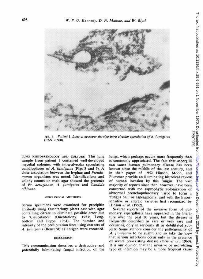
FIG. 9. Patient 1. Lung at necropsy showing intra-alveolar sporulation of A. fumigatus(PAS x 600).
LUNG HISTOPATHOLOGY AND CULTURE The lungsample from patient 1 contained well-developedmycelial colonies, with intra-alveolar sporulatingconidiophores of A. fumigatus (Figs 8 and 9). Aclose association between the hyphae and Pseudo-monas organisms was noted. Identifications andcolony counts on malt agar showed the presenceof Ps. aeruginosa, A. fumigatus and Candidaalbicans.
SEROLOGICAL METHODS
Serum specimens were examined for precipitinantibody using Ouchterlony plates cast with agarcontaining citrate to eliminate possible error dueto 'C-substance' (Ouchterlony, 1953; Long-bottom and Pepys, 1964). The number andintensity of the precipitation lines using extracts ofA. fum71igatus (Bencard) as antigen were recorded.
DISCUSSION
This communication describes a destructive andpotentially fulminating fungal infection of the
lungs, which perhaps occurs more frequently thanis commonly appreciated. The fact that aspergillican cause human pulmonary disease has beenknown since the middle of the last century, andin their paper of 1952 Hinson, Moon, andPlummer provide an illuminating historical reviewof human invasion by this fungus. The vastmajority of reports since then, however, have beenconcerned with the saprophytic colonization ofabnormal bronchopulmonary tissue to form a'fungus baill' or aspergilloma; and with the hyper-sensitive or allergic varieties first recognized byHinson et al. (1952).
Several reports of the invasive form of pul-monary aspergillosis have appeared in the litera-ture over the past 20 years, but the disease isfrequently described as rare or very rare andoccurring only in seriously ill or debilitated sub-jects. Some authors consider the pathogenicity ofA. fumigatus to be slight, and so take the viewthat serious infections occur only in the presenceof severe pre-existing disease (Orie et al., 1960).It is our opinion that the invasive or necrotizingtype of infection may be a more frequent cause
698
copyright. on D
ecember 26, 2019 by guest. P
rotected byhttp://thorax.bm
j.com/
Thorax: first published as 10.1136/thx.25.6.691 on 1 N
of serious pulmonary disease than has previouslybeen realized, as we have seen four separateinstances in a single hospital group within a periodof less than one year.The nature of the invasive process in pulmonary
tissue is controversial. It has been widely held thatpre-existing tissue damage is necessary for fungalcolonization to take place (Hinson et al., 1952;Bech, 1961). Primary aspergillosis in humans isregarded as a rare phenomenon, if it occurs at all.It will always be difficult to confirm that an infec-tion is primary, when it may have taken root insome minute pulmonary lesion such as an infarct(Conen, Walker, Turner, and Field, 1962;Symmers, 1962). Once an infection is established,however, changes occurring in adjacent lung tissueare probably due to diffusible products of fungalmetabolism, producing tissue reactions which maybe inflammatory, granulomatous, necrotic or sup-purative and which may promote further invasionby fungus (Riddell, 1958; Bech, 1961). This viewarises from the observations of Henrici (1939),who extracted from the cell sap of A. fumigatusmycelium a thermolabile endotoxin with a pro-nounced histotoxic effect in laboratory animals.Clayton has also demonstrated the proteolyticactivity of A. fumigatus endotoxin (Campbell andClayton, 1964). Protease systems comparalble totrypsin in proteolytic and fibrinolytic activity havebeen isolated from certain other aspergillus species(Crewther and Lennox, 1950; Stefanini, Marin,Soardi, and Mossa, 1962). There may still be con-siderable variation between species, or evenbetween different strains of a single species, in theproduction of such enzymes (Stefanini and Marin,1958). Despite this, such observations lendcredence to the view that an active growth ofA. fumigatus in the lung may lead directly topulmonary necrosis and further tissue infiltra-tion. Even if the fungus elaborates no true exo-toxin, degenerating fungal elements may liberatetoxic intracellular substances wvhich damagepukmonary tissue (Gowing and Haimlin, 1960).Susceptibility to invasive aspergillosis appearsto be enhanced by antibiotics and by cortico-steroids, by radiotherapy, by cytotoxic drugs,and by a number of systemic and debilitatingconditions (Bech, 1961). Indeed, Sidransky andFriedman (1959) showed experimentally thatthe administration of antibiotics and cortico-steroids to laboratory animals exposed to airbornespores of A. flavus greatly increased the numbersthat became infected. Each of our four patientshad been given multiple antibiotic therapy, andthis may have been a factor which contributed to
the invasive nature of the fungal infection. Inaddition, it is probable that infection with thefungus was secondary to bacterial infection orother lung disease. In patient 1, Ps. aeruginosawas isolated both from the sputum and from lungtissue at necropsy. This organism itself can pro-duce tissue necrosis, and it may be that the bac-terial infection in this patient was largely to blamefor the destruction of lung tissue (Pierce,Edmonson, McGee, Ketchersid, Loudon, andSanford, 1966). The fungus would then beregarded as a secondary invader, fulfilling a sapro-phytic role, although still actively growing withinthe lungs. Okudaira and Schwarz (1962), who haveobserved actual invasion of living bronchial cartil-age by hyphae, concede that at least a part of thecellular reaction accompanying the necrosis inlesions where aspergillus is present may be due toconcomitant bacterial infection.A major difficulty in the diagnosis of pulmonary
aspergillosis is the fact that aspergillus species arecommon laboratory contaminants. The airbornespores of these fungi are present in the atmospherethroughout the year and the demonstration ofaspergillus in sputum is not necessarily an indica-tion of pathogenicity. A diagnosis of aspergillaryinfection should be based on repeated culture, onclinical, radiological, and immunological evidence,and on histological material where possible(Sarkany, 1968). In the present series of fourpatients, histological confirmation was availablein only one case. The three others, however,all developed precipitating antibodies againstA. fumigatus, and all yielded numerous coloniesof the fungus from successive samples of sputum.Such sputum yields may be designated 'fungusabundant' and be regarded as evidence of fungalgrowth in the lungs rather than simple contamina-tion (English and Henderson, 1967).The finding of a mat of tangled hyphae lining
parts of the abscess cavities in the lungs of the firstpatient has similarities to the necropsy appearancesin a case described by Toigo (1960). One of Bech'spatients, too, had abscess cavities containingsloughs of ramifying septate mycelia (Bech, 1961).A feature of the present case was the mannerin which Pseudomonas bacilli and Aspergillusmycelium appeared to co-exist, large numbers ofbacilli lying among the masses of hyphae. Thepossibility of primary invasion by the Gram-negative bacillus, with A. fumigatus playing asaprophytic role, has been discussed; the questionof symbiosis between bacterium and fungus mustalso be considered. Fungal hyphae, particularly inthe medium of soil, are known to present surfaces
699
copyright. on D
ecember 26, 2019 by guest. P
rotected byhttp://thorax.bm
j.com/
Thorax: first published as 10.1136/thx.25.6.691 on 1 N
frequently colonized by bacteria (Warcup, 1965).The reasons for these associations are not wellunderstood, but may range from the action ofbacterial fimbriae on the hyphal surface (Duguid,Smith, Dempster, and Edmunds, 1955) to theexudation of nutrients from fungi or their sub-strates (Tribe, 1957). Conversely, Mangan (1969)has noted that spore germination of A. terreus inbroth cultures is inhibited by Ps. aeruginosa, prob-ably through the elaboration of pyocyanine andfluorescent green pigments which have a fungistaticeffect. The factors which determined the fungal-bacterial association in the present case thereforeremain obscure. Another open question is the asso-
ciation between bronchial neoplasia and fungalinfection, as observed in patient 4. Here again itmay be that an abnormal or devitalized tissuesurface is available for fungal colonization. Weconsider that a true lung abscess, in whichA. fumigatus was actively growing, preceded theappearance of neoplasia in the opposite lung; butthe time interval was probably too short to suggestthat the fungus itself may have had a carcino-genetic effect.
In ill patients, especially those with abnormallungs and those who have received multiple anti-biotic or corticosteroid therapy, it seems likely thatfungal infection will be recognized more often inthe future, if borne in mind and specifically lookedfor. Specialized mycological techniques may benecessary to establish the diagnosis. Once diag-nosed, the fungal infection, if progressive, shouldbe treated actively whether it is regarded as
primary or secondary in nature. Three of thepatients described were treated with natamycin, a
tetraene antifungal antibiotic isolated fromStreptomyces natalensis, and related to otherpolyenes including nystatin and amphotericin(Struyk, Hootte, Drost, Waisvisz, van Eek, andHoogerheide, 1958). It is poorly absorbed fromthe gut but is effective and non-toxic when admin-istered as an aerosol (Edwards and La Touche,1964). Whether inhalation therapy can really con-
trol a deep-seated invasive infection must remainquestionable. Vedder and Schorr (1969) havetreated a 31-year-old girl suffering from primarydisseminated aspergillosis with metastatic infectionof the skin by inhalations of nystatin aerosol; thistreatment produced measurable blood levels ofnystatin, and within a few weeks there was a satis-factory clinical recovery, although the authors are
guarded about the child's ultimate prognosis. Thesynthetic antimycotic clotrimazole, developed inGermany, is said to be absorbed well from thealimentary tract, with few side-effects, and to have
activity against almost all pathogenic fungi(British Medical Journal, 1969). It is possible that-this drug may have an application in broncho-pulmonary aspergillosis.
We wish to thank Dr. 1. W. B. Grant, Dr. M. B.Matthews, and Professor J. A. Strong for allowing usto study their patients. We are indebted to Dr. J. C.Gould, Director, Central Microbiological Laboratories,Western General Hospital, Edinburgh, for the sero-logical studies, and to Dr. J. M. Drennan, ConsultantPathologist, Western General Hospital, for thenecropsy examination. The illustrations were providedby the Medical Photography Unit of the Universityof Edinburgh.The mycological studies were undertaken as part of
a research programme supported by a grant from theScottish Hospitals Endowments Research Trust.
REFERENCESBech, A. 0. (1961). Diffuse bronchopulmonary aspergillosis. Thorax,
16, 144.British Medical Journal (1969). A new synthetic antimycotic (leading
article). Brit. med. J., 4, 444.Campbell, M. J., and Clayton, Y. M. (1964) Bronchopulmonary
aspergillosis. Amer. Rev. resp. Dis., 89, 186.Conen, P. E.,Walker, G. R., Turner,J. A., and Field, P. (1962).
Invasive primary aspergillosis of the lung with cerebral metastasisand complete recovery. Dis. Chest, 42, 88.
Crewther,W. G., and Lennox, F. G. (1950). Preparation of crystalscontaining protease from Aspergillus oryzae. Nature (Lond.),165, 680.
Duguid, J. P., Smith, 1.W., Dempster, G., and Edmunds, P. N.(1955). Non-flagellar filamentous appendages (" fimbriae ")and haemagglutinating activity in Bacterium coli. J. Path. Bact.,70, 335.
Edwards, G., and La Touche, C. J. P. (1964). The treatment ofbronchopulmonary mycoses with a new antibiotic-pimaricin.Lancet, 1, 1349.
English, M. P., and Henderson, A. H. (1967). Significance and inter-pretation of laboratory tests in pulmonary aspergillosis. J.clin.Path., 20, 832.
Gowing, N. F. C., and Hamlin, I. M. E. (1960). Tissue reactions toAspergillus in cases of Hodgkin's disease and leukaemia. J.clin.Path., 13, 396.
Grocott, R. G. (1955). A stain for fungi in tissue sections and smears.Amer. J.clin. Path., 25, 975.
Henrici, A. T. (1939). An endotoxin from Aspergillus fumigatus.J. Immunol., 36, 319.
Hinson, K. F.W., Moon, A. J., and Plummer, N.S. (1952). Broncho-pulmonary aspergillosis. Thorax, 7, 317.
Longbottom,J. L., and Pepys,J. (1964). Pulmonary aspergillosis:diagnostic and immunological significance of antigens andC-substance in Aspergillus fumigatus. J. Path. Bact.,88, 141.
Mangan, A. (1969). Interactions between some aural Aspergillusspecies and bacteria. J. gen. Microbiol., 58, 261.
Okudaira, M., and Schwarz, J. (1962). Tracheobronchopulmonarymycoses caused by opportunistic fungi, with particular refer-ence to aspergillosis. Lab. Invest., 11, 1053.
Orie, N. G. M., deVries, G. A., and Kikstra, A. (1960). Growth ofAspergillus in the human lung. Amer. Rev. resp. Dis., 82, 649.
Ouchterlony, 0. (1953). Antigen-antibody reactions in gels.IV. Typesof reactions in co-ordinated systems of diffusion. Acta path.microbiol. scand., 32, 230.
Pierce, A. K., Edmonson, E. B., McGee, G., Ketchersid,J., Loudon,R. G., and Sanford, J. P. (1966). An analysis of factors predis-posing to gram-negative bacillary necrotizing pneumonia.Amer. Rev. resp. Dis., 94, 309.
Riddell, R.W. (1958). The role of fungi as human pathogens. Proc.roy.Soc. Med., 51, 491.
Sarkany,I. (1968). Systemic mycoses. In Recent Advances in Medicine,ed. Baron, D. N., Compston, N., and Dawson, A. M., 15th ed,pp. 243-273. Churchill, London.
700
copyright. on D
ecember 26, 2019 by guest. P
rotected byhttp://thorax.bm
j.com/
Thorax: first published as 10.1136/thx.25.6.691 on 1 N
Sidransky, H., and Friedman, L. (1959). The effect of cortisone and Toigo, A. (1960). Pulmonary aspergillosis. Amer. Rev. resp. Dis.,antibiotic agents on experimental pulmonary aspergillosis. 81, 392.Amer. J. Path., 35, 169.
Stefanini, M., and Marin, H. M. (1958). Fibrinolysis I. Fibrinolytic Tribe, H. T. (1957). Ecology of micro-organisms in soils as observedactivity of extracts from non-pathogenic fungi. Proc. Soc. exp. during their development upon buried cellulose film. MicrobialBiol., 99, 504. Ecology, edited by R. E. 0. Williams and C. C. Spicer, Uni-- -, Soardi, F., and Mossa, A. (1962). Fibrinolysis IX. The versity Press, Cambridge. Symp. Soc. gen. Microbiol., 7, 287.
comparative activity in vivo of trypsin and aspergillin 0 (moldfibrinolysin). Angiology, 13, 254. Vedder, J. S., and Schorr, W. F. (1969). Primary disseminated pul-
Struyk, A. P., Hoette, I., Drost, G., Waisvisz, J. M., van Eek, T., monary aspergillosis with metastatic skin nodules. J. Amer. med.and Hoogerheide, J. C. (1958). Pimaricin, a new antifungal Ass., 209, 1191.antibiotic. Antibiotics Annual, 1957-58, p. 878. Medical Ency-clopedia, New York. Warcup, J. H. (1965). In Ecology of Soil-borne Plant Pathogens:
Symmers, W. St. C. (1962). Histopathologic aspects of the patho- An International Symposium on Factors determining the Behavgenesis of some opportunistic fungal infections, as exemplified ior of Plant Pathogens in Soil, University of California, 1963,in the pathology of aspergillosis and the phycomycetoses. Lab. Edited by K. E. Baker, W. C. Snyder, and others, p. 59. JohnInvest., 11, 1073. Murray, London.
copyright. on D
ecember 26, 2019 by guest. P
rotected byhttp://thorax.bm
j.com/
Thorax: first published as 10.1136/thx.25.6.691 on 1 N


![Aspergillosis - Youngstown State Universitypeople.ysu.edu/~crcooper01/Aspergillosis[1]- Katie Jacquie Qazi.pdf•People with Aspergillosis are in three distinct groups •Healthy immune](https://static.documents.pub/doc/80x56/5e3883b0e2f2970b7b1c24ad/aspergillosis-youngstown-state-crcooper01aspergillosis1-katie-jacquie-qazipdf.jpg)


