Corresponding author: J.S. Dumler, Departments of Pathology andMicrobiology & Immunology, University of Maryland School of Med-icine, 685 W. Baltimore Street, HSFII 322D, Baltimore, MD 21218,USAE-mail: [email protected]
Introduction
Zoonoses comprise an array of infections, many known
throughout man’s history, and others that were only recentlyrecognized to cause human or animal disease. The phrase
‘neglected infectious disease’ recently emerged based on theperception that neglected infections are largely tropical or
affect large poverty-ridden populations, are chronic, and areperceived to take a steep toll on society and development [1].However, the concept of neglected infections is neither new
nor fixed, and depends on the interpretation context. Fig. 1
shows zoonoses in the USA as reported in Morbidity MortalityWeekly Reports spanning 1981 to 2012 [2] and lacks any priorityneglected zoonosis as cited by the World Health Organization
(WHO) or the U.S. CDC [3,4]. Depending on the source,bacterial infections defined as ‘neglected’ by the Bill & Melinda
Gates Foundation include only trachoma and buruli ulcer [5],whereas WHO and CDC add yaws and leprosy, and the list
expands to include infantile diarrhoea (enterotoxigenic Escher-ichia coli) in the European Union [3,4,6]. The Bill & Melinda
Gates Foundation-sponsored Global Burden of Diseases, In-juries, and Risk Factors Study 2010 list of deaths from globallyimportant infectious diseases includes only a small proportion
that are zoonotic (Fig. 2) [7]. The biased data presentationoccurs largely because neglected diseases cannot be compared
if they are not accurately diagnosed and reported, or assessedbased on disability-adjusted life-years if that factor is unable to
be calculated given the absence of meaningful incidence orprevalence data. Rather the list reflects existing data shaped by
infections for which diagnosis or reporting is easy, or based on
ious Diseases. Published by Elsevier Ltd. All rights reserved
FIG. 1. Zoonoses reported to the CDC and published in Morbidity Mortality Weekly Reports between 1981 and 2012. Not all diseases were reportable
over this interval, and some case definitions changed. No neglected infectious disease as cited by the CDC or WHO appears on this list. Aside from
malaria, which is most often imported, the most commonly reported zoonoses in the USA over this interval were Lyme disease, spotted fever group
rickettsioses, West Nile virus infection, Anaplasma phagocytophilum infection and Ehrlichia chaffeensis infection.
CMI Chikeka and Dumler Neglected bacterial zoonoses 405
evidence estimates that could be biased given the lack of suchdata [8,9]. Much speculation has been made in recent years
about priorities for allocation of resources as malaria comesunder increasing control [10]. A growing number of in-vestigations to discern the aetiology of acute febrile illness in
developed and under-resourced regions illustrate major gaps inour knowledge, and these gaps identify a greater expanse of
neglected infectious diseases, many of which are bacterialzoonoses [11–16]. Such investigations shed light on prevalence
estimates and the likely high human toll of inattention to otherinfections, many bacterial, and the topic of this article.
Bacterial zoonoses occur with transmission via one ofseveral mechanisms: 1) direct contact with animals or infected
materials; 2) animal bites and scratches; 3) bites or mechanicaltransmission by arthropod vectors; and 4) consumption ofcontaminated foods (Table 1). The bacteria that cause the in-
fections can sometimes be acquired by more than one trans-mission mechanism, complicating control measures. Most do
not appear on lists of neglected infections, in part because ofserious problems with definitive aetiological diagnosis and
reporting: most acute febrile illnesses in Sub-Saharan Africa arediagnosed and reported on clinical grounds as malaria, and in
South East Asia, as typhoid fever or dengue virus infection.When investigated objectively using pathogen-specific di-agnostics, infections such as leptospirosis, rickettsioses and
melioidosis are diagnosed as frequently as dengue, typhoid or
malaria [11–13,16,17]. Owing to the biases toward the‘neglected diseases’ and the paucity of objective data accumu-
lated by major public health agencies, this article will focus ononly selected bacterial zoonoses for which their recognition asimportant or relevant is not generally acknowledged, and for
which diagnosis, treatment and research priorities are minimalglobally. Despite the exclusion of certain bacterial zoonoses,
the approach is intended to highlight what is known and notknown as examples of why improvements in the study of these
diseases are needed.
Leptospirosis
Leptospirosis is a zoonosis caused by pathogenic spirochaetes
of the genus Leptospira [18–20]. Leptospires are spiral shaped,motile aerobic spirochaetes distinguished morphologically from
other spirochaetes by characteristic hooked ends [19]. Mem-bers of the Leptospira genus were previously grouped according
to antigenic determinants, with pathogenic leptospiresbelonging to the species Leptospira interrogans and non-pathogenic leptospires grouped under the species Leptospira
biflexa. A new classification system proposes grouping of lep-tospires by DNA relatedness into 20 species: nine pathogenic,
five of intermediate or unclear pathogenicity, and six non-pathogenic saprophytes [21,22].
and Infectious Diseases. Published by Elsevier Ltd. All rights reserved, CMI, 21, 404–415
FIG. 2. The proportional contribution of various infectious disease fatalities as reported in the Global Burden of Diseases, Injuries and Risk Factors
Study 2010 [7]. (a) Pie chart demonstrating all infectious diseases examined by the study. (b) Zoonoses occupy a minority of the reported deaths and
disease burden, and include no bacterial zoonoses.
406 Clinical Microbiology and Infection, Volume 21 Number 5, May 2015 CMI
Animals serve as natural hosts and can be asymptomatic [19].Leptospires are excreted in their urine, contaminating water
and soil where they can remain viable for days to months.Rodents are the most significant reservoirs for transmission andwhen infected can shed the leptospires throughout their life-
time. Humans are accidental hosts, acquiring infection afterexposure of mucous membranes and abraded skin to animal
urine, contaminated water or soil, or infected animal tissue.After entering the bloodstream, the spirochaetes multiply in
organs, most commonly the central nervous system (CNS),kidneys and liver. They are cleared from the blood and most
tissues by the immune response but can persist and multiply inthe tubules of the kidneys.
The distribution of leptospirosis is worldwide but it occurs
with the greatest frequency in tropical and subtropical envi-ronments [18,20]. It is estimated that more than 10 million
cases occur each year around the world and it is a significantcause of morbidity and mortality [21,23]. Leptospirosis is most
ious Diseases. Published by Elsevier Ltd. All rights reserved, CMI, 21, 404–415
TABLE 1. Bacterial zoonoses by transmission mechanism and causative agent(s)
Bacterial zoonoses transmitted by direct contact with animals or infected animal materials Causative agent(s)Anthrax Bacillus anthracisBrucellosis Brucella spp.Cat scratch disease Bartonella spp.Erysipelothrix infections Erysipelothrix rhusiopathiaeGlanders and melioidosis Burkholderia mallei and Burkholderia pseudomalleiLeptospirosis Leptospira interrogans spp.Mycobacterioses Mycobacteria spp.Q fever Coxiella burnetii
Bacterial zoonoses transmitted principally by animal bites or scratchesPasteurellosis Pasteurella multocida and other spp.Capnocytophaga infections Capnocytophaga canimorsusCat scratch disease Bartonella henselaeRat bite fever Spirillum minus and Streptobacillus moniliformis
Vector-borne bacterial zoonosesLyme borreliosis Borrelia burgdorferi sensu lato (incl. Borrelia garinii, Borrelia afzelii)Tick- and louse-borne relapsing fever borreliosis Borrelia recurrentis, Borrelia turicatae, Borrelia hermsii, othersPlague Yersinia pestisTularaemia Francisella tularensisRickettsioses Spotted fever and typhus group Rickettsia speciesEhrlichiosis and Anaplasmosis Ehrlichia chaffeensis, Anaplasma phagocytophilumScrub typhus Orientia tsutsugamushi
CMI Chikeka and Dumler Neglected bacterial zoonoses 407
common in developing urban and rural areas with inadequatesewage disposal and water treatment, yet outbreaks in
temperate regions, including the USA and Europe are welldocumented [21,24]. The burden of disease is highest in theCaribbean, Central and South America, South East Asia, Oce-
ania, India and South Asia, and Eastern Europe. The incidence inthe Seychelles in the Indian Ocean is as high as 432 per
1 000 000 population and globalization continues to occurthrough international travel to these and other similar areas
where leptospirosis is endemic [25]. Although well-validatedglobal data on leptospirosis are lacking, the estimated global
annual incidence of leptospirosis in temperate regions is 0.1–1cases per 100 000 population while the incidence in tropicalclimates is > 10 cases per 100 000 population [26]. These are
likely underestimates because of misdiagnosis and under-reporting, particularly in regions where other diseases with
similar non-specific presentations such as dengue and malariaare prevalent. For example, a cross-sectional study among
hospitalized febrile patients in northeastern Malaysia betweenAugust 2010 and February 2011 found an 8.4% seroprevalence;
but among these, only 31% were correctly diagnosed usingclinical criteria—the remainder were misdiagnosed usually as
dengue/dengue haemorrhagic fever (38%), pneumonia (14%), ortyphoid fever (7%) [27]. Among 3165 sera from acutely febrilepatients with suspected dengue in Jamaica over 2007–2008,
only 38.4% were confirmed to have dengue antibodies, whereas6% were misdiagnosed and had leptospirosis instead, and 1.6%
had serological responses consistent with both infections [28].
Perhaps more directly, Reller et al. showed separately in SriLanka and Nicaragua where leptospirosis is often suspected,
that clinical diagnosis alone is poorly sensitive, 23% and 11%,respectively [14,29]. Likewise, in Sri Lanka, clinical diagnosis ofother acute febrile illnesses is poor, 14% for dengue virus
infection, 3% for rickettsial infections and 0% for Chikungunyavirus infection [13,30,31].
The clinical presentation of leptospirosis is highly variableand non-specific [21,23], explaining the poor predictive value of
clinical assessment for establishing the diagnosis as a guide toappropriate therapy [14,29,32]. Severity ranges from subclinical
to fatal. Clinical illness usually begins abruptly after an incuba-tion period of 2–26 days. The less severe, anicteric form ofleptospirosis resembles an influenza-like illness with fever,
rigors, myalgias, headache, abdominal pain, non-productivecough and conjunctival suffusion, a distinguishing sign [20,21].
The severe icteric form, known as Weil’s disease, occurs in aminority of patients and is associated with jaundice, hepatic
dysfunction, myocarditis with arrhythmias, haemorrhage, uve-itis and multi-organ failure. Both can occur in two phases: an
acute septicaemic phase and an immune phase that can imme-diately follow. Routine laboratory tests are typically non-
specific but can include leucocytosis with a left shift,increased erythrocyte sedimentation rate, mildly elevatedtransaminases, alkaline phosphatase and bilirubin, abnormal
urinalysis and thrombocytopenia. Leptospirosis mimics manyother tropical diseases and diagnosis requires a high degree of
clinical suspicion [14,17,33]. The average annual hospitalization
and Infectious Diseases. Published by Elsevier Ltd. All rights reserved, CMI, 21, 404–415
408 Clinical Microbiology and Infection, Volume 21 Number 5, May 2015 CMI
rate for leptospirosis in the USA from 1998 to 2009 was 0.6/
1 000 000 population and the average length of stay and hospitalcharges were higher in comparison to non-leptospirosis-
associated hospitalizations [34]. In Brazil, untreated leptospi-rosis results in significant social costs in years of potential life
lost and partial hospitalization costs when compared with earlytreatment and prevention [35]. Death occurs in 5–15% of in-fections if untreated, and the proportion increases with age.
Acute disease is the direct result of inflammatory responsesto the presence of leptospires in tissues, including hepatitis and
cholestasis, interstitial nephritis, and meningoencephalitis [21].Pulmonary haemorrhage is increasingly observed, and the
pathogenesis of this process is not defined, but relates to hostimmune response, the production of bacterial factors that lead
to local coagulation abnormalities, or both [18,21]. Disease re-quires the ability of leptospires to enter organs and tissues andspread. Leptospira genome studies show that >1% of genes are
dedicated to motility, and the presence of adhesins and invasins,such as the outer membrane protein Loa22 and the
immunoglobulin-like LigA could contribute to this [21,36]. Inaddition, genes that could affect haemostasis and coagulation are
present, including a platelet-activating factor acetylhydrolase-14(LA2144, pafAH) and von Willebrand factor 15 type A domains
(LB054 and LB055, vwa) [21,36]. The development of dissemi-nated intravascular coagulation with multi-organ failure provides
some evidence to support an immunological basis for severeleptospirosis including the pro-inflammatory response to bac-terial lipopolysaccharide and lipoproteins that stimulate Toll-like
receptor-2. Likewise, pulmonary haemorrhage is more frequentin persons with high antibody titres, and there is evidence that
immunoglobulin and complement fixation on host cells arerelated to the expression of specific leptospiral haemostatic
proteins [36]. The late occurrence of uveitis correlates with the‘immune’ phase and with immunoglobulin and complement
deposition [37].Leptospirosis is diagnosed by serology, culture and molec-
ular tests [18,19,38]. Culture is definitive but slow, lacks
sensitivity, and requires specialized medium, and is thereforenot very useful for diagnostic purposes. Immunohistochemistry
has been used, but is relatively unavailable. Likewise, PCR issensitive but requires broad-range primers that despite the high
degree of genetic diversity, can amplify all variants. Serologicaltests are used most often, particularly the reference standard,
the microscopic agglutination test. This is sensitive as early as5–7 days after onset; a single high titre is acceptable, but
seroconversion is preferred. The pitfall of microscopic agglu-tination test is the requirement for continued growth of mul-tiple serovars, a daunting task for clinical laboratories, and the
delay in detection because antibodies develop later in thecourse of illness.
A recent Cochrane review was unable to discern any sig-
nificant advantages to the use of antibacterial treatments foractive leptospirosis [39]. Yet many believe that antimicrobial
therapy shortens illness duration and reduces shedding of lep-tospires in the urine [33]. Treatment options include oral
doxycycline or azithromycin for mild disease and parenteralpenicillin, doxycycline, or third-generation cephalosporin forsevere disease. Supportive care may be required for Jarisch–
Herxheimer reactions that result in a systemic inflammatoryresponse after lysis of spirochaetes with treatment.
Prevention of leptospirosis focuses on public health efforts,chemoprophylaxis and vaccination. Public health interventions
include identification and modification of risk factors throughpublic education. Human and animal behavioural and environ-
mental risk factors vary by region and include rodent in-festations, particularly rats, exposure to contaminated surfacewater such as in rice paddies, and the presence of skin wounds.
A recent study by Samarakoon and Gunawardena indicates thatin endemic regions the knowledge of leptospirosis prevention
is adequate but does not lead to implementation of personalprotective measures [40]. Prophylaxis with doxycycline
(200 mg once a week) reduces morbidity and mortality duringoutbreaks and other homeoprophylactic measures are re-
ported to provide preventive benefit, but will require exten-sive evaluation [41,42]. There is no human vaccine available but
vaccines do exist for animal reservoirs including dogs andcattle.
Relapsing fever borreliosis
Relapsing fever is an arthropod-borne disease caused by path-ogenic spirochaetes of the genus Borrelia [43,44]. It occurs intwo major forms: tick-borne relapsing fever (TBRF) and louse-
borne relapsing fever (LBRF). LBRF is primarily seen in East andCentral Africa and is a frequent cause of epidemics in areas of
extreme poverty, war, natural disasters and overcrowding. It iswidely perceived that there has been a decline in the incidence
of LBRF secondary to improved standards of living and theintroduction of the insecticide DDT. However, LBRF is still a
major public health issue in East Africa, particularly in Ethiopiaand in surrounding regions where louse infestation iscommonplace. TBRF is found worldwide and is endemic in
many countries including the Americas, Central Asia, theMediterranean region and many parts of Africa. In most rural
areas of Senegal, Mauritania and Mali, TBRF is a common causeof fever with incidence comparable to Plasmodium falciparum
malaria and influenza [45]. The incidence of tick-borne relapsingfever peaks in summer but infection can occur year round,
depending on local climate conditions.
ious Diseases. Published by Elsevier Ltd. All rights reserved, CMI, 21, 404–415
CMI Chikeka and Dumler Neglected bacterial zoonoses 409
Relapsing fever is caused by bacteria of the genus Borrelia.
Borreliae are thin, helical, motile spirochaetes that are unique intheir ability to change their outer membrane surface proteins
by gene conversion to generate antigenic variation [43]. Eachrelapse is associated with the emergence of a unique antigenic
bacterial clone that is subsequently controlled by the immunesystem, only to select for new antigenic variant clones andanother relapse of fever and disease. In fact, some variant
clones have a propensity for dissemination into tissues andorgans such as the CNS.
Louse-borne relapsing fever is caused by Borrelia recurrentis,now believed to be a reduced-genome variant of the TBRF
Borrelia duttonii [46]. The vector for LBRF is the human bodylouse (Pediculus humanus), so humans are the only known
reservoir for this spirochaete [44]. TBRF is transmitted tohumans through the bite of infected soft ticks of the genusOrnithodoros. There are many Borrelia species that cause TBRF,
roughly divided into Old World and New World species, andthey are associated with specific tick species. Unlike the human
body louse that lives for several weeks, Ornithodoros ticks canlive for many years between blood meals, harbour spirochaetes
for prolonged periods, and can transmit the pathogen verticallyto offspring [44].
Lice acquire the spirochaetes by feeding on infected humanswhereas humans acquire LBRF by scratching infected haemo-
lymph of a crushed louse into the skin [44,47]. Ticks becomeinfected by feeding on infected wild rodents and then transmitthe spirochaetes to humans through a tick bite. After entering
the blood, the spirochaetes disseminate, seeding multiple or-gans including the CNS in the case of TBRF.
Relapsing fever is characterized by two or more episodes ofhigh fever (usually >39°C), myalgias, arthralgias, nausea, vom-
and bleeding. Laboratory studies can reveal mild elevations inbilirubin and aminotransferases, thrombocytopenia andanaemia. Later in the illness, jaundice, hepatosplenomegaly and
myocarditis can occur [50]. The initial febrile episode typicallylasts 3–6 days and is followed by an afebrile period of
4–14 days, after which fever and symptoms recur. Subsequentrelapses are usually less severe and can follow at 1- to 2-week
intervals.Because the WHO advises a presumptive diagnosis of ma-
laria in endemic regions, or collects ‘verbal autopsy’ reports fordeaths, relapsing fever often goes undetected, particularly in
parts of Africa where malaria is highly prevalent. A study byNordstrand et al. in Togo, West Africa between 2002 and 2004found that among febrile patients originally diagnosed and
treated for malaria, the prevalence of TBRF was 8.8%,compared with 63.1% for malaria; 4.5% of patients had both
malaria and TBRF [51]. Similarly, among a cohort of individuals
participating in a study of mass treatment to eradicate trachomain Tanzania, 17% of febrile episodes were caused by either
Plasmodium falciparum or Plasmodium vivax, whereas 4% wereattributed to relapsing fever [52], underscoring how uncor-
roborated clinical diagnosis can lead to erroneous reportingcoupled with inflation of the incidence of reportable infectionsat the expense of other aetiologies.
Although the incidence of relapsing fever has declined in thedeveloped world, it continues to be a formidable public health
issue in certain regions. In Tanzania, for example, infection withTBRF is associated with significant morbidity and mortality,
especially in women and children, where it results in highperinatal mortality (436/1000 births) and is frequently listed as
one of the top ten causes of mortality in children under 5 yearsof age [53,54]. A retrospective study conducted between 2009and 2012 of patients with LBRF-like symptoms who were
admitted to a referral hospital in Ethiopia found the prevalenceto be 4.9% and case fatality rates ranged from 2 to 6% [55].
Relapsing fever can account for up to 27% of hospitalizations insome regions [56]. There still exists the possibility of significant
re-emergence, particularly as travel patterns continue tochange and imported cases of relapsing fever are described.
There is also evidence of a resurgence of louse infestationamong certain groups. In Marseille, France, Brouqui et al. found
a high prevalence of louse-borne infections in the homeless anda high level of exposure to tick-borne diseases [57].
Relapsing fever is diagnosed by visualization of spirochaetes
in the blood using dark-field microscopy or Wright or Giemsastaining on thin and thick blood smears [49]. Serological tests
were typically unreliable, but have improved with the devel-opment of assays based on detection of antibodies to relapsing
fever spirochaete-specific GlpQ [58]. Culture requires tech-niques that are not available in most laboratories. Nucleic acid
amplification tests are available in some public health facilities,but not generally accessible to primary healthcare workers, oraccessible in under-resourced regions where relapsing fever is
often present.Few careful studies of in vitro susceptibility for relapsing fever
Borrelia species are published to guide treatment. LBRF wasshown to be effectively treated with a single dose of oral
tetracycline or doxycycline, or intramuscular penicillin G pro-caine [43,49], but over one-third of patients had recurrent fe-
ver, suggesting that a prolonged regimen is preferable. Similarly,the relapse rate of TBRF is at least 20% after single-dose
therapy; hence, the treatment for TBRF is extended to7–10 days. Relapses yield as many as 108 bacteria/mL of blood,so antibiotic therapy can trigger a Jarisch–Herxheimer reac-
tion, particularly with LBRF, and its occurrence is associatedwith high mortality. Tetracyclines are highly effective at clearing
and Infectious Diseases. Published by Elsevier Ltd. All rights reserved, CMI, 21, 404–415
410 Clinical Microbiology and Infection, Volume 21 Number 5, May 2015 CMI
spirochaetes with no or few relapses, but their use is associated
with more frequent and severe episodes of Jarisch–Herxheimer reactions [59,60]. Therefore, preferred treatment
employs either single-dose doxycycline or tetracycline, or asingle dose of procaine penicillin intramuscularly followed by 2
or more days of oral doxycycline or tetracycline. Prevention ofinfection requires good personal hygiene, delousing, launderingat high temperatures and application of insecticides to clothing
and bedding to diminish louse infestations for LBRF. TBRFprevention includes exclusion of rodents that host the argasid
tick vectors, avoiding habitats that harbour the ticks them-selves, and the use of tick repellents.
Scrub typhus, murine typhus and spottedfever rickettsiosis
Bacteria in the genera Orientia, Rickettsia, Ehrlichia and Ana-plasma are obligate intracellular α2 proteobacteria in the Order
Rickettsiales and Families Rickettsiaceae and Anaplasmataceae[61]. Genome sequences illustrate that these agents adapted to
an obligatory intracellular lifestyle through the loss of genes andpathways required for extracellular growth [62]. Regardless,
each contains DNA, RNA, ribosomes, divides by binary fissionand possesses a cell wall with ultrastructural characteristics ofGram-negative bacteria. Rickettsiaceae members (Rickettsia and
Orientia) target endothelial cells in mammals and reside withinthe cytosol. In contrast, Anaplasmataceae family members
usually reside in haematopoietic-derived cells, such as leuco-cytes in mammals where they propagate within pathogen-
modified endosomes. All Rickettsiales have at least part oftheir life cycle in vectors, and for Rickettsia, Orientia, Ehrlichia
and Anaplasma these are usually arthropods, often ticks, fleas,lice or mites.
The genus Rickettsia is divided into spotted fever (SFGR) and
typhus groups. Genome studies illustrate diversity with SFGR,yet these cluster separately from typhus group rickettsiae [62].
Several species occupy intermediate positions sometimes called‘transitional’ or ‘ancestral’. There are over 25 recognized SFGR
species and two typhus group species; most are human path-ogens, yet at least several ‘transitional’ and ‘ancestral’ rickettsiae
are questionably pathogenic or never associated with knownhuman or animal disease. The genus Orientia has a similar de-
gree of genetic diversity as for SFGR, yet has only two namedspecies, Orientia tsutsugamushi and Orientia chuto, both humanpathogens. Although many Anaplasmataceae are animal patho-
gens, only a limited range are human pathogens.Among the rickettsial agents, the most neglected for which
sufficient evidence exists that there are large global diseaseburdens include O. tsutsugamushi, the agent of scrub typhus
[10,12,13,16], Rickettsia typhi, the aetiological agent of murine
typhus [11–13], and various SFGR, especially Rickettsia rickettsii(Rocky Mountain spotted fever [RMSF]) [63,64], Rickettsia con-
orii (Mediterranean spotted fever), Rickettsia africae (African tickbite fever), and likely others in Asia and Australia (Rickettsia
sibirica, Rickettsia heilongjiangensis, Rickettsia japonica, Rickettsiahonei and Rickettsia australis) [65,66]. Although the emergenceof ehrlichiosis and anaplasmosis argues for their importance as
globally neglected infections [67], for simplicity, only scrub ty-phus, murine typhus and ‘generic’ SFGR will be considered
here.
Scrub typhus
Orientia tsutsugamushi is transmitted by larval trombiculid mites
(genus Leptotrombidium), which are distributed throughout Asiaand parts of Australia. The range of scrub typhus extends from
northeast Asia to Papua/New Guinea and Northern Australia inthe southeast, the Maldives and Réunion Islands in the south-
west and to Pakistan and Afghanistan in the northwest [68].This distribution encompasses regions where over 2 billionpeople live. Several recent studies provide preliminary evidence
that scrub typhus could be endemic outside the previouslyestablished ranges, and extend into Africa and as far as South
America, although much more study is needed [10,69,70].Based on the wide ecological distribution of the pathogen, the
density of populations in these geographic regions and the highseroprevalence and incidence of infection, it is conservatively
estimated that there are more than 1 million cases of scrubtyphus yearly [10].
Scrub typhus usually presents as undifferentiated fever[68,71]. Patients also often demonstrate headache, myalgias,rash, among other manifestations, and an eschar at the site of
the mite bite is a key sign (Table 2). Laboratory studies areusually not revealing, except for a normal leucocyte count and
left shift with thrombocytopenia, and moderate increases inserum hepatic transaminase activities (Table 2), all common
features of infections described here. The diagnosis of scrubtyphus relies on clinical suspicion for early treatment [72].
Diagnostic approaches during acute disease include immuno-histochemical demonstration of O. tsutsugamushi in eschar bi-opsies, and although much less sensitive, PCR amplification of
O. tsutsugamushi-specific nucleic acids from blood [72,73].Serology is most often employed and includes specific indirect
immunofluorescence assays for O. tsutsugamushi IgG and IgM,although the latter is associated with false positive tests in the
absence of a concurrent IgG seroconversion [60,74]. Point-of-care assays are not readily available or well validated. Patients
with scrub typhus are best treated with doxycycline, and
ious Diseases. Published by Elsevier Ltd. All rights reserved, CMI, 21, 404–415
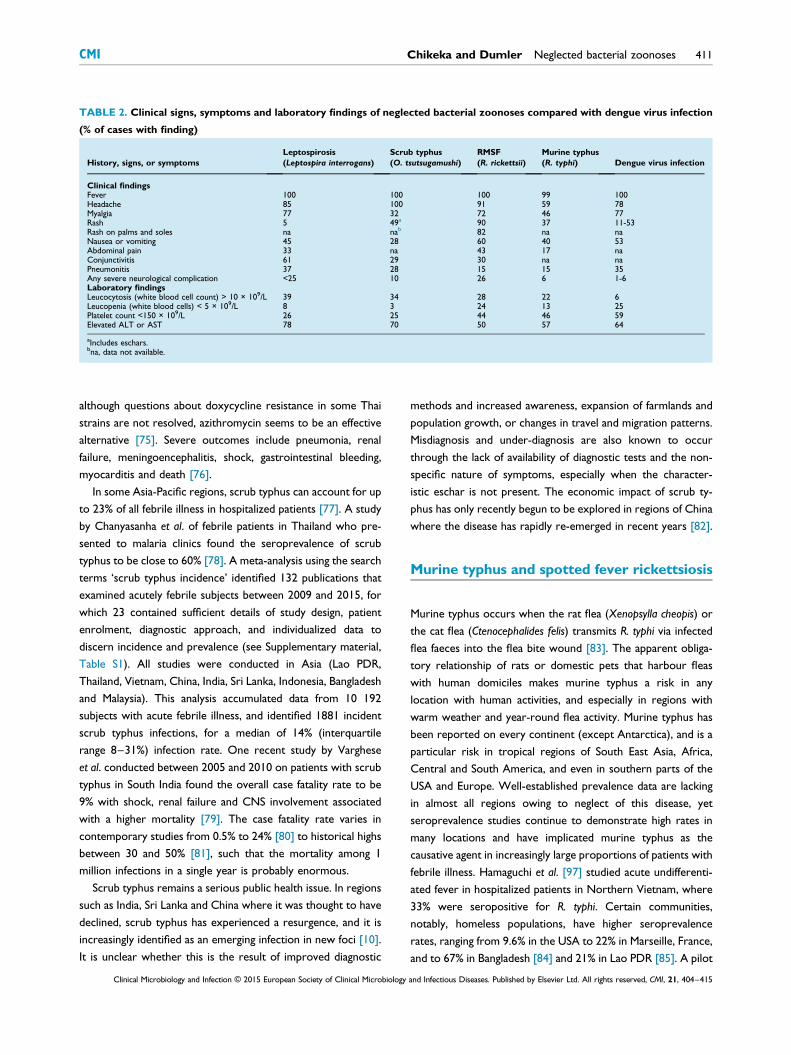
TABLE 2. Clinical signs, symptoms and laboratory findings of neglected bacterial zoonoses compared with dengue virus infection
(% of cases with finding)
History, signs, or symptomsLeptospirosis(Leptospira interrogans)
Scrub typhus(O. tsutsugamushi)
RMSF(R. rickettsii)
Murine typhus(R. typhi) Dengue virus infection
Clinical findingsFever 100 100 100 99 100Headache 85 100 91 59 78Myalgia 77 32 72 46 77Rash 5 49a 90 37 11-53Rash on palms and soles na nab 82 na naNausea or vomiting 45 28 60 40 53Abdominal pain 33 na 43 17 naConjunctivitis 61 29 30 na naPneumonitis 37 28 15 15 35Any severe neurological complication <25 10 26 6 1-6Laboratory findingsLeucocytosis (white blood cell count) > 10 × 109/L 39 34 28 22 6Leucopenia (white blood cells) < 5 × 10⁹/L 8 3 24 13 25Platelet count <150 × 109/L 26 25 44 46 59Elevated ALT or AST 78 70 50 57 64
aIncludes eschars.bna, data not available.
CMI Chikeka and Dumler Neglected bacterial zoonoses 411
although questions about doxycycline resistance in some Thai
strains are not resolved, azithromycin seems to be an effectivealternative [75]. Severe outcomes include pneumonia, renal
failure, meningoencephalitis, shock, gastrointestinal bleeding,myocarditis and death [76].
In some Asia-Pacific regions, scrub typhus can account for upto 23% of all febrile illness in hospitalized patients [77]. A studyby Chanyasanha et al. of febrile patients in Thailand who pre-
sented to malaria clinics found the seroprevalence of scrubtyphus to be close to 60% [78]. A meta-analysis using the search
terms ‘scrub typhus incidence’ identified 132 publications thatexamined acutely febrile subjects between 2009 and 2015, for
which 23 contained sufficient details of study design, patientenrolment, diagnostic approach, and individualized data to
discern incidence and prevalence (see Supplementary material,Table S1). All studies were conducted in Asia (Lao PDR,
Thailand, Vietnam, China, India, Sri Lanka, Indonesia, Bangladeshand Malaysia). This analysis accumulated data from 10 192subjects with acute febrile illness, and identified 1881 incident
scrub typhus infections, for a median of 14% (interquartilerange 8–31%) infection rate. One recent study by Varghese
et al. conducted between 2005 and 2010 on patients with scrubtyphus in South India found the overall case fatality rate to be
9% with shock, renal failure and CNS involvement associatedwith a higher mortality [79]. The case fatality rate varies in
contemporary studies from 0.5% to 24% [80] to historical highsbetween 30 and 50% [81], such that the mortality among 1million infections in a single year is probably enormous.
Scrub typhus remains a serious public health issue. In regionssuch as India, Sri Lanka and China where it was thought to have
declined, scrub typhus has experienced a resurgence, and it isincreasingly identified as an emerging infection in new foci [10].
It is unclear whether this is the result of improved diagnostic
methods and increased awareness, expansion of farmlands and
population growth, or changes in travel and migration patterns.Misdiagnosis and under-diagnosis are also known to occur
through the lack of availability of diagnostic tests and the non-specific nature of symptoms, especially when the character-
istic eschar is not present. The economic impact of scrub ty-phus has only recently begun to be explored in regions of Chinawhere the disease has rapidly re-emerged in recent years [82].
Murine typhus and spotted fever rickettsiosis
Murine typhus occurs when the rat flea (Xenopsylla cheopis) or
the cat flea (Ctenocephalides felis) transmits R. typhi via infectedflea faeces into the flea bite wound [83]. The apparent obliga-tory relationship of rats or domestic pets that harbour fleas
with human domiciles makes murine typhus a risk in anylocation with human activities, and especially in regions with
warm weather and year-round flea activity. Murine typhus hasbeen reported on every continent (except Antarctica), and is a
particular risk in tropical regions of South East Asia, Africa,Central and South America, and even in southern parts of the
USA and Europe. Well-established prevalence data are lackingin almost all regions owing to neglect of this disease, yetseroprevalence studies continue to demonstrate high rates in
many locations and have implicated murine typhus as thecausative agent in increasingly large proportions of patients with
febrile illness. Hamaguchi et al. [97] studied acute undifferenti-ated fever in hospitalized patients in Northern Vietnam, where
33% were seropositive for R. typhi. Certain communities,notably, homeless populations, have higher seroprevalence
rates, ranging from 9.6% in the USA to 22% in Marseille, France,and to 67% in Bangladesh [84] and 21% in Lao PDR [85]. A pilot
and Infectious Diseases. Published by Elsevier Ltd. All rights reserved, CMI, 21, 404–415
412 Clinical Microbiology and Infection, Volume 21 Number 5, May 2015 CMI
meta-analysis conducted using the search terms ‘murine
typhus + incidence’, ‘murine typhus + acute febrile illness’, and‘murine typhus’ retrieved 880 articles since 1934 and only the
325 articles dating to 2001 were reviewed for adequacy ofdetails and methodology, including geographic location for in-
clusion in the study, and with sole focus on studies of incidenceamong prospective acute febrile disease studies (see Supple-mentary material, Table S2). Selected studies originated from
every continent except Antarctica. Of the 325 articlesreviewed, 26 were selected for inclusion, comprising a total of
8642 patients, among which 1486 (17.2%) were deemed tohave murine typhus based on serology and PCR and culture.
The median proportion of murine typhus cases among thestudies was 7.9% (interquartile range 4.2–14.5%).
As with scrub typhus, the clinical presentation is most oftenundifferentiated fever, but lacking an eschar (Table 2) [86–88].A rash can develop in half of infected patients. Complications
are similar to those of other rickettsioses. The case fatality rateis variably between 0.5 and 4% in contemporary series and
most infections have a benign clinical course; however,morbidity can be significant particularly in cases of delayed
diagnosis and in the elderly who, even with proper treatment,suffer a greater number of complications.
Spotted fever group rickettsiosis is best known because ofthe severity of RMSF (R. rickettsii infection) [89–91]. Whereas
RMSF occurs only in the western hemisphere, other SFGRoccur globally. Individual species vary in pathogenicity, but theunderlying pathology for SFGR is endothelial cell infection,
vasculitis and increased systemic and pulmonary vascularpermeability [92]. The majority of SFGR are transmitted by tick
bites; for R. rickettsii this includes Dermacentor variabilis, Der-macentor andersoni and Rhipicephalus sanguineus in North
America, and Amblyomma species in Central and South Amer-ica. Rickettsia conorii is vectored by Rhipicephalus sanguineus in
the Mediterranean region, whereas R. africae is transmitted viaAmblyomma tick bites in Sub-Saharan Africa. A few SFGR arevectored by other arthropods (mites for rickettsial pox (Rick-
ettsia akari); fleas for cat flea typhus (Rickettsia felis)).Spotted fever group rickettsioses are among the most viru-
lent of all human infections, especially RMSF for which historicalcase fatality rates of 25–80% are recorded [92]. Contemporary
data describe case fatality rates between 0.5 and 8–12% in theUSA and up to 35% in Brazil. In Beja, Portugal in 1997, the case
fatality rate in hospitalized patients with Mediterranean spottedfever was 32.3% and in the USA, the American Indian popula-
tion is especially affected by RMSF with both incidence and casefatality rates significantly higher than that of other racial groups;this incidence continues to increase at a disproportionate rate.
Of particular interest is the high seroprevalence of SFGR inmany regions around the world where human SFGR are not
known to exist [91,93]. In southern Taiwan, a region where
spotted fever is poorly characterized, an investigation of 413febrile patients found that 49 (11.9%) were seropositive for
spotted fever group rickettsiae. Spotted fever is also increas-ingly diagnosed in South Asia, including Sri Lanka where spotted
fever rickettsiosis occurred in 10% of febrile patients, second innumber only to leptospirosis [13], and the seroprevalence was33% for any rickettsial infection. Despite inadequate data
collection, reported cases of SFGR are at historical highs in theUSA and globally [64,91]. The reasons for this are not clear, but
could include greater clinical suspicion, case definition anddiagnostic test changes, or real increases in disease incidence
and prevalence. The latter is in part likely given the increasinglydefined distribution of infected vectors [91].
Clinical manifestations of RMSF and other SFGR includefever, and variably headache, myalgias, rash, (macular, mac-ulopapular or petechial), abdominal pain, nausea, vomiting,
diarrhoea, and in severe cases, renal failure, non-cardiogenicpulmonary oedema, shock and multi-organ failure, and CNS
involvement (meningoencephalitis, cerebral oedema, hernia-tion) (Table 2). Most SFGR do not demonstrate this degree of
severity, but it is possible in any single case. Many patients haveeschars (except in RMSF), and some develop vesicular rashes.
Major factors for ineffective diagnosis and delayed therapyinclude absence of a typical rash, presentation during non-peak
tick activity season, and presentation during first 3 days ofillness when a rash may not be present [94].
Murine typhus and SFGR diagnosis requires clinical suspicion
because adequate diagnostic tests in the acute phase of diseaseare not available [95]. If rash is present, rickettsiae can be
demonstrated by immunohistochemistry in rash lesion biopsiesin SFGR and murine typhus. Although PCR sensitivity in blood
is low (<25%), accumulating data suggest that skin biopsy PCRcould be sensitive [91]. The most sensitive diagnostic test,
immunofluorescence assay, is only confirmatory because itrequires acute and convalescent serum antibody titres. How-ever, in general, immunofluorescence assays cannot distinguish
SFGR species or cross-reactions with typhus group rickettsiae.Cultivation is not timely and is generally considered dangerous
and unacceptable for clinical laboratories. Perhaps most chal-lenging of all is the fact that serodiagnosis only works well with
paired sera separated by at least 14 days, precluding its use fordiagnosis and management at the time of active infection. Given
the marked but mostly unrecognized incidence of these in-fections worldwide, and their high case fatality rates, readily
available accurate and sensitive point-of-care diagnostic devicesare desperately needed.
Both SFGR and murine typhus are treated with doxycycline;
however, in a study that examined treatment practices in theUSA, fewer than 40% of healthcare providers correctly chose
ious Diseases. Published by Elsevier Ltd. All rights reserved, CMI, 21, 404–415
CMI Chikeka and Dumler Neglected bacterial zoonoses 413
doxycycline as the treatment of choice for RMSF for children
<8 years of age, a potential cause of the increased case fatalityrates in this age category [95]. Controlled clinical trials have not
been conducted to adequately define efficacy among antibioticsexcept for R. conorii but some evidence exists to support the
use of fluoroquinolones for murine typhus and forms of SFGRother than RMSF, though their use has become controversial[96]. Currently, no vaccines exist for spotted fever rickettsiosis,
murine typhus or scrub typhus. Prevention requires avoidanceof bites by ticks, fleas and chiggers by avoiding infested loca-
tions, by wearing clothing that potentially excludes their abilityto access the skin, or by use of repellents. Prophylactic anti-
biotic treatment is not well assessed and currently cannot beadvocated.
Conclusions
Other than leptospirosis, relapsing fever, scrub typhus andrickettsioses are not yet recognized as neglected diseases by the
WHO, despite data that illustrate their high incidence andprevalence and the potentially dramatic morbidity and costs to
human life. Future goals should include establishing their inci-dence and prevalence comparative to other infections that
seemingly occur at similar rates. Development of easilydeployed point-of-care diagnostics will be critical to assist inaccurately collecting these data and in directing appropriate
care and treatments. Careful clinical and epidemiological/ecological studies, including the roles of animal or vector res-
ervoirs that increase risk will identify opportunities to preventinfection. Study of human infections could identify new loca-
tions where other preventative methods, including vaccinationmight be used. A basic analysis of disability-adjusted life-years
will depend on careful collection of such data and could drivedeployment of critically short resources in areas where theseinfections cause more harm than others commonly believed to
have the greatest impact on humans and their environments.
Appendix A. Supplementary data
Supplementary data related to this article can be found at http://dx.doi.org/10.1016/j.cmi.2015.04.022.
References
[1] Utzinger J, Becker SL, Knopp S, et al. Neglected tropical diseases:diagnosis, clinical management, treatment and control. Swiss Med Wkly2012;142:w13727.
[2] Centers for Disease Control. Summary of notifiable diseases—UnitedStates, 2012. Morbid Mortal Wkly Rep 2014;61:1–121.
[3] World Health Organization. Neglected tropical diseases. 2014. http://www.who.int/neglected_diseases/diseases/en/.
[4] Centers for Disease Control. Neglected tropical diseases. 2014. http://www.cdc.gov/globalhealth/ntd/diseases/index.html.
[5] Bill & Melinda Gates Foundation. Neglected infectious diseases. Seattle,WA: Bill & Melinda Gates Foundation; 2014. http://www.gatesfoundation.org/What-We-Do/Global-Health/Neglected-Infectious-Diseases#bodyregion_0_interiorarticle_0_strategysections_2_strategysubsections45615201ec9a4a6f8b6ec35aec7d7911_3_lnkHeader.
[6] European Commission. Neglected infectious diseases neglected nomore. 2014. http://ec.europa.eu/research/health/infectious-diseases/neglected-diseases/pdf/nid-leaflet_en.pdf.
[7] Lozano R, Naghavi M, Foreman K, et al. Global and regional mortalityfrom 235 causes of death for 20 age groups in 1990 and 2010: a sys-tematic analysis for the global burden of disease study 2010. Lancet2012;380:2095–128.
[8] Joshi R, Kengne AP, Neal B. Methodological trends in studies based onverbal autopsies before and after published guidelines. Bull WorldHealth Organ 2009;87:678–82.
[9] Grosse SD, Lollar DJ, Campbell VA, Chamie M. Disability anddisability-adjusted life years: not the same. Pub Health Rpt 2009;124:197–202.
[10] Paris DH, Shelite TR, Day NP, Walker DH. Unresolved problemsrelated to scrub typhus: a seriously neglected life-threatening disease.Am J Trop Med Hyg 2013;89:301–7.
[11] Acestor N, Cooksey R, Newton PN, et al. Mapping the aetiology ofnon-malarial febrile illness in southeast asia through a systematic re-view—terra incognita impairing treatment policies. PLoS ONE 2012;7:e44269.
[12] Mayxay M, Castonguay-Vanier J, Chansamouth V, et al. Causes of non-malarial fever in laos: a prospective study. Lancet Glob Health 2013;1:e46–54.
[13] Reller ME, Bodinayake C, Nagahawatte A, et al. Unsuspected rick-ettsioses among patients with acute febrile illness, Sri Lanka, 2007.Emerg Infect Dis 2012;18:825–9.
[14] Reller ME, Bodinayake C, Nagahawatte A, et al. Leptospirosis asfrequent cause of acute febrile illness in southern Sri Lanka. EmergInfect Dis 2011;17:1678–84.
[15] Murdoch DR, Woods CW, Zimmerman MD, et al. The etiology offebrile illness in adults presenting to patan hospital in Kathmandu,.Nepal Am J Trop Med Hyg 2004;70:670–5.
[16] Chheng K, Carter MJ, Emary K, et al. A prospective study of the causesof febrile illness requiring hospitalization in children in Cambodia. PLoSONE 2013;8:e60634.
[17] Punjabi NH, Taylor WR, Murphy GS, et al. Etiology of acute, non-malaria, febrile illnesses in Jayapura, northeastern Papua, Indonesia.Am J Trop Med Hyg 2012;86:46–51.
[18] Bharti AR, Nally JE, Ricaldi JN, et al. Leptospirosis: a zoonotic diseaseof global importance. Lancet Infect Dis 2003;3:757–71.
[19] Levett PN. Leptospirosis. Clin Microbiol Rev 2001;14:296–326.[20] Hartskeerl RA, Collares-Pereira M, Ellis WA. Emergence, control and
re-emerging leptospirosis: dynamics of infection in the changing world.Clin Microbiol Infect 2011;17:494–501.
[21] Ko AI, Goarant C, Picardeau M. Leptospira: the dawn of the moleculargenetics era for an emerging zoonotic pathogen. Nat Rev Microbiol2009;7:736–47.
[22] Brenner DJ, Kaufmann AF, Sulzer KR, Steigerwalt AG, Rogers FC,Weyant RS. Further determination of DNA relatedness betweenserogroups and serovars in the family leptospiraceae with a proposalfor Leptospira alexanderi sp. nov. and four new Leptospira genomo-species. Int J Syst Bacteriol 1999;49(Pt 2):839–58.
and Infectious Diseases. Published by Elsevier Ltd. All rights reserved, CMI, 21, 404–415
414 Clinical Microbiology and Infection, Volume 21 Number 5, May 2015 CMI
[23] Schreier S, Doungchawee G, Chadsuthi S, Triampo D, Triampo W.Leptospirosis: Current situation and trends of specific laboratory tests.Expert Rev Clin Immunol 2013;9:263–80.
[24] Katz AR, Ansdell VE, Effler PV, Middleton CR, Sasaki DM. Assessmentof the clinical presentation and treatment of 353 cases of laboratory-confirmed leptospirosis in Hawaii, 1974–1998. Clin Infect Dis2001;33:1834–41.
[27] Rafizah AA, Aziah BD, Azwany YN, et al. A hospital-based study onseroprevalence of leptospirosis among febrile cases in northeasternMalaysia. Int J Infect Dis 2013;17:e394–7.
[28] Lindo J, Brown PD, Vickers I, Brown M, Jackson ST, Lewis-Fuller E.Leptospirosis and malaria as causes of febrile illness during a dengueepidemic in Jamaica. Pathog Glob Health 2013;107:329–34.
[29] Reller ME, Wunder Jr EA, Miles JJ, et al. Unsuspected leptospirosis is acause of acute febrile illness in Nicaragua. PLoS Negl Trop Dis 2014;8:e2941.
[30] Reller ME, Bodinayake C, Nagahawatte A, et al. Unsuspected dengueand acute febrile illness in rural and semi-urban southern Sri Lanka.Emerg Infect Dis 2012;18:256–63.
[31] Reller ME, Akoroda U, Nagahawatte A, et al. Chikungunya as a cause ofacute febrile illness in southern Sri Lanka. PLoS ONE 2013;8:e82259.
[32] Rafizah AA, Aziah BD, Azwany YN, et al. Leptospirosis in northeasternMalaysia: misdiagnosed or coinfection? Int J Collab Res Intern MedPublic Health 2012;4:1419–27.
[33] Suputtamongkol Y, Niwattayakul K, Suttinont C, et al. An open, ran-domized, controlled trial of penicillin, doxycycline, and cefotaxime forpatients with severe leptospirosis. Clin Infect Dis 2004;39:1417–24.
[34] Traxler RM, Callinan LS, Holman RC, Steiner C, Guerra MA. Lepto-spirosis-associated hospitalizations, United States, 1998–2009. EmergInfect Dis 2014;20:1273–9.
[35] Souza VM, Arsky Mde L, Castro AP, Araujo WN. Years of potential lifelost and hospitalization costs associated with leptospirosis in Brazil.Revista de saude publica 2011;45:1001–8.
[36] Adler B, Lo M, Seemann T, Murray GL. Pathogenesis of leptospirosis:the influence of genomics. Vet Microbiol 2011;153:73–81.
[37] Verma A, Stevenson B. Leptospiral uveitis—there is more to it thanmeets the eye! Zoonoses Public Health 2012;59(Suppl. 2):132–41.
[38] Bajani MD, Ashford DA, Bragg SL, et al. Evaluation of four commer-cially available rapid serologic tests for diagnosis of leptospirosis. J ClinMicrobiol 2003;41:803–9.
[39] Brett-Major DM, Coldren R. Antibiotics for leptospirosis. CochraneDatabase Syst Rev 2012;2:CD008264.
[40] Samarakoon YM, Gunawardena N. Knowledge and self-reportedpractices regarding leptospirosis among adolescent school children ina highly endemic rural area in sri lanka. Rural Remote Health 2013;13:2360.
[41] Sehgal SC, Sugunan AP, Murhekar MV, Sharma S, Vijayachari P. Ran-domized controlled trial of doxycycline prophylaxis against leptospi-rosis in an endemic area. Int J Antimicrob Agents 2000;13:249–55.
[42] Bracho G, Varela E, Fernandez R, et al. Large-scale application ofhighly-diluted bacteria for leptospirosis epidemic control. Homeopathy2010;99:156–66.
[43] Barbour AG, Hayes SF. Biology of Borrelia species. Microbiol Rev1986;50:381–400.
[46] Lescot M, Audic S, Robert C, et al. The genome of Borrelia recurrentis,the agent of deadly louse-borne relapsing fever, is a degraded subset oftick-borne Borrelia duttonii. PLoS Genet 2008;4:e1000185.
[47] Houhamdi L, Raoult D. Excretion of living Borrelia recurrentis in feces ofinfected human body lice. J Infect Dis 2005;191:1898–906.
[48] Larsson C, Andersson M, Bergstrom S. Current issues in relapsingfever. Curr Op Infect Dis 2009;22:443–9.
[49] Dworkin MS, Schwan TG, Anderson Jr DE, Borchardt SM. Tick-bornerelapsing fever. Infect Dis Clin North Am 2008;22. 449–68, viii.
[50] Mayegga E, Ljøstad U, Mygland Å, Monstad P. Absence of focalneurological involvement in tick-borne relapsing fever in northernTanzania. Eur J Neurol 2005;12:449–52.
[51] Nordstrand A, Bunikis I, Larsson C, et al. Tickborne relapsing feverdiagnosis obscured by malaria, Togo. Emerg Infect Dis 2007;13:117–23.
[52] Reller ME, Chen WH, Dalton J, Lichay MA, Dumler JS. Multiplex 5’nuclease quantitative real-time pcr for clinical diagnosis of malaria andspecies-level identification and epidemiologic evaluation of malaria-causing parasites, including Plasmodium knowlesi. J Clin Microbiol2013;51:2931–8.
[53] McConnell J. Tick-borne relapsing fever under-reported. Lancet InfectDis 2003;3:604.
[54] Barclay AJ, Coulter JB. Tick-borne relapsing fever in central Tanzania.Trans R Soc Trop Med Hyg 1990;84:852–6.
[55] Yimer M, Mulu W, Ayalew W, Abera B. Louse-borne relapsing feverprofile at Felegehiwot referral hospital, Bahir Dar City, Ethiopia: aretrospective study. BMC Res Notes 2014;7:250.
[56] Cutler SJ, Abdissa A, Trape JF. New concepts for the old challenge ofAfrican relapsing fever borreliosis. Clin Microbiol Infect 2009;15:400–6.
[57] Brouqui P, Stein A, Dupont HT, et al. Ectoparasitism and vector-bornediseases in 930 homeless people from Marseilles. Medicine (Baltimore)2005;84:61–8.
[58] Porcella SF, Raffel SJ, Schrumpf ME, Schriefer ME, Dennis DT,Schwan TG. Serodiagnosis of louse-borne relapsing fever with glyc-erophosphodiester phosphodiesterase (Glpq) from Borrelia recurrentis.J Clin Microbiol 2000;38:3561–71.
[59] Seboxa T, Fekade D. Borrelia species (relapsing fever). In: Yu VL,Weber R, Raoult D, editors. Antimicrobial therapy and vaccines. 2nded. New York: Apple Trees Productions, LLC; 2002. p. 117–20.
[60] Blacksell SD, Jenjaroen K, Phetsouvanh R, et al. Accuracy of rapid IgM-based immunochromatographic and immunoblot assays for diagnosisof acute scrub typhus and murine typhus infections in Laos. Am J TropMed Hyg 2010;83:365–9.
[61] Dumler JS, Barbet AF, Bekker CP, et al. Reorganization of genera in thefamilies Rickettsiaceae and Anaplasmataceae in the order Rickettsiales:unification of some species of Ehrlichia with Anaplasma, Cowdria withEhrlichia and Ehrlichia with Neorickettsia, descriptions of six newspecies combinations and designation of Ehrlichia equi and ’HGE agent’as subjective synonyms of Ehrlichia phagocytophila. Int J Syst EvolMicrobiol 2001;51:2145–65.
[62] Gillespie JJ, Williams K, Shukla M, et al. Rickettsia phylogenomics: un-winding the intricacies of obligate intracellular life. PLoS ONE 2008;3:e2018.
[63] Dahlgren FS, Moonesinghe R, McQuiston JH. Short report: race andrickettsiae: a United States perspective. Am J Trop Med Hyg 2011;85:1124–5.
[64] Openshaw JJ, Swerdlow DL, Krebs JW, et al. Rocky Mountain spottedfever in the United States, 2000–2007: interpreting contemporaryincreases in incidence. Am J Trop Med Hyg 2010;83:174–82.
[65] Aung AK, Spelman DW, Murray RJ, Graves S. Rickettsial infections inSoutheast Asia: implications for local populace and febrile returnedtravelers. Am J Trop Med Hyg 2014;91:451–60.
ious Diseases. Published by Elsevier Ltd. All rights reserved, CMI, 21, 404–415
CMI Chikeka and Dumler Neglected bacterial zoonoses 415
[66] Zhang L, Shan A, Mathew B, et al. Rickettsial seroepidemiology amongfarm workers, Tianjin, People’s Republic of China. Emerg Infect Dis2008;14:938–40.
[67] Walker DH, Paddock CD, Dumler JS. Emerging and re-emerging tick-transmitted rickettsial and ehrlichial infections. Med Clin North Am2008;92. 1345–61, x.
[68] Kelly DJ, Fuerst PA, Ching WM, Richards AL. Scrub typhus: thegeographic distribution of phenotypic and genotypic variants of Orientiatsutsugamushi. Clin Infect Dis 2009;48(Suppl. 3):S203–30.
[69] Thiga JW, Mutai BK, Eyako WK, et al. High seroprevalence of anti-bodies against spotted fever and scrub typhus bacteria in patients withfebrile illness, Kenya. Emerg Infect Dis 2015;21:688–91.
[72] Koh GC, Maude RJ, Paris DH, Newton PN, Blacksell SD. Diagnosis ofscrub typhus. Am J Trop Med Hyg 2010;82:368–70.
[73] Paris DH, Blacksell SD, Stenos J, et al. Real-time multiplex pcr assay fordetection and differentiation of rickettsiae and orientiae. Trans RoyalSoc Trop Med Hyg 2008;102:186–93.
[74] Phetsouvanh R, Thojaikong T, Phoumin P, et al. Inter- and intra-operator variability in the reading of indirect immunofluorescenceassays for the serological diagnosis of scrub typhus and murine typhus.Am J Trop Med Hyg 2013;88:932–6.
[75] Jang MO, Jang HC, Kim UJ, et al. Outcome of intravenous azithromycintherapy in patients with complicated scrub typhus compared with thatof doxycycline therapy using propensity-matched analysis. AntimicrobAgents Chemother 2014;58:1488–93.
[76] Jang MO, Kim JE, Kim UJ, et al. Differences in the clinical presentationand the frequency of complications between elderly and non-elderlyscrub typhus patients. Arch Gerontol Geriatr 2014;58:196–200.
[77] Brown GW, Robinson DM, Huxsoll DL, Ng TS, Lim KJ. Scrub typhus: acommon cause of illness in indigenous populations. Trans Royal SocTrop Med Hyg 1976;70:444–8.
[78] Chanyasanha C, Kaeburong K, Chenchittikul M, Sujirarat D. Sero-prevalence of scrub typhus infection in patients with pyrexia at somemalaria clinics in three western provinces of Thailand. Asian Pac JAllergy Immunol 1998;16:119–25.
[79] Varghese GM, Trowbridge P, Janardhanan J, et al. Clinical profile andimproving mortality trend of scrub typhus in South India. Int J InfectDis 2014;23:39–43.
[80] Griffith M, Peter JV, Karthik G, et al. Profile of organ dysfunctionand predictors of mortality in severe scrub typhus infectionrequiring intensive care admission. Indian J Crit Care Med 2014;18:497–502.
[81] Kawamura Jr A, Tanaka H. Rickettsiosis in Japan. Jpn J Exp Med1988;58:169–84.
[82] Yang LP, Liang SY, Wang XJ, Li XJ, Wu YL, Ma W. Burden of diseasemeasured by disability-adjusted life years and a disease forecasting time
series model of scrub typhus in Laiwu, China. PLoS Negl Trop Dis2015;9:e3420.
[83] Gillespie JJ, Ammerman NC, Beier-Sexton M, Sobral BS, Azad AF.Louse- and flea-borne rickettsioses: Biological and genomic analyses.Vet Res 2009;40:12.
[84] Maude RR, Maude RJ, Ghose A, et al. Serosurveillance of Orientiatsutsugamushi and Rickettsia typhi in Bangladesh. Am J Trop Med Hyg2014;91:580–3.
[85] Vallee J, Thaojaikong T, Moore CE, et al. Contrasting spatial distribu-tion and risk factors for past infection with scrub typhus and murinetyphus in Vientiane city, Lao PDR. PLoS Negl Trop Dis 2010;4:e909.
[86] Gikas A, Doukakis S, Pediaditis J, Kastanakis S, Psaroulaki A, Tselentis Y.Murine typhus in Greece: epidemiological, clinical, and therapeutic datafrom 83 cases. Trans Royal Soc Trop Med Hyg 2002;96:250–3.
[87] Silpapojakul K, Chayakul P, Krisanapan S, Silpapojakul K. Murine typhusin Thailand: clinical features, diagnosis and treatment. Q J Med 1993;86:43–7.
[88] Dumler JS, Taylor JP, Walker DH. Clinical and laboratory features ofmurine typhus in South Texas, 1980 through 1987. JAMA 1991;266:1365–70.
[89] Walker DH, Ismail N. Emerging and re-emerging rickettsioses: endo-thelial cell infection and early disease events. Nat Rev Microbiol2008;6:375–86.
[90] Cazorla C, Socolovschi C, Jensenius M, Parola P. Tick-borne diseases:tick-borne spotted fever rickettsioses in Africa. Infect Dis Clin NorthAm 2008;22. 531–44, ix–x.
[91] Parola P, Paddock CD, Socolovschi C, et al. Update on tick-bornerickettsioses around the world: a geographic approach. Clin Micro-biol Rev 2013;26:657–702.
[92] Ismail N, Walker DH, Ghose P, Tang YW. Immune mediators ofprotective and pathogenic immune responses in patients with mild andfatal human monocytotropic ehrlichiosis. BMC Immunol 2012;13:26.
[93] Susilawati TN,McBrideWJ.Acute undifferentiated fever inAsia: a reviewof the literature. SE Asian J Trop Med Public Health 2014;45:719–26.
[94] Kirkland KB, Wilkinson WE, Sexton DJ. Therapeutic delay and mor-tality in cases of Rocky Mountain spotted fever. Clin Infect Dis1995;20:1118–21.
[95] Chapman AS, Bakken JS, Folk SM, et al. Diagnosis and management oftickborne rickettsial diseases: Rocky Mountain spotted fever, ehrli-chioses, and anaplasmosis—United States: a practical guide for phy-sicians and other health-care and public health professionals. MMWRRecomm Rep 2006;55:1–27.
[96] Botelho-Nevers E, Rovery C, Richet H, Raoult D. Analysis of riskfactors for malignant mediterranean spotted fever indicates that fluo-roquinolone treatment has a deleterious effect. J Antimicrob Che-mother 2011;66:1821–30.
[97] Hamaguchi S, Cuong NC, Tra DT, et al. Clinical and epidemiologicalcharacteristics of scrub typhus and murine typhus among hospitalizedpatients with acute undifferentiated fever in northern vietnam. Am JTrop Med Hyg 2015;92:972–8.
and Infectious Diseases. Published by Elsevier Ltd. All rights reserved, CMI, 21, 404–415