Page 1
Neogenin1 is a Sonic Hedgehog target in
medulloblastoma and is necessary for cell cycle
progression.
Running title: Neo1 is a novel direct Shh downstream mediator in cerebellar
growth and in Shh-driven medulloblastoma
Luis A. Milla1,2,7
, Andrea Arros1, Natalie Espinoza
1, Marc Remke
4, Marcel Kool
5,
Michael D. Taylor4, Stefan M. Pfister
5,6, Brandon J. Wainwright
3, Verónica
Palma1,2*
1 Faculty of Sciences, University of Chile, Santiago, Chile
2 FONDAP Center for Genome Regulation, Santiago, Chile
3 Institute for Molecular Bioscience, University of Queensland, Brisbane,
Australia
4 Developmental & Stem Cell Biology Program, The Hospital for Sick Children,
Toronto, Ontario M5G 1L7, Canada
5 Division of Pediatric Neurooncology, German Cancer Research Center (DKFZ),
Heidelberg, Germany
6 Department of Pediatric Hematology and Oncology, Heidelberg University
Hospital, Heidelberg, Germany
7Present address: Stanford University School of Medicine, Stanford, CA, USA
*Corresponding author: [email protected]
Abstract : 240
Text: 4500
Novelty and Impact statement:
In this study we identified Neogenin-1 as a new direct SHH downstream
regulator mediating proliferation in both cerebellar progenitors and SHH
derived medulloblastoma suggesting that Neogenin-1 could be a promising
alternative avenue for therapeutic intervention to anti-medulloblastoma
therapies.
Keywords: Medulloblastoma, Sonic Hedgehog, Neogenin 1, Gli, cancer
International Journal of Cancer
This article has been accepted for publication and undergone full peer review but has not beenthrough the copyediting, typesetting, pagination and proofreading process which may lead todifferences between this version and the Version of Record. Please cite this article as an‘Accepted Article’, doi: 10.1002/ijc.28330
Page 2
2
Abstract
The canonical Sonic Hedgehog (Shh)/Gli pathway plays multiples roles during
Central Nervous System (CNS) development. In order to elucidate the molecular
repertoire of Shh mediators, we have recently described novel transcriptional
targets in response to Shh pathway modulation. Among them, we were able to
identify Neogenin1 (Neo1), a death dependence receptor, as a new direct Shh
downstream regulator in neural precursor proliferation. Since appropriate Shh
signaling is required for cerebellar growth and alterations cause Shh-driven
medulloblastoma (MB), here we have addressed the role of the Shh/Neogenin1
interaction in the context of cerebellar development and cancer. We demonstrate
that the Shh pathway regulates Neogenin1 expression in mouse models that
recapitulate the Shh MB subtype. We show that the canonical Shh pathway
directly regulates the Neo1 gene, acting through an upstream sequence in its
promoter both in vitro, and in vivo in granule neuron precursor cells (GNPs). We
also identified and characterized a functional Gli Binding site in the first intron of
the human NEO1 gene. Gene expression profiling of more than 300 MB shows
that NEO1 is indeed upregulated in SHH tumors compared to the other MB
subgroups. Finally, we provide evidence that NEO1 is necessary for cell cycle
progression in a human MB cell line, since a loss of function of NEO1 arrests
cells in the G2/M phase. Taken together, these results highlight Neogenin1 as a
novel downstream effector of the Shh pathway in MB, and a possible therapeutic
target.
Page 2 of 32
John Wiley & Sons, Inc.
International Journal of Cancer
Page 3
3
Introduction
Medulloblastoma (MB), a primitive neuroepithelial tumor, is the most
common malignant childhood primary CNS tumor. Current treatment protocols
encompassing surgical resection, chemotherapy, and radiotherapy, contributed to
a better prognosis of MB patients. However, approximately one third of the MB
patients remain incurable and recurrence is still frequent 1.
Four major MB subgroups including WNT, SHH, Group 3 and Group 4,
have been described showing different genetic alterations, pathological features,
and cerebellar locations. WNT and SHH define the signaling pathways that are
deregulated in those subgroups 2. The molecular pathogenesis of the remaining
two groups is less well known, although recent experiments suggest
overexpression of Myc appears to drive “Group 3” MB 3 . Importantly, up to 30%
of human MBs provide evidence of abnormal Shh pathway activation 4.
The Shh pathway has multiple functions throughout development in
various tissues. It plays a role in cellular survival, proliferation, tissue
morphogenesis and differentiation 5. Appropriate Shh signaling is also required
for normal cerebellar development where SHH in the early postnatal period is
produced by the Purkinje cells (PC) to drive the expansion of granule neuron
precursors (GNP) in the external germinal layer (EGL). The EGL has also been
shown to be one of the origins of SHH MB.
Shh is a secreted glycoprotein that activates signaling in target cells by
binding to its 12-pass transmembrane receptor Patched 1 (Ptc1/Ptch1), which then
derepresses Smoothened (Smo), a seven-pass transmembrane protein and G-
Page 3 of 32
John Wiley & Sons, Inc.
International Journal of Cancer
Page 4
4
coupled co-receptor leading to activation of downstream pathway signaling. This
signaling converges in the Gli family of transcription factors (Gli1-3), activating
target gene transcription 6. The Shh pathway was first implicated in the
development of MB through the discovery of PTCH1 mutations in a subset of
patients 7
8 and mouse models with loss of Ptc1 that developed MB
9
10.
Subsequently, mutations in other members of the Shh pathway such as Supressor
of Fused (SuFu) 11
, and Smo 12
have been identified in MB as well. Understanding
the cellular response to Shh pathway activation in the cerebellum is therefore
critical to our understanding of MB formation and treatment.
In order to address the molecular repertoire of Shh target genes, we and
other groups have identified a number of transcriptional targets active during
embryonic development and in cancer 13
14
. In the cerebellum these Shh targets
include Ptc1, Ptc2, Gli1, Nmyc, CyclinD1, Bmi, Gpr153, Foxo6, Yap1 and Ncor2
15 14 16 17 18. In a recent study, we reported neogenin 1 (neo1) as a gene controlled
by the Shh pathway both in vitro and in vivo in zebrafish 13
. Neo1, a member of
the death dependence family of receptors and part of the immunoglobulin (ig)
superfamily, was originally isolated from embryonic chicken cerebellum as a
‘deleted in colorectal cancer’ (DCC) homolog. The protein contains 4
immunoglobulin-like domains followed by 6 fibronectin domains, a
transmembrane domain, and an intracellular domain 19
. It binds both Netrins and
the Repulsive Guidance Molecule (RGM) protein 20
and has many functions,
including axon guidance during vertebrate embryonic development 21
, controlling
cell survival and differentiation 22
23
. How Neo 1 regulates these processes is not
clear although it has been demonstrated that under some circumstances the Neo1
intracellular domain is cleaved and transported to the nucleus where it is capable
of directly regulating transcription. 24
.
Page 4 of 32
John Wiley & Sons, Inc.
International Journal of Cancer
Page 5
5
Here, we demonstrate that the Shh pathway regulates Neo1 expression in
mouse models that recapitulate the Shh MB subgroup. Our data indicate that
canonical Shh pathway activation directly regulates the Neo1 gene in mice and
humans, acting through Gli binding sites present in Neo1 regulatory regions.
Mutation of these sites abolishes responsiveness in murine GNPs primary cultures
as well as human cancer cell lines. Gene expression analysis performed on a total
of 343 patient samples identifies NEO1 as being upregulated in the Shh MB
subgroup. Finally, we present evidence that NEO1 is necessary for cell cycle
progression in a human MB cell line, where a loss of function of NEO1 arrests
cells in the G2/M phase. Taken together, these results highlight Neo1 as a possible
novel therapeutic target in Shh MB.
Page 5 of 32
John Wiley & Sons, Inc.
International Journal of Cancer
Page 6
6
Results
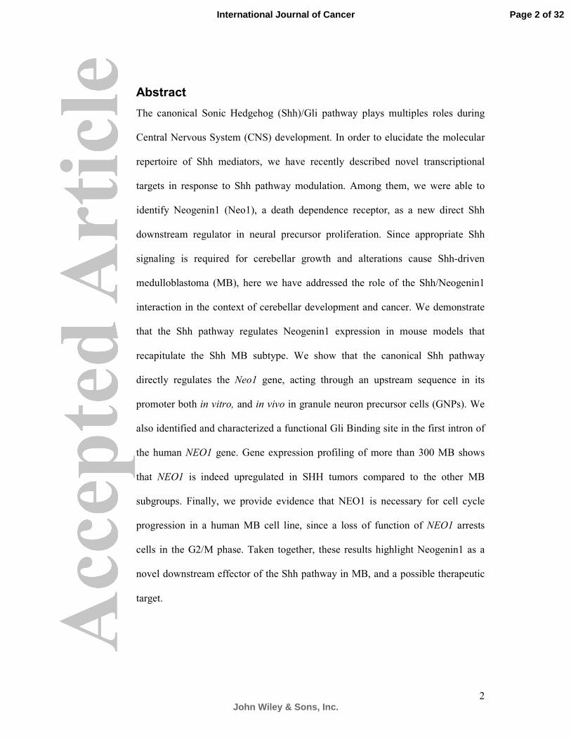
Neo1 expression is restricted to the proliferative EGL of the developing cerebellum
Neo1 is a cell adhesion molecule, and is expressed in a variety of
developing tissues, including the CNS. To determine Neo1 positive regions during
mouse cerebellar development, we evaluated Neo1 expression levels in the E18.5,
P8 and P14 cerebellum (Figure 1 and S1). Neo1 expression is found in the
developing EGL in all stages analyzed. In the mouse cerebellum the EGL reaches
a maximum thickness during the first postnatal week (P7-P8). During this stage,
Neo1 expression on cell somata and processes is mostly restricted to the most
outer group of cells in the EGL (o-EGL) where it colocalizes with the
proliferative nuclear marker PCNA (Figure 1A, C, white brackets). The inner
EGL (i-EGL), positive for the neuronal marker NeuN (Figure 1A) as well as the
internal granular layer (IGL), positive for NeuN and the granule neuron marker
Zic2 (Figure 1E), shows low Neo1 expression. Neo1 expression does not co-
localize with Bergman glia within the cerebellum (Figure 1G). Overall, these data
show that Neo1 is restricted largely to the o-EGL sublayer and when GNPs
postmitotically migrate inwards the cerebellum to form granule neurons, these
cells no longer express Neo1.
Neo1 is overexpressed in Shh-driven cerebellar tumor mouse models
and granule neuron precursors (GNP)
We have previously shown that Neo1 expression can be regulated by the
Shh pathway 13
. Yet, the relationship between the expression of Neo1 and the
growth of MB has not yet been defined. Given that Shh is the major mitogenic
pathway for cerebellar development 25
, we hypothesized that Shh could regulate
Page 6 of 32
John Wiley & Sons, Inc.
International Journal of Cancer
Page 7
7
Neo1 in this context, and examined this using two different Shh pathway driven
tumor murine models. The first one corresponds to the conditional hGFAP-Cre
mediated deletion of Ptc1 (hGFAP-Cre/Ptc1lox/lox
) resulting in activated Shh
signaling in neural stem and progenitor cells. In this model, the Ptc1 function is
ablated in the ventricular zone between E14.5 and E16.5, resulting in a thickened
and disorganized EGL filled with GNP like cells in later stages 10
. Neo1
expression is evident in the E18.5 EGL in the developing mutant cerebellum in a
pattern that resembles the wild type (wt) expression (Figure 2A and S1C). At P8
aberrant Shh activation in committed GNPs results in the formation of tumor
masses with strong Neo1 labeling that is co-expressed with the proliferation
marker PCNA. By P14, the tumors cover the entire cerebellum and have extensive
proliferation zones, clearly defined by Neo1 expression. This result contrasts with
expression of Neo1 in wt at P14, which is restricted to the EGL (Figure 2B-C and
S1D). Notably, the Neo1 positive cells in tumors are negative for the marker
NeuN (Figure 2D).
To evaluate relative differences in transcript levels for Neo1, we next
performed real-time PCR, comparing P7 samples, normal cerebella and isolated
GNPs from both wt and mutant mice. Of note, Neo1 RNA levels are substantially
higher in P14 tumors from the hGFAP-Cre/Ptc1lox/lox
than from wt GNPs (Figure
2E, right panel). Similar relative differences in RNA levels are seen when
comparing Gli1 levels, a well-known canonical Shh pathway readout gene (Figure
2E, left panel). The Neo1 RNA levels range from five to twelve fold higher in the
transgenic mouse cerebella compared to wild type whereas Gli1 ranges from eight
to thirteen fold higher. Thus, we conclude that Neo1 is highly expressed in GNP
like cells, and is likely overexpressed in this Shh driven MB mouse model.
Page 7 of 32
John Wiley & Sons, Inc.
International Journal of Cancer
Page 8
8
SHH pathway-activated MBs are thought to arise from GNPs in the
developing cerebellum that depend on Shh signaling for their expansion during
development. As shown in Figure 2F, proliferation, measured by BrdU
incorporation of GNPs cultures (P7), decreased significantly in the presence of the
Smoothened inhibitor cyclopamine in a dose dependent manner with 10 µM being
more effective than 5 µM. Importantly, when we analyzed GNP protein lysates by
Western blotting after 48 h of inhibitor treatment, we detected low levels of Neo1.
Neo1 showed a similar dose-dependent reduction in protein levels in response to
the drug as seen for Ptc1, a well-known hedgehog target (Figure 2 G).
Given our observations on GNPs we hypothesized that a specific increase
in Shh signaling in this cell population could account for the expanded Neo1
positive cells in vivo. Therefore, we used a second mouse model, the N2-
Cre/SmoA1, that forms MB due to the expression of a constitutively active form
of SmoA1 under a 1-kb human NeuroD2 promoter that drives the expression in
GNP 12
, resulting in early cerebellar hyperproliferation. It has been reported that
MB form in 94% of homozygous Smo/Smo mice by 2 months of age. Consistent
with the thickened EGL that is observed at P8 in N2-Cre/SmoA1 mice (Figure 2,
H, M), Neo1 and PCNA expression is also expanded (Figure 2 H- Q).
Thus, taken together, our data indicate that the Shh pathway modulates
Neo1 expression in the cerebellum, and that a tumorigenic deregulation results in
increased Neo1 levels in GNPs.
Shh/Gli directly regulates Neo1
To investigate whether the apparent transcriptional regulation of Neo1 by Shh
was direct or indirect, we applied a web-based bioinformatic approach to identify
putative Gli Binding Sites (GBS) in the Neo1 promoter. We detected two possible
Page 8 of 32
John Wiley & Sons, Inc.
International Journal of Cancer
Page 9
9
sites that possess the consensus GBS sequences at 18.3 kb (mGBS1) and 5.5 kb
(mGBS2) upstream of the Neo1 gene (Figure 3A). Given these observations, we
next addressed whether the putative GBS were likely to be functional in GNPs.
Accordingly, we performed chromatin immunoprecipitation (ChIP) in P7 GNPs
using antibodies to both murine Gli1 and Gli2. Gli2 but not Gli1 bound to the
mGBS1 while the mGBS2 did not bind Gli2 in this context (Figure 3B and data
not shown). To evaluate their functionality as enhancers, we isolated and cloned
these fragments upstream of a minimal promoter driving the luciferase reporter
gene with mutated versions in the consensus core as controls. In order to evaluate
the Gli dependence, a full-length form of Gli2 was utilized that activates a GBS
tandem repeat, as already described 6. Using the Shh responding cell line
CH310T1/2 it was verified that Gli2 expression was able to generate luciferase
activation using the 18.3kb sequence. As predicted from the ChIP data, the
putative 5.5kb GBS did not activate the reporter in response to Gli2 expression
(Figure S3 and 3C). Overall, these results support the previous ChIP findings and
corroborate that Gli2 factor is able to bind to the Neo1 promoter in P7 GNPs.
Further, these data suggest that the Shh pathway regulates Neo1 expression in the
cerebellum during development.
To further extend our findings to the human Neogenin1 gene we performed an
in silico analysis revealing three putative GBS in the NEO1 sequence: hGBS 1,
located 18.6 kb upstream from the translation start, and hGBS 2 and hGBS 3,
which are located within the first intron, 39.1 kb and 54.4 kb from the translation
start, respectively (Figure 3E). The GLI transcriptional factor recognition and
binding to these putative GBS was analyzed through ChIP using the human
neuroblastoma cell line SH-SY5Y. Whereas both Gli1 and Gli2 bind to a GBS
present in the promoter of PTCH1 (Figure S2), we found that Gli2 recognized
Page 9 of 32
John Wiley & Sons, Inc.
International Journal of Cancer
Page 10
10
only hGBS 3 (Figure 3F). Finally, in order to evaluate the NEO1 GBS
functionality, each site was placed upstream of the luciferase reporter gene.
Concordant with the ChIP result, only the hGBS3, induced luciferase expression
upon Gli2 stimulation whilst in mutated versions this effect was abolished (Figure
3G).
These results support the ChIP findings and corroborate the data from our analysis
of murine Neo1 that the Gli2 transcription factor is able to bind to the Neo1
promoter in P7 GNPs. Further, they suggest that Shh control of Neo1 expression
is likely conserved between murine and human species.
Neo1 is overexpressed in human SHH-MB
Next, we carried out gene expression analysis in a MB cohort of 343 samples
consisting of 40 WNT, 103 SHH, 79 Group 3 and 121 Group 4 tumors. NEO1 is
overexpressed in SHH-driven MBs as compared with other MB subgroups (Figure
4) suggesting NEO1 as a target of Shh signaling, and further underlining the data
from our murine MB studies. For comparison we also present the expression
profile in those same tumors for the established SHH target genes Gli1, Gli2 and
PTCH1 indicating that the expression pattern distribution of NEO1 is consistent
with being a defining feature of the SHH tumor group. We considered the
possibility that within the SHH medulloblastoma group there existed a sub-group
where NEO1 expression might define clinic-pathological features. We applied a
number of standard statistical approaches to this question and no significant
associations were found (data not shown).
Taken together, these data comprise the first observations of NEO1 up-
regulation in human medulloblastoma.
Page 10 of 32
John Wiley & Sons, Inc.
International Journal of Cancer
Page 11
11
NEO1 loss of function induces cell cycle arrest at G2/M.
To elucidate the possible role of Neo1 in MB, we performed NEO1 knock-down
experiments using lentiviral shRNA and the human medulloblastoma cell line
DAOY, which is predicted to be of the SHH subtype. Silencing NEO1 expression
(Figure 5A,B) resulted in an increased number of cells in G2/M cell cycle stage
compared to the control scrambled counterpart and fluorescent shNEO1
transduced cells demonstrated a reduced rate of BrdU incorporation (Figure 5C,
D). To examine whether NEO1 knock-down induces cell cycle arrest in late G2 or
M phase 26
, we quantified the number of histone H3 phosphorylated (H3P)
positive cells. Interestingly, shNEO1 cells accumulated H3P label relative to the
scrambled control (Figure S3A-C, left panel), even when there are fewer cells
expressing the shNEO1 lentivirus after 48 hours of treatment (Figure S3C, right
panel), indicating that cells are likely in a prolonged state G2/M arrest. Despite
the observed G2/M arrest, we still found mitotic figures and condensed
chromosomes (Figure S3D-F). Finally, we verified that NEO1 knock-down does
not induce premature cell differentiation (data not shown).
Page 11 of 32
John Wiley & Sons, Inc.
International Journal of Cancer
Page 12
12
Discussion
In the last few years, detailed genomic information along with the
engineering of different murine models have helped to uncover important
mechanisms in MB etiology, which may be exploited for therapeutic purposes.
Here we show that Neo1 is a novel direct Shh downstream mediator in cerebellar
growth and in Shh-driven MB.
Neo1, is a Shh regulated target expressed in the o-EGL
During cerebellar development, Shh produced by the Purkinje cells acts as
a potent mitogen, signaling to the EGL and increasing the number of the
proliferating GNPs, thus promoting the growth and foliation of the complete
cerebellum. Expression of Neo1 is spatially restricted to the proliferative o-EGL
during postnatal cerebellum development. This, in principle, was unexpected due
to the previously defined roles of Neo1 in axon guidance. However, there is
evidence that Neo1 is expressed in proliferating CNS zones such neurogenic
progenitors 27
and in cells displaying stem cell characteristics within the adult
human SVZ 28
. Within the EGL, a number of hedgehog targets, previously
demonstrated to promote cellular growth, are expressed including Gli1 25
, Nmyc,
CyclinD1 15
, C-Myc 29
, and Bmi1 17
. Neo1 localization studies presented here
suggest that its expression is downregulated in the mitotically-quiescent i-EGL
population when the inward migration of maturing granule neurons begins.
Therefore, the spatial expression pattern of Neo1 is consistent with that of a
putative Shh target gene. A functional mGBS for Neo1 was located at -18.3kb of
Neo1 origin, whereas for human NEO1 we defined a similar element in the first
intron. Long distance enhancers have been described for the members of the Shh
Page 12 of 32
John Wiley & Sons, Inc.
International Journal of Cancer
Page 13
13
pathway 30
31
, and many GBS located near to the gene do not appear to contribute
to transcriptional events 14
, as was the case of the -5.5 kb consensus mGBS
identified here for Neo1. It has been reported that weak GBS, can be acting along
with other non-consensus GBS to drive strong transcriptional activation 32
. This
possibility, or the utilization of the -5.5kb consensus mGBS in a different cellular
context cannot be ruled out. In the o-EGL and i-EGl interface, for instance, a Gli-
mediated differential regulation might contribute to fine-tuning the Neo1 spatial
restriction. Other direct Shh/Gli targets could be following a similar regulatory
pattern. Interestingly, we identified that Neo1 was likely directly regulated by
Gli2 and not by Gli1, which, if confirmed in other systems, would make Neo1 the
first Gli2 specific target identified to date.
Possible roles of Neo1 in normal cerebellar development and
medulloblastoma tumorigenesis
Apart from its initial role as a Netrin receptor participating in pathfinding
axon guidance, there appear to be multiple roles for Neo1 in different aspects of
embryonic development. For instance, as a signal for neural tube closure and
dorsal brain formation 33
, or as a regulator of gene transcription 24
. From the
discovery that the RGM ligand interacts with Neo1 20
, it has been suggested that
RGM/Neo1 interactions function as a dependence ligand/receptor couple,
regulating cell survival through a DAP kinase-dependent mechanism 22
34
.
Importantly, RGM A is expressed in the ventricular zone throughout the
embryonic brain. Thus, RGM A-Neo1 interactions may regulate progenitor
survival or proliferation within the proliferative zones of the developing CNS
(28).
Page 13 of 32
John Wiley & Sons, Inc.
International Journal of Cancer
Page 14
14
In studies of breast cancer there has been a suggestion that NEO1
expression is inversely correlated with mammary carcinogenicity 35
, there is also
evidence that shows a growth inhibition in NEO1 loss of function in human
ovarian epithelial cells 36
. Although there are reports indicating that NEO1 is
expressed in different human MB cell lines 37
to date no detailed analysis has been
performed.
Importantly, we found that Neo1 is expressed in a Shh-dependant murine
MB model, and that Neo1 is necessary to permit the cell cycle progression (Fig. 5
E, F). Indicating conserved roles between development and MB, we also observed
that Shh-induced human MB possessed high NEO1 levels.
Crosstalk of different, previously unrelated pathways may drive
tumorigenesis, probably recapitulating physiological developmental processes.
Notably, the ability of Neo1 to trigger apoptosis in absence of its ligands has been
considered as a “safeguard” mechanism preventing primary tumor proliferation
within a tissue depleted of its ligands (Netrin, RGM) 38
. Tumor cells constitutively
overexpressing the ligand could therefore escape this propoptotic regulation. It
would be interesting to ascertain if the Shh and the Netrin-RGM-Neo1 pathway
are related in different contexts. Further research is required to elucidate
potentially additional roles of NEO1 in cancer progression, such as regulation of
invasiveness, angionenesis or cooperation with the tumor formation.
Neo1 as an interactor with the Shh pathway
Shh signaling controls the brain size partly by controlling the proliferation
of neural stem/progenitor cells. Here, we demonstrate that in MB, Shh acts
through Gli2, to transcriptionally regulate Neo1 expression within the nucleus.
Page 14 of 32
John Wiley & Sons, Inc.
International Journal of Cancer
Page 15
15
Neogenin1 may be acting by sustaining the cell cycle completion during abnormal
cell growth in an opposite manner as the cell cycle interaction reported for Ptc1
and cyclin B (Fig 5B)39
. In other contexts, as for example in the neural tube,
Neo1/RGM act as a dependence receptor/ligand system 22
. Thus, the roles of Neo1
appear to be dependent on the complex biologic or cellular context.
A hallmark of Shh signaling is the upregulation of Ptc1, which functions as a
negative feedback that restricts the activity of Shh. We and other authors have
shown that Neo1 is expressed in proliferative zones of dorsal brain where the Shh
pathway is active. It has been recently reported that Neo1 acts as a negative
regulator for the Shh pathway during limb development 40
. Neo1 could therefore
also be part of a negative feedback, in the Shh signal transduction cascade. Neo1
induced by Shh could act in parallel with Ptc1 to attenuate Shh signaling in the
CNS. The upregulation of Neo1 we observed in the hGFAP-Cre/Ptc1lox/lox
and the
N2-Cre/SmoA1 cerebellum could be the result of a deregulated Shh dependent
activation of the Neo1 pathway leaving this inhibitory loop to no longer be
functional in tumors. It will be of interest to address the mechanism and
regulation of this process and how generally this interaction could be operating to
regulate cell growth and differentiation.
Neo1 pathway as a new therapeutic target for MB
In several recent studies others and we have shown that MB is not a single
disease, but in fact comprises clinically and molecularly diverse tumor subgroups
(51)2.
Currently, the most attractive target for rational therapy of SHH-MB is the SHH
pathway itself. Indeed, multiple pharmaceutical companies have developed small-
Page 15 of 32
John Wiley & Sons, Inc.
International Journal of Cancer
Page 16
16
molecule inhibitors of the SHH pathway co-receptor SMO. Nevertheless,
administration of Smo inhibitors provide only temporary anti-tumor activity 2.
Targeting of additional pathway components, in combination to Smo inhibition
seems to be pivotal to avoid drug resistance. Here, we have demonstrated that Shh
is a direct regulator for the multifunctional receptor Neo1, present in tumor cells,
and is necessary for cell cycle progression in a MB cell line. Our data strongly
suggest that in vivo loss of function will result in mitotic arrest of
medulloblastoma cells, and that this approach would be applicable to the
treatment of SHH subtypes, if not more broadly. This may be a particular
opportunity since the majority of tumors occur after the cerebellum has matured
and NEO1 expressing GNPs would not be targeted by an anti-NEO1 based
therapeutic approach. However, as been discussed, the mechanism of action of
Neogenin1 is unclear and nuclear transcriptional targets have not been defined so
with current knowledge envisaging a specific NEO1 based treatment will require
a much more detailed understanding of the function of NEO1 and its ligands.
Experimental Procedures
Mouse Models
All work involving mice was performed with approval and according to
guidelines of the University of Chile and University of Queensland Ethics
Committee. Mouse models used were C57BL/6, Ptc1 conditional mice 41
crossed
with the GFAP-Cre line42
and SmoA1 conditional mice crossed with the N2-Cre
line 12
.
RNA extraction and real time PCR assays
Total RNA was isolated from cerebella at postnatal day 7 (P7) using RNeasy kit
(Qiagen Hilden, Germany) and stored at -80°C until further processing. Total
Page 16 of 32
John Wiley & Sons, Inc.
International Journal of Cancer
Page 17
17
RNA (2µg) was reverse transcribed using Superscript III system (Invitrogen,
Carlsbad, CA). The quantitative PCR reaction were carried out using custom
Taqman probes using hprt as internal control (Life Technologies, Grand Island,
NY) for the evaluated genes, and quantified by the comparative C(T) method 43
using a AB 7500 Real-Time PCR system (Applied Biosystems, Foster City, CA)
according to manufacturer suggestions.
Immunohistochemistry and immunofluorescence (IHC/IF)
IHC analysis was carried out on 6-µm thick paraffin sections of cerebellum at
E18.5 or P14. Brain samples were fixed in 4% paraformaldehyde over night.
Antigen retrieval of deparaffinised wax tissue sections or defrosted cryosections
was performed by boiling in antigen unmasking solution (Vector Laboratories,
Burlingame, CA). Sections were blocked in 4% horse serum, 1%BSA and 0.2%
Triton-X in PBS prior to primary antibody incubation over night at 4°C. Slides
were incubated with secondary antibodies for 1h at room temperature. For IF,
DAPI counterstain (Sigma Aldrich, St Louis, MO) was performed for 5 min prior
to mounting with Fluorescence Mounting Media (Dako, Carpentaria, CA). For
histological analysis deparaffinized and rehydrated sections were stained in
Haematoxylin (Vector Laboratories) and Eosin Y (Sigma Aldrich, St Louis, MO)
and mounted.
Antibodies
Antibodies used were anti-BrdU (Dako), anti BLBP (Abcam, Cambridge, MA),
anti Neo1 (H-175 and C20, Santa Cruz Biotechnology), anti betaIII tubulin
(Promega Corporation, Madison, WI), anti Phospho-Histone H3 (Cell Signaling,
Danvers, MA), anti zic 2 (kindly provided by Dr. R. Segal, Harvard Medical
Page 17 of 32
John Wiley & Sons, Inc.
International Journal of Cancer
Page 18
18
School), anti Ptc11 (G-19, Santa Cruz Biotechnology), and anti PCNA
(Invitrogen). Fluorescent secondary antibodies used were anti-rabbit Alexa488
(Invitrogen), anti-mouse Alexa555 (Invitrogen).
Chromatin Immunoprecipitations
This assay was performed as described, with several modifications 44
. Briefly, the
tissue was crosslinked in 1% formaldehyde, homogenised and sonicated on ice.
The cell extracts were harvested by centrifugation and immunoprecipitated with
anti-Gli1, anti-Gli2 or anti-IgG, and Protein-A-Agarose (Santa Cruz
Biotechnology). The precipitated DNA fragments were purified by
phenol/chloroform extraction and used for PCR using the following primers:
mGliBS1 (Forward 5’- GCTTTCCCAGAACTTGCTATG- 3’; Reverse 5’
ACAGACAGACCCACCAGGAC- 3’); mGliBS2 (Forward 5’-
AACCAGTTTTCCACCCAGAA-3; Reverse 5’-
TCTGGGCTACAAACCACCTC-3’); hGliBS1(Forward 5’-
GGTCTCCACCTGCTTACCTG 3’; Reverse 5’-
CCAACTCCATACCCCAAAGA-3’); hGliBS2 (Forward 5´-
GCCAGGATTTGTGATTACCG-3’; Reverse 5´-
GGTGACTAATCCAGGGAACAGA -3’); hGliBS3 (Forward 5’-
AAGGTGATCTCGAAGATTGATGA -3’; Reverse 5’-
GGACATCTCCTTTGCAAAACTT -3’). An independent ChIP positive control
was performed with the human PTCH1 promoter, using the following primers:
(Forward 5’- GAAGCCGAGGATGCACAC -3; Reverse 5’-
CTGTCAGATGGCTTCGGTTT -3’).
Reporter contructs.
Fragments from the mouse and human neo1 enhancers were PCR-cloned driving a
Page 18 of 32
John Wiley & Sons, Inc.
International Journal of Cancer
Page 19
19
minimal promoter and luciferase reporter gene in the reporter vector pGL3-
Promoter (Promega, Madison, WI). The mutated versions m5.5GliBS and
m18.3GliBS were created using Quikchange II (Agilent Technologies, Santa
Clara, CA) according to manufacturer instructions.
Lentivirus preparation
Lentivirus were prepared, amplified and purified using 45
as a reference. Briefly,
HEK 293T cells were triple transfected with pCMV-VSV-G, p8.91, and pGIPZ-
shRNA (Openbiosystems, Huntsville, AL). Viral supernatant was harvested 48 h
after transfection, filtered through a 0.45-mm cellulose acetate filter and
ultracentrifuged at 25 000 g for 2 h at 4 1C in a Beckman refrigerated centrifuge.
The viral pellet was resuspended in 0.5 ml of phosphate-buffered saline and stored
at -80°C. Western blot were performed using anti Neo1- C20 (Santa Cruz
Biotechnology) and anti -alpha Tubulin (Sigma).
Luciferase reporter assays
Firefly Luciferase assays were performed using the Dual- Luciferase Reporter
Assay System (Promega), and included a Renilla (Ren) luciferase construct (pRL-
SV40; Promega) as an internal control.
GNP cell isolation
GNP primary cultures were prepared from pooled P7/P8 cerebella of mice
according to the procedures described in 46
and used without further passages.
Microscopy
Confocal images were taken on a Zeiss LSM 510 META. Fluorescence
microscopy was performed using an Olympus BX-51 microscope.
Page 19 of 32
John Wiley & Sons, Inc.
International Journal of Cancer
Page 20
20
FACS analysis
DAOY cells were treated with lentiviral NEO1 shRNA or control sh-scramble for
24 and 48 h. The treated cells were fixed and stained with propidium iodide. At
least 20000 stained cells were analyzed using FACS. The percentages were
calculated after eliminating the cell debris, using the FlowJo software, under
Dean-Jett-Fox (DJF) fitting model, obtaining G0/G1 and G2/M peaks.
Human tumor collection and expression analysis We used publicly available
47,
48,
49 and newly generated gene expression profiles
of in total 343 cases (Kool and Pfister, unpublished data) to analyze the
expression of NEO1. All expression profiles were generated using total RNA
isolated from fresh frozen tumor material hybridized to Affymetrix U133 plus2.0
arrays according to manufacturer’s instructions. Gene expression profiling and
data analysis for NEO1 with tumor subgrouping were performed using the R2
software (http://r2.amc.nl).
Acknowledgements
The authors thank Dr. James Olson for the ND2-Cre driven SmoA1 mice, Dr.
Ariel Ruiz i Altaba for CMV-Gli2, Dr. Rosalind Segal for Zic-2 antibody, Dr.
Hiroshi Sasaki for the 8XGliBSLuc and mut8XGliBSluc, Lena Constantin
for technical support in qPCRs and help with GNP cultures, and Dr. Pilar Sánchez
for FACS analysis and helpful discussion. We would also like to give a special
thanks to the Pew Foundation for their continuous support.
Page 20 of 32
John Wiley & Sons, Inc.
International Journal of Cancer
Page 21
21
Grant Support
This work was supported by FONDAP 15090007 (VP), Fondecyt grant 1110237
(VP), Fondecyt Postdoctoral 3100045 (LAM), Dr. Mildred-Scheel foundation /
German Cancer Aid (MR).
Conflict of Interest
The authors declare that they have no competing financial interests of other
conflicts in relation to the work described in this paper.
Figure Legends
Figure 1: Neo1 is expressed in the EGL of P8 developing cerebellum. High
magnification views of the EGL show Neo1 expression in the o-EGL, with less
expression at the i-EGL (A, C). Postmitotic neuronal markers NeuN (A), and Zic2
(E) show colocalization with Neo1 in the i-EGL. The proliferative o-EGL shows
strong colocalization for Neo1 and the mitotic marker PCNA (C). The radial
processes of the Bergmann Glia are labeled in G (BLBP marker). (B, D, F, H),
nuclear staining for (A, C, E, G), DAPI or TO-PRO3. Bars= 50mm(A-D), and
20mm (E-H). Brackets delimitates o-EGL and i-EGL boundary.
Figure 2: Shh pathway activation upregulates Neo1 in cerebellum. E18.5 GFAP-
Cre/Ptc1lox/lox
mutant EGL is thicker and disorganized in comparison to its wt
counterpart. The EGL expresses PCNA as well as Neo1 (A, A’, compare with Fig
2B, B’). At P8, extensive proliferative PCNA-positive regions inside the
developing tumor can be identified (B, B’). By P14, the tumors cover the entire
cerebellum and have extensive proliferation zones, well delimited by Neo1
expression. NeuN, is completely excluded from the Neo1 labelling (D, white
Page 21 of 32
John Wiley & Sons, Inc.
International Journal of Cancer
Page 22
22
asterisks), confirming that, even in tumors, differentiating cells do not express
Neo1. (A’,B’,C’, hematoxylin and eosin staining. Gray asterisks; Negative
staining for Neo1-PCNA. Bar= 100mm (A); 50mm (B-D). Isolated wt GNPs from
P7 express higher levels of neo1 compared with total cerebellum, the highest
expression, however is seen in P20 GFAP-Cre/Ptc1lox/lox
tumors (E). (F)
Quantification of positive cells for BrdU over total count of DAPI(+) cells.
Treatment of P7 GNP cultures with cyclopamine blocks proliferation in a dose
dependent manner. Five different fields were considered in each case, in three
independent experimental rounds. (G) Western blot showing Neo1 and Ptc1 levels
in GNPs from control and cyclopamine treated samples. For (F, G) Statistically
significant differences are indicated as (*) P < 0.05; (***) P < 0.001. Bars=
50mm. cyclopamine (cyc). (M-Q) P8 EGL from ND2-Cre/SmoA1lox/lox
transgenic
mice MB (white brackets in E, J) shows extended Neo1 and PCNA double
positive territories, compared with wt (H-L).
Figure 3: Canonical Shh pathway directly regulates neo1. Bioinformatic analysis
reveals 2 putative consensus Gli binding sites (GBS) in the 5` proximal regulatory
region of mneo1; mutant sites (underlined) were designed to test the Gli activation
specificity (A). ChIP analysis from P8 GNPs demonstrates in vivo binding of
Gli2 to the 18.3kb mGliBS1. (B). The 18.3kb mGBS1 from the neo1 promoter is
able to enhance luciferase expression in a promoter context under Gli2 expression,
using the Hh responding cell line CH310T1/2(C). Site directed mutagenesis in the
core GBS abolishes luciferase expression. The -5.5kb (mGBS2) does not drive
luciferase expression (D). Bioinformatic analysis reveals 3 putative non-
consensus Gli binding sites (hGBS) in the regulatory regions of hneo1; mutant
Page 22 of 32
John Wiley & Sons, Inc.
International Journal of Cancer
Page 23
23
sites (underlined) were designed to test the Gli activation specificity (E). ChIP
analysis from SH-SY5Y cells demonstrates in vivo binding of Gli2 to the hGBS3
(F). Once stimulated by Gli2 only hGBS3 was able to drive luciferase activity;
mutation of the hGBS3 core abolishes this induction (G). IgG, Immunoglobulin
G, control.
Figure 4: Upregulation of NEO1 expression in human MB. Box plot showing
NEO1 mRNA expression obtained from an array profiling of 343 human
medulloblastomas in comparison to fetal and adult normal cerebella. Note high
levels of expression in the SHH-associated MB. Expression of other hedgehog
target genes (Gli1, Gli2, PTCH1) confirms that the expression pattern distribution
of NEO1 is consistent with being a defining feature of the SHH tumor group.
Figure 5: NEO1 loss of function arrests cells in G2/M. The fluorescent (B)
lentiviral NEO1shRNA downregulates NEO1 expression in Western Blot analysis
(A). FACS analysis shows an increase in G2/M cells under NEO1 shRNA
lentiviral infection. For the control cells the percentage values were G1: 53.3,
S:24.98, G2:15.27. For the sh-NEO1 transduced cells data were: G1: 40.26,
S:26.13, G2:28.3. (C). BrdU incorporation decreases under NEO1 shRNA
treatment (D, right panel). p<0.0001. (E) During cerebellar development,
activated SHH pathway, upregulates Neo1, through the binding of GLI2 to a
specific regulatory site. We cannot rule out the possibility of other GLI binding in
the same or new sites in other cellular contexts. This NEO1 upregulation is
enhanced in the case of MB or other cancer types. (F) The SHH pathway has been
connected with different cell cycle pathway components as CyclinD1 and
CyclinB, in G1 and G2 phases, respectively. We propose that Neo1 is necessary
Page 23 of 32
John Wiley & Sons, Inc.
International Journal of Cancer
Page 24
24
for cell cycle progression at G2-M level, and that this relationship might be
regulated by the Shh pathway.
Figure S1: Neogenin expression in the developing brain. At E14.5 Neo1 is co-
expressed with the proliferative marker PCNA in the tectal neuroepithelia (A, A’),
but is also present in neurons (A’’, closer view). Neo1 is also expressed in
neocortical neural progenitors (27
and data not shown). Cerebellar Neo1
expression is weakly detected in the ventricular zone (VZ) and in the EGL, and
co-expressed with PCNA at E14.5 (B). At E18.5 Neo1 is expressed in the EGL (C
and C’’, closer view). By P14, the EGL still expresses Neo1 in the proliferative,
PCNA-positive cells (D, D’). The IGL cells also have a clear Neo1 expression.
(A’-C’, hematoxylin and eosin stainings).
Figure S2: Controls for Gli2 activation systems in C3H10T1/2 and SH-SY5Y
cells. The CMV-Gli2 acts in a dosage-dependent manner (A) to activate the 8X-
GliBS-Luc (lower panel) and not the mutated version (upper panel). The system is
active in C3H10T1/2 (B) and SH-SY5Y (C) cells. (***) P < 0.001. The Gli1 and
Gli2 antibodies are able to immunoprecipitate GBS from the PTCH1 promoter
(D).
Figure S3: NEO1 loss of function arrests cells in G2/M. The mitotic marker
phospho-histone 3 increases with NEO1 loss of function (A-C, left panel)
p<0.0310, bar=50nm. Total cell number in culture also decreases (C) p<0.0315.
The mitotic figures (D-E, white arrowheads) number does not change in the
treatments (F).
Page 24 of 32
John Wiley & Sons, Inc.
International Journal of Cancer
Page 25
25
References
1. Taylor MD, Northcott PA, Korshunov A, Remke M, Cho YJ, Clifford SC, Eberhart CG,
Parsons DW, Rutkowski S, Gajjar A, Ellison DW, Lichter P, et al. Molecular subgroups of
medulloblastoma: the current consensus. Acta Neuropathol 2012;123:465-72.
2. Northcott PA, Korshunov A, Pfister SM, Taylor MD. The clinical implications of
medulloblastoma subgroups. Nat Rev Neurol 2012.
3. Pei Y, Moore CE, Wang J, Tewari AK, Eroshkin A, Cho YJ, Witt H, Korshunov A, Read
TA, Sun JL, Schmitt EM, Miller CR, et al. An animal model of MYC-driven medulloblastoma.
Cancer Cell 2012;21:155-67.
4. Thompson MC, Fuller C, Hogg TL, Dalton J, Finkelstein D, Lau CC, Chintagumpala
M, Adesina A, Ashley DM, Kellie SJ, Taylor MD, Curran T, et al. Genomics identifies
medulloblastoma subgroups that are enriched for specific genetic alterations. J Clin Oncol
2006;24:1924-31.
5. Fuccillo M, Joyner AL, Fishell G. Morphogen to mitogen: the multiple roles of
hedgehog signalling in vertebrate neural development. Nat Rev Neurosci 2006;7:772-83.
6. Sasaki H, Hui C, Nakafuku M, Kondoh H. A binding site for Gli proteins is essential
for HNF-3beta floor plate enhancer activity in transgenics and can respond to Shh in vitro.
Development 1997;124:1313-22.
7. Raffel C, Jenkins RB, Frederick L, Hebrink D, Alderete B, Fults DW, James CD.
Sporadic medulloblastomas contain PTCH mutations. Cancer Res 1997;57:842-5.
8. Pietsch T, Waha A, Koch A, Kraus J, Albrecht S, Tonn J, Sorensen N, Berthold F,
Henk B, Schmandt N, Wolf HK, von Deimling A, et al. Medulloblastomas of the desmoplastic
variant carry mutations of the human homologue of Drosophila patched. Cancer Res
1997;57:2085-8.
9. Goodrich LV, Milenkovic L, Higgins KM, Scott MP. Altered neural cell fates and
medulloblastoma in mouse patched mutants. Science 1997;277:1109-13.
10. Yang ZJ, Ellis T, Markant SL, Read TA, Kessler JD, Bourboulas M, Schuller U,
Machold R, Fishell G, Rowitch DH, Wainwright BJ, Wechsler-Reya RJ. Medulloblastoma can be
initiated by deletion of Patched in lineage-restricted progenitors or stem cells. Cancer Cell
2008;14:135-45.
11. Taylor MD, Liu L, Raffel C, Hui CC, Mainprize TG, Zhang X, Agatep R, Chiappa S,
Gao L, Lowrance A, Hao A, Goldstein AM, et al. Mutations in SUFU predispose to
medulloblastoma. Nat Genet 2002;31:306-10.
12. Hallahan AR, Pritchard JI, Hansen S, Benson M, Stoeck J, Hatton BA, Russell TL,
Ellenbogen RG, Bernstein ID, Beachy PA, Olson JM. The SmoA1 mouse model reveals that
notch signaling is critical for the growth and survival of sonic hedgehog-induced
medulloblastomas. Cancer Res 2004;64:7794-800.
13. Milla LA, Cortes CR, Hodar C, Onate MG, Cambiazo V, Burgess SM, Palma V. Yeast-
based assay identifies novel Shh/Gli target genes in vertebrate development. BMC Genomics
2012;13:2.
14. Lee EY, Ji H, Ouyang Z, Zhou B, Ma W, Vokes SA, McMahon AP, Wong WH, Scott
MP. Hedgehog pathway-regulated gene networks in cerebellum development and
tumorigenesis. Proc Natl Acad Sci U S A 2010;107:9736-41.
15. Kenney AM, Cole MD, Rowitch DH. Nmyc upregulation by sonic hedgehog
signaling promotes proliferation in developing cerebellar granule neuron precursors.
Development 2003;130:15-28.
16. Lee Y, Miller HL, Jensen P, Hernan R, Connelly M, Wetmore C, Zindy F, Roussel
MF, Curran T, Gilbertson RJ, McKinnon PJ. A molecular fingerprint for medulloblastoma.
Cancer Res 2003;63:5428-37.
Page 25 of 32
John Wiley & Sons, Inc.
International Journal of Cancer
Page 26
26
17. Wang X, Venugopal C, Manoranjan B, McFarlane N, O'Farrell E, Nolte S,
Gunnarsson T, Hollenberg R, Kwiecien J, Northcott P, Taylor MD, Hawkins C, et al. Sonic
hedgehog regulates Bmi1 in human medulloblastoma brain tumor-initiating cells. Oncogene
2012;31:187-99.
18. Fernandez LA, Northcott PA, Dalton J, Fraga C, Ellison D, Angers S, Taylor MD,
Kenney AM. YAP1 is amplified and up-regulated in hedgehog-associated medulloblastomas
and mediates Sonic hedgehog-driven neural precursor proliferation. Genes Dev
2009;23:2729-41.
19. Vielmetter J, Kayyem JF, Roman JM, Dreyer WJ. Neogenin, an avian cell surface
protein expressed during terminal neuronal differentiation, is closely related to the human
tumor suppressor molecule deleted in colorectal cancer. J Cell Biol 1994;127:2009-20.
20. Rajagopalan S, Deitinghoff L, Davis D, Conrad S, Skutella T, Chedotal A, Mueller
BK, Strittmatter SM. Neogenin mediates the action of repulsive guidance molecule. Nat Cell
Biol 2004;6:756-62.
21. Wilson NH, Key B. Neogenin interacts with RGMa and netrin-1 to guide axons
within the embryonic vertebrate forebrain. Dev Biol 2006;296:485-98.
22. Matsunaga E, Tauszig-Delamasure S, Monnier PP, Mueller BK, Strittmatter SM,
Mehlen P, Chedotal A. RGM and its receptor neogenin regulate neuronal survival. Nat Cell
Biol 2004;6:749-55.
23. Hagihara M, Endo M, Hata K, Higuchi C, Takaoka K, Yoshikawa H, Yamashita T.
Neogenin, a receptor for bone morphogenetic proteins. J Biol Chem 2011;286:5157-65.
24. Goldschneider D, Rama N, Guix C, Mehlen P. The neogenin intracellular domain
regulates gene transcription via nuclear translocation. Mol Cell Biol 2008;28:4068-79.
25. Dahmane N, Ruiz i Altaba A. Sonic hedgehog regulates the growth and patterning
of the cerebellum. Development 1999;126:3089-100.
26. Hans F, Dimitrov S. Histone H3 phosphorylation and cell division. Oncogene
2001;20:3021-7.
27. Shoemaker LD, Orozco NM, Geschwind DH, Whitelegge JP, Faull KF, Kornblum HI.
Identification of differentially expressed proteins in murine embryonic and postnatal cortical
neural progenitors. PLoS One 2010;5:e9121.
28. Bradford D, Faull RL, Curtis MA, Cooper HM. Characterization of the
netrin/RGMa receptor neogenin in neurogenic regions of the mouse and human adult
forebrain. J Comp Neurol 2010;518:3237-53.
29. Ruppert C, Goldowitz D, Wille W. Proto-oncogene c-myc is expressed in
cerebellar neurons at different developmental stages. Embo J 1986;5:1897-901.
30. Lettice LA, Horikoshi T, Heaney SJ, van Baren MJ, van der Linde HC, Breedveld GJ,
Joosse M, Akarsu N, Oostra BA, Endo N, Shibata M, Suzuki M, et al. Disruption of a long-range
cis-acting regulator for Shh causes preaxial polydactyly. Proc Natl Acad Sci U S A
2002;99:7548-53.
31. Vokes SA, Ji H, Wong WH, McMahon AP. A genome-scale analysis of the cis-
regulatory circuitry underlying sonic hedgehog-mediated patterning of the mammalian limb.
Genes Dev 2008;22:2651-63.
32. Winklmayr M, Schmid C, Laner-Plamberger S, Kaser A, Aberger F, Eichberger T,
Frischauf AM. Non-consensus GLI binding sites in Hedgehog target gene regulation. BMC Mol
Biol 2010;11:2.
33. De Vries M, Cooper HM. Emerging roles for neogenin and its ligands in CNS
development. J Neurochem 2008;106:1483-92.
34. Fujita Y, Taniguchi J, Uchikawa M, Endo M, Hata K, Kubo T, Mueller BK,
Yamashita T. Neogenin regulates neuronal survival through DAP kinase. Cell Death Differ
2008;15:1593-608.
Page 26 of 32
John Wiley & Sons, Inc.
International Journal of Cancer
Page 27
27
35. Lee JE, Kim HJ, Bae JY, Kim SW, Park JS, Shin HJ, Han W, Kang KS, Noh DY.
Neogenin expression may be inversely correlated to the tumorigenicity of human breast
cancer. BMC Cancer 2005;5:154.
36. Ho SM, Lau KM, Mok SC, Syed V. Profiling follicle stimulating hormone-induced
gene expression changes in normal and malignant human ovarian surface epithelial cells.
Oncogene 2003;22:4243-56.
37. Jarjour AA, Durko M, Luk TL, Marcal N, Shekarabi M, Kennedy TE. Autocrine
netrin function inhibits glioma cell motility and promotes focal adhesion formation. PLoS
One 2011;6:e25408.
38. Wu X, Li Y, Wan X, Kayira TM, Cao R, Ju X, Zhu X, Zhao G. Down-Regulation of
Neogenin Accelerated Glioma Progression through Promoter Methylation and Its
Overexpression in SHG-44 Induced Apoptosis. PLoS One 2012;7:e38074.
39. Barnes EA, Kong M, Ollendorff V, Donoghue DJ. Patched1 interacts with cyclin B1
to regulate cell cycle progression. Embo J 2001;20:2214-23.
40. Hong M, Schachter KA, Jiang G, Krauss RS. Neogenin regulates Sonic Hedgehog
pathway activity during digit patterning. Dev Dyn 2012;241:627-37.
41. Ellis T, Smyth I, Riley E, Graham S, Elliot K, Narang M, Kay GF, Wicking C,
Wainwright B. Patched 1 conditional null allele in mice. Genesis 2003;36:158-61.
42. Zhuo L, Theis M, Alvarez-Maya I, Brenner M, Willecke K, Messing A. hGFAP-cre
transgenic mice for manipulation of glial and neuronal function in vivo. Genesis 2001;31:85-
94.
43. Schmittgen TD, Livak KJ. Analyzing real-time PCR data by the comparative C(T)
method. Nat Protoc 2008;3:1101-8.
44. Soutoglou E, Talianidis I. Coordination of PIC assembly and chromatin
remodeling during differentiation-induced gene activation. Science 2002;295:1901-4.
45. Sena-Esteves M, Tebbets JC, Steffens S, Crombleholme T, Flake AW. Optimized
large-scale production of high titer lentivirus vector pseudotypes. J Virol Methods
2004;122:131-9.
46. Wechsler-Reya RJ, Scott MP. Control of neuronal precursor proliferation in the
cerebellum by Sonic Hedgehog. Neuron 1999;22:103-14.
47. Kool M, Koster J, Bunt J, Hasselt NE, Lakeman A, van Sluis P, Troost D, Meeteren
NS, Caron HN, Cloos J, Mrsic A, Ylstra B, et al. Integrated genomics identifies five
medulloblastoma subtypes with distinct genetic profiles, pathway signatures and
clinicopathological features. PLoS One 2008;3:e3088.
48. Fattet S, Haberler C, Legoix P, Varlet P, Lellouch-Tubiana A, Lair S, Manie E,
Raquin MA, Bours D, Carpentier S, Barillot E, Grill J, et al. Beta-catenin status in paediatric
medulloblastomas: correlation of immunohistochemical expression with mutational status,
genetic profiles, and clinical characteristics. J Pathol 2009;218:86-94.
49. Robinson G, Parker M, Kranenburg TA, Lu C, Chen X, Ding L, Phoenix TN, Hedlund
E, Wei L, Zhu X, Chalhoub N, Baker SJ, et al. Novel mutations target distinct subgroups of
medulloblastoma. Nature 2012;488:43-8.
Page 27 of 32
John Wiley & Sons, Inc.
International Journal of Cancer
Page 28
600x288mm (150 x 150 DPI)
Page 28 of 32
John Wiley & Sons, Inc.
International Journal of Cancer
Page 29
675x345mm (150 x 150 DPI)
Page 29 of 32
John Wiley & Sons, Inc.
International Journal of Cancer
Page 30
313x405mm (150 x 150 DPI)
Page 30 of 32
John Wiley & Sons, Inc.
International Journal of Cancer
Page 31
361x270mm (150 x 150 DPI)
Page 31 of 32
John Wiley & Sons, Inc.
International Journal of Cancer
Page 32
Neo1 and cell cycle
204x280mm (150 x 150 DPI)
Page 32 of 32
John Wiley & Sons, Inc.
International Journal of Cancer
Page 33
Figure S1: Neogenin expression in the developing brain. At E14.5 Neo1 is co-expressed
with the proliferative marker PCNA in the tectal neuroepithelia (A, A’), but is also
present in neurons (A’’, closer view). Neo1 is also expressed in neocortical neural
progenitors (27 and data not shown). Cerebellar Neo1 expression is weakly detected in the
ventricular zone (VZ) and in the EGL, and co-expressed with PCNA at E14.5 (B). At
E18.5 Neo1 is expressed in the EGL (C and C’’, closer view). By P14, the EGL still
expresses Neo1 in the proliferative, PCNA-positive cells (D, D’). The IGL cells also
have a clear Neo1 expression. (A’-C’, hematoxylin and eosin stainings).
Figure S2: Controls for Gli2 activation systems in C3H10T1/2 and SH-SY5Y cells. The
CMV-Gli2 acts in a dosage-dependent manner (A) to activate the 8X-GliBS-Luc (lower
panel) and not the mutated version (upper panel). The system is active in C3H10T1/2 (B)
and SH-SY5Y (C) cells. (***) P < 0.001. The Gli1 and Gli2 antibodies are able to
immunoprecipitate GBS from the PTCH1 promoter (D).
Figure S3: NEO1 loss of function arrests cells in G2/M. The mitotic marker phospho-
histone 3 increases with NEO1 loss of function (A-C, left panel) p<0.0310, bar=50nm.
Total cell number in culture also decreases (C) p<0.0315. The mitotic figures (D-E, white
arrowheads) number does not change in the treatments (F).