Jaundice in the Newborns Jaundice is the most common morbidity in the first week of life, occurring in 60% of term and 80% of preterm newborn. Jaundice is the most common cause of readmission after discharge from birth hospitalization. 1 Jaundice in neonates is visible in skin and eyes when total serum bilirubin (TSB) concentration exceeds 5 to 7 mg/dL. In contrast, adults have jaundice visible in eyes (but not in skin) when TSB concentration exceeds 2 mg/dL. Increased TSB concentration in neonate results from varying contributions of three factors namely increased production from degradation of red cells, decreased clearance by the immature hepatic mechanisms and reabsoption by enterohepatic circulation (EHC). High serum bilirubin levels carry a potential to cause neurological impairment with serious consequences in a small fraction of jaundiced babies. In most cases, jaundice is benign and no intervention is required. Approximately 5-10% of them have clinically significant jaundice that require treatment to lower serum bilirubin levels in order to prevent neurotoxicity. Physiological versus pathological jaundice Jaundice attributable to physiological immaturity of neonates to handle increased bilirubin production is termed as ‘physiological jaundice’. Visible jaundice usually appears between 24 to 72 hours of age. TSB level usually rises in term infants to a peak level of 12 to 15 mg/dL by 3 days of age and then falls. In preterm infants, the peak level occurs on the 3 to 7 days of age and TSB can rise over 15 mg/dL. It may take weeks before the TSB levels falls under 2 mg/dL in both term and preterm infants. ‘Pathological jaundice’ is said to be present when TSB concentrations are not in ‘physiological jaundice’ range, which is defined arbitrarily and loosely as more than 5 mg/dL on first day, 10 mg/dL on second day, and 12-13 mg/dL thereafter in term neonates. 2 Any TSB value of 17 mg/dL or more should be regarded as pathologic and should be evaluated for the cause, and possible intervention, such as phototherapy. 3 It may be noted that the differentiation between ‘pathological’ and ‘physiological’ is rather arbitrary, and is not clearly defined. Presence of one or more of following conditions would qualify a neonate to have pathological jaundice 2 : 1. Visible jaundice in first 24 hours of life. However slight jaundice on face at the end of first day (say 18 to 24 hr) is common and can be considered physiological. 2. Presence of jaundice on arms and legs on day 2 3. Yellow palms and soles anytime 4. Serum bilirubin concentration increasing more than 0.2 mg/dL/hour or more than 5 mg/dL in 24 hours 5. If TSB concentration more than 95 th centile as per age-specific bilirubin nomogram 6. Signs of acute bilirubin encephalopathy or kernicterus 7. Direct bilirubin more than 1.5 to 2 mg/dL at any age 8. Clinical jaundice persisting beyond 2 weeks in term and 3 weeks in preterm neonates
Transcript
Jaundice in the Newborns
Jaundice is the most common morbidity in the first week of life, occurring in 60% of term and 80% of preterm newborn. Jaundice is the most common cause of readmission after discharge from birth hospitalization.1
Jaundice in neonates is visible in skin and eyes when total serum bilirubin (TSB) concentration exceeds 5 to 7 mg/dL. In contrast, adults have jaundice visible in eyes (but not in skin) when TSB concentration exceeds 2 mg/dL. Increased TSB concentration in neonate results from varying contributions of three factors namely increased production from degradation of red cells, decreased clearance by the immature hepatic mechanisms and reabsoption by enterohepatic circulation (EHC).
High serum bilirubin levels carry a potential to cause neurological impairment with serious consequences in a small fraction of jaundiced babies. In most cases, jaundice is benign and no intervention is required. Approximately 5-10% of them have clinically significant jaundice that require treatment to lower serum bilirubin levels in order to prevent neurotoxicity.
Physiological versus pathological jaundice
Jaundice attributable to physiological immaturity of neonates to handle increased bilirubin production is termed as ‘physiological jaundice’. Visible jaundice usually appears between 24 to 72 hours of age. TSB level usually rises in term infants to a peak level of 12 to 15 mg/dL by 3 days of age and then falls. In preterm infants, the peak level occurs on the 3 to 7 days of age and TSB can rise over 15 mg/dL. It may take weeks before the TSB levels falls under 2 mg/dL in both term and preterm infants.
‘Pathological jaundice’ is said to be present when TSB concentrations are not in ‘physiological jaundice’ range, which is defined arbitrarily and loosely as more than 5 mg/dL on first day, 10 mg/dL on second day, and 12-13 mg/dL thereafter in term neonates.2 Any TSB value of 17 mg/dL or more should be regarded as pathologic and should be evaluated for the cause, and possible intervention, such as phototherapy.3
It may be noted that the differentiation between ‘pathological’ and ‘physiological’ is rather arbitrary, and is not clearly defined. Presence of one or more of following conditions would qualify a neonate to have pathological jaundice2:
1. Visible jaundice in first 24 hours of life. However slight jaundice on face at the end of first day (say 18 to 24 hr) is common and can be considered physiological.
2. Presence of jaundice on arms and legs on day 2 3. Yellow palms and soles anytime 4. Serum bilirubin concentration increasing more than 0.2 mg/dL/hour or more than 5 mg/dL in 24
hours 5. If TSB concentration more than 95th centile as per age-specific bilirubin nomogram 6. Signs of acute bilirubin encephalopathy or kernicterus 7. Direct bilirubin more than 1.5 to 2 mg/dL at any age 8. Clinical jaundice persisting beyond 2 weeks in term and 3 weeks in preterm neonates
Causes of pathological jaundice Common causes of pathological jaundice include:
1. Hemolysis: blood group incompatibility such as those of ABO, Rh and minor groups, enzyme deficiencies such as G6PD deficiency, autoimmune hemolytic anemia
2. Decreased conjugation such as prematurity 3. Increased enterohepatic circulation such as lack of adequate enteral feeding that includes
insufficient breastfeeding or the infant not being fed because of illness, GI obstruction 4. Extravasated blood: cephalhematoma, extensive bruising etc
Clinical assessment of jaundice
The parents should be counselled regarding benign nature of jaundice in most neonates, and for the need to be watchful and seek help if baby appears too yellow. The parents should be explained about how to see for jaundice in babies (in natural light and without any yellow background).
Visual inspection of jaundice (Panel 1) is believed to be unreliable, but if it is performed properly (ie examining a naked baby in bright natural light and in absence of yellow background), it has reasonable accuracy particularly when TSB is less than 12 to 14 mg/dL or so. Absence of jaundice on visual inspection reliably excludes the jaundice. At higher TSBs, visual inspection is unreliable and, therefore, TSB should be measured to ascertain the level of jaundice.4
All the neonates should be examined at every opportunity but not lesser than every 12 hr until first 3 to 5 days of life for occurrence of jaundice. The babies being discharged from the hospital at 48 to 72 hours should be seen again after 48 to 72 hours of discharge.
The neonates at higher risk of jaundice should be identified at birth and kept under enhanced surveillance for occurrence and progression of jaundice. These infants include5:
o Gestation<38 wk o Previous baby with significant jaundice o Visible jaundice in first 24 hr o Age specific TSB level being above 95th centile (if measured)
Inadequacy of breastfeeding is a common cause of exaggerated jaundice during initial few days (breastfeeding jaundice). Breastfeeding problems such as improper positioning and attachment, cracked or sore nipple, engorgement, perceived inadequacy of milk production are very common and require intense and sustained support from health professionals caring mothers and babies. Breastfeeding support must include, in addition to providing adequate information, actual helping the mothers to learn proper positioning and attachment, and adequate measures to address breastfeeding problems.
Panel 1 Visual inspection of jaundice
1. Examine the baby in bright natural light. Alternatively, the baby can be examined in white fluorescent light. Make sure there is no yellow or off white background.
2. Make sure the baby is naked. 3. Examine blanched skin and gums, and sclerae 4. Note the extent of jaundice (Kramer’s rule)6
o Face 5-7 mg/dL o Chest 8-10 mg/dL o Lower abdomen/thigh 12 -15 mg/dL o Soles/Palms >15 mg/dL
5. Depth of jaundice (degree of yellowness) should be carefully noted as it is an important indicator of level of jaundice and it does not figure out in Karmer’s rule. A deep yellow staining (even in absence of yellow soles or palms) is often associated with sever jaundice and therefore TSB should be estimated in such circumstances.
Measurement of serum bilirubin
1. Transcutaneus bilirubinometry (TcB)4
a. TcB is a useful adjunct to TSB measurement, and routine employment of TcB can reduce need for blood sampling by nearly 30%. However, current devices are costly and has a significant recurring cost of consumables such as disposable tips etc.
b. TcB can be used in infants of 35 weeks or more of gestation after 24 hr. TcB is unreliable in infants less than 35 weeks gestation and during initial 24 hr of age. TcB has a good correlation with TSB at lower levels, but it becomes unreliable once TSB level goes beyond 14 mg/dL.
c. Hour specific TcB can be used for prediction of subsequent hyperbilirubinemia. TcB value below 50th centile for age would rule out the risk of subsequent hyperbilirubinemia with high probability (high negative predictive value)7
d. Trends in TcB values by measuring 12 hr apart would have a better predictive value than a single value.8
e. We routinely perform TcB measurement in infants of 35 wk or more gestation to screen for hyperbilirubinemia. A TcB value of greater than 12 to 14 mg/dL is confirmed by TSB measurement.
2. Measurement of TSB
a. Indication of TSB measurement: i. Jaundice in first 24 hour
ii. Beyond 24 hr: if visually assessed jaundice is likely to be more than 12 to 14 mg/dL (as beyond this TSB level, visual assessment becomes unreliable) or approaching the phototherapy range or beyond.
iii. If you are unsure about visual assessment iv. During phototherapy, for monitoring progress and after phototherapy to check
for rebound in select cases (such as those with hemolytic jaundice)
b. Frequency of TSB measurement depends upon the underlying cause (hemolytic versus non-hemolytic) and severity of jaundice as well as host factors such as age and gestation. In general, in nonhemolytic jaundice in term babies with TSB levels being below 20 to 22 mg/dL, TSB can be performed every 12 to 24 hr depending upon age of the baby. As opposed to this, a baby with Rh isoimmunisation would require TSB measurement every 6 to 8 hours during initial 24 to 48 hours or so.
c. Methods of TSB measurements
i. Biochemical: High performance liquid chromatography (HPLC) remains the gold standard for estimation of TSB. However, this test is not universally available and laboratory estimation of TSB is usually performed by Vanden Bergh reaction. It has marked interlaboratory variability with coefficient of variation being up to 10 to 12 percent for TSB and over 20 percent for conjugated fraction.10
ii. Micro method for TSB estimation: It is based on spectrophotometry and estimates TSB on a micro blood sample. It is useful in neonates, as bilirubin is predominantly unconjugated.
Approach to a jaundiced neonate:
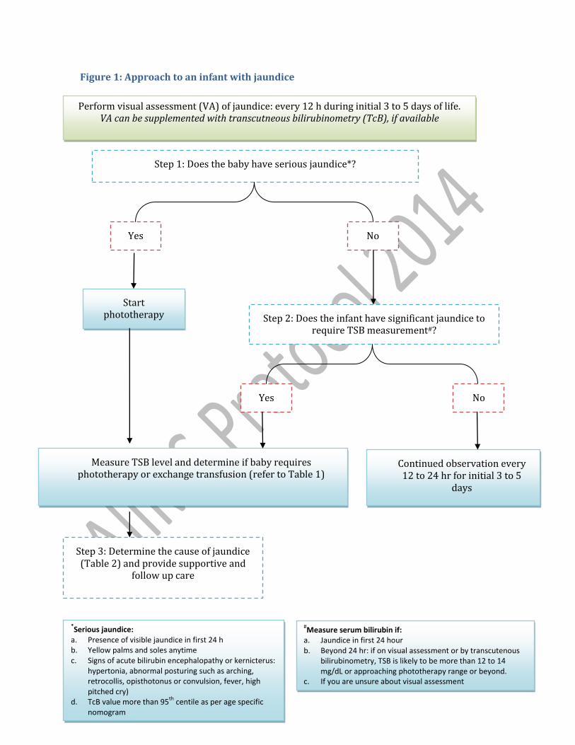
A step wise approach should be employed for managing jaundice in neonates (Figure 1).
All the neonates should be visually inspected for jaundice every 12 hr during initial 3 to 5 days of life. TcB can be used as an aid for initial screening of infants. Visual assessment (when performed properly) and TcB have reasonable sensitivity for initial assessment of jaundice.
As a first step, serious jaundice should be ruled out. Phototherapy should be initiated if the infant meets the criteria for serious jaundice. TSB should be determined subsequently in these infants to determine further course of action.
Though recommended by AAP5, screening of all infants with TSB in order to predict the risk of subsequent hyperbilirubinemia does not seem to be a feasible option in resource restricted settings.
Perform visual assessment (VA) of jaundice: every 12 h during initial 3 to 5 days of life. VA can be supplemented with transcutneous bilirubinometry (TcB), if available
Step 1: Does the baby have serious jaundice*?
Yes No
Start phototherapy
Measure TSB level and determine if baby requires phototherapy or exchange transfusion (refer to Table 1)
No Yes
Continued observation every 12 to 24 hr for initial 3 to 5
days
Step 3: Determine the cause of jaundice (Table 2) and provide supportive and
follow up care
Step 2: Does the infant have significant jaundice to require TSB measurement#?
*Serious jaundice:
a. Presence of visible jaundice in first 24 h b. Yellow palms and soles anytime
c. Signs of acute bilirubin encephalopathy or kernicterus: hypertonia, abnormal posturing such as arching, retrocollis, opisthotonus or convulsion, fever, high pitched cry)
d. TcB value more than 95th
centile as per age specific nomogram
#Measure serum bilirubin if:
a. Jaundice in first 24 hour b. Beyond 24 hr: if on visual assessment or by transcutenous
bilirubinometry, TSB is likely to be more than 12 to 14 mg/dL or approaching phototherapy range or beyond.
c. If you are unsure about visual assessment
Figure 1: Approach to an infant with jaundice
Management of jaundice
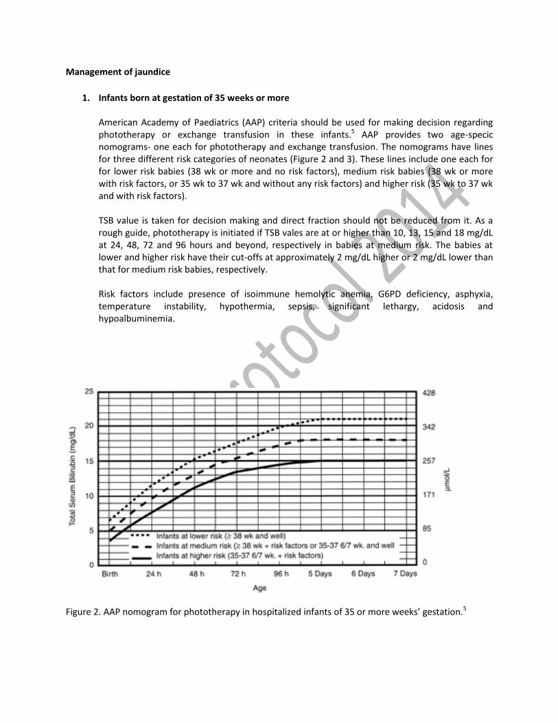
1. Infants born at gestation of 35 weeks or more American Academy of Paediatrics (AAP) criteria should be used for making decision regarding phototherapy or exchange transfusion in these infants.5 AAP provides two age-specic nomograms- one each for phototherapy and exchange transfusion. The nomograms have lines for three different risk categories of neonates (Figure 2 and 3). These lines include one each for for lower risk babies (38 wk or more and no risk factors), medium risk babies (38 wk or more with risk factors, or 35 wk to 37 wk and without any risk factors) and higher risk (35 wk to 37 wk and with risk factors). TSB value is taken for decision making and direct fraction should not be reduced from it. As a rough guide, phototherapy is initiated if TSB vales are at or higher than 10, 13, 15 and 18 mg/dL at 24, 48, 72 and 96 hours and beyond, respectively in babies at medium risk. The babies at lower and higher risk have their cut-offs at approximately 2 mg/dL higher or 2 mg/dL lower than that for medium risk babies, respectively. Risk factors include presence of isoimmune hemolytic anemia, G6PD deficiency, asphyxia, temperature instability, hypothermia, sepsis, significant lethargy, acidosis and hypoalbuminemia.
Figure 2. AAP nomogram for phototherapy in hospitalized infants of 35 or more weeks’ gestation.5
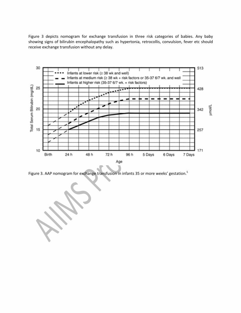
Figure 3 depicts nomogram for exchange transfusion in three risk categories of babies. Any baby showing signs of bilirubin encephalopathy such as hypertonia, retrocollis, convulsion, fever etc should receive exchange transfusion without any delay.
Figure 3. AAP nomogram for exchange transfusion in infants 35 or more weeks’ gestation.5
2. Preterm babies
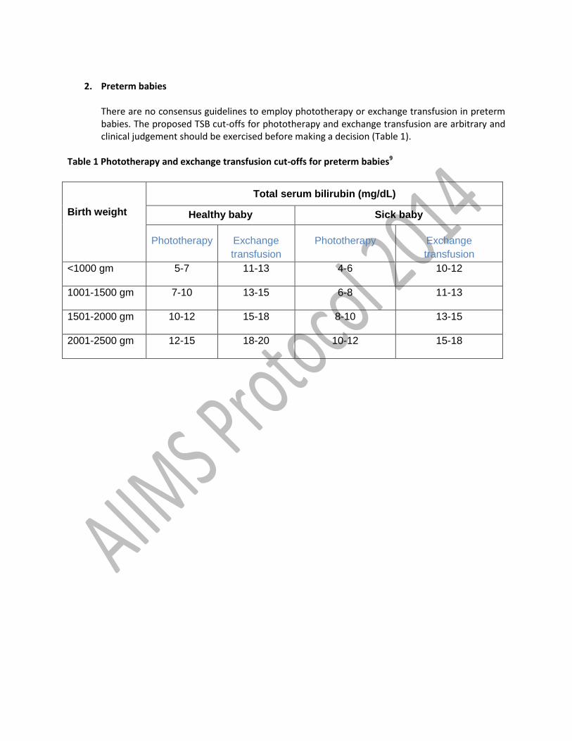
There are no consensus guidelines to employ phototherapy or exchange transfusion in preterm babies. The proposed TSB cut-offs for phototherapy and exchange transfusion are arbitrary and clinical judgement should be exercised before making a decision (Table 1).
Table 1 Phototherapy and exchange transfusion cut-offs for preterm babies9
Birth weight
Total serum bilirubin (mg/dL)
Healthy baby Sick baby
Phototherapy Exchange
transfusion
Phototherapy Exchange
transfusion
<1000 gm 5-7 11-13 4-6 10-12
1001-1500 gm 7-10 13-15 6-8 11-13
1501-2000 gm 10-12 15-18 8-10 13-15
2001-2500 gm 12-15 18-20 10-12 15-18
Therapeutic options
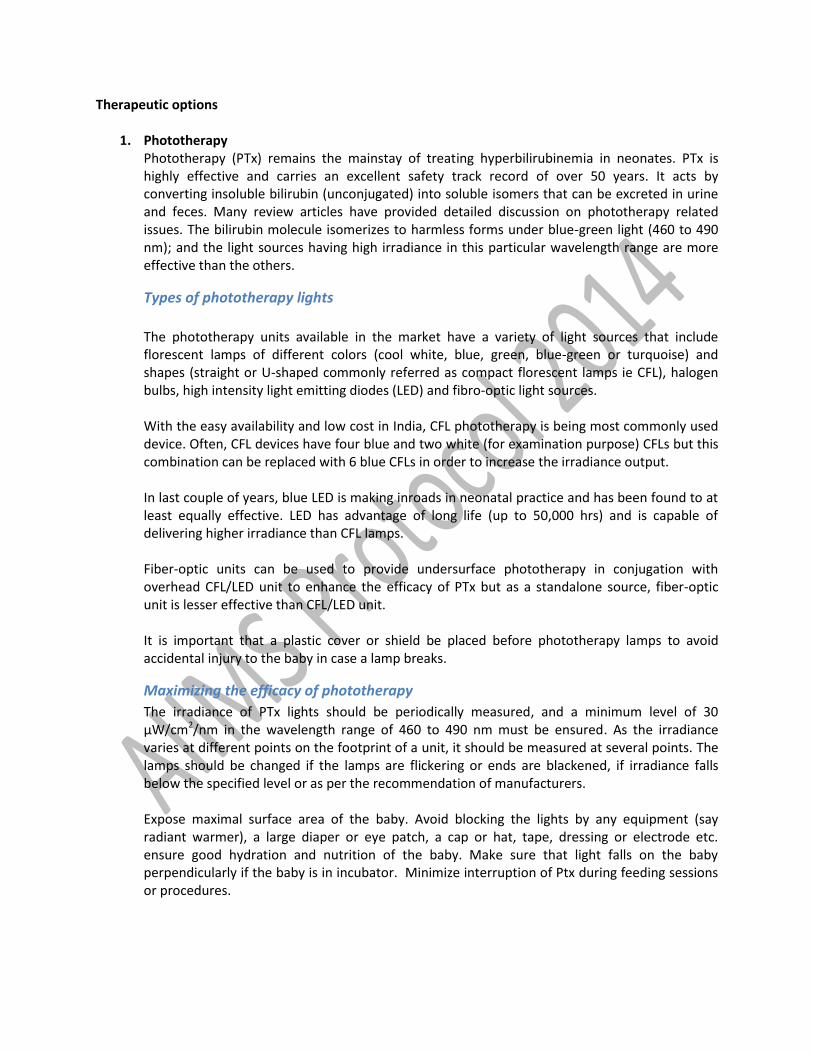
1. Phototherapy Phototherapy (PTx) remains the mainstay of treating hyperbilirubinemia in neonates. PTx is highly effective and carries an excellent safety track record of over 50 years. It acts by converting insoluble bilirubin (unconjugated) into soluble isomers that can be excreted in urine and feces. Many review articles have provided detailed discussion on phototherapy related issues. The bilirubin molecule isomerizes to harmless forms under blue-green light (460 to 490 nm); and the light sources having high irradiance in this particular wavelength range are more effective than the others.
Types of phototherapy lights
The phototherapy units available in the market have a variety of light sources that include florescent lamps of different colors (cool white, blue, green, blue-green or turquoise) and shapes (straight or U-shaped commonly referred as compact florescent lamps ie CFL), halogen bulbs, high intensity light emitting diodes (LED) and fibro-optic light sources. With the easy availability and low cost in India, CFL phototherapy is being most commonly used device. Often, CFL devices have four blue and two white (for examination purpose) CFLs but this combination can be replaced with 6 blue CFLs in order to increase the irradiance output. In last couple of years, blue LED is making inroads in neonatal practice and has been found to at least equally effective. LED has advantage of long life (up to 50,000 hrs) and is capable of delivering higher irradiance than CFL lamps. Fiber-optic units can be used to provide undersurface phototherapy in conjugation with overhead CFL/LED unit to enhance the efficacy of PTx but as a standalone source, fiber-optic unit is lesser effective than CFL/LED unit. It is important that a plastic cover or shield be placed before phototherapy lamps to avoid accidental injury to the baby in case a lamp breaks.
Maximizing the efficacy of phototherapy
The irradiance of PTx lights should be periodically measured, and a minimum level of 30 μW/cm2/nm in the wavelength range of 460 to 490 nm must be ensured. As the irradiance varies at different points on the footprint of a unit, it should be measured at several points. The lamps should be changed if the lamps are flickering or ends are blackened, if irradiance falls below the specified level or as per the recommendation of manufacturers. Expose maximal surface area of the baby. Avoid blocking the lights by any equipment (say radiant warmer), a large diaper or eye patch, a cap or hat, tape, dressing or electrode etc. ensure good hydration and nutrition of the baby. Make sure that light falls on the baby perpendicularly if the baby is in incubator. Minimize interruption of Ptx during feeding sessions or procedures.
Administering phototherapy
Make sure that ambient room temperature is optimum (250 to 280) to prevent hypothermia or hyperthermia in the baby. Remove all clothes of the baby except the diaper. Cover the baby’s eyes with patches, ensuring that the patches do not block the baby’s nostrils. Place the naked baby under the lights in a cot or bassinet if weight is more than 2 kg or in an incubator or radiant warmer if the baby is small (<2 kg). Keep the distance between baby and light 30 to 45 cm (or as per manufacturer recommendation). Ensure optimum breastfeeding. Baby can be taken out for breastfeeding sessions and the eye patch can be removed for better mother-infant interaction. However, minimize interruption to enhance effectiveness of phototherapy. There is no need to supplement or replace breast milk with any other types of feed or fluid (e.g. breast-milk substitute, water, sugar water, etc.)
Monitoring & stopping phototherapy
Monitor temperature of the baby every 2 to 4 hr. Measure TSB level every 12 to 24 hours. Discontinue PTx once two TSB values 12 hr apart fall below current age specific cut offs. The infant should be monitored clinically for rebound bilirubin rise within 24 hours after stopping phototherapy for babies with hemolytic disorders.
Role of sunlight
Exposing the baby to sunlight does not help in treatment of jaundice and is associated with risk of sunburn and therefore should be avoided.
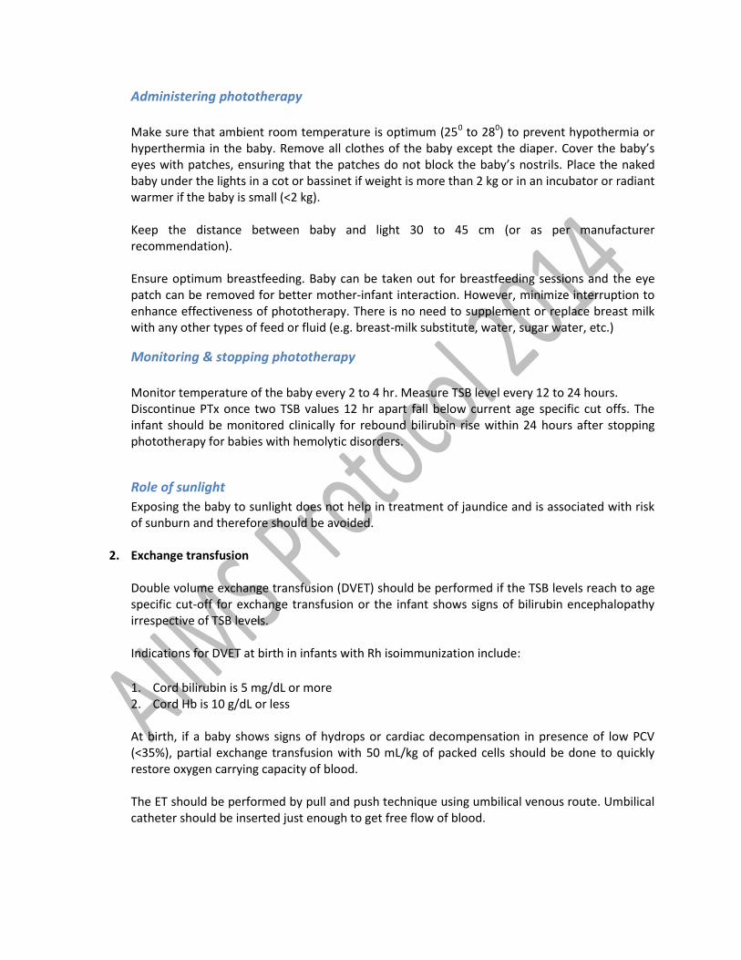
2. Exchange transfusion Double volume exchange transfusion (DVET) should be performed if the TSB levels reach to age specific cut-off for exchange transfusion or the infant shows signs of bilirubin encephalopathy irrespective of TSB levels. Indications for DVET at birth in infants with Rh isoimmunization include:
1. Cord bilirubin is 5 mg/dL or more 2. Cord Hb is 10 g/dL or less
At birth, if a baby shows signs of hydrops or cardiac decompensation in presence of low PCV (<35%), partial exchange transfusion with 50 mL/kg of packed cells should be done to quickly restore oxygen carrying capacity of blood. The ET should be performed by pull and push technique using umbilical venous route. Umbilical catheter should be inserted just enough to get free flow of blood.
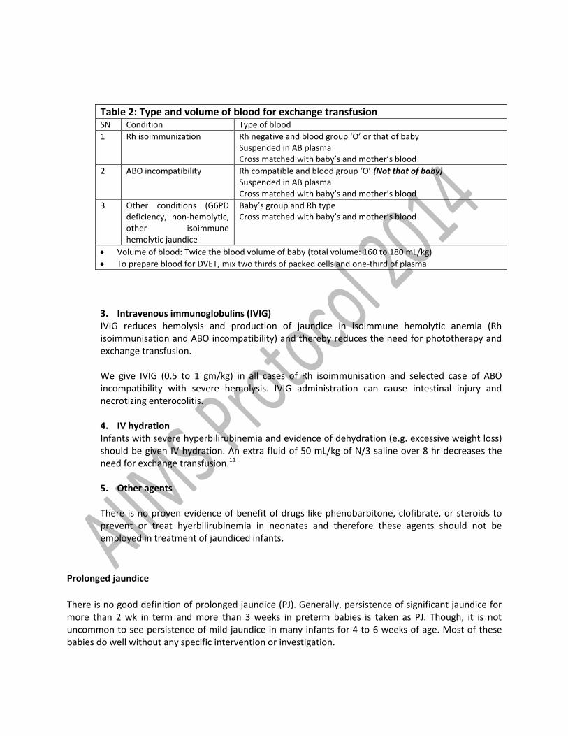
Table 2: Type and volume of blood for exchange transfusion SN Condition Type of blood
1 Rh isoimmunization Rh negative and blood group ‘O’ or that of baby Suspended in AB plasma Cross matched with baby’s and mother’s blood
2 ABO incompatibility Rh compatible and blood group ‘O’ (Not that of baby) Suspended in AB plasma Cross matched with baby’s and mother’s blood
3 Other conditions (G6PD deficiency, non-hemolytic, other isoimmune hemolytic jaundice
Baby’s group and Rh type Cross matched with baby’s and mother’s blood
Volume of blood: Twice the blood volume of baby (total volume: 160 to 180 mL/kg)
To prepare blood for DVET, mix two thirds of packed cells and one-third of plasma
3. Intravenous immunoglobulins (IVIG) IVIG reduces hemolysis and production of jaundice in isoimmune hemolytic anemia (Rh isoimmunisation and ABO incompatibility) and thereby reduces the need for phototherapy and exchange transfusion. We give IVIG (0.5 to 1 gm/kg) in all cases of Rh isoimmunisation and selected case of ABO incompatibility with severe hemolysis. IVIG administration can cause intestinal injury and necrotizing enterocolitis. 4. IV hydration Infants with severe hyperbilirubinemia and evidence of dehydration (e.g. excessive weight loss) should be given IV hydration. An extra fluid of 50 mL/kg of N/3 saline over 8 hr decreases the need for exchange transfusion.11 5. Other agents There is no proven evidence of benefit of drugs like phenobarbitone, clofibrate, or steroids to prevent or treat hyerbilirubinemia in neonates and therefore these agents should not be employed in treatment of jaundiced infants.
Prolonged jaundice
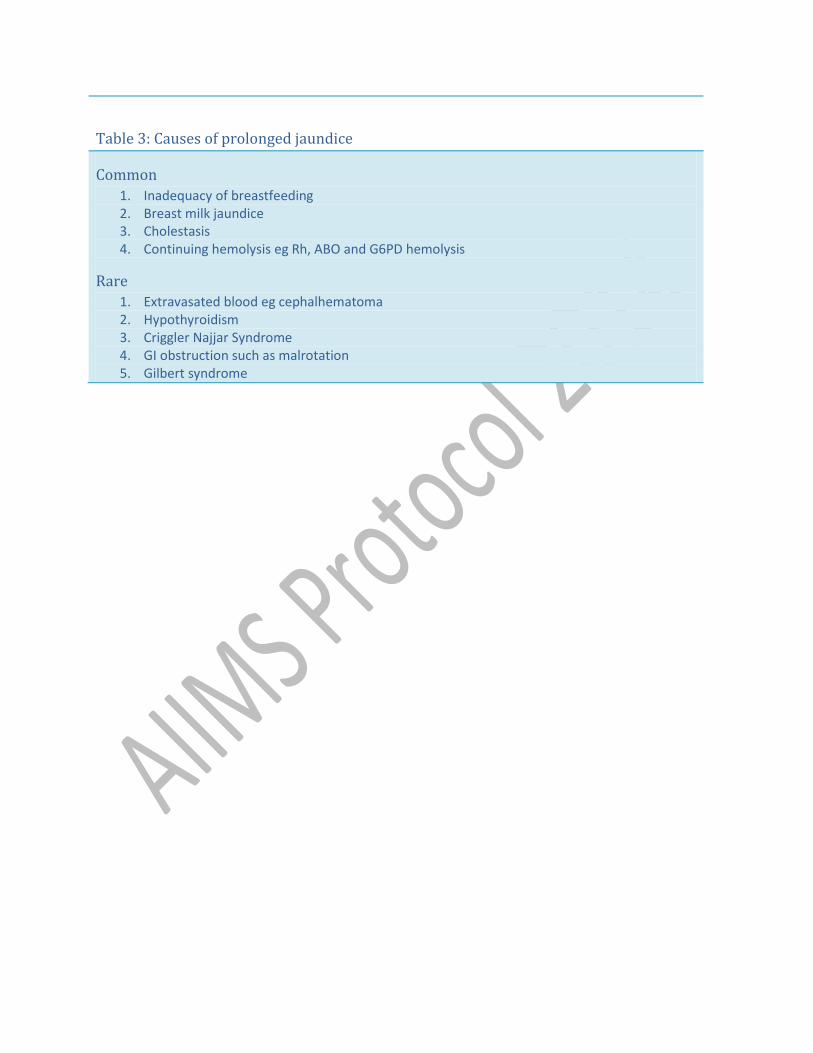
There is no good definition of prolonged jaundice (PJ). Generally, persistence of significant jaundice for more than 2 wk in term and more than 3 weeks in preterm babies is taken as PJ. Though, it is not uncommon to see persistence of mild jaundice in many infants for 4 to 6 weeks of age. Most of these babies do well without any specific intervention or investigation.
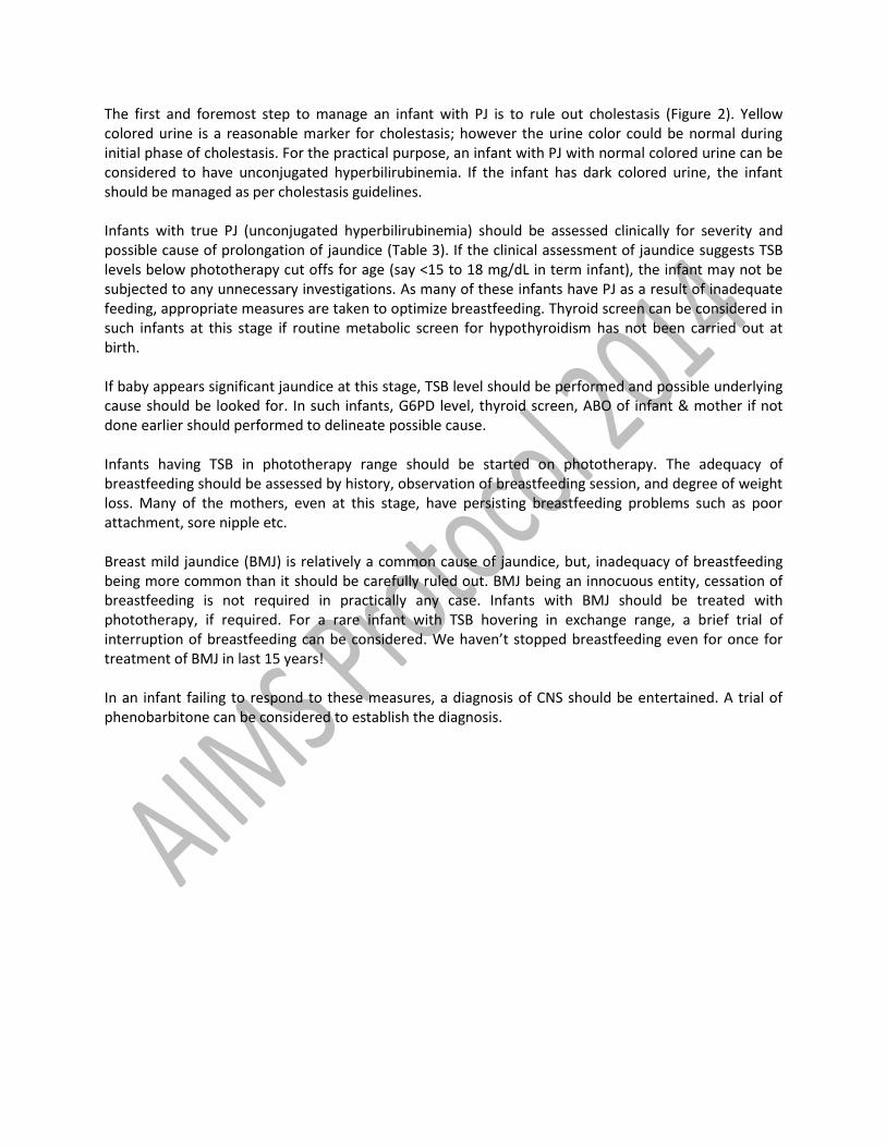
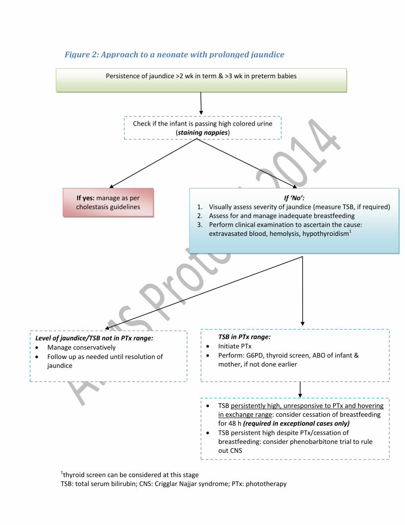
The first and foremost step to manage an infant with PJ is to rule out cholestasis (Figure 2). Yellow colored urine is a reasonable marker for cholestasis; however the urine color could be normal during initial phase of cholestasis. For the practical purpose, an infant with PJ with normal colored urine can be considered to have unconjugated hyperbilirubinemia. If the infant has dark colored urine, the infant should be managed as per cholestasis guidelines. Infants with true PJ (unconjugated hyperbilirubinemia) should be assessed clinically for severity and possible cause of prolongation of jaundice (Table 3). If the clinical assessment of jaundice suggests TSB levels below phototherapy cut offs for age (say <15 to 18 mg/dL in term infant), the infant may not be subjected to any unnecessary investigations. As many of these infants have PJ as a result of inadequate feeding, appropriate measures are taken to optimize breastfeeding. Thyroid screen can be considered in such infants at this stage if routine metabolic screen for hypothyroidism has not been carried out at birth. If baby appears significant jaundice at this stage, TSB level should be performed and possible underlying cause should be looked for. In such infants, G6PD level, thyroid screen, ABO of infant & mother if not done earlier should performed to delineate possible cause. Infants having TSB in phototherapy range should be started on phototherapy. The adequacy of breastfeeding should be assessed by history, observation of breastfeeding session, and degree of weight loss. Many of the mothers, even at this stage, have persisting breastfeeding problems such as poor attachment, sore nipple etc. Breast mild jaundice (BMJ) is relatively a common cause of jaundice, but, inadequacy of breastfeeding being more common than it should be carefully ruled out. BMJ being an innocuous entity, cessation of breastfeeding is not required in practically any case. Infants with BMJ should be treated with phototherapy, if required. For a rare infant with TSB hovering in exchange range, a brief trial of interruption of breastfeeding can be considered. We haven’t stopped breastfeeding even for once for treatment of BMJ in last 15 years! In an infant failing to respond to these measures, a diagnosis of CNS should be entertained. A trial of phenobarbitone can be considered to establish the diagnosis.
Table 3: Causes of prolonged jaundice
Common
1. Inadequacy of breastfeeding 2. Breast milk jaundice 3. Cholestasis 4. Continuing hemolysis eg Rh, ABO and G6PD hemolysis
Rare
1. Extravasated blood eg cephalhematoma 2. Hypothyroidism 3. Criggler Najjar Syndrome 4. GI obstruction such as malrotation 5. Gilbert syndrome
Figure 2: Approach to a neonate with prolonged jaundice
Check if the infant is passing high colored urine (staining nappies)
1thyroid screen can be considered at this stage TSB: total serum bilirubin; CNS: Crigglar Najjar syndrome; PTx: phototherapy
Persistence of jaundice >2 wk in term & >3 wk in preterm babies
If yes: manage as per cholestasis guidelines
guidelines
If ‘No’: 1. Visually assess severity of jaundice (measure TSB, if required) 2. Assess for and manage inadequate breastfeeding 3. Perform clinical examination to ascertain the cause:
extravasated blood, hemolysis, hypothyroidism1
Level of jaundice/TSB not in PTx range:
Manage conservatively
Follow up as needed until resolution of jaundice
TSB in PTx range:
Initiate PTx
Perform: G6PD, thyroid screen, ABO of infant & mother, if not done earlier
TSB persistently high, unresponsive to PTx and hovering in exchange range: consider cessation of breastfeeding for 48 h (required in exceptional cases only)
TSB persistent high despite PTx/cessation of breastfeeding: consider phenobarbitone trial to rule out CNS
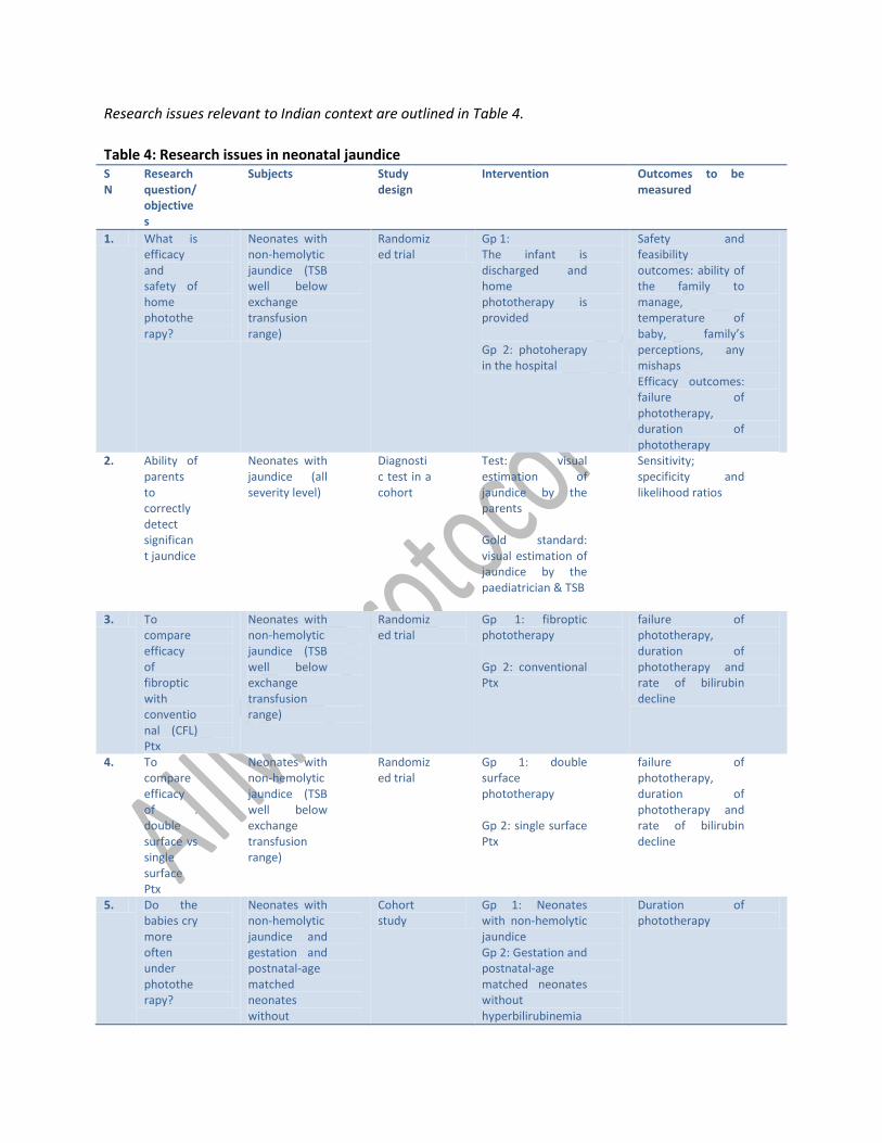
Research issues relevant to Indian context are outlined in Table 4.
Table 4: Research issues in neonatal jaundice SN
Research question/objectives
Subjects Study design
Intervention Outcomes to be measured
1. What is efficacy and safety of home phototherapy?
Neonates with non-hemolytic jaundice (TSB well below exchange transfusion range)
Randomized trial
Gp 1: The infant is discharged and home phototherapy is provided Gp 2: photoherapy in the hospital
Safety and feasibility outcomes: ability of the family to manage, temperature of baby, family’s perceptions, any mishaps Efficacy outcomes: failure of phototherapy, duration of phototherapy
2. Ability of parents to correctly detect significant jaundice
Neonates with jaundice (all severity level)
Diagnostic test in a cohort
Test: visual estimation of jaundice by the parents Gold standard: visual estimation of jaundice by the paediatrician & TSB
Sensitivity; specificity and likelihood ratios
3. To compare efficacy of fibroptic with conventional (CFL) Ptx
Neonates with non-hemolytic jaundice (TSB well below exchange transfusion range)
Randomized trial
Gp 1: fibroptic phototherapy Gp 2: conventional Ptx
failure of phototherapy, duration of phototherapy and rate of bilirubin decline
4. To compare efficacy of . double surface vs single surface Ptx
Neonates with non-hemolytic jaundice (TSB well below exchange transfusion range)
Randomized trial
Gp 1: double surface phototherapy Gp 2: single surface Ptx
failure of phototherapy, duration of phototherapy and rate of bilirubin decline
5. Do the babies cry more often under phototherapy?
Neonates with non-hemolytic jaundice and gestation and postnatal-age matched neonates without
Cohort study
Gp 1: Neonates with non-hemolytic jaundice Gp 2: Gestation and postnatal-age matched neonates without hyperbilirubinemia
Duration of phototherapy
hyperbilirubinemia
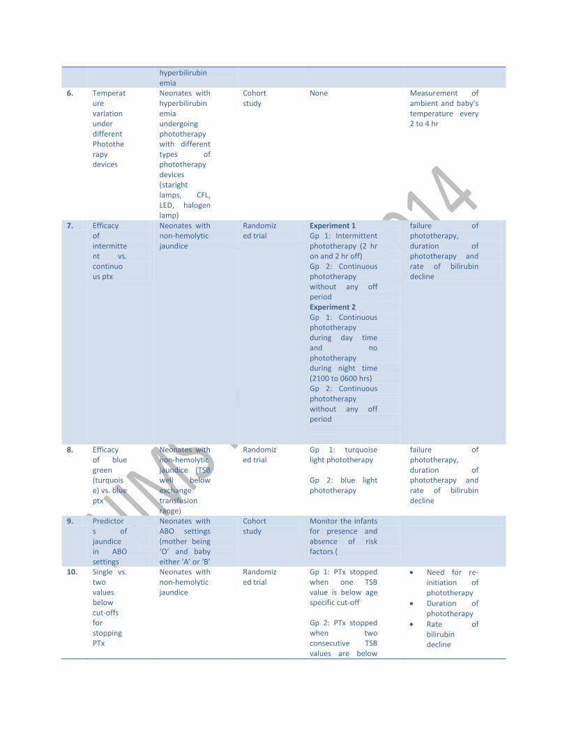
6. Temperature variation under different Phototherapy devices
Neonates with hyperbilirubinemia undergoing phototherapy with different types of phototherapy devices (staright lamps, CFL, LED, halogen lamp)
Cohort study
None Measurement of ambient and baby’s temperature every 2 to 4 hr
7. Efficacy of intermittent vs. continuous ptx
Neonates with non-hemolytic jaundice
Randomized trial
Experiment 1 Gp 1: Intermittent phototherapy (2 hr on and 2 hr off) Gp 2: Continuous phototherapy without any off period Experiment 2 Gp 1: Continuous phototherapy during day time and no phototherapy during night time (2100 to 0600 hrs) Gp 2: Continuous phototherapy without any off period
failure of phototherapy, duration of phototherapy and rate of bilirubin decline
8. Efficacy of blue green (turquoise) vs. blue ptx
Neonates with non-hemolytic jaundice (TSB well below exchange transfusion range)
Randomized trial
Gp 1: turquoise light phototherapy Gp 2: blue light phototherapy
failure of phototherapy, duration of phototherapy and rate of bilirubin decline
9. Predictors of jaundice in ABO settings
Neonates with ABO settings (mother being ‘O’ and baby either ‘A’ or ‘B’
Cohort study
Monitor the infants for presence and absence of risk factors (
10. Single vs. two values below cut-offs for stopping PTx
Neonates with non-hemolytic jaundice
Randomized trial
Gp 1: PTx stopped when one TSB value is below age specific cut-off Gp 2: PTx stopped when two consecutive TSB values are below
Need for re-initiation of phototherapy
Duration of phototherapy
Rate of bilirubin decline
age specific cut-off
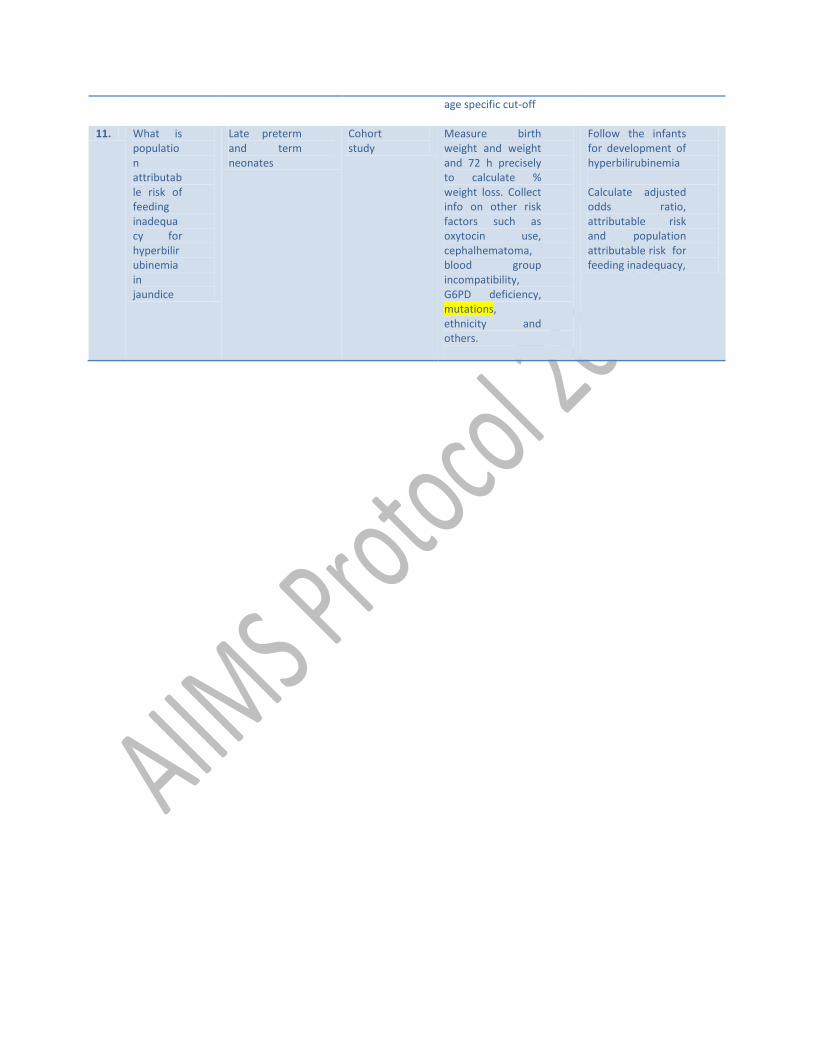
11. 1 What is population attributable risk of feeding inadequacy for hyperbilirubinemia in jaundice
Late preterm and term neonates
Cohort study
Measure birth weight and weight and 72 h precisely to calculate % weight loss. Collect info on other risk factors such as oxytocin use, cephalhematoma, blood group incompatibility, G6PD deficiency, mutations, ethnicity and others.
Follow the infants for development of hyperbilirubinemia Calculate adjusted odds ratio, attributable risk and population attributable risk for feeding inadequacy,
References
1. Young Infants Clinical Signs Study Group. Clinical signs that predict severe illness in children under age 2 months: a multicentre study. Lancet 2008;371:135-42.
2. Madan A, Mac Mohan JR, Stevenson DK.Neonatal Hyperbilrubinemia. In: Avery’s Diseases of the Newborn. Eds: Taeush HW, Ballard RA, Gleason CA. 8
3. Maisels MJ, Gifford K: Normal serum bilirubin levels in newborns and effect of breast-feeding. Pediatrics 78:837-43, 1986.
4. Rennie J, Burman-Roy S, Murphy MS; Guideline Development Group. Neonatal jaundice: summary of NICE guidance. BMJ. 2010 May 19;340:c2409. doi:10.1136/bmj.c2409.
5. American Academy of Pediatrics Subcommittee on Hyperbilirubinemia. Management of hyperbilirubinemia in the newborn infant 35 or more weeks of gestation. Pediatrics 2004;114:297-316.
6. Kramer LI. Advancement of dermal icterus in jaundiced newborn. Am J Dis Child 1969;118:454-8.
7. Kaur S, Chawla D, Pathak U, Jain S. Predischarge non-invasive risk assessment for prediction of significant hyperbilirubinemia in term and late preterm neonates. J Perinatol. 2011 Nov 17. doi: 10.1038/jp.2011.170.
8. Dalal SS, Mishra S, Agarwal R, Deorari AK, Paul V. Does measuring the changes in TcB value offer better prediction of Hyperbilirubinemia in healthy neonates? Pediatrics 2009;124:e851-7.
9. Halamek LP, Stevenson DK. Neonatal Jaundice. In Fanroff AA, Martin RJ (Eds): Neonatal Perinatal Medicine. Diseases of the fetus and Infant. 7ed. St louis, Mosby Year Book 2002. pp 1335.
10. van Imhoff DE, Dijk PH, Weykamp CW, Cobbaert CM, Hulzebos CV; BARTrial Study Group. Measurements of neonatal bilirubin and albumin concentrations: a need for improvement and quality control. Eur J Pediatr 2011;170:977-82.
11. Mehta S, Kumar P, Narang A. A randomized controlled trial of fluid supplementation in term neonates with severe hyperbilirubinemia. J Pediatr 2005;147:781-5.