40
Neonatal Surgical Neonatal Surgical Issues Issues (Part 1) (Part 1) Sue Ann Smith, MD Sue Ann Smith, MD Neonatologist Neonatologist
| Date post: | 20-Dec-2015 |
| Category: |
Documents |
| View: | 216 times |
| Download: | 0 times |
Neonatal Surgical IssuesNeonatal Surgical Issues(Part 1)(Part 1)
Sue Ann Smith, MDSue Ann Smith, MD
NeonatologistNeonatologist
An anatomic surveyAn anatomic survey
Head and Neck lesionsHead and Neck lesionsChest lesionsChest lesionsAbdomenAbdomen
Abdominal wall defects and infectionAbdominal wall defects and infection
The NoseThe Nose
Choanal atresia – bilateral atresia Choanal atresia – bilateral atresia Respiratory distress resolves with cryingRespiratory distress resolves with crying Treat with oral airway until surgical repairTreat with oral airway until surgical repair CT scan often used in surgical planningCT scan often used in surgical planning ENT surgeons make opening in bony plate and stent ENT surgeons make opening in bony plate and stent
open during healingopen during healing
Nasolacrimal duct cysts – large and bilatNasolacrimal duct cysts – large and bilat Respiratory distress resolves with cryingRespiratory distress resolves with crying Treat with oral airwayTreat with oral airway Can usually be seen with otoscopeCan usually be seen with otoscope
Robin sequenceRobin sequence
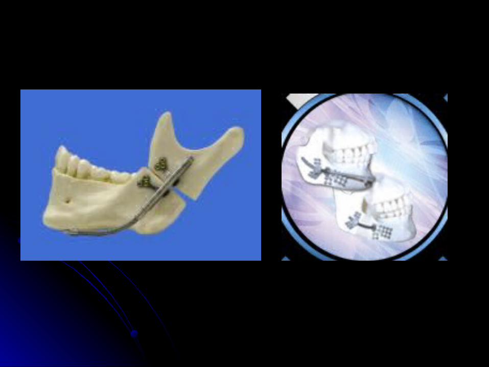
AKA Pierre Robin syndromeAKA Pierre Robin syndromeHypoplastic mandible with U-shaped Hypoplastic mandible with U-shaped
midline cleft palatemidline cleft palateRespiratory and feeding difficultiesRespiratory and feeding difficultiesPosition prone, may require nasopharyngeal Position prone, may require nasopharyngeal
tube, oral airway, LMA, or endotracheal tubetube, oral airway, LMA, or endotracheal tubeMandibular distraction is now treatment of Mandibular distraction is now treatment of
choice at OHSUchoice at OHSU
The UnusualThe Unusual
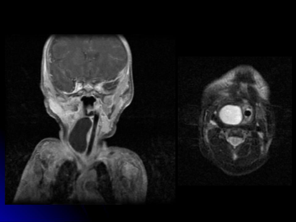
Laryngotracheal cleftsLaryngotracheal cleftsLaryngeal websLaryngeal websTracheal agenesis – frequently lethalTracheal agenesis – frequently lethalNeck massesNeck masses
Foregut duplication cystForegut duplication cyst lymphangiomalymphangioma
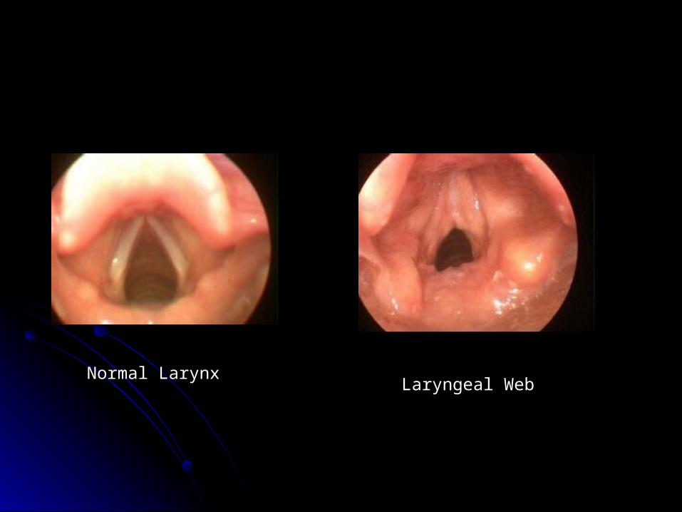
Normal LarynxLaryngeal Web
Congenital Chest LesionsCongenital Chest Lesions
Tracheo-esophageal fistulaTracheo-esophageal fistulaDiaphragmatic Hernia (briefly)Diaphragmatic Hernia (briefly)Congenital lobar emphysemaCongenital lobar emphysemaCystic adenomatoid malformationCystic adenomatoid malformationVascular ringsVascular rings
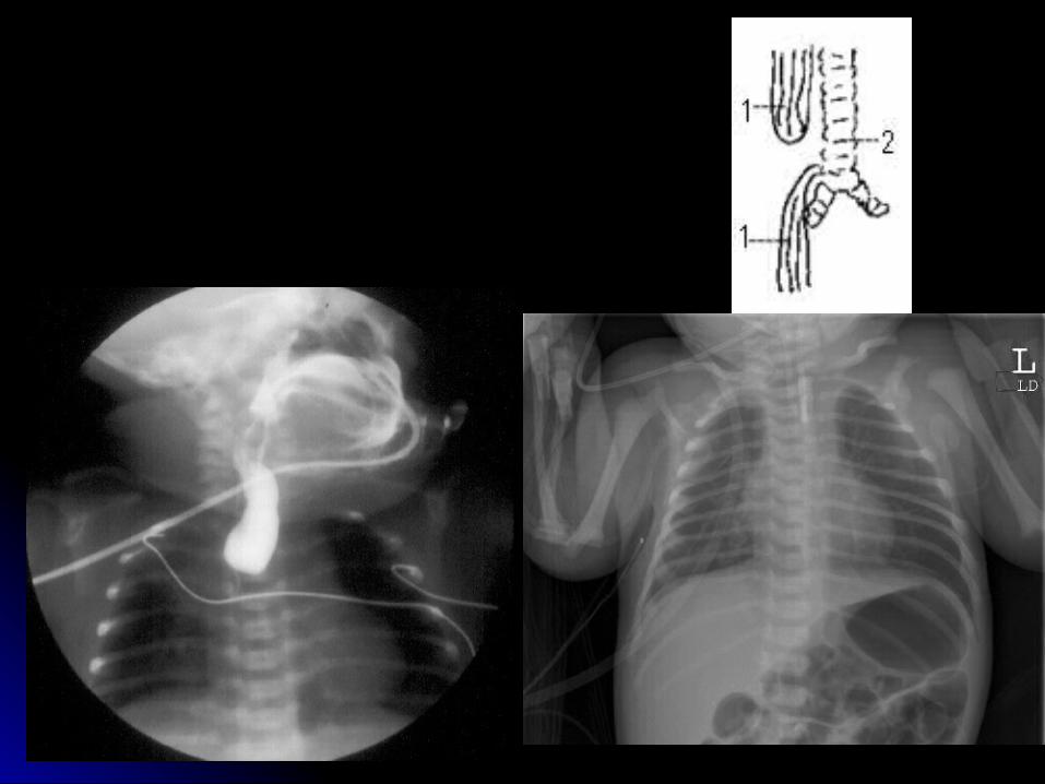
Tracheo-Esophageal Fistula (TEF)Tracheo-Esophageal Fistula (TEF)
Esophageal atresia with TEF is most Esophageal atresia with TEF is most common (85%).common (85%).Diagnosis may be suspected antenatal with Diagnosis may be suspected antenatal with
absence of stomach bubble and absence of stomach bubble and polyhydramnios. (*Caution: also seen with polyhydramnios. (*Caution: also seen with conditions that lead to poor swallowing)conditions that lead to poor swallowing)
Often associated with other anomalies: Often associated with other anomalies: VATER and chromosomalVATER and chromosomal
Tracheo-Esophageal Fistula (TEF) Tracheo-Esophageal Fistula (TEF) (cont)(cont)
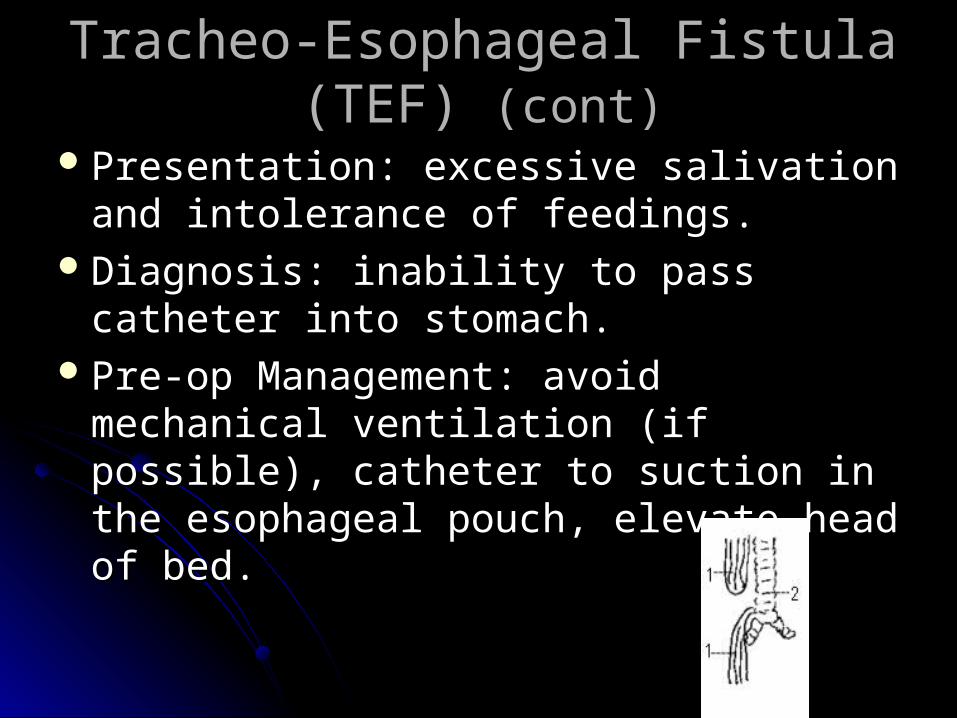
Presentation: excessive salivation and Presentation: excessive salivation and intolerance of feedings. intolerance of feedings.
Diagnosis: inability to pass catheter into Diagnosis: inability to pass catheter into stomach.stomach.
Pre-op Management: avoid mechanical Pre-op Management: avoid mechanical ventilation (if possible), catheter to suction ventilation (if possible), catheter to suction in the esophageal pouch, elevate head of in the esophageal pouch, elevate head of bed. bed.
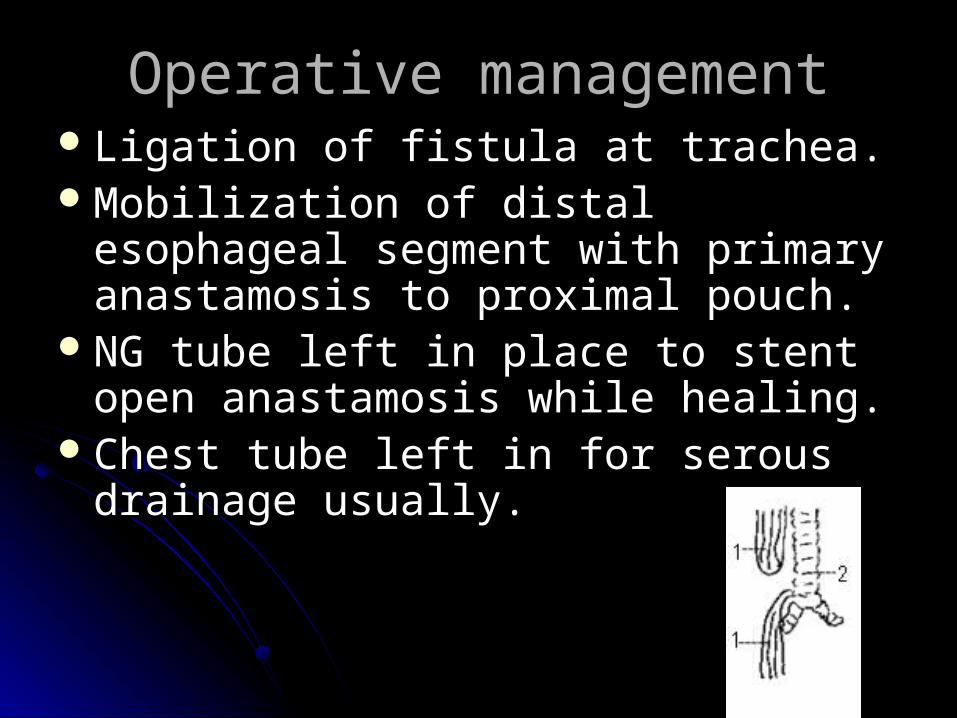
Operative managementOperative managementLigation of fistula at trachea. Ligation of fistula at trachea. Mobilization of distal esophageal segment Mobilization of distal esophageal segment
with primary anastamosis to proximal with primary anastamosis to proximal pouch. pouch.
NG tube left in place to stent open NG tube left in place to stent open anastamosis while healing.anastamosis while healing.
Chest tube left in for serous drainage Chest tube left in for serous drainage usually. usually.

Post-operative ManagementPost-operative Management
Careful airway management to prevent Careful airway management to prevent trauma to the fistula ligation site in the trauma to the fistula ligation site in the trachea. trachea.
Prior to feedings, must make sure that the Prior to feedings, must make sure that the esophageal anastamosis does not leak. esophageal anastamosis does not leak. (swallow study)(swallow study)
Often have on going feeding problems. Often have on going feeding problems. May need dilation procedures periodicallyMay need dilation procedures periodically
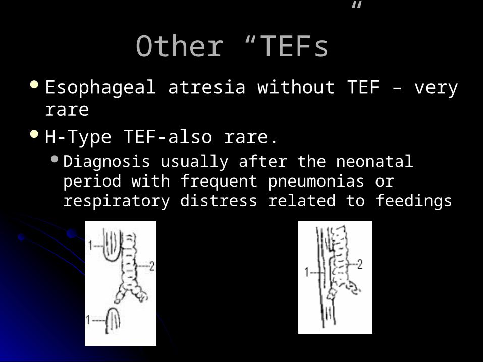
Other “TEFs”Other “TEFs”Esophageal atresia without TEF – very rareEsophageal atresia without TEF – very rareH-Type TEF-also rare. H-Type TEF-also rare.
Diagnosis usually after the neonatal period with Diagnosis usually after the neonatal period with frequent pneumonias or respiratory distress frequent pneumonias or respiratory distress related to feedingsrelated to feedings
Congenital Diaphragmatic Hernia Congenital Diaphragmatic Hernia (CDH)(CDH)
Most commonly on left sideMost commonly on left side Incidence 1:2000 to 1:5000Incidence 1:2000 to 1:5000Often associated with other malformationsOften associated with other malformationsFrequently diagnosed prenatallyFrequently diagnosed prenatallyAvoid bag-mask PPVAvoid bag-mask PPV
Pre-op CDHPre-op CDH
Delayed surgical repair – usually after 72 Delayed surgical repair – usually after 72 hrs of agehrs of age
NG drainage tube to keep bowel NG drainage tube to keep bowel decompresseddecompressed
Treat aggressively for pulmonary Treat aggressively for pulmonary hypoplasia and Persistent Pulmonary hypoplasia and Persistent Pulmonary Hypertension – including ECMO(?). Hypertension – including ECMO(?).
Surfactant therapy is now controversialSurfactant therapy is now controversial
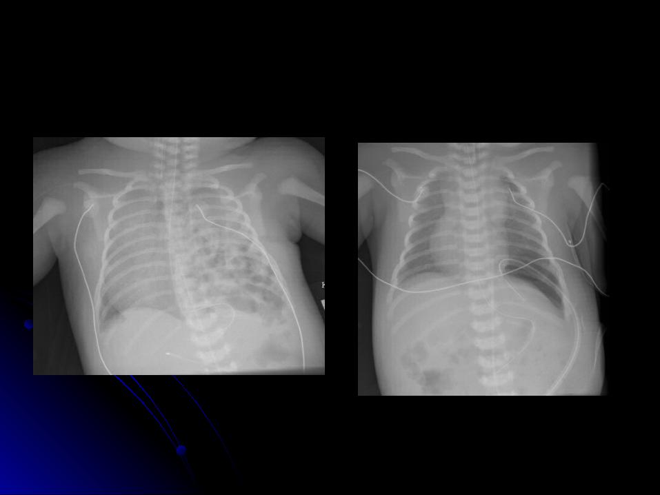
Post-Op CDHPost-Op CDH
““Anatomy is destiny”Anatomy is destiny”Survival continues to be around 40-50%.Survival continues to be around 40-50%.Feeding difficulties Feeding difficulties
Congenital lobar emphysemaCongenital lobar emphysema
Lesions that cause air trapping, with Lesions that cause air trapping, with compression of surrounding tissue compression of surrounding tissue
Most common in left upper, right middle Most common in left upper, right middle and right upper lobesand right upper lobes
Usually attempt low volume ventilation. Usually attempt low volume ventilation. Sometimes selective intubation of other Sometimes selective intubation of other bronchusbronchus
May require surgical resectionMay require surgical resection
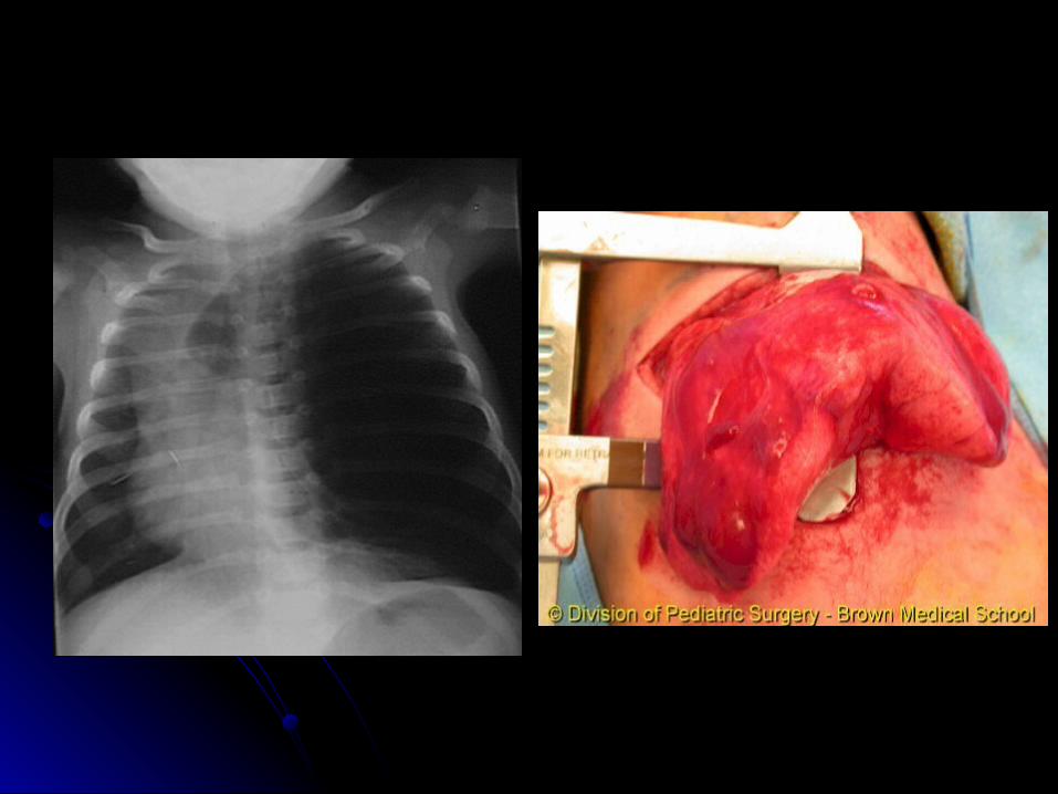
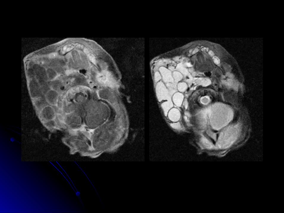
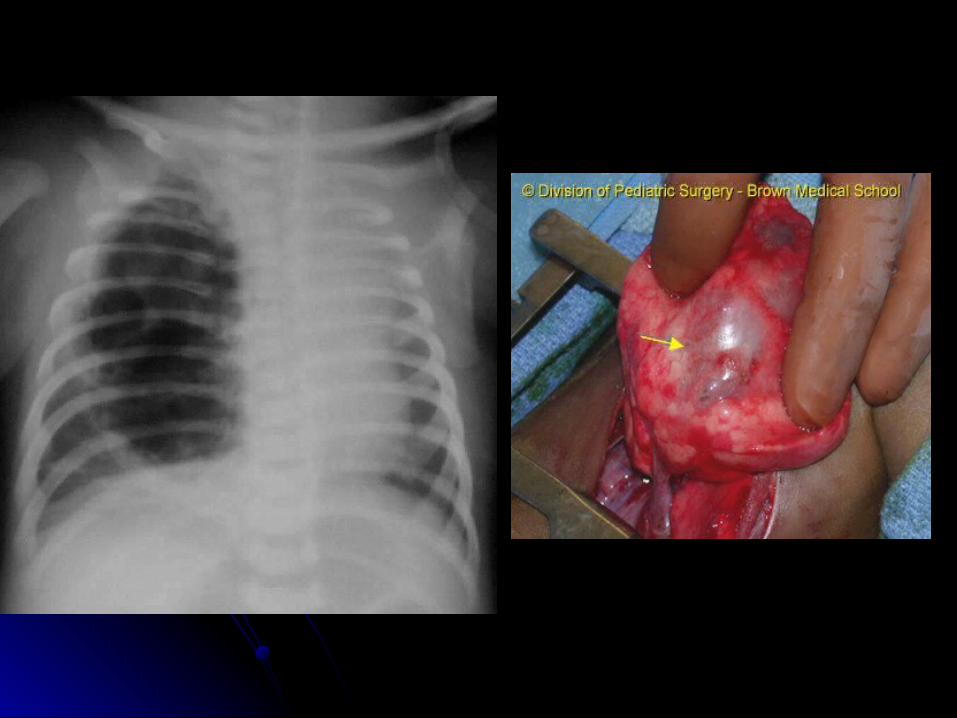
Congenital Cystic Adenomatoid Congenital Cystic Adenomatoid Malformation (CCAM)Malformation (CCAM)
May be confused with CDHMay be confused with CDHAbnormal lung tissue that forms fluid filled Abnormal lung tissue that forms fluid filled
cysts. May be large cysts, or many small cysts. May be large cysts, or many small cysts and solid areascysts and solid areas
Space occupying lesionSpace occupying lesionMay cause shifting of mediastiumMay cause shifting of mediastiumMay spontaneously regress in fetusMay spontaneously regress in fetusMay require surgical removalMay require surgical removal
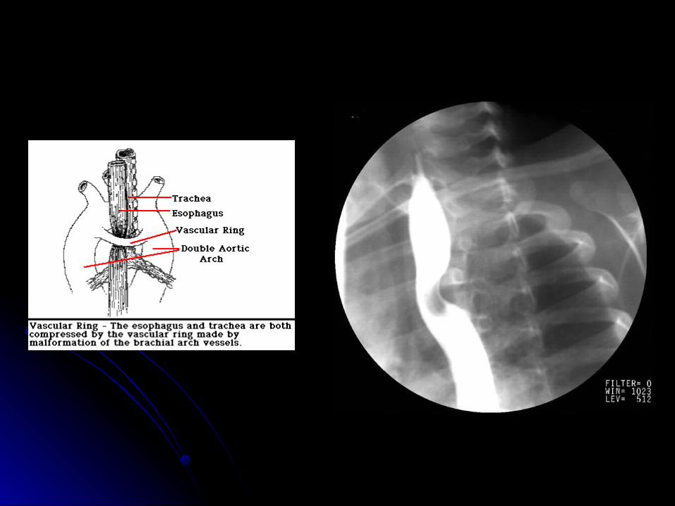
Vascular RingsVascular Rings
UncommonUncommonSigns include stridor, vomiting and difficulty Signs include stridor, vomiting and difficulty
swallowing.swallowing.Barium swallow can be diagnostic, but may Barium swallow can be diagnostic, but may
need chest MRI.need chest MRI.Sometimes may need cardiac Sometimes may need cardiac
catheterizationcatheterization
The AbdomenThe AbdomenAbdominal wall Abdominal wall
defects defects infectioninfection
BowelBowelObstructionsObstructions
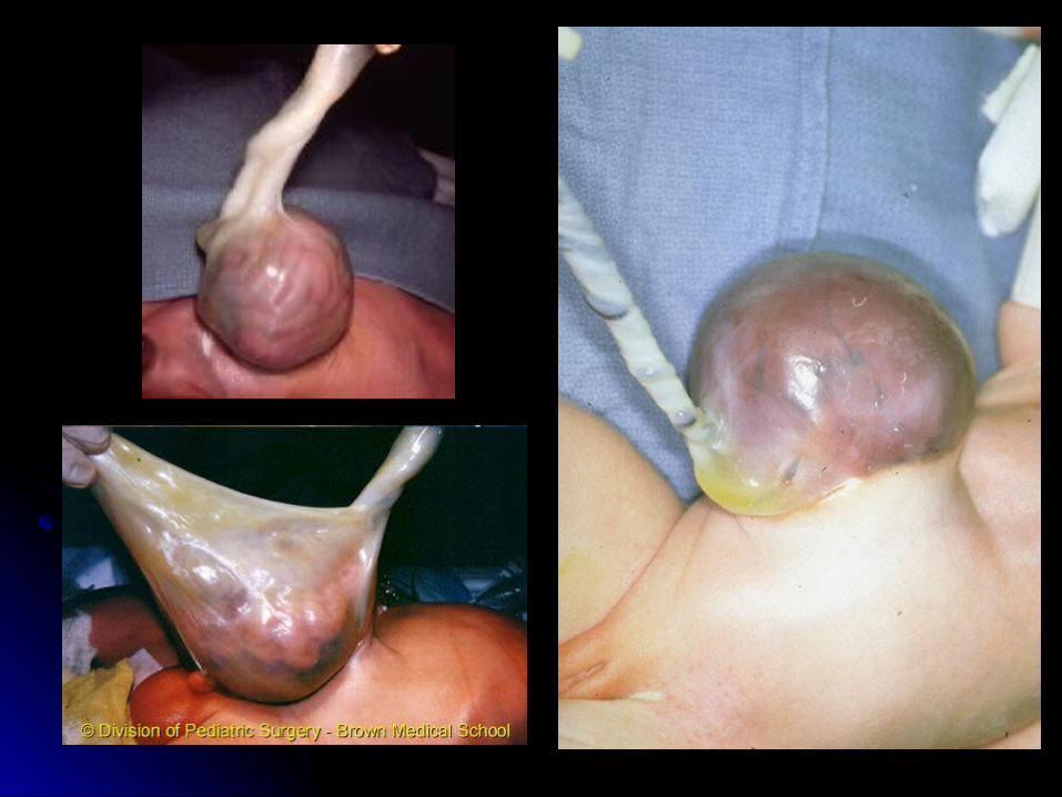
GastroschisisGastroschisis
Abdominal wall defect to right of umbilicus with Abdominal wall defect to right of umbilicus with no covering over intestinesno covering over intestines
Rarely associated with other anomaliesRarely associated with other anomalies Most babies are SGA and born to young Most babies are SGA and born to young
mothers (why?)mothers (why?) 10% will have intestinal atresias10% will have intestinal atresias Rarely will have significant infarction of most of Rarely will have significant infarction of most of
small bowel (i.e. lethal)small bowel (i.e. lethal) Most will have “meconium” stained amniotic fluid Most will have “meconium” stained amniotic fluid
(really bile)(really bile)
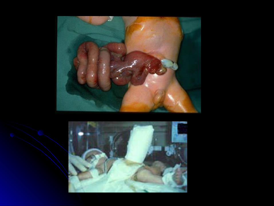
Gastroschisis Pre-opGastroschisis Pre-op
Empty stomach (usually lots of bilious fluid)Empty stomach (usually lots of bilious fluid)NG tube for decompressionNG tube for decompressionPlace in bowel bag or wrap in warm saline Place in bowel bag or wrap in warm saline
soaked gauze and saran wrapsoaked gauze and saran wrapSupport the bowel so as to maintain Support the bowel so as to maintain
perfusionperfusion

Gastroschisis (post-op)Gastroschisis (post-op)
Primary closure is attemptedPrimary closure is attemptedMay require silo with slow return of May require silo with slow return of
intestine into small abdominal cavityintestine into small abdominal cavityMaintain perfusionMaintain perfusionFeeding difficulties are main post-op Feeding difficulties are main post-op
problemproblemAt risk for adhesions throughout lifeAt risk for adhesions throughout life

OmphaloceleOmphaloceleAbdominal wall defect at umbilicus with Abdominal wall defect at umbilicus with
covering (sac may rupture)covering (sac may rupture)Frequently associated with other Frequently associated with other
anomalies anomalies Giant omphaloceles: respiratory issues Giant omphaloceles: respiratory issues
with misshaped chest and airway malaciaswith misshaped chest and airway malacias
OmphaloceleOmphalocele
Decompress stomach initiallyDecompress stomach initiallyCareful eval for other anomaliesCareful eval for other anomalies Intact sac may defer operation for yearsIntact sac may defer operation for years
““paint” membrane with betadine to toughen paint” membrane with betadine to toughen into a “rind”into a “rind”
Ruptured sac – repair similar to Ruptured sac – repair similar to gastroschisisgastroschisis
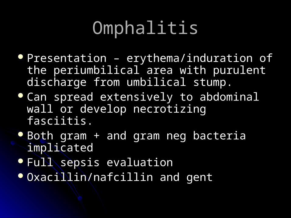
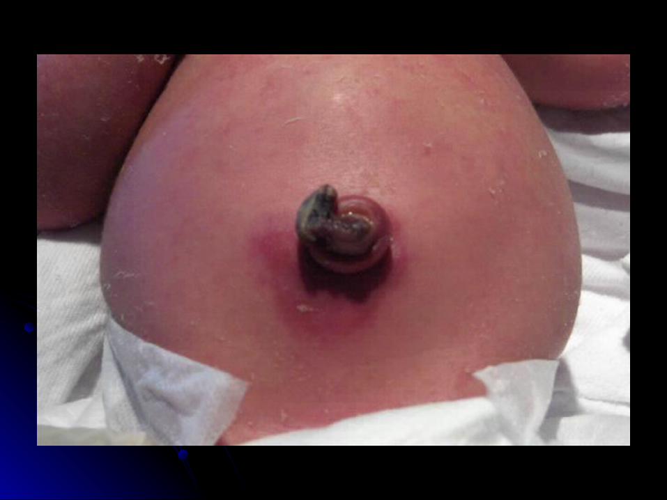
OmphalitisOmphalitis
Presentation – erythema/induration of the Presentation – erythema/induration of the periumbilical area with purulent discharge periumbilical area with purulent discharge from umbilical stump.from umbilical stump.
Can spread extensively to abdominal wall Can spread extensively to abdominal wall or develop necrotizing fasciitis.or develop necrotizing fasciitis.
Both gram + and gram neg bacteria Both gram + and gram neg bacteria implicatedimplicated
Full sepsis evaluationFull sepsis evaluationOxacillin/nafcillin and gentOxacillin/nafcillin and gent


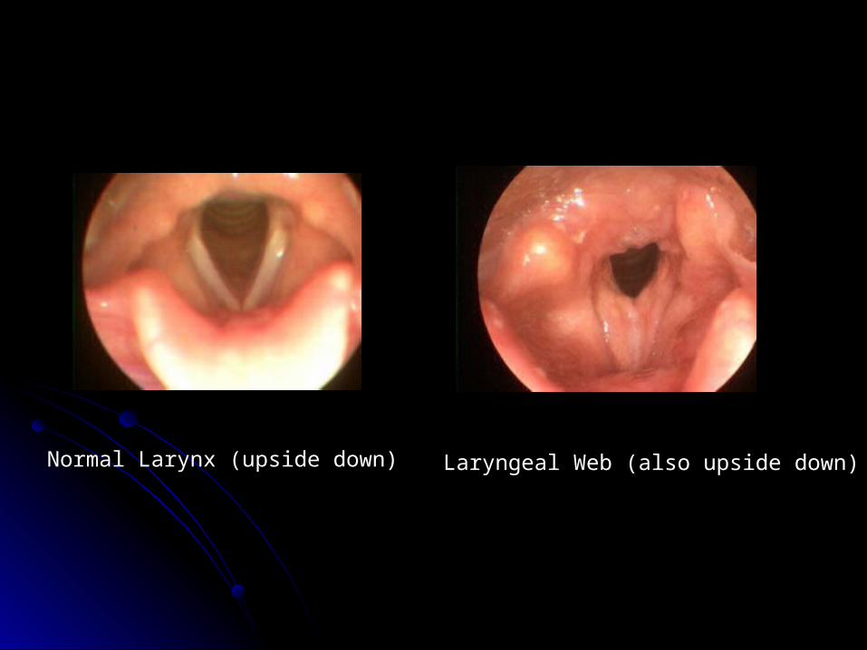
Normal Larynx (upside down) Laryngeal Web (also upside down)


