Page 1
Neonatale aanpak van het kind met een
anorectale malformatie
B. D’hondt, M. Miserez
Department of Abdominal Surgery
University Hospitals Leuven, Belgium
Interuniversitaire Vergadering Neonatologie
Gent, 17 januari 2014
Page 2
• Incidence: 1/4000-5000 live births
•Occuring ≤ 6th-7th week of gestation
• Very heterogenous group of severity• Anatomy of the malformation
• high vs low: supralevator vs infralevator
• +/- fistula = ectopic anal canal (IAS?)
• Associated anomalies (2/3)
• chromosomal defects: 8%
• Pelvic floor function
• anatomy
• innervation
Anorectal malformations
Page 3
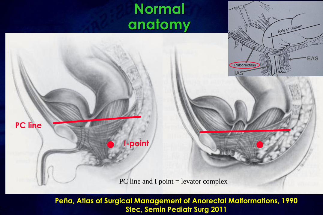
Peña, Atlas of Surgical Management of Anorectal Malformations, 1990
Stec, Semin Pediatr Surg 2011
Normal anatomy
Page 4
Peña, Atlas of Surgical Management of Anorectal Malformations, 1990
Stec, Semin Pediatr Surg 2011
IAS
EAS
Normal anatomy
PC line
I-point
PC line and I point = levator complex
Page 5
Determinants of fecal continence in ARM
•Type of malformation
•Associated anomalies
•Surgical aspects
•Postoperative care
Page 6
• Anatomical sfincter defects
• Decreased/absent anorectal sensitivity
• Motility disorders
• Chronic rectal distention
• Lumbosacral nerve abnormalities
Determinants of fecal continence in ARM
Page 7
Which classification for ARM
•Many classifications – difficult comparison• Ladd and Gross 1934
• Stephens and Smith 1963 (high vs. low)
• Santulli 1964
• “International” Melbourne Classification 1970 (nearly 40 subtypes)
• Wingspread Classification 1984
• Peña 1995
Page 8
Wingspread Classification
• Fistula vs. no fistula
• Low vshigh vsintermediate
Page 9
To develop a system for comparable follow-up studies
Page 10
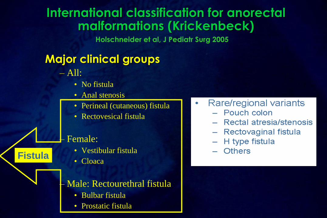
International classification for anorectalmalformations (Krickenbeck)
Major clinical groups– All:
• No fistula
• Anal stenosis
• Perineal (cutaneous) fistula
• Rectovesical fistula
– Female:
• Vestibular fistula
• Cloaca
– Male: Rectourethral fistula
• Bulbar fistula
• Prostatic fistula
Holschneider et al, J Pediatr Surg 2005
Fistula
Page 11
International classification for anorectalmalformations (Krickenbeck)
Major clinical groups– All:
• No fistula
• Anal stenosis
• Perineal (cutaneous) fistula
• Rectovesical fistula
– Female:
• Vestibular fistula
• Cloaca
– Male: Rectourethral fistula
• Bulbar fistula
• Prostatic fistula
Holschneider et al, J Pediatr Surg 2005
> 50%
Page 12
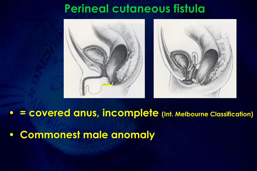
Perineal cutaneous fistula
• = covered anus, incomplete (Int. Melbourne Classification)
• Commonest male anomaly
Page 13
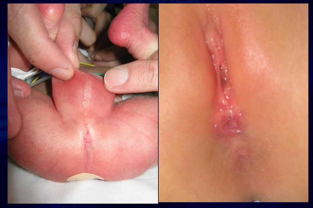
Perineal cutaneous fistula
Black ribbon“White epithelial
pearl”
Page 15
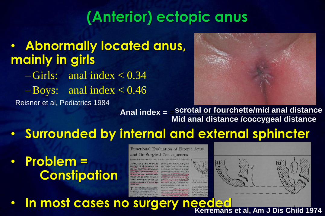
(Anterior) ectopic anus
• Abnormally located anus,mainly in girls
– Girls: anal index < 0.34
– Boys: anal index < 0.46
• Surrounded by internal and external sphincter
• Problem = Constipation
• In most cases no surgery neededKerremans et al, Am J Dis Child 1974
Anal index = = scrotal or fourchette/mid anal distanceMid anal distance /coccygeal distance
Reisner et al, Pediatrics 1984
Page 16
Vestibular fistula
• Common wall betweenrectum and vagina
Page 17
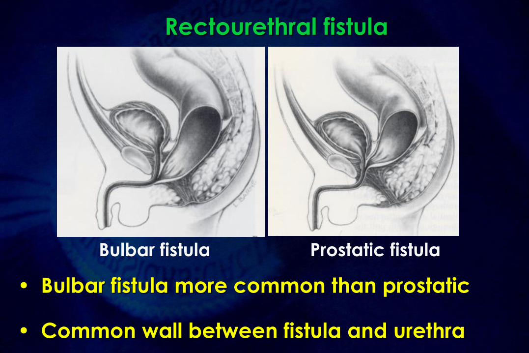
Rectourethral fistula
• Bulbar fistula more common than prostatic
• Common wall between fistula and urethra
Bulbar fistula Prostatic fistula
Page 18
International classification for anorectalmalformations (Krickenbeck)
Major clinical groups– All:
• No fistula
• Anal stenosis
• Perineal (cutaneous) fistula
• Rectovesical fistula
– Female:
• Vestibular fistula
• Cloaca
– Male: Rectourethral fistula
• Bulbar fistula
• Prostatic fistula
Holschneider et al, J Pediatr Surg 2005
The other major
clinical groups
Page 19
Anal stenosis
• Covered anus:
• (Partial) covering by genital folds
(hypertrophic raphe, bucket handle)
• Anal membrane
Bucket
handle
Page 20
Rectovesical fistula
• = rectobladder neck fistula• Flat perineum• Sacrum and pelvis can appear dysmorphic
Page 21
Associated anomalies (1)
• More associated anomalies in high lesions
• Serious, potentially lethal defects
• Vertebral - spinal deformities – hemivertebrae
– sacral deformities
– caudal regression syndrome
– tethered cord?• skin lesion: lump, vascular nevus, sinus, angioma on the midline
of the back, hypertrichosis, skin dimple, sacral lipoma
• neurological or neuro-orthopedic abnormalities of
lower extremities, scoliosis
• bladder and bowel dysfunction
permanent
colostomy
Neural tube defect of terminal spinal cord:
- Vertebral abnormalities
- Flat buttocks
- Lower limb neurological deficit
- Neurogenic bladder
- ARM
Page 22
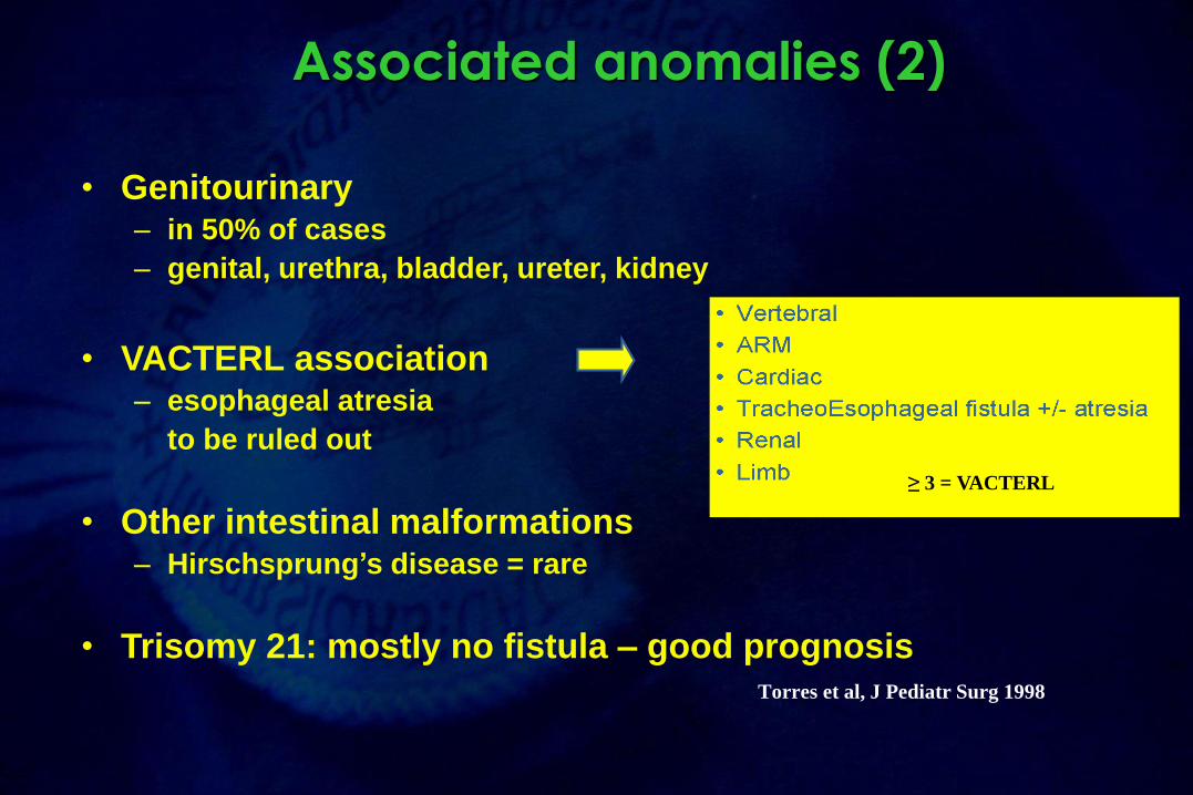
Associated anomalies (2)
• Genitourinary– in 50% of cases
– genital, urethra, bladder, ureter, kidney
• VACTERL association– esophageal atresia
to be ruled out
• Other intestinal malformations– Hirschsprung’s disease = rare
• Trisomy 21: mostly no fistula – good prognosisTorres et al, J Pediatr Surg 1998
≥ 3 = VACTERL
Page 23
Initial assessment of the newborn
• Clinical assessment
– Perineal inspection
– Associated anomalies
• Imaging
– Cross-table lateral film
– Ultrasound, cystogram, spinal examination
Page 24
Perineal inspection
• Anal dimple = EAS (cutaneoanal reflex, EMG)
• Fistula visible?
• Midline groove between buttocks and anal dimple (“flat buttocks”)
decreased prominence ~ height of the fistula
• Boys: – meconium or squamous epithelium in urine = fistula
– white epithelial pearls = fistula
• Girls: number of orifices + probing
Page 25
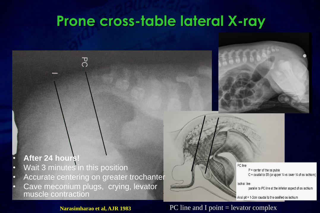
Prone cross-table lateral X-ray
• After 24 hours!
• Wait 3 minutes in this position
• Accurate centering on greater trochanter
• Cave meconium plugs, crying, levator muscle contraction
PC line and I point = levator complexPC line and I point = levator complexNarasimharao et al, AJR 1983
Page 26
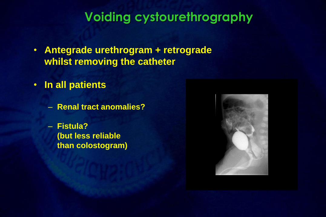
Voiding cystourethrography
• Antegrade urethrogram + retrograde
whilst removing the catheter
• In all patients
– Renal tract anomalies?
– Fistula?
(but less reliable
than colostogram)
Page 27
Imaging
– Prone cross-table lateral film
– Cystourethrography
– Ultrasound abdomen• Kidney, pelvis (genitourinary)
– Transperineal ultrasonography for location of distal rectal pouch (> 15 vs. < 15mm) and fistula Kim et al, 2000; Haber et al, 2007
– Echocardiography
– Spine• X-ray spine
• X-ray pelvis-sacrum
• US spine for detection of spinal dysraphism
• MR spine
Page 28
Sacral ratio
Normal sacral ratio: a/b ≥ 0.74 (anteroposterior)
If sacral ratio < 0.5: bad prognostic sign
for ultimate continence
cave pelvic tilting
Page 29
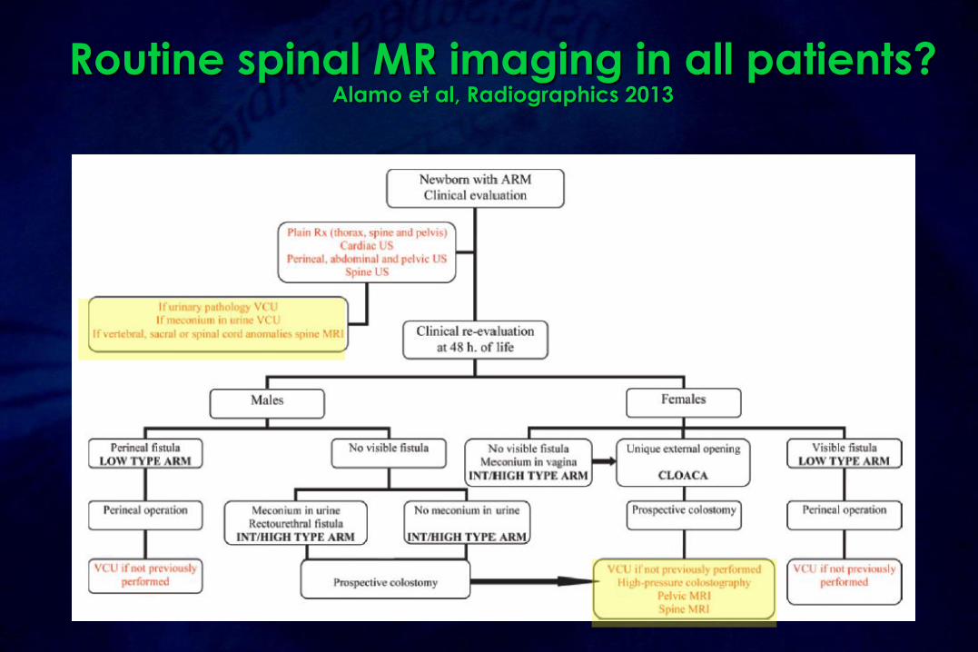
Routine spinal MR imaging in all patients?Alamo et al, Radiographics 2013
Page 30
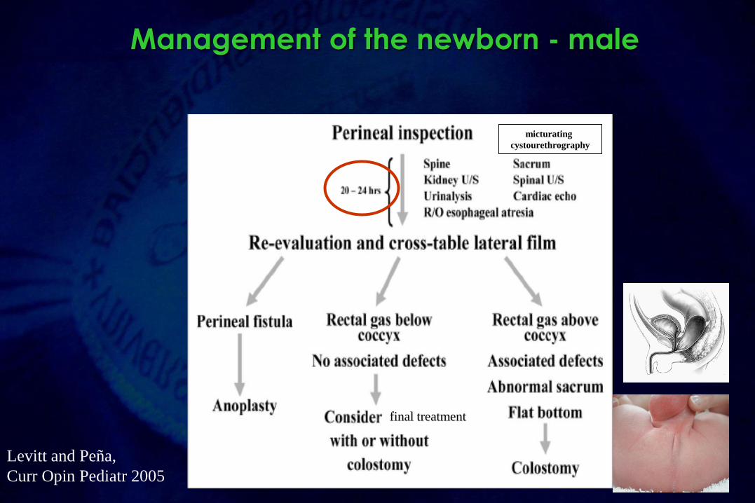
Management of the newborn - male
Levitt and Peña,
Curr Opin Pediatr 2005
micturating
cystourethrography
final treatment
Page 31
Perineal repair - male
Cutback anoplasty
Page 32
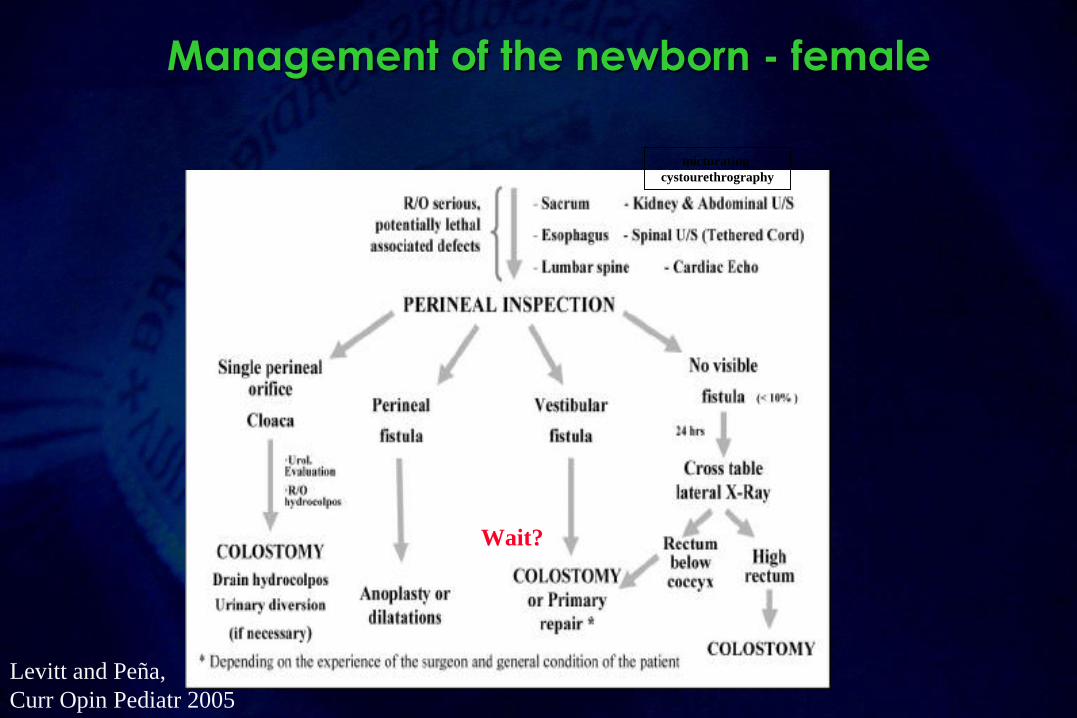
Management of the newborn - female
Levitt and Peña,
Curr Opin Pediatr 2005
micturating
cystourethrography
Wait?
Page 33
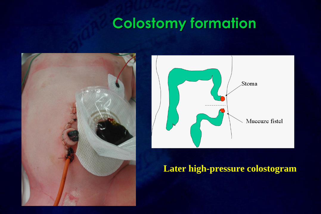
Colostomy formation
Later high-pressure colostogram
Page 34
International grouping of surgicalprocedures (Krickenbeck)
• Pull-through abdominoperineal (Rhoads, 1948)
• Sacroperineal approach (Stephens 1953)
• Perineal operation (Browne, Potts 1954)
– Cutback anoplasty (Browne)
– “Potts transfer anoplasty”
• Pull-through abdominosacroperineal (Kiesewetter 1966 , Rehbein 1967)
• PSARP (Peña-deVries, 1982)
•Anterior sagittal approach (Mollard, 1989, Okada, 1992)
• Pull-through laparoscopic-assisted (Georgeson, 2000)
Holschneider et al, J Pediatr Surg 2005
GOLD
STANDARD
Page 36
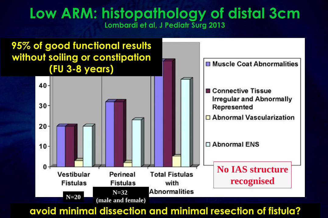
Low ARM: histopathology of distal 3cmLombardi et al, J Pediatr Surg 2013
N=20N=32
(male and female)
No IAS structure
recognised
avoid minimal dissection and minimal resection of fistula?
95% of good functional results
without soiling or constipation
(FU 3-8 years)
Page 37
Indicators of prognosis for bowel control in ARM patients
Levitt et al, J Pediatr Surg 2010
Page 38
Conclusions•A good, simple classification is crucial for adequate comparison of data
• Surgical and prognostic relevance
• “high and low” is too general
• Ectopic anus is not the same as ectopic anal canal
•Multidisciplinary collaboration
• Pediatrician, pediatric surgeon, radiologist, nursing team,
psychologist, physiotherapy, stoma nurse…
• Preoperative work-up
• Meticulous surgical technique
• Postoperative follow-up into adulthood
• GI, urinary, sexual, psychosocial


![48 Marketing RT04 17 14 3[1] - Wepublic Retailtrends.pdf · marketingmix. Of, zoals ceo Howard Schultz van Starbucks zegt: ÒDeze onzekere tijden vragen om een andere aanpak dan voorheen.Ó](https://static.documents.pub/doc/80x56/5e6a1edffea7ec7bbb3c2d6d/48-marketing-rt04-17-14-31-wepublic-retailtrendspdf-marketingmix-of-zoals.jpg)


