This study examined amplitude integrated electroencephalogram (aEEG) characteristics in term neonates undergoing treatment for Neo-natal Abstinence Syndrome (NAS). Twenty mothers consented to par-ticipate and eight infants with an estimated gestational age !37 weeks and undergoing treatment for NAS were placed on aEEG for the first 72 hours of life and then, when possible, for 48 hours every week thereaf-ter until discharge. The average length of time for monitoring was 174 hours. Abnormal progression of cycling as well as presence of electro-graphic seizures was identified during the study period. The number of aEEG seizures identified ranged from two to a maximum of nine in one infant. The presence of aEEG seizure activity in addition to abnormal cycling patterns without physical manifestation of seizures may provide significant additional clinical information to withdrawal scores in infants with NAS.
Keywords: seizure, aEEG, neonatal abstinence syndrome, drug with-drawal
Introduction
Illicit drug use is prevalent in women during pregnancy. From 2000-2009, 16.2% of pregnant teens and 7.4% of pregnant women between 18 and 25 years used illicit drugs in the month before participation in drug use interviews. Concomitantly, rates of Neonatal Abstinence Syn-drome (NAS) have increased in the United States from 1.3 per 1000 births in 2000 to 3.3 per 1000 births in 2009, with the approximate num-ber of newborns with NAS in the U.S. of 13,500. From 2000 to 2009, hospital charges for NAS increased from an estimate of $190 million to $720 million dollars when adjusted for inflation.1 Background
The physical signs of withdrawal can be debilitating and include evi-dence of central nervous system and gastrointestinal dysfunction and neurologic excitability as described in Table 1. Seizures may accom-pany the withdrawal process in as many as 2%-11% of infants with-drawing from opiates. Abnormal electroencephalogram (EEG) findings have been reported; however for more than 30% of these infants, no overt seizure activity was noted.2,3 The onset of withdrawal from opi-ates, including methadone, is typically within the first 24 hours to up to 72 hours of age, depending on the opiate used. First symptoms may also occur as late as 7 days after birth.4,5
Traditionally, symptoms of NAS have been attributed to the abrupt cessation of drug use. However, recent evidence suggests there may be interplay of other genetic, epigenetic and environmental fac-
tors. Polysubstance abuse concomitant with psychological comor-bidities within circumstances of abuse, poor nutrition, and lack of prenatal care appears to create significant risk for NAS.6
Over time, understanding of the pathophysiology for infants present-ing with NAS has expanded.6 Evidence suggests that fetal pro-gramming may play a significant role in whether infants will exhibit symptoms of withdrawal as a result of in utero stressors. Proposed is that the fetus adapts to the unfavorable intrauterine environment by alteration of physiologic systems as a response. In utero re-sponses result in ex utero maladaption, manifested as symptoms of NAS. In 1980, Dinges, et al., studied infants born to mothers on various amounts of narcotics. The authors performed a sleep study that re-corded electroencephalogram, electrooculogram, electromyogram, respiration, and behavioral activity to evaluate these newborns, and found that the opiate-exposed infants exhibited less quiet sleep and more active REM sleep than their non-exposed counterparts.7 In 1988, Pinto, et al., published a case series report on 13 infants with neonatal abstinence syndrome and examined their sleep at the end of their sec-ond week of life and then again in the fourth and fifth week after the abstinence syndrome was treated. Again, NAS babies demonstrated decreases in quiet sleep.8
In contrast, Sarfi and colleagues examined the sleep patterns of three month-old babies who were born to mothers taking either methadone or buprenorphine as compared to a control group and found no differ-ences between the groups at this age.9 However, in both reports, sleep was evaluated at only one point in time. In fact, there is no evidence describing the progression of aEEG background pattern, bandwidth and time to develop cycling in term infants born to a mother on opioids. Therefore, the goal of this study was to describe aEEG characteristics in the term neonate undergoing treatment for NAS. The study was ap-proved by the Institutional Review Board and was not funded by any organization or company.
Methods
All infants with an estimated gestational age (GA) of >37+0 weeks born to mothers with known narcotic use, no prenatal care, or who had a positive maternal urine drug screen had urine and meconium analyzed for illicit drugs and methadone were invited to participate. They were monitored with abstinence scores using the modified Fin-negan scoring system every 8 hours.
When a diagnosis of NAS was made, the baby was admitted to the Neonatal Intensive Care Unit (NICU). Informed parental consent for study participation was obtained. Each subject was assigned an indi-vidual study number and aEEG was started. Day of Life (DOL) was used to describe the number of 24 hour periods after birth with the day of birth as DOL 0.
NEONATOLOGY TODAYN e w s a n d I n f o r m a t i o n f o r B C / B E N e o n a t o l o g i s t s a n d P e r i n a t o l o g i s t s
Babies with Neonatal Abstinence Syndrome Have Electrographic Seizures and Altered Sleep on Amplitude-Integrated EEGBy R. Edwin Spitzmiller, DO; Tracy Morrison, RN, BSN; Robert White, MD
NEONATOLOGY TODAY ! www.NeonatologyToday.net ! REPRINTED WITH PERMISSION
Continuous bedside cerebral function monitoring – providing actionable information when you need it most…
newborn care
www.natus.com
Monitoring the “State of the Brain”
Designed speci�cally for use in the NICU — for neonatologists and NICU nurses
Provides instant, long-term aEEG/EEG monitoring information at the bedside
Available with web-based remote view to assist with clinical collaboration
NOW FEATURING AUTOMATIC SEIZURE DETECTION
CFMOlympic Brainz Monitor
Natus...Where Babies Come First.™
Visit our NERVE Center® education portal at nervecenter.natus.com
For more information or to schedule a demonstration of the Olympic Brainz Monitor call Natus Medical at 1-800-303-0306 or 1-650-802-0400 or visit us at www.natus.com
Natus Medical Incorporated 1501 Industrial RoadSan Carlos, CA 94070 USA 1-800-303-0306 • +1-650-802-0400www.natus.com
Amplitude integrated EEG, was monitored using either the Natus Medical CFM-6000, manufactured by Olympic Medical, Seattle, Washington, USA, or the Natus Medical BRM3 manufactured by Xltek, Oakville, On-tario, Canada. The aEEG was initially placed by nurses for 72 hours and then reapplied by nurses every 7 days for a 48 hour period until discharge or withdrawal from the study. Ini-tially the study was designed to include the use of hydrogel and/or needle electrodes, but the increased physical activity of infants with NAS precluded the use of the hydrogel elec-trodes due to the difficulty in obtaining artifact free tracings. To reduce measurement error, only a small team of 12 experienced nurses was recruited for electrode needle insertion and maintenance. Verification of aEEG elec-trode application was accomplished through simulation so that each RN used the same application techniques.
All infants with NAS received standard of care according to NICU protocols. Education re-garding the study was provided in staff meet-ings, by unit newsletter, and in person by the study nurses. Families were encouraged to ask questions and continue to fully participate in the care of their infants. There was always a study team member in house or on call to initiate study monitoring during the study pe-riod.
Infants with NAS were treated with metha-done; with the dose and frequency modified based on Finnegan scores over the previous 24 to 48 hours. The Finnegan scoring sys-tem is a list of symptoms with accompanying numbers to reflect the severity of each sys-tem, and has been well-described and util-ized. A score of 8 or more indicates the need for treatment.10,11 The American Acad-
emy of Pediatrics states the use of a scoring system is necessary as it results in more objective criteria to determine whether pharmacologic treatment is necessary to begin and whether the dose of the medica-tion should be altered.4
Reduction in methadone dose or frequency was not made more frequently than every 48 hours as was the care protocol for the NICU. Dosage adjustments were made at the discre-tion of the attending neonatologist based on Finnegan scores.
Instrumentation
The (aEEG) is a bedside monitor in which cerebral electrical activity is recorded from either 1 or 2 channels. The aEEG has been d e s c r i b e d i n d e t a i l i n s e v e r a l publications.12,13,14,15,16 Briefly, the electrical signal obtained is rectified, smoothed, and recorded on a semi-logarithmic scale. Inter-pretation is based on recognition of the back-ground pattern as defined by the height of the upper and lower margins of the band, the width of the electrical band, the continuity of the signal, and the changes in the cyclical activity in the recording.12,13 This allows for easy interpretation at the bedside without extensive training.17
Data Analysis
Normal sleep-wake cycling (SWC) is charac-terized by sinusoidal variations in the mini-mal and maximal amplitude of the aEEG which reflects patterns of alternating periods of sleep and wakefulness. A broad band-width represents discontinuous background activity during quiet sleep while a narrow bandwidth represents wakefulness and ac-tive sleep.15,18 The patterns on aEEG repre-senting active sleep and wakefulness cannot be easily distinguished.19
Since the aEEG monitoring system does not provide summation scores for monitored events, aEEG bandwidth of the aEEG tracing was calculated by drawing lines across the upper and lower margins of the band and subtracting voltage of the latter from that of the former. Sleep-wake cycling (SWC) was defined as a regular pattern of wider band-width alternating with narrower bandwidth pattern every 40–90 minutes.15,18 The quiet portion of the cycle time was found by count-ing the minutes from the beginning of the widened portion of the tracing to the end of the same portion. Seizures were defined as an abrupt upstroke of the lower margin in the aEEG pattern with regularly occurring associ-ated high amplitude spikes in the raw EEG for a minimum of 10 seconds.20 See the accom-panying screen shots from the monitors used during the study. Below each screen shot is the tracing type and the definition of each tracing for definitions and screen images from
20 mothers approached
14 consented 10 babies withdrawn at
the request of the parents, but the
IRB allowed the use of data obtained
from the tracings
5 infants were recordedfor less than 48 hours
4 babies monitored to discharge
9 tracings available for review
Table 1: Symptoms of Neon natal Abstinence Syndrome
Adapted from Jansson, L.M. 2008; American Aca ademy of Pediatrics Committee on Drugs, 1998.
NEONATOLOGY TODAY ! www.NeonatologyToday.net ! REPRINTED WITH PERMISSION
the two monitors used. All infant tracings were reviewed by the two physician investigators. In the event of a disagreement, all aspects of the tracing measurement or interpretation were discussed until an agreement between the two reviewers was reached.
Results
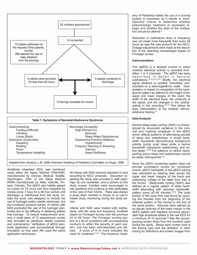
Twenty mothers between May 1, 2009 and April 30, 2011 were ap-proached to participate in the study and fourteen consented to participa-tion of their infants. Infants were enrolled in the study for 5 to 66 days with a mean of 27.4 days, a standard deviation of 18.5 days and a me-dian of 15 days. The average length of monitoring was 174 hours with a range of 95 – 344 hours. Subjects not included for analysis were one infant determined to be 36 weeks gestation by exam and ten babies were withdrawn from monitoring prior to discharge at the request of their parent(s). After request, the IRB granted permission to use the data ob-tained from early study withdrawal babies for analysis. Five infants had recordings for less than 72 hours; therefore, information from these trac-ings was not used in the final analysis. Three babies were monitored until discharge. For the final analysis, aEEG results were used for analy-sis from a total of 8 infants, as described in Figure 1.
All eight babies had background patterns consistent with continuous normal voltage (CNV) on Day One of monitoring. The bandwidth ranged from 10 to 15 microvolts ("v) with a mean of 12 "v. Seven of the eight (88%) babies had electrographic seizure activity, without clinically ap-parent seizures. The seizures were not treated due to retrospective re-view of the tracings. The number of aEEG seizures seen ranged from 1 to a maximum of 9 seizures in 1 baby.
Evidence of cycling was seen on DOL 1 in 3 babies, DOL 2 in 3 babies, and DOL 3 in 2 babies. There was no change in cycling for the duration of monitoring in 5 babies; however, in 3 babies the pattern changed over time, with prolonged active sleep/awake pattern in the first 24 hours, which then normalized over a period of days to weeks. All babies spent more time in either awake or active sleep than in quiet sleep during monitoring. The average awake/active sleep period was approximately 67% of the total recorded time which is summarized in Table 2. There did not appear to be a correlation between the presence of seizure activ-ity and the timing of the appearance of a normal sleep-wake cycling pat-tern or the total awake time and the NAS scores.
Discussion
To our knowledge, the current descriptive study is the first study to document abnormal quiet-active cycling as well as the presence of sub-
clinical seizure activity on aEEG in term newborns undergoing treatment for NAS.
We found the basic and predominant background tracing to be continu-ous normal voltage in all subjects. One of the most surprising findings, however, was the number of seizures seen on aEEG. As described, 88% of the babies had at least one aEEG seizure during the monitoring period without a physical manifestation. One infant had up to 9 seizures without overt clinical evidence. This is the first time electrographic sei-zures via aEEG monitoring have been described in the infant with NAS.
The SWC was not consistent in these infants. Some of the babies de-veloped a regular SWC from the time the aEEG was first placed, and then became irregular; other babies did not develop a regular SWC pat-tern until much later. There did not appear to be any distinct correlation between the NAS scores the development of regular SWC. These find-ings are consistent with the literature; however this is the first time it has been described using aEEG.
There are 5 conscious states of the term newborn: wakefulness, drowsiness, active sleep, quiet sleep, and indeterminate sleep. To properly determine the sleep/wake state of the newborn, Kidokoro, et al., concluded that one must use physiological parameters such
Tracing Type: SeizureDefinition: A rapid upstroke in the lower margin.From Subject #12. This baby was 4 weeks 5 days old.
Tracing Type: BandwidthDefinition: The difference between the upper and lower margins of the tracing.
Tracing Type: Continuous Normal VoltageFrom Subject #12. This also demonstrates normal sleep-wake cylcing.
NEONATOLOGY TODAY ! www.NeonatologyToday.net ! REPRINTED WITH PERMISSION
as rapid eye movement, body movements, and respirations in addi-tion to the aEEG.18 The development of the sleep cycle of the nor-mal newborn infant has been well reviewed.18,21 An infant at 39-41 weeks’ gestation spends approximately 65% of its time in active sleep, which steadily decreases as the infant ages. The two month-old spends approximately 55% of its sleep time in active sleep. It is possible that if more babies remained enrolled for the duration of their hospitalization, there would have been an even higher per-centage of time in active sleep/awake.
Polydrug-exposed infants have been shown to have more total awake time, less total sleep time and more arousals during active sleep than non-exposed infants.22, 23 Although the babies in our study did spend about 67% of the time either in active sleep or awake, this figure repre-sents an average. We did not differentiate based on the age of the baby or whether the baby was asleep or awake. Several of the tracings showed prolonged active sleep/awake time in the earlier part of the study, which then transitioned to a normal cycle over time.
Polydrug-exposed infants have been shown to have more total awake time, less total sleep time and more arousals during active sleep than non-exposed infants.22,23 One study utilized aEEG sleep recordings for 2 hours at two time points22 while the other observed respiratory con-trol and behavior overnight on postnatal days 3, 4 and 5. 23 Both stud-ies involved babies exposed to polydrug abuse including cocaine.
The presence of abnormal cycling should be considered in the treat-ment of the withdrawing infant. In our study, four of the eight infants had periods of normal cycling interspersed with periods of prolonged active phase. The presence of these prolonged active patterns, when paired with physical signs of wakefulness, could be used to deter-mine medication adjustments during treatment.
The incidence of visible seizures in the newborn is 1 to 3.5 per 1000 live births. Electrographic-only seizures are not uncommon; one re-port quotes 80% of electrographic seizures were not associated with clinical findings and they occurred in 1% of 1200 neonates consid-ered high risk for seizures.24 Although clinically apparent seizures are known to occur during opiate withdrawal we observed solely electrographic-only seizures.2, 3, 25
Implications
One limitation of this study is the small number of infants enrolled. Our results need to be duplicated on a larger scale. Another limita-
tion was the inability to monitor all infants until discharge due to parental withdrawal of consent in the later stages of their treatment. A third limitation is that we did not specifically evaluate the amount of active sleep time, awake time or time in quiet sleep; rather, we compared active sleep/awake versus quiet sleep. While there has been no study validating aEEG for quantification of sleep-wake cy-cling, Kidokoro, et al state the presence of cycling on aEEG corre-sponds to the presence of alternate changes of continuous and discontinuous patterns on conventional EEG.18 Since the aEEG trace is the result of a filtered, amplified, rectified, smoothed and compressed raw EEG displayed on an semilogrithmic scale, it is the best way to follow cycling for a prolonged period while a baby is in the NICU. Our findings of electrographic-only seizures and the ab-normal SWC patterns are the first to be described. These findings should be considered an additional, previously undescribed mani-festation of NAS, with possible implications for treatment. More studies with larger numbers of subjects need to be done.
Tracing Type: Continuous normal voltageDefinition: Voltage margins between 10 and 50 microvoltsFrom Subject #6. This shows lower and upper margins above 10 and 50 µV. There is not sleep-wake cycling present during this 4 hour tracing.
Tabl le 2: Demographic cs of Babies Who Were Used in the e Final Analysis
NEONATOLOGY TODAY ! www.NeonatologyToday.net ! REPRINTED WITH PERMISSION
Conclusion
The aEEG may be a useful adjunct tool in the evaluation of the full-term neonate who is ex-hibiting signs of NAS. Some of these babies have less sleep – wake cycling and many ba-bies exhibit electrographic-only seizure activity, the significance of which is yet to be estab-lished. More studies with larger numbers of infants are needed to answer these questions.
We propose the aEEG be used as an adjunct during the treatment of Neonatal Abstinence Syndrome. The presence of seizures on aEEG or the absence of a normal sleep wake cycling pattern should provide added information to the withdrawal score and may be valuable to determine pharmacologic therapy adjustment.
Acknowledgments
We wish to thank Marc Belcastro, DO; Tamisha Samiec, MD; Alan Spitzer MD; and Pat O’Mal-ley, PhD, RN, CNS for their valuable input on this manuscript. Thanks also to the ECMO nurses at Miami Valley Hospital for their com-mitment to this project and to Sue Mackey for her assistance with IRB submission.
Author Contributions
Ed Spitzmiller wrote the majority of the manu-script and Tracy Morrison wrote the majority of the methods section. Robert White contributed valuable editorial advice and wrote smaller sections as well as serving a reviewer of aEEG tracings.
Declaration of Conflicting Interests
None of the authors have any conflicting inter-ests to declare.
Funding
The authors received no funding for this re-search.
Ethical Approval
This study was approved by the institutional review board at Miami Valley Hospital.
References
1. Patrick SW, Schumacher RE, Benney-worth BD, Krans EE, McAllister JM, Davis MM. Neonatal abstinence syndrome and associated health care expenditures: United states, 2000-2009. JAMA. 2012; 307(18): 1934-1940.
2. Zelson C, Rubio E, Wasserman E. Neo-natal narcotic addiction: 10 year observa-tion. Pediatrics. 1971; 48(2):178-89.
3. Kandall SR, Gartner LM. Late presenta-tion of drug withdrawal symptoms in new-b o r n s . A m J D i s C h i l d . 1 9 7 4 ; 127(1):58-61.
4. American Academy of Pediatrics Com-mittee on Drugs. Neonatal drug with-drawal. Pediatrics. 1998;101(6):1079-88.
6. Jansson LM, Choo R, Velez ML, Harrow C, Schroeder JR, Shakleya DM, et al. Methadone maintenance and breastfeed-ing in the neonatal period. Pediatrics. 2008; 121(1):106-14.
7. Dinges DF, Davis MM, Glass P. Fetal ex-posure to narcotics: Neonatal sleep as a measure of nervous system disturbance. Science. 1980; 209(4456):619-21.
8. Pinto F, Torrioli MG, Casella G, Tempesta E, Fundaro C. Sleep in babies born to chronically heroin addicted mothers. A follow-up study. Drug Alcohol Depend. 1988; 21(1):43-7.
9. Sarfi M, Martinsen H, Bakstad B, Roislien J, Waal H. Patterns in sleep-wakefulness in three-month old infants exposed to methadone or buprenorphine. Early Hum Dev. 2009;85(12):773-8.
10. Finnegan LP, Connaughton JF, Jr., Kron RE, Emich JP. Neonatal abstinence syn-drome: Assessment and management. Addict Dis. 1975; 2(1-2):141-58.
11. Burgos AE, Burke BLJ. Neonatal absti-nence syndrome. Neoreviews. 2009; 10 (5 ) : e222 -229 . Ava i l ab l e a t http://neoreviews.aappublications.org/. Accessed July 25, 2013.
12. Thornberg E, Thiringer K. Normal pattern of the cerebral function monitor trace in term and preterm neonates. Acta Paediatr Scand. 1990; 79(1):20-5.
13. al Naqeeb N, Edwards AD, Cowan FM, Azzopardi D. Assessment of neonatal encephalopathy by amplitude-integrated electroencephalography. Pediatrics. 1999; 103(6):1263-71.
14. Hellstrom-Westas L, Rosen I. Amplitude-integrated electroencephalogram in new-born infants for clinical and research pur-p o s e s . A c t a P a e d i a t r . 2 0 0 2 ; 91(10):1028-30.
15. Rosen I. The physiological basis for con-tinuous electroencephalogram monitoring in the neona te . C l in Per ina to l . 2006;33(3):593,611, v.
16. Spitzmiller RE, Phillips T, Meinzen-Derr J, Hoath SB. Amplitude-integrated EEG is useful in predicting neurodevelopmental outcome in full-term infants with hypoxic-ischemic encephalopathy: A meta-a n a l y s i s . J C h i l d N e u r o l . 2007;22(9):1069-78.
17. Rennie JM, Chorley G, Boylan GB, Pressler R, Nguyen Y, Hooper R. Non-expert use of the cerebral function monitor for neonatal seizure detection. Arch Dis C h i l d F e t a l N e o n a t a l E d . 2004:89(1):F37-40.
18. Kidokoro H, Inder T, Okumura A, Watan-abe K. What does cyclicity on amplitude-
19. Hellstrom-Westas L. Continuous electro-encephalography monitoring of the pre-term infant. Clin Perinatol. 2006; 33(3):633, 47, vi.
20. Hellstrom-Westas L, de Vries LS, Rosen I. An atlas of ammplitude - integrated EEGs in the newborn. New York, NY: Parthenon Publishing; 2003.
21. Curzi-Dascalova L, Peirano P, Morel-Kahn F. Development of sleep states in normal premature and full-term newborns. Dev Psychobiol. 1988;21(5):431-44.
22. Scher MS, Richardson GA, Day NL. Ef-fects of prenatal cocaine/crack and other drug exposure on electroencephalo-graphic sleep studies at birth and one year. Pediatrics. 2000; 105(1 Pt 1):39-48.
23. Gingras JL, Feibel JB, Dalley LB, Muele-naer A, Knight CG. Maternal polydrug use including cocaine and postnatal infant sleep architecture: Preliminary observa-tions and implications for respiratory control and behavior. Early Hum Dev. 1995; 43(3):197-204.
24. Seshia SS, Huntsman RJ, Lowry NJ, Seshia M, Yager JY, Sankaran K. Neona-tal seizures: Diagnosis and management. Zhongguo Dang Dai Er Ke Za Zhi. 2011; 13(2):81-100.