54
Neoplasia Kazan (Volga region) Federal University Institute of Fundamental Medicine and Biology Department of Morphology and General Pathology Lecturer Olga N. Chernova
Neoplasia
Kazan (Volga region) Federal University
Institute of Fundamental Medicine and Biology
Department of Morphology and General Pathology
Lecturer
Olga N. Chernova
Neoplasia
Literally “new growth”
• In common medical usage, a neoplasm often is referred to as a tumor, and the study of tumors is called oncology (from oncos, “tumor,” and logos, “study of”)
2
Characteristic of cancer
• Cancer is a genetic disorder caused by DNA mutations that are (for the most part) acquired spontaneously or induced by environmental insults
• These genetic alterations are heritable, being passed to daughter cells upon cell division. As a result, cells harboring these alterations are subject to darwinian selection
• Accumulation of mutations gives rise to a set of properties that have been called hallmarks of cancer
3
4
INTRODUCTION & DEFINITIONS
• Neoplasia (Latin, new growth) is an abnormality of cellular differentiation, maturation, and control of growth.
• Neoplasms are commonly recognized by the formation of masses of abnormal tissue (tumors).
• The term tumor can be applied to any swelling- and in that context is one of the cardinal signs of inflammation-but today it is used most commonly to denote suspected neoplasm.

5
• Neoplasms are benign or malignant depending on several features, chiefly the ability of malignant neoplasms to spread from the site of origin.
• Benign neoplasms grow but remain localized.
• Cancer denotes a malignant neoplasm (the term is thought to derive from the way in which the tumor grips the surrounding tissues with claw-like extensions, much like a crab). This feature led Hippocrates to call such tumors karkinoma after karkinos.
6
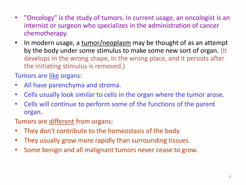
• "Oncology" is the study of tumors. In current usage, an oncologist is an internist or surgeon who specializes in the administration of cancer chemotherapy.
• In modern usage, a tumor/neoplasm may be thought of as an attempt by the body under some stimulus to make some new sort of organ. (It develops in the wrong shape, in the wrong place, and it persists after the initiating stimulus is removed.)
Tumors are like organs:
• All have parenchyma and stroma.
• Cells usually look similar to cells in the organ where the tumor arose.
• Cells will continue to perform some of the functions of the parent organ.
Tumors are different from organs:
• They don't contribute to the homeostasis of the body.
• They usually grow more rapidly than surrounding tissues.
• Some benign and all malignant tumors never cease to grow.

7
BIOLOGIC BEHAVIOR OF NEOPLASMS
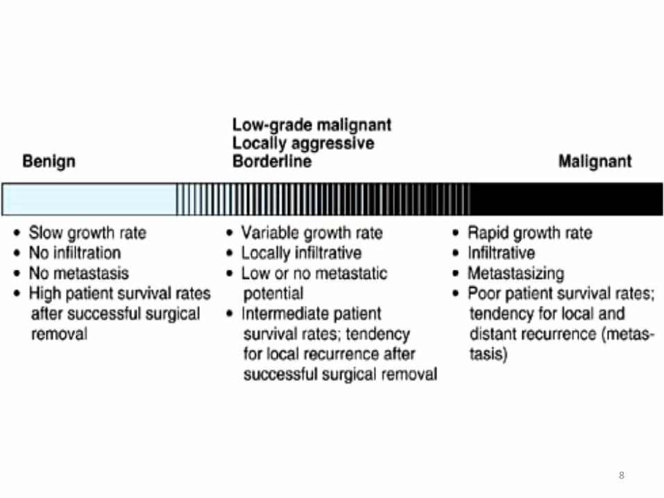
• The biologic behavior of neoplasms constitutes a spectrum with two extremes: Benign and Malignant.
• Benign: benign neoplasms grow slowly and do not invade surrounding tissues or spread to distant sites (ie, no metastasis).
• Benign neoplasms are rarely life-threatening but may become so because of hormone secretion or critical location, eg, a benign neoplasm can cause death if it arises in a cranial nerve and compresses the medulla spinalis.
8

9
• Malignant: Malignant neoplasms grow rapidly, infiltrate and destroy surrounding tissues, and metastasize throughout the body, often with lethal results.
• Between Benign and Malignant; is a smaller third group of neoplasms that are locally invasive but have low metastatic potential.
• Such neoplasms are borderline neoplasms or locally aggressive neoplasms or low-grade malignant neoplasms. An example is basal cell carcinoma of the skin and serous borderline neoplasm of the ovaries.
10
• 1. Rate of Growth:
• Malignant neoplasms generally grow more rapidly than benign ones, but there is no critical rate that distinguishes malignant from benign.
• Assessment of the growth rate is based upon clinical information (eg, change in size of the mass in serial examinations).
• On microscopic examination, the number of mitotic figures and the metabolically active appearance of nuclei (enlarged, dispersed chromatin, large nucleoli) correlate positively with the growth rate of the neoplasm.

11
• 2. Size:
• The size of a neoplasm usually has no bearing on its biologic behavior.
• Many benign neoplasms become very large; conversely, highly malignant neoplasms may be lethal by virtue of extensive dissemination even though the original primary tumor is still small.
• In a few neoplasms (such as endocrine neoplasms), however, size is the deciding factor in distinguishing benign from malignant growths.
12
• 3. Degree of Differentiation:
• Denotes the degree to which a neoplastic cell resembles the normal mature cells of the tissue in question; this meaning is distinct from the more general use of the word to describe passage of a cell down a particular maturation pathway.
• Benign neoplasms are fully (well) differentiated, ie, they closely resemble normal tissue.
• Malignant neoplasms, show variable degrees of differentiation and frequently demonstrate little resemblance to normal tissue (ie, they are poorly differentiated).
13
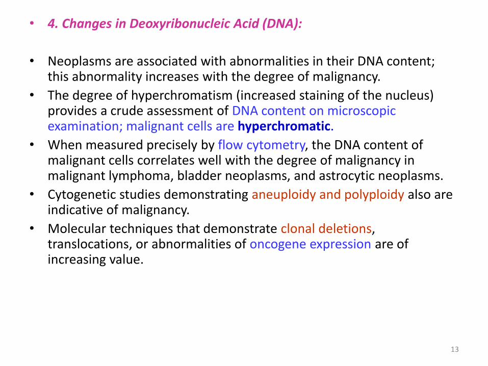
• 4. Changes in Deoxyribonucleic Acid (DNA):
• Neoplasms are associated with abnormalities in their DNA content; this abnormality increases with the degree of malignancy.
• The degree of hyperchromatism (increased staining of the nucleus) provides a crude assessment of DNA content on microscopic examination; malignant cells are hyperchromatic.
• When measured precisely by flow cytometry, the DNA content of malignant cells correlates well with the degree of malignancy in malignant lymphoma, bladder neoplasms, and astrocytic neoplasms.
• Cytogenetic studies demonstrating aneuploidy and polyploidy also are indicative of malignancy.
• Molecular techniques that demonstrate clonal deletions, translocations, or abnormalities of oncogene expression are of increasing value.
14
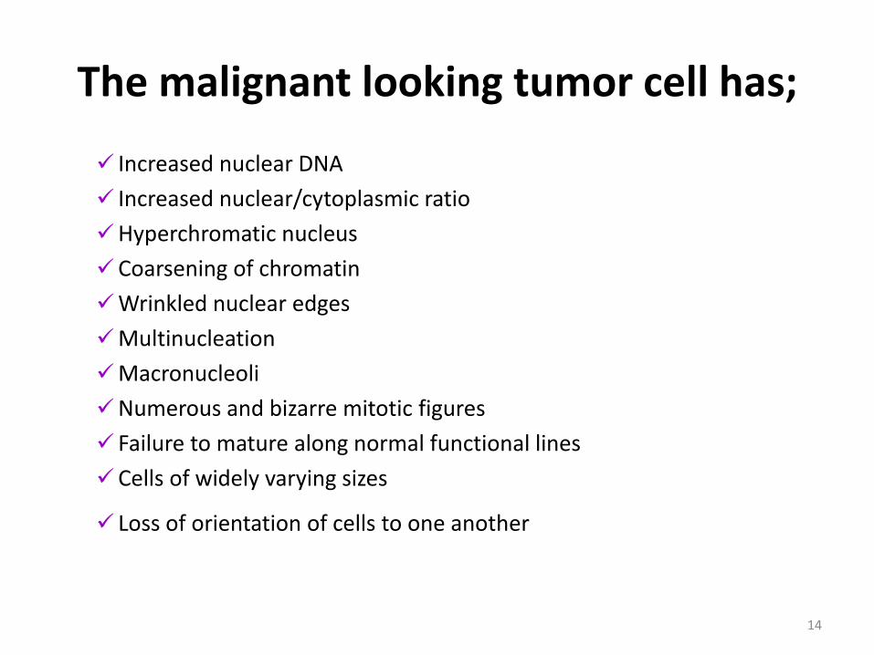
The malignant looking tumor cell has;
Increased nuclear DNA
Increased nuclear/cytoplasmic ratio
Hyperchromatic nucleus
Coarsening of chromatin
Wrinkled nuclear edges
Multinucleation
Macronucleoli
Numerous and bizarre mitotic figures
Failure to mature along normal functional lines
Cells of widely varying sizes
Loss of orientation of cells to one another
15
16
• 5. Infiltration and Invasion:
• Benign neoplasms are generally non-infiltrative and are surrounded by a capsule of compressed and fibrotic normal tissue.
• Malignant neoplasms, have infiltrating margins. Some exceptions to this rule exist, and some benign neoplasms (eg, granular cell tumor, dermatofibroma, and carcinoid tumors) lack a capsule and have an infiltrative margin.
17
• 6. Metastasis:
• The occurrence of metastasis (noncontiguous or distant growth of tumor) is absolute evidence of malignancy.
• The major reason for distinguishing benign from malignant neoplasms is to be able to predict their ability to metastasize before they do so.
• Gross and microscopic examination of a neoplasm usually enables a trained pathologist to classify most neoplasms as benign or malignant.
• In some instances, however, this identification is difficult, and the only reliable evidence of a neoplasm's biologic behavior is the occurrence of metastasis; about 90% of pheochromocytomas are benign, but there are no reliable criteria for identifying the 10% that will metastasize.

Metastatic cascade

Six hallmarks of cancer

20
Oncogenes & Tumor Suppressor Genes
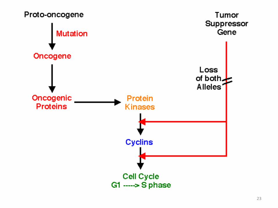
• There are two main categories of genes that regulate cell growth, and the abnormal action of either or both may lead to neoplasia.
• Proto-oncogenes (cellular oncogenes: c-onc) code for a variety of growth factors, receptors, and signal-relay or transcription factors, which act in concert to control entry into the cell cycle (eg, the growth promoter effect).

21
• The action of these genes is opposed by the action of tumor suppressor genes, which serve to down-regulate the cell cycle. A net increase in the production of stimulatory (promoter) factors, a decrease in inhibitory (suppressor) growth factors, or the production of functionally abnormal factors may lead to uncontrolled cell growth.

22
23

24
Oncogene Associated Neoplasms
c-erb-B2 Breast and ovarian carcinomas
ras Many carcinomas and leukemias
c-sis Gliomas
c-abl Chronic myelogenous leukemia, acute lymphocytic leukemia
c-myc Lymphomas
BRCA-1 Breast and ovarian carcinomas
APC Colonic adenocarcinomas
NF-1 Neurofibromas and neurofibrosarcomas
Rb Retinoblastomas, osteosarcomas, small cell lung carcinomas
p53 Many carcinomas
bcl-2 Chronic lymphocytic leukemia, lymphomas

26
Viral Oncogene Hypothesis
• Certain RNA viruses contain nucleic acid sequences that are complementary to a protooncogene and can (by reverse transcriptase) produce a viral DNA sequence that is essentially identical, lacking only the introns of the animal host cell.
• These sequences are termed viral oncogenes (v-onc).

27
28
Epigenetic hypothesis
• The main evidence for the role of epigenetic mechanisms in neoplasia comes from cancers produced by chemicals that have no known effect on the genetic apparatus of the cell.
• It is postulated that these chemicals may serve as promoters by binding various growth regulatory proteins, thus rendering them inactive.
29
Hypothesis of Failure of Immune Surveillance
• (1) Neoplastic changes frequently occur in the cells of the body.
• (2) As a result of alteration in their DNA, neoplastic cells produce new molecules (neoantigens, tumor-associated antigens).
• (3) The immune system of the body recognizes these neoantigens as foreign and mounts a cytotoxic immune response that destroys the neoplastic cells.
• (4) Neoplastic cells produce clinically detectable neoplasms only if they escape recognition and destruction by the immune system

30
AGENTS CAUSING NEOPLASMS (Oncogenic Agents; Carcinogens)
• An agent that causes neoplasms is an oncogenic agent;
an agent causing a malignant neoplasm (cancer) is a carcinogenic agent.
• 1) the cause of most common human cancers is unknown;
• 2) most cases of cancer are probably multifactorial in origin; and
• 3) except for cigarette smoking, the agents discussed below have been implicated in only a small percentage of cases.
31
Carcinogen
• A cancer-causing agent
• Three classes:
– Chemical carcinogens (endogenous/exogenous)
– Physical carcinogens (UV, radiation, asbestos)
– Oncogenic microbes (mainly viruses)
32
Chemical Carcinogens as Mutagens
• Mutagen: an agent that can permanently alter genetic constitution of a cell
• 90% of known carcinogens are mutagenic
• Most mutagens are carcinogens
33
Promoters in Human Cancers
• Cigarettes
• UV
• High Fat Diet
• Hormones
• Viral Infections
34
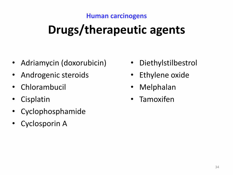
Human carcinogens Drugs/therapeutic agents
• Adriamycin (doxorubicin)
• Androgenic steroids
• Chlorambucil
• Cisplatin
• Cyclophosphamide
• Cyclosporin A
• Diethylstilbestrol
• Ethylene oxide
• Melphalan
• Tamoxifen
35
Direct-acting carcinogens
• Nitrogen mustard
• Nitrosomethylurea
• Benzyl chloride
36
Indirect-acting carcinogens
• Polycyclic aromatic hydrocarbons (PAH)
• Produced by incomplete combustion of organic materials
• Present in – chimney soot,
– charcoal grilled meats,
– auto exhaust,
– cigarette smoke.
37
Physical Carcinogens
• Ultraviolet light
• Asbestos
• Foreign body carcinogenesis
• Ionizing radiation (X-rays), radioisotopes, nuclear bomb
38
Cancers caused by UV exposure
• Squamous cell carcinoma
• Basal cell carcinoma
• Malignant Melanoma
39
Asbestos
• Widely used in construction, insulation, and manufacturing
• Family of related fibrous silicates
• Chrysotile (serpentine form - flexible)
• Crocidolite (amphibole form - rigid rods)
40
Ionizing radiation
• Death of pioneer radiation researchers from neoplasms
• High incidence of leukemia among radiologists recognized in 1940s
• Osteosarcoma incidence in radium dial painters

41
Viral Carcinogenesis
• Viral infections account for an estimated one in seven human cancers worldwide
• Majority of these are due to infection with two DNA viruses
• HBV – linked to hepatocellular carcinonoma
• HPV – linked to cervical carcinoma
42
HPV
• HPV 6,11 – low risk viruses
• HPV – 16, 18, 31, 33, 35, 39, 45 – High risk viruses
• 85% of cervical carcinomas that are HPV-positive contain a high risk HPV (70% have HPV 16 or 18)
43
EBV – involvement in human tumors
• African Burkitt lymphoma
• B-cell lymphomas of immunosuppressed patients
• Some cases of Hodgkin disease
• Nasopharyngeal carcinomas
44
How do viruses like HPV and HBV cause cancer?
• Very small viruses
• Can integrate their viral DNA into host genome
• They code for viral proteins which block tumor suppressor proteins in cell

45
Helicobacter pylori
• Gastric infection linked to gastric lymphomas and gastric carcinomas
• Detection of H. pylori in majority of cases of gastric lymphomas
• Antibiotic treatment results in gastric lymphoma regression in most cases
46
Human Herpesvirus 8
• Kaposi sarcoma - a vascular neoplasm originally described in eastern Europe
• KS is today most common neoplasm associated with AIDS
• Cells contain HHV8 (also called KS-associated herpesvirus) KSHV
47
Factors Influencing Chemical Carcinogenesis
• Metabolism
• Sex and Hormonal Status
• Diet
48
Nutritional Oncogenesis
• A diet high in animal fat has been associated statistically with an increased incidence of cancer of the colon and with breast cancer; this observation remains unexplained.

49
Nutritional Oncogenesis
• A diet high in animal fat has been associated statistically with an increased incidence of cancer of the colon and with breast cancer; this observation remains unexplained.
50
Hormonal Oncogenesis • Estrogens. causes endometrial hyperplasia, which is followed first by
cytologic dysplasia and then by neoplasia.
• Hormones and breast cancer. patients taking oral contraceptives have shown that the risk of breast cancer is minimally increased in patients taking preparations with high estrogen content. The current low-estrogen contraceptives are not thought to increase the risk of breast cancer.
• Diethylstilbestrol (DES). Female children who were exposed to diethylstilbestrol in utero have a greatly increased incidence of clear-cell adenocarcinoma, a rare vaginal cancer that develops in young women between 15 and 30 years of age.
• Steroid hormones. Use of oral contraceptives and anabolic steroids is rarely associated with development of benign liver cell adenomas. A few cases of liver cell carcinoma have been reported.
51
Hormone dependent neoplasms
• Prostate ca
• Breast ca
• Thyroid ca

52
Tumor Cell Products
• The synthesis and secretion of various tumor cell products are important for two reasons:
• (1) their presence may indicate the existence of a neoplasm in the body (ie, they act as tumor markers); and
• (2) they may produce clinical effects (paraneoplastic syndromes) unrelated to direct involvement of tissue by the tumor.
53
PARANEOPLASTIC SYNDROMES
• Fever (lymphomas, acute leukemias, sarcomas, renal cell carcinomas
(Grawitz tumors), and digestive malignancies (including the liver).
• Leukemoid reactions (lymphomas or cancers of the lung, breast, or stomach)
• Erythrocytosis or anemia, thrombocytosis (many types of cancers)
• Disseminated intravascular coagulation (many types of cancers)
• Cryoglobulinemia (lung cancer, pleural mesothelioma)
• Paraneoplastic arthropathies - rheumatic polyarthritis (myelomas; lymphomas; acute leukemia; malignant histiocytosis; and tumors of the colon, pancreas, prostate, CNS
• Hypertrophic osteoarthropathy (lung cancers)
• Scleroderma (breast, uterus, and lung)
• SLE (lung, breast, gonads)
54
PARANEOPLASTIC SYNDROMES
• Amyloidosis (myeloma, renal carcinoma, and lymphomas)
• Nephrotic syndrome (Hodgkin lymphoma (HL); non-Hodgkin lymphoma (NHL); leukemias; melanomas; or malignancies of lung, thyroid, colon, breast, ovary, or pancreatic head)
• Watery diarrhea - electrolyte imbalance (medullary thyroid carcinomas, proctosigmoid tumors, melanomas, myelomas, ovarian tumors, pineal body tumors, and lung metastases)
• Acanthosis nigricans, dermic melanosis (metastatic melanomas or pancreatic tumors
• Cushing syndrome (ectopic production of ACTH or ACTH-like molecules from many tumors, eg, small cell cancer of the lung)
• Neuromuscular disorders (ovarian and pulmonary cancers)


