45
Neoplasia Dr D S O’Briain April 2008
| Date post: | 29-Dec-2015 |
| Category: |
Documents |
| Upload: | lucy-mcdowell |
| View: | 216 times |
| Download: | 2 times |

Neoplasia
Dr D S O’Briain
April 2008
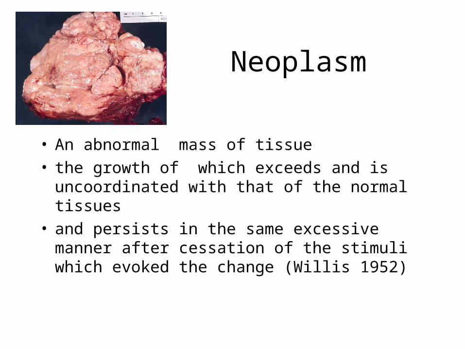
Neoplasm
• An abnormal mass of tissue• the growth of which exceeds and is
uncoordinated with that of the normal tissues • and persists in the same excessive manner after
cessation of the stimuli which evoked the change (Willis 1952)
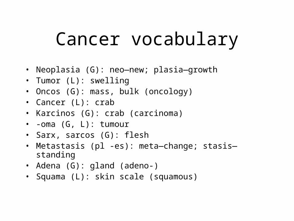
Cancer vocabulary
• Neoplasia (G): neo—new; plasia—growth• Tumor (L): swelling• Oncos (G): mass, bulk (oncology)• Cancer (L): crab• Karcinos (G): crab (carcinoma)• -oma (G, L): tumour• Sarx, sarcos (G): flesh• Metastasis (pl -es): meta—change; stasis—standing• Adena (G): gland (adeno-)• Squama (L): skin scale (squamous)

Cellular dysplasia
• Nuclear size enlarged, variable
• Chromatin increased, variable
• Nucleolus enlarged, malformed
• Mitoses increased, abnormal
• Nucleus/Cytoplasm increased ratio

Differentiation
Resembles adult tissue in
• Cell morphology
• Architecture
• Function
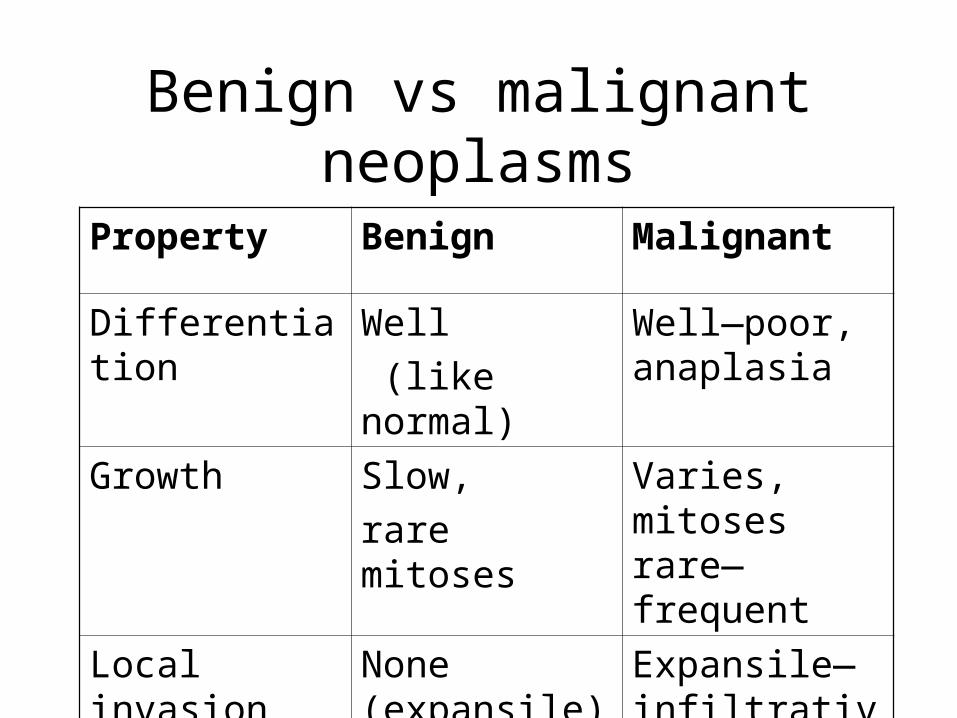
Benign vs malignant neoplasms
Property Benign Malignant
Differentiation Well
(like normal)
Well—poor, anaplasia
Growth Slow,
rare mitoses
Varies, mitoses rare—frequent
Local invasion None (expansile)
Expansile— infiltrative
Metastasis Absent Absent—present

Growth and proliferation
Cell proliferation/growth,
Regeneration (repair)
Hyperplasia
Tumour
Purposeful Purposeless
Physiological stimuli ? Stimuli
Feedback control Reduced/absent feedback control
Reversible Irreversible
Beneficial deleterious
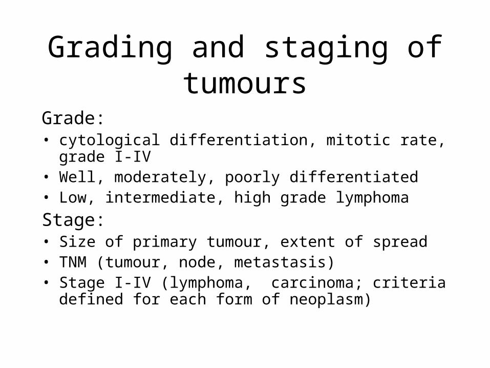
Grading and staging of tumours
Grade:• cytological differentiation, mitotic rate, grade I-IV • Well, moderately, poorly differentiated• Low, intermediate, high grade lymphoma
Stage:• Size of primary tumour, extent of spread• TNM (tumour, node, metastasis)• Stage I-IV (lymphoma, carcinoma; criteria defined for
each form of neoplasm)
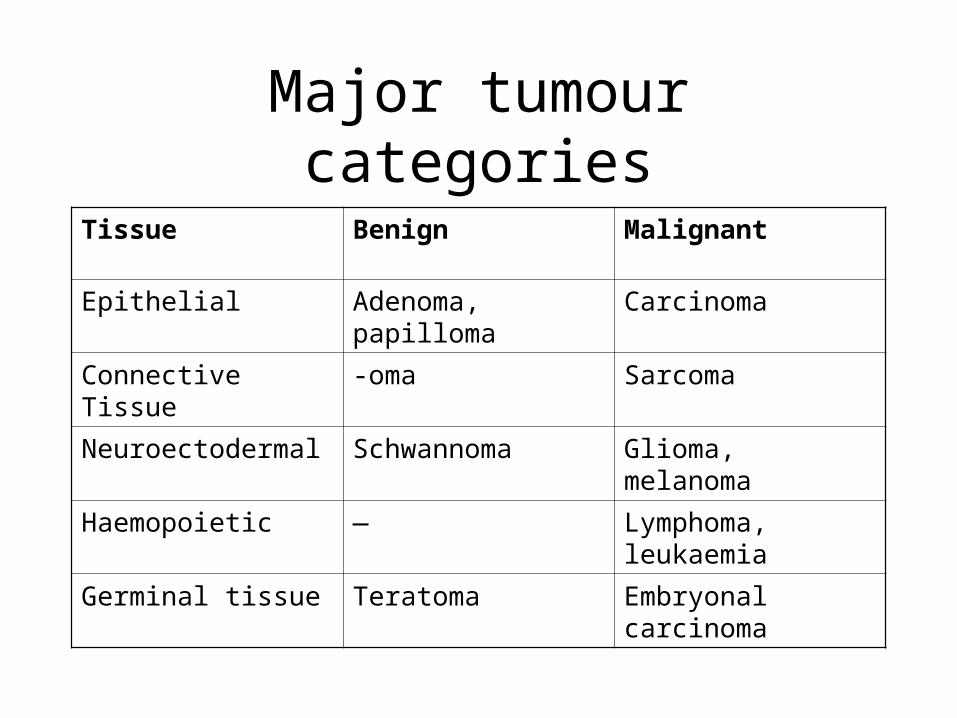
Major tumour categories
Tissue Benign Malignant
Epithelial Adenoma, papilloma Carcinoma
Connective Tissue -oma Sarcoma
Neuroectodermal Schwannoma Glioma, melanoma
Haemopoietic — Lymphoma, leukaemia
Germinal tissue Teratoma Embryonal carcinoma

Epithelial tumoursBenign: • papillomas, adenomas (polyp), tubulovillous
Malignant: stroma (desmoplasia), scirrhous (hard), encephaloid, medullary (soft)
• Adenocarcinoma; mucinous, papillary, cystadenocarcinoma (renal, hepatic)
• Squamous (contains keratin pearls, prickles) carcinoma• Basal cell, transitional cell, clear cell carcinomas• Endocrine (carcinoid, islet cell tumour, small cell carcinoma)
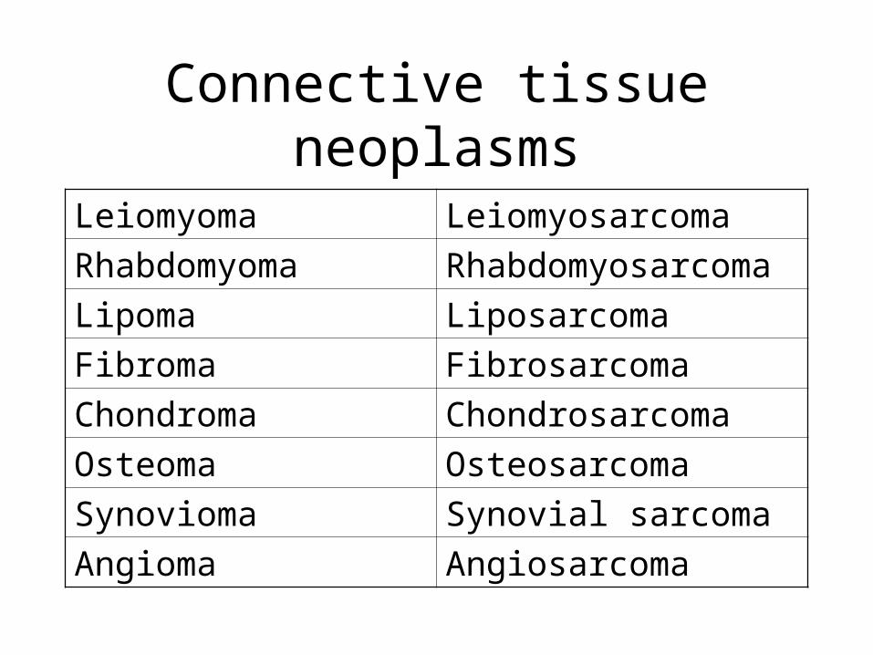
Connective tissue neoplasms
Leiomyoma Leiomyosarcoma
Rhabdomyoma Rhabdomyosarcoma
Lipoma Liposarcoma
Fibroma Fibrosarcoma
Chondroma Chondrosarcoma
Osteoma Osteosarcoma
Synovioma Synovial sarcoma
Angioma Angiosarcoma

Neuroectodermal tumours
Central• Glial: astrocytoma, oligodendroglioma,
ependyoma• Neural: medulloblastoma, neuroblastoma,
ganglioneuroma, meningomaPeripheral• Schwannoma, Neurofibroma, • Naevus, Melanoma
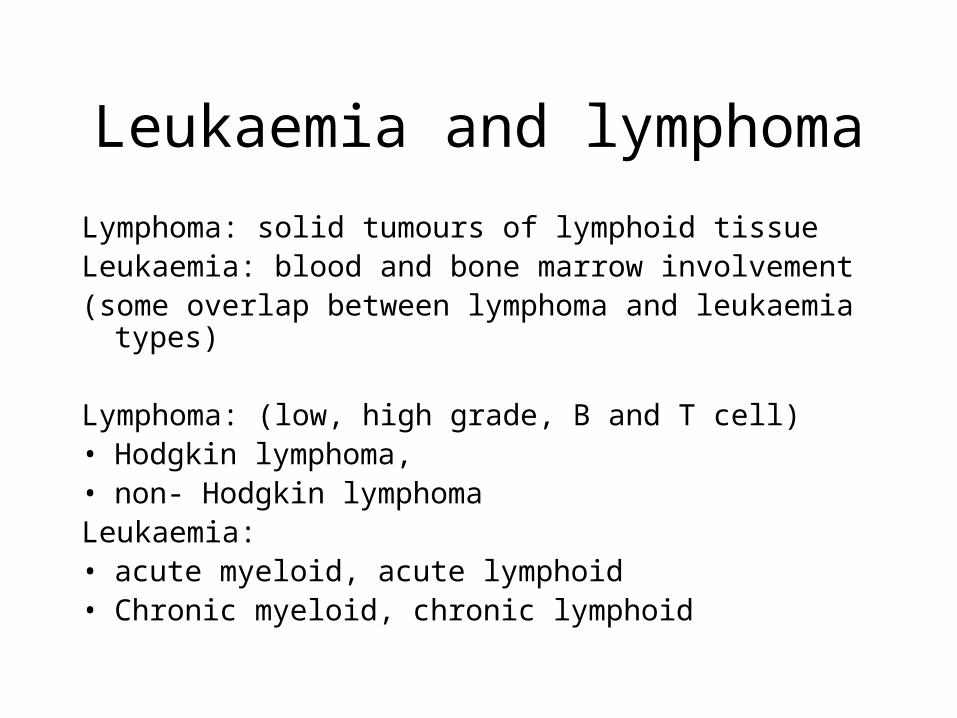
Leukaemia and lymphoma
Lymphoma: solid tumours of lymphoid tissueLeukaemia: blood and bone marrow involvement (some overlap between lymphoma and leukaemia types)
Lymphoma: (low, high grade, B and T cell)• Hodgkin lymphoma, • non- Hodgkin lymphoma Leukaemia: • acute myeloid, acute lymphoid• Chronic myeloid, chronic lymphoid

Germinal tumours
• Teratoma:
Benign: mature, cystic
Malignant: malignant, embryonal carcinoma, choriocarcinoma
• Seminoma, germinoma
• Sex cord stromal tumours
• Placenta: hydatidiform mole, choriocarcinoma

Mixed tumour
Not germinal tumours
• adenosquamous carcinoma,
• fibroadenoma, carcinosarcoma, mixed mullerian tumour
• pleomorphic adenoma
• paediatric blastomas
• metaplastic tumours
• collision tumours

Effects of cancer
• Damage or obstruct critical structures (location)
• Infiltrates nerves—pain
• Functioning activity (hormones)
• Bleeding
• Ulcerate—infected
• Infarct
• Cachexia and paraneoplastic effects

Spread of Cancer• Expansion—compression of surrounding tissue—formation of
(pseudo)capsule• Local invasion (infiltrative)
Follows line of least resistanceDamages surrounding tissueAlters function of tissueMakes excision difficultCauses pain (nerve invasion)
• Lymphatic spread: permeates lymphatics, metastasizes to node• Haematogenous spread: anatomical (portal/systemic) retrograde,
venous• Intracavity: meningeal, serosal cavities• Recurrence: residual tumour, second primary

Site of metastases
Anatomic factors• Sarcoma ˛ lung ˛ general circulation• Carcinoma ˛local lymph nodes ˛ lung, bone, brain• GI primary ˛ liver• Prostate, breast ˛ local venous plexus• Ovary ˛ peritoneumHumoral factors (homing, soil)• Local; preferential adhesion, preferential growth, local
diffusion of attractants (migratory directors)

Metastases—metastatic cascade
• 70% of newly diagnosed cancers have metastases• Millions of tumour cells circulate daily• Less than 0.01% initiate colonies
1. Adhere to endothelium: fibrin platelets, clotting factors, receptors basement membrane
2. Dissolve basement membrane: proteases, collagenases IV, cathepsins
3. Locomotion: initiation (random), directed by autocrine and host growth factors

Paraneoplastic effects
• Endocrinopathies: Cushings syndrome, hyponatremia (SIADH), hypercalcemia, hyperthyroidism, hypoglycemia
• Neuromuscular: cerebellar ataxia, Eaton Lambert syndrome
• Skin disorders: acanthosis nigricans, dermatomyositis
• Blood/vascular: venous thrombosis, DIC, marantic endocarditis, cytopenias, cythemias
• Kidney: renal failure, nephrotic syndrome
• Extermities: digital clubbing, hypertrophic osteoarthropathy

Tumour markers
Class Type Tumour
Antibodies Protein M band Myeloma, lymphoma
Antigens (oncofetal) Carcinoembryonic antigen Colon Ca, others
Alpha-fetoprotein Germ cell tumour, liver Ca
Prostate specific antigen Prostate carcinoma
Hormones HCG Germ cell
Calcitonin Medullary carcinoma thyroid
Ectopic ACTH, ADH Lung Ca, others
Enzymes Acid phosphatase Prostate Ca
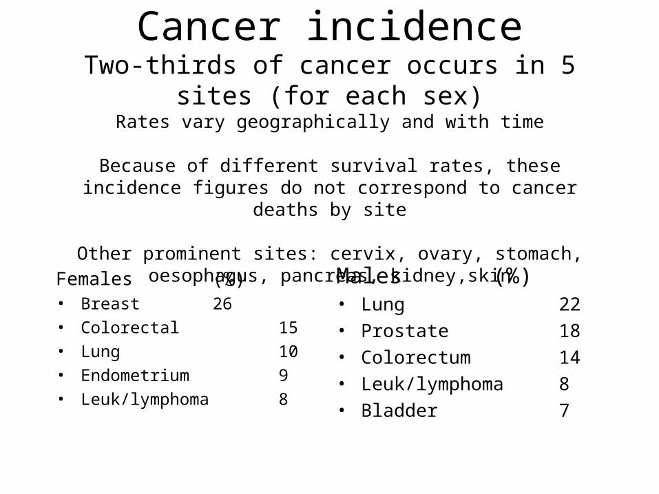
Cancer incidenceTwo-thirds of cancer occurs in 5 sites (for each sex)
Rates vary geographically and with time
Because of different survival rates, these incidence figures do not correspond to cancer deaths by site
Other prominent sites: cervix, ovary, stomach, oesophagus, pancreas, kidney,skin
Females (%)• Breast 26
• Colorectal 15
• Lung 10
• Endometrium 9
• Leuk/lymphoma 8
Males (%)• Lung 22
• Prostate 18
• Colorectum 14
• Leuk/lymphoma 8
• Bladder 7

Cancer incidence—secular changes
Increasing• Lung• Pancreas• (Cervix, ovary,
leukaemia)
Decreasing• Stomach• Endometrium• Liver• Colorectal (females)

Cancer deaths (per 100000) geographic variation
Tumour High incidence
Rate Low incidence
Rate
Colorectal Hungary 36 Mexico 5
Lung Hungary 84m/22f Mexico 17m/7f
Ireland 38m/18f
Breast Ireland 26f China 6f
Cervix Zimbabwe 43f Australia 3f
Stomach China 33m/15f USA 4m/2f
Prostate Norway 28 China 1
Oesophagus China 22m/10f Greece 1m/0.4f
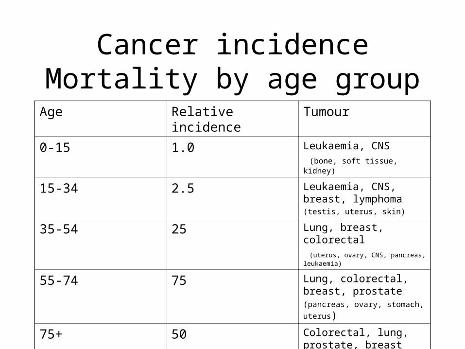
Cancer incidenceMortality by age group
Age Relative incidence Tumour
0-15 1.0 Leukaemia, CNS
(bone, soft tissue, kidney)
15-34 2.5 Leukaemia, CNS, breast, lymphoma (testis, uterus, skin)
35-54 25 Lung, breast, colorectal (uterus, ovary, CNS, pancreas, leukaemia)
55-74 75 Lung, colorectal, breast, prostate
(pancreas, ovary, stomach, uterus)
75+ 50 Colorectal, lung, prostate, breast
(pancreas, bladder, uterus)

Cancer in Ireland—rank among 50 countries (age adjusted death rates per 100 000 population (2002)
Site Female
Rank Incidence
Male
Rank incidence
All sites 6 124 18 168
Breast 3 26
Liver 42 2 42 3
Colorectal 12 14 5 24
Oesophagus 8 4 11 8
Lung 8 18 27 38
Prostate 14 20
Stomach 34 5 38 9
Uterus 40 2

What is meant by cancer cure?
• Optimal result: remission of disease, absence of relapse, good quality of life, normal life expectance, death from another cause
In practical terms cure is expressed by:• Mean survival (average duration of survival—for all patients, or by
stage)• 1 year (or 5 year, 10 year) survival—for all or by stage• Actuarial correction (correct for age related mortality) Kaplan Meyer
curveOther parameters: disease free survival, quality of life, disease or therapy
related morbidity, death from other causes• Spontaneous remission, miracles

Cancer therapycurative—palliative
Surgery Conservative—radical
Debulking, limb sparing, organ transplant
Radiotherapy External, implant
nuclides
Chemotherapy Single agent—combination
Induction—maintenance
Other forms Immunotherapy, novel forms, alternative therapy

5-year survival (all stages)
• <20% group: liver (3%), pancreas (3%), oesophagus (6%), lung (13%), stomach (16%)
• 20-40% group: brain (22%), myeloma (24%), leukaemia (34%), ovary (37%)
• 50% group: colorectal, kidney, oral cavity/pharynx, non-Hodgkin lymphoma
• 75% group: breast, prostate, cervix, larynx, Hodgkin lympoma, bladder, melanoma, endometrium, (testis, thyroid)
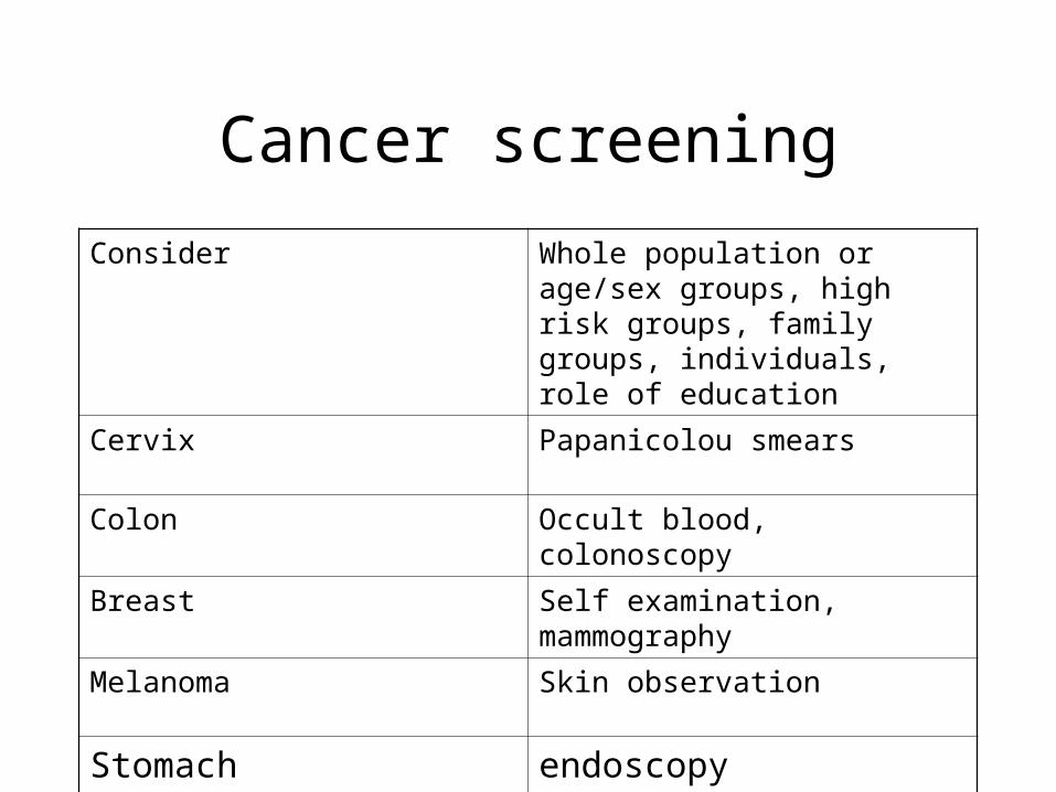
Cancer screening
Consider Whole population or age/sex groups, high risk groups, family groups, individuals, role of education
Cervix Papanicolou smears
Colon Occult blood, colonoscopy
Breast Self examination, mammography
Melanoma Skin observation
Stomach endoscopy
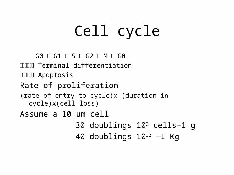
Cell cycle
G0 ˛ G1 ˛ S ˛ G2 ˛ M ˛ G0
˛ Terminal differentiation
˛ Apoptosis
Rate of proliferation(rate of entry to cycle)x (duration in cycle)x(cell loss)
Assume a 10 um cell
30 doublings 109 cells—1 g
40 doublings 1012 —I Kg
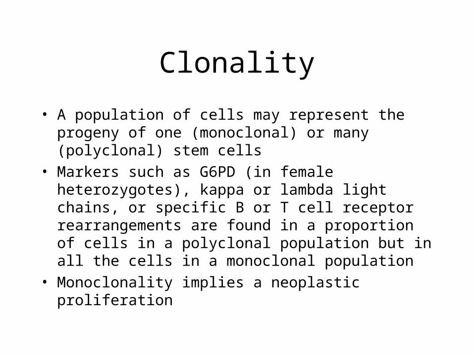
Clonality
• A population of cells may represent the progeny of one (monoclonal) or many (polyclonal) stem cells
• Markers such as G6PD (in female heterozygotes), kappa or lambda light chains, or specific B or T cell receptor rearrangements are found in a proportion of cells in a polyclonal population but in all the cells in a monoclonal population
• Monoclonality implies a neoplastic proliferation

Cancer; a disease caused by alteration of a cell’s genes
Gene categories
• Proliferation
• Suppressor
• Apoptosis control
• DNA repair

Proto-oncogenes
Cell growth and proliferation involve:
• External factors (first messengers) which bind receptor—transmembrane signal conduction
• Signal is transmitted to nucleus by second messenger
• DNA transcription begins, leading to cell division
Proto-oncogene products are involved in all of these reactions

Activation of proto-oncogenes
• Proto-oncogenes are part of the normal genome and under normal control
• When activated they are termed c-onc (they have introns and exons)
• Identical or similar genes occur in viruses (v-onc) these have only exons
• Gene function altered by: Point mutation, Chromosome translocations, Gene amplification, Deletions, Altered expression: eg methylation

Retrovirus
3 types of oncogenic retrovirus1) acute transforming
2) slow transforming
3) human T cell leukaemia virus

Oncogenic DNA viruses
Produce transforming proteins—
Human papilloma virus (HPV): about 40 types skin warts (benign), condylomas (venereal warts, benign) verrucous
carcinoma, carcinoma of cervix, penis
Herpes virus• Epstein Barr virus: infectious mononucleosis, Burkitt lymphoma, Hodgkin
lymphoma, nasopharyngeal carcinoma HHV8: kaposi sarcoma
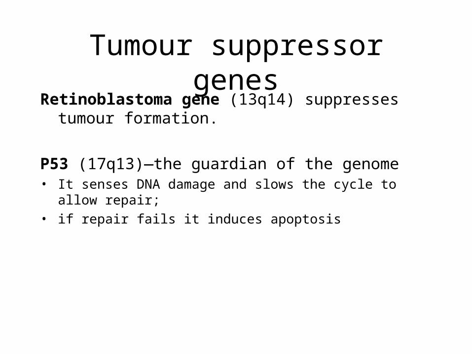
Tumour suppressor genesRetinoblastoma gene (13q14) suppresses tumour formation.
P53 (17q13)—the guardian of the genome• It senses DNA damage and slows the cycle to allow repair;
• if repair fails it induces apoptosis

Apoptosis genes
• Loss of apoptosis leads to cell accumulation
• Bcl-2 anti-apoptosis
• Bax pro-apoptosis

DNA repair genes
These repair errors in DNA transcription
• Hereditary non-polyposis colon carcer (usully right colon tumours)
• (important in these rare disorders: xeroderma pigmentosum, Bloom’s syndrome, ataxia telangiectasia, Fanconi anaemia

Cancer aetiological factors
• Smoking
• Diet
• Environment
• Hormonal agents
• Age

Canceraetiological factors
Observed factors
• Smoking
• Diet
• Environment
• Hormonal status/gender
• Age
• Genetic
Aetiology
• Chemical
• Radiation
• Virus
• Genetic– Oncogenes

Cancer—chemical inductionPott—scrotal carcinoma in sweeps
coal tar—skin cancer
Polycyclic aromatic hydrocarbons
Benzpyrene
Heterocyclic hydrocarbons Aflatoxin
Aromatic amines Azo dyes
Nitrosamines
Metals Ni, Pb, Cd, Co, Ba
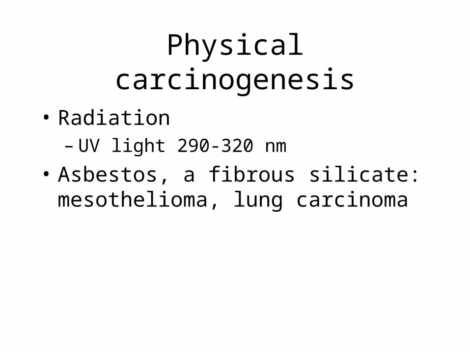
Physical carcinogenesis
• Radiation– UV light 290-320 nm
• Asbestos, a fibrous silicate: mesothelioma, lung carcinoma
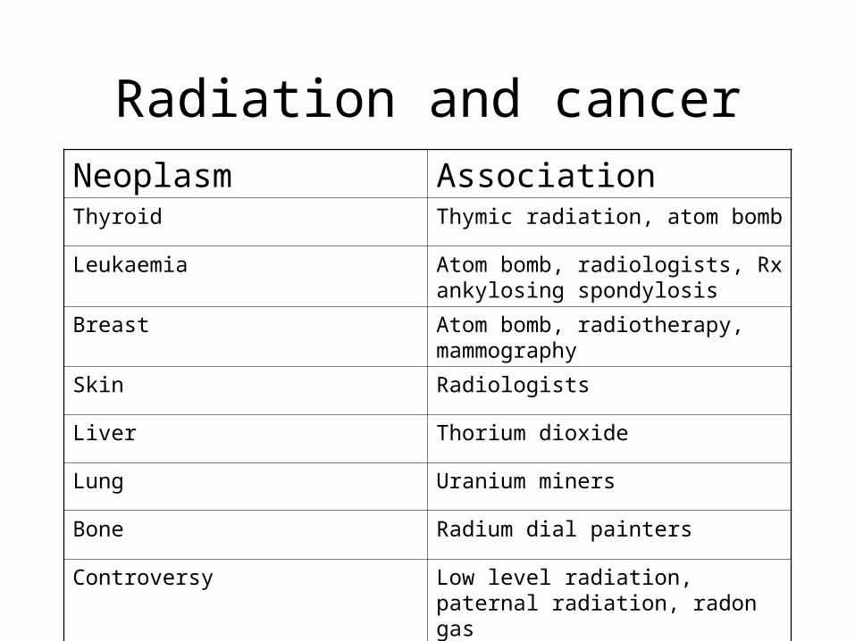
Radiation and cancerNeoplasm AssociationThyroid Thymic radiation, atom bomb
Leukaemia Atom bomb, radiologists, Rx ankylosing spondylosis
Breast Atom bomb, radiotherapy, mammography
Skin Radiologists
Liver Thorium dioxide
Lung Uranium miners
Bone Radium dial painters
Controversy Low level radiation, paternal radiation, radon gas


