80
| Date post: | 14-Dec-2014 |
| Category: |
Documents |
| Upload: | van016bunny |
| View: | 391 times |
| Download: | 16 times |

Nephrotic SyndromeNephrotic syndrome is primarily a pediatric
disorder and is 15 times more common in children than adults.
The incidence is 2–3/100,000 children per year, and the vast majority of affected children will have steroid-sensitive minimal change disease.

The characteristic features of nephrotic syndrome are: heavy proteinuria (>3.5?g/24?hr in adults or
40?mg/m2 /hr in children)
hypoalbuminemia (<2.5?g/dL)Edemahyperlipidemia

Etiology
Most children (90%) with nephrotic syndrome have a form of the idiopathic nephrotic syndrome.

EtiologyCauses of idiopathic nephrotic syndrome
include:
minimal change disease (85%)mesangial proliferation (5%)focal segmental glomerulosclerosis (10%).
EtiologyThe remaining 10% of children with nephrotic
syndrome have secondary nephrotic syndrome related to glomerular diseases such as:
membranous nephropathy or membranoproliferative glomerulonephritis

PathophysiologyThe underlying abnormality in nephrotic syndrome is an increase in permeability of
the glomerular capillary wall
which leads to massive proteinuria andhypoalbuminemia.

The cause of the increased permeability is not well understood.
In minimal change disease, it is possible that T-cell dysfunction leads to alteration of cytokines, which causes a loss of negatively charged glycoproteins within the glomerular capillary wall.

The mechanism of edema formation in nephrotic syndrome is incompletely understood
In most instances: urinary protein loss leads tohypoalbuminemia
which causes a decrease in the plasma oncotic pressure and transudation of fluid from the intravascular
compartment to the interstitial space.
The reduction in intravascular volume decreases renal perfusion pressure,
activating the renin-angiotensin-aldosterone system, which stimulates tubular reabsorption of sodium.

The reduced intravascular volume also stimulates the release of antidiuretic
hormone,
which enhances the reabsorption of water in the collecting duct.
Because of the decreased plasma oncotic pressure, fluid shifts into the interstitial
space, exacerbating the edema.

Approximately 90% of children with nephrotic syndrome have idiopathic nephrotic syndrome.
Idiopathic nephrotic syndrome includes three histologic types: minimal change disease mesangial proliferation,focal segmental glomerulosclerosis.

PathologyMinimal change disease (85% of total
cases)the glomeruli appear normal or show a minimal increase in mesangial cells and matrix.
Findings on immunofluorescence microscopy are typically negative, and electron microscopy simply
reveals effacement of the epithelial cell foot processes.
More than 95% ofchildren with minimal change disease respond to corticosteroid therapy.

PathologyMesangial proliferation (5% of total cases)
is characterized by a diffuse increase in mesangial cells and matrix on light microscopy.
Immunofluorescence microscopymay reveal trace to 1+ mesangial IgM and/or IgA staining.
Electron microscopy reveals increased numbers of mesangial cells and matrix as well as effacement of the epithelial cell foot processes.
Approximately 50% of patients with this histologic lesion respond to corticosteroid therapy.

Focal segmental glomerulosclerosis (10% of total cases), glomeruli show mesangial proliferation and segmental scarring on light microscopy.
Immunofluorescence microscopy shows IgM and C3 staining in the areas of segmental sclerosis.
Electron microscopy shows segmental scarring of the glomerular tuft with obliteration of the glomerular capillary lumen.

A similar lesion may be seen with HIV infection, vesicoureteral reflux, and intravenous heroin abuse.
Approximately 20% of patients with focal segmental glomerulosclerosis respond to prednisone.
The disease is frequently progressive, ultimately involving all glomeruli, and leads to end-stage renal failure in most patients.

Clinical ManifestationsThe idiopathic nephrotic syndrome is more
common in males than in females (2:1) and most commonly appears between the ages of 2 and 6 yr.
It has been reported as early as 6 mo of age and throughout adulthood

The initial episode and subsequent relapses may follow minor infections and, occasionally, reactions to insect bites, bee stings, or poison ivy.
Children usually present with mild edema, which is initially noted around the eyes and in the lower extremities.

Nephrotic syndrome may initially be misdiagnosed as an allergic disorder because of the periorbital swelling that decreases throughout the day.
The edema becomes generalized, with the development of ascites, pleural effusions, and genital edema.
Anorexia, irritability, abdominal pain, and diarrhea are common
Hypertension and gross hematuria are uncommon.

Differential diagnosis Includes :
Protein-losing enteropathy, hepatic failure, congestive heart failure, acute or chronic glomerulonephritis, and protein malnutrition.
Diagnosis
The urinalysis reveals 3+ or 4+ proteinuria
Microscopic hematuria may be present in 20% of children
Urinary protein excretion exceeds 3.5?g/24?hr in adults and 40?mg/m2 /hr in children
Spot urine protein to creatinine ratio exceeds 2.0
The serum creatinine value is usually normal, but it may be increased because of diminished renal perfusion resulting from contraction of the intravascular volume.
The serum albumin level is generally less than 2.5?g/dL
Serum cholesterol and triglyceride levels are elevated
C3 and C4 levels are normal.
Renal biopsy is not required for diagnosis in most children

Secondary Nephrotic SyndromeNephrotic syndrome also occurs as a
secondary feature of many forms of glomerular disease.
Membranous nephropathy, membranoproliferative glomerulonephritis, postinfectious glomerulonephritis, lupus nephritis, and Henoch-Schönlein purpura nephritis may all have a nephrotic component.

Membranous nephropathy
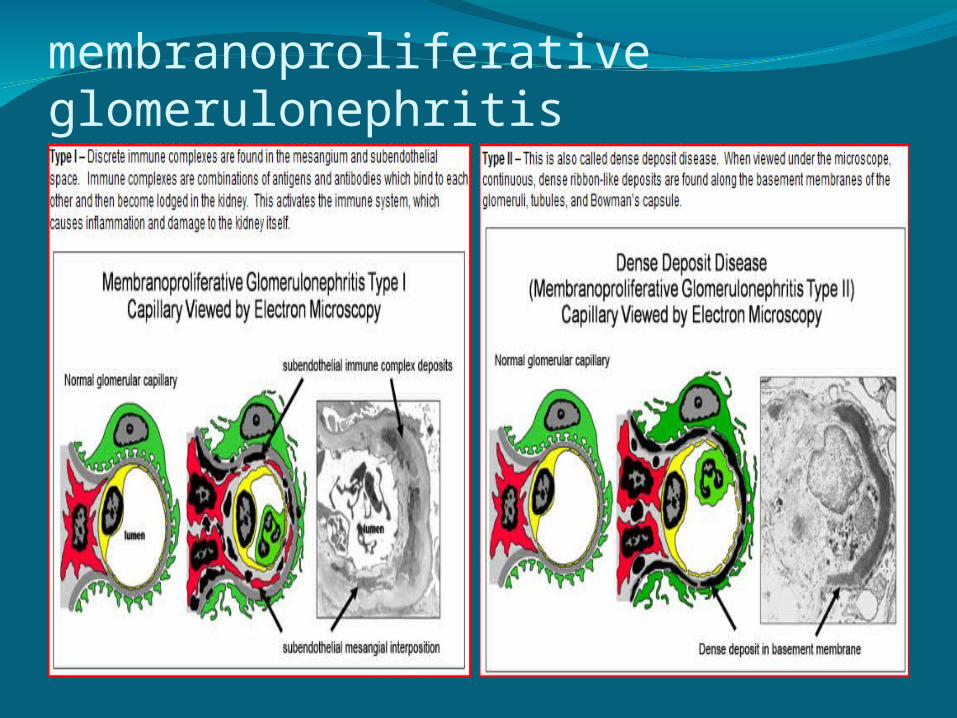
membranoproliferative glomerulonephritis

postinfectious glomerulonephritis

Henoch-Schönlein purpura nephritis

In general, secondary nephrotic syndrome should be suspected in patients with:
age > 8 yrHypertensionHematuriaRenal dysfunctionExtrarenal symptomatology (rash,
arthralgias, etc.), ordepressed serum complement levels.

Acute GlomerulonephritisAcute Poststreptococcal
GlomerulonephritisThis disease is a classic example of the acute
nephritic syndrome characterized by :
Sudden onset of gross hematuriaEdemaHypertensionRenal insufficiency.


.Etiology and Epidemiology
Acute poststreptococcal glomerulonephritis follows infection of the throat or skin by certain “nephritogenic” strains of group A ß-hemolytic streptococci.

The factors that allow only certain strains of streptococci to be nephritogenic remain unclear.
Poststreptococcal glomerulonephritis commonly follows streptococcal pharyngitis during cold weather months and streptococcal skin infections or pyoderma during warm weather months.
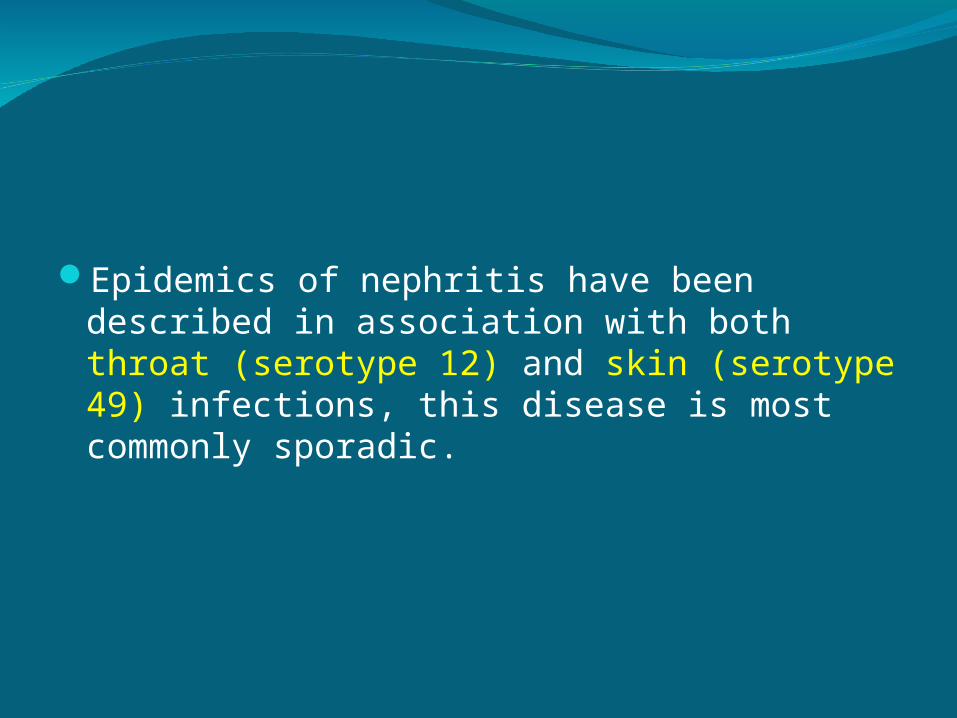
Epidemics of nephritis have been described in association with both throat (serotype 12) and skin (serotype 49) infections, this disease is most commonly sporadic.
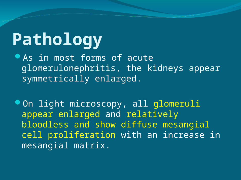
PathologyAs in most forms of acute glomerulonephritis,
the kidneys appear symmetrically enlarged.
On light microscopy, all glomeruli appear enlarged and relatively bloodless and show diffuse mesangial cell proliferation with an increase in mesangial matrix.

.Pathogenesis
Although morphologic studies and a depression in the serum complement (C3) level strongly suggest that post-streptococcal glomerulonephritis is mediated by immune complexes
The precise mechanisms by which nephritogenic streptococci induce complex formation remain to be determined..

Clinical Manifestations
Poststreptococcal glomerulonephritis is most common in children aged 5–12 yr and uncommon before the age of 3 yr.
The typical patient develops an acute nephritic
syndrome 1–2 wk after an antecedent streptococcal pharyngitis or 3–6 wk after a streptococcal pyoderma.

The severity of renal involvement varies from asymptomatic microscopic hematuria with normal renal function to acute renal failure.
Depending on the severity of renal involvement, patients may develop various degrees of :edemahypertension oliguria

Patients may develop encephalopathy and/or heart failure owing to hypertension or hypervolemia.
Encephalopathy may also result directly from the toxic effects of the streptococcal bacteria on the central nervous system.

Edema typically results from salt and water retention and nephrotic syndrome may develop in 10–20% of cases.
Specific symptoms such as:MalaiseLethargyAbdominal or flank painFever

Acute subglottic edema and airway compromise
The acute phase generally resolves within 6–8 wk.
Urinary protein excretion and hypertension usually normalize by 4–6 wk after onset,
Persistent microscopic hematuria may persist for 1–2 yr after the initial presentation
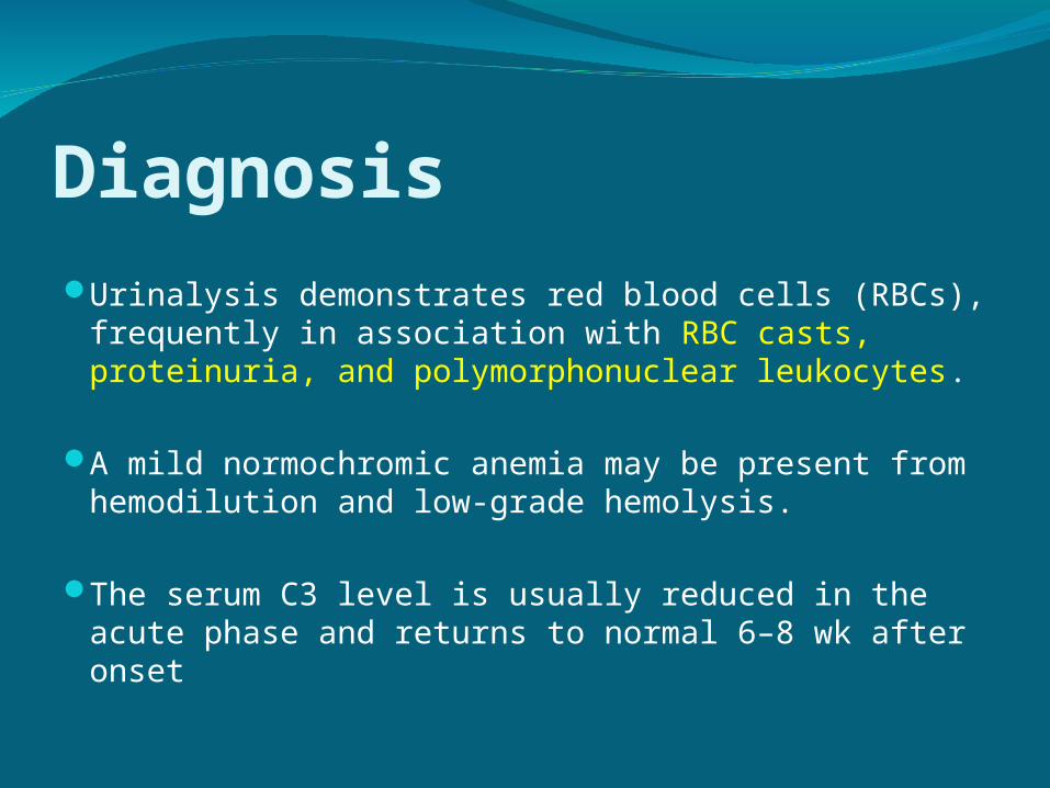
DiagnosisUrinalysis demonstrates red blood cells (RBCs),
frequently in association with RBC casts, proteinuria, and polymorphonuclear leukocytes.
A mild normochromic anemia may be present from hemodilution and low-grade hemolysis.
The serum C3 level is usually reduced in the acute phase and returns to normal 6–8 wk after onset
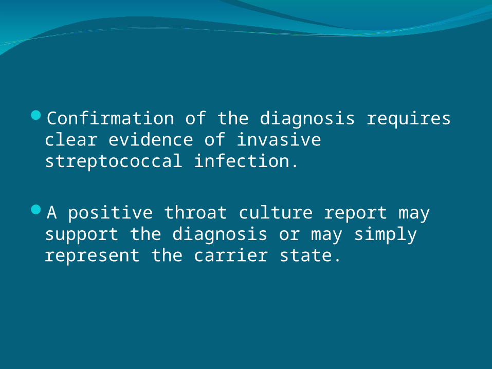
Confirmation of the diagnosis requires clear evidence of invasive streptococcal infection.
A positive throat culture report may support the diagnosis or may simply represent the carrier state.
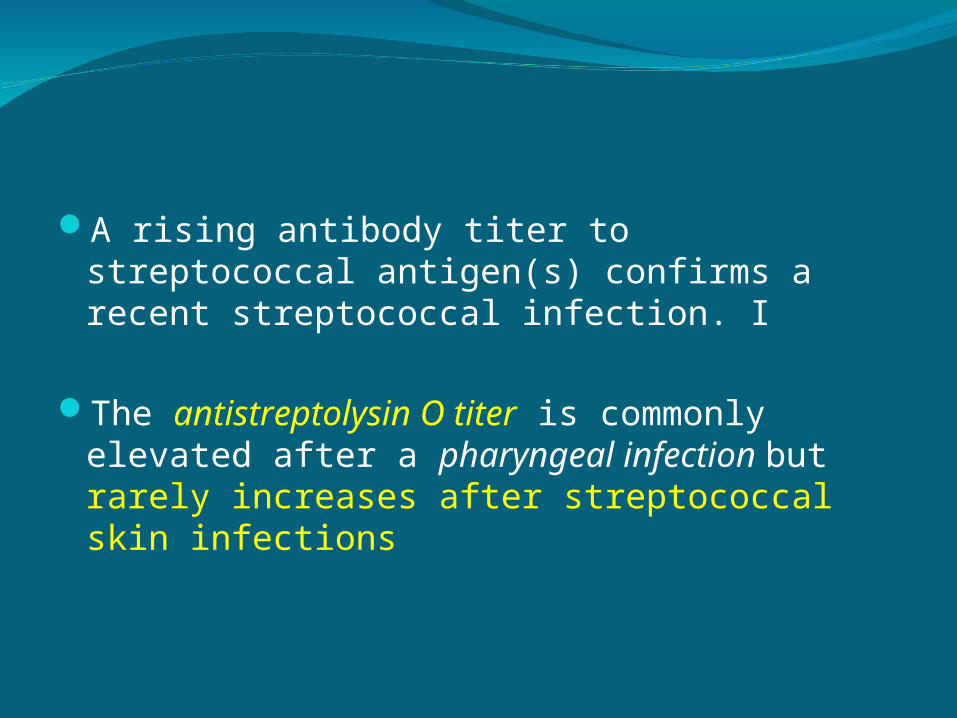
A rising antibody titer to streptococcal antigen(s) confirms a recent streptococcal infection. I
The antistreptolysin O titer is commonly elevated after a pharyngeal infection but rarely increases after streptococcal skin infections

The clinical diagnosis of poststreptococcal glomerulonephritis is quite likely in a child presenting with acute nephritic syndrome, evidence of recent streptococcal infection, and a low C3 level.
It is important to consider other diagnoses such as systemic lupus erythematosus and an acute exacerbation of chronic glomerulonephritis

Renal biopsy should be considered only in the presence of:
acute renal failurenephrotic syndrome absence of evidence for streptococcal infectionor normal complement levels

DifferentialsLow serum complement level
Systemic diseases SLE (focal, 75%; diffuse, 90%) Subacute bacterial endocarditis (90%) Visceral abscess "Shunt" nephritis (90%) Cryoglobulinemia (58%)
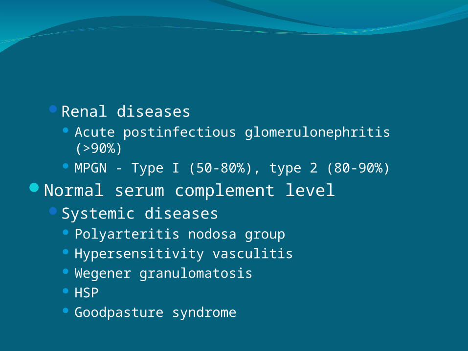
Renal diseases Acute postinfectious glomerulonephritis (>90%) MPGN - Type I (50-80%), type 2 (80-90%)
Normal serum complement levelSystemic diseases
Polyarteritis nodosa group Hypersensitivity vasculitis Wegener granulomatosis HSP Goodpasture syndrome

Renal diseases IgA (or IgG-IgA) nephropathy Idiopathic rapidly progressive glomerulonephritis
(RPGN) Anti-glomerular basement membrane (GBM)
disease Negative immunofluorescence findings Immune complex disease
ComplicationsAcute complications of this disease result
primarily from hypertension and acute renal dysfunction.
Hypertension is seen in 60% of patients and may be associated with hypertensive encephalopathy in 10% of cases.
Other potential complications include heart failure, hyperkalemia, hyperphosphatemia, hypocalcemia, acidosis, seizures, and uremia.

Prevention
Early systemic antibiotic therapy for streptococcal throat and skin infections does not eliminate the risk of glomerulonephritis.
Family members of patients with acute glomerulonephritis should be cultured for group A ß-hemolytic streptococci and treated if culture positive.

.TreatmentManagement is directed at treating the acute
effects of renal insufficiency and hypertension.
Although a 10-day course of systemic antibiotic therapy with penicillin is recommended to limit the spread of the nephritogenic organisms, antibiotic therapy does not affect the natural history of glomerulonephritis.

Sodium restriction, diuresis, and pharmacotherapy with calcium channel antagonists, vasodilators, or angiotensin-converting enzyme inhibitors are standard therapies used to treat hypertension.

treatment:antibiotics
Penicillin V 50-100KU/kg/day x10 days Erythromycin
sodium and fluid restriction (20cc/kg/day)diuresis (Furosemide 1 mkdose)correct hypertension
Nifedipine 0.25-0.5 mkday TID Captopril 0.5-2.0 mkday TID Sodium nitroprusside 0.5-1.0 mcg/kg/min Labetolol 0.2-1.0 mkdose Hydralazine 0.2-0.4 mkdose

.PrognosisComplete recovery occurs in more than 95%
of children with acute poststreptococcal glomerulonephritis.
Mortality in the acute stage can be avoided by appropriate management of acute renal failure, cardiac failure, and hypertension.
The acute phase may be severe and lead to glomerular hyalinization and chronic renal insufficiency.
However, the diagnosis of acute poststreptococcal glomerulonephritis must be questioned in patients with chronic renal dysfunction because other diagnoses such as membranoproliferative glomerulonephritis may be present.
Recurrences are extremely rare.

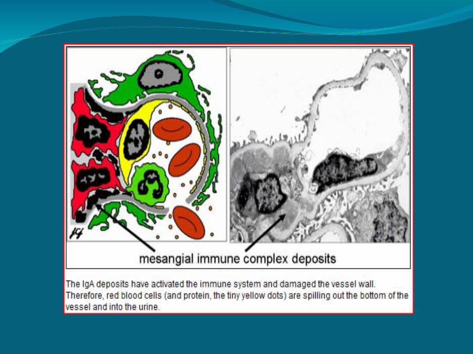
IgA Nephropathy
aka Berger Nephropathymost common chronic glomerular diseasepredominance of IgA within mesangial
deposits of the glomerulus in the absence of systemic diseases
abN in the IgA immune systemlinked to genetic abN (6q22-23)

IgA Nephropathy
manifestations:mild to moderate hypertensionedemagross or microscopic hematuriaproteinurianormal complement levels elevated serum IgA level

prognosis:progressive kidney disease develops in 20-
30%, 15-20 years after onsetpoor prognostic indicators:
persistent hypertension diminished renal function heavy or prolonged proteinuria

treatment:blood pressure controlimmunosuppressantsrenal transplantation

Hereditary Nephritis
aka Alport Syndromecaused by mutations in the genes coding for
type IV collagen85% are X-linked

Hereditary Nephritis
pathology:mesangial proliferation and capillary wall
thickeningtubular atrophy, interstitial inflammation and
fibrosispresence of foam cells

Hereditary Nephritis
manifestations:asymptomatic microscopic hematuriaprogressive proteinuria extrarenal:
sensorineural hearing loss visual abnormalities (anterior lenticonus, macular
flecks, recurrent corneal erosions) leiomyomatosis

Hereditary Nephritis
prognosis:progressive renal dysfunction occurs in 75%
treatment:no specific treatmentcareful management of hypertension, anemia,
and electrolyte imbalancedialysisrenal transplantation
Lupus Nephritis
SLE is characterized by fever, weight loss, rash, hematologic abN, arthritis, involvement of the heart, lungs, CNS, and kidneys
common among adolescent femalesoccurs in 30-70% of all casesWHO classification is based on:
light microscopyimmunofluorescenceelectron microscopy

Lupus Nephritis
Class Ino histologic abN
Class II (mesangiopathy)mesangial widening and hypercellularity
Class III (focal segmental LN)mesangial deposits in all glomerulicapillary wall necrosis, crescent formationsclerosing lesions
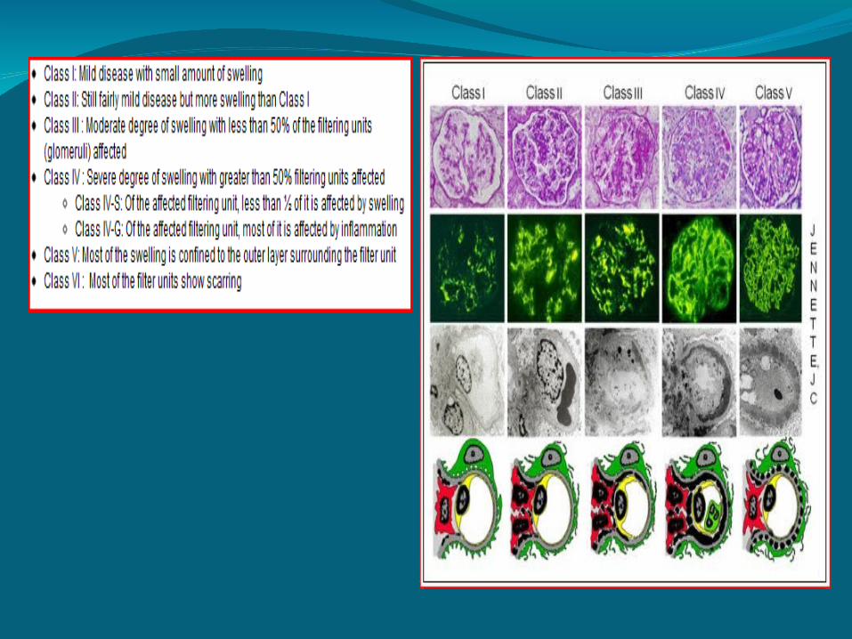
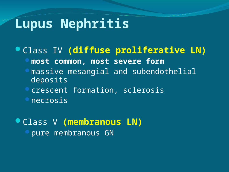
Lupus Nephritis
Class IV (diffuse proliferative LN)most common, most severe formmassive mesangial and subendothelial
depositscrescent formation, sclerosisnecrosis
Class V (membranous LN)pure membranous GN

Lupus Nephritis
manifestations:asymptomatic hematuria or proteinuria,acute nephritic syndrome, ornephrotic syndrome

Lupus Nephritis
diagnosis:SLE criteriahigh ANA titerhigh anti-dsDNA titer low serum C3 levelrenal biopsy
should be performed in all patients with SLE
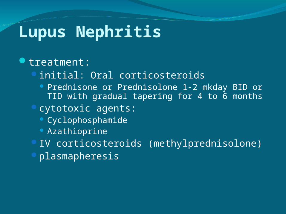
Lupus Nephritis
treatment:initial: Oral corticosteroids
Prednisone or Prednisolone 1-2 mkday BID or TID with gradual tapering for 4 to 6 months
cytotoxic agents: Cyclophosphamide Azathioprine
IV corticosteroids (methylprednisolone)plasmapheresis

Lupus Nephritis
prognosis:renal failure is the most common cause of death
among patients with SLE
complicationsprolonged steroid use (growth retardation,
hypertension, obesity, osteoporosis, diabetes mellitus)
cytotoxic effects (malignancy, infertility)intercurrent infectionsthromboses
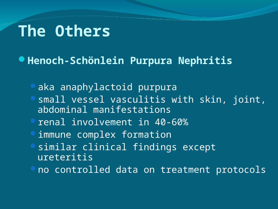
The Others
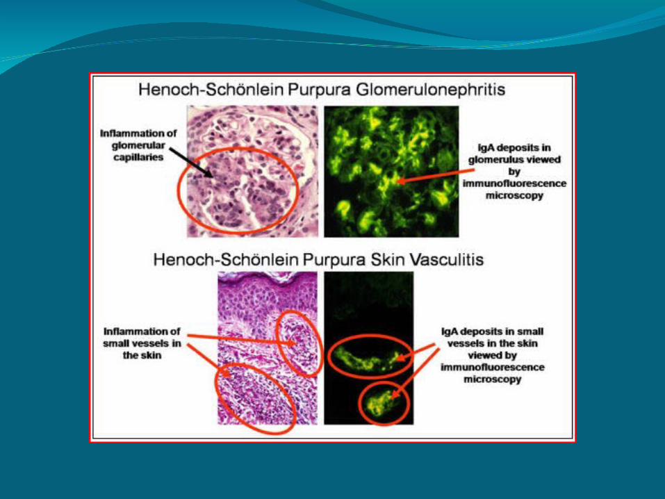
Henoch-Schönlein Purpura Nephritis
aka anaphylactoid purpurasmall vessel vasculitis with skin, joint,
abdominal manifestationsrenal involvement in 40-60%immune complex formationsimilar clinical findings except ureteritisno controlled data on treatment protocols

The Others
Polyarteritis nodosalarge vessel vasculitis with rash,
arthralgia, and nephritismalignant hypertension and weight loss renal involvement in 40-70%
capillary thrombosis fibrinoid necrosis capsular infiltration with crescent formation
treatment: supportive (steroids, anticoagulants, cytotoxic
agents) antihypertensives