Nerve Endings With Structural Characteristics ofMechanoreceptors in the Human Scleral Spur
Ernst R. Tamm, Cassandra Fliigel, Fritz H. Stefani* and Elke Lutjen-Drecoll
Purpose. The innervation of the scleral spur region was investigated to learn whether mechano-receptors are present in this region.
Methods. Serial tangential sections and whole-mount preparations of the scleral spur region of18 human eyes of different ages were investigated with electronmicroscopic and immunohisto-chemical methods. For immunohistochemistry antibodies against neurofilament-proteins, syn-aptophysin, substance P (SP), calcitonin gene-related peptide (CGRP), vasoactive intestinalpolypeptide (VIP), neuropeptide Y (NPY), tyrosine-hydroxylase, dopamine-/3-hydroxylase, andacetylcholinesterase were used.
Results. Club- or bulb-shaped nerve endings with a diameter of 5 /xm to 25 fim were identifiedin the scleral spur region throughout the whole circumference of the eyes. The terminalsderive from myelinated axons with a diameter of approximately 3 m and stain with antibodiesagainst neurofilament-proteins and synaptophysin but do not stain for tyrosine-hydroxylase,dopamine-j8-hydroxylase, acetylcholinesterase, NPY, VIP, SP, or CGRP. Electronmicroscopi-cally, the endings contain abundant neurofilaments, granular and agranular vesicles of differ-ent sizes, numerous mitochondria, and lysosome-like lamellated structures. The endings areincompletely ensheathed by Schwann cells. Those areas of the cell membrane of the endingsthat are not covered by Schwann cells are in intimate contact with the fibrillar connective tissueelements of the scleral spur.
Conclusion. These structural features are highly characteristic for mechanoreceptive nerveendings in other tissues of the human body. The authors therefore hypothesize that the club-or bulb-shaped nerve endings in the human scleral spur are afferent mechanoreceptors thatmeasure stress or strain in the connective tissue elements of the scleral spur. Such changesmight be induced by ciliary muscle contraction and/or by changes in intraocular pressure.Invest Ophthalmol Vis Sci. 1994; 35:1157-1166.
A he inner layers of the mammalian eye are suppliedby sensory nerves that originate from the trigeminalganglion. The majority of these trigeminal fibers con-sist of unmyelinated fibers of the C-type.1"4 Immuno-histochemical studies indicate that the terminals of thefibers may contain certain neuropeptides, such as sub-stance P or calcitonin gene-related peptide, that areknown to play an important role in the irritative re-
From the Department of Anatomy II, University of Erlangen-Nurnberg, Erlangen,and the *Eye Hospital of the University of Munich, Munich, Germany.Presented in part at the annual meeting of the Association for Research in Visionand Ophthalmology, Sarasota, Florida, May 2-7, 1993.Supported by grants from the Deutsche Forschungsgemeinschaft (Ta 115/5-1) andthe Academy for Science and Literature, Mainz, Germany.Submitted for publication March 16, 1993; revised September 21, 1993; acceptedSeptember 22, 1993.Proprietary interest category: N.Reprint requests: Dr. Ernst R. Tamm, Department of Anatomy II, University ofErlangen-Nurnberg, Universitatsstrasse 19, 91054 Erlangen, Germany.
sponse of the eye5 7 It is not known whether some ofthe ocular sensory nerves function as visceral mecha-noreceptors. In general, visceral mechanoreceptorsare stretch-receptors that are able to measure stress orstrain in their surrounding connective tissue ele-ments8"10 The afferent terminals of mechanoreceptorsshow several structural characteristics, such as a highcontent of mitochondria, granular, and agranular vesi-cles of different sizes, lysosome-like lamellated bodies,numerous neurofilaments, and a close contact of theircell membrane with connective tissue fibrils11"15 In thechamber angle, visceral mechanoreceptors might beimportant to monitor ciliary muscle tone and/or varia-tions in intraocular pressure (IOP). The morphologicsearch for such specialized receptors in the chamberangle, and in other regions of the primate eye as well,has been without clear result.216 In the present study,
we investigated the scleral spur region in human eyeswith ultrastructural and immunocytochemical meth-ods. We report on a group of nerve endings, not previ-ously described, that share typical structural charac-teristics with mechanoreceptive nerve endings in vis-ceral organs such as the carotid sinus,17""21 aorticarch,22 respiratory system,23-24, atrial endocardium,25
and esophagus.26'27 We therefore suggest that the hu-man scleral spur region contains mechanoreceptors.Although their functional role is not yet clarified, thepossibility exists that these sensory receptors are in-volved in the regulation of IOP.
MATERIALS AND METHODS
Ten pairs of human eyes (age range, 28 to 87 years)obtained after autopsy and eight eyes enucleated be-cause of posterior choroidal melanoma (age range, 33to 81 years) were investigated. The melanoma eyeswere obtained from the Eye Hospital of the Universityof Munich, Germany. None of the donors had any his-tory of abnormality in the chamber angle. Methods forsecuring human tissue were humane, included properconsent and approval, and complied with the Declara-tion of Helsinki.
The eyes were cut equatorially behind the oraserrata, and the anterior segment was dissected inquadrants. From each quadrant, wedge-shaped piecescontaining trabecular meshwork and the anterior cili-ary muscle were cut. The specimens were immersed inZamboni's28 or Ito's solution29 for 24 hours at 4°C. Allspecimens from autopsy eyes were placed in fixativewithin 4 hours after death; specimens from two pairsof these autopsy eyes (ages 33 and 68 years) were fixedwithin 20 minutes after death.
Electron Microscopy
Specimens fixed in Ito's solution were processed forelectron microscopy. After fixation with 1% osmiumtetroxide, the specimens were dehydrated with gradedalcohols and embedded in Epon (Roth, Karlsruhe,Germany). Serial meridional, frontal, and tangentialsemithin and ultrathin sections were cut on a micro-tome. Figure 1 shows the sectional plane of the tan-gential sections. Semithin sections were stained withRichardson's stain.30 Ultrathin sections were treatedwith lead citrate and uranyl acetate and viewed using aZeiss EM 902 microscope (Zeiss, Oberkochen, Ger-many).
IMMUNOHISTOCHEMISTRY
Specimens fixed in Zamboni's solution were washedfor 24 hours in phosphate-buffered saline (PSB) andquick frozen in isopentane, precooled with liquid ni-trogen, or embedded in paraffin. Meridional and se-rial tangential (Fig. 1) cryostat sections were cut at a
FIGURE l. Schematic drawing of the anterior segment of theeye showing the sectional plane of the tangential sectionsthrough the scleral spur region.
thickness of 20 or 50 /an, and paraffin sections werecut at a thickness of 5 /xm. The sections were placed onslides covered with 0.1% poly-L-lysine and preincu-bated for 45 minutes in Blotto's dry milk solution.31
After preincubation, the sections were incubatedovernight at room temperature with the primary anti-bodies. Final dilutions of all antibodies contained0.3% Triton X-100 and 2% bovine serum albumin. Fordemonstration of neurofilament proteins, monoclonalantibodies from Dakopatts (Hamburg, Germany,clone 2F11, 1:100) and Boehringer (Mannheim, Ger-many, clone RT97, 1:25)32 were used. For synaptophy-sin, a monoclonal antibody from Dakopatts (cloneSY38)33 was applied to the slides at a dilution of 1:10.Demonstration of tyrosine-hydroxylase, dopamine-/3-hydroxylase, and substance P was performed usingpolyclonal rabbit antibodies from Eugene Tech (Ram-sey, NJ, 1:200). Acetylcholinesterase was visualizedwith rabbit antibodies from Chemicon (Temecula, CA,1:200). Vasoactive intestinal polypeptide and calci-tonin gene-related peptide were stained with polyclo-nal rabbit antibodies from Medscand Diagnostics(Lund, Sweden, 1:1000), whereas neuropeptide Y wasvisualized with polyclonal rabbit antibodies fromAmersham Buchler (Braunschweig, Germany, 1:80).After overnight incubation, the sections were washedin PBS, reacted for 1 hour with biotinylated secondaryantibodies (Amersham), washed again, and coveredwith streptavidin-FITC (Dakopatts).
Double-staining experiments were performed byincubating sections with a combination of synaptophy-sin antibodies and polyclonal rabbit antibodies againstneurofilament proteins (Sigma, St. Louis, MO). Bind-ing of the monoclonal antibody was visualized usingthe biotin-streptavidin-FITC system as describedabove, and the rabbit antiserum was stained with aTexas red conjugated anti-rabbit IgG (Amersham).
After washing in PBS, the sections were mountedin Entellan (Merck, Darmstadt, Germany) containing
Scleral Spur Mechanoreceptors 1159
1,4-Diazabicyclo [2,2,2] octan (DABCO, Merck)34 andviewed with a Leitz Aristoplan microscope (Ernst LeitzGmbH, Wetzlar, Germany). Some of the sections wereviewed with a BioRad MRC 600 confocal laser scan-ning microscope (BioRad Microscience Ltd., HemelHempstead, UK). A Kodak T-max 400 film was usedfor photography.
For whole-mount staining, the anterior meri-dional ciliary muscle portion, scleral spur, and trabecu-lar meshwork were dissected and separated from otherocular structures. The tissues were passed throughgraded alcohols to xylene (30 minutes each step) andreturned through alcohols to xylene35 The free float-ing tissue preparations were incubated in primary an-tibody for 24 hours at 4°C, followed by incubationwith biotinylated antibodies against mouse Ig (Amer-sham) and a streptAB-Complex (Amersham). After de-velopment in diaminobenzidine hydrogen peroxidesolution, the tissues were mounted on microscopeslides.
Staining with antibodies against the different neu-ropeptides visualized varicose terminals with the typi-cal spatial distribution, as described for each of thesepeptides in the chamber angle of human eyes.5'6 Nega-tive control experiments were performed using eitherPBS or mouse or rabbit pre-immune serum substi-tuted for the primary antibody.
In the eyes of four donors (ages 28, 33, 68, and 81years), the number of club- or bulb-shaped terminalspositively stained for neurofilament proteins and thedistance between them were quantified. Wholemounts of the temporal sector of the scleral spurwith 10 mm circumferential length were viewed with alight microscope using a magnification of X 20 andanalyzed with an image processing and analysissystem (Quantimed 500, Leica Cambridge Ltd, Cam-bridge, UK).
RESULTS
Light Microscopy
Neurofilament Proteins. Antibodies against neuro-filament proteins label myelinated nerve fibers as wellas larger unmyelinated axons. Thus, immunostainingfor neurofilament proteins, applied to serial sectionsand whole-mount preparations containing the ante-rior meridional ciliary muscle, scleral spur, and trabec-ular meshwork, visualizes the architecture of all largeraxons in this area. Both monoclonal and polyclonalantibodies against neurofilament proteins give similarresults.
Numerous axons are positively stained for neuro-filament proteins in the meridional portion of the cili-ary muscle, near its insertion to the scleral spur. Theaxons run meridionally, e.g., parallel to the ciliary
muscle bundles. In the interstitial or intermuscularspaces between the muscle bundles, solitary myelin-ated axons with a diameter of 2 to 3.5 /xm predomi-nate. Most of these axons branch and give rise to thin-ner unmyelinated axons that enter the muscle bundlesand terminate between the individual muscle cells.Some of the myelinated axons do not branch in themuscle but continue toward the anterior insertion ofthe muscle at the scleral spur. Having passed the ante-rior insertion of the ciliary muscle, these axons turncircumferentially into the scleral spur to run parallelto the elastic and collagenous fibers of the spur. Thus,the axons give rise to a loose network of circumferen-tially oriented myelinated axons in the scleral spur.Myelinated axons that do not pass through the ciliarymuscle but run in a meridional direction in the supra-ciliary space (e.g., the uveoscleral interface) also turncircumferentially at the spur and join the network ofcircumferential spur axons. Most of these spur axonsare observed at the inner aspect of the scleral spur.Some of the axons pass forward to the uveal mesh-work, others branch and provide both spur and mesh-work. In places, the myelinated spur axons lose theirmyelin sheath, branch profusely, and terminate by ex-panding to club- Or bulb-like structures with a range insize of 5 to 10 /xm (Figs. 2, 3). In eyes of humansyounger than 40 years of age, these structures are reg-ularly observed in all quadrants of the circumference.Quantitative analysis in the eyes of two young donors(ages 28 and 32 years), using whole-mounts of a tem-poral sector of the scleral spur with 10 mm circumfer-ential length, shows an average distance between thenerve endings of 1.11 ± 0.4 mm and 1.32 ± 0,33 mm(Table 1). Most of these endings are observed at theinner aspect of the spur (Fig. 3). Some of these club-shaped endings, however, are also found deeper in thespur tissue, near the anterior insertion of the ciliarymuscle, or in the posterior parts of the uveal trabecu-lar meshwork.
In the eyes of humans older than 70 years of age,the club-shaped terminals are larger, with a range insize of 20 to 25 /urn (Figs. 2B, 2C). In addition, theseendings are more frequently observed in older ratherthan in younger donors. Quantitative analysis in theeyes of two old donors (ages 68 and 81 years) shows anaverage distance between nerve endings of 0.3 ± 0.23mm and 0.44 ± 0.22 (Table 1). Similar to the eyes ofyoung humans, the terminals are regularly observed inall quadrants of the circumference.
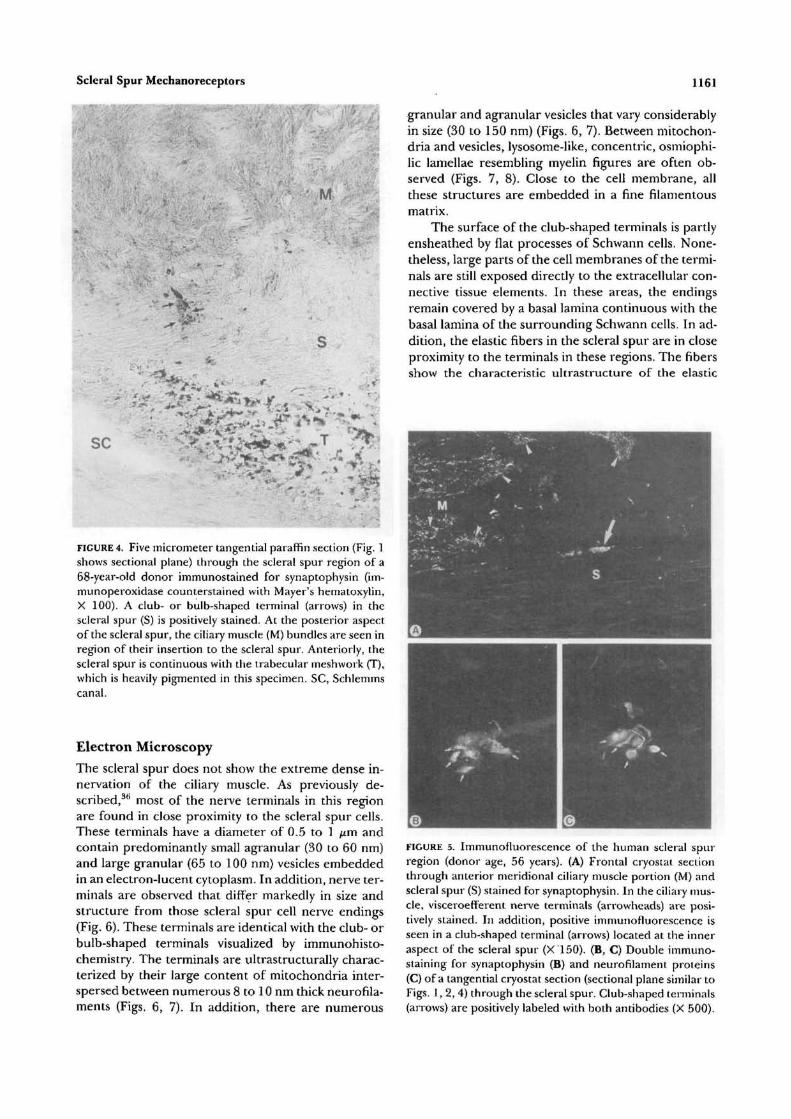
Synaptophysin. In the ciliary muscle, antibodiesagainst synaptophysin intensely label the visceroeffer-ent nerve endings. Each individual smooth muscle cellis surrounded by approximately 10 to 15 of thesenerve endings, which measure 0.5 to 1 ^m in diameter(Fig. 5A). In the scleral spur and the trabecular mesh-work, positive staining of a considerable number of
FIGURE 3. Schematic drawing of the anterior segment of theeye showing the localization of the club- or bulb-shapednerve terminals in the scleral spur. Myelinated axons passthrough the ciliary muscle or through the supraciliary space(arrows). In the scleral spur, the axons lose their myelinsheath, branch profusely, and terminate by expanding toclub- or bulb-like structures (arrowheads).
FIGURE 2. (A) Tangential cryostat section (Fig. 1 shows sec-tional plane) (B, C) and whole-mount preparation of a sectorof the scleral spur with 0.5 mm circumferential length im-munostained with antibodies against neurofilament pro-teins. The orientation of the micrographs is similar to that inFigure 4. The anterior insertion of the ciliary muscle is situ-ated beyond the top of the individual micrographs, the tra-becular meshwork beyond the bottom, (A) 50 im tangentialsection of the scleral spur of a 32-year-old donor viewedwith confocal laser scanning microscope. Numerous posi-tively stained axons are seen in the scleral spur. One of theseaxons (arrowheads) branches profusely and terminates byexpanding to club- or bulb-like structures (arrows, immuno-fluorescence, overlay image of 22 serial sections of 2 fimthickness, X 150). (B, C) Whole mount of the scleral spur ofa 67-year-old donor. Both lightmicrographs were takenfrom the same specimen in different planes of focus. Twoclub- or bulb-shaped terminals are seen (arrows). In the eyesof older donors, the club-shaped nerve terminals are larger,ranging in size from 20 to 25 fim, and are more numerousthan in the eyes of younger donors (immunoperoxidase, X150).
varicose terminals, which derive from unmyelinatedaxons, is observed. In contrast, the myelinated axonsin this area do not stain for synaptophysin. However,the club- or bulb-shaped terminals, which originate
from these axons, express intense positive immunore-activity for synaptophysin (Figs. 4, 5A).
Double staining with antibodies against both neu-rofilament and synaptophysin show that staining forneurofilament proteins is more confined to the centralarea of these endings, whereas staining for synapto-physin predominates in their peripheral parts (Figs.5B, 5C). The varicose axons of the unmyelinated axonsin scleral spur and trabecular meshwork stain only forsynaptophysin but not for neurofilament. The same istrue for the visceroefferent terminals around the cili-ary muscle cells.
The enlarged, club-shaped endings in the eyes ofolder donors show intense immunoreactivity for neu-rofilament proteins, whereas staining for synaptophy-sin is usually weaker than it is in the eyes of youngdonors. In addition, in the older eyes some of the end-ings stain for neurofilament proteins but not for syn-aptophysin.
TABLE l. Quantitative Evaluation of theClub-Shaped Terminals in the ScleralSpur RegionAge(Yr) No. of Terminals
Distance Between Terminals(mm) (Mean ± SD)
2833• § »
81
98
3121
1.11 ±0.41.32 ±0.350.30 ± 0.230.44 ± 0.22
In the eyes of four human donors, whole-mount preparations ofthe temporal sector of the scleral spur region were immunostainedfor neurofilament proteins. The specimens had a total circumfer-ential length of 10 mm and were analyzed with an image process-ing mid analysis system.
Scleral Spur Mechanoreceptors 1161
SC
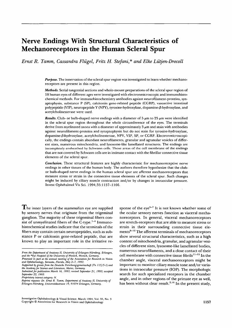
FIGURE 4. Five micrometer tangential paraffin section (Fig. 1shows sectional plane) through the scleral spur region of a68-year-old donor immunostained for synaptophysin (im-munoperoxidase counterstained with Mayer's hematoxylin,X 100). A club- or bulb-shaped terminal (arrows) in thescleral spur (S) is positively stained. At the posterior aspectof the scleral spur, the ciliary muscle (M) bundles are seen inregion of their insertion to the scleral spur. Anteriorly, thescleral spur is continuous with the trabecular meshwork (T),which is heavily pigmented in this specimen. SC, Schlemmscanal.
Electron Microscopy
The scleral spur does not show the extreme dense in-nervation of the ciliary muscle. As previously de-scribed,3*1 most of the nerve terminals in this regionare found in close proximity to the scleral spur cells.These terminals have a diameter of 0.5 to 1 jum andcontain predominantly small agranular (30 to 60 nm)and large granular (65 to 100 nm) vesicles embeddedin an electron-lucent cytoplasm. In addition, nerve ter-minals are observed that differ markedly in size andstructure from those scleral spur cell nerve endings(Fig. 6). These terminals are identical with the club- orbulb-shaped terminals visualized by immunohisto-chemistry. The terminals are ultrastructurally charac-terized by their large content of mitochondria inter-spersed between numerous 8 to 10 nm thick neurofila-ments (Figs. 6, 7). In addition, there are numerous
granular and agranular vesicles that vary considerablyin size (30 to 150 nm) (Figs. 6, 7). Between mitochon-dria and vesicles, lysosome-like, concentric, osmiophi-lic lamellae resembling myelin figures are often ob-served (Figs. 7, 8). Close to the cell membrane, allthese structures are embedded in a fine filamentousmatrix.
The surface of the club-shaped terminals is partlyensheathed by flat processes of Schwann cells. None-theless, large parts of the cell membranes of the termi-nals are still exposed directly to the extracellular con-nective tissue elements. In these areas, the endingsremain covered by a basal lamina continuous with thebasal lamina of the surrounding Schwann cells. In ad-dition, the elastic fibers in the scleral spur are in closeproximity to the terminals in these regions. The fibersshow the characteristic ultrastructure of the elastic
FIGURE 5. Immunofluorescence of the human scleral spurregion (donor age, 56 years). (A) Frontal cryostat sectionthrough anterior meridional ciliary muscle portion (M) andscleral spur (S) stained for synaptophysin. In the ciliary mus-cle, visceroefferent nerve terminals (arrowheads) are posi-tively stained. In addition, positive immunofluorescence isseen in a club-shaped terminal (arrows) located at the inneraspect of the scleral spur (X 150). (B, C) Double immuno-staining for synaptophysin (B) and neurofilament proteins(C) of a tangential cryostat section (sectional plane similar toFigs. 1, 2, 4) through the scleral spur. Club-shaped terminals(arrows) are positively labeled with both antibodies (X 500).
FIGURE 6. Electronmicrograph of a club-shaped terminal inthe scleral spur (donor age, 38 years). The terminal has alength of approximately 25 fim and is densely surrounded bycollagen (C) and elastic fibers (E). The surface of the termi-nal is partly ensheathed by flat processes of Schwann celts(S). Nonetheless, large parts of the cell membrane are stillexposed directly to the extracellular fibrils (arrows, X 6300).
fibers in the chamber angle tissues3738 because theycomprise an electron-dense amorphous central coreand a sheath consisting of microfibrils and cross-banded material with a periodicity of 50 to 60 nm. The50 to 60 nm cross-banded sheath material of the elas-tic fibers merges with the basal lamina and contacts thecell membrane of the endings directly (Figs. 7, 8, 9). Insome areas of contact with the cross-banded sheathmaterial, the cell membrane of the terminals has spe-
FIGURE 7. Higher magnification of Figure 3 (X 14,000). Thecytoplasm of the terminal contains numerous mitochondria(M) as well as agranular (black arrows) and granular vesicles(white arrows) that are embedded in a filamentous matrix.The 50 to 60 nm cross-banded sheath material of an elasticfiber (E) merges with the basal lamina of the terminal andcontacts the cell membrane (arrowheads).
cialized electron-dense areas (Fig. 10). Some of theterminals form protrusions that surround larger partsof elastic fibers (Figs. 8, 9). Serial ultrathin sectionsconfirm that the club-shaped endings derive from my-elinated axons with a diameter of about 3 to 3.5 /on.The axons lose their myelin sheath and form distinc-
FIGURE 8. Tangential section of a scleral spur terminal (elec-tron migrograph X 5,000; donor age, 56 years). The terminalis embedded in a meshwork of numerous, circularly orientedelastic fibers (E) that are in close contact with the cell mem-brane of the terminal (arrowheads). In addition, the termi-nal forms protrusions that surround larger parts of the elas-tic fibers (arrows).
Scleral Spur Mechanoreceptors 1163
> f
FIGURE 9. Meridional section of a club-shaped terminal inthe scleral spur (donor age, 34 years). The terminal containsnumerous mitochondria, vesicles, and a filamentous matrix.An elastic fiber (asterisk) is wrapped by cytoplasmic protru-sions of the terminal. The terminal is ensheathed by flat pro-cesses of terminal Schwan cells (arrowheads). Nevertheless,in regions of contact with the the elastic fiber, the cell mem-brane of the terminal is not covered by such Schwann cellsbut is in direct contact with the cross-banded sheath materialof the elastic fiber (arrows, electronmicrograph X 5,800).
tive half nodes of Ranvier (Fig. 11). Before expandingto the typical terminals, the unmyelinated preterminalaxons continue their course in the scleral spur tissuefor several micrometers.
Tyrosine-Hydroxylase, Dopamine-/?-Hydroxylase, Acetylcholinesterase, SP, CGRP,VIP, NPYAntibodies against tyrosine-hydroxylase and dopa-mine-/3-hydroxylase stain some presumably adrenergicvaricose terminals in ciliary muscle, scleral spur, andtrabecular meshwork, whereas staining for acetylcho-linesterase is strictly confined to the ciliary musclebundles. Staining with antibodies against the differentneuropeptides visualizes varicose terminals with thetypical spatial distribution, as described for each ofthese peptides in the chamber angle of human eyes.56
FIGURE 10. High-power electronmicrograph of a club-shaped terminal (X 33,000). In some areas of contact withthe 50 to 60 nm cross-banded sheath material of the elasticfibers (E), the cell membrane of the terminal (asterisk) hasspecialized electron-dense areas (arrow).
FIGURE 11. The club-shaped terminals derive from myelin-ated axons with a diameter of 3 to 3.5 pm. The axons (aster-isk) lose their myelin sheath (arrows) and form distinctivehalf nodes of Ranvier (electronmicrograph, X 7,000).
The club-shaped nerve endings are not stained withany of these antibodies.
DISCUSSIONThe presence of nerve fibers in the anterior chamberangle of monkey and human eyes is well docu-mented.630 In the trabecular meshwork of both spe-cies, most of the nerve fibers were reported to be un-myelinated, whereas in the scleral spur myelinatedaxons also were observed.40'41
Our study confirms the presence of myelinatedaxons in the scleral spur and provides evidence that aconsiderable number of these axons terminate as dis-tinctive club- or bulb-shaped structures. These termi-nals might be identical to nervous aggregates de-scribed in earlier studies by means of silver-impregna-tion techniques and discussed as baroreceptors.4243
Our results show that in common with the viscero-efferent nerve endings of the ciliary muscle cells andthe terminals of the unmyelinated varicose axons inscleral spur and trabecular meshwork, the club-shapedterminals stain positively for synaptophysin. Synapto-physin is a transmembranous glycoprotein specificallylocalized to synaptic vesicles and the vesicular struc-tures of neuroendocrine cells.44 The immunoreactivityfor synaptophysin is not confined just to efferentnerve endings but is also observed in afferent termi-nals.45 It has been suggested that the small agranularvesicles present in such afferent terminals are mostprobably stained.45
In contrast to the ciliary muscle terminals and theterminals of the unmyelinated axons, the club-shapedendings show intense immunoreactivity with antibod-ies against neurofilament proteins. It is generallyagreed upon that neurofilaments predominate in my-elinated axons.46 It has been demonstrated in rat sen-sory ganglia that one of the antibodies (RT97) used inthe present study specifically stains large neurons ofthe A type, which have a fast conducting myelinatedaxon.4748
Ultrastructurally, the club-shaped nerve endingsshow characteristic features that are regarded as typi-cal for the receptive areas of afferent nerve endings inskin, tendons, and joint capsules,11121549'50 or invarious visceral organs.17~19>22'27 These common char-acteristics are as follows: a high content of mitochon-dria; granular and agranular vesicles of different sizes;lysosome-like lamellated bodies; numerous neurofila-ments; and a fine filamentous matrix in region of thereceptive cell membrane, the so-called "receptor ma-trix."13-14 We therefore suggest that the club-shapednerve endings in the human scleral spur are terminalsof primary afferent neurons. Indeed, degenerationstudies have shown that 20% of the axons in the scleralspur of cynomolgus monkeys have a sensory originfrom the trigeminal ganglion.41 So far, however, ultra-structural studies have failed to demonstrate distinc-tive sensory nerve endings in the human chamber an-gle comparable to those described in our study.
Characteristically, the club-shaped terminals inthe scleral spur region are in close contact with theextracellular fibrillar material of their surroundingconnective tissue. It is primarily the numerous elasticfibers in the spur that are in close proximity to the cellmembrane of the endings. In general, such contactsbetween extracellular fibrils and sensory nerve termi-nals are characteristic of mechanoreceptors. Theyhave been described for the mechanoreceptive nerveendings of the Golgi tendon organ,5152 the encapsu-lated Ruffini corpuscles of the skin,53 and for visceralmechanoreceptors such as the branched lanceolate orruffini-like corpuscles of the respiratory system,23-24
the dura mater encephali,54 the periodontal liga-ment, 55~58' and vascular structures such as the carotidsinus,1718 the aortic arch,22 or the atrial endocar-dium.25
It seems probable that the club-shaped spur termi-nals serve a mechanoreceptive function. In general,mechanoreceptors are in contact with accessory struc-tures that transfer a mechanical disturbance in the lo-cal environment to a mechanosensitive region of thereceptor.59 The mechanisms of mechanotransductionin visceral mechanoreceptors, which measure stretchand distention of the organs of the gastrointestinal,respiratory, urogenital, and vascular system, are notfully understood. The current hypothesis is thatstretch in surrounding connective tissue elements thatare in close contact with the cell membrane of thereceptor influences stretch-sensitive ion channels inthe membrane.81059 The mechanical linkage betweenchannel and membrane is thought to be provided bycytoskeletal strings that pull the membrane open whenthe membrane is stretched. Such cytoskeletal couplingmay explain the prominent occurrence of microfila-ments in the transducing region of this type of mecha-noreceptor.59 Mechanoreceptors or stretch-receptorsin the scleral spur should measure stress or strain inthe connective tissue elements of the spur. Because
these factors are surely influenced by changes in ciliarymuscle tone, the nerve endings might represent pro-prioreceptive "tendon organs" of the ciliary muscle.In addition, contraction of the scleral spur cells, whichform microtendon-like connections with the elasticfibers of the spur,36 might modulate the tension of thefibers. On the other hand, the scleral spur, with itscircumferentially arranged collagen and elasticfibers,60 is the innermost part of the outer coat of theeye, the sclera. Changes in intraocular pressure exertinfluence on stress or strain of the sclera and probablyalso on stress of the scleral spur. Thus, mechanorecep-tors that measure stretch of the scleral spur might alsohave a baroreceptive function. Interestingly, most vis-ceral receptors that measure distention of the gastroin-testinal and urogenital tracts and vascular system aredirectly or indirectly associated with smooth muscles.Contraction of smooth muscle can modulate the excit-ability or may lead to excitation of visceral receptorsby changing visceral compliance.8 Physiological stud-ies suggest that baroreceptive nerve terminals mightexist in the eye because sensory discharges in the cili-ary nerves related to IOP changes have been ob-served.61"65 Belmonte et al64 suggested that the ciliarynerves in the cat eye contain afferent fibers that re-spond tonically within the normal range of IOP andoriginate from specific, slowly adapting mechanore-ceptors sensitive to variations in IOP.
Morphologically, such mechanoreceptors havenot been described in mammalian eyes. An exceptionare the eyes of such aquatic mammals as whales anddolphins.66-67 In these animals, lamellated pacciniancorpuscles are present in the chamber angle and mayrepresent specialized structures important to adapt tochanges between aquatic and atmospheric environ-ments.39
The putative mechanoreceptive nerve endings inthe human scleral spur show structural changes withage that include an increase in diameter. Interestingly,silver impregnation studies by Wolter,68 Vrabec,69 andValu,70 described an axonal swelling of nerve fibers inthe chamber angle as typical senile change of this tis-sue, which was more pronounced in glaucomatouseyes.68 It might be that these studies also visualized anage- or disease-related enlargement of the scleral spurterminals.
In summary, we hypothesize that mechanorecep-tive nerve endings are present in the human scleralspur. The physiological role of these structures re-mains to be clarified.
Key Wordsmechanoreceptor, sensory nerve terminal, scleral spur,chamber angle, human eye
Acknowledgments
The authors thank Angelika Hauser and Simone Klein forexpert assistance with immunohistochemistry and electron-microscopy, and Marco G6/3wein and Anette Gach for prepa-
Scleral Spur Mechanoreceptors 1165
ration of the photographs and drawings. They also thankBioRad Microsciences Ltd., Hemel Hempstead, UK, for theuse of the confocal laser scanning microscope to examinespecimens.
References
1. Holland MG, von Sallmann L, Collins EM. A study ofthe innervation of the chamber angle: II: The originof trabecular axons revealed by degeneration. Am JOphthalmol. 1957;44:206-221.
2. Bergmannson JPG. The ophthalmic innervation of theuvea in monkeys. Exp Eye Res. 1977;24:225-240.
3. Ten Tuscher MPM, Klooster J, Van der Want JJL,Lamers WPMA, Vrensen GFJM. The allocation ofnerve fibers to the anterior segment and peripheralganglia of rats: I: The sensory innervation. Brain Res.1989;494:95-104.
4. Beckers HJM, Klooster J, Vrensen GFJM, LamersWPMA. Ultrastructural identification of trigeminalnerve endings in the rat cornea and iris. Invest Opthal-mol VisSci. 1992;33:1979-1986.
5. Stone RA, Kuwayama Y, Laties AM. Regulatory pep-tides in the eye. Experientia. 1987;43:791-800.
6. Stone RA, Kuwayama Y. The nervous system and intra-ocular pressure. In: Ritch R, Shields MB, Krupin T,eds. The glaucomas. St. Louis: CV Mosby; 1989:257-279.
7. Bill A. The 1990 Endre Balazs Lecture: Effects ofsome neuropeptides on the uvea. Exp Eye Res.1991;53:3-11.
8. Janig W, Morrison JFB. Functional properties of spi-nal visceral afferents supplying abdominal and pelvicorgans, with special emphasis on visceral nociception.In: Cervero F, Morrison JFB, eds. Progress in BrainReserach. Vol. 67. Amsterdam: Elsevier; 1986:87-114.
13. Andres KH. Morphological criteria for the differen-tiation of mechanoreceptors in vertebrates. In:Schwartzkopff J, ed. Symposium Mechanorezeption,Abhdlg. Rhein. Westf. Akad. Wiss. 53. Opladen: West-deutscher Verlag; 1974:135-152.
14. Andres KH, von During M. Morphology of cutaneousreceptors. In: Iggo A, ed. Handbook of sensory physiol-ogy. Berlin, Heidelberg, New York: Springer Verlag;1973:3-28.
15. Iggo A, Andres KH. Morphology of cutaneous recep-tors. Annu Rev Neurosci. 1982; 5:1-31.
16. Ruskell GL. Innervation of the anterior segment ofthe eye. In: Liitjen-Drecoll E, ed. Basic Aspects of Glau-coma Research. Stuttgart: Schattauer Verlag; 1982:49-66.
17. Rees PM. Observations on the fine structure and dis-
tribution of presumptive baroreceptor nerves at thecarotid sinus. J Comp Neurol. 1967; 131:517-548.
18. Bock P, Gorgas K. Fine structure of baroreceptor ter-minals in the carotid sinus of guinea pigs and mice.Cell Tissue Res. 1976; 170:95-112.
19. Knoche H, Addicks K. Electron microscopic studies ofthe pressoreceptor fields of the carotid sinus of thedog. Cell Tissue Res. 1976; 173:77-94.
20. Knoche H, Walther-Wenke G, Addicks K. Die Fein-struktur der barorezeptorischen Nervenendigungenin der Wand des Sinus caroticus der Katze. Acta Anat.1977; 97:403-418. (In German.)
21. Knoche H, Wiesner-Menzel L, Addicks K. Ultrastruc-ture of baroreceptors in the carotid sinus of the rab-bit. Acta Anat. 1980; 106:63-83.
22. Krauhs JM. Structure of rat aortic baroreceptors andtheir relationship to connective tissue. J Neurocytol.1979;8:401-404.
23. von During M, Andres KH, Iravani J. The fine struc-ture of the pulmonary stretch receptor in the rat. ZAnat Entwickl-Gesch. 1974; 143:215-222.
24. von During M, Andres KH. Structure and functionalanatomy of visceroreceptors in the mammalian respi-ratory system. In: Hamann W, Iggo A, eds. Progress inBrain Research. Vol. 74. Amsterdam: Elsevier;1988:139-154.
25. Tranum-Jensen J. The ultrastructure of the sensoryend-organs (baroreceptors) in the atrial endocardiumof young mini-pigs. J Anat. 1975; 119:255-275.
26. Neuhuber WL. Sensory vagal innervation of the raiesophagus and cardia: A light and electron micro-scopic anterograde tracing study. J Auton New Syst.1987;20:243-255.
27. Neuhuber WL, Clerc N. Afferent innervation of theesophagus in cat and rat. In: Zenker W, NeuhuberWL, eds. The Primary Afferent Neuron. New York: Ple-num; 1990:93-107.
28. Stefanini M, de Martino C, Zamboni C. Fixation ofejaculated spermatozoa for electron microscopy.Neuroscience. 1967; 216:173-174.
29. Ito S, Karnovsky MJ. Formaldehyde-glutaraldehydefixatives containing trinitro compounds. J Cell Biol.1968;39:168A-169A.
30. Richardson KC, Jarret L, Finke H. Embedding inepoxy resins for ultrathin sectioning in electron mi-croscopy. Stain Technol. 1960; 35:313-323.
31. Duhamel RC, Johnson DA. Use of nonfat dry milk toblock nonspecific nuclear and membrane staining byavidin conjugates. J Histochem Cytochem. 1985; 33:711-714.
33. Wiedenmann B, Franke WW. Identification and local-ization of synaptophysin, an integral membrane glyco-protein of Mr 38000 characteristic of presynaptic vesi-cles. Cell. 1985;41:10l7-1028.
34. Johnson GD, Davidson RS, McNamee KC, Rusel G,Goodwin D, Holborow EJ. Fading of immunofluores-cence during microscopy: A study of the phenomenonand its remedy•. J Immunol Methods. 1982;55:231-242.
35. Costa M, Furness JB, Solcia E. Immunohistochemicallocalization of polypeptides in peripheral autonomic
nerves using whole mount preparations. Histochemis-try. 1080;65:157-165.
36. Tamm E, Flugel C, Stefani FH, Rohen JW. Contractilecells in the human scleral spur. Exp Eye Res.1992;54:531-543.
37. Vegge T, Ringvold A. The ultrastructure of the extra-cellular components of the trabecular meshwork inthe human eye. Z Zellforsch. 1971; 11:361-376.
38. Rohen JW, Futa R, Lutjen-Drecoll E. The fine struc-ture of the cribriform meshwork in normal and glau-comatous eyes as seen in tangential sections. InvestOpthalmol Vis Sci. 1981;21:574-585.
39. Stone RA, Laties AM. Neuroanatomy and neuroendo-crinology of the chamber-angle. In: Krieglstein GK,ed. Glaucoma Update III. Berlin: Springer Verlag;1987:1-16.
40. Feeney L. Ultrastructure of the nerves in the humantrabecular region. Invest Opthalmol Vis Sci. 1962;1:462-473.
41. Ruskell GL. The source of nerve fibres of the trabecu-lae and adjacent structures in monkey eyes. Exp EyeRes. 1976; 23:449-459.
42. Kurus E. Uber ein Ganglienzellsystem der menschli-chen Aderhaut. Klin Mbl Augenheilk. 1955; 127:198-206. (In German.)
43. Castro-Correira J. Studies on the innervation of theuveal tract. Ophthalmologica. 1967; 154:497-520.
44. Wiedenmann B, Huttner WB. Synaptophysin andchromogranins/secretogranins: Widespread constitu-ents of distinct types of neuroendocrine vesicles andnew tools in tumor diagnostics. Virchows Archiv B CellPathol. 1989;58:95-121.
45. De Camilli P, Vitadello M, Canevini MP, Zanoni R,Jahn R, Gorio A. The synaptic vesicle proteins synap-sin I and synaptophysin (protein p38) are concen-trated both in efferent and afferent nerve endings ofthe skeletal muscle. / Neurosci. 1988;8:1625-1631.
46. Lawson SN, Harper AA, Harper El, Garson JA, An-derton BH. A monoclonal antibody against neurofila-ment protein specifically labels a subpopulation of ratsensory neurons./ Comp Neurol. 1984;228:263-272.
47. McCarthy PW, Lawson SN. Cell type and conductionvelocity of rat primary sensory neurons with substanceP-like immunoreactivity. Neuroscience. 1989;28:745—753.
48. McCarthy PW, Lawson SN. Cell type and conductionvelocity of rat primary sensory neurons with calcitoningene-related peptide-like immunoreactivity. Neuro-science. 1990; 34:623-632.
49. Halata Z, Rettig T, Schulze W. The ultrastructure ofsensory nerve endings in the human knee joint cap-sule. Anat Embryol. 1985; 172:265-275.
50. Halata Z. The ultrastructure of the sensory nerve end-ings in the articular capsule of the domestic cat (Ruf-fini corpuscles and Pacinian corpuscles). J Anat.1977;124:7l7-729.
51. Schoultz TW, Swett JE. The fine structure of the Golgitendon organ. J Neurocytol. 1972; 1:1-26.
52. Schoultz TW, Swett JE. Ultrastructural organizationof the sensory fibers innervating the Golgi tendon or-gan. Anat Rec. 1974; 179:147-162.
53. Chambers MR, von During M, Iggo A. The structure
and function of the slowly adapting type II mechanore-ceptor in hairy skin. QJ Exp Physiol. 1972; 57:417-445.
54. Andres KH, von During M, Muszynski K, Schmidt RF.Nerve fibers and their terminals of the dura materencephali of the rat. Anat Embryol. 1987; 175:289-301.
55. Everts V, Beertsen W, Van den Hoof A. Fine structureof an end organ in the periodontal ligament of themouse incisor. Anat Rec. 1977; 189:73-90.
56. Maeda T, Sato O, Kobayashi S, Iwanaga T, Fujita T.The ultrastructure of ruffini endings in the periodon-tal ligament of rat incisors with special reference tothe terminal Schwann cells (K-cells). Anat Rec.1989;223:95-103.
57. Kannari K, Sato O, Maeda T, Iwanaga T, Fujita T. Apossible mechanism of mechanoreception in ruffiniendings in the periodontal ligament of hamster inci-sors./ Comp Neurol. 1991;313:368-376.
58. Sato O, Maeda T, Kannari K, Kawahara I, Iwanaga T,Takano Y. Innervation of the periodontal-ligament inthe dog with special reference to the morphology ofruffini endings. Arch Histol Cytol. 1992; 55:21-30.
60. Moses RA, Grodzki WJ, Starcher BC, Galione MJ.Elastin content of the scleral spur, trabecular meshand sclera. Invest Opthalmol Vis Sci. 1978; 17:817-818.
61. von Sallmann L, Fuortes MGF, Macri FJ, Grimes P.Study of afferent electric impulses induced by intraoc-ular pressure changes. Am J Ophthalmol. 1958;45:211-220.
62. Lele PP, Grimes P. The role of neural mechanisms inthe regulation of intraocular pressure in the cat. ExpNeurol. 1960; 2:199-220.
64. Belmonte C, Simon J, Gallego A. Effects of intraocu-lar pressure changes on the afferent activity of ciliarynerves. Exp Eye Res. 1971; 12:342-355.
65. Zuazo A, Ibanez J, Belmonte C. Sensory nerve re-sponses elicited by experimental ocular hypertension.Exp Eye Res. 1986;43:759-769.
66. Vrabec F. Encapsulated sensory corpuscles in thesclerocorneal boundary tissues of the killer whale Or-cinus orca L. Ada Anat. 1972; 81:23-29.
67. Wickham MG. Irido-corneal angle of mammalianeyes: Comparative morphology of encapsulated cor-puscles in odontocete cetaceans. Cell Tissue Res.1980;210:501-515.
68. Wolter JR. Neuropathology of the trabeculum inopen-angle glaucoma. Arch Ophthalmol. 1959;62:99-111.
69. Vrabec F. On the development and senile changes ofthe innervation of the trabecular meshwork in hu-mans. In: Rohen JW, ed. The structure of the eye. Vol. 2.Stuttgart: Schattauer Verlag; 1965:215-222.
70. Valu L. Uber die Innervation des Uvea-Trabekel-Sys-tems. Graefe's Arch Clin Exp Ophthalmol. 1962; 164:496-502. (In German.)