Nerve Transfer Surgery to Improve Hand Function in Spinal Cord Injury: Multidisciplinary Evaluation and Management Ida K. Fox, MD Division of Plastic and Reconstructive Surgery Co-Authors: Davidge KM, Novak CB, Kahn LC, Juknis N, Ruvinskaya R, Mackinnon SE
Transcript
Nerve Transfer Surgery to Improve
Hand Function in Spinal Cord Injury:
Multidisciplinary Evaluation and Management
Ida K. Fox, MD
Division of Plastic and Reconstructive Surgery
Co-Authors: Davidge KM, Novak CB, Kahn LC,
Juknis N, Ruvinskaya R, Mackinnon SE
Disclosures
This continuing education activity is managed and accredited by Professional Education Services Group in cooperation with the Paralyzed Veterans of America. Neither PESG nor PVA nor any accrediting organization supports or endorses any product or service mentioned in this activity.
PESG Staff and the Program Planning Committee have no financial interest to disclose.
Commercial Support was not received for this activity.
Disclosures (IKF) • Received funding from Henry M. Jackson Foundation
for the Advancement of Military Medicine for research (nervesurgery.wustl.edu).
• Received funding from Axogen (industry) in past for research.
• Current NIH funding for breast cancer research.
Disclosures
Disclosures (LCK)
• Consultant on current Missouri Spinal Cord Injury Research Project (MO SCIRP) grant.
Disclosures
• Case Presentation
• Background
• Patient Evaluation and Management
• Surgery and Intraoperative Technique
• Post-surgical Rehabilitation
• Results
• Future Directions
• Summary
Outline
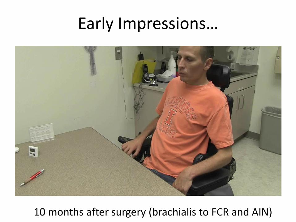
Early Impressions…
10 months after surgery (brachialis to FCR and AIN)
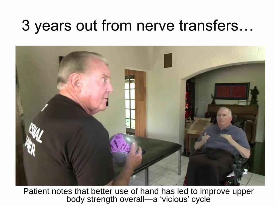
3 years out from nerve transfers…
Patient notes that better use of hand has led to improve upper body strength overall—a ‘vicious’ cycle
• Describe the proper evaluation of patients with cervical level spinal cord injury (SCI) for possible nerve transfer surgery to improve upper extremity function.
• Explain the physiology and surgical principles of the brachialis to anterior interosseous nerve transfer to restore volitional prehension in patients with a C6 or C7 motor level SCI.
• Begin to discuss the barriers to surgical care for patients with cervical SCI.
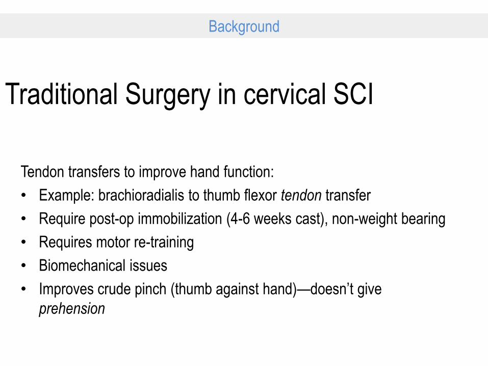
• Improves crude pinch (thumb against hand)—doesn’t give
prehension
Background
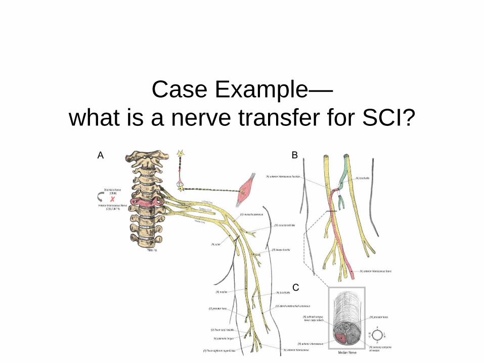
New area—nerve transfers
What are Nerve transfers?
• Robbing Peter to pay Paul…
• Take something that is working and re-
wire into something that is not
• In SCI that means restoring volitional
control to muscles by stealing from
redundant muscles that they can still
control—example:
– Take an extra elbow flexor
– Give back finger function
Background
Nerve transfers in general
Nerve transfers:
• Used to treat peripheral nerve injury (example brachial plexus)
• Key differences: – In peripheral nerve, TIME=MUSCLE (if the nerve is cut, you must reinnervate
within 1 year before muscle becomes unresponsive)
– In SCI, muscle below injury is ‘kept alive’, need to restore control over that nerve/muscle unit (nerve transfer allows that—surgeon creates the peripheral nerve injury by cutting and re-splicing nerve—so need to find a working nerve to steal that is near to the muscle you need it to grow back to)
– Some SCI patients DO have ‘peripheral or direct LMN’ injuries too
Background
Nerve transfers in SCI
• Surgery is done on nerve in the arm (not spinal cord)
• RE-routing of nerves under volitional control
Background
Nerve transfers in SCI
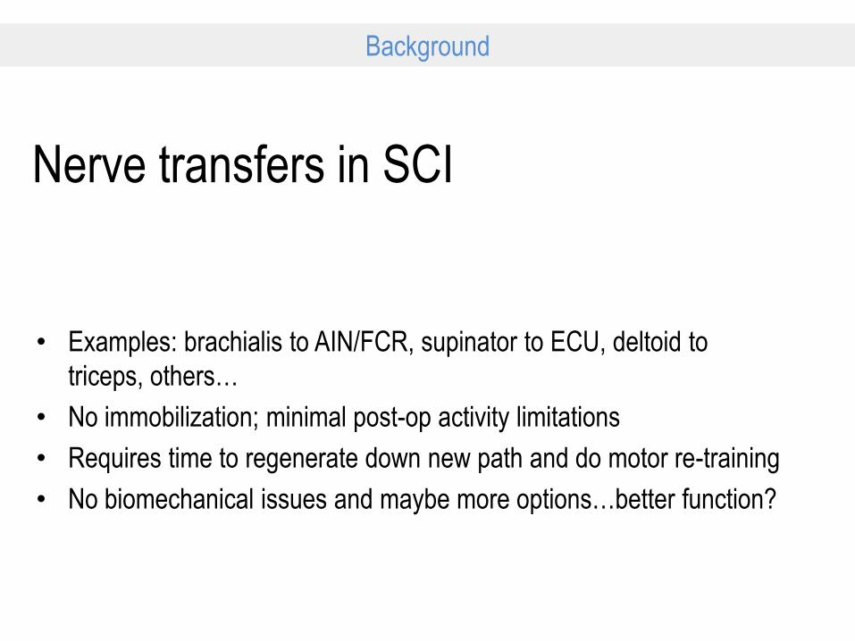
• Examples: brachialis to AIN/FCR, supinator to ECU, deltoid to
triceps, others…
• No immobilization; minimal post-op activity limitations
• Requires time to regenerate down new path and do motor re-training
• No biomechanical issues and maybe more options…better function?
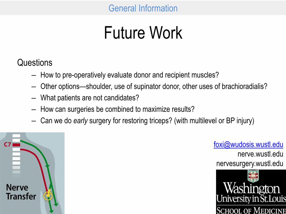
we How we came to do the brachialis to AIN nerve transfer in patients with cervical
spinal cord injury:
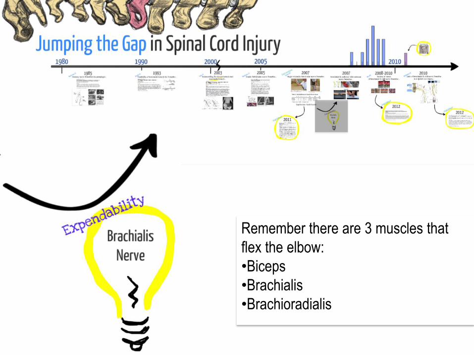
•Question 1: Is brachialis (elbow flexor expendable)?
•Question 2: How can we get hand function back?
Remember there are 3 muscles that
flex the elbow:
•Biceps
•Brachialis
•Brachioradialis
For patients with brachial plexus and peripheral
nerve injury.
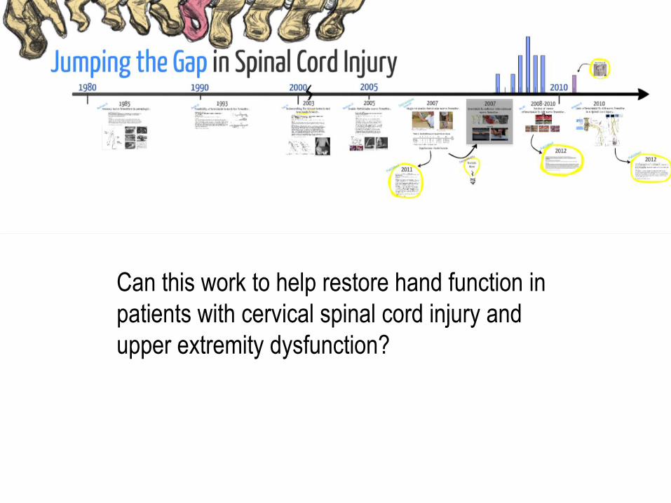
Can this work to help restore hand function in
patients with cervical spinal cord injury and
upper extremity dysfunction?
I
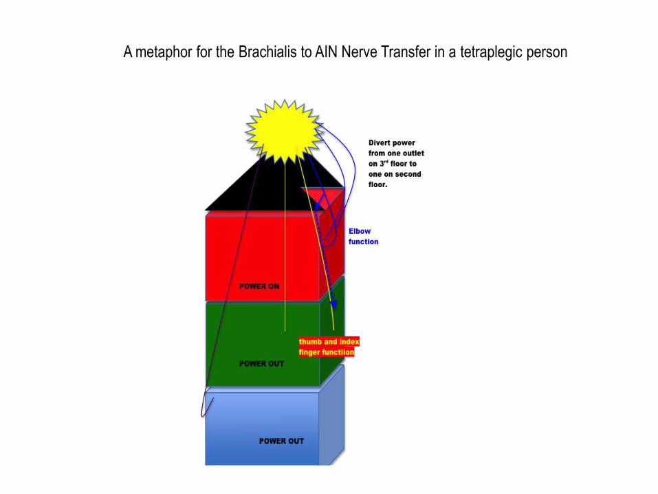
In SCI, unlike peripheral nerve injury, the cell body to muscle connection is often intact…
So we can do a nerve transfer (in the arm) to restore new volitional function:
A metaphor for the Brachialis to AIN Nerve Transfer in a tetraplegic person
In Caveats include:
• Limited knowledge of what will/won’t work
• Careful to not do too much at once
• Do not downgrade function (elbow flexion, pronation, tenodesis)
• Save a back-up plan
• It takes a long time to see the outcome; patience is key!
Build on what we know from traditional surgeries: Bunnel, Moberg, Lamb, Hentz, House, Zancolli
Other work on nerve transfer in SCI: Friden, Bertelli, etc.
In summary: Excellent possibilities for restoring function
without significant down time
Purpose
So what did we do?: Our approach Washington University in Saint Louis
Division of Plastic Surgery
Plan to offer nerve transfers to improve function in cervical SCI:
• Bring nerve transfers from our peripheral nerve injury patient
population to patients with cervical SCI
• Key differences
• Safety first (primum non nocere)—must NOT downgrade function
• Multidisciplinary approach—comprehensive assessment with multiple
practitioners input and multiple modalities of testing
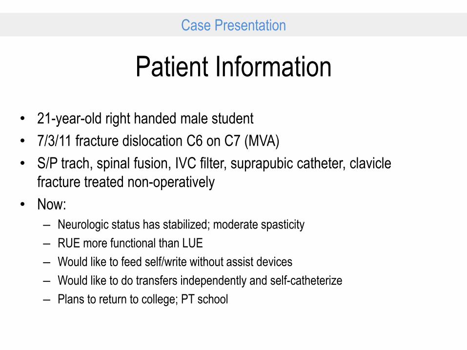
Back to the Case Presentation
The challenge—improve function with
minimal downtime.
Comprehensive Assessment Plan
So how did we get to that plan?
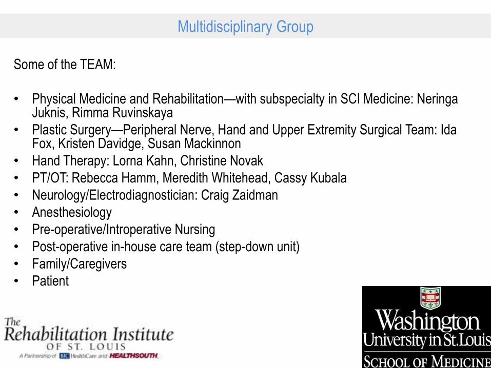
Multidisciplinary Group
Some of the TEAM:
• Physical Medicine and Rehabilitation—with subspecialty in SCI Medicine: Neringa Juknis, Rimma Ruvinskaya
• Plastic Surgery—Peripheral Nerve, Hand and Upper Extremity Surgical Team: Ida Fox, Kristen Davidge, Susan Mackinnon
• Post-operative in-house care team (step-down unit)
• Family/Caregivers
• Patient
Clinical Algorithm
Multidisciplinary Group
Evaluation
Evaluation: Inclusion Criteria • Cervical level spinal cord injury- with loss of (primarily) wrist and/or hand function
• Timing since spinal cord injury- minimum of 6-12 months, maximum 11 years?
• Condition of the upper extremities- – Baseline motor function (must have adequate expendable donor)
– Joint stability
– Range of motion- AROM and PROM
– Spasticity
– Contracture
• Assess current physical therapy/rehabilitation program – Access to physical therapy
– Patient participation and compliance
• Other considerations – Social support
– Financial support for perioperative care
– Psychological well-being
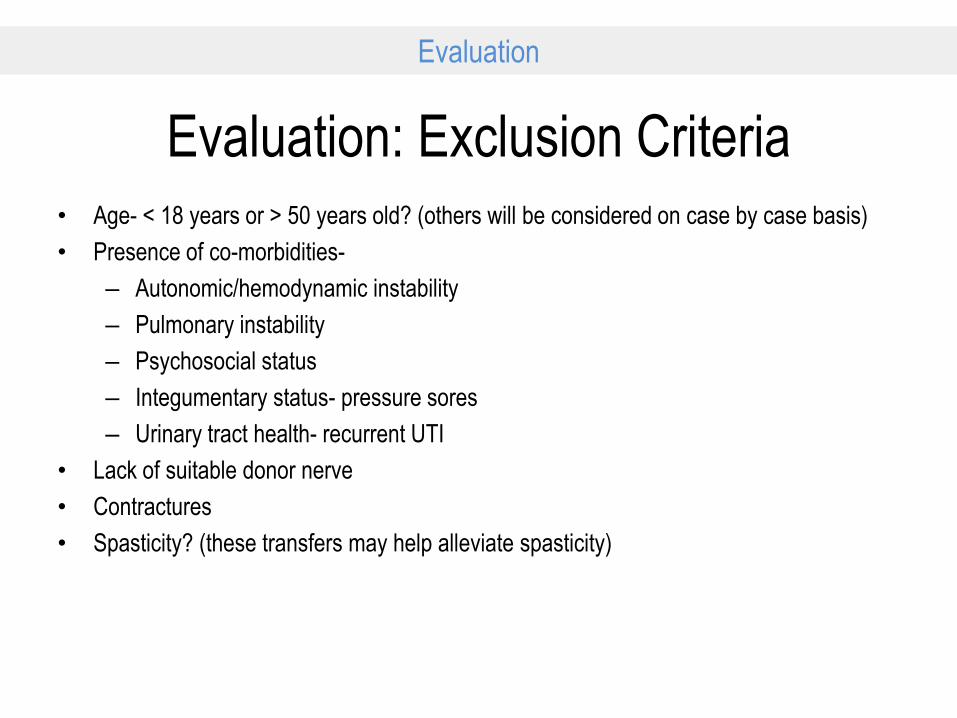
Evaluation
Evaluation: Exclusion Criteria • Age- < 18 years or > 50 years old? (others will be considered on case by case basis)
• Presence of co-morbidities-
– Autonomic/hemodynamic instability
– Pulmonary instability
– Psychosocial status
– Integumentary status- pressure sores
– Urinary tract health- recurrent UTI
• Lack of suitable donor nerve
• Contractures
• Spasticity? (these transfers may help alleviate spasticity)
Lorna C. Kahn, CHT/PT
Milliken Hand Rehabilitation Center
The Rehabilitation Institute of St. Louis
Saint Louis, Missouri
Nerve Transfers in Tetraplegia
Physical Therapy
EVALUATION
Evaluation
SUBJECTIVE HISTORY – history of injury
– social situation/support
– pain
– goals
– work history/plans
PROM bilateral UE
MMT bilateral UE
SENSATION: SWM bilateral UE
QUALITATIVE GRASSP TEST
(prehension skill level)
QUANTITATIVE GRASSP TEST
PROM
Assess for joint contractures and limitations which may hinder progress
following the nerve transfer
Address joint restriction preoperatively with ROM and splinting
MUSCLE TESTING
Complete manual muscle testing of bilateral upper extremities
-assess potential “donor” muscles
-confirm level of hand and arm strength deficits
COMPARISON OF ALL THREE ELBOW FLEXOR MUSCLES
Biceps tested in forearm supination
Palpate tendon
Brachioradialis tested in forearm neutral
Palpate muscle
Brachialis tested in forearm pronation
Palpate muscle by pinching beneath the biceps
SENSORY TESTING
Semmes Weinstein Monofilaments: test for the presence
of touch sensibility
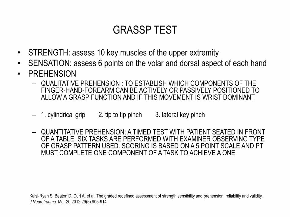
GRASSP TEST
• STRENGTH: assess 10 key muscles of the upper extremity
• SENSATION: assess 6 points on the volar and dorsal aspect of each hand
• PREHENSION – QUALITATIVE PREHENSION : TO ESTABLISH WHICH COMPONENTS OF THE
FINGER-HAND-FOREARM CAN BE ACTIVELY OR PASSIVELY POSITIONED TO ALLOW A GRASP FUNCTION AND IF THIS MOVEMENT IS WRIST DOMINANT
– 1. cylindrical grip 2. tip to tip pinch 3. lateral key pinch
– QUANTITATIVE PREHENSION: A TIMED TEST WITH PATIENT SEATED IN FRONT OF A TABLE. SIX TASKS ARE PERFORMED WITH EXAMINER OBSERVING TYPE OF GRASP PATTERN USED. SCORING IS BASED ON A 5 POINT SCALE AND PT MUST COMPLETE ONE COMPONENT OF A TASK TO ACHIEVE A ONE.
Kalsi-Ryan S, Beaton D, Curt A, et al. The graded redefined assessment of strength sensibility and prehension: reliability and validity.
J Neurotrauma. Mar 20 2012;29(5):905-914
GRASSP TEST QUANTITATIVE PREHENSION TESTING
KEY PINCH CYLINDRICAL GRIP
SPHERICAL
GRASP
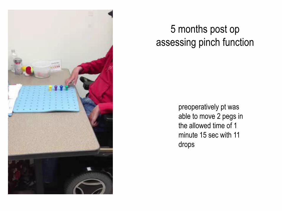
PENNIES/TIP TO TIP PINCH PEGBOARD/TRIPOD PINCH
Evaluation
Evaluation: further work-up
• Check electrodiagnostics—Often see mixed injury
– Want normal EMG of donors
– Check for direct lower motor neuron cell body injury to recipients
– Coexisting peripheral nerve injury—assessment of conduction of median and
ulnar nerves, etc.
(nerve transfer ineffective if >1 year post-SCI if there is LMN injury—can we rescue
muscles with combined injury with early intervention—area of future investigation?)
• Consider imaging
– U/S to assess muscle quality—fatty replacement/fibrosis
Example EMG study—not a good candidate for nerve transfer
Management
Evaluation: take home points
• Want biologic, psycho, and social stability
– As edema resolves, reconditioning occurs can see improved function
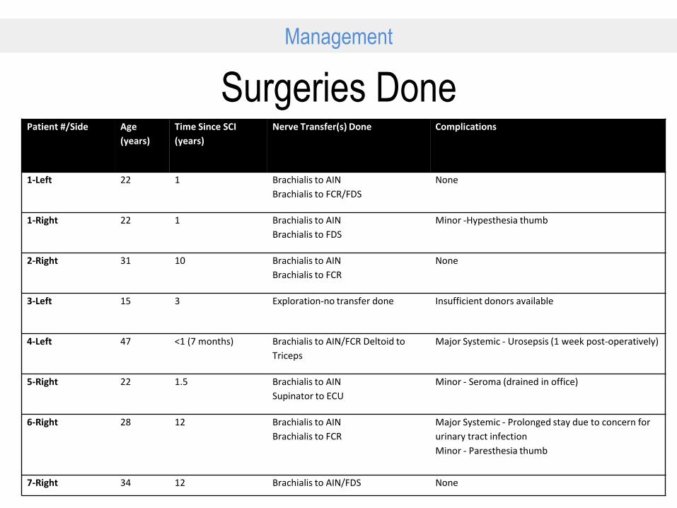
3-Left 15 3 Exploration-no transfer done Insufficient donors available
4-Left 47 <1 (7 months) Brachialis to AIN/FCR Deltoid to
Triceps
Major Systemic - Urosepsis (1 week post-operatively)
5-Right 22 1.5 Brachialis to AIN
Supinator to ECU
Minor - Seroma (drained in office)
6-Right 28 12 Brachialis to AIN
Brachialis to FCR
Major Systemic - Prolonged stay due to concern for
urinary tract infection
Minor - Paresthesia thumb
7-Right 34 12 Brachialis to AIN/FDS None
Back to the Case Example
Surgical Plan
Left Side
Right Side
• – Brachialis (1st branch) to AIN – Brachialis (2nd branch) to FCR/FDS (median nerve motor component fascicle) – Exclusion of PT (volitional control) thenar fascicles (too far), sensory fascicles (wasteful)
• – Brachialis (1st branch) to AIN – Brachialis (2nd branch) to FCR/FDS (median nerve motor component fascicle) – Transfer to FCR/FDS was done end to side to avoid downgrading volitional existing FCR
function – Exclusion of PT (volitional control) thenar fascicles (too far), sensory fascicles (wasteful)
Surgical Technique
Brachialis nerve branches to AIN and FCR nerve
(N) medial antebrachial cutaneous
(N) median (N) median (N) lateral antebrachial cutaneous
(N) brachialis (N) brachialis
(N) median
(N) thenar
(N) anterior interosseous and FCR/PL/FDS
(N) pronator teres
(N) sensory component
(N) brachialis
(N) brachialis
(N) flexor carpi radialis
(N) anterior interosseous
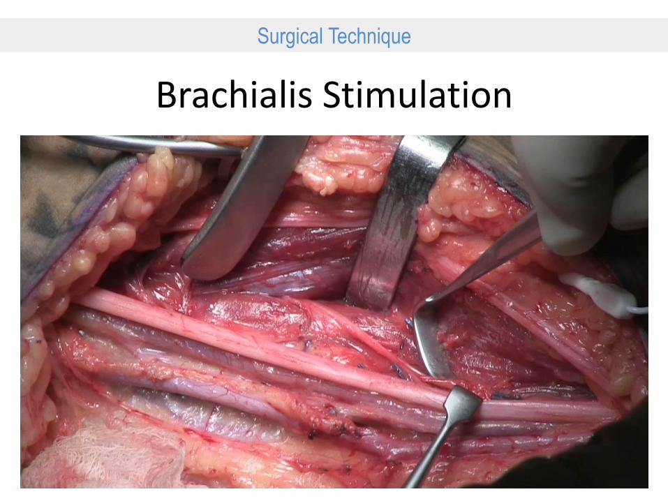
Surgical Technique
Brachialis Stimulation
Surgical Technique
Recipient Nerve Stimulation
Surgical Technique
Nerve Transfers Done
Surgical Options—before. . .
SCI level Missing function Reconstructive options
High
(C5) Elbow extension 1. Deltoid to triceps tendon transfer
2. Biceps to triceps tendon transfer
Wrist extension BR to ECRB or ECRL tendon transfer
Pinch 1. FPL tenodesis to distal radius
2. Thumb IPJ fusion
Mid
(C6-7)
Pinch
Thumb:
1. BR to FPL tendon transfer
2. PT to FPL tendon transfer
3. FPL tenodesis
4. Thumb fusion
Index finger:
ECRL to FDP Index tendon transfer
Grasp ECRL to FDPs of all digits tendon transfer
Wrist flexion 1. Gravity
2. PT to FCR tendon transfer
Finger extension 1. EDC tenodesis to radius
2. BR to EDC tendon transfer
Thumb extension 1. EPL tenodesis to radius
2. Side-to-side transfer of EPL to EDC
Intrinsics Zancolli anti-claw lasso
Low (C8)
Intrinsics 1. Opponensplasty
2. Zancolli anti-claw lasso
Traditional Tendon Transfers:
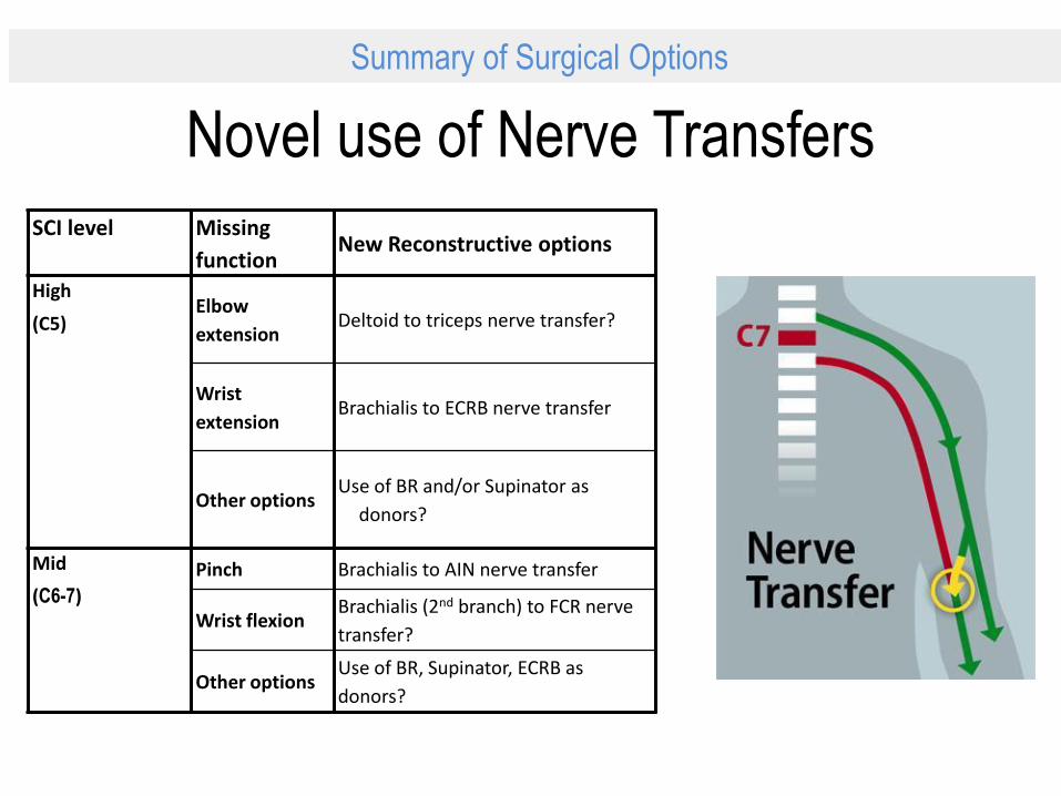
Summary of Surgical Options
Novel use of Nerve Transfers
SCI level Missing
function New Reconstructive options
High
(C5) Elbow
extension Deltoid to triceps nerve transfer?
Wrist
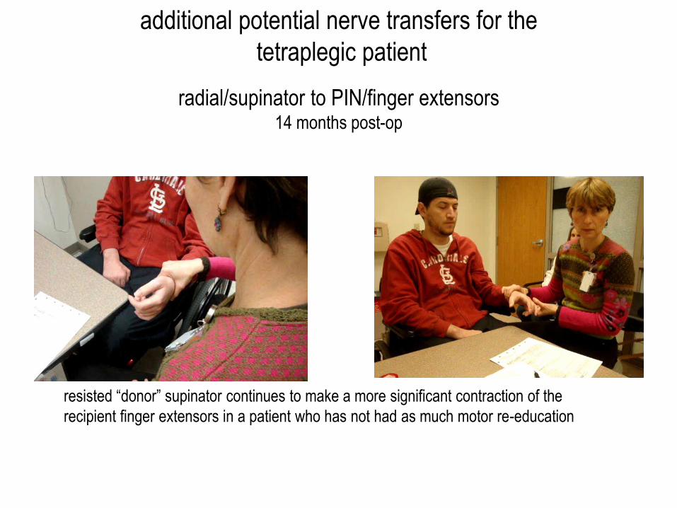
extension Brachialis to ECRB nerve transfer
Other options Use of BR and/or Supinator as
donors?
Mid
(C6-7)
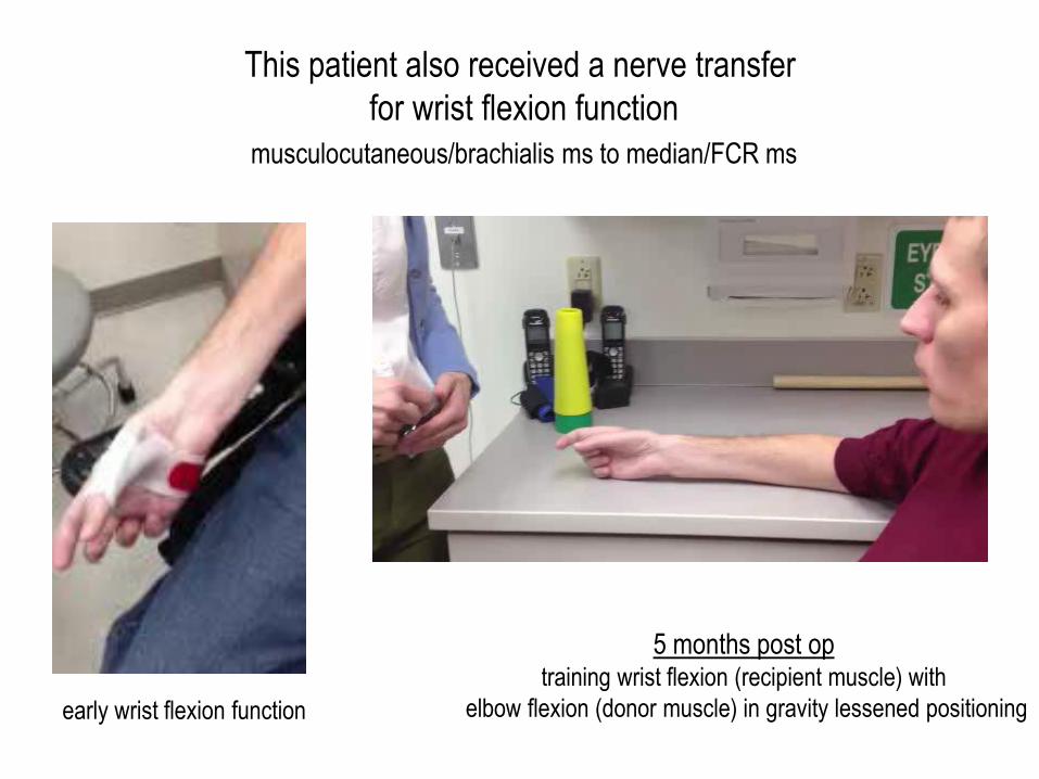
Pinch Brachialis to AIN nerve transfer
Wrist flexion Brachialis (2nd branch) to FCR nerve
transfer?
Other options Use of BR, Supinator, ECRB as
donors?
General Information
Further Surgical Refinement • Double check exam intra-operatively as well
– Confirm donor—are biceps and brachialis both OK?
– Confirm recipient—do recipient muscles, when stimulated, produce some
motion?
• First do no harm
– Abort surgery if there is any possibility that function will be downgraded
– Case example:
• C5 motor level
• Patient with very weak deltoid 2/5, somewhat weak elbow flexion 4-/5
• Goal: restore wrist extension (and hand use via tenodesis) by brachialis to ECRL transfer*
*Friden J, Gohritz A. Brachialis-to-extensor carpi radialis longus selective nerve transfer to restore wrist extension in tetraplegia: case
report. J Hand Surg Am. Aug 2012;37(8):1606-1608.
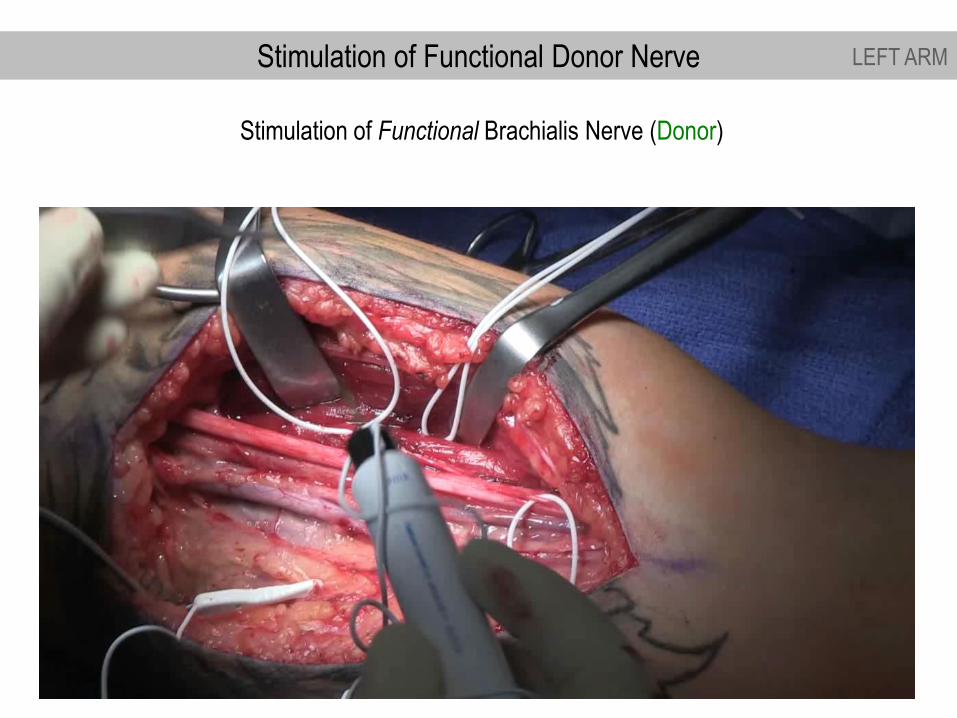
Stimulation of Functional Donor Nerve
Stimulation of Functional Brachialis Nerve (Donor)
Retrospective Case Review of Aborted Nerve Transfer
Stimulation of Musculocutaneous Nerve (Donor) – Video #1
General Information
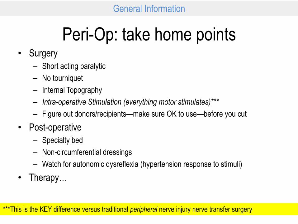
Peri-Op: take home points • Surgery
– Short acting paralytic
– No tourniquet
– Internal Topography
– Intra-operative Stimulation (everything motor stimulates)***
– Figure out donors/recipients—make sure OK to use—before you cut
• Post-operative
– Specialty bed
– Non-circumferential dressings
– Watch for autonomic dysreflexia (hypertension response to stimuli)
• Therapy…
***This is the KEY difference versus traditional peripheral nerve injury nerve transfer surgery
Lorna C. Kahn, CHT/PT
Milliken Hand Rehabilitation Center
The Rehabilitation Institute of St. Louis
Saint Louis, Missouri
Nerve Transfers in Tetraplegia
Rehabilitation
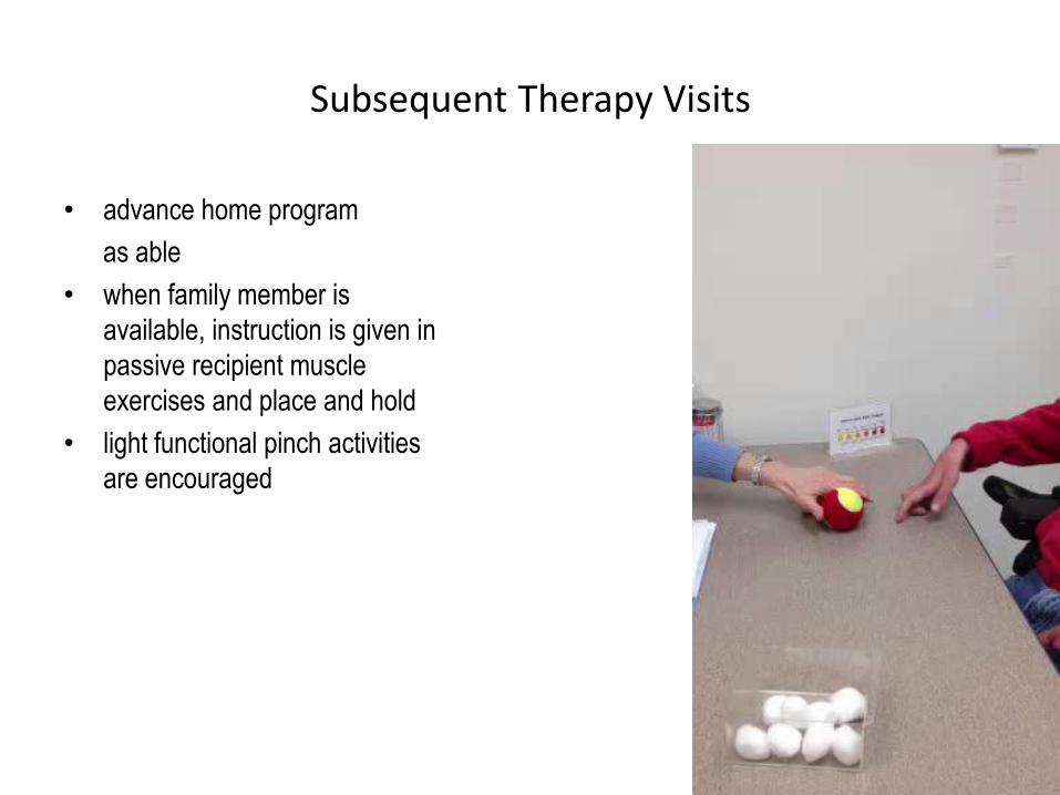
Management
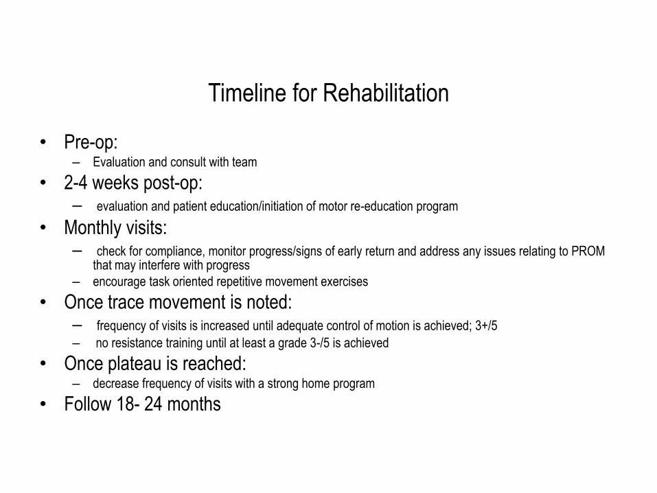
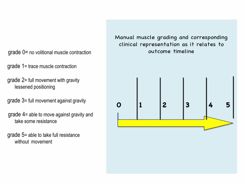
Timeline for Rehabilitation
• Pre-op: – Evaluation and consult with team
• 2-4 weeks post-op: – evaluation and patient education/initiation of motor re-education program
• Monthly visits: – check for compliance, monitor progress/signs of early return and address any issues relating to PROM
that may interfere with progress
– encourage task oriented repetitive movement exercises
• Once trace movement is noted: – frequency of visits is increased until adequate control of motion is achieved; 3+/5
– no resistance training until at least a grade 3-/5 is achieved
• Once plateau is reached: – decrease frequency of visits with a strong home program
• Follow 18- 24 months
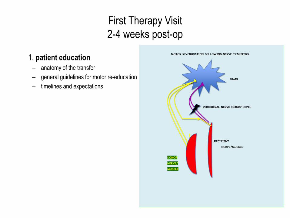
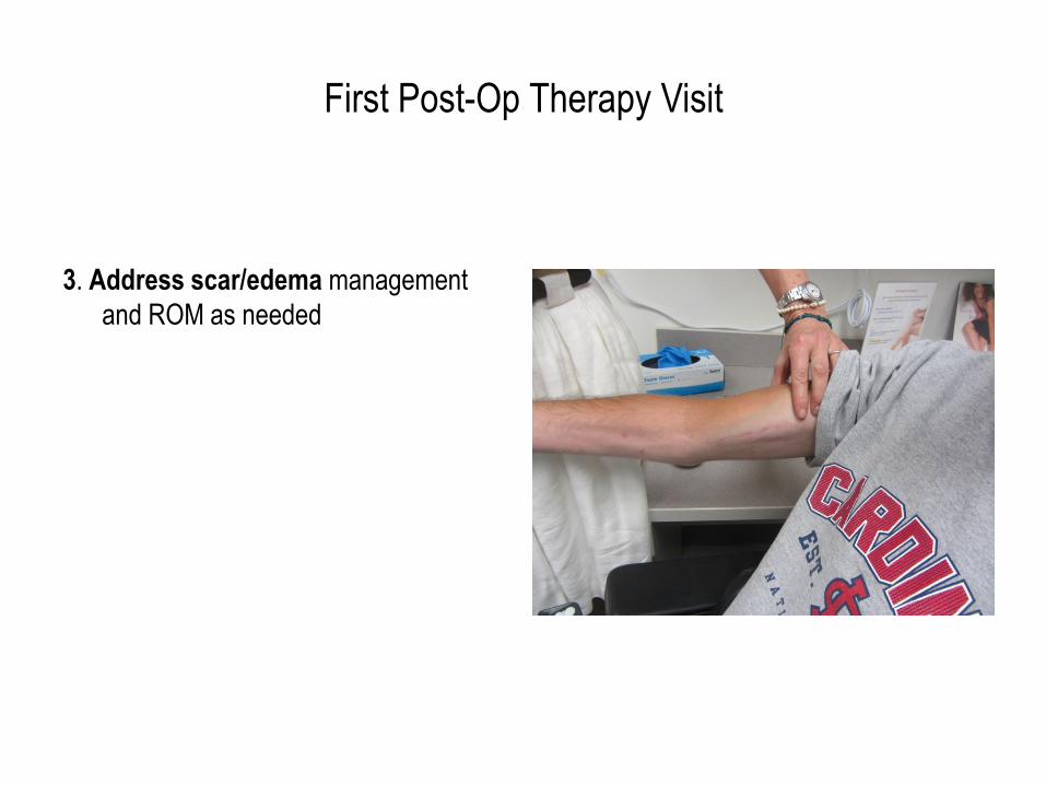
First Therapy Visit
2-4 weeks post-op
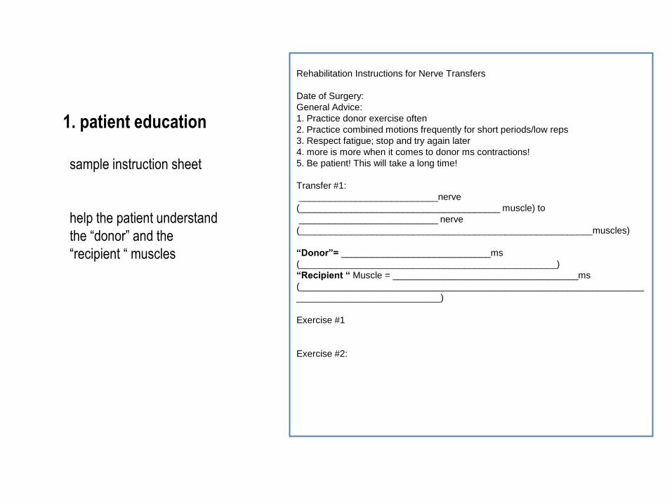
1. patient education
– anatomy of the transfer
– general guidelines for motor re-education
– timelines and expectations
grade 0= no volitional muscle contraction
grade 1= trace muscle contraction
grade 2= full movement with gravity
lessened positioning
grade 3= full movement against gravity
grade 4= able to move against gravity and
take some resistance
grade 5= able to take full resistance
without movement
Rehabilitation Instructions for Nerve Transfers
Date of Surgery:
General Advice:
1. Practice donor exercise often
2. Practice combined motions frequently for short periods/low reps
3. Respect fatigue; stop and try again later
4. more is more when it comes to donor ms contractions!
5. Be patient! This will take a long time!
Transfer #1:
___________________________nerve
(_______________________________________ muscle) to