72
NERVOUS SYSTEM

NERVOUS SYSTEM
Homeostasis controlled by
• Nervous system
• Endocrine system

Divisions of the nervous system
• Central nervous system (CNS)
• Spinal cord• brain
• Peripheral Nervous system (PNS)
• Nerves that carry impulses to and from CNS
Functions
• Monitoring changes inside and outside of body – (stimuli) and gathers sensory input
• Integration – processes and interprets the sensory input and makes decision about what is to be done
• Effects a response by activating a muscle or gland – response is called motor output
Neuron
Nerve cell

Parts of a neuron
• Dendrite – carries impulses to the cell body• Axon – carries impulses from the cell body• Myelin sheath – protective layer formed by
Schwann cells• Nodes of Ranvier – gaps between the
sheaths ( not present in all neurons)• Axonal terminals – branches located at the
terminal end of the neuron
• Neurilemma – sheath of Schwann
• Collateral branch – branch arising from the axon ( not present in all axons)
• Oligodendrocytes – take the place of the Schwann cells in the CNS
SUPPORTING CELLS - neuroglia
• Astrocytes – numerous projections with swollen ends that cling to neurons. Anchoring the neurons to their blood supply
• Microglia – phagocytes that dispose of debris such as dead brain cells, bacteria, ect.
• Ependymal cells = line cavities of the brain and spinal cord. Have cilia that beat to circulate the cerebrospinal fluid
• Oligodendrocytes• Satellite cells – protective and cushion nerve cells

TYPES OF NEURONS

1. Afferent
• Also called sensory
• Carry nerve impulses toward CNS

2. Efferent
• Also called motor neuron
• Carries impulses away from CNS

3. Interneuron
• Also called association neuron
• Connect motor and sensory neuron in neural pathways
CONDUCTION OF A NERVE IMPULSE
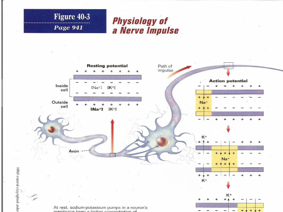
RESTING POTENTIAL
• Membrane is polarized• Not conducting an impulse• -65mV to –70 mV• Na+ greater concentration on outside• K+ greater concentration on inside• Overall charge is + on outside and – on inside• Na/K pumps maintains this difference

DEPOLARIZATION
• Activates the neuron to transmit and action potential (AP) also called a nerve impulse
• All or nothing response – AP is either propagated or not
• Threshold must be reached –55mv• Na+ gates open and Na+ flows inside
neuron• Voltage changes from –65 to +40
REPOLARIZATION
• Na+ gate closes
• K+ gate open and K+ flows to outside
• Voltage change from +40 to –65
• Na/K pumps return Na+ and K+ back to normal
• Thus neuron returns to resting potential
REFRACTORY PERIOD
• AP has passed
• Na gate can’t open
• Keeps AP from traveling in the wrong direction

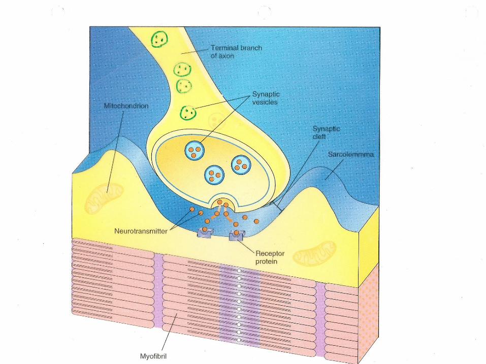
CROSSING THE SYNAPSE
• Synapse – gap between the presynaptic and postsynaptic membrane
• AP arrives at the presynaptic membrane
• Presynaptic membrane becomes permeable to Ca++
• Vesicles move to the presynaptic mb.
• They release neurotransmitter

Myelinated neurons
• Conduction occurs faster because the nerve impulses jump from node to node along the length of the fiber
• Saltatory conduction – faster conduction in myelinated neurons

External impairment of conduction
• Alcohol, sedatives, and anesthetics block nerve impulses by reducing membrane permeability to sodium ions
• Is no Na ions can enter the neuron then no AP will occur
• Cold and pressure hinder impulse conduction because the interrupt blood circulation

• Neurotransmitter diffuses across the synaptic cleft
• At the postsynaptic mb. The neurotransmitter merges with receptor sites
• AP starts at the postsynaptic mb
• Neurotransmitters may be broken down by enzymes, washed away, or recycles

• Axons may synapse with many other neurons
25 different neurotransmitters
acetyocholine
norepinephrine
NERVE
A bundle of axons
also called nerve fibers
REFLES ARC
• Rapid predictable and involuntary responses to stimuli

PERIPHERAL NERVOUS SYSTEM
• Made up of nerves that lie outside the CNS
• cell bodies (called ganglia) found outside the CNS
• 2 structural types– cranial nerves– spinal nerves

Cranial nerves
• Carry impulses to and from the brain
• 12 pairs
• some are sensory
• some are motor
• some are mixed
Spinal nerves
• Carry impulses to and from spinal cord
• all are mixed
• 31 pairs

Functional classification of PNS

Sensory
• Carries information to CNS from sense organs and sensory receptors
• example: eye, Merkel discs
Motor
• Somatic - allows one to consciously control skeletal muscles
• autonomic nervous system - regulates activity of smooth and cardiac muscles and glands

2 branches of the autonomic nervous system
• Sympathetic nervous system
• parasympathetic nervous system
SYMPATHETIC
• Controls body’s response to emergency situation and stress
• increase blood glucose levels in blood• in heart rate• increases oxygen uptake• decrease activity of digestive system and urine
output• dilates pupils• goosebumps/ perspiration
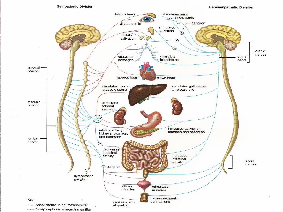
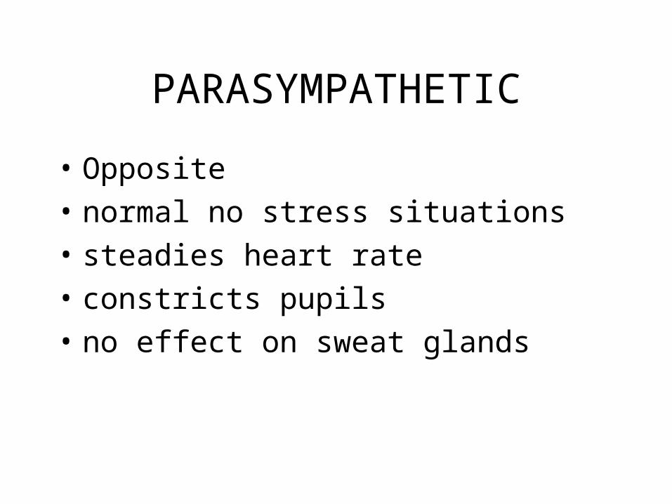
PARASYMPATHETIC
• Opposite
• normal no stress situations
• steadies heart rate
• constricts pupils
• no effect on sweat glands
CNS
PROTECTION
• Meninges - connective tissue membranes around the brain
• cerebrospinal fluid - cushions,– formed by choroids plexuses,– fluid continually moves through ventricles and
between the brain and spinal cord, – 1/2 cup of fluid

SPINAL CORD
• 17 inches long
• 2 way connection with brain
• ends at lumbar vertebrae #2
• rest of column filled with spinal fluid
• has gray and white matter

• Caudal equina – collection of spinal nerves that extend from the end of the spinal cord
• Paralysis – result of injury to cord
• Severity due to location on spinal cord
GRAY MATTER
• Contains cell bodies and nonmyelinated fibers
• contains portions of sensory and motor neurons

WHITE MATTER
• Myelinated axons of interneurons that run to the brain and from one side of the SC to the other side ( called tracts)
PARTS OF THE BRAIN
http://www.psych.ualberta.ca/~ITL/brain/
Ventricles
• Interconnecting cavities filled with CSF
• 4: 4th, 3rd, and 2 lateral ventricles
Medulla Oblongata
• Regulates heart beat, blood pressure, and breathing,
• Has reflex centers for swallowing, coughing, sneezing, hiccupping and vomiting
• Has nerve tracts between the spinal cord and brain

Pons
• Works with medulla to regulate breathing rate
• Mostly a fiber tract
MIDBRAIN
• Relay station for tracts passing between the cerebrum and the spinal cord or cerebellum
• Has reflex centers for visual, auditory, and tactile responses
• Contains the cerebral peduncles and corpora quadrigemina
CEREBELLUM
• Maintains normal muscle tone, posture, balance
• Ensures that all of the skeletal muscles work together to produce smooth and coordinated movements
• Essential for skills such as playing the piano or hitting a baseball.
hypothalamus
• Forms floor of the 3rd ventricle• Maintains homeostasis by regulating hunger,
sleep, thirst, body temperature and water balance.• Sex, pain , and pleasure centers are located here • Part of the limbic system• Called emotion visceral brain• Regulates the pituitary gland therefore it is the link
between the nervous system and the endocrine system

THALAMUS
• Serves as a relay station for sensory impulses traveling upward to other parts of the brain to the cerebrum
• Involved in arousal and higher mental functions such as memory and emotion
• Gives one a crude awareness of whether the sensation will be pleasant or not

Pineal gland
• Secretes the hormone melatonin
• At night the pineal gland produces melatonin - causes one to fall asleep
LIMBIC SYSTEM
• System of tracts and nuclei
• Surround the brain stem
• Called emotional brain
• Blends higher mental functions and primitive emotions into a whole
• Area that makes eating and sexual behavior seem pleasant
Parts of limbic system
• Hippocampus
• Amygdala
• Thalamus
• Hypothalamus
• Frontal lobe
hippocampus
• Area through which incoming sensory signals generate particular limbic response
• example

Amygdala
• Associated with fear conditioning and associating danger with sensory stimuli
• May be responsible for controlling human aggression
Frontal lobe
• Keeps limbic system in check
• It uses reason to keep us from acting out strong feelings
• Alcohol suppresses the frontal lobe and the limbic system takes over
RETICULAR FORMATION
• Nuclei and fibers that extend the length of the brain stem
• Reticular activating system
• Controls the sleep/wake cycle
• Severe damaged can cause one to be comatose
CEREBRUM

CEREBRUM
• Largest portion of the brain
• Divided into left and right cerebral hemispheres
• Surface has gyri – elevated ridges of tissue
• Sulci – shallow grooves
• Fissures – deeper grooves
• Divided into lobes
• Lobes named after cranial bones
• Gray matter
• Center for association, integration, and learning

CORPUS CALLOSUM
• Bridge of nerve tracts that connect the right and left hemispheres
• Tracts cross – left controlled by right side of brain
• White matter – tracts that carry information from 1 part of cerebrum to another part
• Basal nuclei – relay stations that help to regulate motor activities – islands of gray matter found deep within the white matter

LOBES
• Occipital – vision
• Temporal – hearing
• Frontal - olfactory and higher learning
• Parietal
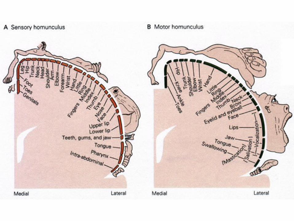
Functional areas of the cerebral cortex
• Somatic sensory area– Impulses from the body’s sensory receptors,
except for the special senses, are localized and interpreted in this area of the brain
• Primary motor area– Allows us to consciously move our skeletal
muscles


GENERAL INFORMATION

Falling in love
• Pleasure center in the hypothalamus
• Brain neurotransmitters – norepinephrine and dopamine
• They give brain a pleasure flush (cousins of amphetamines)
• Falling in love – pleasure center is bathed with dopamine and norepinephrine
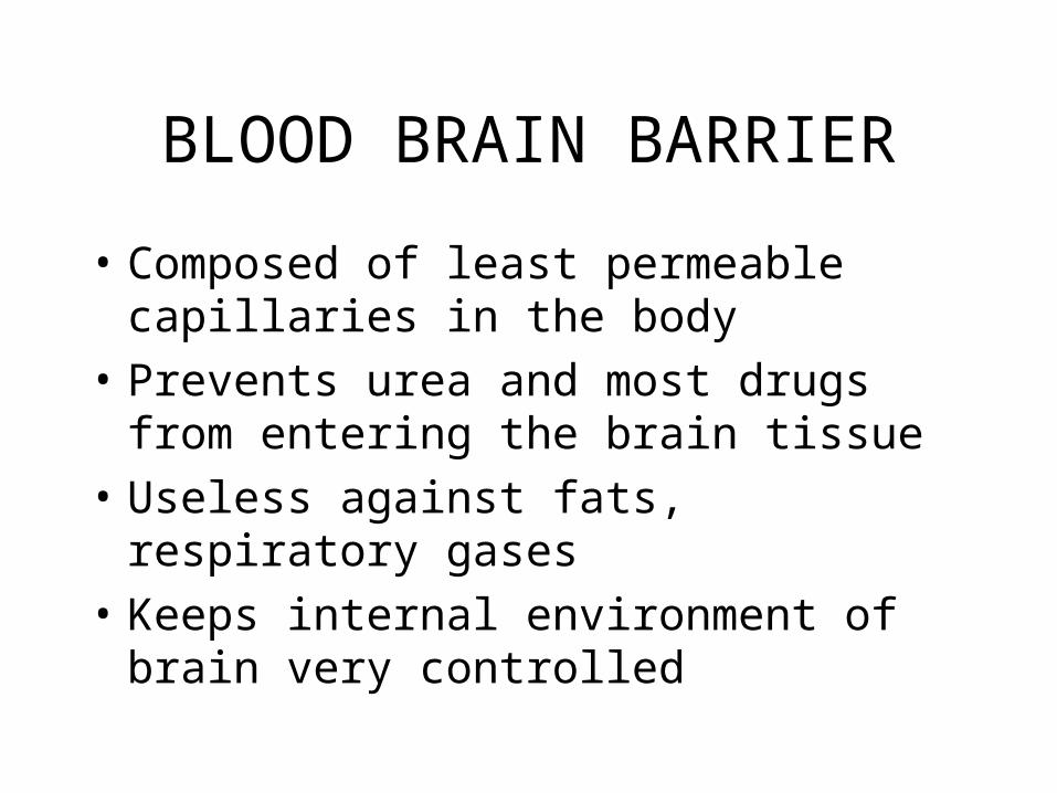
BLOOD BRAIN BARRIER
• Composed of least permeable capillaries in the body
• Prevents urea and most drugs from entering the brain tissue
• Useless against fats, respiratory gases
• Keeps internal environment of brain very controlled
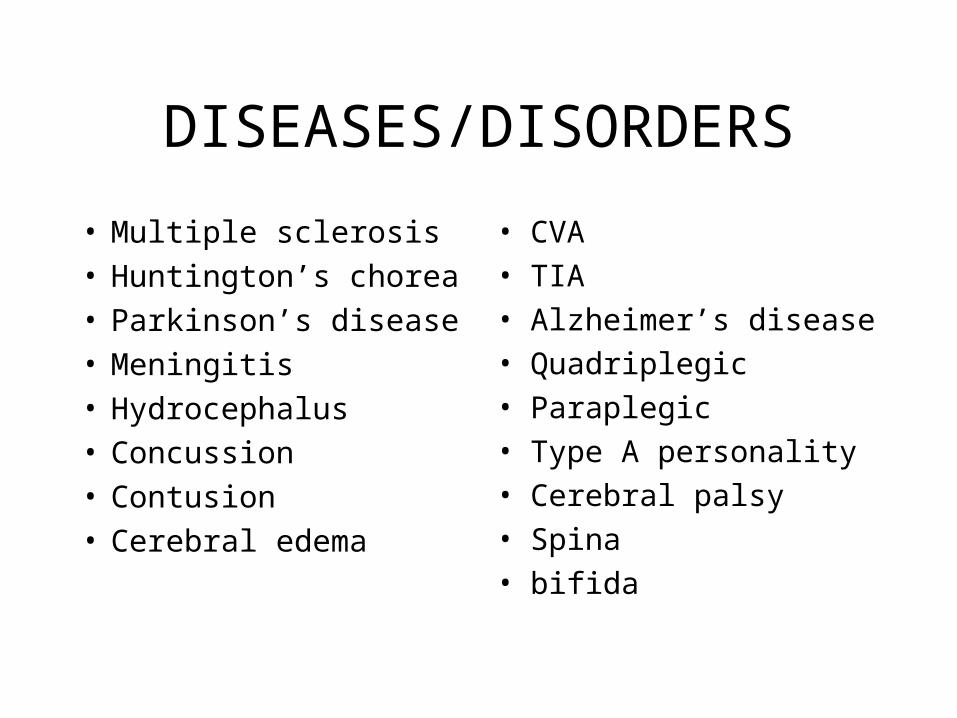
DISEASES/DISORDERS
• Multiple sclerosis• Huntington’s chorea• Parkinson’s disease• Meningitis• Hydrocephalus• Concussion• Contusion• Cerebral edema
• CVA
• TIA
• Alzheimer’s disease
• Quadriplegic
• Paraplegic
• Type A personality
• Cerebral palsy
• Spina
• bifida


