SPECIFIC LANGUAGE IMPAIRMENT/SPEECH SOUND DISORDERS (P VAN LIESHOUT, SECTION EDITOR) Neural Correlates of Developmental Speech and Language Disorders: Evidence from Neuroimaging Frédérique Liégeois & Angela Mayes & Angela Morgan Published online: 7 June 2014 # The Author(s) 2014. This article is published with open access at Springerlink.com Abstract Disorders of speech and language arise out of a complex interaction of genetic, environmental, and neural factors. Little is understood about the neural bases of these disorders. Here we systematically reviewed neuroimaging findings in Speech disorders (SD) and Language disorders (LD) over the last five years (2008–2013; 10 articles). In participants with SD, structural and functional anomalies in the left supramarginal gyrus suggest a possible deficit in sensory feedback or integration. In LD, cortical and subcorti- cal anomalies were reported in a widespread language net- work, with little consistency across studies except in the superior temporal gyri. In summary, both functional and structural anomalies are associated with LD and SD, including greater activity and volumes relative to controls. The variabil- ity in neuroimaging approach and heterogeneity within and across participant samples restricts our full understanding of the neurobiology of these conditions— reducing the potential for devising novel interventions targeted at the underlying pathology. Keywords Speech disorder . Language disorder . Specific language impairment . Speech delay . Speech sound errors . Childhood apraxia of speech . Motor speech disorder . MRI . Functional MRI . Diffusion-weighted MRI . Communications disorders . Childhood Introduction Developmental communication disorders are prevalent, af- fecting over 10 % of school aged children [1]. Here we focus on two common subtypes, namely Language (LD) and Speech (SD) disorders. Whilst some symptoms may “resolve” or be compensated for into adolescence [2], there is increasing evidence for persistent life-long negative impacts of SD and LD on literacy, educational, employment, and psychosocial outcomes [3–5, 6•, 7]. Traditionally, both LD and SD have been defined as idiopathic (of unknown origin). Clearly the term idiopathic implies that the disorders cannot be explained by neurological or sensory deficits, nor are they associated with frank brain abnormalities on clinical MRI. Advances in neuroimaging methods over past decades however, have un- covered both functional and sub-macroscopic structural brain anomalies associated with these disorders. Language Disorders (LDs) are defined as a failure to de- velop age appropriate language skills despite normal sensory abilities and environmental exposure, and affect between 7 % and 20 % of pre-schoolers [8, 9]. A spectrum of LD profiles exists, dependent upon which aspect of language processing is most impaired (e.g., syntax, semantics) [10]. LDs have in the past also been termed “Specific Language Impairments” or SLI, but the “specific” aspect of the disorder remains contro- versial [11•]. Speech Disorders (SDs) is also an umbrella term, encompassing numerous subtypes of developmental speech disorder. Several classification methods have been proposed for SDs [12••, 13]. Here we consider studies that focus on Electronic supplementary material The online version of this article (doi:10.1007/s40474-014-0019-1) contains supplementary material, which is available to authorized users. F. Liégeois (*) UCL Institute of Child Health, Cognitive Neuroscience and Neuropsychiatry Section, 30 Guilford Street, London WC1N 1EH, UK e-mail: [email protected]A. Mayes : A. Morgan Language & Literacy Group, Murdoch Childrens Research Institute, Flemington Road, Parkville, Victoria, Australia A. Morgan Department of Paediatrics, University of Melbourne, Parkville, Victoria, Australia Curr Dev Disord Rep (2014) 1:215–227 DOI 10.1007/s40474-014-0019-1
Transcript
SPECIFIC LANGUAGE IMPAIRMENT/SPEECH SOUND DISORDERS (P VAN LIESHOUT, SECTION EDITOR)
Neural Correlates of Developmental Speech and LanguageDisorders: Evidence from Neuroimaging
Frédérique Liégeois & Angela Mayes & Angela Morgan
Published online: 7 June 2014# The Author(s) 2014. This article is published with open access at Springerlink.com
Abstract Disorders of speech and language arise out of acomplex interaction of genetic, environmental, and neuralfactors. Little is understood about the neural bases of thesedisorders. Here we systematically reviewed neuroimagingfindings in Speech disorders (SD) and Language disorders(LD) over the last five years (2008–2013; 10 articles). Inparticipants with SD, structural and functional anomalies inthe left supramarginal gyrus suggest a possible deficit insensory feedback or integration. In LD, cortical and subcorti-cal anomalies were reported in a widespread language net-work, with little consistency across studies except in thesuperior temporal gyri. In summary, both functional andstructural anomalies are associated with LD and SD, includinggreater activity and volumes relative to controls. The variabil-ity in neuroimaging approach and heterogeneity within andacross participant samples restricts our full understanding ofthe neurobiology of these conditions— reducing the potentialfor devising novel interventions targeted at the underlyingpathology.
Developmental communication disorders are prevalent, af-fecting over 10 % of school aged children [1]. Here we focuson two common subtypes, namely Language (LD) and Speech(SD) disorders. Whilst some symptoms may “resolve” or becompensated for into adolescence [2], there is increasingevidence for persistent life-long negative impacts of SD andLD on literacy, educational, employment, and psychosocialoutcomes [3–5, 6•, 7]. Traditionally, both LD and SD havebeen defined as idiopathic (of unknown origin). Clearly theterm idiopathic implies that the disorders cannot be explainedby neurological or sensory deficits, nor are they associatedwith frank brain abnormalities on clinical MRI. Advances inneuroimaging methods over past decades however, have un-covered both functional and sub-macroscopic structural brainanomalies associated with these disorders.
Language Disorders (LDs) are defined as a failure to de-velop age appropriate language skills despite normal sensoryabilities and environmental exposure, and affect between 7 %and 20 % of pre-schoolers [8, 9]. A spectrum of LD profilesexists, dependent uponwhich aspect of language processing ismost impaired (e.g., syntax, semantics) [10]. LDs have in thepast also been termed “Specific Language Impairments” orSLI, but the “specific” aspect of the disorder remains contro-versial [11•]. Speech Disorders (SDs) is also an umbrella term,encompassing numerous subtypes of developmental speechdisorder. Several classification methods have been proposedfor SDs [12••, 13]. Here we consider studies that focus on
Electronic supplementary material The online version of this article(doi:10.1007/s40474-014-0019-1) contains supplementary material,which is available to authorized users.
F. Liégeois (*)UCL Institute of Child Health, Cognitive Neuroscience andNeuropsychiatry Section, 30 Guilford Street,London WC1N 1EH, UKe-mail: [email protected]
A. Mayes :A. MorganLanguage & Literacy Group, Murdoch Childrens Research Institute,Flemington Road, Parkville, Victoria, Australia
A. MorganDepartment of Paediatrics, University of Melbourne, Parkville,Victoria, Australia
Curr Dev Disord Rep (2014) 1:215–227DOI 10.1007/s40474-014-0019-1
subtypes of articulation disorder (phonetic based or motorexecution errors), phonological disorder (phonemic based orcognitive-linguistic errors), and childhood apraxia of speech(CAS, motor planning and programming errors), as well asthose that use the less explicit diagnostic terms of speecherrors and speech delay. Although behavioral assessments ofdeficits are crucial, neuroimaging studies can provide us witha different level of explanation of symptoms, and may offer anovel way of classifying subtypes of SDs and LDs.
To date, the most extensive neuroimaging studies of a de-velopmental speech and language disorder have been carriedout in the affected members of the KE family, who have a raremutation in the FOXP2 gene, with a seminal imaging studypublished on this family in 1998 [14]. Affected members of theKE family present with both speech (verbal and orofacial praxisand dysarthria) and language impairments, affecting speechintelligibility as well as the use of morphosyntax and thecomprehension of complex grammatical structures [15, 16]. Itis critical to note that the phenotypic marker, co-segregatingaffected and unaffected familymembers, is a diagnosis of CAS.Since the early KE studies, examination of the neural basis ofSD and LD has been limited and is still an emerging field.
Here we systematically reviewed all articles publishedbetween 2008 and 2013 in individuals (adults or children)diagnosed with developmental forms of SD or LD.1 Wepresent functional and structural MRI findings to ask whetherwe are any closer to answering the following question: whichbrain anomalies are associated with atypical development ofspeech and language?
Methods
Search Strategy
A computerized systematic search was conducted of relevantdatabases: EMBASE (1996 to August 2013), OVIDMEDLINE (1996 to August 2013), PubMed (searched Au-gust 2013). The following MeSH terms were used to identifySD and LD papers of interest: (speech disorder OR articula-tion disorder OR phonetic disorder OR speech delay ORphonological impairment OR language disorder OR languagedevelopment disorder) AND (magnetic resonance imagingOR diffusion magnetic resonance imaging OR echo planerimaging OR computerized positron emission tomography ORsingle photon OR brain) NOT (dyslexia OR Asperger syn-drome OR autistic disorder OR aphasia OR Broca’s aphasiaOR Wernicke’s aphasia OR primary progressive aphasia ORconduction primary progressive non fluent aphasia OR
electroencephalography). Of note, the MeSH terms for SDsand LDs were kept broad to encompass all relevant terminol-ogy (e.g., speech delay, speech sound disorder, SLI). Searcheswere limited to papers written in English between 2008 andpresent (August 2013) with human participants. Manualsearches were completed in relevant journals publishingbrain-behavior relationships in this field (i.e., Brain and Lan-guage, Brain Topography).
Inclusion Criteria
Studies were included if they reported results of individualswith either SD or LD, together with a MRI neuroimagingmethod to investigate brain structure or function. Full textarticles were required to be available and published in English.Failure to meet one of the above criteria resulted in exclusion.
Data Extraction
A total of 2,602 abstracts were identified. An additional fourwere located in a manual search. Two stages of exclusion wereconducted (Supplementary Fig. 1). Firstly, papers were excludedbased on title only, including any duplicates (n=2,573) by oneauthor (A. Mayes). Secondly, papers were excluded based onindependent review of the abstract and/or full text article (n=23)by all three authors, using the following criteria: Participantselection criteria (excluding studies with children who havebrain injury); imaging methods (excluding studies without im-aging); analysis method (excluding studies with no quantitativeanalysis). Disagreements were resolved by discussion (one arti-cle). All three authors manually searched for additional publica-tions relevant to the field published between 2008 and 2013 andlisted within the reference list of each selected paper.
Critical Appraisal
To examine the level of evidence provided, we used theNHMRC (National Health and Medical Research Council,Australia) classification (http://sydney.edu.au/medicine/21st-century/presentations/2013/NHMRC-hierarchy-of-evidence.pdfref/Appendix) [17]. This classification system allows agrading from the poorest level of evidence (Grade IV, Casesseries studies) to the highest (e.g., Grade I, systematic reviewof randomized controlled trials).
Results
Overview of Articles: Methodological Considerationsand Critical appraisal
Ten articles (see Supplementary Fig. 1) were included, five onSD and five on LD. All were case-control studies (NHMRC
1 Authors were requested to review literature in this field over the past12 months. Given the scarcity of literature, the authors extended thesearch to encompass the past 5 years.
evidence level III-2) [17]. Effect sizes for group comparisonswere available for two studies (Preston et al., [18], Table 1;Lee et al., [19], Table 2) and could not be calculated for theremainder as standard deviations were not provided. Agebands were relatively narrow (1–3 years) for studies on chil-dren with SD, but wider in studies on children with LD, wherethree out of four studies reported on groups spanning nineyears or greater. Only Verhoeven et al., [20] focused on anarrow age band (all cases were 10 year olds).
Another observation is that the recruitment samples in bothSD and LD studies were heterogeneous with regard to diag-nosis. Liegeois et al., [21] and Kadis et al., [22] focused onCAS, although the former was focused on FOXP2 associatedCAS in adults and the latter included young idiopathic cases.
Tkach et al., [23] considered both phonetic (articulatory)and phonemic (phonological process analysis) level errors.Preston et al., [18, 24] report on phonetic level errors only,using largely the same sample of children with persistent SD(17 of the 23 in 2014 were from the original study [24]).Phonological process analysis was not reported in either Pres-ton et al., study, however CTOPP results were reported, i.e., ameasure of phonological awareness, rather than productionper se. No group differences were reported on the CTOPP as awhole in Preston et al., [24], but moderate effect sizes werereported on CTOPP subtests of Elision and Blending words inthe later study [18]. Hence, it is challenging to interpret thelevel of phonological deficit, if any, in these participants whoare denoted as having “Speech Sound Errors” (SSE). Allstudies included participants with persistent SD, with theexception of Tkach et al., [23], who focused on a sample witha history of SD. Only one of the six cases in Tkach et al., hadpersistent SD.
Thus, overall, across the five SSD studies, it appears thatone focused on persistent speech motor programming deficits(CAS) [22]; one on persistent speech programming and exe-cution deficits associated with FOXP2 mutation (CAS anddysarthria) [21]; two on persistent phonetic level (i.e., articu-latory) deficits [18, 24]; and one on a history of phonetic and/or phonemic level (i.e., articulatory/phonological) deficits[23].
Similarly, for the LD studies, inclusion criteria and diagno-ses were highly varied. Some even included several subtypesof impairment within LD groups. Verhoeven et al., [20],included children with a history of language delay and whoscored <10th percentile on at least one of three language testsbeyond the age of four. At the time of testing, the SLI groupscored more than one standard deviation below the normativemean on both receptive and expressive subtests of the DutchCELF. The study by Soriano-Mas et al., [25] included childrenwith speech programming, phonological-syntactic, lexical, ormixed deficits according to the Rapin criteria [26]. At the timeof testing, the SLI group scored more than one standarddeviation below the normative population mean on three
language measures. The notable exception is the study by deGuibert et al., [27], which claims to focus exclusively onyoung people with “structural” language impairment. Partic-ipants with LD showed deficits in phonology (assessed usingunfamiliar word repetition, which is arguably not a pure test ofphonological ability), morphosyntax (tested using a sentencecompletion test), or both. Finally, the study on adults [19]included participants diagnosed with LI as children, and whoas an adult group scored 1.5 standard deviation below thenormative mean on a composite language score. It is notewor-thy that the classification of LD is still a matter of debate, withthe question of a continuum vs. discrete entities still unan-swered [11•, 28].
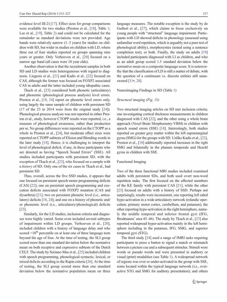
Neuroimaging Findings in SD (Table 1)
Structural imaging (Fig. 1b)
Two structural imaging articles on SD met inclusion criteria;one investigating cortical thickness measurements in childrendiagnosed with CAS [22], and the other using a whole brainapproach (Voxel Brain Morphometry-VBM) in children withspeech sound errors (SSE) [18]. Interestingly, both studiesreported on greater grey matter within the left supramarginalgyrus (SMG) for the groups with SD. Unlike Kadis et al., [22],Preston et al., [18] additionally reported increases in the rightSMG and bilaterally in the planum temporale and Heschlgyrus in children with SSE.
Functional Imaging
Two of the three functional MRI studies included examinedadults with persistent SDs, and both used overt non-wordrepetition tasks. The first focused on the affected membersof the KE family with persistent CAS [21], while the other[23] focused on adults with a history of SSD. Perhaps notsurprisingly, results were inconsistent, one reporting left sidedhypo-activation in a wide articulatory network (rolandic oper-culum, primary motor cortex, cerebellum, and putamen); theother reporting hypo-activation in the right hemisphere, name-ly the middle temporal and inferior frontal gyri (IFG,Brodmanns’ area 45–46). The study by Tkach et al., [23] alsoreported widespread hyper-activation mainly in the left hemi-sphere including in the putamen, IFG, SMG, and superiortemporal gyri (STG).
The third study [24] used a range of fMRI tasks requiringparticipants to press a button to signal a match or mismatchbetween a picture cue and a subsequent stimulus. Stimuli werewords or pseudo words and were presented in auditory orvisual (print) modalities (see Table 1). Awidespread networkof regions was over or under-activated in the group with SSE,some located within the typical language network (i.e., over-active STG and SMG for auditory presentation); and others
external to this network (e.g., underactive orbital gyri, over-active middle frontal gyrus and posterior cingulate for audito-ry presentation). Of note, participants with SSE showed moreactivation in the left inferior/middle frontal gyrus when pre-sented with words rather than with pseudo-words, while thecontrol group showed the opposite trend.
Neuroimaging Findings in LD (Table 2)
Structural Imaging (Fig. 1a)
Of the four structural studies included [19, 20, 25, 29], all butone focused on child participants [19]. Two VBM studiesreported abnormalities in LD participants within the temporalregion, with some degree of anatomical inconsistency. Onereported reduced grey matter in the right posterior superiorand middle temporal gyri and left posterior superior temporalsulcus [29]. The other, on the contrary, reported increasedregional volumes within a right posterior “perisylvian” areaextending from the posterior STG to the angular and SMG [25].
Subcortical structures were also found to develop atypical-ly in participants with LD, with again contrasting findings forthe caudate nucleus (reductions in two papers [19, 29]; andincreases in one [25]). Of note, reductions were also found inLD participants’ unaffected siblings [29]. The same study alsofound that caudate nucleus volume was negatively correlatedwith non-word repetition scores in children with LD [29].Larger relative volumes (i.e., corrected for intracranial vol-ume) were reported bilaterally in the putamen for children
with LD in one study, with a larger putamen associated withpoorer language performance [19].
Soriano-Mas et al., [25] examined white matter usingVBM. They reported morphological increases in white matterbilaterally in the middle temporal gyrus and an anterior clusterin the medial frontal lobe for the younger SLI group. Twostudies used diffusion-weighted MRI to examine microstruc-tural abnormalities in LD. Verhoeven et al., [20] focused onthe superior longitudinal fasciculus, and reported reducedfractional anisotropy (FA) values (a measure of white mattermicrostructure) for children with LD. Additionally, FA valueswere negatively correlated with language measures includingword class receptive and expressive sub tests. In contrast, thestudy by Lee et al., [19] focused on grey matter. They reportedvolumetric reductions in most of the subcortical and corticalROIs examined, and FA reductions in the cortex, but no FAreductions in the caudate or putamen. Poor language perfor-mance was only associated with FA reductions across thewhole brain.
Functional Imaging
Two fMRI studies of children with LD were included. Oneemployed covert lexical semantic and phonological tasks [27],and the other a covert auditory response naming task [29]; andboth reported hypo-activation of the posterior STG. In onestudy, right sided hyper-activation was seen within the rightinsula extending to IFG -pars opercularis/pars triangularis,and caudate head for children with LD in response to aphonological difference task (i.e., where the children see a
a)
b)
L R
L R
Fig. 1 Morphological grey matter (GM) differences in individuals with(a), Language Disorder (LD) and (b), Speech Disorder (SD), relative totypically developing participants Colour code: GM volume decreases inLD: Badcock et al., [29] = blue; GM increases in LD: Badcock et al., [29]= green; Lee et al. [19], relative volumes) = yellow; Soriano Mas et al.,
[25] = purple; GM increases in SD: Kadis et al. 2013 = light blue, Prestonet al., [18] = red. GM decreases in SD: Preston et al., [18] = orange. Note:Fractional anisotropy (FA) differences (Lee et al.) are not illustrated hereas changes were observed across the whole brain (Table 2)
220 Curr Dev Disord Rep (2014) 1:215–227
Tab
le2
Neuroim
agingstudieson
LD
Article
Studygroupand
selectioncriteria
Samplesize
(males)
Meanage(range)
inyears
Methods
Brain
behaviourcorrelation
Decreases
instudy
group(effectsize)
Increasesin
study
group(effectsize)
Badcock
etal
2012
[29]
SLI
<10th
percentileon
≤2l
anguageor
literacy
tests(A
x:CCC-2
[59]
orCC-A
[60],
TROG-2
[61],T
OWRE
[62],N
EPS
Y[63],and
≥80WASI
[64].
SLI(n=10;9
M);
SIB(n=6;
4M);
TD(n=16;7
M)
1.SL
I:13.5(8–17)
2.SIB:1
8(12–22),
3.TD:1
2.50
(6–25)
VBM
(wholebrain)
fMRI
Silent
wordassociation
task
(“Sp
eech”)
Vs.Passivelistening
toreversed
speech
Not
exam
ined
Reduced
grey
matter:
SLI<TD:M
edialfrontal
pole,L
+RpS
TSext.to
RST
G,R
medialsuperior
parietalcortex,L
occipital
pole,R
caudatenucleus,
Rsubstantianigra;RpM
TG
SLI<SIB:L
+Rparoperculum
cortex,L
occipitalp
ole
Reduced
brainactiv
itySpeechcondition:
SLI<TD:L
IFG(pars
orbitalis
);SL
I<SIB:L
IFG(parsorbitalis);R
IFG(parstriangularis);
LpS
TG;
Speech>ReversedSp
eech:
SLI<TD:L
pSTG;R
putamen;
SLI<SIB:L
IFG
(parsorbitalis)
Increasedgrey
matter:
SLI>TD:L
frontal
operculum,R
anterior
insula,L
aIPS
;SL
I>SIB:L
aIPS
Increasedbrainactiv
ityNone
Verhoeven
etal,
2012
[20]
SLI(m
ixed
receptive-expressive)
<3rdcentile
≥1of
3subtests
ofReynellTaaloontwik-
kelin
gsschalen[65],
Taaltestsvoor
Kinderen
[66],orSchlichtingTest
voor
Taalproductie
[67],
andPIQor
FSIQ
>80
[68]
SLI(n=13;1
0M)
TD(n=12;8
M)
SLI:10.1(SD=0.4)
TDforSL
I:10.2
(SD=0.3);
DTI
Tractography
(Superior
Longitudinal
Fasciculus,
SLF)
InSL
Igroup:
WCR
subtestand
FAboth
LandRSL
F;WCE
andleftSL
F
SLI<allT
D:R
educed
Fractio
nalanisotropyin
SLF
NootherROImeasured
deGuibertetal,
2011
[27]
SLI
>1S
Dbelowmeanfor
phonology,sentence
repetition,and
morphosyntacticintegration
[69,70],andWISC/W
AIS
[71,72]≥
70
SLI(n=21,9
M)
TD(n=18,9
M)
SLI:11.4(7–18)
TD:1
2.7(8.7–17.7)
fMRI
Silent
generatio
nor
naming.
ROIanalyses
Reduced
brainactiv
ity:
Audito
ryResponseNam
ing:
LpS
TG/SMGjunctio
n
Increasedbrainactiv
ityPh
onologicaldifference
task:
Ranterior
insulaextto
IFGopercularis/triangularis
andcaudatehead.
Soriano-M
asetal
2009
[25]
DLI
Rapin
[26]
classificatio
n:Sp
eech
programming
deficit(n=5),phonological-
syntactic
deficit
(n=18),lexicald
eficit
(n=8),m
ixed
(n=5)
>1S
DbelowmeanPP
VT[73],
TTFC
[74],ITPA
[75],and
WISC-III[76].
IQ>85.
SLI(n=36;2
4M)
TD(n=36;2
4M)
SLI:10.58(5–17)
TD:1
0.88
(5–17)
VBM
Older
SLI:n
egative
correlationbtwverbal
IQandGM
Rperisylvian
region,P
PVT+GM
occipitalp
etalia
None
Increasedgrey
matter
Globalv
olum
e;Rposterior
perisylvian,LMOG
(occipitalp
etalia)
Young
SLI>Young
TD:L
+R
entorhinal,L
+Rtemporopolar,
L+Rcaudatenucleus,L+R
precentralgyrus,Lprecuneus,
LmedialM
OG
Increasedwhitematter
Globalv
olum
e;Young
SLI>
Young
TD:R
medialfront
cortex,L
+RMTG
Lee
etal.,
2013
[19]
DevelopmentalL
anguage
Impairment(DLI)
>1.5SDbelowmeanof
language
composite(w
ordderivatio
ns–
subtesto
fTOAL-4,P
PVT-4,
tokentest)[77,78].
WASI
PIQassessed
notu
sed
ascriteria
DLI(n=12;4
M)
TD(n=12;4
M)
DLI:21.99
TD:2
2.06
Overallrange
19–25
DTI+Volum
etricROIs:
Caudatenucleus;
putamen;n
ucleus
accumbens;g
lobus
pallidus;thalam
us;
occipital,parietal,
temporal,frontal
lobes;hippocam
pus
negativ
ecorrelationbtw
nucleusaccumbens;
globus
palladius;
putamen,hippocampus
ROIs,w
holebrainFA
+language
composite
Reduced
grey
mattervolumes:
ICV,L
+Rcaudatenucleus
(d=-1.21),L
+Rthalam
us(d=-1.57),occipitallobe
(d=-1.54),parietallobe
(d=-1.47),tem
porallobe
(d=-1.32),frontallobe
(d=-1.50).
Reduced
FA:
Wholebrain(d=-2.00);g
lobus
pallidus(d=-0.96)
thalam
us
Increasedgrey
matterVolum
es:
WhenROIvolumecorrectedfor
ICV:p
utam
en(d=1.07),
nucleusaccumbens
(d=1.0);
hippocam
pus(d=1.70).
IncreasedFA
:None
Curr Dev Disord Rep (2014) 1:215–227 221
picture and silently generate names of three objects, each witha different initial phoneme) [27].
Discussion
All studies reported significant developmental anomalies ofbrain structure or function in relatively small groups of chil-dren with SD and LD as revealed by quantitative imaging.Here we discuss the most consistent findings, but emphasizethe need for caution in interpretation given methodologicalvariability across studies.
Neural Basis of SD
Morphological Anomalies
Converging evidence for abnormal increases in the left SMGwas noted in two studies. The authors hypothesized thatincreases in this region reflect “immaturity or altered devel-opment” [22] or “reduced synaptic pruning” [18]. In addition,this similar finding points to possible commonalities betweenthe aetiology of speech sound disorders of articulation andphonology and CAS, despite these conditions being tradition-ally viewed as distinct clinical diagnoses. There may also havebeen overlap of symptoms between participants from thesetwo studies. In adult neuroanatomical models, the SMG isassumed to play a crucial role in auditory motor and sensori-motor [30] integration. This is a critical region in the somato-sensory feedback loop in both the DIVA [31] and HSFC [32]computational models of speech production. Further, a recentrepetitive TMS study highlighted the importance of this infe-rior parietal region and its connections to frontal and motoroutput areas, in learning and adapting sensorimotor patternsfor speech [33]. Structural anomalies in the left SMG aretherefore consistent with the hypothesis that SDs arise fromabnormal somatosensory feedback or dysfunctional integra-tion between sensory and auditory motor systems.
Morphological anomalies in the STG, a region traditionallyinvolved in auditory processing [30, 34] were reported bilat-erally in children with SD [24], but not in the CAS study [22].In adult models [31, 32], these superior temporal regions arepart of the auditory feedback control subsystem. Preston andcolleagues argue that children with SD may therefore sufferfrom abnormal auditory perceptual networks. The observedcorrelation between speech sound production accuracy andSTG volumes in the whole sample (but not the SD subgroup)was seen to support this hypothesis. Yet no data were availableon participants’ speech processing performance.
Altogether, the limited structural imaging findings on chil-dren with SD converge toward a tendency for atypical in-creases of grey matter in regions crucial to the system offeedback control during speech production. If confirmed inT
larger future studies, these findings may indicate that SDs,other than CAS, are associated with both auditory and so-matosensory feedback, whereas CAS occurs mainly due tosomatosensory feedback deficits. This conclusion remainsspeculative given that the two groups studied here differedon age and seemingly severity, which may also account forthese differences.
In CAS [22], the lack of evidence for morphological anom-alies within the typical planning regions (e.g., Broca’s area,insula, ventral premotor cortex) contrasts with both findingson adults with apraxia of speech after stroke [35, 36], andmodels suggesting these regions play a crucial role in storingmotor programs [31, 32]. These neuroanatomical differencescould indicate that CAS and Apraxia of Speech are distinct,despite sharing some symptomatology (although see [37] forfurther discussion of developmental and acquired apraxia).Alternatively, one could argue that the regions involved inspeech planning/programming early in speech acquisition andin adulthood differ. Finally, the differences seen between adultand child studies with apraxia of speech may reflect differ-ences in compensation strategies, functional, or structuralreorganization patterns. To our knowledge, only Terbandand colleagues [38••] have attempted to model childhoodmotor speech disorders and have begun to predict the possibleeffects of auditory vs. motor processing deficits on speecherrors based on assumptions underlying the DIVA model.
Functional Anomalies
There was little consistency, and even contrasting findings,between the fMRI studies that used nonsense word repetition[21, 23]. The discrepancies could arise from several causes,the most important being the different phenotype. The studyby Liegeois et al., focused on individuals with severe andpersistent CAS concomitant with dysarthria and oraldyspraxia; while the other examined individuals with a historyof moderate–severe articulation/phonological disorder (whereonly one individual made speech errors at the time of testing)[23]. Therefore, the hyper-activity of the left hemispherefound in the case of a milder phenotype may be explainedby efficient compensatory mechanisms. The authors them-selves conclude that adults with “speech sound disorders” relymore on dorsal speech regions [23]. Given that little is knownon the exact type of speech errors made by participants (e.g.,articulation vs. phonological), generalization of findings toother SD populations remains difficult.
Finally, there was also little agreement on functional anom-alies within the cortico-striatal circuits, with both hypo-activity and hyper-activity in the putamen and inferior frontalregions across the three fMRI studies reviewed here— againpossibly as a result of different speech symptoms (betweenand within studies), or different fMRI tasks used.
Neural basis of LD
Morphological Anomalies
Discussion of results remains speculative given the heteroge-neity in studies reviewed here. Nevertheless, converging evi-dence of morphological reductions in the STG/superior tem-poral sulcus (STS) in either hemisphere suggests an importantrole for intact auditory processing during typical languagedevelopment. In the Dual Stream model developed by Hickokand Poeppel [39], the STG and STS are at the interfacebetween the dorsal and ventral routes. A significant body ofliterature has focused on the hypothesis that language disor-ders may be born from auditory processing deficits or differ-ences [40••]. The auditory system is obviously critical tohealthy speech and language processing, but the exact rela-tionship between language impairment and auditory process-ing is far from clear [40••]. None of the imaging studiesreported here measured auditory processing skills usingstraight behavioral measures or electrophysiological ap-proaches, making it challenging to interpret the relationshipbetween morphological anomalies of the auditory system andSD or LD any further.
Volumetric reductions in the caudate nucleus [19, 29] (butsee [25] for an increase) are consistent with previous findingsin the affected members of the KE family [15, 41], wherenegative correlations with non-word repetition [15] have beenreported. Another striatal structure, the putamen, was alsofound to be enlarged in one study [19], as in the affected KEfamily members [15] (but see [41]), with larger putamenvolumes correlating with poorer language performance. Sev-eral models do consider the basal ganglia as crucial to lan-guage acquisition given its role in procedural learning [42,43], but little consensus is evident regarding the specificity ofthe basal ganglia for language related functions (e.g., grammarlearning) [44] vs. more general cognitive development [45]. Inaddition, although cortico-cortical interaction may be crucialto language acquisition (see section “Commonalities betweenLD and SD” for further discussion on the basal ganglia),whether cortical or subcortical abnormalities are the primarybiomarkers of LD remains unknown.
In addition to whole-brain analyses such as VBM, ad-vances in diffusion weighted imaging and tractographymethods now allow us to identify tracts important to thetypical development of language. The reductions in FA inthe SLF [20] for LD children is noteworthy, and points toatypical development of the dorsal stream [39]; possibly con-sistent with increased volume in the middle temporal whitematter in young children with LD using VBM [25]. It isdifficult to conclude whether the relationship between theSLF and language outcome is specific in the tractographystudy [20], as no other tracts were examined and no correla-tion with other cognitive functions were conducted. This
Curr Dev Disord Rep (2014) 1:215–227 223
approach is promising however, as it allows examination oflanguage functions at the network level [46••].
Functional Anomalies
The most consistent findings of reduced brain activity in theleft posterior STG points to both functional and morphologi-cal anomalies in this region for people with LD. As mentionedabove, this finding would be consistent with abnormal audi-tory processing in people with SD, although the fact that thisregion is at the interface between ventral and dorsal streamscould explain a wide range of language deficits.
One study also reported hypo-activity in the right putamenand right inferior frontal gyrus— a finding similar to thatreported in the left hemisphere of affected KE members[47]. In contrast, increased fMRI activation was noted in theright IFG and caudate in another study [27] (but see [24] forincreases in the left IFG). As seen for SD, the inconsistency infMRI results concerning basal ganglia and inferior frontalactivity therefore makes it difficult to disentangle findingsassociated with compensatory vs. deficit-related brain re-sponses in LD.
Commonalities and Differences Between LD and SD
The discrepancy in study designs and findings across studiesallows us to draw only preliminary conclusions that must beconsidered with caution.
Although activation in the STG was reported to be abnor-mally increased in the SD focused studies, reductions werereported in the LD literature. These contrasting findings couldimply distinct mechanisms of atypical cortical development inthe two conditions. One common finding between LD and SDwas the limited evidence for structural abnormalities IFG-pars opercularis or triangularis, alongside an important rolefor the temporo-parietal junction in SD and LD. However,again discrepant findings were reported across studies exam-ined here, such as increased grey matter volume in the righthemisphere in LD [25] vs. a left increase in CAS [22].
Findings relating to subcortical structures were also incon-sistent between LD and SD populations. Although striatalmorphological and functional anomalies were reported acrossa handful of LD studies, the putamen and caudate nucleuswere either not examined or not reported as abnormal in thestudies that focused on SD, except in the affected KE familymembers [21]. Paradoxically, given the putative role of striatalstructures in motor learning, more evidence is therefore avail-able for subcortical abnormalities in LD than in SD. Drawingparallels with the KE family findings remains difficult, as theaffected members have both SD (primarily childhood apraxiaof speech) and LD. In 2005, Ullman & Pierpont [42] sug-gested that SLI is associated with impaired procedural learn-ing. Reaction time experiments seem to indicate that people
with LD have poorer procedural learning skills than theirpeers (see [48] for a meta-analysis), and that grammaticalskills correlate strongly with long-term consolidation of learn-ing [49]. The neuroimaging studies reviewed here presentinconsistent results regarding basal ganglia abnormalities,with puzzling negative correlations with language perfor-mance. We cannot rule out that subcortical structural abnor-malities may be linked to atypical language development, buta causal relationship remains difficult to establish.
In summary, perhaps as predicted from the low co-occurrence of SD and LD, at least in middle childhood[50], the recent neuroimaging evidence does not point to-wards an obvious common causal pathway for these twoconditions.
General Considerations
The diversity of neuroimaging methods is likely to increaseour understanding of developmental SDs and LDs and, in thelong term, hopefully provide some answers relevant to thepathways leading from genes to brain to symptomatology.Each method has limitations however, e.g., task-based func-tional imaging findings are heavily dependent on the taskused, and regions of hyper-activation remain difficult to inter-pret. In VBM analyses, a recurrent question is whether tocorrect for global volumetric differences or not. Finally, ourunderstanding of both SD and LD is hampered by a lack ofdevelopmental models relating how speech and languagefunctions are established between early childhood and intoadulthood. Neuroimaging studies have suggested for instancethat language processing shifts from an inter-hemispheric toan intra-hemispheric network during development [51], andhave revealed asynchrony between the development of ventraland dorsal pathways [52•]. Practically, this means that focus-ing on left hemisphere regions or tracts may be misleading,especially in younger age groups (see [53] for further evidenceof developmental changes in language networks).
Future Directions
Only large scale prospective longitudinal studies of well-defined clinical subtypes will lead to a more informed pictureof the neural bases of LD and SD. Given the change in clinicalpresentation throughout development [54, 55, 56•], discrimi-nant analyses may also be useful. In addition, functional andeffective connectivity approaches have not been used in thesepopulations (yet see [57, 58] for examples in Dyslexia re-search). These approaches may shed some light on possiblenetwork property abnormalities in SD and LD.
224 Curr Dev Disord Rep (2014) 1:215–227
Conclusion
Structural anomalies in SD and LD include a combination ofatypical progressive (e.g., “pathologically” larger or thickergrey matter structures) and regressive (e.g., FA and volumetricreductions) processes relative to individuals with typicalspeech development. Unfortunately, the current lack of con-sistency in approaches, selection criteria, and age bands makeit difficult to extract a consistent developmental trajectory forthese conditions.
Acknowledgements Angela Morgan is supported by National Healthand Medical Research Council (NHMRC) career development grant607315. Angela Mayes is supported by NHMRC grant 1023493. Thiswork was supported by the Victorian Government’s Operational Infra-structure Support Program.
Compliance with Ethics Guidelines
Conflict of Interest Frederique Liegeois, Angela Mayes, and AngelaMorgan declare that they have no conflict of interest.
Human and Animal Rights and Informed Consent This article doesnot contain any studies with human or animal subjects performed by anyof the authors.
Open Access This article is distributed under the terms of the CreativeCommons Attribution License which permits any use, distribution, andreproduction in any medium, provided the original author(s) and thesource are credited.
References
Papers of particular interest, published recently, have beenhighlighted as:• Of importance•• Of major importance
1. McLeod S,McKinnonDH. Prevalence of communication disorderscompared with other learning needs in primary and secondaryschool students. Int J Lang Commun Disord. 2007;42 Suppl 1:37–59.
2. Glogowska M, Roulstone S, Peters TJ, Enderby P. Early speech-and language-impaired children: linguistic literacy, and social out-comes. Dev Med Child Neurol. 2006;48(6):489–94.
3. Conti-Ramsden G, Durkin K. Postschool educational and employ-ment experiences of young people with specific language impair-ment. Lang Speech Hear Serv Sch. 2012;43(4):507–20.
4. Durkin K, Mok PL, Conti-Ramsden G. Severity of specific lan-guage impairment predicts delayed development in number skills.Front Psychol. 2013;4:581.
5. Conti- Ramsden G, Mok PL, Pickles A, Durkin K. Adolescentswith a history of specific language impairment (SLI): strengths anddifficulties in social, emotional and behavioural functioning. ResDev Disabil. 2013;34(11):4161–9.
6.• Elbro C, Dalby M, Maarbjerg S. Language-learning impairments: a30-year follow-up of language-impaired children with and withoutpsychiatric, neurological and cognitive difficulties. Int J Lang
Commun Disord. 2011;46(4):437–48. A long-term study of individ-uals with LD that examines a breadth of outcomes.
7. Skebo CM, Lewis BA, Freebairn LA, Tag J, Avrich Ciesla A, SteinCM. Reading skills of students with speech sound disorders at threestages of literacy development. Lang Speech Hear Serv Sch.2013;44(4):360–73.
8. Reilly S,WakeM, Ukoumunne OC, Bavin E, Prior M, Cini E, et al.Predicting language outcomes at 4 years of age: findings from earlylanguage in Victoria study. Pediatrics. 2010;126(6):e1530–7.
9. Tomblin JB, Records NL, Buckwalter P, Zhang X, Smith E,O'Brien M. Prevalence of specific language impairment in kinder-garten children. J Speech Lang Hear Res. 1997;40(6):1245–60.
10. Bishop DV. Language impairment. Listening out for subtle deficits.Nature. 1997;387(6629):129–30.
11.• Reilly S, Tomblin B, Law J, McKean C, Mensah FK, Morgan A,Goldfeld S, Nicholson JM, Wake M. Specific LanguageImpairment: a convenient label for whom? Int J Lang CommDisord, In Press. A commentary on the use of the term “SLI”.
12.•• Waring R, Knight R. How should children with speech sounddisorders be classified? A review and critical evaluation of currentclassification systems. Int J Lang Commun Disord. 2013;48(1):25–40. An overview of classification systems for speech sound disor-ders that highlights the need for a universal system.
13. Morgan AT, Liégeois F. Re-thinking diagnostic classification of thedysarthrias: a developmental perspective. Folia Phoniatr Logop.2010;62(3):120–6.
14. Vargha-Khadem F, Watkins KE, Price CJ, Ashburner J, AlcockJ, Connelly A, et al. Neural basis of an inherited speech andlanguage disorder. Proc Natl Acad Sci U S A. 1998;95(21):12695–700.
15. Watkins KE, Vargha-Khadem F, Ashburner J, Passingham RE,Connelly A, Friston KJ, et al. MRI analysis of an inherited speechand language disorder: structural brain abnormalities. Brain.2002;125(Pt 3):465–78.
16. Morgan AT, Liegeois F, Vargha-Khadem F. Motor speech outcomeas a function of the site of brain pathology: A developmentalperspective. Chapter 6. In: Maassen B, van Lieshout P, editors.Speech motor control: New developments in basic and appliedresearch. Oxford: Oxford University Press; 2010. p. 95–115.
17. National Health and Medical Research Council. NHMRC addition-al levels of evidence and grades for recommendations for devel-opers of guidelines. Canberra: Commonwealth Government; 2009.
18. Preston JL, Molfese PJ, Mencl WE, Frost SJ, Hoeft F, FulbrightRK, et al. Structural brain differences in school-age children withresidual speech sound errors. Brain Lang. 2014;128(1):25–33.
19. Lee JC, Nopoulos PC, Bruce TJ. Abnormal subcortical componentsof the corticostriatal system in young adults with DLI: a combinedstructural MRI and DTI study. Neuropsychologia. 2013;51(11):2154–61.
20. Verhoeven JS, Rommel N, Prodi E, Leemans A, Zink I, VandewalleE, et al. Is there a common neuroanatomical substrate of languagedeficit between autism spectrum disorder and specific languageimpairment? Cereb Cortex. 2012;22(10):2263–71.
21. Liégeois F, Morgan AT, Connelly A, Vargha-Khadem F.Endophenotypes of FOXP2: dysfunction within the human articu-latory network. Eur J Paediatr Neurol. 2011;15(4):283–8.
22. Kadis DS, Goshulak D, Namasivayam A, Pukonen M, Kroll R, DeNil LF, et al. Cortical thickness in children receiving intensivetherapy for idiopathic apraxia of speech. Brain Topogr.2014;27(2):240–7.
23. Tkach JA, Chen X, Freebairn LA, Schmithorst VJ, Holland SK,Lewis BA. Neural correlates of phonological processing in speechsound disorder: a functional magnetic resonance imaging study.Brain Lang. 2011;119(1):42–9.
24. Preston JL, Felsenfeld S, Frost SJ, Mencl WE, Fulbright RK,Grigorenko EL, et al. Functional brain activation differences in
Curr Dev Disord Rep (2014) 1:215–227 225
school-age children with speech sound errors: speech and printprocessing. J Speech Lang Hear Res. 2012;55(4):1068–82.
25. Soriano-Mas C, Pujol J, Ortiz H, Deus J, López-Sala A, Sans A.Age-related brain structural alterations in children with specificlanguage impairment. Hum Brain Mapp. 2009;30(5):1626–36.
26. Rapin I. Practitioner review: developmental language disorders: aclinical update. J Child Psychol Psychiatry. 1996;37:643–55.
27. de Guibert C,Maumet C, Jannin P, Ferré JC, Tréguier C, Barillot C,et al. Abnormal functional lateralization and activity of languagebrain areas in typical specific language impairment (developmentaldysphasia). Brain. 2011;134(Pt 10):3044–58.
28. Bishop D. Ten questions about terminology for children with un-explained language problems. Int J of Lang & Comm Disord. InPress.
29. Badcock NA, Bishop DV, Hardiman MJ, Barry JG, Watkins KE.Co-localisation of abnormal brain structure and function in specificlanguage impairment. Brain Lang. 2012;120(3):310–20.
30. Gow DW. The cortical organization of lexical knowledge: a duallexicon model of spoken language processing. Brain Lang.2012;121(3):273–88.
31. Golfinopoulos E, Touville JA, Guenther FH. The integration oflarge-scale neural network modelling and functional brain imagingin speech motor control. Neuroimage. 2010;52(3):862–72.
32. Hickok G. Computational neuroanatomy of speech production. NatRev Neurosci. 2012;13(2):135–45.
33. Shum M, SHiller DM, Baum SR, Gracco VL. Sensorimotor inte-gration for speech motor learning involves the inferior parietalcortex. Eur J Neurosci. 2011;34(11):1817–22.
34. Price CJ. Review and synthesis of the first 20 years of PET andfMRI studies of heard speech, spoken language and reading.Neuroimage. 2012;62(2):816–47.
35. Dronkers NF. A new brain region for coordinating speech articula-tion. Nature. 1996;384(6605):159–61.
36. Hillis AE,WitykRJ, Barker PB, BeauchampNJ, Gailloud P,MurphyK, et al. Subcortical aphasia and neglect in acute stroke: the role ofcortical hypoperfusion. Brain. 2002;125(Pt 5):1094–104.
37. Maassen B. Issues contrasting adult acquired versus developmentalapraxia of speech. Semin Speech Lang. 2002;23(4):257–66.
38.•• Terband H, Maassen B, Guenther FH, Brumberg J. Auditory-motori n t e r ac t i ons in ped i a t r i c mo to r speech d i so rde r s :Neurocomputational modelling of disordered development. JCommun Disord. 2014;47:17–33. A first attempt to model motorspeech disorders within the DIVA framework.
39. Hickok G, Poeppel D. The cortical organization of speech process-ing. Nat Rev Neurosci. 2007;8(5):393–402.
40.•• Protopapas A. From temporal processing to developmental lan-guage disorders: mind the gap. Philos Trans R Soc Lond B BiolSci. 2013;369(1634):20130090. An article reviewing the evidencefor a link between temporal auditory processing deficits and devel-opmental language and literacy disorders.
41. Belton E, Salmond CH, Watkins KE, Vargha-Khadem F, GadianDG. Bilateral brain abnormalities associated with dominantlyinherited verbal and orofacial dyspraxia. Hum Brain Mapp.2003;18(3):194–200.
42. Ullman MT, Pierpont EI. Specific language impairment is notspecific to language: the procedural deficit hypothesis. Cortex.2005;41(3):399e433.
43. Vargha-Khadem F, Gadian DG, Copp A, Mishkin M. FOXP2 andthe neuroanatomy of speech and language. Nat Rev Neurosci.2005;6(2):131–8.
44. Folia V, Petersson KM. Implicit structured sequence learning: anfMRI study of the structural mere-exposure effect. Front Psychol.2014;5:41.
45. Leisman G, Braun-Benjamin O, Melillo R. Cognitive-motor inter-actions of the basal ganglia in development. Front Syst Neurosci.2014;8:16.
46.•• Dick AS, Bernal B, Tremblay P. The Language Connectome: NewPathways, New Concepts. Neuroscientist. 2013 Dec 15. [Epub aheadof print]. A recent review of the anatomy of the language circuitry,including cortico-cortical and cortico-subcortical connections.
47. Liegeois F, Baldeweg T, Connelly A, Gadian DG, Mishkin M,Vargha-Khadem F. Language fMRI abnormalities associated withFOXP2 gene mutation. Nat Neurosci. 2003;6(11):1230–7.
48. Lum JA, Conti-Ramsden G, Morgan AT, Ullman MT. Procedurallearning deficits in specific language impairment (SLI): a meta-analysis of serial reaction time task performance. Cortex.2014;51C:1–10.
49. HedeniusM, Persson J, TremblayA,Adi-Japha E, Veríssimo J, DyeCD, et al. Grammar predicts procedural learning and consolidationdeficits in children with specific language impairment. Res DevDisabil. 2011;32(6):2362–75.
50. Shriberg LD, Tomblin JB, McSweeny JL. Prevalence of speechdelay in 6-year-old children and comorbidity with language impair-ment. J Speech Lang Hear Res. 1999;42(6):1461–81.
51. Friederici AD, Brauer J, Lohmann G. Maturation of the languagenetwork: from inter- to intrahemispheric connectivities. PLoS One.2011;6(6):e20726.
53.• Brauer J, Anwander A, Perani D, Friederici AD. Dorsal and ventralpathways in language development. Brain Lang. 2013;127(2):289–95. A study comparing ventral and dorsal language pathways innewborns, children, and adults.
53. Lidzba K, Schwilling E, Grodd W, Krägeloh-Mann I, Wilke M.Language comprehension vs language production: age effects onfMRI activation. Brain Lang. 2011;119(1):6–15.
54. Law J, Tomblin B, Zhang X. Characterising the growth trajectoriesof language-impaired children between 7 and 11 years of age. JSpeech Lang Hear Res. 2008;51(3):739–49.
55. UkoumunneOC,WakeM, Carlin J, Bavin EL, Lum J, Skeat J, et al.Profiles of language development in pre-school children: a longitu-dinal latent class analysis of data from the early language in Victoriastudy. Child Care Health Dev. 2012;38(3):341–9.
57.• Conti-Ramsden G, St Clair MC, Pickles A, Durkin K.Developmental trajectories of verbal and nonverbal skills in indi-viduals with a history of specific language impairment: from child-hood to adolescence. J Speech Lang Hear Res. 2012;55(6):1716–35. A longterm longitudinal study examining both verbal andnonverbal skills in people with SLI.
57. Finn ES, Shen X, Holahan JM, Scheinost D, Lacadie C, PapademetrisX, Shaywitz SE, Shaywitz BA, Constable RT. Disruption of function-al networks in Dyslexia: A whole-brain, data driven analysis ofconnectivity. Biol Psychiatry. 2013; Epub ahead of print.
58. Hosseini SM, Black JM, Soriano T, Cugescu N,Martinez R, RamanMM, et al. Topological properties of large-scale structural brainnetworks in children with familial risk for reading difficulties.Neuroimage. 2013;71:260–74.
59. Bishop DVM. The children’s communication checklist, version 2(CCC-2). London: The Psychological Corporation; 1998.
61. Bishop DVM. Test for reception of grammar (version 2). London:The Psychological Corporation; 2003.
62. Torgesen JK, Wagner RK, Rashotte CA. Test of word readingefficiency. Austin: Pro-ed; 1999.
63. Korkman M, Bortolini U, Kemp SI. NEPSY: A developmentalneuropsychological assessment. San Antonio: The PsychologicalCorporation; 1998.
64. Wechsler D, Chen HY. Wechsler abbreviated scale of intelligence.New York: The Psychological Corporation; 1999.
65. Schaerlaekens A, Zink I, Van Ommeslaeghe K. ReynellTaalonwikkelingsschalen, Handleiding, tweede versie. [ReynellLanguage Developmental Scales: Manual, 2nd ed.]. Utrecht (TheNetherlands): De Tijdsstroom; 2003.
226 Curr Dev Disord Rep (2014) 1:215–227
66. van BonWHJ, Hoekstra JG. Taaltests voor Kinderen: Handleiding.[Language Tests for Children: Manual]. Lisse: Swets & Zeitlinger;1982.
67. Schlichting JEPT, van EldikMCM, Spelberg HCL, van der MeulenS, van der Meulen BF. Schlichting Test voor Taalproductie:Handleiding, derde gewijzigde druk. [Schlichting Test forLanguage Production: Manual, 3rd rev. ed.]. Nijmegen (TheNetherlands): Berkhout; 2003.
68. Kort W, Schittekatte M, Dekker PH, Verhaeghe P, Compaan EL,Bosmans M, et al. Wechsler Intelligence Scale for Children. In:Webshler D, editor. Handleiding en Verantwoording. Amsterdam:Harcourt Test Publishers; 2005.
69. Chevrie-Muller C, Plaza M. Nouvelles Epreuves pour l’Examen duLangage (N-EEL). Paris: Editions du Centre de PsychologieAppliquée; 2001.
70. Chevrie-Muller C, Simon AM, Fournier S. Batterie Langage oral etécrit. Mémoire. Attention (L2MA). Paris: Editions du Centre dePsychologie Appliquée; 1997.
71. Wechsler D. Wechsler Intelligence Scale for Children—Fourth Edition. San Antonio: Psychological Corporation;2003.
72. Wechsler D. WAIS-III administration and scoring manual. SanAntonio: The Psychological Corporation; 1997.
73. Dunn LM, Dunn LM. Peabody Picture Vocabulary Test. 3rd ed.Circle Pines: AGS Publishing; 1997.
74. DiSimoni F. Token Test for Children (TTFC). Austin: PROED;1978.
75. Kirk S, MCCarthy J, Kirk W. Illinois Test of PsycholinguisticAbilities. Urbana: Illinois University Press; 1968.
76. Wechsler D. Wechsler Intelligence Scale for Children. 3rd ed. SanAntonio: Psychological Corporation; 1991.
77. Hammill DD, Brown VL, Larsen SC, Wiederholt JL. Test ofadolescent and adult language. Austin: Pro-Ed; 2007.
78. Dunn LM, Dunn DM. Peabody picture vocabulary test-fourthedition (PPVT-4).MN. Pearson; 2007.
79. Shriberg LD, Austin D, Lewis BA, McSweeny JL,Wilson DL. Thepercentage of consonants correct (PCC) metric: extensions andreliability data. J Speech Lang Hear Res. 1997;40:708–22.
80. Wagner RK, Torgesen JK, Rashotte CA. Comprehensive Test ofPhonological Processing. Austin: Pro-Ed; 1999.
81. Goldman R, Fristoe M. Goldman–Fristoe test of articulation 2. 2nded. San Antonio: Pearson; 2000.


![[SfN 2013] Neural correlates of flow](https://static.documents.pub/doc/80x56/5564e56bd8b42afb268b4978/sfn-2013-neural-correlates-of-flow.jpg)


