J. exp. Biol. (1976), 64, 34S-3S6 245 With 6 figures rrinted in Great Britain NEURAL REGULATION OF THE HEART MUSCLE IN AN ISOPOD CRUSTACEAN: ACCELERATION AND PERIPHERAL INHIBITION BY J. C. DELALEU AND A. HOLLEY Laboratoire d'Electrophysiologie, Universitd Claude Bernard, 69621 Lyon-Villewbanne, France {Received 17 July 1975) SUMMARY 1. In the neurogenic heart of the isopod crustacean Porcellio dilatatus, repetitive electrical stimulation of the cardiac nerves elicited either cardio- acceleratory or cardio-inhibitory effects depending on the stimulation parameters. 2. Acceleratory effects were accompanied by a decrease of membrane potential and by changes in the contour of the spontaneous electrical responses: increase in the speed of the rising phase and enhancement of the plateau phase. 3. Inhibitory stimulation acted on rhythmicity and/or contour of spon- taneous responses. At stimulation pulse frequencies beyond 25/s a hyper- polarization appeared after the cessation of the inhibitory train. 4. Inhibitory stimulation elicited IJPs in the myocardium. Their reversal potential was found to be close to the value of the resting membrane potential. During inhibitory stimulation, the membrane resistance of the heart muscle was frequently decreased. 5. The effects of changing the external chloride content, and of adding GABA and picrotoxin support the hypothesis that the inhibitory impulses increased the myocardium permeability to Cl~. 6. On the basis of these findings it is assumed that cardio-inhibitory fibres act on both cardiac ganglion and myocardium. 7. Comparisons are established between the wood-louse's heart and the skeletal or heart muscle of some arthropods. The functional significance of peripheral inhibition is further discussed in relation to the nature of the spontaneous electrical responses and to contraction. INTRODUCTION The main investigations of the extrinsic nervous regulation of the neurogenic heart of Crustacea have been undertaken in Decapoda and Stomatopoda. Some work has been concerned with the effects of cardio-regulatory impulses on the heartbeat (Wiersma & Novitski, 1942; Smith, 1947; Maynard, 1953; Florey, i960) while a number of extensive studies have dealt with the action of the regulatory nerve stimula- tion on the cardiac ganglion responsible for the automaticity (Terzuolo & Bullock, 1958; Watanabe, Obara & Akiyama, 1968, 1969).
NEURAL REGULATION OF THE HEART MUSCLEIN AN ISOPOD CRUSTACEAN: ACCELERATION AND
PERIPHERAL INHIBITION
BY J. C. DELALEU AND A. HOLLEY
Laboratoire d'Electrophysiologie, Universitd Claude Bernard,69621 Lyon-Villewbanne, France
{Received 17 July 1975)
SUMMARY
1. In the neurogenic heart of the isopod crustacean Porcellio dilatatus,repetitive electrical stimulation of the cardiac nerves elicited either cardio-acceleratory or cardio-inhibitory effects depending on the stimulationparameters.
2. Acceleratory effects were accompanied by a decrease of membranepotential and by changes in the contour of the spontaneous electricalresponses: increase in the speed of the rising phase and enhancement ofthe plateau phase.
3. Inhibitory stimulation acted on rhythmicity and/or contour of spon-taneous responses. At stimulation pulse frequencies beyond 25/s a hyper-polarization appeared after the cessation of the inhibitory train.
4. Inhibitory stimulation elicited IJPs in the myocardium. Their reversalpotential was found to be close to the value of the resting membranepotential. During inhibitory stimulation, the membrane resistance of theheart muscle was frequently decreased.
5. The effects of changing the external chloride content, and of addingGABA and picrotoxin support the hypothesis that the inhibitory impulsesincreased the myocardium permeability to Cl~.
6. On the basis of these findings it is assumed that cardio-inhibitoryfibres act on both cardiac ganglion and myocardium.
7. Comparisons are established between the wood-louse's heart and theskeletal or heart muscle of some arthropods. The functional significance ofperipheral inhibition is further discussed in relation to the nature of thespontaneous electrical responses and to contraction.
INTRODUCTION
The main investigations of the extrinsic nervous regulation of the neurogenic heartof Crustacea have been undertaken in Decapoda and Stomatopoda. Some work hasbeen concerned with the effects of cardio-regulatory impulses on the heartbeat(Wiersma & Novitski, 1942; Smith, 1947; Maynard, 1953; Florey, i960) while anumber of extensive studies have dealt with the action of the regulatory nerve stimula-tion on the cardiac ganglion responsible for the automaticity (Terzuolo & Bullock,1958; Watanabe, Obara & Akiyama, 1968, 1969).
346 J. C. DELALEU AND A. HOLLEY
It has generally been assumed that the cardio-regulatory nerves acted on the cardia*ganglion rather than on the heart muscle. However, Hagiwara (1961) and Maynard(1961) put forward the hypothesis that, in addition to their effects on the ganglion,the regulatory impulses might act on the myocardial membrane.
Peripheral inhibition has been proved to be a particularity of the skeletal muscleof a variety of Arthropoda (Insecta: Usherwood & Grundfest, 1965; Crustacea:Boistel & Fatt, 1958; Dudel & Kuffler, 1961; Atwood, 1968; Arachnida: Brenner,1972; Merostomata: Parnas et al. 1968). To our knowledge, peripheral inhibitionhas not been shown in the heart of Decapoda. On the contrary, Hallet (1971) pointedout that, in the lobster heart, the stimulation of the cardiac regulator fibres (eitheracceleratory or inhibitory) did not induce direct modification of the myocardial mem-brane activity, the effect of the stimulation being explained by indirect action throughthe cardiac ganglion. On the other hand, in the heart of the terrestrial isopod crusta-cean, Porcellio dilatatus (wood-louse), previous results suggest the existence ofperipheral inhibition (Holley, 1968).
The aim of the present investigations was to study the cardio-regulatory mechanismsin the heart of the wood-louse and particularly the functional significance of peripheralinhibition in the control of electrogenesis and contraction. Preliminary reports of thiswork have been published already (Delaleu & Holley, 1973; Delaleu, 1974).
METHODSPreparation
Detailed studies using light and electron microscopy have been undertaken in theheart of Porcellio (Delaleu, 1970, 1974). The heart of the wood-louse is neurogenic,and a cardiac ganglion containing neurones lies at the dorsal inner limit of the myocar-dium wall. The ganglion is connected to a pair of fine cardiac nerves, originating fromthe central nervous system and from the stomatogastric system and running alongthe'aorta'.
The methods of dissection and the composition of the bathing medium have beendescribed elsewhere (Holley & Delaleu, 1972). In these experiments, the heart tubewas completely isolated from the posterior part of the exoskeleton but the 'aorta',which prolongs the heart, remained in situ, attached to the tergites of the first fourthoracic segments. This prevented the cardiac nerves being damaged.
Stimulation and electrical recording
The stimulation of the cardiac nerves was achieved by means of two Ag-AgClwires, 100 /an in diameter, isolated by polyethylene tubes, except at the tips. Thestimulating electrodes were placed on the rostral part of the aorta, which helpedlocate the nearly invisible nerves. They were connected to a stimulator through anisolation unit. The stimulations were trains of 1 ms square pulses at various fre-quencies. The electrical activity of the heart was recorded with conventional glassmicroelectrodes, rilled either with 3 M-KC1 or o-6 M KaSO4, impaled in the anteriorpart of the heart. The distance between the foci of stimulation and recording wasapproximately 2 mm. Measurements of membrane resistance or electrical polariza-
Neural regulation in an isopod crustacean
(a) (h)
Ill
347
• 5 •
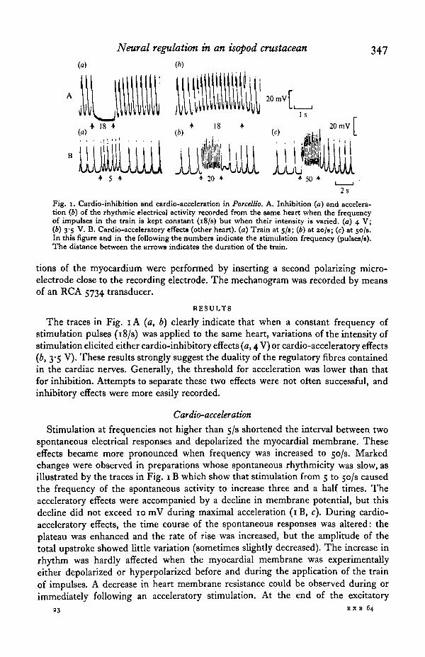
Fig. I. Cardio-inhibition and cardio-acceleration in PorceUio. A. Inhibition (a) and accelera-tion (6) of the rhythmic electrical activity recorded from the same heart when the frequencyof impulses in the train is kept constant (i8/s) but when their intensity is varied, (a) 4 V;(b) 3-5 V. B. Cardio-acceleratory effects (other heart), (a) Train at 5/s; (6) at 2o/s; (c) at 50/8.In this figure and in the following the numbers indicate the stimulation frequency (pulses/s).The distance between the arrows indicates the duration of the train.
tions of the myocardium were performed by inserting a second polarizing micro-electrode close to the recording electrode. The mechanogram was recorded by meansof an RCA 5734 transducer.
RESULTS
The traces in Fig. 1A (a, b) clearly indicate that when a constant frequency ofstimulation pulses (18/s) was applied to the same heart, variations of the intensity ofstimulation elicited either cardio-inhibitory effects (a, 4 V) or cardio-acceleratory effects{b, 3-5 V). These results strongly suggest the duality of the regulatory fibres containedin the cardiac nerves. Generally, the threshold for acceleration was lower than thatfor inhibition. Attempts to separate these two effects were not often successful, andinhibitory effects were more easily recorded.
Cardio-acceleration
Stimulation at frequencies not higher than 5/3 shortened the interval between twospontaneous electrical responses and depolarized the myocardial membrane. Theseeffects became more pronounced when frequency was increased to 50/s. Markedchanges were observed in preparations whose spontaneous rhythmicity was slow, asillustrated by the traces in Fig. 1B which show that stimulation from 5 to 50/8 causedthe frequency of the spontaneous activity to increase three and a hah0 times. Theacceleratory effects were accompanied by a decline in membrane potential, but thisdecline did not exceed 10 mV during maximal acceleration (iB, c). During cardio-acceleratory effects, the time course of the spontaneous responses was altered: theplateau was enhanced and the rate of rise was increased, but the amplitude of thetotal upstroke showed little variation (sometimes slightly decreased). The increase inrhythm was hardly affected when the myocardial membrane was experimentallyeither depolarized or hyperpolarized before and during the application of the trainof impulses. A decrease in heart membrane resistance could be observed during orimmediately following an acceleratory stimulation. At the end of the excitatory
23 EX B 64
348 J. C. DELALEU AND A. HOLLEY
(a) (b) (c)
. . 20-1A \mV I • I ^ ^ ^ ^ B |20mV
15 + | f PSH
Is 400 ms ^ 10 s
Fig. 2. The cardio-inhibitory effects. A. Changes in spontaneous activity when frequencyis progressively increased, (a) io/s; (6) 12/s; (c) 14/s. B and C. Effect of inhibitory trainson two hearts whose spontaneous electrical responses displayed humped plateaus. D. Oneexample of a post-stimulus hyperpolarization (PSH) recorded when applying a 58 duration50/s train (horizontal bar).
stimulation, marked after-effects were not generally observed; membrane potentialreturned to its resting value and the original rhythm rapidly recovered.
Cardio-inhibitionGeneral aspects of inhibition
According to the frequencies used, the inhibitory stimulation altered the rhythm,the time course of the spontaneous responses, and the resting potential. At lowfrequencies (less than 12/s) these parameters were not identically affected in all thehearts tested. In some instances the contour and the amplitude of the responseswere modified before there was any marked change in rate of beating as examplifiedby traces (a), (b), (c) on Fig. 2 A. It should be noted that myocardial membranehyperpolarized during the stimulation. In other instances (Fig. 2B), the changes inrhythm preceded any reduction of the initial upstroke, and electrical stimulation didnot cause any change in resting membrane potential. This trace and trace C in thesame figure show that the time course of the plateau could be affected by stimulatingat frequencies that did not noticeably reduce the amplitude of the upstroke. Thispoint is worth emphasizing as it suggests the heterogeneity of the components(upstroke and plateau) of the electrical response.
The cessation of the activity occurred at frequencies from 12 to 20/s dependingon the hearts studied but occurred at constant frequency for a given heart. At moderatefrequencies, up to 15/s, the after-effects were slight but nevertheless they were moreimportant than those observed during cardio-acceleration. For example, after theapplication of the stimulation the plateau was transiently enhanced and prolonged.
At frequencies higher than 25/s the post-stimulation effects became more pro-nounced. In particular, following the end of the stimulating pulse trains, a long-lastinghyperpolarization was observed (Fig. 2D). This post-stimulation hyperpolarization(PSH) varied in amplitude and duration with the frequency and duration of thestimulating volley. For instance, trains of pulses at 50/s, lasting 5 s, resulted in
Neural regulation in an isopod crustacean 349
A B
(") I V («)
(c) 10/s
Fig. 3. Synaptic potentials recorded in the myocardium. A. The inhibitory potentials (IJPs),depolarizing at normal resting potential, are reversed by the application of outward currents(a) and increased in sire when the myocardial membrane was experimentally hyperpolarized(6). Intensity of current: ± 7 x io~*A; train at 15/8. In this experiment £jjp= —58 mV. B.IJPs in presence of tetraethylammonium chloride (10 mM/1). (a) Effect of progressive incre-ments in frequency (from IO/B up to 30/s) on hyperpolariiing potentials. (6), (c) and (d)Summation of depolarizing potentials (frequency, respectively 5, 10 and 30/8).
a PSH of 10 mV and lasting 25 s (means of 40 experiments). This PSH exhibitedunusual properties, namely a paradoxical increase in membrane resistance. Detailedstudy showed that this PSH was not a simple development of the inhibitory processesbrought into play during stimulation, but was a phenomenon justifying separateinvestigation. Accordingly, the data reported in this paper mainly deal with theinhibitory processes induced by stimulation not exceeding 25/s, i.e. frequencies atwhich the special phenomenon represented by the PSH is thought to be absent orminimal.
Inhibitory functional potentials
Intracellular records from myocardial fibres during the cardiac nerve stimulationrevealed the presence of inhibitory junctional potentials (IJPs) which could be eitherdepolarizing or, more rarely, hyperpolarizing. In some hearts no detectable IJPswere recorded unless the membrane potential was varied experimentally under con-ditioning current injection. As illustrated in Fig. 3 A, this method was used to deter-mine the reversal potential of the IJPs. In most cases this reversal potential wasfound to be roughly equal to that of the resting potential or more positive by a fewmillivolts.
Whereas the amplitude of IJPs did not exceed 2 mV in normal saline, nervestimulation applied in presence of tetraethylammonium chloride (TEA, 10 mM/1)elicited synaptic potentials whose amplitude could reach 4-5 mV. Contrasting tothe situation in normal saline, TEA allowed us to record hyperpolarizing IJPs morefrequently than depolarizing IJPs. This can be partly attributed to the depolarizingeffect of TEA in the wood-louse's myocardium (Delaleu et al. 1972). In Fig. 3B,trace (a) shows the hyperpolarizing IJPs summated during a progressive increase in
23-2
350
(»)
J. C. DELALEU AND A. HOLLEY
(*) W (d)
+ 25 + 125
20 mV
+ 25 +
20 mV
10 s
Fig. 4. The cardio-inhibitory effects in presence of Cl~ -deficient solutions and/or GAB A.A. Shift of the level of the summated IJPs in presence of a Cl"-deficient (methylsulphate)saline (6) after 1 min, (c) after 2 min. The normal saline controls are in (a) and (d). Note the'paradoxical' acceleration in (c). Trains at 25/s. Resting membrane potential: —60 mV.B. Inhibition of the spontaneous activity and membrane potential changes, successively inpresence of a Cl~-deficient solution containing GABA (between the arrows) and in thenormal saline containing GABA (o-i mM/1).
stimulation frequency in TEA medium. At the beginning of the stimulation at 10/s,the synaptic potentials exhibited some degree of facilitation. Conversely, tracings(b), (c), (d), represent depolarizing IJPs recorded in another heart also perfused witha TEA solution. Under optical examination, the myocardium was seen to contractunder the summated IJPs. The ability of the inhibitory stimulations to clamp themembrane potential close to its resting value was further illustrated under particularconditions. As previously reported (cf. the above reference), in presence of TEA themyocardium membrane potential could be suddenly blocked close to electrical zero.It was observed that stimulation of the cardiac nerves at 50/s caused the myocardialmembrane to repolarize in an all-or-none manner.
Effect of varying the external chloride concentration and action of GABASince the heart of Porcellio was shown to have common features with crustacean
skeletal muscle, we suspected Cl~ ions to be implicated in the IJPs. To test thispossibility, the external Cl~ content ([Cl~]0) was varied. Gamma-aminobutyric acid(GABA), the transmitter that increases the chloride permeability at the crustaceaninhibitory neuromuscular junction, was added in the bathing medium. In Cl~-deficient solutions, the cardiac nerves were stimulated at 25/s, and we observed thelevel of the membrane potential, determined by the summated IJPs.
When the inhibitory stimulation was applied 60 s after the introduction of themodified saline (90 % of the external Cl~ was replaced by the large methylsulphateanion), the level of the summated IJPs shifted by 15-20 mV in the positive direction(Fig. 4 A, b). This was observed about fifty times in twelve hearts. If the same experi-ment was performed one minute later, the same inhibitory stimulation did not causethe heart to stop immediately, but a marked acceleration of the heartbeats could be
Neural regulation in an isopod crustacean 351
20raV
Fig. 5. Membrane resistance changes during inhibitory stimulation, (a) Control. (6), (c)and (d) Effect of inhibitory trains on the electrotonic potential resulting from the applicationof an intracellular current pulse ( — 8x io~* A) to the myocardium. The horizontal barsindicate the pulse duration.
observed (c). Higher frequencies were then required to obtain an arrest. When thenormal saline was replaced the previous stimulation again induced cessation of thespontaneous activity (d).
The traces in Fig. 4 B show that when a Cl~-deficient solution containing GABA(o*i mM/1) was introduced (first arrow) the electrical activity stopped and the myo-cardial membrane depolarized by 15 mV. If the normal saline containing GABAwas replaced (second arrow) the membrane potential returned close to its normalresting value. In one preparation, the introduction of the Cl--deficient solutioncontaining GABA resulted in a temporary acceleration of the heartbeat. Thus GABAmimicked the effects of the inhibitory stimulation in both normal and Cl~-deficientsalines.
Changes in membrane resistance during inhibitory stimulation
The inhibitory stimulation decreased the membrane resistance (RM) (15 experi-ments). The traces in Fig. 5 represent the variations of the hyperpolarizing effectsof a similar current pulse applied during stimulation of cardiac nerves at differentrates. The reduction in size of the electrotonic potentials indicates a decrease inmembrane resistance caused by stimulation. This decrease in i?M was a function ofthe stimulation frequency and, in this experiment, RM was halved by stimulation at30/s. It should be noted that inhibitory stimulations did not always result in such adiminution of i?M. Actually, in some experiments no variation in /?M was recordedalthough the stimulation was efficient, at least on the pacemaker neurones, as judgedby the cessation of the spontaneous activity. Furthermore, in a few instances a slightincrease in i?M even appeared. The lack of consistency of the results concerning thechanges in Ru, especially the paradoxical increase in RM, reflected the dual effectof the stimulation.
Inhibitory stimulation and contraction
In so far as mechanical activity is controlled by the myocardial membrane polariza-tion (Holley & Delaleu, 1972), the inhibitory stimulation that modifies the electricalresponses and the resting potential can be expected to act on the contraction and theheart tonus. This could be verified: stimulation of the cardiac nerves at rates thatdid not cause an arrest of the heart, but simply reduced the amplitude of the
352 J. C. DELALEU AND A. HOLLEY
(a) (6)
14 •
Fig. 6. Cardio-inhibitory effects and contraction. Effect of inhibitory stimulation on electricalactivity (upper traces) and on mechanogram (lower traces). Frequency of stimulation (a),9/s; (6), 14/s-
spontaneous electrical responses, markedly affected the mechanogram. The contractionswere reduced as soon as the plateau of the responses was altered even though theinitial upstroke was not yet affected. The diastolic tonus of the heart was also modifiedby the stimulation as illustrated in Fig. 6. In this case the stimulation that hyper-polarized the heart membrane induced a marked reduction of the diastolic tonus. Itcan be seen that a hyperpolarization of less than 5 mV (b) during stimulation at 14/scaused a decrease in mechanical tension equal to the amplitude of the normal con-tractions. This confirms the presence of a large diastolic tonus in normal conditions.
DISCUSSION
Stimulation of the cardiac nerves led to either acceleration or, more frequently,inhibition of the heartbeat, which agrees with previous mechanogram examination(Holley & Delaleu, 1967; Holley, 1967). It is probable that each cardiac nerve con-tains two categories of fibres. Selective stimulation of only one type of fibre wasattempted by finely adjusting the stimulation parameters, together with changes inplacement of the stimulating electrodes, and apparently was sometimes achieved,for example when clear acceleratory effects were recorded. Under these conditions aweak increase in the intensity of stimulation (but not in frequency) frequently causeda complete inversion of the effects, suggesting that the fibres were scarce, and thatthe inversion was due to recruitment of an inhibitory fibre. Histological studiesrevealed the scarceness of the fibres.
Cardio-acceleration
The increase in the rhythm and rate of rise of the spontaneous electrical responsesmight at first be attributed to an acceleration of the periodical activity of the cardiacganglion cells along with an increase in the spike discharge frequency within eachburst. Well-known studies on the crustacean heart support this hypothesis (seereview by Maynard, i960).
The question arises as to whether the cardio-acceleratory fibres also acted on themyocardial membrane. In Porcellio, acceleratory effects decreased the membrane
Neural regulation in an isopod crustacean 353
potential and caused a sustained depolarization that looked like that observed byWatanabe et al. (1969) in pacemaker neurones of Squilla during stimulation of the/? nerves. However, in our opinion, this change in membrane polarization of thewood-louse heart does not represent proof for a direct peripheral effect, for thisfinding may be explained as being the consequence of an acceleration causing a'tetanization', hence preventing full repolarization before the appearance of a newdepolarizing response. Several attempts to attribute a change in membrane resistanceto the functioning of ' acceleratory' junctions on the myocardium were unsuccessful.Changes in membrane resistance were explainable as a consequence of the increasein rhythm of the electrical responses, which partly consist of excitatory junctionalpotentials. The direct effect of the cardio-acceleratory stimulation on the myocardiumcould have been demonstrated by the presence of junctional potentials, but we failedto identify such potentials.
Cardio-inhibition
In addition to the action of inhibitory stimulation on the cardiac ganglion, thereis convincing evidence for direct inhibitory action on the myocardial membrane, asindicated by the presence of IJPs and the changes in membrane resistance duringstimulation. Therefore the physiology of the heart muscle of Porcellio presentsfeatures in common with the crustacean skeletal muscle in which this mode of post-synaptic inhibition has been frequently observed (Boistel & Fatt, 1968; Atwood,1968). On the other hand, the heart of this isopod differs from that of the lobster inwhich Hallet (1971) failed to record synaptic potentials when stimulating the cardio-inhibitory nerves.
The value of the reversal potential for the IJPs, close to that of the membranepotential, supports the comparison of the heart of Porcellio with the skeletal muscleof Crustacea studied by Fatt & Katz (1953), and Hoyle & Wiersma (1958). The shiftin the level of the summated IJPs in Cl~-deficient solutions strongly suggests thatduring inhibition there was an increase in chloride conductance, the level of mem-brane polarization during the stimulation presumably tending to follow the shift inthe equilibrium potential for Cl" (2?a-). I*1 other words the effect of cardio-inhibitoryimpulses would be to clamp the membrane potential close to E^-.
In some preparations the efficiency of inhibitory stimulation decreased in methyl-sulphate solutions. In addition, an acceleration of the spontaneous responses wasnoted under inhibitory stimulation. This paradoxical phenomenon may be attributedto an inversion, in low [Cl~]0, of the effects of inhibitory transmission at the level ofthe pacemaker cells of the cardiac ganglion. These results argue that the chlorideconductance was normally involved in 'central' ganglion inhibition as in peripheralinhibition. The observed acceleration probably resulted from the depolarizing effectof the stimulation that increased chloride permeability when Ea- had shifted in thepositive direction (decrease in [Cl~]0). There have been similar findings with molluscanneurones during iontophoretic application of acetylcholine in chloride-deficientsolutions (Kerkut & Thomas, 1963).
Several hypotheses may be proposed to account for the increase in IJP amplitudein TEA solutions. Since this substance increases the membrane resistance (Delaleuet al. 1972), it is conceivable that this effect could induce larger junctional potentials.Another tentative explanation would be that TEA increased the amount of
354 J- C. DELALEU AND A. HOLLEY
transmitter released through a lengthening of the presynaptic action potentials 3*nobserved by Kusano et al. (1967).
GABA mimicked the effects of the stimulation of the inhibitory fibres since itabolished the spontaneous activity; without doubt it acted on the cardiac ganglion.GABA also acted on the myocardial membrane, as indicated by the shift in membranepotential recorded in GABA-methylsulphate solution. This change in membranepotential was somewhat comparable with that observed when the inhibitory fibreswere stimulated in methylsulphate saline. This is consistent with an increased Cl~conductance caused by GABA. The decrease in i?M in the presence of GABA reportedin a previous paper (Holley & Delaleu, 1972) strengthens this interpretation.
The acceleration sometimes observed in GABA-methylsulphate solutions could beexplained by assuming that the Cl~ conductance mediated the action of GABA atthe level of the pacemaker cells. The action of the inhibitory fibres at this level wasshown to depend on the same mediation. While awaiting further analysis of theinhibitory process in Porcellio, it should be emphasized that picrotoxin, known as anantagonist of GABA, antagonized the inhibitory effect of the cardiac nerve stimulation.A close relationship is indicated between the properties of GABA and those of thenatural inhibitory transmitter; both these substances could act on the same receptorof the postsynaptic membrane or on receptors coupled with a common ionophore.
The changes in Ru recorded during the inhibitory stimulation suggest an actionof the inhibitory fibres on the myocardium, and confirm the hypothesis of peripheralinhibition put forward by Holley (1968). Our results provide further evidence thatthe heart of the wood-louse shares some physiological properties with the skeletalmuscle of some arthropods (see reviews by Atwood, 1968 and Pearson, 1973). Theincrease in RM that was sometimes recorded in Porcellio, but not in the skeletal muscle,may be explained by stimulation of the cardio-regulatory nerves having two effects,A and B, with opposite action on RM. The A effect would be the synaptic processesleading to an increase in chloride conductance, as analysed in the present paper.The B effect appears essentially as an increase in membrane resistance resulting ina post-stimulation hyperpolarization (PSH) at rather high rates of stimulation.This hypothesis will be presented and discussed in a subsequent paper. Since theA and B effects lead to opposite changes in membrane resistance it is conceivablethat their combination in various proportions determines various values of the globalmembrane resistance. It must be further stated that picrotoxin depressed the increasein chloride conductance (A effect), thus revealing the decrease in conductancecharacterizing the B effect, at the very beginning of the cardiac nerve stimulation.
Whereas the myocardium of the wood-louse shares some properties of the skeletalmuscle of decapods it seems to differ from their heart muscle, since peripheralinhibition could not be demonstrated in the lobster (Hallet, 1971). The significanceof these important differences in cardiac regulation mechanisms in the two orderscan be understood if we refer to the difference in nature of the processes regulatingcontraction, i.e. the spontaneous electrical activity. The normal spontaneous responseof the heart of Homarus has been interpreted as a temporal summation of JPs resultingfrom the synaptic action of the cardiac ganglion on passive membrane conductances(see review by Anderson, 1973). On the other hand, the responses in Porcellio showappreciable differences, since the plateau was thought to bring into play active
Neural regulation in an isopod crustacean 355
membrane conductances. If the contractile activity depends directly on the amplitudeand duration of the depolarization in both species (Hallet, 1971; Holley & Delaleu,1972; Delaleu et al. 1972), the regulation of the contraction may be specificallyachieved through a control of the plateau. In the lobster, this control can be performedby the action of the extrinsic regulatory fibres on the unique cardiac ganglion, sincethe postsynaptic electrical responses closely depend on the characteristics (numberof spikes, frequency, duration) of the ganglion bursts. In the wood-louse the problemis different: the plateau appears to depend strongly on the myocardial membraneproperties and it is then conceivable that the plateau and the related contraction areregulated at the peripheral level. As a matter of fact it could be observed in PorcelUothat inhibitory stimulation had a specific depressive action on the sustained depolari-zation and consequently on the contraction at low frequencies that were not out ofa possible physiological range.
In conclusion, inhibitory regulation operates at two different levels in the heartof PorceUio: the frequency of beating is regulated by the effect of inhibitory impulseson the cardiac ganglion, while the tonus and the amplitude of the contraction arechiefly modulated at a peripheral level by impulses reaching the myocardial membrane.
This work constitutes partial fulfilments of the requirements for a Doctorat esSciences, no. CNRS AO 9785, Lyon.
REFERENCES
ANDERSON, M. (1973). Neuromuscular systems in neurogenic Arthropod hearts. Amer. Zool. 13, 291-8.ATWOOD, H. L. (1968). Peripheral inhibition in Crustacean muscle. Experientia 24, 753-63.BOISTEL, J. & FATT, P. (1938). Membrane permeability change during inhibitory transmitter action
in crustacean muscle. J. Pltytiol., Land. 144, 176-91.BRENNER, H. R. (1972). Evidence for peripheral inhibition in an arachnid muscle. J. Comp. Phyriol.
80, 227-31.DELALEU, J. C. (1970). Le systeme nerveux intrapdricardique et ses relations avec le systeme nerveux
central chez trois Oniscoldcs: PorceUio dilatatus (Brandt), Helleria brcvicornis (Ebner) et Ligiaoceamca (L.). Bull. Soc. Zool. France 95, 2, 201-10.
DELALEU, J. C. (1974). Structure et physiologie d'un coeur a automatisme neurogene (Crustace' Isopode):myocarde, relations neuromusculaires, facteurs de la regulation. These de Sciences, A.O. 9875, Lyon.
DELALEU, J. C , BLONDBAU, A. & HOLLEY, A. (1972). Electrophysiology of the heart of an isopodcrustacean: PorcelUo dilatatus. II. Effect of ions and membrane permeability inhibitors. J. exp.Biol. 57, 609-31.
DELALEU, J. C. & HOLLEY, A. (1973). Modalit^s et mecanismes de l'inhibition pe'ripherique dans lecoeur d'un Crustace' Isopode. J. Pkytioi, Paris 67,184 A
DUDEL, J. & KUFFLER, S. W. (1961). Presynaptic inhibition at the crayfish neuromuscular junction.J. Physiol., Lond. 155, 543-62.
FATT, P. & KATZ, B. (1953). The effect of inhibitory nerve impulses on a crustacean muscle fibre.J. Physiol., Lond. xai, 374-88.
FLOREY, E. (i960). Studies on the nervous regulation of the heart beat in Decapod Crustacea. J. gen.Pltysiol. 43, 1061-81.
HAGIWARA, S. (1961). Nervous activities of the heart in Crustacea. Ergebn. Biol. 24, 287-311.HALLET, M. (1971). Lobster heart: electrophysiology of single cells including effect of the regulator
nerves. Comp. Biocliem. Physiol. 39 A, 643-8.HOLLEY, A. (1967). Inhibition cardiaque chez un Crustace' Isopode: e'tude electrophysiologique.
C. r. Soc. Biol., Paris 161, 1786-92.HOLLEY, A. (1968). Physiologie du coeur d'un Crustace' Isopode {PorcelUo dilatatus): activity me'canique,
activity electrique, regulation nerveuse. These de Sciences, A.O. 2379, Poitiers.HOLLEY, A. & DELALEU, J. C. (1967). Etude de la regulation nerveuse du cceur d'un Crustac6 Isopode
(PorcelUo dilatatus, Brandt). C. r. Soc. Biol., Paris 161, 891-5.HOLLEY, A. & DELALEU, J. C. (1972). Electrophysiology of the heart of an isopod crustacean: PorcelUo
dilatatus. I. General properties. J. exp. Biol. $7, 589-608.
356 J. C. DELALEU AND A. HOLLEY
HOYLB, G. & WIERSMA, C. A. G. (1958). Inhibition at neuromuscular junction in Crustacea. J. Physiol.-Land. 143, 426-40.
KERKUT, G. A. & THOMAS, R. C. (1963). Acetylcholine and the spontaneous inhibitory post-synapticpotentials in the snail neurone. Comp. Biochem. Phytiol. 8, 39-45.
KUSANO, K., LrvENOOOD, D. R. & WERMAN, R. (1967). Tetraethylammonium ions: effect of pre-synaptic injection on synaptic transmission. Science 155, 1257-9.
MAYNARD, D. M. (1953). Activity in a crustacean ganglion. I. Cardioinhibition and acceleration inPanuUnu argus. Biol. Bull. mar. biol. Lab., Woods Hole 164, 156-70.
MAYNARD, D. M. (i960). Circulation and heart function. In Tlie Physiology of Crustacea, vol. 1(ed. T.Waterman), pp. 1, 151-226. New York and London: Academic Press.
MAYNARD, D. M. (1961). Cardiac inhibition in decapod Crustacea. In Nervous Inhibition (ed. E. Florey),pp. 144-78. Oxford, London, New York, Paris: Pergamon Press.
PARNAS, I., ABBOTT, B. C , SHAPIRO, B. & LANG, F. (1968). Neuromuscular system of Limulus legcloser muscle. Comp. Biochem. Physiol. a6, 467-78.
PEARSON, K. G. (1973). Function of peripheral inhibitory axons in insects. Atner. Zool. 13, 321-30.SMITH, R. I. (1947). The action of electrical stimulation and of certain drugs on cardiac nerves of
the crab Cancer irroratus. Biol. Bull. mar. biol. Lab., Woods Hole 93, 72-88.TERZUOLO, C. A. & BULLOCK, T. H. (1958). Acceleration and inhibition in crustacean ganglion cells.
Archs ital. Biol. 96, 117-34.USHERWOOD, P. N. R. & GRUNDFEST, H. (1965). Peripheral inhibition in skeletal muscle of insects.
J. Neurophysiol. 38, 497-518.WATANABE, A., OBARA, S. & AKIYAMA, T. (1968). Inhibitory synapses on pacemaker neurons in the
heart ganglion of a Stomatopod. Squilla oratorio. J. gen. Physiol. 5a, 908-24.WATANABE, A., OBARA, S. & AKIYAMA, T. (1969). Acceleratory synapses on pacemaker neurons in the
heart ganglion of a Stomatopod, Squilla oratorio. J. gen. Physiol. 54, 212-31.WIERSMA, C. A. G. & NOVITSKI, E. (1942). The mechanism of the nervous regulation of the crayfish