Ischemic optic neuropathy q Sohan Singh Hayreh * Department of Ophthalmology and Visual Sciences, College of Medicine, University of Iowa, 200 Hawkins Drive, Iowa City, IA 52242-1091, USA Keywords: Anterior ischemic optic neuropathy Giant cell arteritis Ischemia Ischemic optic neuropathy Optic nerve Optic nerve head Posterior ischemic optic neuropathy abstract Ischemic optic neuropathy is one of the major causes of blindness or seriously impaired vision, yet there is disagreement as to its pathogenesis, clinical features and especially its management. This is because ischemic optic neuropathy is not one disease but a spectrum of several different types, each with its own etiology, pathogenesis, clinical features and management. They cannot be lumped together. Ischemic optic neuropathy is primarily of two types: anterior (AION) and posterior (PION), involving the optic nerve head (ONH) and the rest of the optic nerve respectively. Furthermore, both AION and PION have different subtypes. AION comprises arteritic (A-AION – due to giant cell arteritis) and, non-arteritic (NA-AION – due to causes other than giant cell arteritis); NA-AION can be further classified into classical NA-AION and incipient NA-AION. PION consists of arteritic (A-PION – due to giant cell arteritis), non- arteritic (NA-PION – due to causes other than giant cell arteritis), and surgical (a complication of several systemic surgical procedures). Thus, ischemic optic neuropathy consists of six distinct types of clinical entities. NA-AION is by far the most common type and one of the most prevalent and visually crippling diseases in the middle-aged and elderly. A-AION, though less common, is an ocular emergency and requires early diagnosis and immediate treatment with systemic high dose corticosteroids to prevent further visual loss, which is entirely preventable. Controversy exists regarding the pathogenesis, clinical features and especially management of the various types of ischemic optic neuropathy because there are multiple misconceptions about its many fundamental aspects. Recently emerging information on the various factors that influence the optic nerve circulation, and also the various systemic and local risk factors which play important roles in the development of various types of ischemic optic neuropathy have given us a better understanding of their pathogeneses, clinical features and management. This knowledge should help us not only to manage them better but also to reduce their incidence. For example, clinically, the evidence that about 40% of NA- AION eyes experience spontaneous improvement in visual acuity and that systemic steroid therapy during early stages in both NA-AION and NA-PION has a significant beneficial effect for visual outcome are encouraging developments. This review discusses the current concepts on various issues related to various types of ischemic optic neuropathy. Ó 2008 Elsevier Ltd. All rights reserved. Contents 1. Introduction ........................................................................................................................35 2. Terminology .........................................................................................................................35 2.1. Anterior ischemic optic neuropathy (AION) ...................................................................................... 35 2.2. Posterior ischemic optic neuropathy (PION) ..................................................................................... 35 3. Blood supply of the optic nerve ...................................................................................................... 36 3.1. Anterior part of the optic nerve (ONH) .......................................................................................... 36 3.1.1. Arterial supply (A) ..................................................................................................... 36 3.1.2. Venous drainage (B) ................................................................................................... 37 3.2. Posterior part of the optic nerve (Figs. ) ......................................................................................... 37 3.2.1. Arterial supply ........................................................................................................ 37 3.2.2. Venous drainage (B) . . . . . . . . . . . . . . . . . . . . . . . . . . . . . . . . . . . . . . . . . . . . . . . . . . . . .. . . . . . . . . . . . . . . . . . . . . . . . . . . . . . . . . . . . . . . . . . . . . . 37 q Supported by grants EY-1151 and 1576 from the National Institutes of Health, and in part by unrestricted grant from Research to Prevent Blindness, Inc., New York. * Corresponding author. Tel.: þ1 319 356 2947; fax: þ1 319 353 7996. E-mail address: [email protected]Contents lists available at ScienceDirect Progress in Retinal and Eye Research journal homepage: www.elsevier.com/locate/prer 1350-9462/$ – see front matter Ó 2008 Elsevier Ltd. All rights reserved. doi:10.1016/j.preteyeres.2008.11.002 Progress in Retinal and Eye Research 28 (2009) 34–62
Transcript
lable at ScienceDirect
Progress in Retinal and Eye Research 28 (2009) 34–62
Contents lists avai
Progress in Retinal and Eye Research
journal homepage: www.elsevier .com/locate/prer
Ischemic optic neuropathyq
Sohan Singh Hayreh*
Department of Ophthalmology and Visual Sciences, College of Medicine, University of Iowa, 200 Hawkins Drive, Iowa City, IA 52242-1091, USA
1350-9462/$ – see front matter � 2008 Elsevier Ltd.doi:10.1016/j.preteyeres.2008.11.002
a b s t r a c t
Ischemic optic neuropathy is one of the major causes of blindness or seriously impaired vision, yet thereis disagreement as to its pathogenesis, clinical features and especially its management. This is becauseischemic optic neuropathy is not one disease but a spectrum of several different types, each with its ownetiology, pathogenesis, clinical features and management. They cannot be lumped together. Ischemicoptic neuropathy is primarily of two types: anterior (AION) and posterior (PION), involving the opticnerve head (ONH) and the rest of the optic nerve respectively. Furthermore, both AION and PION havedifferent subtypes. AION comprises arteritic (A-AION – due to giant cell arteritis) and, non-arteritic(NA-AION – due to causes other than giant cell arteritis); NA-AION can be further classified into classicalNA-AION and incipient NA-AION. PION consists of arteritic (A-PION – due to giant cell arteritis), non-arteritic (NA-PION – due to causes other than giant cell arteritis), and surgical (a complication of severalsystemic surgical procedures). Thus, ischemic optic neuropathy consists of six distinct types of clinicalentities. NA-AION is by far the most common type and one of the most prevalent and visually cripplingdiseases in the middle-aged and elderly. A-AION, though less common, is an ocular emergency andrequires early diagnosis and immediate treatment with systemic high dose corticosteroids to preventfurther visual loss, which is entirely preventable.Controversy exists regarding the pathogenesis, clinical features and especially management of thevarious types of ischemic optic neuropathy because there are multiple misconceptions about its manyfundamental aspects. Recently emerging information on the various factors that influence the optic nervecirculation, and also the various systemic and local risk factors which play important roles in thedevelopment of various types of ischemic optic neuropathy have given us a better understanding of theirpathogeneses, clinical features and management. This knowledge should help us not only to managethem better but also to reduce their incidence. For example, clinically, the evidence that about 40% of NA-AION eyes experience spontaneous improvement in visual acuity and that systemic steroid therapyduring early stages in both NA-AION and NA-PION has a significant beneficial effect for visual outcomeare encouraging developments. This review discusses the current concepts on various issues related tovarious types of ischemic optic neuropathy.
Ischemic optic neuropathy constitutes one of the major causesof blindness or seriously impaired vision among the middle-agedand elderly population, although no age is immune. Its pathogen-esis, clinical features and management have been subjects of a gooddeal of controversy and confusion. I have conducted basic, experi-mental and clinical research on the blood supply of the optic nerveand on various aspects of ischemic optic neuropathy since 1955.This review is based on the cumulative information drawn fromthose studies, as well as from a PubMed search of the literature onthe subject.
2. Terminology
Before 1974, this condition was described under differenteponyms, including optic neuritis, arteriosclerotic papillitis, senilepapillopathy, papillary apoplexy, vascular pseudo-papillitis, optico-malacia, ischemic neuritis of papilla, ischemic papillopathy andischemic optic neuritis and so on (Hayreh, 1975a). Since studieshave shown that it is an acute ischemic disorder of the optic nerve,the proper terminology for this disease is ‘‘ischemic optic neurop-athy’’. Based on the blood supply pattern of the optic nerve, and myclinical and experimental studies, in 1974 I defined ischemic opticneuropathy into the following two distinct clinical entities.
2.1. Anterior ischemic optic neuropathy (AION)
This is due to ischemia of the anterior part of the optic nerve,which is supplied by the posterior ciliary artery (PCA) circulation(Hayreh, 1969, 1995, 2001b) (Fig. 1A). In view of that I named it‘‘anterior ischemic optic neuropathy’’ (Hayreh, 1974b).
2.2. Posterior ischemic optic neuropathy (PION)
I first described this clinical entity in 1981 (Hayreh, 1981b); it isdue to ischemia of a segment of the posterior part of the opticnerve, which is supplied by multiple sources but not the PCA (Figs.1B and 2).
Of the two types, AION is far more common than PION. Patho-genetically and clinically AION and PION are quite distinct clinicalentities; thus, the common practice of calling AION simply‘‘ischemic optic neuropathy’’ is incorrect, and ‘‘ischemic opticneuritis’’ is worse still, since there is no evidence of inflammation.
From the basic scientific facts about the disease process, one canlogically deduce its pathogenesis, clinical features and manage-ment. The basic sciences are the foundation of Medicine. Tocomprehend the scientific basis of the pathogeneses, various clin-ical features and management of AION and PION, the first essentialis to have a good understanding of the various basic scientific issuesinvolved. Since this is an ischemic disorder of the optic nerve, the
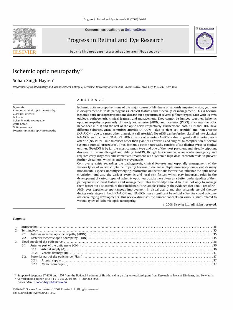
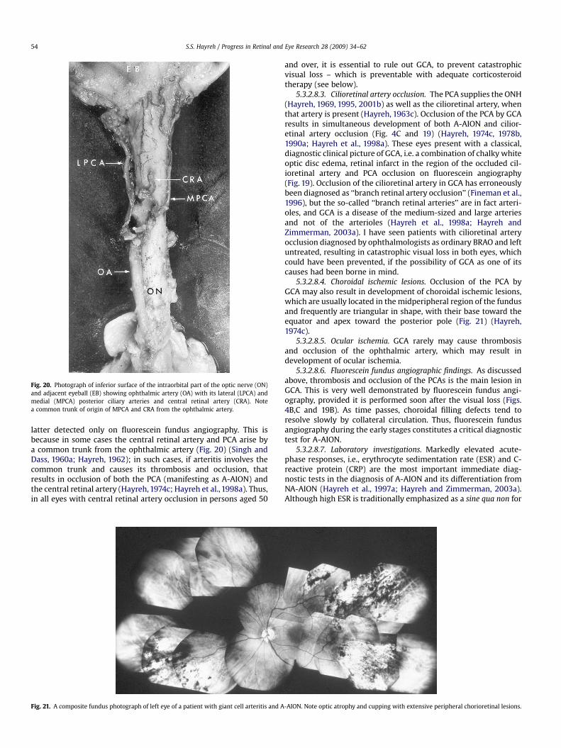
Fig. 1. Schematic representation of blood supply of: (A) the optic nerve head and (B) the optic nerve (B modified from Hayreh, S.S. (1974) Trans. Am. Acad. Ophthalmol. Otolaryngol.78, OP240–OP254. A reproduced from Hayreh, 1978a). Abbreviations: A ¼ arachnoid; Ant. Sup. Hyp. Art. ¼ anterior superior hypophyseal artery; C ¼ choroid; CAR and CRA ¼ centralretinal artery; Col. Br. ¼ collateral branches; CRV ¼ central retinal vein; CZ ¼ circle of Zinn and Haller; D ¼ dura; ICA ¼ internal carotid artery; LC ¼ lamina cribrosa; LPCA ¼ lateralposterior ciliary artery; Med. Mus. ¼medial muscular artery; MPCA ¼medial posterior ciliary artery; NFL ¼ surface nerve fiber layer of the disc; OA ¼ ophthalmic artery; OD ¼ opticdisc; ON ¼ optic nerve; P ¼ pia; PCA ¼ posterior ciliary artery; PR and PLR ¼ prelaminar region; R ¼ retina; RA ¼ retinal arteriole; Rec. Br. CZ ¼ recurrent pial branches fromperipapillary choroid/CZ; S ¼ sclera; SAS ¼ subarachnoid space.
S.S. Hayreh / Progress in Retinal and Eye Research 28 (2009) 34–6236
first basic issue is the blood supply of the optic nerve and the role ofvarious factors in the production of acute optic nerve ischemia.
3. Blood supply of the optic nerve
Based on its blood supply pattern, the optic nerve can be dividedinto two distinct regions: (a) anterior (also called optic nerve head –ONH); and (b) posterior.
3.1. Anterior part of the optic nerve (ONH)
Blood supply of this part is described in detail elsewhere(Hayreh, 1969, 1995, 2001b). Following is a brief summary.
3.1.1. Arterial supply (Fig. 1A)Anatomically ONH consists of, from front to back: (i) surface
nerve fiber layer; (ii) prelaminar region; (iii) lamina cribrosa region;and (iv) retrolaminar region (Hayreh and Vrabec, 1966; Hayreh,1972) (Fig. 1A).
3.1.1.1. The surface nerve fiber layer. This is mostly supplied by theretinal arterioles. In some cases, its temporal region may instead besupplied by the PCA circulation from the deeper prelaminar region.The cilioretinal artery (rarely a tiny cilio-papillary artery), whenpresent, usually supplies the corresponding sector of the surface layer.
3.1.1.2. The prelaminar region. This is situated between the surfacenerve fiber layer and the lamina cribrosa. It is supplied by finecentripetal branches from the peripapillary choroid. The centralretinal artery gives no branches in this region. The blood supply inthis region is sectoral in nature, similar to the overall segmentaldistribution of the PCA circulation (Hayreh, 1975b, 2004a).
3.1.1.3. The lamina cribrosa region. This is supplied by centripetalbranches from the short PCAs either directly or by the so-calledarterial circle of Zinn and Haller, when that is present. The centralretinal artery gives off no branches in this region. In the laminacribrosa, the blood vessels, 10–20 m in diameter, lie in the fibroussepta and form a dense capillary plexus that makes this part ofthe ONH a highly vascular structure (Levitzky and Henkind,1969).
3.1.1.4. The retrolaminar region. This lies immediately behind thelamina cribrosa. This part of the ONH may have a dual source ofblood supply (Figs. 1A,B and 2).
3.1.1.4.1. The peripheral centripetal vascular system. This isalways present and forms the major source of supply here. It isformed by recurrent pial branches arising from the peripapillarychoroid and circle of Zinn and Haller (when present) or the shortPCAs instead. In addition, pial branches from the central retinalartery and other orbital arteries also supply this part (Hayreh, 1958;
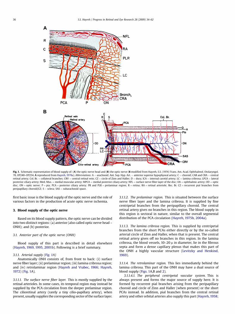
Fig. 2. Diagrammatic representation of blood supply of the various parts of the opticnerve, and location of the circle of Haller and Zinn (CZ), as seen from above. Forabbreviations see Fig. 1. (Reproduced from Hayreh, 1963b).
S.S. Hayreh / Progress in Retinal and Eye Research 28 (2009) 34–62 37
Singh and Dass, 1960b). The pial vessels give off centripetalbranches, running in the septa of the nerve.
3.1.1.4.2. The axial centrifugal vascular system. This is notpresent in all eyes. When present, it is formed by branches arisingfrom the intraneural part of the central retinal artery (Hayreh, 1958;Singh and Dass, 1960b)
Thus, the main source of blood supply to the ONH is the PCAcirculation via the peripapillary choroid and the short PCAs (or thecircle of Zinn and Haller). The blood supply in the ONH has a sec-toral distribution, which helps to explain the segmental visual lossin ONH ischemic disorders.
3.1.2. Venous drainage (Fig. 1B)This is essentially via the central retinal vein except that the
prelaminar region also drains into the peripapillary choroidal veins(Hayreh, 1969). This latter communication assumes importance indeveloping retinociliary collaterals (misnamed optociliary shunts)in the event of central retinal vein occlusion behind the laminacribrosa.
3.2. Posterior part of the optic nerve (Figs. 1B and 2)
For purposes of description of the blood supply, the posteriorpart of the optic nerve can be divided into intraorbital, intra-canalicular and intracranial parts (Hayreh, 1963a).
3.2.1. Arterial supply3.2.1.1. Intraorbital part. This is further subdivided by point of entryof the central retinal artery in the optic nerve into: (a) anterior; and(b) posterior segments (Figs. 1B and 2) (Singh and Dass, 1960b;Hayreh, 1963a,b).
3.2.1.1.1. Anterior segment. This is between the ONH and the siteof entry of the central retinal artery into the nerve (Fig. 1B). It hastwo vascular systems for its supply.
3.2.1.1.1.1. Peripheral centripetal vascular system. This is presentin all cases and is formed by pial vascular plexus, supplied bymultiple pial branches originating from the peripapillary choroid,circle of Zinn and Haller, central retinal artery, ophthalmic arteryand other orbital arteries (Figs. 1B and 2).
3.2.1.1.1.2. Axial centrifugal vascular system. This is present in75% of the nerves and supplied by one to eight intraneural branchesof the central retinal artery (Fig. 1B).
3.2.1.1.2. Posterior segment. This is primarily supplied by theperipheral centripetal vascular system formed by the pial vascularplexus, supplied by multiple small collateral arteries usuallyarising directly from the ophthalmic artery and less often fromother orbital arteries (Figs. 1B and 2) (Hayreh, 1963a). In about10% of optic nerves there may be an axial centrifugal vascularsystem extending backward for a variable distance, formed byintraneural branches of the central retinal artery (Fig. 3) (Hayreh,1958).
3.2.1.2. Intracanalicular part. This has only the peripheral centrip-etal system, supplied almost entirely by fine collateral branchesfrom the ophthalmic artery lying inferior to the optic nerve (Fig. 2)(Hayreh, 1963a,b).
3.2.1.3. Intracranial part. This once again has only a pial vascularplexus, supplied by a variable number of fine branches coming fromvarious surrounding arteries, including the anterior superiorhypophyseal, anterior cerebral, anterior communicating andophthalmic arteries (Fig. 2) (Hayreh, 1963a,b).
3.2.2. Venous drainage (Fig. 1B)This is by the central retinal vein and also many other small
venous tributaries draining into the various orbital veins.
3.3. Interindividual variations in the blood supply of the optic nerve
There exists a general impression that the pattern of bloodsupply of the optic nerve is almost identical in all eyes, and that allischemic lesions are explainable from one standard vascularpattern. This is a fundamental error, which is responsible for muchconfusion. This is particularly so for the blood supply of the ONH,which shows marked interindividual variations, as discussed atlength elsewhere (Hayreh, 1985, 1995, 2001b). Briefly, the followingfactors are responsible for the interindividual variations in theblood supply of the ONH.
3.3.1. Variations in the anatomical patternThis has wide variations, so much so that in Hayreh’s (1958,
1962; Singh and Dass, 1960a,b) anatomical studies no two speci-mens had identical patterns, not even the two eyes of the sameindividual. So the anatomical vascular pattern of the optic nerve isfar from standard in all humans.
3.3.2. Variations in the pattern of PCA circulationThese are produced by several factors.
3.3.2.1. Variations in number of PCAs supplying an eye. There maybe one to five PCAs, usually two to three (Hayreh, 1962, 1995,2001b).
Fig. 3. Diagram (based on camera lucida drawings) showing one of the intraneural branches of the central retinal artery running backward in the axial part of the optic nerveposterior to the central retinal artery. From one of the specimens in my anatomical study on the central retinal artery in humans. For abbreviations see Fig. 1. (Reproduced fromHayreh, 1958).
S.S. Hayreh / Progress in Retinal and Eye Research 28 (2009) 34–6238
3.3.2.2. Variations in the area supplied by various PCAs. In humans,this shows marked interindividual variation (Hayreh, 1990b,2004a). PCAs and their branches have a segmental distributionin vivo, in the choroid as well as in the ONH (Hayreh, 1975b,1985, 1990b, 2004a). Therefore, with the interindividual vari-ation in number and distribution by the various PCAs, we canget an extremely variable pattern of distribution by the PCAs
Fig. 4. Fluorescein fundus angiograms of three eyes showing areas of supply by the occludedfor giant cell arteritis), showing normal filling of the area supplied by the lateral PCA (includ(including the nasal half of optic disc). (Reproduced from Hayreh, 1985). (B) Right eye withtemporal ¼ of the optic disc) but no filling of the area supplied by the medial PCA (includingassociated with cilioretinal artery occlusion, showing normal filling of the area supplied by tPCA or of the cilioretinal artery (arrow). (Reproduced from Hayreh, 1978b).
in both the choroid and the ONH – a key fact to be borne inmind in any consideration of ischemic disorders of the ONH,since PCAs are its main source of blood supply. For example,Fig. 4 shows three such variations in the supply by the medialand lateral PCAs in the choroid and the ONH; the part of theONH involved depends upon the area supplied by the occludedPCA.
PCA and the patent PCA. (A) Right eye with NA-AION (negative temporal artery biopsying the temporal half of optic disc) but no filling of the area supplied by the medial PCAA-AION, showing normal filling of the area supplied by the lateral PCA (including thethe nasal 3⁄4 of the disc). (Reproduced from Hayreh, 1978b). (C) Left eye with A-AION
he lateral PCA, but no filling of the choroid and entire optic disc supplied by the medial
S.S. Hayreh / Progress in Retinal and Eye Research 28 (2009) 34–62 39
3.3.2.3. Variation in location of watershed zones between the PCAs inrelation to the ONH. This again plays an important role in ischemicdisorders of the ONH, because the location of the watershed zonedetermines the vulnerability of the corresponding part of the ONHto ischemia (Hayreh, 1985, 1990b). In the event of a fall of perfusionpressure in the PCAs or their branches, the part of the ONH locatedin the watershed zone becomes vulnerable to ischemia. Forexample, Fig. 5 shows four variations in the location of watershedzone between the medial and lateral PCAs; the part of the ONHinvolved depends upon the relationship of ONH to the location ofthe watershed zone.
3.3.2.4. Variation in mean blood pressure in various PCAs as well asshort PCAs. This may occur in health as well as in disease (Hayreh,1985). In the event of a fall of perfusion pressure, the vascular bedsupplied by one artery may be affected earlier and more than the others.
4. Factors influencing the blood flow in the optic nerve head
These factors are critical to understanding the pathogenesis ofischemic disorders of the ONH. This subject is discussed at lengthelsewhere (Hayreh, 2001c). Following is a brief summary of that.
4.1. Blood flow formula
The blood flow in the ONH is calculated by the followingformula:
Fig. 5. Fluorescein fundus angiograms of four eyes with AION showing different locations ofthe watershed zone lying temporal to the optic disc. (B) Right eye with the watershed zonchoroid. (C) Left eye with the optic disc lying in the center of the watershed zone. (D) Left eyeperipapillary choroid. (Reproduced from Hayreh, 1985).
Blood flow ¼ Perfusion pressure
Resistance to flow
Perfusion pressure ¼mean BP minus intraocular pressure (IOP).Mean BP ¼ diastolic BP þ1/3 (systolic minus diastolic BP).
Thus the blood flow in the ONH depends upon: (i) resistance toblood flow; (ii) BP; and (iii) IOP.
4.1.1. Resistance to blood flowA large number of factors can influence resistance to blood flow
in the ONH. These include: (a) efficiency of autoregulation of theONH blood flow; (b) vascular changes in the arteries and arteriolessupplying the ONH circulation; and (c) rheological properties of theblood.
4.1.1.1. Autoregulation of blood flow in the ONH. This plays animportant role (discussed at length elsewhere (Hayreh, 2001c)).Briefly, the goal of autoregulation in a tissue is to maintain rela-tively constant blood flow, capillary pressure and nutrient supply inspite of changes in perfusion pressure. Autoregulation of blood flowis due to alteration in the resistance to blood flow and that in turn isdue to changes in the tone of the blood vessels. It is generallythought that the terminal arterioles regulate the resistance to flow,i.e. they dilate to increase the blood flow when the perfusionpressure falls and constrict to reduce the blood flow in arterial
the watershed zone (vertical dark bands) in relation to the optic disc. (A) Right eye withe passing through the temporal part of the disc and adjacent temporal peripapillarywith the watershed zone passing through the nasal part of the disc and adjacent nasal
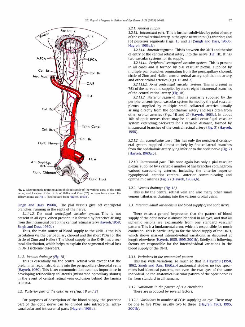
Fig. 6. A diagrammatic representation of blood flow autoregulation range at differentperfusion pressures in normal persons. Absent and present denote absence or presenceof the autoregulation.
S.S. Hayreh / Progress in Retinal and Eye Research 28 (2009) 34–6240
hypertension. Recent studies have shown that pericytes in thecapillaries may also play a role in regulation of the blood flowautoregulation by virtue of the presence of contractile proteinsactin and myosin. Since there is a limit to how far the terminalarterioles or capillaries can constrict or dilate, the autoregulationoperates only within a certain critical range of perfusion pressure,and breaks down when the perfusion pressure goes below or abovethis critical range (Fig. 6).
The exact mechanism of blood flow autoregulation is still notknown, and various hypotheses have been put forward (Hayreh,2001c). Briefly, there are three hypotheses. (i) Metabolic hypoth-esis: according to this, local arteriolar smooth muscle tone isregulated by local concentration of metabolic products, pO2 andpCO2, and they play a role in maintaining autoregulation. (ii)Myogenic hypothesis: according to this, the rise of intra-vascularpressure causes vasoconstriction. (iii) Neurogenic hypothesis: wedo not have much evidence of this in the ONH, since vessels in theretina and ONH have no autonomic nerve supply but both still haveautoregulation. The choroid, by contrast, is richly supplied by theautonomic nerves and yet has no appreciable autoregulation.
4.1.1.1.1. What is the range of perfusion pressure over which ONHautoregulation operates?. Autoregulation operates only over a crit-ical range of perfusion pressure, so that with a rise or fall ofperfusion pressure beyond the critical range, the autoregulationbecomes ineffective and breaks down (Fig. 6). In normal monkeys,autoregulation in the ONH has been reported to be normal ata perfusion pressure of �30 mmHg by Bill and co-workers (Geijerand Bill, 1979; Sperber and Bill, 1985) and >50 mmHg by Ernest(1976), but it definitely breaks down below 30 mmHg (Bill andSperber, 1987). In old, atherosclerotic rhesus monkeys, ONHautoregulation was already defective at 30–35 mmHg perfusion
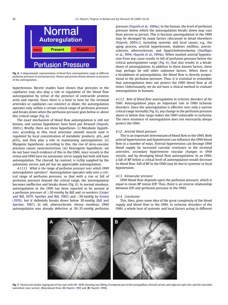
Fig. 7. Fluorescein fundus angiogram of two eyes with NA- AION showing non-filling of temwatershed zone (arrow). [Reproduced from (A) Hayreh, 1985 and (B) Hayreh, 1996].
pressure (Hayreh et al., 1994a). In the human, the level of perfusionpressure below which the autoregulation breaks down may varyfrom person to person. This is because autoregulation in the ONHmay be deranged by many factors (discussed in detail elsewhere(Hayreh, 2001c)), including systemic and local causes, e.g., theaging process, arterial hypertension, diabetes mellitus, arterio-sclerosis, atherosclerosis, and hypercholesterolemia (Haefligeret al., 1994; Hayreh et al., 1994a). When marked arterial hypoten-sion from any cause results in fall of perfusion pressure below thecritical autoregulation range (Fig. 6), that also results in a break-down of autoregulation. In addition to these known factors, theremay perhaps be still other unknown factors. When there isa breakdown of autoregulation, the blood flow is directly propor-tional to the perfusion pressure. Thus, it is essential to rememberthat autoregulation does not protect the ONH blood flow at alltimes. Unfortunately, we do not have a clinical method to evaluateautoregulation in humans.
4.1.1.2. Role of blood flow autoregulation in ischemic disorders of theONH. Autoregulation plays an important role in ONH ischemicdisorders. Since the autoregulation is effective over only a narrowcritical range normally (Fig. 6), any change in the perfusion pressureabove or below that range makes the ONH vulnerable to ischemia.The mere existence of autoregulation does not necessarily alwaysprotect the ONH.
4.1.2. Arterial blood pressureThis is an important determinant of blood flow in the ONH. Both
arterial hypertension and hypotension can influence the ONH bloodflow in a number of ways. Arterial hypertension can derange ONHblood supply by increased vascular resistance in the terminalarterioles, secondary hypertensive vascular changes in ONHvessels, and by deranging blood flow autoregulation. In an ONH,a fall of BP below a critical level of autoregulation would decreaseits blood flow. Fall of BP in the ONH may be due to systemic or localhypotension.
4.1.3. Intraocular pressureONH blood flow depends upon the perfusion pressure, which is
equal to mean BP minus IOP. Thus, there is an inverse relationshipbetween IOP and perfusion pressure in the ONH.
4.1.4. ConclusionThis, then, gives some idea of the great complexity of the blood
supply and blood flow in the ONH. In ischemic disorders of theONH, a whole host of systemic and local factors acting in different
poral part of the peripapillary choroid (arrow) and adjacent optic disc and the choroidal
S.S. Hayreh / Progress in Retinal and Eye Research 28 (2009) 34–62 41
combinations and to different extents may derange the circulationin the ONH, with some making the ONH susceptible to ischemiawhile others act as the final insult in one case and vice versa inanother. Moreover, one set of factors may be responsible for ONHischemia in one case and a totally different set in another, and so on.In such a scenario, a particular factor may be present in one caseand not in another. Thus ONH ischemic disorders are multifactorialin nature, particularly in AION, according to the available evidence(Hayreh et al., 1994b,c). Each patient with non-arteritic AION orother ONH ischemic disorders may have a unique combination ofsystemic and local factors which together produce ONH ischemicdamage (Hayreh, 1996). It is the lack of awareness of thiscomplexity of ONH blood flow which is responsible for most of thecontroversy and confusion about AION.
5. Ischemic optic neuropathy
5.1. Classification
As discussed above, based on the two very distinct patterns ofthe blood supply patterns of the ONH and rest of the optic nerve,ischemic optic neuropathy is of two distinct types (Hayreh, 1974c).
5.1.1. Anterior ischemic optic neuropathy (AION)This is due to ischemia of the ONH. Etiologically and pathoge-
netically, AION is of two types (Hayreh, 1974c, 1978b, 1981c, 1990a).
5.1.1.1. Arteritic AION (A-AION). This is due to giant cell arteritis(GCA).
5.1.1.2. Non-arteritic AION (NA-AION). This type is not due to GCA.
This is the most common type of ischemic optic neuropathy, andhas attracted the most controversy as to its pathogenesis andmanagement.
5.2.1. PathogenesisThis is discussed at length elsewhere (Hayreh, 1996). Following
is a brief account:NA-AION is due to acute ischemia of the ONH (Hayreh, 1974b,
1981c, 1985), whose main source of blood supply is from the PCAcirculation (Fig. 1A). Therefore, NA-AION represents an ischemicdisorder of PCA circulation in the ONH. Marked interindividualvariations in blood supply of the ONH (see above) and its blood flowpatterns profoundly influence the pathogenesis and clinicalfeatures of NA-AION. This entire subject is very controversial andrequires a detailed discussion to place the various relevant issues inproper perspective.
Etiologically and pathogenetically NA-AION is of two types.
5.2.1.1. Due to transient non-perfusion or hypoperfusion of the ONHcirculation. This is by far the commonest cause of NA-AION (Hayreh,1996; Hayreh et al.,1994c,1997b,1999). There is almost a universallyheld belief among ophthalmologists and neurologists that NA-AIONhas a pathogenesis like that of a stroke which is a thromboembolicdisorder; however, in the vast majority of NA-AION cases there is noevidence of that, as indicated by the following evidence.
(i) First and foremost, if NA-AION were a thromboembolicdisorder, like A-AION, fluorescein fundus angiography during
the early stages of onset of visual loss must almost invariablyshow evidence of complete occlusion of the vessels supplyingthe ONH (as is the case in A-AION – see below); however, nosuch occlusion is seen in NA-AION. Fluorescein fundus angi-ography soon after the onset of NA-AION shows only a delayedand slow filling of the peripapillary choroid and/or choroidalwatershed zones, but no permanent occlusion (Figs. 5A,B,Dand 7) which provides a definite proof that NA-AION is nota thromboembolic occlusive disorder.
(ii) The severity of ONH ischemic damage depends upon theseverity and the duration of the ONH ischemia; the latterdetermines the extent of recovery of visual function followingthe acute episode. In NA-AION, because there is only transientnon-perfusion or hypoperfusion the ONH circulation, there isusually much less severe and less extensive ONH damage thanin A-AION, in which there is thrombotic occlusion of the PCA.Two large studies (Ischemic Optic Neuropathy DecompressionTrial Research Group, 1995; Hayreh and Zimmerman, 2008a)have shown that in NA-AION 41% of the eyes show sponta-neous visual improvement. In sharp contrast to that, in A-AION no such visual improvement is seen (Hayreh andZimmerman, 2003b).
(iii) In NA-AION patients, compared to age-matched controls,transcranial Doppler did not reveal an increased incidence ofembolic events, which further confirms that NA-AION is nota thromboembolic disorder (Kosmorsky et al., 1998).
Thus, all the available evidence indicates that NA-AION is nota thromboembolic disorder. Naturally the question arises: what isthe mechanism of transient non-perfusion or hypoperfusion of theONH circulation in NA-AION? It can be caused by a variety of factors.Available evidence indicates that in the vast majority of cases it isa transient fall of blood pressure, most commonly during sleep(nocturnal arterial hypotension – see below) or a nap during theday (Hayreh et al., 1994c, 1999), and more rarely ocular ischemia,severe internal carotid artery and/or ophthalmic artery stenosis orocclusion during sleep (Mizener et al., 1997). Any kind of shock canalso cause a transient fall of blood pressure. A sharp rise in the IOPto high levels (e.g., in neovascular glaucoma associated with ocularischemia, or angle closure glaucoma) can also cause a transient fallin perfusion pressure (perfusion pressure is equal to mean bloodpressure minus IOP).
A transient fall of perfusion pressure in the ONH vessels resultsin transient non-perfusion or hypoperfusion of those vessels. Asdiscussed above, a fall in perfusion pressure in the capillaries of theONH below the critical autoregulatory range level (Fig. 6), insusceptible persons (see below), results in ischemia of the ONH anddevelopment of NA-AION. The severity of ONH ischemia may varyfrom mild to marked, depending upon the severity and theduration of the transient ischemia and other factors influencing theblood flow in the ONH (see above).
5.2.1.2. Due to embolic lesions of the arteries/arterioles feeding theONH. This is only an occasional cause of NA-AION. Multiple emboliin the vessels of the anterior part of the optic nerve have beendemonstrated histopathologically in AION (Lieberman et al., 1978).This has also been shown on fluorescein fundus angiography(Fig. 4A) (Hayreh, 1985). Compared to the hypotensive type of NA-AION, the extent of ONH damage in this type is usually massive,severe, and permanent (similar to that in A-AION – see below),depending upon the size of the artery involved and the area of thenerve supplied by the occluded artery.
5.2.2. Risk factors for development of NA-AIONAll the available evidence indicates that NA-AION is multifac-
torial in nature. The risk factors fall into two main categories.
S.S. Hayreh / Progress in Retinal and Eye Research 28 (2009) 34–6242
5.2.2.1. Predisposing risk factors. These may be systemic or local inthe eye and/or ONH, and they may make the ONH susceptible toischemic disorders but do not necessarily produce NA-AION ontheir own.
5.2.2.1.1. Systemic risk factors. Various studies have showna significantly high prevalence of arterial hypertension, nocturnalarterial hypotension, diabetes mellitus, ischemic heart disease,hyperlipidemia, atherosclerosis and arteriosclerosis in NA-AIONpatients compared to the general population (Repka et al., 1983;Guyer et al., 1985; Hayreh et al., 1994b; Hayreh, 1996; Jacobsonet al., 1997; Hayreh and Zimmerman, 2008c). Other associatedsystemic diseases have also been reported, including sleep apnea(Hayreh, 1996; Mojon et al., 2002; Palombi et al., 2006; Li et al.,2007), arterial hypotension due to a variety of causes including,shock, cardiopulmonary bypass surgery and hemodialysis, massiveor recurrent hemorrhages (Hayreh, 1987) and malignant arterialhypertension (Hayreh et al., 1986a). The possibility that in anoccasional patient embolism from thrombophilic factors may causeNA-AION cannot be ruled out (Hayreh, 2008d). Similarly, other rarecauses include, migraine, defective cardiovascular autoregulation,‘‘Type A personality’’ (Hayreh, 1996), and carotid dissection (Bio-usse et al., 1998). In addition, the literature is full of anecdotal casereports of the association of ‘‘anterior ischemic optic neuropathy’’with a large variety of systemic diseases and causes, but it is notpossible to establish a cause-and-effect relationship in all of them.However, these rare diseases have no role in the vast majority ofNA-AION cases, since it is primarily a hypotensive disorder.
5.2.2.1.2. Ocular and ONH risk factors. A significant associationof NA-AION has been seen with a number of ocular and ONHconditions. These include absent or small cup in the optic disc (Becket al., 1987; Hayreh and Zimmerman, 2008d), angle closure glau-coma or other causes of markedly raised IOP (Hayreh, 1980),marked optic disc edema due to any cause (Hayreh, 1977b), locationof the watershed zone of the PCAs in relation to the optic disc(Hayreh, 1990b), and vascular disorders in the nutrient vessels ofthe ONH (Hayreh, 1995), optic disc drusen and cataract extraction(Hayreh, 1980). There are a few reports of delayed development ofNA-AION in the fellow eyes long after cataract extraction in oneeye (Nguyen et al., 2006; Lam et al., 2007), implying that cataractextraction per se is a risk factor; there is no evidence in support ofthat. This is because a person who has the required risk factors, is atrisk of developing NA-AION irrespective of whether he/she hascataract extraction or not – the fact that he/she developed NA-AIONafter cataract extraction in the first eye indicates the presence ofthose predisposing risk factors in him/her. Optic disc related visualfield defects detected after vitrectomy (Taban et al., 2007) are mostprobably due to development of NA-AION in those eyes duringvitrectomy, due to intra- and/or postoperative raised IOP, alongwith other associated systemic risk factors mentioned above.
5.2.2.1.2.1. The role of an absent or small cup in the pathogenesisof development of NA-AION. Since 1974, several studies have shownthat in eyes with NA-AION there is a significantly higher prevalenceof absent or small cup than in the general population (Hayreh,1974a; Hayreh and Zimmerman, 2008d). This has resulted ina misconception in the ophthalmic community that a small orabsent cup is actually the primary factor in the development of thedisease; this has resulted in terms like ‘‘disc at risk’’. The role of anabsent or small cup in the pathogenesis of development of NA-AION is discussed in detail elsewhere (Beck et al., 1987; Hayreh andZimmerman, 2008d). Briefly, in the multifactorial scenario of thepathogenesis of NA-AION, one has to consider the role of thefollowing two factors relevant to cup/disc ratio. (a) Absent or smallcup is associated with a small scleral canal and small opening in theBruch’s membrane, resulting in crowding of the optic nerve fibersas they pass through the restricted space in the optic disc andlamina cribrosa. (b) Ischemia or hypoxia of the axons in the ONH
causes axoplasmic flow stasis, which in turn results in swollenaxons (Hayreh, 1977a). Axoplasmic flow stasis causes swelling ofthe axons and that is responsible for optic disc edema in ischemicoptic neuropathy (McLeod et al., 1980). If the optic disc has a cup,the swollen axons can expand into that without compressing anyother tissues in the optic disc. But when there is no cup or onlya small cup, the swollen axons are crowded in a restricted space inthe optic disc, and they can expand only by compressing thesurrounding tissues. The tissues that are most vulnerable tocompression here are capillaries and other fine vessels lying amongthe nerve fibers. Thus, swollen axons in restricted space within theoptic disc produce secondary vascular changes (Hayreh, 1977a). Ithas been shown that asymptomatic optic disc edema is the earliestsign of NA-AION (Hayreh, 1981a; Hayreh and Zimmerman, 2007a).It has also been demonstrated that nocturnal arterial hypotensionprecipitates the development of NA-AION (Hayreh et al., 1997b).Thus, the available evidence indicates that the sequence of eventsin the development of NA-AION are as follows: subclinical ischemia(hypoxia) of the optic nerve head / axoplasmic flow stasis in theoptic nerve fibers / axonal swelling / asymptomatic optic discedema (incipient NA-AION (Hayreh, 1981a; Hayreh and Zimmer-man, 2007a)) / compression of the intervening capillaries byswollen axons in a crowded disc / setting up a vicious cycle: thegreater the compression of capillaries, the greater the blood flowcompromise, the greater the axoplasmic flow stasis and the morethe axonal swelling. Since compression of the optic disc capillariescompromises their blood flow, a fall of blood pressure must furtherderange their blood flow. Therefore, in this situation, a fall ofperfusion pressure in the optic disc capillaries due to nocturnalarterial hypotension results in marked ischemia and that precipi-tates visual loss (symptomatic NA-AION), which is usually discov-ered on waking up in the morning (Hayreh et al., 1997b).
From this sequence of events, it is evident that in the multi-factorial scenario of pathogenesis of NA-AION, contrary to theprevalent impression, an absent or small cup is simply a secondarycontributing factor, ONCE the process of NA-AION has started, andNOT a primary factor (Beck et al., 1987; Hayreh and Zimmerman,2007a,b).
5.2.2.2. Precipitating risk factor(s). In a person with a predisposingrisk factor already present, these risk factors act as the final insult(‘‘last straw’’), resulting in ischemia of the ONH and NA-AION.Nocturnal arterial hypotension is the most important factor in thiscategory (Hayreh et al., 1994c, 1997b, 1999). Studies have shownthat patients with NA-AION and often also those with A-AIONtypically complain of discovering visual loss on waking in themorning. In NA-AION, 73% gave a definite history of discoveringthe visual loss on waking up in the morning or from a nap, or firstopportunity in the day to use vision critically (Hayreh et al.,1997b). The incidence may actually be much higher than 73%because many others who became aware of visual loss later on inthe day could not be certain when it had occurred. Hayreh et al.’s(1994c, 1999) 24-h ambulatory blood pressure monitoring hasshown development of marked nocturnal arterial hypotension insuch patients. For example, the 24-h ambulatory blood pressuremonitoring pressure graph in Fig. 8 shows a steep drop in bloodpressure on falling asleep at night and recovery to normal onwaking in the morning. Studies have also shown that arterialhypertensives on oral hypotensive therapy have a significant(p ¼ 0.004) association between progressive visual field deterio-ration in NA-AION and nocturnal hypotension (Hayreh et al.,1994c, 1999). The fall of blood pressure during sleep is a physio-logical phenomenon, but it is influenced by many factors,including the various arterial hypotensive drugs taken for arterialhypertension or other cardiovascular disorders, particularly thenumber and amount of drugs taken and the time of day they are
Fig. 8. Ambulatory BP and heart rate monitoring records (based on individual readings) over a 24-h period, starting from about 11 a.m., in a 58-year old woman with bilateralNA-AION, and on no medication. The BP is perfectly normal during the waking hours but there is marked nocturnal arterial hypotension during sleep. (Reproduced fromHayreh et al., 1999).
S.S. Hayreh / Progress in Retinal and Eye Research 28 (2009) 34–62 43
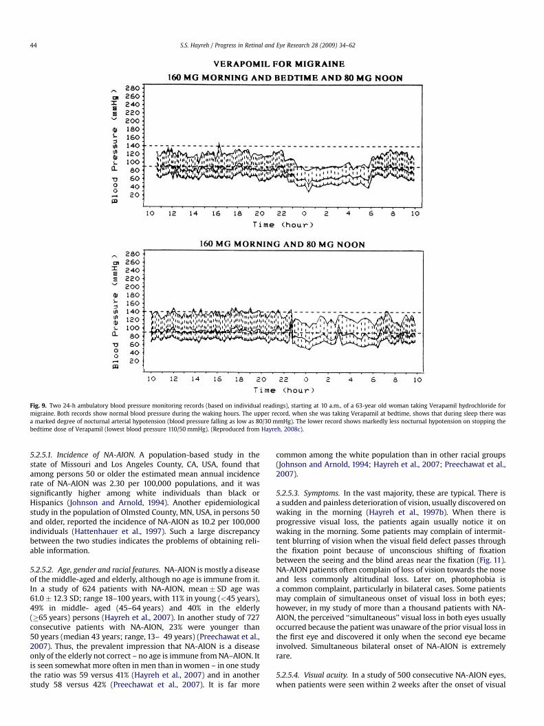
taken. When these drugs were taken at bedtime, they produceda far more marked degree of nocturnal hypotension than whentaken in the morning, because they aggravate the naturallyoccurring fall of blood pressure during sleep (Fig. 9). There are,however, some patients who develop marked nocturnal hypo-tension even without any medication (presumably due to defec-tive cardiovascular autoregulation), as can be seen in Fig. 8.
5.2.2.3. Conclusion. From this brief discussion, the greatcomplexity of mechanisms of development of NA-AION, and therole of nocturnal hypotension in it become clear. A whole host ofsystemic and local factors, acting in different combinations and todifferent extents may derange the ONH circulation, with somemaking the ONH susceptible to ischemia and others acting as thefinal insult. Nocturnal hypotension seems to be an importantprecipitating factor in the susceptible patient.
The pathogenesis of NA-AION is complex but not, as oftenstated, unknown.
Recently Levin and Danesh-Meyer (2008) have publisheda hypothesis dealing with pathogenesis of NA-AION. According tothis hypothesis, ‘‘Anatomical or functional occlusion of CRV (centralretinal vein) tributaries within the anterior optic nerve would causevenous congestion of the optic nerve parenchyma, subsequentcytotoxic and vasogenic edema, and consequent further compres-sion of venules feeding the CRV. Venous congestion can causesecondary constriction of small arterioles via the venoarteriolarresponse.’’ Based on my studies on the anatomy and blood of theoptic nerve, and experimental and clinical studies on variousaspects of NA-AION and central retinal vein occlusion, I find thishypothesis invalid on several counts (Hayreh, in press b). Venousocclusion has no role whatsoever in the development of NA-AION.
5.2.3. NA-AION and cerebral stroke are not similar in natureThere is a common perception among ophthalmologists and
neurologists that NA-AION and cerebral stroke are similar in naturepathogenetically and in management. This has resulted in majorcontroversy on pathogenesis and management of NA-AION. Thefollowing evidence, however, indicates that NA-AION pathoge-netically is a distinct clinical entity.
5.2.3.1. Difference in association of smoking. There is a huge volumeof literature showing a significant association between smokingand cerebrovascular accident (a thromboembolic disorder) (Dag-enais et al., 2005). No association has been found between smokingand NA-AION (Newman et al., 2002; Hayreh et al., 2007).
5.2.3.2. Difference in response to aspirin. While the beneficial effectof aspirin in cerebrovascular accident (usually a thromboembolicdisorder) is well-established, NA-AION studies have shown thataspirin has no beneficial effects in NA-AION (being a hypotensivedisorder) (Newman et al., 2002; Beck et al.,1997; Botelho et al.,1996).
5.2.3.3. Difference in association between thrombophilic risk fac-tors. While an association has been reported between thrombo-philic risk factors and cerebrovascular accident, no significantassociation has been found between NA-AION and thrombophilicrisk factors for the same reason (Salomon et al., 1999; Hayreh,2001a, 2008e; Abu-Amero and Bosley, 2006).
5.2.3.4. A hypotensive disorder. As discussed above, other findingsshow that NA-AION is primarily a hypotensive disorder and nota thromboembolic disorder in the vast majority.
5.2.4. Histopathologic findings in ischemic optic neuropathyKnox et al. (2000) reported these in 193 eyes with ischemic optic
neuropathy. They concluded, ‘‘ischemic optic nerve lesions areinitially acellular and later show macrophage infiltration.Cavernous lesions with MPS (mucopolysaccharide) are present4 weeks or longer after vision loss. The location of MPS posteriorlyand along the internal margin suggests that MPS is produced at theedges of lesions.’’ The experimental model of AION by Hayreh andBaines (1972) showed similar histopathological findings (Fig. 10).
5.2.5. Clinical features of classical NA-AIONNA-AION is the most common type of ischemic optic neurop-
athy. It usually has classical symptoms and signs which make it easyto diagnose. The subject is discussed at length elsewhere (Hayreh,1996). Following is a very brief account of the clinical features ofNA-AION.
Fig. 9. Two 24-h ambulatory blood pressure monitoring records (based on individual readings), starting at 10 a.m., of a 63-year old woman taking Verapamil hydrochloride formigraine. Both records show normal blood pressure during the waking hours. The upper record, when she was taking Verapamil at bedtime, shows that during sleep there wasa marked degree of nocturnal arterial hypotension (blood pressure falling as low as 80/30 mmHg). The lower record shows markedly less nocturnal hypotension on stopping thebedtime dose of Verapamil (lowest blood pressure 110/50 mmHg). (Reproduced from Hayreh, 2008c).
S.S. Hayreh / Progress in Retinal and Eye Research 28 (2009) 34–6244
5.2.5.1. Incidence of NA-AION. A population-based study in thestate of Missouri and Los Angeles County, CA, USA, found thatamong persons 50 or older the estimated mean annual incidencerate of NA-AION was 2.30 per 100,000 populations, and it wassignificantly higher among white individuals than black orHispanics (Johnson and Arnold, 1994). Another epidemiologicalstudy in the population of Olmsted County, MN, USA, in persons 50and older, reported the incidence of NA-AION as 10.2 per 100,000individuals (Hattenhauer et al., 1997). Such a large discrepancybetween the two studies indicates the problems of obtaining reli-able information.
5.2.5.2. Age, gender and racial features. NA-AION is mostly a diseaseof the middle-aged and elderly, although no age is immune from it.In a study of 624 patients with NA-AION, mean � SD age was61.0 � 12.3 SD; range 18–100 years, with 11% in young (<45 years),49% in middle- aged (45–64 years) and 40% in the elderly(�65 years) persons (Hayreh et al., 2007). In another study of 727consecutive patients with NA-AION, 23% were younger than50 years (median 43 years; range, 13– 49 years) (Preechawat et al.,2007). Thus, the prevalent impression that NA-AION is a diseaseonly of the elderly not correct – no age is immune from NA–AION. Itis seen somewhat more often in men than in women – in one studythe ratio was 59 versus 41% (Hayreh et al., 2007) and in anotherstudy 58 versus 42% (Preechawat et al., 2007). It is far more
common among the white population than in other racial groups(Johnson and Arnold, 1994; Hayreh et al., 2007; Preechawat et al.,2007).
5.2.5.3. Symptoms. In the vast majority, these are typical. There isa sudden and painless deterioration of vision, usually discovered onwaking in the morning (Hayreh et al., 1997b). When there isprogressive visual loss, the patients again usually notice it onwaking in the morning. Some patients may complain of intermit-tent blurring of vision when the visual field defect passes throughthe fixation point because of unconscious shifting of fixationbetween the seeing and the blind areas near the fixation (Fig. 11).NA-AION patients often complain of loss of vision towards the noseand less commonly altitudinal loss. Later on, photophobia isa common complaint, particularly in bilateral cases. Some patientsmay complain of simultaneous onset of visual loss in both eyes;however, in my study of more than a thousand patients with NA-AION, the perceived ‘‘simultaneous’’ visual loss in both eyes usuallyoccurred because the patient was unaware of the prior visual loss inthe first eye and discovered it only when the second eye becameinvolved. Simultaneous bilateral onset of NA-AION is extremelyrare.
5.2.5.4. Visual acuity. In a study of 500 consecutive NA-AION eyes,when patients were seen within 2 weeks after the onset of visual
Fig. 10. Photomicrographs of the optic nerve head and retrolaminar optic nerve inrhesus monkey 36 days after occlusion of all PCAs (Masson’s Trichrome stain) showing:(A) atrophy and degenerative changes in retrolaminar region, and (B) higher magni-fication showing marked degeneration of neural tissue in retrolaminar region,producing an appearance resembling cavernous degeneration. (Reproduced fromHayreh and Baines, 1972).
S.S. Hayreh / Progress in Retinal and Eye Research 28 (2009) 34–62 45
loss, initial visual acuity was 20/20 in 33%, better than 20/40 in 51%,and 20/200 or worse in 21% (Hayreh and Zimmerman, 2008a,b).This shows that the presence of normal visual acuity does not ruleout NA-AION. Comparison of refraction in NA-AION patients50 years and older with that of an age-matched general populationshowed no significant difference between the two groups (Hayrehand Zimmerman, 2008d).
5.2.5.5. Visual fields. In contrast to visual acuity, which can bewithin normal limits in almost half of the eyes with NA-AION,visual field defects are a universal occurrence. Therefore, perimetryis the most important and essential visual function test to evaluatethe visual loss. These eyes can present with a variety of optic nerverelated visual field defects. In a visual field study of 312 consecutiveNA-AION eyes, manual kinetic perimetry (i.e. visual fields plotted
with a Goldmann perimeter) showed an overall prevalence ofgeneral visual field defects in 83% with I-2e, 79% with I-4e and 69%with V-4e, and of scotoma(s) within the central 30� in 55, 49 and36% respectively (Hayreh and Zimmerman, 2005). Central scotomawas seen in 49% with I-2e, 44% with I-4e, and 29% with V-4e.Relative inferior altitudinal defect was common (35% with I-2e; and22% with I-4e), but absolute inferior altitudinal defect was seen inonly 8%. By contrast, absolute inferior nasal sector visual loss wasthe most common defect detected in NA-AION (22%). Thus,a combination of a relative inferior altitudinal defect with absoluteinferior nasal defect is the most common pattern in NA-AION(Fig. 11A). This contradicts the commonly held belief that inferioraltitudinal visual field defect is typical of NA-AION.
Currently visual fields are usually plotted using automatedperimetry. Unfortunately, automated perimetry provides infor-mation on only up to about 24–30� in the periphery. Kineticperimetry, by contrast, provides peripheral visual field informationall the way to about 80–90� temporally, 70� inferiorly, 60–70�
nasally and 50–60� superiorly. This has two importantimplications.
5.2.5.5.1. Peripheral visual field defects. It is well -establishedthat the constant tracking provided by the peripheral visual fields isessential for sensory input to our day-to-day activity. For example,the peripheral visual field is vital for driving and ‘‘navigating’’ in theworld. In view of that, to assess the visual function disabilityproduced by NA-AION, it is important to have complete informa-tion about the peripheral visual fields and any impairment in them.While kinetic perimetry provides that information reliably, auto-mated perimetry does not. This is big drawback in automatedperimetry to evaluate visual function in NA-AION.
5.2.5.5.2. Central visual field defects. Large scotomas in thecentral 24–30� on automated perimetry may be misinterpreted asaltitudinal when in fact the eye still may have intact peripheral fieldoutside the central 24–30�. This may be a factor in the widespreaderroneous belief that inferior altitudinal field defect is the mostcommon defect in NA-AION.
Therefore, in NA-AION the visual field plotted with manualkinetic perimetry provides far superior information about type ofvisual field defect and the peripheral field, and for evaluating visualfunctional disability (Hayreh and Zimmerman, 2005).
5.2.5.6. Natural history of visual outcome in NA-AION. There are twoprospective studies that have evaluated this, one based on 125 eyes(Ischemic Optic Neuropathy Decompression Trial Research Group,1995) and the other on 386 eyes (Hayreh and Zimmerman, 2008a);both arrived at the same conclusion. Both studies showed that inpatients seen within 2 weeks of onset of visual loss and initial visualacuity of 20/70 or worse, there was improvement in 41–43% andworsening in 15–19% at 6 months. One of these studies that eval-uated visual fields with kinetic perimetry showed that 26% of thosewho were first seen �2 weeks of onset with moderate to severevisual field defect, showed improvement at 6 months (Hayreh andZimmerman, 2008a). Visual acuity and visual fields showedimprovement or further deterioration mainly up to 6 months, withno significant change after that (Hayreh and Zimmerman, 2008a).When NA-AION develops in the second eye, there is no correlationin the visual outcome in the two eyes.
5.2.5.7. Anterior segment of the eye. This invariably shows noabnormality except for the presence of relative afferent pupillarydefect in unilateral NA-AION eyes, and in some there may beraised IOP.
5.2.5.8. Optic disc changes. At the onset of visual loss, there isalways optic disc edema (ODE) (Hayreh and Zimmerman, 2007b).There are several misconceptions about ODE in NA-AION. The most
Fig. 11. Visual field defects in NA-AION, plotted with Goldmann perimeter (using I-2e, I-4e and V-4e targets). (A) Shows inferior altitudinal defect with I-2e and inferior nasal defectwith I-4e and V-4e. (B) Shows absolute inferior altitudinal defect with I-2e, I-4e and V-4e. The visual acuity in both eyes was 20/20. (Reproduced from Hayreh and Zimmerman,2005).
S.S. Hayreh / Progress in Retinal and Eye Research 28 (2009) 34–6246
common one is that in NA-AION the ODE is always pale – that is nottrue at all initially, because the color of ODE in NA-ION initially doesnot differ from ODE due to other causes – in some cases there mayeven be hyperemia of the optic disc (Figs. 12, 13 and 14B). A splinterhemorrhage at disc margin is common (Fig. 13). ODE starts todevelop pallor about 2–3 weeks after the onset of NA-AION (Hayrehand Zimmerman, 2007b). A study based on evaluation of variousaspects of ODE in 749 eyes showed that the overall median time(25th–75th percentile) to spontaneous resolution of ODE from theonset of visual loss was 7.9 (5.8 – 11.4) weeks (Hayreh and Zim-merman, 2007b). The time it took to resolve depended on severalfactors, e.g., longer in diabetics than in non-diabetics, and worseinitial visual field defect and visual acuity were associated witha faster resolution of ODE. When patients were treated with steroidtherapy within 2 weeks after onset of NA-AION, there wasa significantly faster ODE resolution than in the untreated cases.There is a characteristic evolutionary pattern of ODE in NA-AION(Hayreh and Zimmerman, 2007b). Initially the involved part of thedisc (i.e. corresponding to the location of visual field loss) hasedema, with the rest of the disc normal or showing much lessedema / after several days the entire disc may show generalized
Fig. 12. Left fundus photograph showing optic disc edema and hyperemia during theacute phase of NA-AION.
edema / still later, the optic disc in the originally involved partbegins to develop pallor and the edema gradually starts toregress in that part, so that the uninvolved part (correspondingto normal visual field) may have more edema than the ischemicpart / then the involved part has pallor but is not edematousany more, while the rest of the disc may show mild edema andeven some pallor / the ODE gradually resolves / pallor of theinvolved region only or the entire disc (Fig. 14), and in the lattercase the pallor may or may not be more marked in the involvedpart. Therefore, the sector of the optic disc showing edemausually corresponds to the location of the visual field defect onlyduring the very early stage, and not later on. Similarly, Arnoldand Hepler (1994b) recorded ‘‘no consistent correlation’’ of thesector of the disc edema with the visual field defect. On resolu-tion of ODE, the distribution of optic disc pallor does not alwayscorrespond with the extent and location of visual and nerve fiberloss (Hayreh and Zimmerman, 2007b).
In the fellow normal eye, optic disc usually shows either no cupor small cup (see above). This can be a helpful clue in the diagnosisof NA-AION in doubtful cases. If originally both eyes have small disccups, I have seen that in unilateral NAION, once the disc edema
Fig. 13. Right fundus photograph showing optic disc edema and hyperemia, witha splinter hemorrhage (arrow) during the acute phase of NA-AION.
Fig. 14. Fundus photographs of left eye of a 53-year-old man. (A) Normal disc before developing NA-AION, (B) with optic disc edema during the active phase of NA-AION, and (C)after resolution of optic disc edema and development of optic disc pallor – more marked in temporal part than nasal part.
S.S. Hayreh / Progress in Retinal and Eye Research 28 (2009) 34–62 47
resolves, the cup in the involved eye may become slightly largerthan the fellow eye because of loss of nerve fibers. This has alsobeen reported by others (Saito et al., 2008).
In occasional cases, where NA-AION is due to embolism into thePCA, the ODE, unlike in the classical NA-AION, usually has a chalkywhite appearance.
In diabetics, optic disc changes in NA-AION may have somecharacteristic diagnostic features. During the initial stages, the ODE
Fig. 15. Fundus photographs of the left eye, of a 19½ year-old white male juvenile diabetic. (Amultiple punctate peripapillary and macular retinal hemorrhages, engorged retinal veins.hemorrhages on resolution. (Reproduced from Hayreh, 1978b).
is usually (but not always) associated with characteristic prom-inent, dilated and frequently telangiectatic vessels over the disc,and much more numerous peripapillary retinal hemorrhages thanin non-diabetics (Fig. 15A) (Hayreh and Zahoruk, 1981; Hayreh andZimmerman, 2008c). These findings may easily be mistaken forproliferative diabetic retinopathy associated with optic disc neo-vascularization. When the ODE resolves spontaneously, theseprominent telangiectatic disc vessels and retinal hemorrhages also
) Shows massive optic disc edema with marked telangiectatic vessels on the optic disc,(B) Shows normal-looking optic disc, no abnormal vessels on the disc, and no retinal
S.S. Hayreh / Progress in Retinal and Eye Research 28 (2009) 34–6248
resolve spontaneously (Fig. 15B). The presence of these character-istic fundus changes in some diabetics with NA-AION has resultedin a good deal of controversy because it has been thought to bea separate clinical entity – described under different eponyms, themost common being ‘‘diabetic papillopathy’’, when in fact it is NA-AION (Hayreh and Zimmerman, 2008c).
5.2.5.9. Other fundus changes. The presence of a few splinterhemorrhages on optic disc or immediate peripapillary region iscommon in association with the ODE (Fig. 13); those resolve sponta-neously with ODE resolution. Diabetics tend to have more peripapillaryretinal hemorrhages than non-diabetics. (Hayreh and Zahoruk, 1981;Hayreh and Zimmerman, 2008c). Occasionally, I have seen mild serousretinal detachment between the optic disc and macula and that mayeven extend to the macular region to produce macular edema (Fig.16).This has also been reported by other authors (Tomsak and Zakov,1998;Hedges et al., 2008). Because of ODE, there is a certain amount of retinalvenous engorgement. In some eyes, as the ODE resolves, some lipiddeposits are seen in the peripapillary or macular region.
5.2.5.10. Fluorescein fundus angiographic findings. It is only angi-ography during the very early arterial phase of dye filling in thefundus that demonstrates the tell-tale impaired circulation and itslocation in NA-AION. In my studies, there is almost invariably fillingdefect/delay in the prelaminar region and in the peripapillarychoroid (Fig. 7) and/or choroidal watershed zones (Figs. 5A,B,D) atonset of NA-AION (Hayreh, 1985). This has also been shown byothers (Arnold and Hepler, 1994a; Arnold et al., 1996). In the occa-sional case, where NA-AION is due to embolism into the PCA, thepart of the choroid supplied by the occluded PCA or short PCA doesnot fill (Fig. 4A). Late optic disc staining is a non-specific finding ofoptic disc edema, and has no diagnostic importance for NA-AION.
5.2.5.11. Bilateral NA-AION. The cumulative probability of thefellow eye developing NA-AION has varied among different studies:
Fig. 16. Fundus photograph (A) and OCT (B) of right eye with NA-AION and serous retinal delipid deposits in the central part of the macula.
25% within 3 years in 438 patients (Beri et al., 1987), 17% in 5 yearsin 431 patients (Beck et al., 1997) and 15% over 5 years in 326patients (Newman et al., 2002); however, different criteria wereused to determine the probability, which may explain the differ-ences. According to one study (Beri et al., 1987), the risk is greater inmen, particularly young diabetic men, while according to anotherstudy (Newman et al., 2002) increased incidence was associatedwith poor baseline visual acuity and diabetes mellitus. The risk ofthe second eye getting involved by NA-AION was evaluated in 655patients (206 diabetics and 449 non-diabetics) and that showeda significantly (p ¼ 0.003) greater risk in diabetics than in non-diabetics (Hayreh and Zimmerman, 2008c); that study also showedthat the median (25th–75th percentile) time to involvement of thefellow eye by NA-AION was 6.9 (0.4–16.9) years in diabetics and 9.1(1.8–19.0) years in non-diabetics.
There are reports in the literature of simultaneous onset ofbilateral NA-AION. As discussed above, in my study of more thana thousand patients with NA-AION, the reported ‘‘simultaneous’’visual loss in both eyes due to NA-AION usually occurred becausethe patient was unaware of the NA-AION in the first eye until thesecond eye got involved. Pattern of ODE during the initial stages ofNA-AION is different from that later on (see above), and that ishelpful to time the onset in bilateral NA-AION. Simultaneousbilateral onset of NA-AION is extremely rare, except in patients whodevelop sudden, severe arterial hypotension, e.g. during hemodi-alysis or surgical shock.
5.2.5.12. Recurrence of NA-AION in the same eye. Such recurrencesmentioned in the literature are often more a progression of NA-AION during the acute stage rather than actual new episodes afterthe first episode has resolved completely. In a study of 829 NA-AION eyes, the overall cumulative percentage of recurrence of NA-AION in the same eye was at 3 months 1.0 � 0.4% (SE), at 6 months2.7 � 0.7%, at 1 year 4.1 � 0.9%, and 2 years 5.8 � 1.1% (Hayreh et al.,2001). The only significant association for recurrence of NA-AION
tachment between the optic disc and the macula. In (A) arrows indicate the presence of
S.S. Hayreh / Progress in Retinal and Eye Research 28 (2009) 34–62 49
was with nocturnal arterial hypotension. Thus, this study indicatedthat nocturnal diastolic arterial hypotension might be a risk factorfor recurrence of NA-AION; however, since NA-AION is a multifac-torial disease, other risk factors so far unknown may also play a role.
5.2.5.13. NA-AION in diabetics versus non-diabetics. In a study of655 consecutive NA-AION patients (931 eyes) – 206 patients withdiabetes and 449 without – comparison of various clinical featuresof NA-AION in diabetics and non-diabetics showed no significantdifference in age, but diabetics had slightly more women than men(45 versus 38%; p ¼ 0.078), and a higher prevalence of arterialhypertension (p < 0.0001), ischemic heart disease (p ¼ 0.0001),transient ischemic attacks (p ¼ 0.0003), and second eye involve-ment by NA-AION (p ¼ 0.003) (Hayreh and Zimmerman, 2008c).Initial visual acuity did not differ significantly between diabeticsand non-diabetics; however, of those seen within 2 weeks of onsetof NA-AION, diabetics had less severe visual field defect (p ¼ 0.010).At 6 months after onset, there was no significant difference invisual acuity and visual field improvement between diabetics andnon-diabetics. Time to optic disc edema resolution was (p ¼ 0.003)longer in diabetics than non-diabetics. As discussed above, the ODEin diabetics usually has characteristic, diagnostic dilated telangi-ectatic vessels during early stages of NA-AION.
5.2.5.14. NA-AION and phosphodiesterase-5 (PDE5) inhibitors. Thesubject is discussed at length elsewhere (Hayreh, 2005; Hayreh,2008c). Briefly, most patients reported to have developed NA-AIONfollowing the use of these drugs are middle-aged or elderly menwho already had various predisposing risk factors for NA-AION (seeabove). These drugs are mostly taken in the evening for sexualintercourse. PDE5 inhibitors result in fall of blood pressure; whentaken in the evening, as discussed above, there is high chance ofthem producing abnormal nocturnal arterial hypotension, whichmay be further aggravated if the person is taking other arterialhypotensive drugs for arterial hypertension or other cardiovasculardisorders. Like the vast majority of NA-AION patients, most of thepatients reporting NA-AION following ingestion of PDE5 discoveredvisual loss upon awakening in the morning. A critical review of allthe reported cases shows a usually good temporal relationshipbetween the ingestion of these drugs and onset of NA-AION. Whenall the above evidence is put together, it suggests that Viagra andother PDE5 inhibitors can result in development of NA-AION inpersons who already have predisposing risk factors.
5.2.5.15. Amiodarone and NA-AION. There is a universal belief thatamiodarone causes optic neuropathy, called ‘‘amiodarone-inducedoptic neuropathy’’. However, the following facts do not support thatview (Hayreh, 2006).
(i) Patients who take amiodarone have cardiovascular disorders,which are per se well-established risk factors for the devel-opment of NA-AION (Hayreh et al., 1994b, 1996). Many of thesepatients also have other risk factors (arterial hypertension,diabetes mellitus, hyperlipidemia and ischemic heart disease)(Hayreh et al., 1994b; Hayreh, 1996). They are candidates forNA-AION whether they are taking amiodarone or not.
(ii) Patients on amiodarone often also take other drugs (beta-blockers, calcium channel blockers, ACE inhibitors) thatinfluence the cardiovascular system. As discussed above,patients on these drugs are at high risk of developingnocturnal arterial hypotension, which is a common precipi-tating factor for the development of NA-AION (Hayreh, 1996;Hayreh et al., 1994c, 1999).
(iii) One of the arguments put forward to differentiate amiodar-one-induced optic neuropathy from NA-AION is that somepatients taking amiodarone develop asymptomatic ODE that
may later progress to visual loss. Asymptomatic ODE has beenknown since 1981 as an early sign of NA-AION (Hayreh, 1981a)and a recent paper reported 60 eyes with ‘‘incipient NA-AION’’that had asymptomatic ODE to begin with (Hayreh and Zim-merman, 2007a). Many of them progressed to visual loss, butnot all. Not one of those patients was taking amiodarone. ODEfor various reasons can persist much longer than the 6–8 weeks usually seen for typical NA-AION (Hayreh and Zim-merman, 2007b). Moreover, there are reports of patients withasymptomatic ODE progressing to visual loss after amiodaronehad been discontinued (Murphy and Murphy, 2005).
(iv) Most importantly, the clinical features of the optic neuropathyin patients taking amiodarone are typical of NA-AION ratherthan a toxic optic neuropathy.
Thus, in the multifactorial scenario of NA-AION, it is thesystemic cardiovascular risk factors rather than amiodarone thatcause NA-AION.
5.2.5.16. Familial NA-AION. There are five reports in the literaturerepresenting 10 unrelated families in which more than onemember developed NA-AION (Berggren et al., 1974; Manor, 1990;Wang et al., 1999; Fingert et al., 2007; Hayreh et al., 2008). Hayrehet al. (2008) have shown that this rare entity of familial NA-AION isclinically similar to the classical non-familial NA-AION, with theexception that familial NA-AION occurred in younger patients andhad much higher involvement of both eyes than the classical NA-AION. The role of genetic factors in familial NA-AION is not known.In fact, it could be argued that since NA-AION is a common disease,the possibility of occasional occurrence of clusters in familieswithout any genetic abnormality cannot be ruled out. The potentialrole of genetic factors in familial NA-AION remains to be clarified byadditional research. One preliminary study has suggested that theG4132A mitochondrial mutation may be associated with disease inat least one pedigree with familial NA-AION (Fingert et al., 2007),but the report by Hayreh et al. (2008) indicates that this mutation isnot associated with disease in two of the three familial NA-AIONpedigrees in the study, nor is this mutation associated with themuch more common classical non-familial NA-AION.
5.2.6. Management of NA-AIONThis has been a highly controversial subject. A number of
treatments have been advocated, principally the following.
5.2.6.1. Optic nerve sheath decompression. Sergott et al. (1989)claimed that optic nerve sheath decompression improved visualfunction in ‘‘progressive’’ NA-AION. But from various studies ondifferent basic aspects of the subject, Hayreh (1990c) concludedthat there was no scientific rationale for doing optic nerve sheathdecompression in NA-AION and that the procedure can be harmful.After the report by Sergott et al. (1989) and a few other anecdotalreports (Kelman and Elman, 1991; Spoor et al., 1991), the proceduregained world-wide favor not only in ‘‘progressive’’ but also in alltypes of NA-AION. A multicenter clinical trial conducted by theNational Institutes of Health subsequently established that thisprocedure is ‘‘not effective’’ and ‘‘not an appropriate treatment fornon-arteritic AION’’ and ‘‘may be harmful’’, because 24% of the eyeswith the optic nerve sheath decompression suffered further visualloss as compared to only 12% simply left alone (Ischemic OpticNeuropathy Decompression Trial Research Group, 1995). This studyalso showed that 42% of cases showed improvement in visual acuityspontaneously, without any procedure.
5.2.6.2. Aspirin. One study, based on 131 patients, claimed thataspirin prevented the development of NA-AION in the fellow eye(Sanderson et al., 1995). A much larger study, based on 431 patients
S.S. Hayreh / Progress in Retinal and Eye Research 28 (2009) 34–6250
with unilateral NA-AION, revealed no long-term benefit fromaspirin in reducing the risk of NA-AION in the fellow eye (Beck et al.,1997). Similarly, Newman et al. (2002) found no associationbetween regular aspirin use and incidence of new NAION in thefellow eye. Botelho et al. (1996) also found that use of aspirin doesnot improve the visual outcome in NA-AION patients. These find-ings are not surprising since NA-AION is NOT a thromboembolicdisorder but a hypotensive disorder and aspirin has no effect on theblood pressure or nocturnal arterial hypotension.
5.2.6.3. Systemic corticosteroid therapy. Two small reports almostfour decades ago suggested that systemic corticosteroids givenduring the very early stages of the disease may help to improve thevisual function in some patients (Foulds, 1970; Hayreh, 1974d)However, there was a good deal of skepticism in the neuro-ophthalmic community about any use of steroid therapy in NA-AION. A recent large, prospective study (Hayreh and Zimmerman,2008b), based on 696 eyes, comparing the visual outcome intreated (364 eyes) versus untreated control (332 eyes) groups,suggested that NA-AION eyes treated during the acute phase (i.e. solong as ODE was present) with systemic corticosteroids hada significantly higher probability of improvement in visual acuity(p ¼ 0.001) and visual field (p ¼ 0.005) compared to the untreatedgroup. In eyes with initial visual acuity of 20/70 or worse, seenwithin 2 weeks of onset, there was visual acuity improvement in70% in the treated group compared to 41% in the untreated group(odds ratio of improvement: 3.39; 95% CI:1.62, 7.11; p ¼ 0.001).Similarly, among those seen within 2 weeks of NA-AION onset withmoderate to severe initial visual field defect, there was improve-ment in 40% of the treated group and 25% of the untreated group(odds ratio: 2.06, 95% CI: 1.24, 3.40; p ¼ 0.005). In both treated anduntreated groups, the visual acuity and visual fields kept improvingfor up to about 6 months after the onset of NA-AION, but very littlethereafter. A comparison of treated versus untreated groups alsoshowed that ODE resolved significantly (p ¼ 0.0006) faster in thetreated group.
5.2.6.4. Use of intravitreal triamcinolone acetonide for treatment ofNA-AION. There have recently been two contradictory studies onthis topic. Jonas et al. (2007), in three patients, found that it had nobeneficial effect on visual acuity. Kaderli et al. (2007), in four eyes,reported visual acuity improvement, but without any improvementin visual fields. However, the study of Kaderli et al. (2007) has somenotable flaws which are discussed in detail elsewhere (Hayreh,2008a). Briefly, these include: (a) their study was based on onlyfour eyes. (b) Two large natural history studies have shown spon-taneous visual acuity improvement in 41 – 43% of eyes with NA-AION (Ischemic Optic Neuropathy Decompression Trial ResearchGroup, 1995; Hayreh and Zimmerman, 2008a). (c) More impor-tantly, none of the eyes in the study by Kaderli et al. (2007) showedimprovement in visual fields and all had altitudinal visual fielddefects. Studies have shown that in NA-AION and A-AION apparentvisual acuity improvement without visual field improvement is dueto the patient learning to fixate eccentrically, rather than beinga genuine visual improvement (Hayreh et al., 2002; Hayreh andZimmerman, 2008a). In Kaderli et al.’s (2007) study, eccentricfixation may explain why the visual acuity of the patients appar-ently improved, while the visual fields did not.
Most importantly, intravitreal triamcinolone injection in NA-AION eyes can be harmful. ONH circulation depends upon theperfusion pressure (mean blood pressure minus IOP). Intravitrealinjection increases the volume in the eyeball, thereby resulting ina transient rise of IOP. In addition, there are many reports showinga substantial rise in IOP a few days or weeks after intravitrealtriamcinolone. In NA-AION, with already precarious optic nervehead circulation, even a small rise in IOP for any reason can further
compromise the circulation and result in further visual loss. Oralsteroid therapy for NA-AION by contrast, did not have that effect onIOP during a short-term treatment (Hayreh and Zimmerman,2008b). Thus, one cannot equate oral and intravitreal steroidtherapy in NA-AION.
5.2.6.5. Use of intravitreal Bevacizumab for treatment of NA-AION. There is an anecdotal case report claiming reduction of ODEand visual improvement after an intravitreal injection of Bev-acizumab (Avastin) 3 weeks after the onset of NA-AION in one eye(Bennett et al., 2007). The authors claim that Bevacizumab –a vascular endothelial growth factor inhibitory drug – improvedvisual acuity in the eye by reducing ODE. It is impossible to judgethe effectiveness of a mode of treatment from one eye when 41 –43% of NA-AION eyes show spontaneous visual acuity improve-ment. Moreover, as discussed above, intravitreal injection causesa rise in IOP, which in an already precarious ONH circulation in NA-AION may act as the last straw to compromise the circulation andresult in further loss of vision.
5.2.6.6. Reduction of risk factors. The usual advice given byophthalmologists and neurologists to NA-AION patients is thatnothing can be done. Having dealt with more than a thousandpatients with NA-AION and having investigated various aspects ofNA-AION over the years, I find that is an inadequate response. Asdiscussed above, NA- AION is a multifactorial disease and manyrisk factors contribute to it. The correct strategy is to try to reduceas many risk factors (discussed above) as possible, to reduce therisk of NA-AION in the second eye or any further episode in thesame eye.
As discussed above, nocturnal arterial hypotension is a majorrisk factor in NA-AION patients who already have predisposing riskfactors. Since the 1960s many highly potent drugs with arterialhypotensive effect have emerged to treat arterial hypertension,other cardiovascular diseases, benign prostatic hyperplasia andother diseases; those drugs are currently widely used. It may not becoincidental that the incidence of NA-AION has progressivelyincreased since the 1960s, so that it has now become a commonvisually disabling disease. This strongly suggests that NA-AION maybe emerging as an iatrogenic disease, stemming from the aggres-sive use of the very potent arterial hypotensive agents now avail-able. In view of this, management of nocturnal arterial hypotensionseems to be an important step both in the management of NA-AIONand in the prevention of its development in the second eye.Therefore, I strongly recommend that when a patient is at risk ofdeveloping ocular and ONH ischemic and vascular disorders, or hasthe following: (a) NA-AION or history of NA-AION in one eye; (b)active giant cell arteritis; (c) normal-tension glaucoma; (d) occlu-sion or severe stenosis of internal carotid artery; (e) low centralretinal artery pressure; or (f) chronic ODE due to any cause, thetreating physician should be made aware of the potential risks ofintensive arterial hypotensive therapy, particularly giving that inthe evening.
5.2.7. Incipient non-arteritic anterior ischemic optic neuropathyHayreh (1981a), in 1981 reported that ‘‘symptomless ODE
precedes the visual loss and may be the earliest sign of AION (NA-AION)’’. In 2007, based on a detailed study of a series of 60 eyes withsymptomless ODE, he described this as a distinct clinical entityunder the name of ‘‘incipient non-arteritic anterior ischemic opticneuropathy’’ (Hayreh and Zimmerman, 2007a). This clinical entityinitially presents with asymptomatic ODE and no visual lossattributable to NA-AION. Available evidence indicates that itrepresents the earliest, asymptomatic clinical stage in the evolutionof the NA-AION disease process; therefore, it shares most clinicalfeatures with classical NA-AION except for the visual loss.
S.S. Hayreh / Progress in Retinal and Eye Research 28 (2009) 34–62 51
5.2.7.1. Clinical features of incipient NA-AION. A recent study, basedon 60 eyes with incipient NA-AION, described its various clinicalfeatures. In that study, the mean (SD) age of the patients was58.7 � 15.9 years (range 16–85 years) (Hayreh and Zimmerman,2007a). At initial visit, all had ODE without any visual loss attrib-utable to NA-AION. In 55% the fellow eye had classical NA-AION, in25% incipient progressed to classical NA-AION (after a median timeof 5.8 weeks), and 20% developed classical NA-AION after resolu-tion of a first episode of incipient NA-AION. Median time to reso-lution of ODE in the group that progressed was 5.8 weeks versus9.6 weeks in those that did not. Patients with incipient NA-AIONhad a greater prevalence of diabetes mellitus than classical NA-AION; therefore, this has often been misdiagnosed as ‘‘diabeticpapillopathy’’ or ‘‘diabetic papillitis’’, which has created confusionand controversy. Patients who progressed to classical NA-AIONwere significantly younger than those who did not. Similarly,incipient NA-AION progressing to classical NA-AION has also beenmisdiagnosed as ‘‘amiodarone-induced optic neuropathy’’ inpatients who happen to be on amiodarone therapy for cardiovas-cular disorders (Hayreh, 2006).
5.2.7.2. Management of incipient NA-AION. When a patient pres-ents with asymptomatic ODE, incipient NA-AION must be borne inmind as a strong possibility for those who have had classical NA-AION in the fellow eye, for diabetics of all ages, and for those withhigh risk factors for NA-AION. This can avoid unnecessary andexpensive investigations.
To reduce the risk of progression of incipient to classical NA-AION, immediately steps should be taken to try to eliminate riskfactors for development of NA-AION. These include the following:
1. Since nocturnal arterial hypotension is usually the precipi-tating risk factor in development of NA-AION by reducing theperfusion pressure in the ONH vessels, these measuresshould include: shifting the blood pressure lowering medi-cines from night or evening to morning, stopping any drugsthat could cause a decrease in blood pressure during sleep(sleep medications, sedatives, alcohol, pain medications,alpha 1 blockers used for benign prostatic hypertrophy inmen and bladder problems in women, and erectile dysfunc-tion drugs.).
2. Immediate evaluation for sleep apnea, if the history indicates it.3. If IOP is high or borderline high, it would also be advisable to
try to lower IOP to improve perfusion pressure in the ONH.4. Systemic steroid therapy may be useful. An original pilot study
on incipient NA-AION suggested that systemic steroid therapymight help to reduce the risk of progression of incipient NA-AION to classical NA-AION (Hayreh, 1981a). However, theresults of a subsequent study in 60 eyes showed that thistherapy did not make any significant difference in theprogression (Hayreh and Zimmerman, 2007a). Nevertheless,steroid therapy resulted in: (a) significantly faster resolution ofODE in the treated NA-AION group (Hayreh and Zimmerman,2007b); and (b) a significant visual improvement in classicalNA-AION compared to the natural history as discussed above(Hayreh and Zimmerman, 2008b). The disparity may be due toeither nature of outcome measures being used in the two typesof NA-AION, or possibly due to the much small number (60eyes) of cases in the incipient NA-AION study compared toa much larger number (696 eyes) in the classical NA-AION.
5.2.7.3. Misconceptions about incipient NA-AION. Since this entity ismore common in diabetics than non-diabetics, it has often beenmisdiagnosed as ‘‘diabetic papillopathy’’ or even ‘‘proliferativediabetic retinopathy’’ and treated with panretinal photocoagulation
which is not indicated and can be harmful (Hayreh and Zahoruk,1981; Hayreh and Zimmerman, 2008c).
5.2.8. Animal model of NA-AIONA group claims that they have produced a rodent (Bernstein
et al., 2003) and primate (Chen et al., 2008) animal model, which,according to them, ‘‘is clinically, angiographically, electrophysio-logically, and histopathologically similar to human NAION’’.However, multiple flaws have been reported in that model, whichinvalidate their claim (Hayreh, 2008b; Hayreh, in press a).
5.2.9. Misconceptions about NA-AIONThe subject of NA-AION is plagued with multiple misconcep-
tions, resulting in controversy and confusion. Following are themajor misconceptions.
1. That NA-AION and cerebral stroke are similar in nature. Asdiscussed above, cerebral stroke is a thromboembolic disorderwhereas NA-AION is primarily a hypotensive disorder.
2. That absence of optic disc cup is the main cause of develop-ment of NA-AION. As discussed above, an absent or small cup issimply a secondary contributing factor, ONCE the process ofNA-AION has started, and NOT a primary factor.
3. That there is no spontaneous visual improvement in NA-AION.Two large prospective natural history studies have shown thatvisual acuity improves spontaneously in 41 – 43% of the eyes.
4. That NA-AION is not seen in young persons. As discussed above,two large studies have disproved this myth.
5. That all eyes with NA-AION initially have pale ODE. Disc palloractually starts to develop only 2–3 weeks after the onset ofvisual loss; before that there is no pale ODE.
6. That inferior altitudinal defect is the classical diagnostic visualfield defect in NA-AION. As discussed above, a study of 312 NA-AION eyes showed that inferior nasal field defect is the mostcommon defect.
7. That all eyes with NA-AION have poor visual acuity at onset. Ina study of 237 eyes seen within 2 weeks of onset, 33% had 20/20 or better visual acuity.
8. That steroid therapy has no role in the management of NA-AION. As discussed above, in a study of 696 NA-AION eyes (364treated versus 332 controls) the treated group showed signif-icantly more visual acuity improvement than the control group(70 versus 41%).
9. That smoking is a risk factor for development of NA-AION. Twolarge prospective studies have shown that this is not true.
10. That aspirin reduces the risk of second eye involvement by NA-AION. Two large studies have disproved this belief.
11. That all patients with NA-AION should be investigated forthrombophilia. As discussed above, NA-AION is not a throm-boembolic disorder in the vast majority of cases.
This almost invariably is due to GCA, although rarely other typesof vasculitis can also cause it. A-AION in one or both eyes is the mostcommon cause of visual loss in GCA.
5.3.1. PathogenesisGCA is the primary cause of A-AION. Other rare causes include
other types of vasculitis, e.g., polyarteritis nodosa, systemic lupuserythematosus, and herpes zoster.
GCA is a systemic vasculitis, and it preferentially involvesmedium-sized and large arteries. Weyand and Goronzy (2003) haverecently reviewed the mechanism of disease process in GCA.According to them, GCA is a T-cell-dependent disease, and the‘‘CD4 þ T -cells that orchestrate the injury of tissues are a sine qua
Fig. 17. Fundus photograph of right eye with A-AION showing chalky white optic discedema during the initial stages.
S.S. Hayreh / Progress in Retinal and Eye Research 28 (2009) 34–6252
non in the vasculitic process. T-cell activation in the non-lymphoidenvironment of the arterial wall requires the activation of special-ized antigen-presenting cells, the dendritic cells.’’ They go on toadd, ‘‘The concept that giant-cell arteritis is the consequence ofantigen-specific T-cell responses in arterial tissue implies threecritical events: T -cells gain access to a site that they usually do notenter, an inciting antigen is accessible, and antigen-presenting cellsthat are capable of T-cell stimulation differentiate. . Tissue-resi-dent T -cells induce and maintain inflammatory infiltrates byreleasing interferon-y’’. It is because of this etiopathogenesis of GCAthat steroid therapy may be efficacious in treatment of GCA.
In the eye, GCA has a special predilection to involve the PCA,resulting in its thrombotic occlusion. Since the PCA is the mainsource of blood supply to the ONH (Hayreh, 1969, 1995, 2001b),occlusion of the PCA results in infarction of a segment or the entireONH, depending upon the area of the ONH supplied by theoccluded PCA (Hayreh, 1975a). That results in development ofA-AION. In a study of fluorescein angiography of 66 eyes during theearly stage of A-AION, there was occlusion of the medial PCA(Fig. 4B,C and 19B) in 24, lateral PCA in five, and both medial andlateral PCAs in 37 eyes – thus medial PCA is the most commonlyinvolved artery by GCA (Hayreh et al., 1998a). When only themedial or lateral PCA is occluded, that usually results in segmentalinfarction of the ONH (Figs. 4B and 19B), but when both PCAs areoccluded or the occluded PCA supplies the entire ONH (Fig. 4C), thatresults in total infarction of the ONH. ONH ischemia is much moresevere in A-AION than in NA-AION, resulting in massive visual lossin one or both eyes.
5.3.2. Clinical features of A-AION5.3.2.1. Age, gender and race. GCA, which is by far the mostcommon cause of A-AION, is a disease of late middle-aged andelderly persons. In the study of 85 GCA patients with A-AION byHayreh et al. (1998a), mean � SD age was 76.2 � 7.0 (range 57–93 years). In that study, 71% were women and 29% men. There isevidence that GCA is far more common among Caucasians thanother races; however, some cases have been reported from China,India, Thailand, Israel, among Arabs, Hispanics (Mexican) andAfrican Americans (Hayreh and Zimmerman, 2003a). These racialdifferences suggest a genetic predisposition to GCA.
5.3.2.2. Symptoms. Amaurosis fugax is an important visualsymptom and an ominous sign of impending visual loss in GCA. Inone series, it was present in 31% of the patients (Hayreh et al.,1998a). Transient visual loss may be brought about by stooping orinduced by postural hypotension. Most patients with GCA developvisual loss suddenly without any warning. Simultaneous bilateralvisual loss has been reported but our study indicated that itgenerally represented cases where the patient is unaware of visionloss in one eye until the second eye is also involved (Hayreh et al.,1998a). The incidence of bilateral involvement depends upon howearly the patient is seen, when the diagnosis is made, and howaggressively systemic corticosteroid therapy is used – the longerthe time interval from the onset of visual symptoms in one eyewithout adequate steroid therapy, the higher the risk of second eyeinvolvement. Other ocular symptoms in our series includeddiplopia in 6% and ocular pain in 8% (Hayreh et al., 1998a). I haveseen a rare patient with GCA suffering from euphoria and evendenying any visual loss.
GCA patients usually present with systemic symptoms,including anorexia, weight loss, jaw claudication, headache, scalptenderness, abnormal temporal artery, neck pain, myalgia, malaiseand anemia. A study, based on 363 patients who had temporalartery biopsy, showed that systemic symptoms showing a signifi-cant association with a positive temporal artery biopsy for GCAwere jaw claudication (odds 9.0 times, p < 0.0001), neck pain (odds
3.4 times, p ¼ 0.0003), and anorexia (p ¼ 0.0005), with no othersystemic symptoms showing significant difference from those witha negative biopsy (Hayreh et al., 1997a). Most interestingly, thatstudy showed that 21.2% of patients with visual loss due to GCA hadoccult GCA, i.e. no systemic symptoms whatsoever, with a positivetemporal artery biopsy and visual loss (Hayreh et al., 1998b). This isan extremely important clinical entity because there is almosta universal belief that all patients with GCA always have systemicsymptoms; that has resulted in missing GCA, with tragic conse-quence of blindness. Thus one in five patients with GCA is at risk ofgoing blind without any systemic symptoms of GCA at all.
5.3.2.3. Visual acuity. In one large series of 123 eyes with visual lossdue to GCA, initial visual acuity was 20/40 or better in 21%, 20/50 –20/100 in 17%, and 20/200 to count fingers in 24% and hand motionto no light perception in 38% (Hayreh et al., 1998a). Thus, althoughusually there is a marked deterioration of visual acuity in GCA,almost normal visual acuity does not rule it out. Visual loss in 76% ofthese eyes was due to A-AION.
5.3.2.4. Visual fields. The extent and severity of visual field defectsdepends upon the extent of optic nerve damage caused byischemia. Compared to NA-AION, the visual defects are much moreextensive and severe in A-AION.
5.3.2.5. Anterior segment of the eye. Usually it is normal except forrelative afferent pupillary defect in unilateral A-AION cases. In anoccasional case, there may be signs of anterior segment ischemia,with ocular hypotony, and/or marked exudation in the anteriorchamber (Hayreh, 1975a) (erroneously diagnosed as anterioruveitis).
5.3.2.6. Extraocular motility disorders. This results in diplopia. Thecause of involvement of extraocular muscles is controversial(Hayreh et al., 1998a). It is often thought to be due to ischemia ofone or more of the three oculomotor nerves or possibly brain stemischemia, but it seems the most likely cause is extraocular muscleischemia caused by thrombotic occlusion of the respectivemuscular artery/arteries.
Fig. 18. Fundus photographs of right eye with A-AION: (A) Before developing A-AION, (B) 1 week after developing A-AION with chalky white optic disc edema and (C) 4 monthslater showing optic disc cupping with a cup/disc ratio of 0.8 (note no cup in A).
S.S. Hayreh / Progress in Retinal and Eye Research 28 (2009) 34–62 53
5.3.2.7. Optic disc changes. ODE, compared to NA-AION, usuallyhas a diagnostic appearance in A-AION, i.e. chalky white color(Hayreh, 1975a, 1978b, 1998a) (seen in 69% – Hayreh et al.,1998a) (Figs. 17, 18B and 19A). When ODE resolves, the optic discin the vast majority shows cupping indistinguishable from thatseen in glaucomatous optic neuropathy (Fig. 18C) (Hayreh,1975a, 1978b, 1998a), except that the disc rim is pale whereas itis of normal color in glaucomatous optic neuropathy. By contrast,in NA-AION no such cupping of the optic disc is seen (Hayreh,1975a, 1978b). Danesh-Meyer et al. (2001) in a study of 92A-AION and 102 NA-AION found optic disc cupping in 92% inA-AION and in only 2% of NA-AION. These authors (Danesh-Meyer et al., 2005) also compared cup size in involved versus the
Fig. 19. Fundus photograph (A) and fluorescein fundus angiogram (B) of right eye with A-Ashows chalky white optic disc edema with retinal infarct in the distribution of occluded cilmedial PCA and no filling of the cilioretinal artery.
uninvolved eyes in patients with unilateral A-AION and unilat-eral NA-AION; in eyes affected with A-AION there was a ‘‘signif-icant excavation and enlargement of the optic cup whencompared with contralateral uninvolved eyes’’ but not so inNA-AION.
5.3.2.8. Other fundus changes. These are as follows in a series of 123eyes (Hayreh et al., 1998a).
5.3.2.8.1. Retinal cotton wool spots. These were seen in one thirdof the eyes with visual loss. They are seen during early stages of thedisease and located at the posterior pole.
5.3.2.8.2. Central retinal artery occlusion. This was seen in 14%.They are almost invariably combined with PCA occlusion – the
ION and cilioretinal artery occlusion during the initial stages. (A) Fundus photographioretinal artery. (B) Fluorescein fundus angiogram shows evidence of occlusion of the
Fig. 20. Photograph of inferior surface of the intraorbital part of the optic nerve (ON)and adjacent eyeball (EB) showing ophthalmic artery (OA) with its lateral (LPCA) andmedial (MPCA) posterior ciliary arteries and central retinal artery (CRA). Notea common trunk of origin of MPCA and CRA from the ophthalmic artery.
S.S. Hayreh / Progress in Retinal and Eye Research 28 (2009) 34–6254
latter detected only on fluorescein fundus angiography. This isbecause in some cases the central retinal artery and PCA arise bya common trunk from the ophthalmic artery (Fig. 20) (Singh andDass, 1960a; Hayreh, 1962); in such cases, if arteritis involves thecommon trunk and causes its thrombosis and occlusion, thatresults in occlusion of both the PCA (manifesting as A-AION) andthe central retinal artery (Hayreh, 1974c; Hayreh et al., 1998a). Thus,in all eyes with central retinal artery occlusion in persons aged 50
Fig. 21. A composite fundus photograph of left eye of a patient with giant cell arteritis and A
and over, it is essential to rule out GCA, to prevent catastrophicvisual loss – which is preventable with adequate corticosteroidtherapy (see below).
5.3.2.8.3. Cilioretinal artery occlusion. The PCA supplies the ONH(Hayreh, 1969, 1995, 2001b) as well as the cilioretinal artery, whenthat artery is present (Hayreh, 1963c). Occlusion of the PCA by GCAresults in simultaneous development of both A-AION and cilior-etinal artery occlusion (Fig. 4C and 19) (Hayreh, 1974c, 1978b,1990a; Hayreh et al., 1998a). These eyes present with a classical,diagnostic clinical picture of GCA, i.e. a combination of chalky whiteoptic disc edema, retinal infarct in the region of the occluded cil-ioretinal artery and PCA occlusion on fluorescein angiography(Fig. 19). Occlusion of the cilioretinal artery in GCA has erroneouslybeen diagnosed as ‘‘branch retinal artery occlusion’’ (Fineman et al.,1996), but the so-called ‘‘branch retinal arteries’’ are in fact arteri-oles, and GCA is a disease of the medium-sized and large arteriesand not of the arterioles (Hayreh et al., 1998a; Hayreh andZimmerman, 2003a). I have seen patients with cilioretinal arteryocclusion diagnosed by ophthalmologists as ordinary BRAO and leftuntreated, resulting in catastrophic visual loss in both eyes, whichcould have been prevented, if the possibility of GCA as one of itscauses had been borne in mind.
5.3.2.8.4. Choroidal ischemic lesions. Occlusion of the PCA byGCA may also result in development of choroidal ischemic lesions,which are usually located in the midperipheral region of the fundusand frequently are triangular in shape, with their base toward theequator and apex toward the posterior pole (Fig. 21) (Hayreh,1974c).
5.3.2.8.5. Ocular ischemia. GCA rarely may cause thrombosisand occlusion of the ophthalmic artery, which may result indevelopment of ocular ischemia.
5.3.2.8.6. Fluorescein fundus angiographic findings. As discussedabove, thrombosis and occlusion of the PCAs is the main lesion inGCA. This is very well demonstrated by fluorescein fundus angi-ography, provided it is performed soon after the visual loss (Figs.4B,C and 19B). As time passes, choroidal filling defects tend toresolve slowly by collateral circulation. Thus, fluorescein fundusangiography during the early stages constitutes a critical diagnostictest for A-AION.
5.3.2.8.7. Laboratory investigations. Markedly elevated acute-phase responses, i.e., erythrocyte sedimentation rate (ESR) and C-reactive protein (CRP) are the most important immediate diag-nostic tests in the diagnosis of A-AION and its differentiation fromNA-AION (Hayreh et al., 1997a; Hayreh and Zimmerman, 2003a).Although high ESR is traditionally emphasized as a sine qua non for
-AION. Note optic atrophy and cupping with extensive peripheral chorioretinal lesions.
S.S. Hayreh / Progress in Retinal and Eye Research 28 (2009) 34–62 55
diagnosis of GCA, there are numerous reports of ‘‘normal’’ or ‘‘low’’ESR in patients with positive temporal artery biopsy for GCA(Hayreh et al., 1997a; Hayreh and Zimmerman, 2003a). In ourseries, we had some patients with ESR as low 4–5 mm/h withpositive biopsy (Hayreh et al., 1997a; Hayreh and Zimmerman,2003a). Thus, the rule is normal ESR does not rule out GCA. CRP, onthe other hand, emerged from our study as a much more reliabletest to diagnose GCA – in our series sensitivity of 100% and speci-ficity of 82% (Hayreh et al., 1997a; Hayreh and Zimmerman, 2003a).A combination of ESR with CRP gave the very best specificity (97%)for detection of GCA. We always use both tests in all our patients,for diagnosis of GCA and monitoring of steroid therapy. Parikh et al.(2006) found sensitivity of ESR 76– 86%, depending on which twoformulas they used, and of elevated CRP 97.5%. The sensitivity of theESR and CRP together was 99%. In that study, two of 119 patients(1.7%) had a ‘‘normal’’ CRP despite an elevated ESR. They recom-mended that ‘‘the use of both tests provides a slightly greatersensitivity for the diagnosis of GCA than the use of either testalone.’’ The difference in sensitivity of CRP found between ourseries (Hayreh et al., 1997a) and that of Parikh et al. (2006) may bedue to the difference in the criterion of ‘‘normal’’ level of CRP, whichvaries widely among different laboratories – the study by Parikhet al. (2006) was based on multiple centers, while that by us wasonly at one center.
Other hematological tests, which can help in the diagnosis ofGCA include the presence of thrombocytosis, anemia, elevatedwhite blood cell count and low hemoglobin and hematocrit levels(Costello et al., 2004). In conclusion, the combined informationprovided by ESR, CRP, platelet and white blood cell count andhemoglobin and hematocrit levels is highly useful in diagnosis ofGCA, although none of them is individually 100% sensitive andspecific.
5.3.3. Management of A-AIONManagement of A-AION is actually management of GCA. Kearns
(1975) rightly stressed that GCA ‘‘ranks as the prime medicalemergency in ophthalmology, there being no other disease inwhich prevention of blindness depends so much on promptrecognition and early treatment.’’ A-AION is the most commoncause of visual loss in GCA. Therefore, to prevent blindness in GCA,two things are crucial: (a) early diagnosis of GCA; and (b) imme-diate and adequate steroid therapy. My studies on GCA haverevealed that there is a difference of perspective between rheu-matologists and ophthalmologists. For ophthalmologists GCA isa blinding disease with tragic consequences, whereas rheumatol-ogists see mainly a disease with rheumatologic complaints, notvery serious. This difference in perspective on GCA has resulted inthe controversy about its diagnosis as well as management. Thissubject is discussed at length elsewhere (Hayreh and Zimmerman,2003a). Following is a brief summary of that.
5.3.3.1. To establish a definite diagnosis of GCA without delay. This isa most critical step in the management of GCA (Hayreh and Zim-merman, 2003a). Classically the ‘‘gold standard’’ considered fordiagnosis of GCA are the five criteria advocated by the AmericanCollege of Rheumatologists (Hunder et al., 1990): (1) age �50 yearsat onset; (2) new onset of localized headache; (3) temporal arterytenderness or decreased temporal artery pulse; (4) elevated ESR(Westergren) �50 mm/h; and (5) positive temporal artery biopsyfor GCA. American College of Rheumatologists stated: ‘‘a patientshall be classified as having GCA if at least three of these five criteriaare met.’’ The study by Hayreh et al. (1997a), dealing with validityand reliability of various diagnostic criteria for GCA, showed thesecriteria to be inadequate to prevent blindness in all GCA patients,particularly patients with occult GCA (21% – Hayreh et al., 1998b)who never develop any systemic symptoms of GCA. Thus, American
College of Rheumatologists’ study criteria are likely to result insome false–negative or false–positive diagnoses of GCA, riskingvisual loss.
The study by Hayreh et al. (1997a), using positive temporalartery biopsy (TAB) as the definite diagnostic criterion for GCA,showed that the odds of a positive TAB were nine times greaterwith jaw claudication (p < 0.0001), 3.4 times with neck pain(p ¼ 0.0085), 2.0 times with ESR (Westergren) 47–107 mm/h rela-tive to those with ESR <47 mm/h (p ¼ 0.0454), and 3.2 times withCRP >2.45 mg/dl compared to CRP �2.45 mg/dl (p ¼ 0.0208), and2.0 times when the patients were aged �75 years as compared tothose <75 years (p ¼ 0.0105). Among the other systemic signs andsymptoms, the only significant one was anorexia/weight loss(p ¼ 0.0005); the rest showed no significant difference from thosewith negative TAB. So the set of clinical criteria most stronglysuggestive of GCA are jaw claudication, CRP >2.45 mg/dl (normalvalue for CRP in our hematology laboratory is �0.5 mg/dl), neckpain and ESR �47 mm/h, in that order. CRP was more sensitive(100%) than ESR (92%), and a combination of ESR with CRP gave thebest specificity (97%) for detection of GCA. As discussed above,a ‘‘normal ESR’’ did not rule out GCA. Notably, in our study 21.2% ofpatients had TAB confirmed GCA without any systemic symptomsor signs of GCA whatsoever, at any stage (i.e. occult GCA) (Hayrehet al., 1998b). To get reliable information about GCA from TAB, wehave found that the following steps are essential. (a) The TABspecimen must be at least one inch long; (b) all TAB specimensmust be examined by serial sectioning (in one of our cases, only oneof 300 sections showed evidence of GCA); (c) if there is a high indexof suspicion of GCA, but TAB is negative on one side, TAB should bedone on the second side, which was positive in 9% in our series(Hayreh et al., 1997a).
5.3.3.1.1. Differentiation of A-AION from NA-AION. Whena patient is diagnosed as having AION, the first crucial step inpatients aged 50 and over is to identify immediately whether it isarteritic or non-arteritic. Differential diagnosis of the two types ofAION is discussed at length elsewhere (Hayreh, 1990a, 1996;Hayreh and Zimmerman, 2003a) and the following is a briefsummary.
Collective information provided by the following criteria helpsto differentiate the two types of AION reliably.
5.3.3.1.1.1. Systemic symptoms of GCA. These are discussed indetail above. However, 21.2% with occult GCA have no systemicsymptoms of any kind, ever, and visual loss is the sole complaint.Patients with NA-AION have no systemic symptoms of GCA.
5.3.3.1.1.2. Visual symptoms. As discussed above, amaurosisfugax is highly suggestive of A-AION and is extremely rare inNA-AION.
5.3.3.1.1.3. Hematologic abnormalities. Immediate evaluation ofESR and CRP is vital in all patients aged 50 and over. As discussedabove, elevated ESR and CRP, particularly CRP, is helpful in thediagnosis of GCA. Patients with NA-AION do not show any of theseabnormalities, except when a patient has some other intercurrentsystemic disease.
5.3.3.1.1.4. Early massive visual loss. In our study, initial visualacuity of count fingers to no light perception was seen in 54% withA-AION (Hayreh et al., 1998a) and in 14% with NA-AION (Hayrehand Zimmerman, 2008a). This shows that early massive visual lossis extremely suggestive of A-AION. However, the presence ofperfectly normal visual acuity does not rule out A-AION (see above).
5.3.3.1.1.5. Chalky white optic disc edema (Figs. 17, 18B and19A). This is almost diagnostic of arteritic AION and is seen in 69%of A-AION eyes. In NA-AION, chalky white optic disc edema occursonly very rarely with embolic occlusion of the PCA.
5.3.3.1.1.6. A-AION associated with cilioretinal artery occlusion(Fig. 19). This is almost diagnostic of A-AION. As discussed above,this is because both the ONH and cilioretinal artery derive their
S.S. Hayreh / Progress in Retinal and Eye Research 28 (2009) 34–6256
blood supply from the PCA, and occlusion of the PCA naturallyresults in both lesions.
5.3.3.1.1.7. Evidence of PCA occlusion on fluorescein fundus angi-ography (Figs. 4B,C and 19B). If angiography is performed duringthe first few days after the onset of A-AION, and the choroidsupplied by one or more of the PCAs does not fill, this once again isalmost diagnostic of A-AION. However, later on, with the estab-lishment of collateral circulation, this information may be lost. Inmy studies of NA-AION, such a non-filling of a PCA on angiographyhas not been seen, except in an extremely rare case when there isembolic occlusion of the PCA (Fig. 4A).
5.3.3.1.1.8. Temporal artery biopsy. This finally establishes thediagnosis and its role has been discussed above.
5.3.3.2. Steroid therapy in GCA to prevent blindness. This is a highlycontroversial subject, because practically all the available informa-tion is from the rheumatological literature. As mentioned above,there is a differing perspective on GCA between rheumatologists andophthalmologists which has influenced their recommendations onsteroid therapy – the regimen advocated by the former primarilyconcerns managing benign rheumatologic symptoms and signs,whereas the latter confronts the probability of blindness (Hayrehand Zimmerman, 2003a). Moreover, I have found that rheumatolo-gists often tend not to differentiate between polymyalgia rheumaticaand GCA in their management. A regimen of steroid therapy, whichis adequate to control rheumatologic symptoms and signs and pol-ymyalgia rheumatica, is often totally inadequate to prevent blind-ness. With this in view, Hayreh and Zimmerman (2003a) did a 27-year prospective study on steroid therapy in GCA, to find a regimenthat would prevent visual loss. That study showed marked differ-ences between the rheumatologic and ophthalmic steroid therapyregimens. In the light of information from that study, the followingare my guidelines to prevent visual loss.
(a) If there is a reasonable index of suspicion of GCA, as judgedfrom systemic symptoms, high ESR and CRP (particularly highCRP) and sudden visual loss from A-AION or central retinalartery occlusion, high doses of systemic corticosteroid therapymust be started IMMEDIATELY, as an EMERGENCY MEASURE.
Fig. 22. Graphs of (A) C-reactive protein levels and (B) erythrocyte sedimentation rates (ESsteroid therapy. (Reproduced from Hayreh and Zimmerman, 2003a).
The physician should not wait for the result of the TAB becauseby the time it is available, the patient may have lost furthervision irreversibly, in one or both eyes. Every minute counts; itis unwarranted to take chances of losing vision by starting witha small dose; once vision is lost, a subsequent higher dose willnot restore it. In my study, the median starting oral Prednisonedose was 80 mg/day, with 40% of patients on �100 mg/day.
(b) A high-dose steroid therapy must be maintained until both theESR and CRP settle down to a stable level which usually takes2–3 weeks – CRP usually settles much earlier than the ESR(Fig. 22).
(c) After that, gradual tapering down of steroid therapy should bestarted. Recently, Salvarani et al. (2008) stated that 2–4 weeksafter the start of initial dose, ‘‘the dose can be graduallyreduced each week or every 2 weeks by a maximum of 10% ofthe total daily dose.’’ The study by Hayreh and Zimmerman(2003a) showed this to be a dangerous formula to preventblindness. According to that study, a titration of the steroiddosage with the levels of ESR and CRP is the only safe andreliable method for tapering down and follow-up of steroidtherapy, and using clinical symptoms and signs of GCA asa guide (often recommended by rheumatologists) isa dangerous practice to prevent blindness (Hayreh, 2000). Inmy experience of managing several hundred GCA patientsduring the past four decades, I have never had any GCA patientwho developed systemic symptoms even when his ESR andCRP went up significantly after steroid therapy was reduced;ESR and CRP are far more sensitive and reliable than systemicsymptoms for managing steroid therapy to prevent visual loss.However, the possibility of systemic symptoms of GCA devel-oping in an extremely rare case while ESR and CRP remainnormal on steroid therapy cannot be entirely ruled out – inMedicine there is no such thing as ‘‘never’’. The relapses of GCAfrequently mentioned in the rheumatologic literature are theresult of using inadequate criteria to monitor and managesteroid therapy and/or inadequate dose of corticosteroids;relapses put the patient at risk of going blind.
(d) Patients with GCA show marked interindividual variation inthe dosage of corticosteroids they require, their response to
R) of six patients with giant cell arteritis, showing their initial responses to high dose
S.S. Hayreh / Progress in Retinal and Eye Research 28 (2009) 34–62 57
steroid therapy, and their therapeutic and tapering regi-mens of steroid therapy, so that therapy must always beindividualized. No generalization is possible; NO one sizefits all.
(e) Salvarani et al. (2008) recommended that ‘‘the necessaryduration of glucocorticosteroid therapy is variable, but in mostcases it can be discontinued within 1–2 years.’’ This recom-mendation is based on the widespread but mistaken beliefamong rheumatologists that GCA is a self-limiting disease andburns itself out within 1–2 years. Following were the relevantfindings of the study by Hayreh and Zimmerman (2003a). Themedian follow-up was 2.43 years, (inter-quartile range of1– 6 years) and in many cases 20 years or more. This studyshowed that the statement by Salvarani et al. (2008) and otherrheumatologists is not valid as regards visual loss. Somepatients went blind after their local rheumatologist stoppedsteroid therapy after a year or two. In addition, repeat TAB hasshown evidence of active GCA after as much as 9 years ofsteroid therapy (Cohen, 1973; Blumberg et al., 1980; Gouetet al., 1986; Bendo et al., 2008). To keep GCA under good controland prevent blindness, study by Hayreh and Zimmerman(2003a) showed that the vast majority of these patients requirea life long low, maintenance dose of corticosteroids, whichwere found to have no systemic side-effects. In that study, themedian time to reach the lowest maintenance dose of Predni-sone (at which the ESR and CRP stayed low and stable) was48.7 months (95% CI: 34.6, 71.4 months), and the medianlowest Prednisone dose achieved was 7 mg/day (interquartilerange of 1– 16 mg/day). A comparison of patients with andwithout initial visual loss showed no significant difference inthe time to attain the lowest dose. In the study, only 10 (sevenwithout visual loss, three with visual loss) of 145 patients wereable to stop the therapy completely and maintain stable ESRand CRP levels without visual loss.
(f) This study found no evidence that intravenous mega-dosesteroid therapy was more effective than oral therapy inimproving vision (Hayreh et al., 2002) or preventing visualdeterioration (Hayreh and Zimmerman, 2003b) due to GCA. Mycurrent recommendation for intravenous steroid therapy is togive initially one intravenous mega dose (equivalent to onegram of Prednisone) followed by high-dose (80–120 mg) oralprednisone in any patient who presents with: (i) a history ofamaurosis fugax; (ii) complete or marked loss of vision in oneeye; or (iii) early signs of involvement of the second eye.
5.3.3.2.1. Risk/benefit ratio of steroid therapy in GCA patents. Theconcept that systemic steroid therapy is dangerous and must begiven in the lowest possible dose and for the minimum period isover-stated in the rheumatologic literature. No doubt, chronicsteroid therapy carries a high risk of variable side-effects, but mostof them are either tolerable or easily manageable once the patient isfully aware of the alternative, i.e. the risk of going blind in one orboth eyes. Once a patient is made aware of the choice between side-effects of steroid therapy versus the risk of going blind, he/she willalways choose the therapy, even at the risk of a certain amount ofside-effects – I have yet to find a single patient in more than40 years of dealing with GCA patients who opted against steroidtherapy and took the risk of going blind.
To reiterate: in my experience of dealing with several hundredGCA patients for about four decades, I have found that if they aretreated promptly and aggressively with an adequate dose of corti-costeroids, and reduction of steroid therapy is regulated by usingESR and CRP as the only criteria, not a single patient suffered anyfurther visual loss 5 days after starting adequate steroid therapy –that is testimony to the effectiveness of my steroid therapyregimen.
5.3.3.2.2. Visual prognosis with adequate steroid therapy. In thestudy of steroid therapy in GCA by Hayreh and Zimmerman(2003a), in spite of start of high dose steroid therapy, only 4% ofGCA patients with visual loss showed any visual improvement(Hayreh et al., 2002), and during the first 5 days from the start ofthe therapy 4% developed further visual loss but there was nofurther visual loss after 5 days (Hayreh and Zimmerman, 2003b).
5.3.4. Misconceptions about A-AION and GCA andpreventing visual loss
1. That to diagnose GCA, the patient must have systemic symp-toms and signs of GCA. A study showed that 21% of GCApatients have no systemic symptoms and signs whatsoever andthe only presenting sign is visual loss, i.e., occult GCA (Hayrehet al., 1998b).
2. That to diagnose GCA, the patient must have elevated ESR. Asdiscussed above, it has been shown that normal ESR does notrule out GCA.
3. That steroid therapy can be tapered according to a set regimen(Salvarani et al., 2008). As discussed above, it has been shownthat there is marked interindividual variation in the taperingregimen of steroid therapy and NO one size fits all.
4. That steroid therapy can be regulated by using clinical symp-toms and signs of GCA. That is not valid to prevent visual loss.The only reliable method is to use ESR and CRP as the guideline.
5. That steroid therapy can be stopped after 1–2 years because thedisease burns itself out. That is not true at all. A vast majority ofpatients require life-long steroid therapy to prevent visual loss.
5.4. Posterior ischemic optic neuropathy
Hayreh (1981b) first described this clinical entity in 1981. Sincethen, many reports have appeared, but they are all anecdotal innature, except for three series – 14 cases by Isayama et al. (1983), 72by Sadda et al. (2001) and 42 by Hayreh (2004b). This shows thatPION is much less common than AION; this may be due tocombination of various factors (Hayreh, 2004b). Since the diagnosisof PION, and especially non-arteritic PION, is usually hard to makewith certainty, it is difficult to ascertain its true incidence. WhenHayreh (1981b) first described this as a distinct clinical entity, hestressed that it is a diagnosis of exclusion. It should be made onlyafter all other possibilities have been carefully ruled out, e.g.,macular and retinal lesions, NA-AION, retrobulbar optic neuritis,compressive optic neuropathy, other optic disc and optic nervelesions, neurological lesions, hysteria, even malingering, and a hostof other lesions.
5.4.1. ClassificationEtiologically, PION can be classified into three types: (1) arteritic
PION (A-PION) due to GCA; (2) non-arteritic PION (NA-PAON) dueto causes other than GCA; and (3) surgical PION as a complication ofa surgical procedure. Incidence of various types of PION reported inthe three large series varies widely. All 14 cases reported byIsayama et al. (1983) had NA-PION. Among the 72 patients in Saddaet al.’s (2001) series, 53% had NA-PION, 8% A-PION and 39% surgicalPION. In Hayreh (2004b) study, of the 42 patients, 65% hadNA-PION, 28% A-PION and 7% surgical PION. The clinical findings inany PION study depend upon not only the number of patients butalso the various types of PION.
5.4.2. PathogenesisThis is discussed at length elsewhere (Hayreh, 2004b). Briefly, it
is as follows.
S.S. Hayreh / Progress in Retinal and Eye Research 28 (2009) 34–6258
5.4.2.1. Arteritic PION. This is due to GCA when arteritis involvesorbital arteries, which supply the posterior part of the optic nerve(Fig. 2). A-PION occurs much less commonly than A-AION. Forexample, in the study by Hayreh et al. (1998a) of 123 eyes withvisual loss due to GCA, A-AION was seen in 94 eyes and A-PION inonly seven.
5.4.2.2. Non-arteritic PION. An association between NA-PION anda variety of systemic diseases has been reported in the literature.The common diseases include arterial hypertension, diabetesmellitus, arteriosclerosis, atherosclerosis, and marked arterialhypotension, and there have been anecdotal case reports of PIONassociated with many other diseases (Sadda et al., 2001; Hayreh,2004b). In Hayreh’s (2004b) series of 42 consecutive patients withPION, when the prevalence of systemic diseases was compared tothe control population, there was a significantly higher prevalenceof arterial hypertension, diabetes mellitus, ischemic heart disease,cerebrovascular disease, carotid artery and peripheral vasculardisease and migraine in NA-PION patients. Thus, the pathogenesisof NA-PION, like NA-AION (Hayreh et al., 1994b), is multifactorial innature, with a variety of systemic diseases, other vascular riskfactors and/or local risk factors predisposing an optic nerve todevelop PION; defective autoregulation of the optic nerve may alsoplay a role. Finally, some precipitating risk factor acts as the ‘‘laststraw’’ to produce PION.
5.4.2.3. Surgical PION. This clinical entity has also been calledpostoperative (Roth and Barach, 2001) or perioperative (Saddaet al., 2001) PION. I have used the term ‘‘surgical PION’’ because it ismore inclusive. Surgical PION usually tends to cause bilateralmassive visual loss or even complete blindness, which is usuallypermanent; therefore, it has great medicolegal importance. A largenumber of surgical PION cases have been reported in the literature(mostly anecdotal), almost invariably associated with prolongedsystemic surgical procedures, for a variety of conditions, includingspinal and other orthopedic surgical procedures, radical neckdissection, venous graft in extremities, coronary artery bypass, hipsurgery, nasal surgery, thoracotomy for hemothorax, penetratingthoracoabdominal injury, cataract surgery, and strabismus surgery(Hayreh, 2004b). Sadda et al. (2001) reported 28 patients followinga variety of procedures.
The pathogenesis of surgical PION is discussed at length else-where (Hayreh, 2004b). Briefly, it is multifactorial in nature. Themain factors include severe and prolonged arterial hypotension(due to prolonged general anesthesia, surgical trauma and massiveblood loss), hemodilution from administration of a large amount ofintravenous fluids to compensate for the blood loss, orbital andperiorbital edema, chemosis and anemia, and rarely even directorbital compression by prone position.
Several authors have equated visual loss due to surgical PION tothat seen in patients with post-hemorrhagic amaurosis (Hayreh,1987), i.e. visual loss after recurrent systemic hemorrhages. Thetwo are actually very different in nature, because in post-hemor-rhagic amaurosis visual loss develops: (a) hours, days or evenweeks after systemic bleeding; (b) very rarely after a singlehemorrhage; (c) when hemoglobin and blood pressure may bewithin normal limits; and (d) usually during sleep, or worsensduring sleep (Hayreh, 1987) The pathogenesis of post-hemorrhagicamaurosis is discussed in detail elsewhere (Hayreh, 1987). Briefly,it is as follows. Considerable evidence has accumulated that bloodloss, with or without arterial hypotension, causes increase inrelease of renin and endogenous vasoconstrictor agents (e.g.,angiotensin, epinephrine, and vasopressin) because of activationof sympathoadrenergic system and vasomotor center. Our exper-imental studies on renovascular malignant hypertension indicatethat endogenous vasoconstrictor agents produce choroidal
ischemia and NA-AION (Hayreh et al., 1986a,b). In view of all theavailable evidence, it is postulated that in the production of NA-AION after blood loss, release of endogenous vasoconstrictoragents is probably a very important factor, with arterial hypoten-sion an additional factor; increased platelet aggregation may alsoplay a role (Hayreh, 1987).
5.4.3. Clinical features of PION5.4.3.1. Age and gender. PION, like NA-AION, is seen mostly in themiddle-aged and elderly population but no age is immune to it. InHayreh’s (2004b) series, median age was 61.5 years in NA-PION, 73.4in A-PION, and 77.3 in surgical PION. The youngest patient in hisseries with NA-PION was 20 years old; in Sadda et al.’s (2001) series18 years old; and Gerber et al.’s (1992) reported this in a 15-year old.A-PION, like A-AION, is more common in women than men.
5.4.3.2. Symptoms. Clinically, patients with A-PION and NA-PIONtypically present with acute, painless visual loss in one or both eyes,sometimes discovered upon waking up in the morning. In someeyes, it may initially be progressive. Patients with surgical PIONdiscover visual loss as soon as they are alert postoperatively, whichmay be several days after surgery. Surgical PION usually tends tocause bilateral massive visual loss or even complete blindness,which is usually permanent.
5.4.3.3. Visual acuity. This depends upon the type of PION. InHayreh’s (2004b) series, in NA-PION, it was 20/20–20/25 in 17%,better than 20/40 in 20%, 20/200 or worse in 69%; in A-PION 29, 43and 50% respectively; and in surgical PION only light perception.
5.4.3.4. Visual fields. A wide variety of optic nerve related visualfield defects have been reported in PION; their type varies with thetype of PION (Sadda et al., 2001; Hayreh, 2004b). In the study byHayreh (2004b), the most common visual field defect was centralvisual loss, alone or in combination with other types of visual fielddefects (Fig. 23). Central visual field defect was present in 84% inNA-PION, 69% in A-PION. A small number of PION eyes show thereverse pattern, i.e., the central field was normal with marked lossof peripheral fields (Fig. 24).
5.4.3.4.1. Pathogenesis of various types of visual field defects seenin PION. To understand these two totally opposite patterns ofvisual field defects, it is essential to have a grasp of: (a) thearrangement of the optic nerve fibers; and (b) vascular pattern inthe posterior part of the optic nerve. The optic nerve fibers rear-range themselves as they travel posteriorly in the optic nerve; forexample, the macular fibers lie in the temporal part of the ONHbut in the central part of the optic nerve posteriorly (Duke-Elderand Wybar, 1961). The implication of this is that segmentalischemia of the ONH is likely to produce a visual field defect verydifferent from that produced by segmental ischemia in theposterior part of the optic nerve.
In most eyes, the blood supply to the posterior part of the opticnerve is by the peripheral vascular system only, with no separateaxial blood supply (Fig. 1B). That makes the axial part of the opticnerve more susceptible to ischemic damage than the peripheralpart. That may explain the common occurrence of central visualfield defect with intact peripheral field in PION (Fig. 23). In about10% of nerves, by contrast, the axial region of the posterior part ofthe optic nerve is supplied by an intraneural branch of the centralretinal artery (Fig. 3) (Hayreh, 1958, 2004b), and that helps toprotect the axial region from ischemic damage. That may explainthe preservation of a tunnel-like central field in PION in spite of lossof peripheral visual field (Fig. 24).
Since each collateral branch supplies a localized area of variablesize in the posterior optic nerve (Figs. 1B and 2), occlusion ofdifferent collaterals can result in a variety of visual field defects
Fig. 23. Four visual fields showing varying sizes and densities of central scotoma and other field defects, with normal peripheral visual fields in non-arteritic PION. (Reproducedfrom Hayreh, 2004b).
S.S. Hayreh / Progress in Retinal and Eye Research 28 (2009) 34–62 59
depending upon their area of supply and the origin of the opticnerve fibers from the retina in that region. Isayama and Takahashi(1983) in their histopathological study in PION found that theischemic lesions could be located in the transverse, peripheral,altitudinal or axial areas of the nerve. That would explain thedifferent types of visual field defects seen in PION.
5.4.3.5. Ophthalmic evaluation. Initially, apart from relativeafferent pupillary defect in unilateral PION, the anterior segment,intraocular pressure, and optic disc and fundus are normal on
Fig. 24. Visual fields of (A) right and (B) left eyes with arteritic PION, showing markedly(Reproduced from Hayreh, 2004b).
ophthalmoscopy and fluorescein fundus angiography. The discdevelops pallor generally within 6–8 weeks, usually more markedin the temporal part. Rarely the optic disc may develop cupping inNA-PION (Hayreh, 2004b). The criteria to differentiate arteriticfrom non-arteritic PION are basically the same as those for arteriticand non-arteritic AION discussed above, except that the optic discand fundus are initially normal in both types in PION.
5.4.3.6. Diagnosis of PION. A combination of the following findingsis highly suggestive of PION: (a) sudden onset of visual deterioration,
constricted central visual fields, with complete loss of peripheral fields in both eyes.
S.S. Hayreh / Progress in Retinal and Eye Research 28 (2009) 34–6260
with or without deterioration of central visual acuity; (b) opticnerve-related visual field defects in the involved eye; (c) presence ofa relative afferent pupillary defect in the involved eye in patientswith a perfectly normal fellow eye; (d) optic disc and fundus initiallynormal on ophthalmoscopy and fluorescein fundus angiography; (e)no other ocular, orbital or neurological abnormality to explain thevisual loss; and (f) development of optic disc pallor, usually within6–8 weeks. The diagnosis of surgical PION, on the other hand, isrelatively straightforward; dramatic visual loss noticed as soon asthe patient is alert enough after a major surgical procedure, with theabove clinical findings. As I stressed in my original paper (Hayreh,1981b) on PION, it is a diagnosis of exclusion.
5.4.3.7. Bilateral PION. In the series of Sadda et al. (2001) PION wasbilateral in 21% of NA-PION, 50% of A-PION and 54% of surgicalPION. In Hayreh’s (2004b) series, the corresponding incidence was25, 17 and 75% respectively.
5.4.4. Management of PIONThis depends upon the type of PION. In all cases other than
surgical PION, as in AION, the most important first step in personsaged 50 years or older is always to rule out GCA (Hayreh, 1990a;Hayreh et al., 1998a).
5.4.4.1. Arteritic PION. Management is similar to that of A-AIONdiscussed above. There is usually no visual improvement withsystemic steroid therapy.
5.4.4.2. Non-arteritic PION. The role of systemic steroid therapy inPION was evaluated in the study by Hayreh (2004b). That showedthat the eyes of patients treated with high dose systemic steroidtherapy during the very early stages of the disease showed signif-icant improvement in visual acuity and visual field, compared tountreated eyes. In addition, the magnitude of visual acuity andvisual field improvement was much greater in the treated groupthan the untreated group. Thus, it is clear that aggressive systemicsteroid therapy has a beneficial effect on visual function during thevery early stages of the disease. However, spontaneous improve-ment in visual acuity and visual field may also occur to some extentin some eyes without steroid therapy. Sadda et al. (2001) reportedthat visual acuity improved in 34%, remained stable in 28% andworsened in 38%; in that study, however, there is no mentionwhether any of their patients were treated or not.
In the management of these patients, since systemic risk factorsmay play a part in the development of NA-PION, one should try toreduce as many risk factors as possible, to reduce the risk of secondeye involvement.
5.4.4.3. Surgical PION. Basically, the management amounts toprophylactic measures to prevent development, because once thevisual loss occurs, it is usually bilateral, severe and irreversible. Notreatment has been found to be effective to recover or improve thelost vision. Prophylactic measures during surgery include avoiding:arterial hypotension, excessive fluid replacement and hemodilution,pressure on the eyeball and orbit, and dependent position of thehead, as well as shortening the duration of surgery to the minimum.Since systemic cardiovascular risk factors may predispose a patientto a higher risk of developing surgical PION, it may be advisable toconsider those factors in the decision to perform surgery.
5.4.5. Visual prognosis in PIONThis varies with the type of PION. Eyes with NA-PION treated with
high dose steroid therapy at onset showed significantly greatervisual improvement than untreated patients (Hayreh, 2004b).Patients with A-PION, if treated urgently and aggressively with highdose steroid therapy, showed no improvement in vision but also
showed no further visual loss. Patients with surgical PION usuallysuffer severe, often bilateral and irreversible visual loss; this doesnot respond to steroid therapy (Sadda et al., 2001; Hayreh, 2004b).
6. Conclusions and future directions
It is now clear that ischemic optic neuropathy is not one diseasebut a spectrum of several different types, each with its ownetiology, pathogenesis and management. Each must be consideredseparately. Overall, they constitute one of the major causes ofblindness or seriously impaired vision, yet there is marked confu-sion and controversy on their pathogeneses, clinical features andespecially their management. This is because the subject is plaguedby misconceptions about many fundamental aspects, discussed inthis review. The ophthalmologist needs to be aware of thesemisconceptions, and also to be armed with the most comprehen-sive, up-to-date basic scientific information. The basic sciences arethe foundation of Medicine. During the recent past, newlyemerging knowledge on basic and clinical aspects of the varioustypes of ischemic optic neuropathy has improved our knowledge oftheir etiology, clinical features and management. Advances inknowledge of the various factors that influence the optic nervecirculation, and also the various systemic and local risk factorswhich play important roles in the development of various types ofischemic optic neuropathy have given us a better understanding oftheir pathogenesis. This knowledge should help us not only tomanage them better but also to reduce their incidence. Forexample, clinically, the evidence that about 40% of NA-AION eyesexperience spontaneous improvement in visual acuity, and thatsystemic steroid therapy during early stages of both NA-AION andNA-PION has a significant beneficial effect on the visual outcomeare encouraging signs for the better management of these blindingdiseases. Knowledge is ever advancing, and with advances in basicscientific knowledge we should be able to manage these blindingdiseases more and more effectively.
Conflict of interest statement
The author has no conflict of interest.
Acknowledgements
I am grateful to many persons who have contributed in one wayor another to the various studies on ischemic optic neuropathy overthe years. The extraordinary biostatistical expertise of ProfessorBridget Zimmerman has been crucial in statistical data analysis.Mrs. Patricia Podhajsky’s help with data management for all theclinical studies has been critical. I am grateful to Mrs. Patricia Duffeland Ms. Georgiane Perret for their invaluable help with bibliog-raphy, and to my wife Shelagh for her help with the manuscript.
References
Abu-Amero, K.K., Bosley, T.M., 2006. Prothrombotic and atherosclerotic risk factorslack significance in NAION patients harbouring mitochondrial DNA mutations.Br. J. Ophthalmol 90, 119–120.
Arnold, A.C., Hepler, R.S., 1994a. Fluorescein angiography in acute nonarteriticanterior ischemic optic neuropathy. Am. J. Ophthalmol 117, 222–230.
Arnold, A.C., Hepler, R.S., 1994b. Natural history of nonarteritic anterior ischemicoptic neuropathy. J. Neuroophthalmol 14, 66–69.
Beck, R.W., Servais, G.E., Hayreh, S.S., 1987. Anterior ischemic optic neuropathy. IX.Cup-to-disc ratio and its role in pathogenesis. Ophthalmology 94, 1503–1508.
Bendo, E., Gilbert, M., Foroozan, R., 2008. Can lightning strike twice? Surv. Oph-thalmol 53, 296–300.
S.S. Hayreh / Progress in Retinal and Eye Research 28 (2009) 34–62 61
Bennett, J.L., Thomas, S., Olson, J.L., Mandava, N., 2007. Treatment of nonarteriticanterior ischemic optic neuropathy with intravitreal bevacizumab. J. Neuro-ophthalmol 27, 238–240.
Berggren, L., Thorburn, W., Fodstad, H., 1974. Three cases of non-inflammatoryischemic optic neuropathy occurring in the same family. Acta Ophthalmol.(Copenh.) 52, 589–595.
Beri, M., Klugman, M.R., Kohler, J.A., Hayreh, S.S., 1987. Anterior ischemic opticneuropathy. VII. Incidence of bilaterality and various influencing factors.Ophthalmology 94, 1020–1028.
Bernstein, S.L., Guo, Y., Kelman, S.E., Flower, R.W., Johnson, M.A., 2003. Functionaland cellular responses in a novel rodent model of anterior ischemic opticneuropathy. Invest. Ophthalmol. Vis. Sci. 44, 4153–4162.
Bill, A., Sperber, G.O., 1987. Blood flow and glucose consumption in the optic nerve:effects of high intraocular pressure. In: Krieglstein, G.K. (Ed.), Glaucoma UpdateIII. Springer-Verlag, Heidelberg, pp. 51–57.
Blumberg, S., Giansiracusa, D.F., Docken, W.P., Kantrowitz, F.G., 1980. Recurrenceof temporal arteritis. Clinical recurrence nine years after initial illness. JAMA244, 1713.
Botelho, P.J., Johnson, L.N., Arnold, A.C., 1996. The effect of aspirin on the visualoutcome of nonarteritic anterior ischemic optic neuropathy. Am. J. Ophthalmol121, 450–451.
Cohen, D.N., 1973. Temporal arteritis: improvement in visual prognosis andmanagement with repeat biopsies. Trans. Am. Acad. Ophthalmol. Otolaryngol77, OP74–OP85.
Costello, F., Zimmerman, M.B., Podhajsky, P.A., Hayreh, S.S., 2004. Role of thrombo-cytosis in diagnosis of giant cell arteritis and differentiation of arteritic fromnon-arteritic anterior ischemic optic neuropathy. Eur. J. Ophthalmol 14, 245–257.
Dagenais, G.R., Yi, Q., Lonn, E., Sleight, P., Yusuf, S., HOPE Trial Investigators, 2005.Impact of cigarette smoking in high-risk patients participating in a clinical trial.A substudy from the Heart Outcomes Prevention Evaluation (HOPE) trial. Eur. J.Cardiovasc. Prev. Rehabil 12, 75–81.
Danesh-Meyer, H.V., Savino, P.J., Sergott, R.C., 2001. The prevalence of cupping inend-stage arteritic and nonarteritic anterior ischemic optic neuropathy.Ophthalmology 108, 593–598.
Danesh-Meyer, H., Savino, P.J., Spaeth, G.L., Gamble, G.D., 2005. Comparison ofarteritic and nonarteritic anterior ischemic optic neuropathies with the Hei-delberg Retina Tomograph. Ophthalmology 112, 1104–1112.
Duke-Elder, S., Wybar, K.C., 1961. System of Ophthalmology, vol. 2. Kimpton, Lon-don, pp. 643-645.
Geijer, C., Bill, A., 1979. Effects of raised intraocular pressure on retinal, prelaminar,laminar, and retrolaminar optic nerve blood flow in monkeys. Invest. Oph-thalmol. Vis. Sci. 18, 1030–1042.
Gerber, C.J., Neil-Dwyer, G., Kennedy, P., 1992. Posterior ischaemic optic neuropathyafter a spontaneous extradural haematoma. J. Neurol. Neurosurg. Psychiatry55, 630.
Gouet, D., Marechaud, R., Le Berre, D., Alcalay, M., Becq-Giraudon, B., Boissonnot, L.,Bontoux, D., Gil, R., Lefevre, J.P., Risse, J.F., et al., 1986. Pronostic de l’arteritetemporale traitee. Etude retrospective de 87 observations. Presse Med 15,603–606.
Guyer, D.R., Miller, N.R., Auer, C.L., Fine, S.L., 1985. The risk of cerebrovascular andcardiovascular disease in patients with anterior ischemic optic neuropathy.Arch Ophthalmol 103, 1136–1142.
Haefliger, I.O., Meyer, P., Flammer, J., Luscher, T.F., 1994. The vascular endothelium asa regulator of the ocular circulation: a new concept in ophthalmology? Surv.Ophthalmol 39, 123–132.
Hayreh, S.S., 1958. A study of the central artery of retina in human beings in itsintra-orbital and intra-neural course: Thesis for Master of Surgery. PanjabUniversity, India.
Hayreh, S.S.,1962. The ophthalmic artery: III. Branches. Br. J. Ophthalmol 46, 212–247.Hayreh, S.S., 1963a. Blood supply and vascular disorders of the optic nerve. Anal.
Inst. Barraquer 4, 7–109.Hayreh, S.S., 1963b. The central artery of the retina. its role in the blood supply of
the optic nerve. Br. J. Ophthalmol 47, 651–663.Hayreh, S.S., 1963c. The cilio-retinal arteries. Br. J. Ophthalmol 47, 71–89.Hayreh, S.S., 1969. Blood supply of the optic nerve head and its role in optic atrophy,
glaucoma, and oedema of the optic disc. Br. J. Ophthalmol 53, 721–748.Hayreh, S.S., 1972. Optic disc changes in glaucoma. Br. J. Ophthalmol 56, 175–185.
Hayreh, S.S.,1974a. Pathogenesis of cupping of the optic disc. Br. J. Ophthalmol 58, 863–876.Hayreh, S.S., 1974b. Anterior ischaemic optic neuropathy. I. Terminology and
pathogenesis. Br. J. Ophthalmol 58, 955–963.Hayreh, S.S., 1974c. Anterior ischaemic optic neuropathy. II. Fundus on ophthal-
moscopy and fluorescein angiography. Br. J. Ophthalmol 58, 964–980.Hayreh, S.S., 1974d. Anterior ischaemic optic neuropathy. III. Treatment, prophy-
laxis, and differential diagnosis. Br. J. Ophthalmol 58, 981–989.Hayreh, S.S., 1975a. Anterior Ischemic Optic Neuropathy. Springer-Verlag,
Heidelberg.Hayreh, S.S., 1975b. Segmental nature of the choroidal vasculature. Br. J. Ophthalmol
59, 631–648.Hayreh, S.S., 1977a. Optic disc edema in raised intracranial pressure. V. Pathogen-
esis. Arch. Ophthalmol 95, 1553–1565.Hayreh, S.S., 1977b. Optic disc edema in raised intracranial pressure. VI. Associated
visual disturbances and their pathogenesis. Arch. Ophthalmol 95, 1566–1579.Hayreh, S.S., 1978a. Fluids in the anterior part of the optic nerve in health and
extraction. Arch. Ophthalmol 98, 1410–1416.Hayreh, S.S., 1981a. Anterior ischemic optic neuropathy. V. Optic disc edema an
early sign. Arch. Ophthalmol 99, 1030–1040.Hayreh, S.S.,1981b. Posterior ischemic optic neuropathy. Ophthalmologica 182, 29–41.Hayreh, S.S., 1981c. Anterior ischemic optic neuropathy. Arch. Neurol 38, 675–678.Hayreh, S.S., 1985. Inter-individual variation in blood supply of the optic nerve head.
Its importance in various ischemic disorders of the optic nerve head, and glau-coma, low-tension glaucoma and allied disorders. Doc. Ophthalmol 59, 217–246.
Hayreh, S.S., 1987. Anterior ischemic optic neuropathy. VIII. Clinical features andpathogenesis of post-hemorrhagic amaurosis. Ophthalmology 94, 1488–1502.
Hayreh, S.S., 1990a. Anterior ischaemic optic neuropathy. Differentiation of arteriticfrom non-arteritic type and its management. Eye 4, 25–41.
Hayreh, S.S., 1990b. In vivo choroidal circulation and its watershed zones. Eye 4,273–289.
Hayreh, S.S., 1990c. The role of optic nerve sheath fenestration in management ofanterior ischemic optic neuropathy. Arch. Ophthalmol 108, 1063–1065.
Hayreh, S.S., 1995. The 1994 Von Sallman Lecture. The optic nerve head circulationin health and disease. Exp. Eye Res. 61, 259–272.
Hayreh, S.S., 1996. Acute ischemic disorders of the optic nerve: pathogenesis,clinical manifestations and management. Ophthalmol. Clin. North Am. 9,407–442.
Hayreh, S.S., 2000. Steroid therapy for visual loss in patients with giant-cell arteritis.Lancet 355, 1572–1573. 356, 434.
Hayreh, S.S., 2001a. Risk factors in AION. Ophthalmology 108, 1717–1718.Hayreh, S.S., 2001b. The blood supply of the optic nerve head and the evaluation of
it – myth and reality. Prog. Retin. Eye Res. 20, 563–593.Hayreh, S.S., 2001c. Blood flow in the optic nerve head and factors that may
influence it. Prog. Retin. Eye Res. 20, 595–624.Hayreh, S.S., 2004a. Posterior ciliary artery circulation in health and disease: the
ogenesis, and management. Eye 18, 1188–1206.Hayreh, S.S., 2005. Erectile dysfunction drugs and non-arteritic anterior ischemic
optic neuropathy: is there a cause and effect relationship? J. Neuroophthalmol25, 295–298.
Hayreh, S.S., 2006. Amiodarone, erectile dysfunction drugs, and non-arteriticischemic optic neuropathy. J. Neuroophthalmol 26, 154–155.
Hayreh, S.S., 2008a. Intravitreal triamcinolone for nonarteritic anterior ischemicoptic neuropathy. J. Neuroophthalmol 28, 77–78.
Hayreh, S.S., 2008b. Animal model for nonarteritic anterior ischemic opticneuropathy. J. Neuroophthalmol 28, 79–80.
Hayreh, S.S., 2008c. Non-arteritic anterior ischemic optic neuropathy and phos-phodiesterase-5 inhibitors. Br. J. Ophthalmol. 92, 1577–1580.
Hayreh, S.S., 2008d. Non-arteritic anterior ischemic optic neuropathy and throm-bophilia. Graefes Arch. Clin. Exp. Ophthalmol 2008 Oct 29 [Epub ahead of print].
Hayreh, S.S., in press a. A primate model of nonarteritic anterior optic neuropathy.Invest. Ophthalmol. Vis. Sci. (In press a).
Hayreh, S.S., in press b. A venous etiology for nonarteritic anterior ischemic opticneuropathy. Arch Ophthalmol (In press b).
Hayreh, S.S., Baines, J.A.B., 1972. Occlusion of the posterior ciliary artery. III. Effectson the optic nerve head. Br. J. Ophthalmol 56, 754–764.
Hayreh, S.S., Vrabec, F., 1966. The structure of the head of the optic nerve in rhesusmonkey. Am. J. Ophthalmol 61, 136–150.
Hayreh, S.S., Zahoruk, R.M., 1981. Anterior ischemic optic neuropathy. VI. In juvenilediabetics. Ophthalmologica 182, 13–28.
Hayreh, S.S., Zimmerman, B., 2003a. Management of giant cell arteritis. Our 27-yearclinical study: new light on old controversies. Ophthalmologica 217, 239–259.
Hayreh, S.S., Zimmerman, B., 2003b. Visual deterioration in giant cell arteritispatients while on high doses of corticosteroid therapy. Ophthalmology 110,1204–1215.
Hayreh, S.S., Zimmerman, B., 2005. Visual field abnormalities in nonarteritic ante-rior ischemic optic neuropathy: their pattern and prevalence at initial exami-nation. Arch. Ophthalmol 123, 1554–1562.
S.S. Hayreh / Progress in Retinal and Eye Research 28 (2009) 34–6262
Hayreh, S.S., Zimmerman, M.B., 2008a. Nonarteritic anterior ischemic opticneuropathy: natural history of visual outcome. Ophthalmology 115, 298–305.
Hayreh, S.S., Zimmerman, M.B., 2008b. Non-arteritic anterior ischemic opticneuropathy: role of systemic corticosteroid therapy. Graefes Arch. Clin. Exp.Ophthalmol 246, 1029–1046.
Hayreh, S.S., Zimmerman, M.B., 2008c. Nonarteritic anterior ischemic opticneuropathy: clinical characteristics in diabetic patients versus nondiabeticpatients. Ophthalmology 2008 May 23 [Epub ahead of print].
Hayreh, S.S., Zimmerman, M.B., 2008d. Nonarteritic anterior ischemic opticneuropathy: refractive error and its relationship to cup/disc ratio. Ophthal-mology 115, 2275–2281.
Hayreh, S.S., Servais, G.E., Virdi, P.S., 1986a. Fundus lesions in malignant hyperten-sion. V. Hypertensive optic neuropathy. Ophthalmology 93, 74–87.
Hayreh, S.S., Servais, G.E., Virdi, P.S., 1986b. Fundus lesions in malignant hyper-tension. V. Hypertensive choroidopathy. Ophthalmology 93, 1383–1400.
Hayreh, S.S., Bill, A., Sperber, G.O., 1994a. Effects of high intraocular pressure on theglucose metabolism in the retina and optic nerve in old atheroscleroticmonkeys. Graefes Arch. Clin. Exp. Ophthalmol 232, 745–752.
Hayreh, S.S., Zimmerman, M.B., Podhajsky, P., Alward, W.L.M., 1994c. Nocturnalarterial hypotension and its role in optic nerve head and ocular ischemicdisorders. Am. J. Ophthalmol 117, 603–624.
Hayreh, S.S., Podhajsky, P.A., Raman, R., Zimmerman, B., 1997a. Giant cell arteritis:validity and reliability of various diagnostic criteria. Am. J. Ophthalmol 123,285–296.
Hayreh, S.S., Podhajsky, P.A., Zimmerman, B., 1997b. Nonarteritic anterior ischemicoptic neuropathy: time of onset of visual loss. Am. J. Ophthalmol 124, 641–647.
Hayreh, S.S., Podhajsky, P.A., Zimmerman, B., 1998a. Ocular manifestations of giantcell arteritis. Am. J. Ophthalmol 125, 509–520.
Hayreh, S.S., Podhajsky, P., Zimmerman, M.B., 1999. Role of nocturnal arterial hypo-tension in optic nerve head ischemic disorders. Ophthalmologica 213, 76–96.
Hayreh, S.S., Podhajsky, P.A., Zimmerman, B., 2001. Ipsilateral recurrence of non-arteritic anterior ischemic optic neuropathy. Am. J. Ophthalmol 132, 734–742.
Hayreh, S.S., Zimmerman, B., Kardon, R.H., 2002. Visual improvement with corti-costeroid therapy in giant cell arteritis. Report of a large study and review ofliterature. Acta Ophthalmol. Scand 80, 355–367.
Hunder, G.G., Bloch, D.A., Michel, B.A., Stevens, M.B., Arend, W.P., Calabrese, L.H.,Edworthy, S.M., Fauci, A.S., Leavitt, R.Y., Lie, J.T., et al., 1990. The AmericanCollege of Rheumatology 1990 criteria for the classification of giant cell arteritis.Arthritis Rheum 33, 1122–1128.
Isayama, Y., Takahashi, T., 1983. Posterior ischemic optic neuropathy. II. Histopa-thology of the idiopathic form. Ophthalmologica 187, 8–18.
Isayama, Y., Takahashi, T., Inoue, M., Jimura, T., 1983. Posterior ischemic opticneuropathy. III. Clinical diagnosis. Ophthalmologica 187, 141–147.
Ischemic Optic Neuropathy Decompression Trial Research Group, 1995. Optic nervedecompression surgery for nonarteritic anterior ischemic optic neuropathy(NAION) is not effective and may be harmful. JAMA 273, 625–632.
Jacobson, D.M., Vierkant, R.A., Belongia, E.A., 1997. Nonarteritic anterior ischemicoptic neuropathy. A case-control study of potential risk factors. Arch. Oph-thalmol 115, 1403–1407.
Johnson, L.N., Arnold, A.C., 1994. Incidence of nonarteritic and arteritic anteriorischemic optic neuropathy. Population-based study in the state of Missouri andLos Angeles County, California. J. Neuroophthalmol 14, 38–44.
Kaderli, B., Avci, R., Yucel, A., Guler, K., Gelisken, O., 2007. Intravitreal triamcinoloneimproves recovery of visual acuity in nonarteritic anterior ischemic opticneuropathy. J. Neuroophthalmol 27, 164–168.
Kearns, T.P., 1975. Collagen and rheumatic diseases: ophthalmic aspects. In:Mausolf, F.A. (Ed.), The Eye and Systemic Disease. Mosby, St. Louis, pp. 105–118.
Kosmorsky, G., Straga, J., Knight, C., Dagirmanjian, A., Davis, D.A., 1998. The role oftranscranial Doppler in nonarteritic ischemic optic neuropathy. Am. J. Oph-thalmol 126, 288–290.
Lieberman, M.F., Shahi, A., Green, W.R., 1978. Embolic ischemic optic neuropathy.Am. J. Ophthalmol 86, 206–210.
Manor, R.S., 1990. Nonarteritic ischemic optic neuropathy in identical female twins:improvement of visual outcome in one by optic nerve decompression. Arch.Ophthalmol 108, 1067–1068.
McLeod, D., Marshall, J., Kohner, E.M., 1980. Role of axoplasmic transport in thepathophysiology of ischaemic disc swelling. Br. J. Ophthalmol 64, 247–261.
Mojon, D.S., Hedges 3rd, T.R., Ehrenberg, B., Karam, E.Z., Goldblum, D., Abou-Chebl, A., Gugger, M., Mathis, J., 2002. Association between sleep apneasyndrome and nonarteritic anterior ischemic optic neuropathy. Arch. Oph-thalmol 120, 601–605.
Murphy, M.A., Murphy, J.F., 2005. Amiodarone and optic neuropathy: the heart ofthe matter. J. Neuroophthalmol 25, 232–236.
Newman, N.J., Scherer, R., Langenberg, P., Kelman, S., Feldon, S., Kaufman, D.,Dickersin, K., 2002. The fellow eye in NAION: report from the Ischemic OpticNeuropathy DecompressionTrial Follow-Up Study. Am. J. Ophthalmol 134, 317–328.
Nguyen, L.T., Taravella, M.J., Pelak, V.S., 2006. Determining whether delayed non-arteritic ischemic optic neuropathy associated with cataract extraction is a trueentity. J. Cataract Refract. Surg 32, 2105–2109.
Palombi, K., Renard, E., Levy, P., Chiquet, Ch., Deschaux, C., Romanet, J.P., Pepin,J.L., 2006. Non-arteritic anterior ischaemic optic neuropathy is nearlysystematically associated with obstructive sleep apnoea. Br. J. Ophthalmol 90,879–882.
Parikh, M., Miller, N.R., Lee, A.G., Savino, P.J., Vacarezza, M.N., Cornblath, W.,Eggenberger, E., Antonio-Santos, A., Golnik, K., Kardon, R., Wall, M., 2006. Prev-alence of a normal C-reactive protein with an elevated erythrocyte sedimenta-tion rate in biopsy-proven giant cell arteritis. Ophthalmology 113, 1842–1845.
Preechawat, P., Bruce, B.B., Newman, N.J., Biousse, V., 2007. Anterior ischemic opticneuropathy in patients younger than 50 years. Am. J. Ophthalmol 144, 953–960.
Repka, M.X., Savino, P.J., Schatz, N.J., Sergott, R.C., 1983. Clinical profile and long-term implications of anterior ischemic optic neuropathy. Am. J. Ophthalmol 96,478–483.
Roth, S., Barach, P., 2001. Postoperative visual loss: still no answersdyet. Anes-thesiology 95, 575–577.
Sadda, S.R., Nee, M., Miller, N.R., Biousse, V., Newman, N.J., Kouzis, A., 2001. Clinicalspectrum of posterior ischemic optic neuropathy. Am. J. Ophthalmol 132, 743–750.
Saito, H., Tomidokoro, A., Tomita, G., Araie, M., Wakakura, M., 2008. Optic disc andperipapillary morphology in unilateral nonarteritic anterior ischemic opticneuropathy and age- and refraction-matched normals. Ophthalmology 115,1585–1590.
Salomon, O., Huna-Baron, R., Kurtz, S., Steinberg, D.M., Moisseiev, J., Rosenberg, N.,Yassur, I., Vidne, O., Zivelin, A., Gitel, S., Davidson, J., Ravid, B., Seligsohn, U.,1999. Analysis of prothrombotic and vascular risk factors in patients withnonarteritic anterior ischemic optic neuropathy. Ophthalmology 106, 739–742.
Sanderson, M., Kupersmith, M., Frohman, L., Jacobs, J., Hirschfeld, J., Ku, C.,Warren, F., 1995. Aspirin reduces anterior ischemic optic neuropathy (AION) inthe second eye (abstract 912). Invest. Ophthalmol. Vis. Sci. 36, S196.
Sergott, R.C., Cohen, M.S., Bosley, T.M., Savino, P.J., 1989. Optic nerve decompressionmay improve the progressive form of nonarteritic ischemic optic neuropathy.Arch. Ophthalmol 107, 1743–1754.
Singh (Hayreh), S., Dass, R., 1960a. The central artery of the retina. I. Origin andcourse. Br. J. Ophthalmol 44, 193–212.
Singh (Hayreh), S., Dass, R., 1960b. The central artery of the retina. II. A study of itsdistribution and anastomoses. Br. J. Ophthalmol 44, 280–299.
Sperber, G.O., Bill, A., 1985. Blood flow and glucose consumption in the optic nerve,retina and brain: effects of high intraocular pressure. Exp. Eye Res. 41, 639–653.
Taban, M., Lewis, H., Lee, M.S., 2007. Nonarteritic anterior ischemic optic neurop-athy and ‘visual field defects’ following vitrectomy: could they be related?Graefes Arch. Clin. Exp. Ophthalmol 245, 600–605.