80
Neuro MRI Basics Dr Cameron Scott Queensland XRay
Neuro MRI Basics
Dr Cameron Scott
Queensland XRay

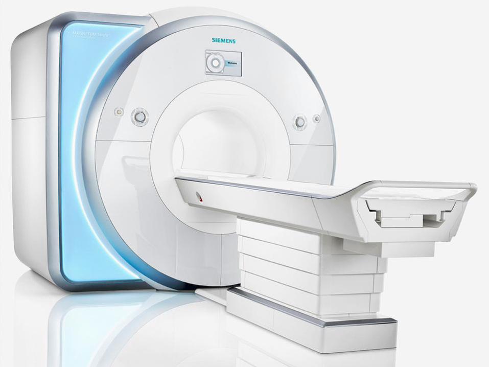
What is an MRI?
• Imaging modality whereby images are produced by pulsing radio waves in a magnetic field.

MRI
• Patient is placed in a high strength magnetic field (1.5T or 3T).

MRI
• The protons (Hydrogen atoms) in the body align with the magnetic field.

MRI
• Radiowaves are then used to de-phase the protons.
• The timing and strength of the waves can be manipulated to produce different tissue weighting characteristics.
MRI Advantages
• Superior soft tissue characterisation
• Free of ionising radiation

MRI Disadvantages
• Time consuming
• Acquire in one plane at a time (CT acquires volume)
• Inferior spatial resolution compared with CT
• Cramped space - claustrophobia
Standard Brain Pulse Sequences
• T1 weighted
• T2 weighted
• FLAIR
• DWI
• SWI
• T1 post contrast
• TOF MRA

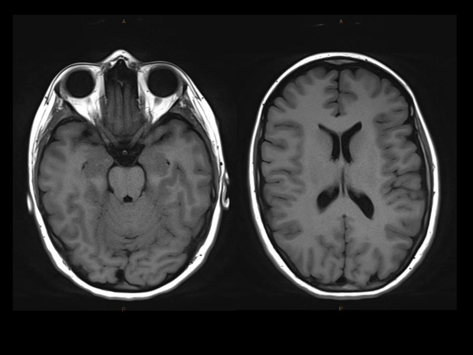
T1
• Fat
• Methaemaglobin (sub-acute haemorrhage)
• Mineralisation
• Slow flow
• Contrast
• Proteinaceous fluid
• Melanin

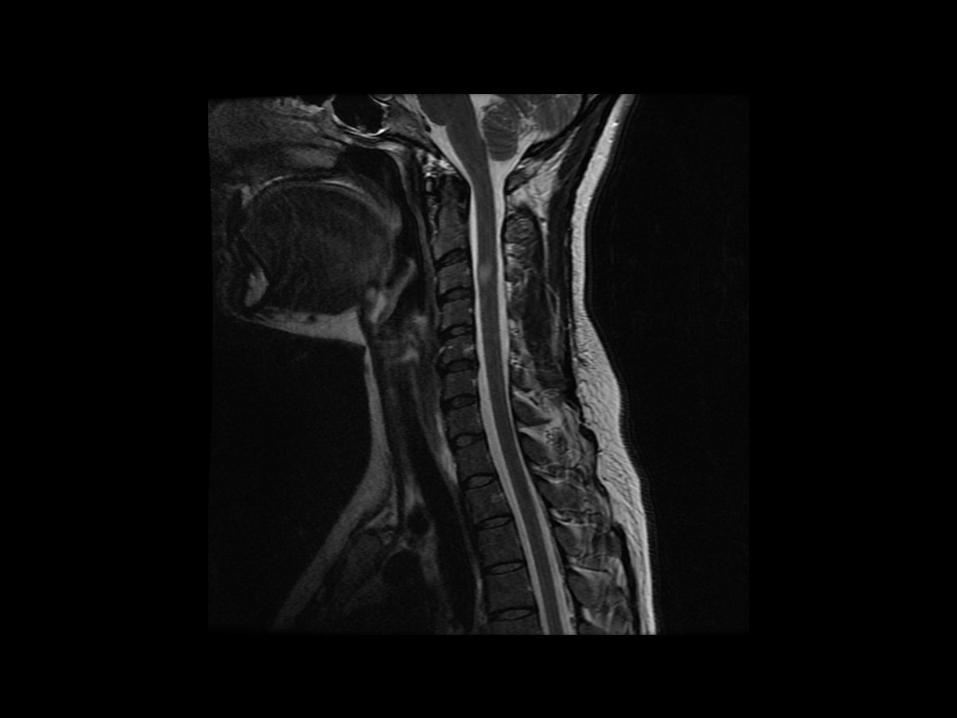
T2
• Fluid
• Fat
• T2 weighted sequences are typically sensitive for pathology


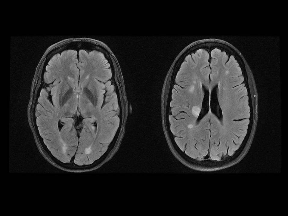
FLAIR
• FLuid Attentuated Inversion Recovery
• T2 weighted sequence with CSF signal supressed


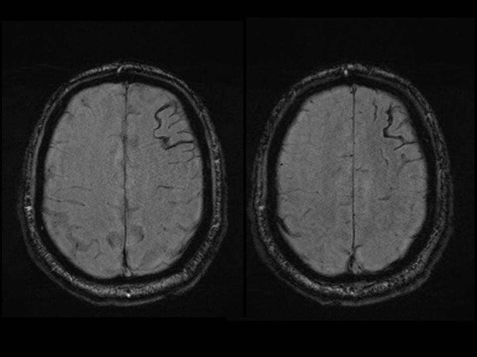
SWI
• Susceptibility Weighted Imaging
• Gradient echo sequence
• Sensitive to
– Venous blood
– Haemorrhage
– Iron

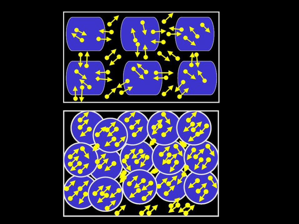
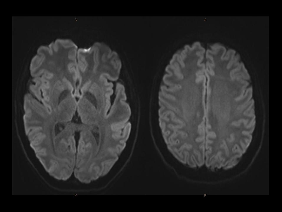
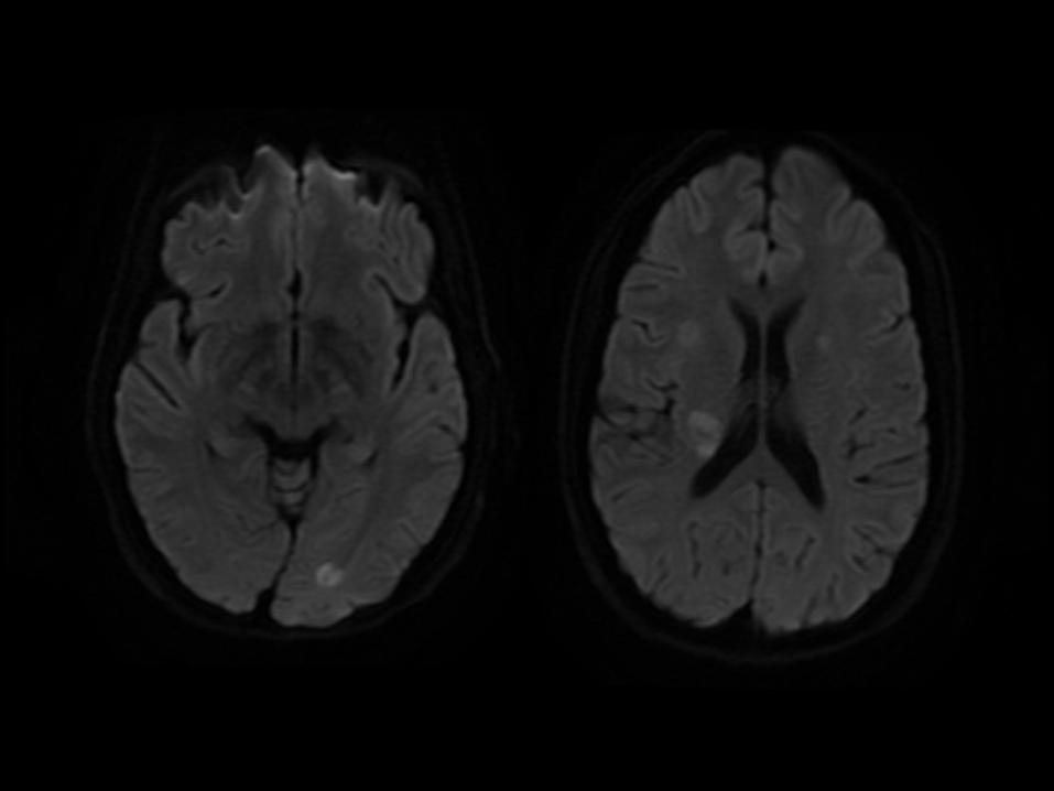
DWI
• Diffusion Weighted Imaging
• Sensitive to restrictoin of Brownian motion of extracellular water
• Sensitive for
– Ischaemia
– Pus
– Epidermoid

Contrast Enhanced T1
• Contrast does not cross the normal BBB
• Contrast is useful for detecting and characterising diseases that disrupt the BBB

MR Angiography
• Contrast enhanced
• Time of Flight
• Lower resolution than CTA but can provide a 4D image (TRICKS/TWIST)

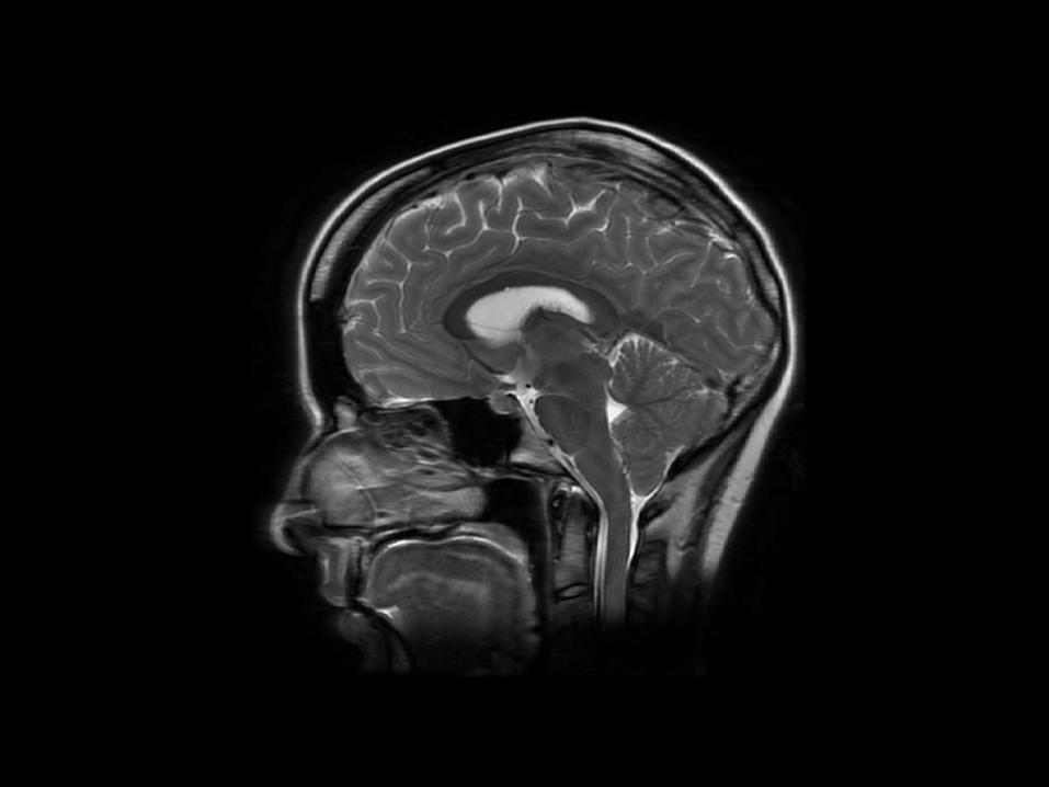
Basic Anatomy
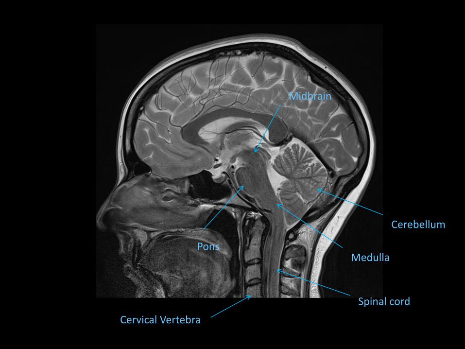
Cervical Vertebra
Spinal cord
Cerebellum
Medulla Pons
Midbrain

Frontal Lobe
Pituitary Gland
Occipital Lobe
Hypothalamus
Corpus Callosum
Parietal Lobe
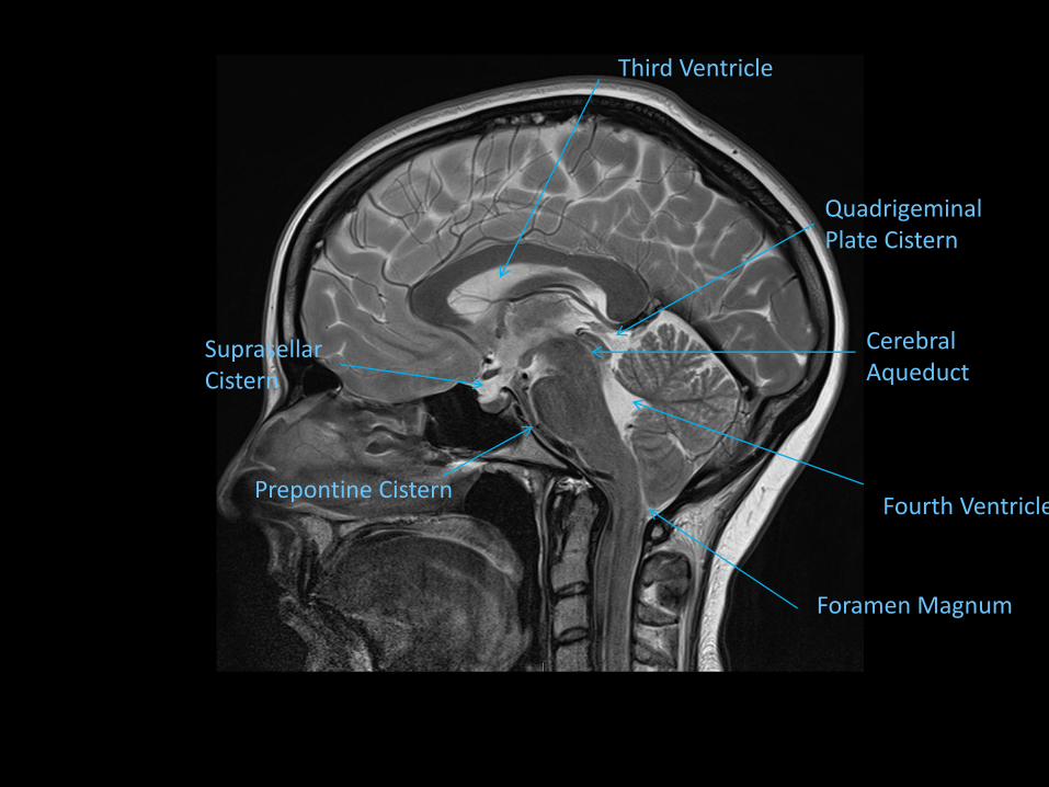
Prepontine Cistern
Quadrigeminal Plate Cistern
Fourth Ventricle
Foramen Magnum
Suprasellar Cistern
Third Ventricle
Cerebral Aqueduct
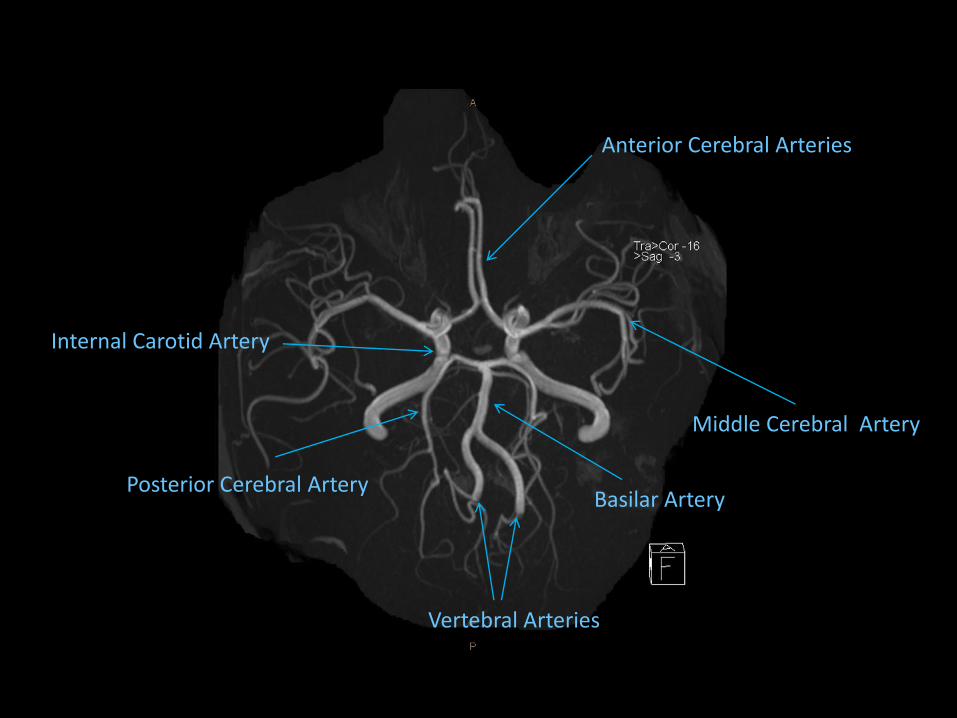
Anterior Cerebral Arteries
Middle Cerebral Artery
Internal Carotid Artery
Vertebral Arteries
Basilar Artery Posterior Cerebral Artery

Hard Palate
Ramus of Mandible
Parotid Gland
Lateral mass C1
Spinal Cord
Odontoid Peg
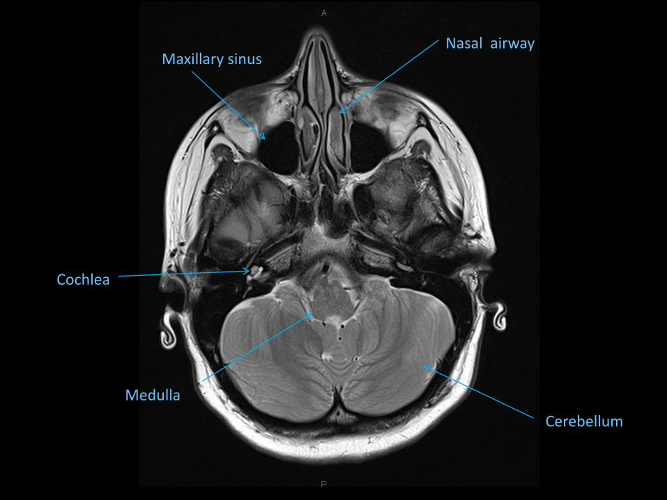
Cochlea
Medulla
Nasal airway Maxillary sinus
Cerebellum

Globe
Pons
Cavernous Sinus
Fourth Ventricle
Pituitary Gland
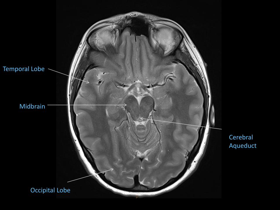
Midbrain
Temporal Lobe
Occipital Lobe
Cerebral Aqueduct
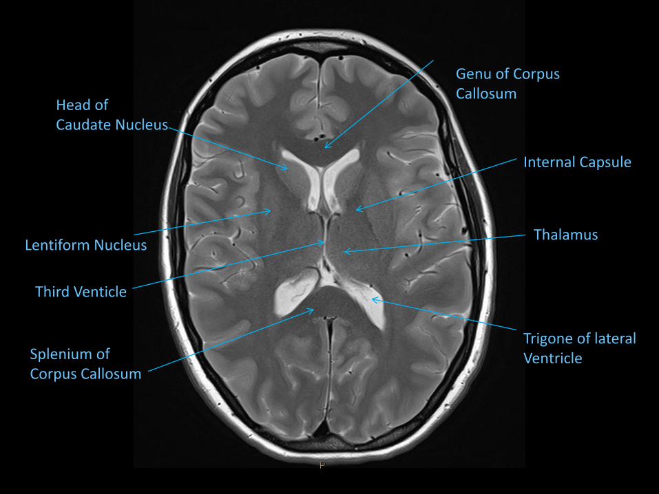
Head of Caudate Nucleus
Lentiform Nucleus
Third Venticle
Splenium of Corpus Callosum
Genu of Corpus Callosum
Internal Capsule
Thalamus
Trigone of lateral Ventricle
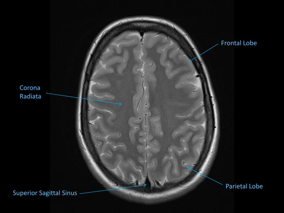
Corona Radiata
Superior Sagittal Sinus Parietal Lobe
Frontal Lobe

Central Sulcus
Pre-central Gyrus
Post-central Gyrus
The Value of the Different Pulse Sequences
Cases
Case 1
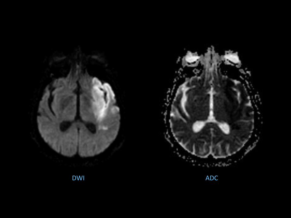
DWI ADC

Case 2


Case 3


Case 4


Case 5

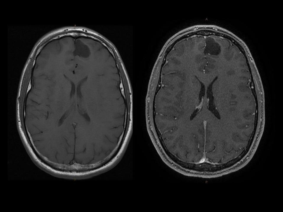

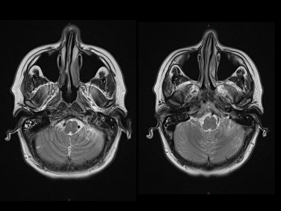
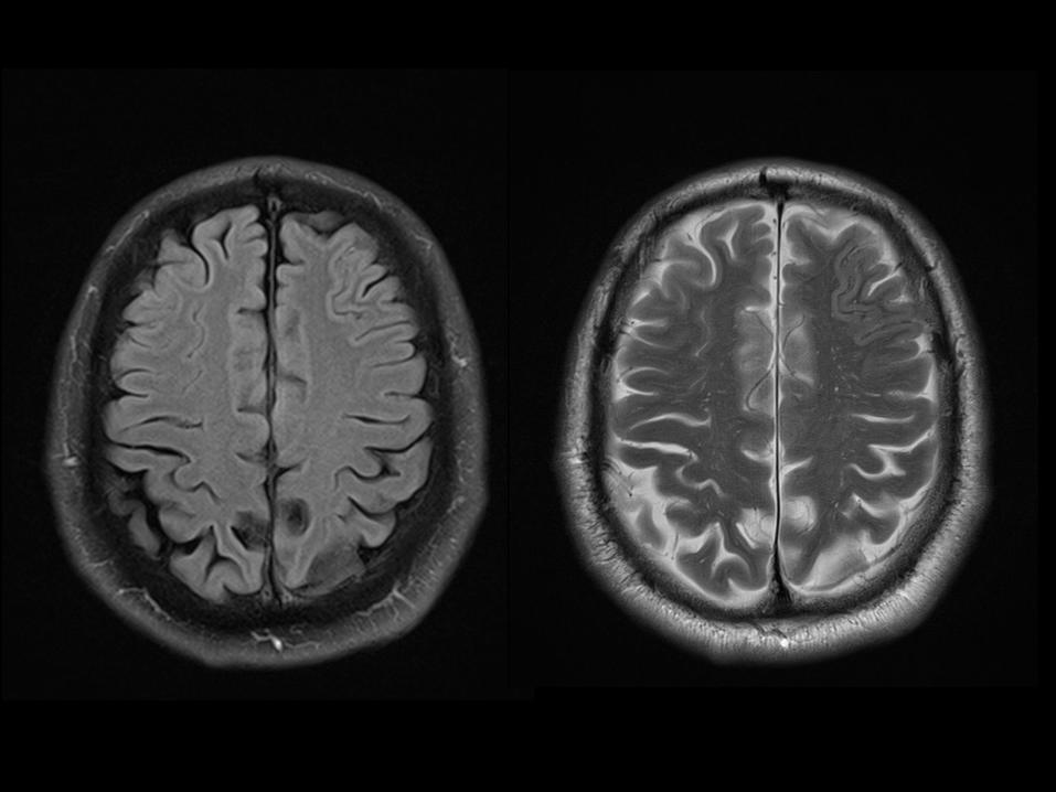
Case 6


Case 7


Case 8


Case 9

Case 10


Case 11


Case 12


Questions?


