NEUROANATOMY Lecture : 6 The Ventricles and Meninges of the Brain , The Cerebro-Spinal Fluid Prepared and presented by : Dr. Iyad Mousa Hussein , MD, Ph.D in Neurology Head of Neurology Department Nasser Hospital
Transcript
NEUROANATOMY
Lecture : 6
The Ventricles and Meninges of the Brain ,The Cerebro-Spinal Fluid
Prepared and presented by:Dr. Iyad Mousa Hussein ,
MD, Ph.D in NeurologyHead of Neurology Department
Nasser Hospital
1. The Ventricles of the Brain.
2. Definition, Site, Communicates, and Parts of the Lateral Ventricle.
3. Definition, Site, Communicates, and Parts of the Third Ventricle.
4. Definition, Site, Communicates. and Parts of the Fourth Ventricle.
5. The Meninges and Intracranial Spaces of the Brain.
6. Dural Nerve Supply.
7. Headaches due to Diseases of the Teeth, Paranasal Sinuses, and
Eyes.
9. Definition, Production, Circulation, Absorption, and Functions of the
Cerebro-Spinal Fluid.
10. Characters of the normal Cerebro-Spinal Fluid.
LECTURE OBJECTIVES:
► The brain and spinal cord float within a protective bath of
cerebrospinal fluid (CSF), which is produced by the
choroid plexus within the ventricles of the brain.
► Each part of the CNS contains a component of the
ventricular system.
► The ventricles contains about 35 ml of CSF.
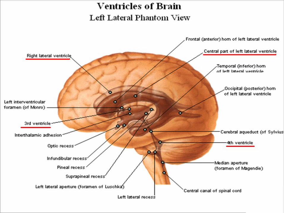
The Ventricles of the Brain
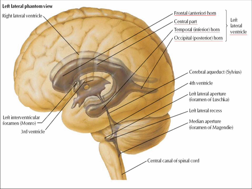
There are four interconnected ventricles in the brain:
1. Two lateral ventricles (Right and Left).
2. Third ventricle.
3. Fourth ventricle.
The Ventricles of the Brain
The Ventricles of the Brain
Definition: it is the cavity of the cerebral hemispheres.
There are two lateral ventricles (Right & Left), one in
each cerebral hemisphere.
Site: it is the cavity present in each cerebral hemisphere.
Communications:
It communicates with the third ventricle through the
interventricular foramen of Monro.
The Lateral Ventricle
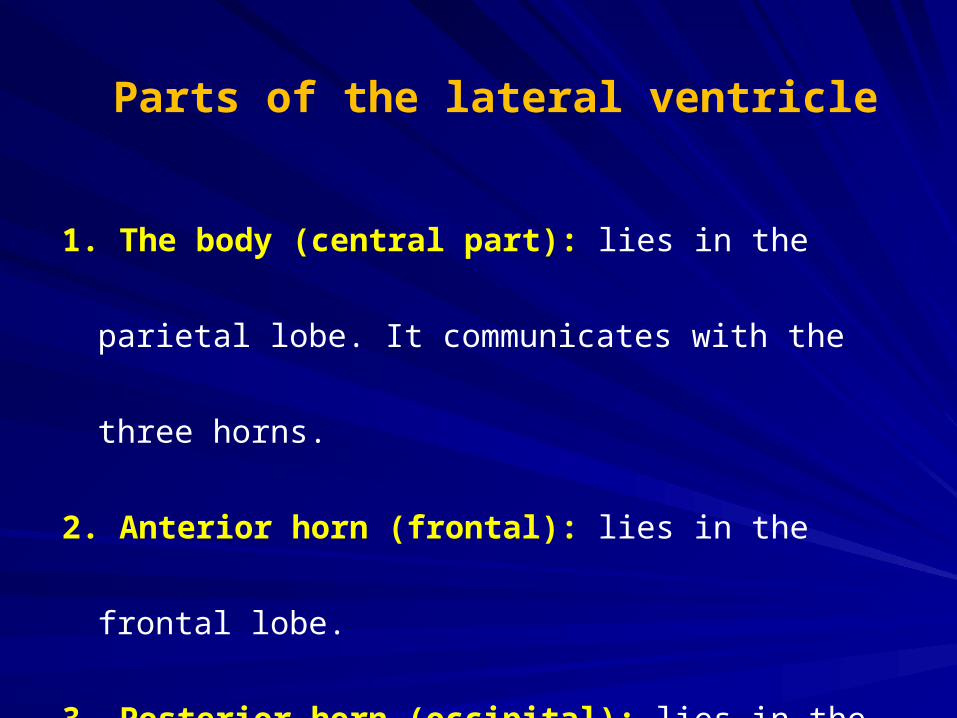
1. The body (central part): lies in the parietal lobe. It
communicates with the three horns.
2. Anterior horn (frontal): lies in the frontal lobe.
3. Posterior horn (occipital): lies in the occipital lobe.
4. Inferior horn (temporal): lies in the temporal lobe.
Parts of the lateral ventricle
The Third VentricleThe Third Ventricle
Definition: It is a narrow cavity situated in the midline
between two thalami.
it is the a cavity of the diencephalon.
Site: it is lying in the median plane between the right
and left thalami.
1. Anterosuperiorly: on each side, it communicates
with each lateral ventricle by an interventricular
foramen of Monro.
2. Posteroinferiorly: in the median plane, it
communicates with the fourth ventricle through the
cerebral aqueduct (of Sylvius), which passes
through the midbrain.
Communications of the Third Ventricle
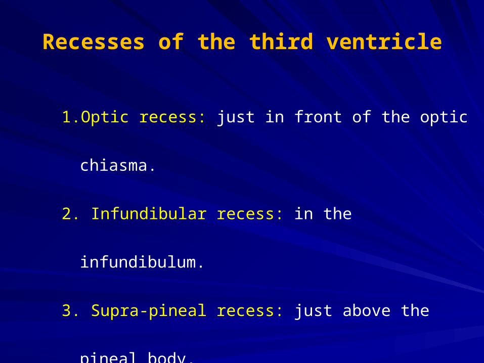
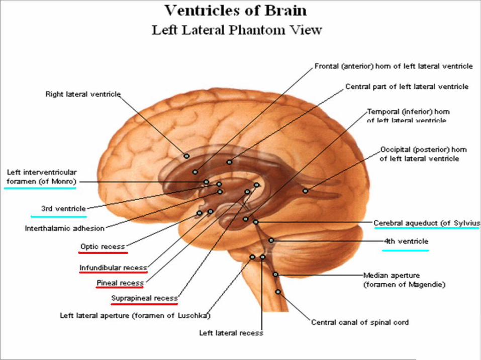
1. Optic recess: just in front of the optic chiasma.
2. Infundibular recess: in the infundibulum.
3. Supra-pineal recess: just above the pineal body.
4. Pineal recess: in the pineal body.
Recesses of the third ventricle
Choroid Plexus of the Lateral and Choroid Plexus of the Lateral and Third Third !!!!!!! !!!!!!! VentriclesVentricles
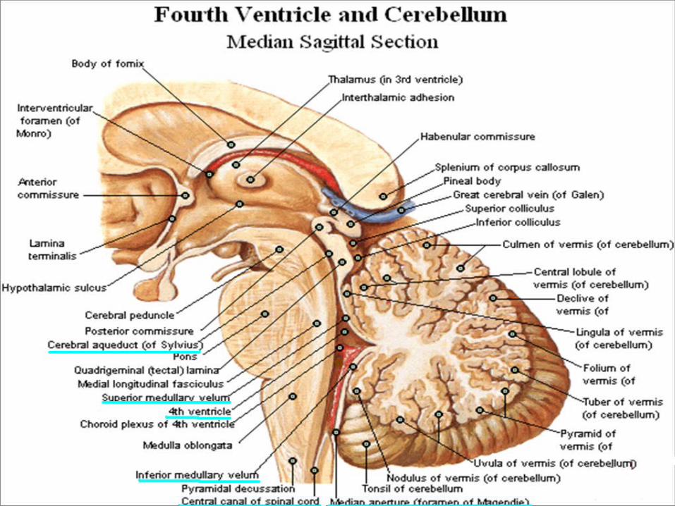
Definition: It is the cavity of the hindbrain (Rhombencephalon)Rhombencephalon).
Position: it lies between:
1. The dorsal surfaces of the pons and upper part of the
medulla anteriorly.
2. The ventral surface of the cerebellum posteriorly.
The Fourth Ventricle
1. Superior angle: where the fourth ventricle is continues
with the aqueduct of the midbrain.
2. Inferior angle: where the fourth ventricle is continuous
with central canal of the closed medulla.
3. Right & Left lateral angle: each lateral recess opens into
the subarachnoid space (by right and left foramen
Luschka).
Angles of the fourth ventricle
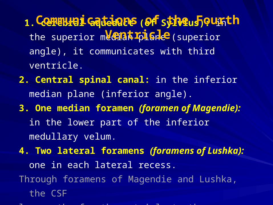
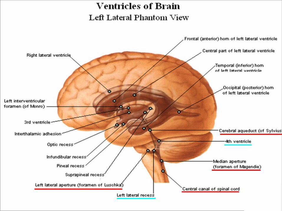
1. cerebral aqueduct (of Sylvius): in the superior
median plane (superior angle), it communicates with
third ventricle.
2. Central spinal canal: in the inferior median plane
(inferior angle).
3. One median foramen (foramen of Magendie): in
the lower part of the inferior medullary velum.
4. Two lateral foramens (foramens of Lushka): one in
each lateral recess.
Through foramens of Magendie and Lushka, the CSF
leaves the fourth ventricle to the subarachnoid space.
Communications of the Fourth Ventricle
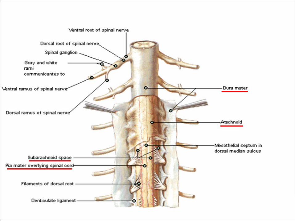
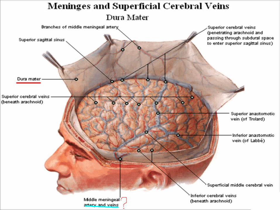
It is protective coverings (membranes) over the surface of
the brain and spinal cord.
The brain is surrounded by 3 membranes, from inside
outwards they are:
1. Pia mater.
2. Arachnoid mater.
3. Dura mater.
The Meninges of the Brain
► It is a vascular membrane forming the inner most
covering membrane of the brain.
► The choroid plexus of the lateral and third ventricles
are formed by invagination of th pia mater.
► It send sheaths around the cranial nerves in the
cranial cavity.
I. The Pia Mater
► It is a thin membrane lying outside the pia mater.
► It send sheaths around the cranial nerves till their
points of exit from the skull.
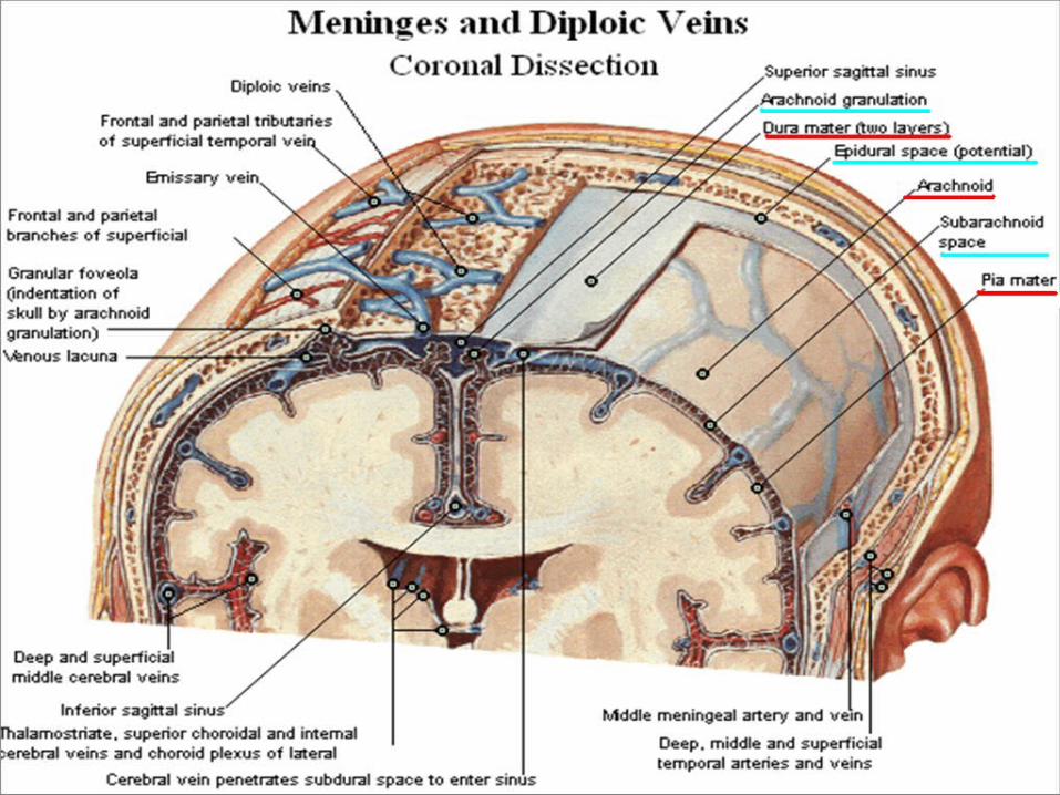
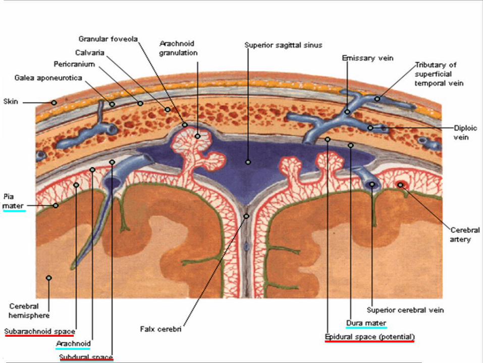
► It is consist of arachnoid villi (microscopic) and
arachnoid granulations (macroscopic).
► It is separated from the pia mater by a narrow space
called subarachnoid space containing CSF.
II. The Arachnoid Mater
► It is a thick membrane forming the outermost
covering of the brain.
► It consist of two layer:
1. Outer or endosteal layer: lines the inner surface
of the skull bones.
2. Inner or meningeal layer.
► Surrounds and protects the brain.
► The two layers of the dura are fused together except
in certain places where they separate to form
venous sinuses.
III. The Dura Mater
1. Epidural space: it is located between the two layers of
the dura mater.
2. Subdural space: it is located between the dura mater and
arachnoid mater. It contains a small amount of serous
fluid.
3. Subarachnoid space: it is located between the arachnoid
and pia mater. It contains CSF and blood vessels.
The Intracranial Spaces
1. Trigeminal nerve:
a. Ophthalmic nerve: supplies the dura mater of
anterior cranial fossa.
b. Maxillary and Mandibular nerves: supply the
dura matter of middle cranial fossa.
2. Vagus and Glossopharyngeal nerves: supply the
dura matter of middle cranial fossa.
3. The first three cervical spinal nerves.
4. Sympathetic trunk.
Dural Nerves Supply
Dental infection and sinusitis are common causes of
headache. The pain is referred to the skin of the face
and head along the branches of the trigeminal nerve.
Headaches due to Diseases of the Teeth, Paranasal Sinuses, and Eyes
Definition: it is the fluid which circulates in the
ventricles and central canals of the CNS and also
fills the subarachnoid space.
Production of the CSF:
It is secreted by the choroid plexuses of lateral,
third and fourth ventricles.
The Cerebro-Spinal Fluid
The CSF in each lateral ventricle → interventricular
foramen of Monro → third ventricle → aqueduct of
midbrain → fourth ventricle → some fluid enter the
central canal of spinal cord but the majority passes
through the 3 foramina (one median of foramen
Magendie & two lateral foramins of Lushka) →
subarachnoid space were it flows over the surface of
the brain and spinal cord.
Circulation of the Cerebro-Spinal Fluid
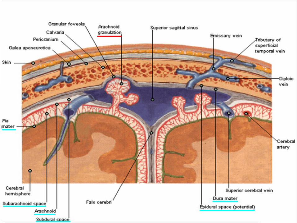
Absorption of the Cerebro-Spinal Fluid
The CSF is absorbed by means of arachnoid villi and
arachnoid granulations which absorbs the CSF from
the subarachnoid space into the dural venous sinuses
to reach the blood stream.
1. Cushions and protects the central nervous system
from trauma.
2. It takes the place of the lymph in the CNS which is
devoid of lymphatic vessels.
3. Regulation of the intracranial pressure.
4. Removes metabolites and toxics from the central
nervous system.
5. Nourishes the central nervous system.
Functions of the Cerebro-Spinal Fluid
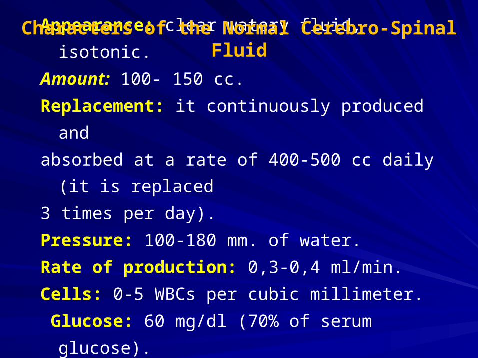
Appearance: clear watery fluid, isotonic.
Amount: 100- 150 cc.
Replacement: it continuously produced and
absorbed at a rate of 400-500 cc daily (it is replaced